Article

Production of New Antibacterial 4-Hydroxy-α-Pyrones by a Marine Fungus Aspergillus niger Cultivated in Solid Medium

1
Li Dak Sum Yip Yio Chin Kenneth Li Marine Biopharmaceutical Research Center, College of Food and Pharmaceutical Sciences, Ningbo University, Ningbo 315832, China
2
Department of Chemistry, College of Chemistry and Materials Science, Jinan University, Guangzhou 510632, China
*
Authors to whom correspondence should be addressed.
Mar. Drugs 2019, 17(6), 344; https://doi.org/10.3390/md17060344
Submission received: 17 May 2019 / Revised: 3 June 2019 / Accepted: 5 June 2019 / Published: 10 June 2019

Abstract

:
Four 4-hydroxy-α-pyrones including three new ones named nipyrones A–C (13) together with one known analogue germicidin C (4) were discovered from a marine sponge-derived fungus Aspergillus niger cultivated in a solid rice culture. Their structures and absolute configurations were elucidated through a combination of spectroscopic data and electronic circular dichroism (ECD) calculations as well as comparison with literature data. Compounds 14 were evaluated for their antibacterial activities against five pathogenic bacteria (Staphylococcus aureus, Escherichia coli, Bacillus subtilis, methicillin-resistant Staphylococcus aureus, and Mycobacterium tuberculosis). Compound 3 showed promising activity against S. aureus and B. subtilis, with minimum inhibitory concentration (MIC) values of 8 μg/mL and 16 μg/mL, respectively, and displayed weak antitubercular activities against M. tuberculosis, with MIC value of 64 μg/mL, while compounds 1 and 2 exhibited moderate antibacterial efficacy against four pathogenic bacteria with MIC values of 32–64 μg/mL.

1. Introduction

In recent years, sponge-derived fungi have represented a potential resource for discovery of novel bioactive molecules [1,2]. Numerous secondary metabolites with a broad spectrum of bioactivities have been isolated from sponge-derived fungi, inclusive of alkaloids [3], terpenoid [4], polyketides [5], and peptides [6]. α-Pyrones are one of polyketide-biosynthetic skeletons, characterized by six-membered unsaturated cyclic scaffold containing a lactone [7]. α-Pyrones can be widely found in the fungi and actinomycetes [8,9,10], and these molecules demonstrated a wide range of extraordinary biological activities, such as antimicrobial [11], anti-inflammatory [12], cytotoxic [13], and quorum sensing signaling molecules [14,15].
Members of the genus Aspergillus are well known to produce chemically diverse secondary metabolites, many of which have been developed to therapeutic leads for human health [16,17,18,19]. During our ongoing search for sponge-derived fungi capable of producing antibiotics, a sponge-derived fungus Aspergillus niger LS24 showed antimicrobial activities. HPLC-UV profile of crude extract of A. niger LS24 grown on solid rice medium indicated the presence of various α-pyrone derivatives with the characteristic UV absorption similar to that of germicidin C [20]. Antibacterial-guided fractionation of the EtOAc extract from a scale-up culture led to the isolation and identification of three new 4-hydroxy-α-pyrones, named nipyrones A–C (13) and one known analogue, germicidin C (4) (Figure 1). Compounds 14 were evaluated for their antibacterial activities against five pathogenic bacteria. Herein, the isolation, structure elucidation, and antibacterial evaluation of these α-pyrones are described.

2. Results and Discussion

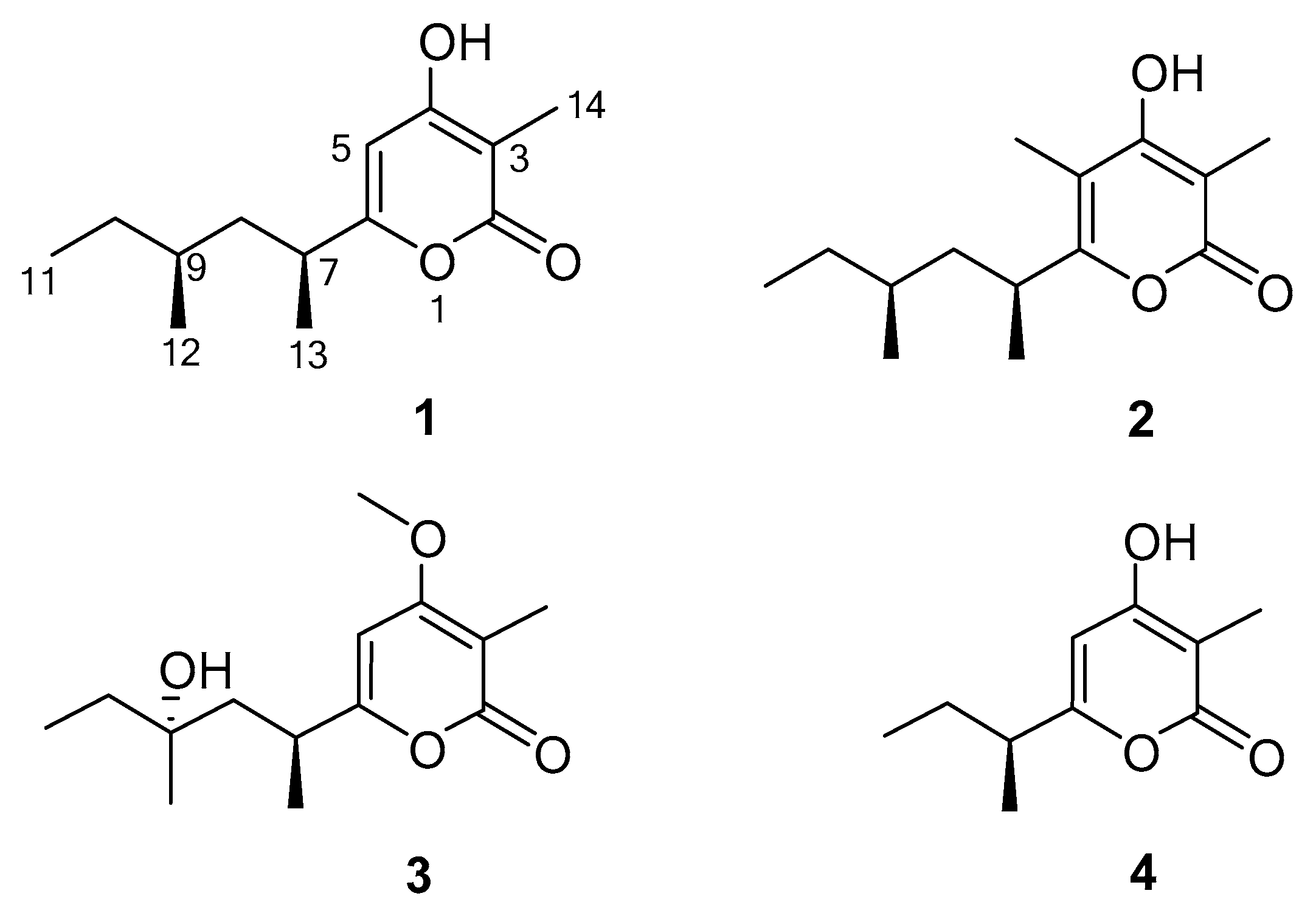
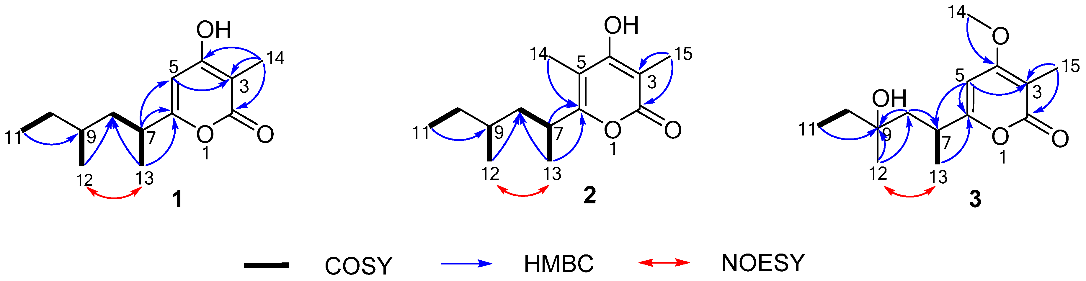
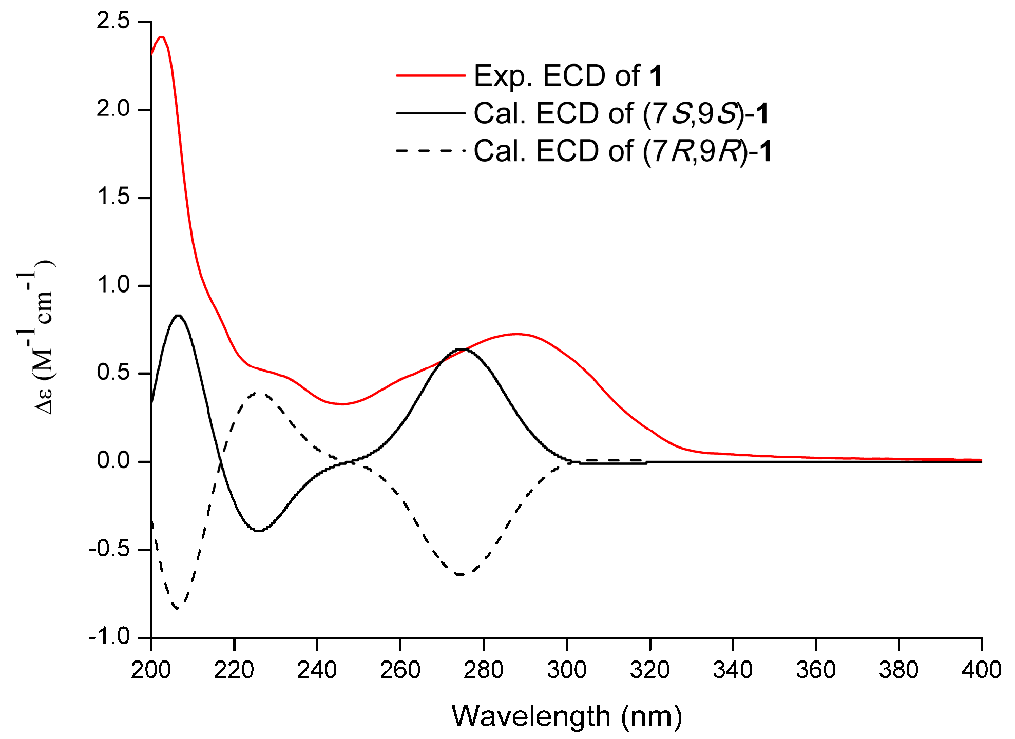
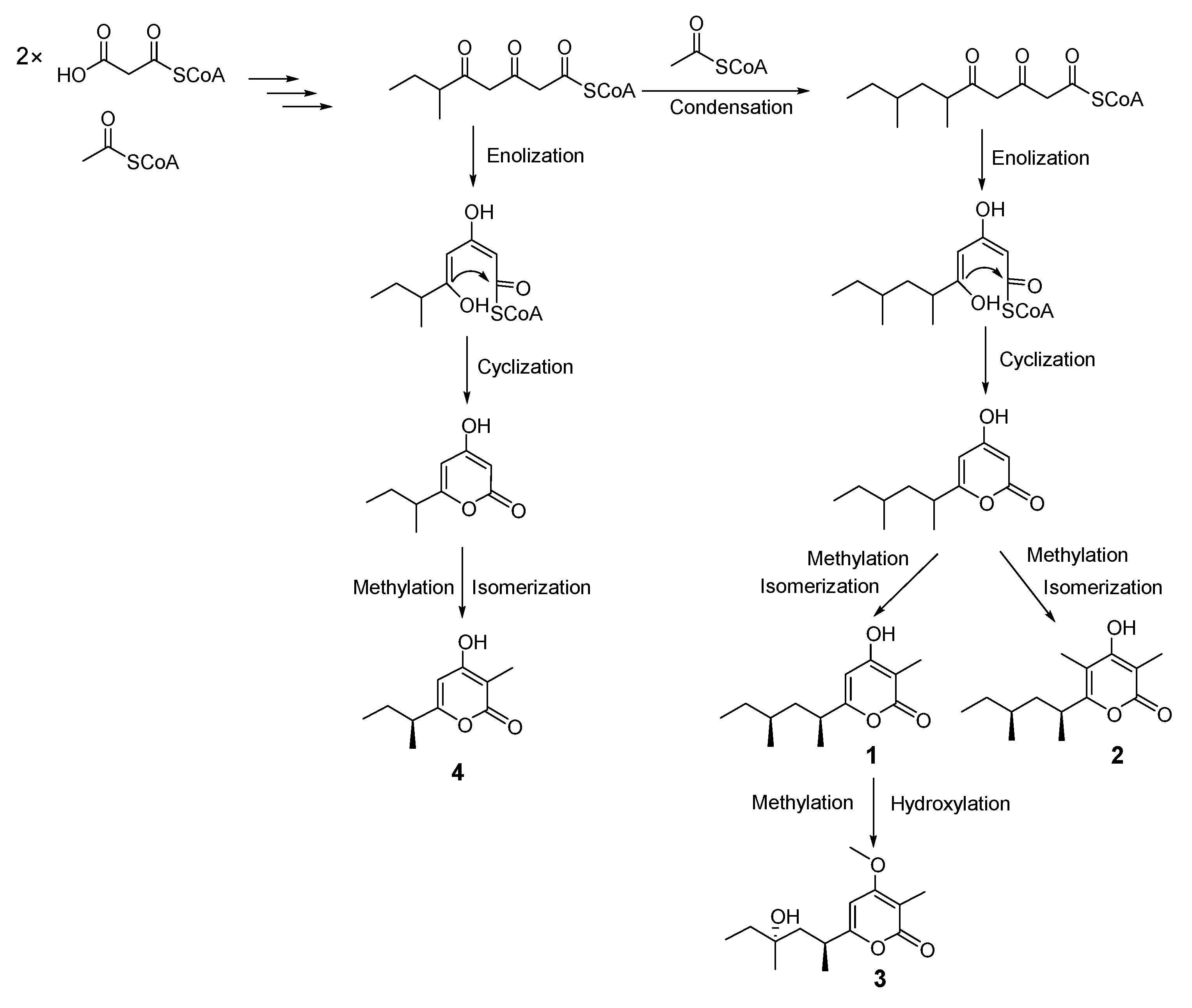
2.1. Structure Elucidation

Nipyrone A (1) was obtained as a colorless oil. The molecular formula of 1 was assigned as C13H20O3 by HRESIMS data and gave an [M + H]+ peak at m/z 225.1487, suggesting four degrees of unsaturation. Its UV absorption at 290 nm indicated the presence of a conjugated α-pyrone chromophore [21] (Figure S2). The 1H-NMR spectrum (Table 1) of 1 displayed one olefinic proton at δH 6.16 (s, H-5), two methylenes at δH 1.70 (m, H-8), 1.19 (m, H-8), 1.27 (m, H-10), and 1.10 (m, H-10), two methines at δH 2.64 (m, H-7) and 1.27 (m, H-9), two methyl doublets at δH 0.84 (d, J = 6.4 Hz, H3-12) and 1.19 (d, J = 6.9 Hz, H3-13), one methyl triplet at δH 0.82 (t, J = 7.3 Hz, H3-11), and one methyl singlet at δH 1.97 (s, H3-14). Analysis of 13C NMR and DEPT spectra of 1 classified the 14 carbons into four methyls, two methylenes, three methines, and four nonprotonated carbons. HMBC correlations (Figure 2) of H3-14/C-2 (δC 167.3), C-3 (δC 98.7), and C-4 (δC 168.7) as well as H-5/C-3 (δC 98.7) in the HMBC spectrum suggested the existence of the 4-hydroxy-3-methyl α-pyrone skeleton (Figure 2 and Figure S7). The structure of a 1,3-dimethylpentyl group was further verified by the COSY correlations (H-7/H2-8/H3-13, H-9/H3-12, and H-10/H3-11) and HMBC correlations from H3-12/H3-13 to C-8 (δC 41.7) and H3-11 to C-9 (δC 32.1). The 1,3-dimethylpentyl group was connected to C-6 (δC 167.3) of 4-hydroxy-3-methyl α-pyrone moiety, supported by the HMBC correlations of H-7/C-5 (δC 100.0) and C-6 together with H3-13/C-6 (Figure 2). Thus, the planar structure of 1 was established as 4-hydroxy-3-methyl-6-(-4-methylhexan-2-yl)-2H-pyran-2-one. The relative configuration of 1 was established by the NOESY data (Figure S9). The NOE correlation between H3-12/H3-13 clarified H3-12 and H3-13 to be cofacial of the side chain (Figure 2). Thus, the absolute configuration at C-7 and C-9 of 1 was identified as 7S,9S or 7R,9R. The absolute configuration of 1 was further determined by comparing the experimental electronic circular dichroism (ECD) spectrum of 1 with the correspondingly time-dependent density functional theory (TDDFT)-calculated one. The Cotton effects of the experimental ECD spectrum of 1 matched very well with the calculated ECD spectrum for the model molecule of 7S,9S at the B3LYP/6-311 + G(d, p) level (Figure 3 and see Supplementary Materials), thus confirming its absolute configuration.
Nipyrone B (2) was also obtained as a colorless oil with a molecular formula of C14H22O3 as determined by HRESIMS data with one CH2 more than 1. The spectroscopic data of 2 (Table 1) were highly identical to that of 1 except for the absence of one olefinic proton and an additional methyl singlet. The additional methyl group was located at C-5 (δC 106.8, Table 1) by HMBC correlation from H3-14 to C-6 (Figure 2 and see Supplementary Materials). The absolute configuration of 2 tentatively led to deduction of the same as 1 based on the biosynthetic consideration and specific rotation.
Nipyrone C (3) was isolated a as a colorless oil, and its molecular formula was assigned as C14H23O4 based on the HRESIMS ([M + H]+ m/z 255.1591) and 13C NMR data. The 1D and 2D NMR spectroscopic data for 3 (Table 1) closely resembled those of 1. Significant differences in the NMR data for 3 were only found in the resonances assigned to an additional hydroxy group and an additional methoxy group. Moreover, the C-9 position in 1 replaced by a hydroxy group in 3 was confirmed by chemical shift C-9 (δC 72.9). The attachment of the methoxy group (C-14, δC 56.1) at C-4 was supported by the HMBC correlation of H3-14/C-4 (see Supplementary Materials). Based on the biosynthetic point of view and specific rotation, the absolute configuration of 3 might be assigned to be 7S,9R.
In addition, compound 4 was identified as germicidin C by comparison of its spectral data with those reported in [20]. Many α-pyrone-based secondary metabolites biosynthesized by polyketide synthetase (PKS) pathway have been widely reported, uncovering their biosynthetic gene clusters [7,22,23]. We proposed that the polyketide chain primed with acetyl-CoA and malonyl-CoA was elongated, enolized, cyclized, methylated, hydroxylated, and released as the corresponding four 4-hydroxy-α-pyrones. A probable biosynthesis pathway of 14 is illustrated in Scheme 1.

2.2. Biological Activities

The antibacterial activities of 14 were evaluated against four bacteria, including Gram-positive and Gram-negative bacteria, using broth micro-dilution method within a concentration range of 256–1 µg/mL. Four pathogenic bacteria, including S. aureus, E. coli, B. subtilis, and methicillin-resistant S. aureus (MRSA) were performed. The results are shown in Table 2. Compound 3 exhibited significant inhibitory activity against S. aureus and B. subtilis, with minimum inhibitory concentration (MIC) values of 8 and 16 µg/mL, respectively. Compounds 1, 2, and 4 exhibited moderate antimicrobial effect against S. aureus, E. coli, and B. subtilis, with MIC values in range of 32–64 µg/mL. Compounds 14 displayed weak antibiotic capacity against MRSA. Compound 3 possessed weak antitubercular activities against M. tuberculosis (MIC, 64 µg/mL).

3. Materials and Methods

3.1. General Experimental Procedures

Optical rotations were determined with a P-2000 digital polarimeter (JASCO, Hachioji, Japan). UV spectra were obtained with a NADE Evolution 201 spectrophotometer (ThermoFisher, Waltham, MA, USA). IR spectra were measured on a Nicolet iS5 spectrometer (ThermoFisher, Waltham, MA, USA). NMR data were carried out at ambient temperature on a Varian 600 MHz (Palo Alto, CA, USA) spectrometer operating at 600 (1H) and 150 (13C) MHz. HRESIMS data were recorded on an Agilent Technologies 6520 Accurate Mass Q-TOF LC/MS spectrometer equipped with an ESI source (Agilent Technologies, Santa Clara, CA, USA). Medium-pressure liquid chromatography (MPLC) was performed on a FLEXA Purification System (Bonna-Agela Technologies Co., Tianjin, China) using a 15 µm ODS column (Santai Technologies, Inc., Changzhou, China). Semi-preparative HPLC was performed on an YMC-Pack Pro C18 RS column (5 μm, 250 × 10 mm id; YMC, Kyoto, Japan) with a Waters 600 separation system coupled with a Waters 2998 Photodiode Array detector (Waters, MA, USA). Column chromatography (CC) was performed on silica gel (200–300 mesh, Qingdao Haiyang Chemical Factory, Qingdao, China) and Sephadex LH-20 (25–100 µm; Pharmacia, Uppsala, Sweden).

3.2. Fungal Material

Marine sponge Haliclona sp. was collected at Lingshui, Hainan Province, China. The sponge tissue was cut into small pieces of about 0.1 cm3 each, which were homogenized with sterile seawater. 20 μL of the diluted homogenate (1:100, sterile seawater) was inoculated in PDA agar plates, which were incubated for periods of 3 days to 4 weeks for purifying fungal colonies. 15 fungal isolates were obtained. Among them, the EtOAc extract of the fungal strain LS24 showed antimicrobial activities. Surprisingly, its extract showed stronger antimicrobial effects (MICs, 32–128 μg/mL against different pathogenic bacteria) when grown on solid rice medium in comparison to liquid medium (MICs, >128 μg/mL against different pathogenic bacteria). The fungal strain LS24 was identified using morphological studies, DNA amplification, and the internal transcribed spacer region (ITS) sequencing (GenBank accession ID: KX290301, 100% similarity). The isolate was stored on PDA medium (potato 200 g, dextrose 20 g, sea salt 35 g and agar 15 g in 1.0 L of H2O, pH 7.4–7.8) slants at 4 °C. A voucher strain was preserved at College of Food and Pharmaceutical Sciences, Ningbo University, Ningbo, China.

3.3. Fermentation

The fungus A. niger was cultured on PDA agar plate at 28 °C for 7 days. The fungal colony was further inoculated into the PDB medium (potato 200 g, dextrose 20 g, and sea salt 35 g in 1.0 L of H2O, pH 7.4–7.8) at 28 °C for 3 days on a rotating shaker (180 rpm). Then, a large-scale fermentation of the strain was performed. The fungal seed broth (20 mL) was added to 10 flasks (1000 mL), each containing 100 g rice and 160 mL water. These flasks were incubated at 28 °C for 30 days under static conditions.

3.4. Extraction and Isolation

All the fermented materials were extracted with 5 L EtOAc three times to afford a brown extract (8 g). The EtOAc extract was subjected to vacuum liquid chromatography (VLC) on a silica gel column (6 × 15 cm, 200–300 mesh) using step gradient elution with petroleum ether/EtOAc (from 20:1 to 0:1, v/v) to obtain seven fractions (Fr.1–7) according to HPLC analysis. Fraction 4 was further chromatographed over a Sephadex LH-20 column, eluted with CH3OH and CH2Cl2 (1:1, v/v), yielding three subfractions (Fr.4.A–C). Fr.4.B (300 mg) was further separated into six subfractions (Fr.4.B.1–6) by ODS silica gel MPLC eluting with MeOH/H2O (30–100%, 120 min, flow rate 20 mL/min) to obtain nine subfractions (Fr.4.B.1–9). Fr.4.B.3 was separated by semipreparative HPLC (35% MeCN/H2O, 2 mL/min, detected at 290 nm) to provide 1 (1.3 mg, tR 32 min) and 2 (1.2 mg, tR 34 min). Meanwhile, Fr.4.B.7 was purified by semipreparative HPLC (40% MeCN/H2O, 2 mL/min, detected at 290 nm) to yield 3 (1.1 mg, tR 30 min) and 4 (3.1 mg, tR 32 min).
Nipyrone A (1): colorless oil; [ α ] D 25 +43 (c 0.23, MeOH); CD λmax (Δε) 203(+2.41), 288 (+0.72) nm; UV (MeOH) (log ε) λmax 290 (4.19) nm; IR (KBr) νmax 3093, 2964, 2930, 2876, 2680, 1662, 1582, 1459, 1415, 1375, 1245, 1152, 1126, 1057, 972, 932, 876, 837, 760, 705 cm−1; 1H and 13C NMR data, see Table 1; HRESIMS m/z 225.1487 [M + H]+ (calcd for C13H21O3, 225.1485).
Nipyrone B (2): colorless oil; [ α ] D 25 +72 (c 1.13, MeOH); UV (MeOH) (log ε) λmax 286 (3.86) nm; IR (KBr) νmax 3219, 2963, 2930, 2875, 1705, 1671, 1606, 1568, 1459, 1408, 1377, 1232, 1156, 1093, 1035, 958, 873, 806, 761 cm−1; 1H and 13C NMR data, see Table 1; HRESIMS m/z 239.1639 [M + H]+ (calcd for C14H23O3, 239.1628).
Nipyrone C (3): colorless oil; [ α ] D 25 +35 (c 0.12, MeOH); UV (MeOH) (log ε) λmax 299 (3.92) nm; IR (KBr) νmax 3429, 3101, 2967, 2929, 2876, 1686, 1642, 1566, 1462, 1406, 1380, 1325, 1251, 1192, 1141, 1099, 1032, 968, 941, 905, 804, 756 cm−1; 1H and 13C NMR data, see Table 1; HRESIMS m/z 255.1591 [M + H]+ (calcd for C14H23O4, 255.1591).

3.5. Antibacterial Assay

The antibacterial effects of compounds 14 were evaluated against Gram-positive bacteria S. aureus ATCC 25923, B. subtilis ATCC 6633, methicillin-resistant S. aureus ATCC 43300 (MRSA), and Gram-negative bacterium E. coli ATCC 25922 according to a previously described method [24]. The test compounds were dissolved in DMSO (1 mg/mL for 14). The minimum inhibitory concentration (MIC) values were defined as the lowest concentration of test compound that inhibited the growth of more than 99% of the bacterial population after overnight incubation in 96-well microtiter plates, as detected by eye. Briefly, 100 μL of each bacterial solution was inoculated in each well (105 CFU/mL) and added with 100 µL of each compound solution and control in triples. Microplates were incubated for 24 h at 37 °C. The final concentrations of each test compound in the wells were in the range of 256–1 μg/mL using sequential 2-fold serial dilutions. The final DMSO concentration was maintained at 0.5% by adding the medium. Chloramphenicol and DMSO were used as the positive control and the negative control, respectively. The detailed method of the antitubercular activity of compounds 14 against M. tuberculosis H37Rv was described by the agar proportion method based on the previous report [25]. The blank control was DMSO. Ethambutol was used as a positive control.

4. Conclusions

In summary, three new 4-hydroxy-α-pyrone derivatives, nipyrones A–C (13) along with one known analogue germicidin C (4) were isolated from a marine sponge-derived fungus A. niger grown on a solid rice culture. These 4-hydroxy-α-pyrone derivatives have in common the differences in functional group substitution and side chain length. Biogenetically, nipyrones A–C (13) are presumably originated from the polyketide pathway. This study further expanded the structural diversity of naturally occurring α-pyrone derivatives. Notably, compound 3 displayed significant inhibitory effects on two pathogenic bacteria, S. aureus and E. coli and may be considered to have potential as an antibiotic agent.

Supplementary Materials

The following are available online at https://www.mdpi.com/1660-3397/17/6/344/s1. HRESIMS, NMR, IR, UV of the new compounds 13; ECD calculation data for 1.

Author Contributions

S.H. and S.X. conceived and designed the experiments. L.D., L.R. and Z.H. performed the experiments. L.D., L.R., S.L., J.S., and S.H. analyzed the data. L.D. and S.H. wrote the paper.

Funding

This study was supported by the National Key Research and Development Program of China (2018YFC0310900), the National Natural Science Foundation of China (41776168, 41706167, 21672084, 41876145), Ningbo Public Service Platform for High-Value Utilization of Marine Biological Resources (NBHY-2017-P2), the National 111 Project of China (D16013), the Li Dak Sum Yip Yio Chin Kenneth Li Marine Biopharmaceutical Development Fund, and the K.C. Wong Magna Fund in Ningbo University.

Conflicts of Interest

The authors declare no conflict of interest.

References

  1. Gu, B.B.; Wu, Y.; Tang, J.; Jiao, W.H.; Li, L.; Sun, F.; Wang, S.P.; Yang, F.; Lin, H.W. Azaphilone and isocoumarin derivatives from the sponge-derived fungus Eupenicillium sp. 6A-9. Tetrahedron Lett. 2018, 59, 3345–3348. [Google Scholar] [CrossRef]
  2. Tian, Y.; Lin, X.; Zhou, X.; Liu, Y. Phenol derivatives from the sponge-derived fungus Didymellaceae sp. SCSIO F46. Front. Chem. 2018, 6, 536. [Google Scholar] [CrossRef] [PubMed]
  3. Buttachon, S.; Ramos, A.A.; Inacio, A.; Dethoup, T.; Gales, L.; Lee, M.; Costa, P.M.; Silva, A.M.S.; Sekeroglu, N.; Rocha, E.; et al. Bis-indolyl benzenoids, hydroxypyrrolidine derivatives and other constituents from cultures of the marine sponge-associated fungus Aspergillus candidus KUFA0062. Mar. Drugs 2018, 16, 119. [Google Scholar] [CrossRef] [PubMed]
  4. Yong, L.; Dong, L.; Cheng, Z.; Proksch, P.; Lin, W. Cytotoxic trichothecene-type sesquiterpenes from the sponge-derived fungus Stachybotrys chartarum with tyrosine kinase inhibition. RSC Adv. 2017, 7, 7259–7267. [Google Scholar]
  5. Tian, Y.Q.; Lin, S.T.; Kumaravel, K.; Zhou, H.; Wang, S.Y.; Liu, Y.H. Polyketide-derived metabolites from the sponge-derived fungus Aspergillus sp. F40. Phytochem. Lett. 2018, 27, 74–77. [Google Scholar] [CrossRef]
  6. Ding, L.J.; Yuan, W.; Liao, X.J.; Han, B.N.; Wang, S.P.; Li, Z.Y.; Xu, S.H.; Zhang, W.; Lin, H.W. Oryzamides A–E, cyclodepsipeptides from the sponge-derived fungus Nigrospora oryzae PF18. J. Nat. Prod. 2016, 79, 2045–2052. [Google Scholar] [CrossRef]
  7. Schäberle, T.F. Biosynthesis of α-pyrones. Beilstein J. Org. Chem. 2016, 12, 571–588. [Google Scholar] [CrossRef]
  8. Liu, D.; Li, X.M.; Meng, L.; Li, C.S.; Gao, S.S.; Shang, Z.; Proksch, P.; Huang, C.G.; Wang, B.G. Nigerapyrones A–H, α-pyrone derivatives from the marine mangrove-derived endophytic fungus Aspergillus niger MA-132. J. Nat. Prod. 2011, 74, 1787–1791. [Google Scholar] [CrossRef]
  9. Ma, Y.M.; Li, T.; Ma, C.C. A new pyrone derivative from an endophytic Aspergillus tubingensis of Lycium ruthenicum. Nat. Prod. Res. 2016, 30, 1499–1503. [Google Scholar] [CrossRef]
  10. Rab, E.; Kekos, D.; Roussis, V.; Ioannou, E. α-pyrone polyketides from Streptomyces ambofaciens BI0048, an endophytic actinobacterial strain isolated from the red alga Laurencia glandulifera. Mar. Drugs 2017, 15, 389. [Google Scholar] [CrossRef]
  11. Zhang, H.; Saurav, K.; Yu, Z.; Mándi, A.; Kurtán, T.; Li, J.; Tian, X.; Zhang, Q.; Zhang, W.; Zhang, C. α-Pyrones with diverse hydroxy substitutions from three marine-derived Nocardiopsis strains. J. Nat. Prod. 2016, 79, 1610–1618. [Google Scholar] [CrossRef] [PubMed]
  12. Lee, J.; Han, C.; Lee, T.G.; Chin, J.; Choi, H.; Lee, W.; Paik, M.J.; Won, D.H.; Jeong, G.; Ko, J. Marinopyrones A–D, α-pyrones from marine-derived actinomycetes of the family Nocardiopsaceae. Tetrahedron Lett. 2016, 57, 1997–2000. [Google Scholar] [CrossRef]
  13. Li, J.; Wu, X.; Ding, G.; Feng, Y.; Jiang, X.; Guo, L.; Che, Y. α-Pyrones and pyranes from the plant pathogenic fungus Pestalotiopsis scirpina. Eur. J. Org. Chem. 2012, 2012, 2445–2452. [Google Scholar] [CrossRef]
  14. Brachmann, A.O.; Brameyer, S.; Kresovic, D.; Hitkova, I.; Kopp, Y.; Manske, C.; Schubert, K.; Bode, H.B.; Heermann, R. Pyrones as bacterial signaling molecules. Nat. Chem. Biol. 2013, 9, 573–578. [Google Scholar] [CrossRef] [PubMed]
  15. Fu, P.; Liu, P.; Gong, Q.; Wang, Y.; Wang, P.; Zhu, W. α-Pyrones from the marine-derived actinomycete Nocardiopsis dassonvillei subsp. dassonvillei XG-8-1. RSA Adv. 2013, 3, 20726–20731. [Google Scholar] [CrossRef]
  16. Nakamura, I.; Kanasaki, R.; Yoshikawa, K.; Furukawa, S.; Fujie, A.; Hamamoto, H.; Sekimizu, K. Discovery of a new antifungal agent ASP2397 using a silkworm model of Aspergillus fumigatus infection. J. Antibiot. 2017, 70, 41–44. [Google Scholar] [CrossRef]
  17. Wang, C.; Guo, L.; Hao, J.; Wang, L.; Zhu, W. α-Glucosidase inhibitors from the marine-derived fungus Aspergillus flavipes HN4-13. J. Nat. Prod. 2016, 79, 2977–2981. [Google Scholar] [CrossRef]
  18. Zhu, H.; Chen, C.; Tong, Q.; Yang, J.; Wei, G.; Xue, Y.; Wang, J.; Luo, Z.; Zhang, Y. Asperflavipine A: A cytochalasan heterotetramer uniquely defined by a highly complex tetradecacyclic ring system from Aspergillus flavipes QCS12. Angew. Chem. Int. Ed. 2017, 56, 5242–5246. [Google Scholar] [CrossRef]
  19. Zhang, X.; Li, Z.; Gao, J. Chemistry and biology of secondary metabolites from Aspergillus genus. Nat. Prod. J. 2018, 8, 275–304. [Google Scholar] [CrossRef]
  20. Aoki, Y.; Matsumoto, D.; Kawaide, H.; Natsume, M. Physiological role of germicidins in spore germination and hyphal elongation in Streptomyces coelicolor A3(2). J. Antibiot. 2011, 64, 607–611. [Google Scholar] [CrossRef]
  21. Barrero, A.F.; Oltra, J.E.; Herrador, M.M.; Cabrera, E.; Sanchez, J.F.; Quílez, J.F.; Rojas, F.J.; Reyes, J.F. Gibepyrones: α-pyrones from Gibberella fujikuroi. Tetrahedron 1993, 49, 141–150. [Google Scholar] [CrossRef]
  22. Kasahara, K.; Fujii, I.; Oikawa, H.; Ebizuka, Y. Expression of Alternaria solani PKSF generates a set of complex reduced-type polyketides with different carbon-lengths and cyclization. Chembiochem 2010, 7, 920–924. [Google Scholar] [CrossRef] [PubMed]
  23. Xu, W.; Cai, X.; Jung, M.E.; Tang, Y. Analysis of intact and dissected fungal polyketide synthase-nonribosomal peptide synthetase in vitro and in Saccharomyces cerevisiae. J. Am. Chem. Soc. 2010, 132, 13604–13607. [Google Scholar] [CrossRef] [PubMed]
  24. An, C.-L.; Kong, F.-D.; Ma, Q.-Y.; Xie, Q.-Y.; Yuan, J.-Z.; Zhou, L.-M.; Dai, H.-F.; Yu, Z.-F.; Zhao, Y.-X. Chemical constituents of the marine-derived fungus Aspergillus sp. SCS-KFD66. Mar. Drugs 2018, 16, 468. [Google Scholar] [CrossRef]
  25. Braña, A.F.; Sarmiento-Vizcaíno, A.; Pérez-Victoria, I.; Martín, J.; Otero, L.; Palacios-Gutiérrez, J.J.; Fernández, J.; Mohamedi, Y.; Fontanil, T.; Salmón, M.; et al. Desertomycin G, a new antibiotic with activity against Mycobacterium tuberculosis and human breast tumor cell lines produced by Streptomyces althioticus MSM3, isolated from the Cantabrian Sea Intertidal macroalgae Ulva sp. Mar. Drugs 2019, 17, 114. [Google Scholar] [CrossRef] [PubMed]
Figure 1. Structures of compounds 14.
Marinedrugs 17 00344 g001
Figure 2. Key HMBC, COSY, and NOESY correlations of 13.
Marinedrugs 17 00344 g002
Figure 3. Experimental and calculated electronic circular dichroism (ECD) spectra of 1 in methanol.
Marinedrugs 17 00344 g003
Scheme 1. Proposed biosynthetic pathway of 14.
Marinedrugs 17 00344 sch001
Table 1. 1H (600 MHz) and 13C NMR (150 MHz) data for compounds 13 (CDCl3).
No.123
δC, TypeδH (J in Hz)δC, TypeδH (J in Hz)δC, TypeδH (J in Hz)
2167.3, C-166.9, C-165.9, C-
398.7, C-98.2, C-100.8, C-
4168.7, C-165.7, C-166.1, C-
5100.0, CH6.16, s106.8, C-93.2, CH6.04, s
6167.3, C-162.1, C-168.6, C-
736.1, CH2.64, m32.3, CH3.00, m35.1, CH2.85, m
841.7, CH21.70, m; 1.19, m41.5, CH21.79, m; 1.19, m45.5, CH22.06, dd (14.7, 5.8);
1.57, dd (14.6, 4.3)
932.1, CH1.27, m32.4, CH1.13, m72.9, C-
1029.6, CH21.27, m; 1.10, m29.9, CH21.27, m; 1.09, m35.3, CH1.46, q (7.5)
1111.2, CH30.82, t (7.3)11.3, CH30.81, t (7.3)8.2, CH30.88, t (7.5)
1219.2, CH30.84, d (6.4)19.2, CH30.83, d (6.2)25.9, CH31.15, s
1319.5, CH31.19, d (6.9)19.3, CH31.16, d (7.0)21.4, CH31.29, d (7.0)
148.3, CH31.97, s9.8, CH32.00, s56.1, CH33.87, s
15--8.8, CH32.02, s8.5, CH31.90, s
Table 2. Antibacterial activities of compounds 14.
CompoundsMIC (µg/mL)
S. aureusE. coliB. subtilisMRSA M. tuberculosis
1643264128128
2646464128128
38641612864
4646432128128
Chloramphenicol8424-
Ethambutol----8

