Communication

Azaphilones from the Marine Sponge-Derived Fungus Penicillium sclerotiorum OUCMDZ-3839

1
Key Laboratory of Marine Drugs, Ministry of Education of China, School of Medicine and Pharmacy, Ocean University of China, Qingdao 266003, China
2
Open Studio for Druggability Research of Marine Natural Products, Laboratory for Marine Drugs and Bioproducts, Pilot National Laboratory for Marine Science and Technology (Qingdao), Qingdao 266003, China
3
College of Computer Science and Engineering, Shandong University of Science and Technology, Qingdao 266590, China
*
Author to whom correspondence should be addressed.
These authors contributed equally to this paper.
Mar. Drugs 2019, 17(5), 260; https://doi.org/10.3390/md17050260
Submission received: 5 April 2019 / Revised: 27 April 2019 / Accepted: 30 April 2019 / Published: 30 April 2019

Abstract

:
Four new azaphilones, sclerotiorins A–D (14), as well as the dimeric sclerotiorin E (5) of which we first determined its absolute configuration, and 12 known analogues (516) were isolated from the fermentation broth of Penicillium sclerotiorum OUCMDZ-3839 associated with a marine sponge Paratetilla sp.. The new structures, including absolute configurations, were elucidated by spectroscopic analyses, optical rotation, ECD spectra, X-ray single-crystal diffraction, and chemical transformations. Compounds 11 and 14 displayed significant inhibitory activity against α-glycosidase, with IC50 values of 17.3 and 166.1 μM, respectively. In addition, compounds 5, 7, 10, 1214, and 16 showed moderate bioactivity against H1N1 virus.

1. Introduction

Marine fungi are important sources of bioactive natural products (NPs) [1]. Accordingly, more than 3000 NPs were discovered from marine fungi in the past decades, accounting for 27% of all the marine-derived NPs [2,3,4,5,6,7]. Azaphilones are a class of biologically active metabolites of fungi and have been reported to display antimicrobial, antiviral, antioxidant, cytotoxic, nematocidal, and anti-inflammatory bioactivities [8,9]. So far, over 430 azaphilones derived from both marine and terrestrial fungi have been reported, such as the cytotoxic chaetomugilins A–O [10,11,12,13] and isochromophilones A–F [14], anti-bacterial penicilones A–H [15,16] and pleosporalones B–C [17], anti-inflammatory monapilol A–D [18], and others.
The chemical investigation of a Paratetilla sp. sponge-derived fungus, Penicillium sclerotiorum OUCMDZ-3839, led to the identification of four new azaphilones, called sclerotiorins A–D (14), as well as 12 known analogues (Figure 1), sclerotiorin E (5) [19], geumsanol G (6) [20,21], (+)sclerotiorin (7) [22,23,24,25], isochromophilone I (11) [26], IV (9) [27], VI (15) [27,28], VIII (8) [29], and IX (16) [28], TL-1-monoactate (10) [30], ochrephilone (12) [24,31], 8-acetyldechloroisochromophilone III (13) [32], and scleratioramine (14) [24,33]. Although the composition of sclerotiorin E (5) was previously reported, its absolute configuration is determined in the current work for the first time. Compounds 5, 7, 10, 1214, and 16 showed stronger antiviral activity against H1N1 in the MDCK cell line than the positive control ribavirin. Furthermore, compounds 11 and 14 displayed significant inhibitory activity against α-glycosidase, with IC50 values of 17.3 and 166.1 μM, respectively.

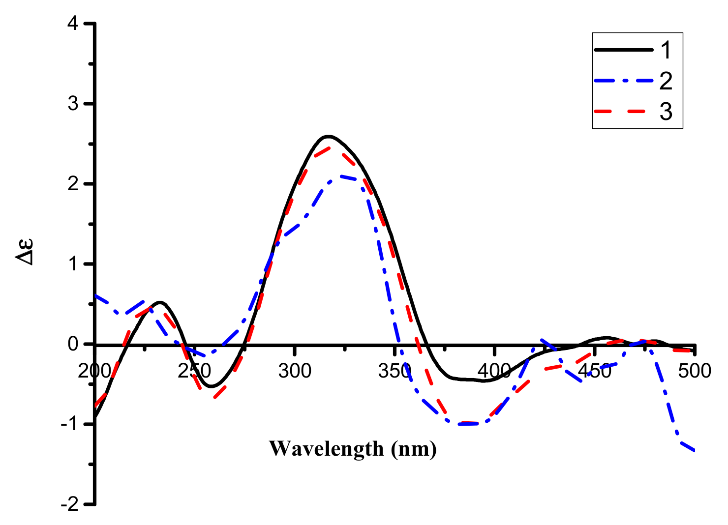
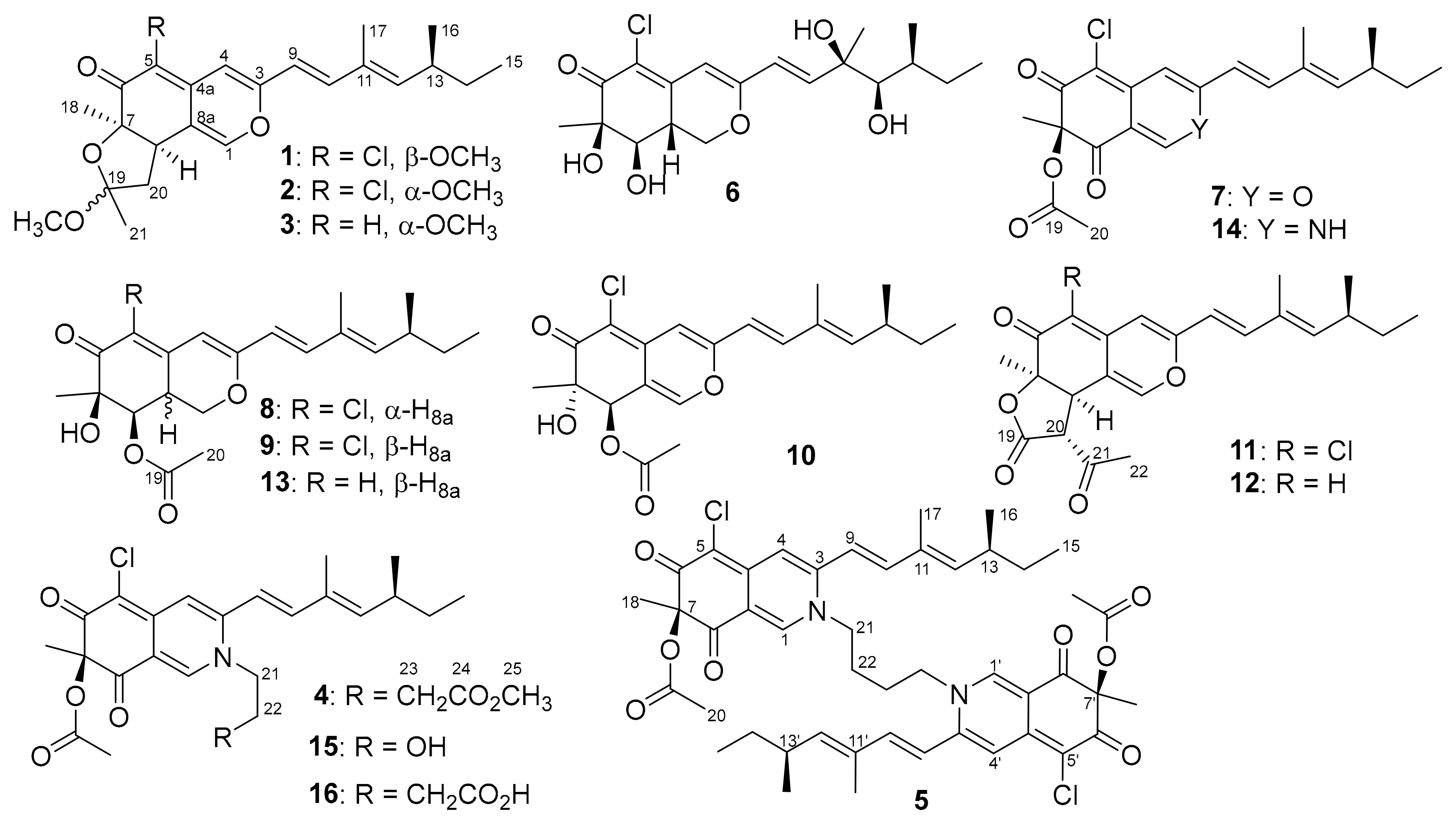
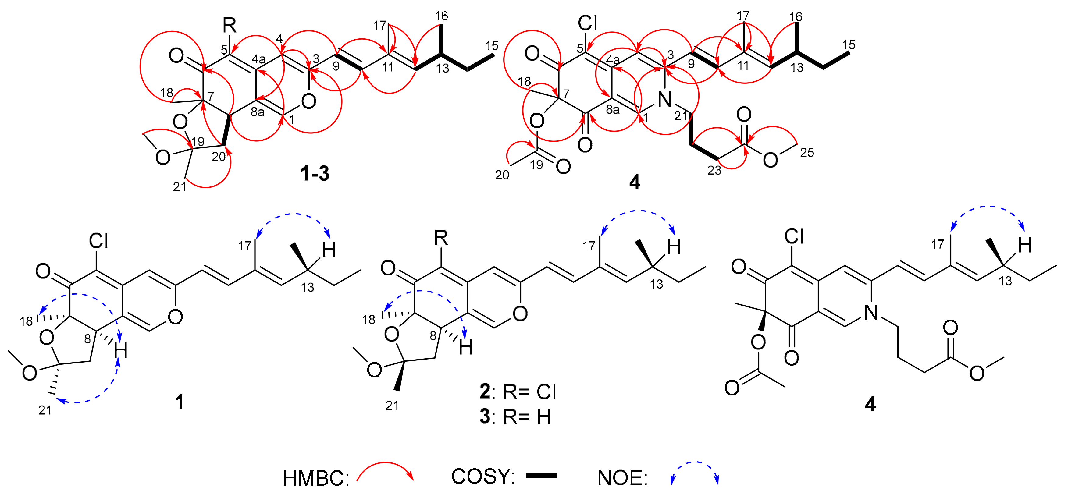
2. Results and Discussion

Sclerotiorin A (1) was obtained as a yellow amorphous powder. The molecular formula of 1 was determined to be C23H29O4Cl by the HRESIMS (high resolution electrospray ionization mass spectroscopy) peak at m/z 405.1834 [M + H]+ (Figure S1), with the 1:3 chlorine isotope peaks. 1H (Table 1, Figure S2), 13C (Table 2, Figure S3) combined with DEPT (distortionless enhancement by polarization transfer, Figure S4) and HSQC (heteronuclear single-quantum correlation, Figure S5) NMR data of 1 revealed the presence of 5 singlet methyls, 1 methoxy, two methylenes, 2 sp3 methines, 2 heteroatom-bonded sp3 non-protonated carbons, 10 olefinic/aromatic carbons, and 1 carbonyl. The HMBC (heteronuclear multiple bond correlation, Figure 2 and Figure S7) from H-1 to C-3 and C-4a, H-4 to C-3, C-5, and C-8a, H-8 to C-1, C-4a, C-6, and C-7 established the core skeleton of azaphilones.8 Moreover, the COSY (correlation spectroscopy) cross peaks (Figure 2 and Figure S6) from H-9 to H-10 and from H-13 to H-12, H-14, H-16, then from H-14 to H-15, along with the HMBC correlations from H-9 to C-11, H-10 to C-17 and C-12, H-12 to C-10 and C-17 demonstrated the presence of the common side chain of azaphilones [8]. The linkage of the unsaturated side chain to C-3 was demonstrated by the HMBC correlations from H-9 to C-3 and C-4 along with that from H-10 to C-3. Additionally, the other HMBC correlations from H-20a to C-7, H-20b to C-19, C-8a ,and C-8, H-β-OCH3 to C-19, and H-21 to C-20 revealed a furan nucleus. The COSY correlations from H-8 to H-20, as well as the HMBC correlations from H-20 to C-7 and C-8, confirmed the connection mode of the furan nucleus to the fused pyrone–quinone core skeleton. Thus, the constitution of sclerotiorin A (1) was determined.
Sclerotiorin B (2) was obtained as a yellow amorphous powder. HRESIMS gave the peak of m/z 405.1836 [M + H]+ (Figure S10) and the chlorine isotope peaks; consequently, the molecular formula was determined to be C23H29O4Cl, the same as that of 1. The NMR data of 2 ( Table 1; Table 2, Figures S11–S14) were similar to those of 1, except that C-8, C-19, and C-20 were shielded and shifted from δC 43.4, 106.0, and 45.8 of 1 to δC 42.7, 105.4, and 44.2 of 2, respectively. The chemical shift changes may be triggered by the difference of the stereochemistry on C-19. Accordingly, compound 2 was identified as a 19-epimer of compound 1, which was further confirmed by the 2D NMR data (Figure 2, Figure S15 and Figure S16).
Sclerotiorin C (3) was also obtained as a yellow powder. The molecular formula was deduced to be C23H30O4 by the m/z 371.2218 [M + H]+ of the HRESIMS (Figure S18), without the chlorine isotope peaks. The NMR data (Table 1 and Table 2, Figures S19–S23) and optical rotation of 3 were similar to those of 2, except for the expected extra CH signal (δC/H 106.1/5.23) on C-5 indicating that the chlorine atom at C-5 in 3 was substituted by a hydrogen atom. The key HMBC correlations (Figure S24) from H-5 (δH 5.23) to C-7 (δC 83.1), C-8a (δC 116.4), and C-4 (δC 107.7) confirmed the position of the extra H-5. Thus, 3 was identified as the dechlorinated derivative of 2, named sclerotiorin C.
Sclerotiorin D (4) was a red powder after purification. The peak m/z 490.1993 [M + H]+ of HRESIMS (Figure S26) indicated the molecular formula was C26H32O6NCl. When comparing the NMR data (Table 1 and Table 2, Figures S27–S30) of compound 4 to those of 16 [28], the most obvious differences were that compound 4 had one more methyl group (δC/H-25 52.2/3.70), and the carbonyl carbon C-24 (δC 172.6) was deshielded. Consequently, compound 4 was supposed to be the methyl ester derivative of 16. A further analysis of 1D and 2D NMR confirmed the chemical composition of compound 4 (Figure 2, Figure S31 and Figure S32).
In all new compounds 14, the large J value between H-9 and H-10 (Table 2) and the NOESY (nuclear Overhauser enhancement spectroscopy) correlations of H-17/H-13 suggested the E-type of Δ9 and Δ11 double bonds (Figure 2, Figures S8, S17, and Figure S25). According to the common Cotton effect of azaphilones established by Steyn and Vleggaar [25,34], the absolute configuration of C-7 in 13 was suggested to be R from the positive Cotton effects at 317 nm (Δε + 2.59, 1), 317 nm (Δε + 2.45, 2), and 324 nm (Δε + 2.09, 3) (Figure 3), which were consistent with those reported for isochromophilones C (314 nm, Δε + 2.85) and D (314 nm, Δε + 3.99) [14]. Then, key NOE (Nuclear Overhauser Effect) enhancements of H-18 (δH 1.19) and H-21 (δH 1.35) were observed after irradiation of H-8 (δH 3.23) in 1 (Figure 2 and Figure S9). However, only the H-8/H-18 correlations were observed on the NOESY spectra of 2 and 3, indicating the opposite configurations of C-19. Therefore, the absolute configurations of sclerotiorin A–C (13) were determined to be 7R/8R/19S, 7R/8R/19R, and 7R/8R/19R, respectively. The absolute configurations of C-13 in 13 were suggested as S by the common biosynthetic pathway of the aliphatic side chain in reported azaphilones [9,14], which were determined by X-ray single-crystal diffraction [35], hydrolysis [36], or ECD (electronic circular dichroism) calculation [14].
Taking into account the structural similarity of 14 with 4, 5, 7, 15, and 16, the absolute configurations of these compounds could be resolved by chemical correlation if a single crystal of 14 could be obtained. Fortunately, a single crystal of compound 14 was obtained, and the X-ray diffraction (Figure 4) clearly determined the absolute configuration of 14 as 7R and 13S from the absolute structure parameter of 0.01(2). Thus, a series of reactions were carried out using (+)-sclerotiorin (7) as a raw material (Scheme 1) [37]. Compounds 1416 were directly produced after the reaction of 7 with ammonium acetate, aminoethanol, and 3-aminobutyric acid, while compounds 4 and 5 resulted from the reactions of 16 and 14 with iodomethane and 1,4-diiodobutane (Figure S39), respectively. The synthetic compounds 4, 5, and 1416 were identified as the natural ones by the same retention times in their co-HPLC profiles, as shown in Figure S37. In addition, compounds 4, 5, and 1416 displayed similar ECD (Figure S38) and the same sign of specific rotation. Therefore, compounds 4, 5, and 1416 had the same (7R,13S) configurations.
Sclerotiorin E (5) was obtained as a red powder. The HRESIMS peak m/z 833.3350 [M + H]+ indicated the molecular formula was C46H54O8N2Cl2 (Figure S33). Although the constitution of 5 [19] was identified to be the same as that reported by NMR (Table S1 and S2, Figures S34–S37), the absolute configuration has not been resolved yet. Expectedly, compound 5 was the dimer of 14 linked by a 1,4-butylidene bridge. As shown in Scheme 1, compound 5 could be semi-synthesized from 14. Thus, the absolute configuration of compound 5 that we named sclerotiorin E was determined to be (7R, 7′R, 13S, 13′S) for the first time.
The anti-H1N1-virus activity of compounds 116 were examined in the MDCK cell line by the CPE (cytopathic effect) + MTT (3-(4,5-dimethyl-2-thiazolyl)-2,5-diphenyl-2-H-tetrazolium bromide) method [38,39]. Compounds 5, 7, 10, 12, 13, 14, and 16 showed better inhibitory activity on H1N1 than the positive control (ribavirin), with IC50 values ranging from 78.6 to 156.8 μM (Table 3). The results displayed the structure–activity relationships of these azaphilones against the H1N1 virus: the chlorine atom at C-5 was not necessary for the activity, the dimer had a stronger anti-viral activity than the monomer, replacement of the oxygen atom connected to C-1 by a nitrogen atom did not affect the anti-H1N1 activity. Furthermore, the α-glycosidase inhibitory activity of compounds 116 was assayed by the PNPG (p-nitrophenyl β-D-glucopyranoside) method [40]. Compounds 11 and 14 displayed good activity against α-glycosidase, with IC50 values of 17.3 and 166.1 μM, respectively (acarbose, 1.1 mM).

3. Experimental Section

3.1. General Experimental Procedures

All the NMR spectra were recorded on a JEOLJN M-ECP 600 spectrometer (JEOL, Tokyo, Japan) or a Bruker Advance 500 spectrometer (Bruker, Fällanden, Switzerland) used TMS (tetramethylsilane) as internal standard. 1H chemical shifts were referenced to the residual CDCl3 or DMSO-d6 signal (δ7.26 and 2.50 ppm, respectively). 13C chemical shifts were referenced to the CDCl3 or DMSO-d6 solvent peak (δ77.16 or 39.52 ppm, respectively). Optical rotations were measured on a JASCO P-1020 digital polarimeter (JASCO Corporation, Tokyo, Japan). ECD data were acquired on a JASCO J-815 spectropolarimeter (JASCO Corporation, Tokyo, Japan). HRESIMS spectra were collected using the Q-TOF ULTIMA GLOBAL GAA076 LC mass spectrometer (Waters Asia Ltd., Singapore). Semi-preparative HPLC was performed using an ODS (octadecylsilyl) column (YMC-pack ODS-A, 10 × 250 mm, 5 μm, 4 mL/min). TLC was performed on plates precoated with silica gel GF254 (10–40 μm, Qingdao Marine Chemical Factory, Qingdao, China). Column chromatography (CC) was carried by silica gel GF254 and Sephadex LH-20 (Amersham Biosciences, Uppsala, Sweden). Vacuum–liquid chromatography (VLC) utilized silica gel H (Qingdao Marine Chemical Factory, Qingdao, China).

3.2. Fungal Material and Fermentation

The fungus OUCMDZ-3839 was isolated from Paratetilla sp. sponges collected from Xisha Island in November 2011. The sponges were firstly clipped, then ground and suspended in sterile distilled water. The suspension was spread on a seawater starch (SWS) agar plate (10 g starch, 1 g protein powder, 20 g agar per liter of sea water). After culturing at 28 °C for several days, the single colony was purified on a potato dextrose agar plate (PDA, 200 g potato, 20 g glucose, 20 g agar per liter of tap water) and maintained at −80 °C. After culture on the PDA plate at 28 °C for 4 days, the mycelium was inoculated into 200 × 1000 mL Erlenmeyer flasks, each containing 300 mL of liquid medium (20 g sorbitol, 20 g maltose, 10 g monosodium glutamate, 0.5 g KH2PO4, 0.3 g MgSO4·7H2O, 0.5 g tryptophan, 3 g yeast extract per liter of sea water pH 6.5). The medium was incubated at 28 °C under static conditions for 41 days.

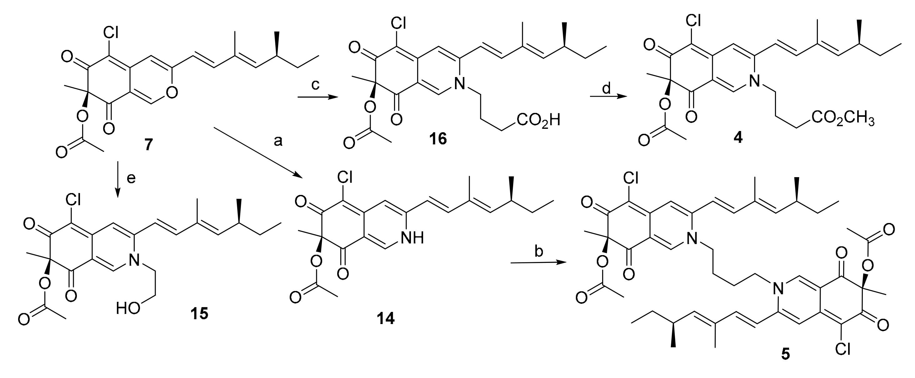
3.3. Extraction and Purification

The mycelium and the aqueous layer of the whole culture (60 L) were separated through cheesecloth. The mycelium was soaked by acetone for three times, and the acetone phases were collected and then evaporated under vacuum until acetone was removed. The residue water layer and the whole aqueous layer were extracted with three volumes of ethyl acetate (EtOAc) for three times. The EtOAc layer was collected and evaporated to dry. A total of 51.4 g of crude extract was obtained. The EtOAc extract was subjected to a silica gel VLC column, eluting with petroleum, petroleum/CH2Cl2 (v/v, 1:1), CH2Cl2, a stepwise gradient of CH2Cl2/MeOH (v/v, 100:1, 75:1,50:1, 25:1, 10:1, 5:1, 2:1, 1:1), and MeOH to give 11 fractions. Fraction 4 (400 mg) was subjected to Sephadex LH-20 chromatography (50 × 1270 mm) to give two fractions (Fr. 4-1–Fr. 4-2). Fraction 4-2 was further purified by HPLC over an ODS column (80% MeOH−H2O, v/v) to give compounds 8 (tR 16.6 min, 3.6 mg) and 9 (tR 20.9 min, 6.3 mg) and a mixture (20 mg) of 7 and 10. The mixture was then subjected to Sephadex LH-20 chromatography (30 × 80 mm) and further purified by HPLC over the ODS column (75% MeOH−H2O, v/v) to give compounds 7 (tR 11.1 min, 12.0 mg) and 10 (tR 26.8 min, 3.0 mg). Fraction 4-1 was subjected to Sephadex LH-20 chromatography (30 × 80 mm) to give Fr. 4-1-1–Fr. 4-1-2. Fraction 4-1-1 was then purified by HPLC over an ODS column (25% MeOH−H2O, v/v) to give compound 12 (tR 15.2 min, 26.0 mg). Fraction 4-1-2 was further subjected to Sephadex LH-20 chromatography (20 × 78 mm) to give 4 fractions (Fr. 4-1-2-1–Fr. 4-1-2-4). Fraction 4-1-2-1 was purified by HPLC over an ODS column (70% MeOH−H2O, v/v) to give compounds 1 (tR 35.4 min, 3.0 mg), 2 (tR 42.2 min, 4.8 mg), and 3 (tR 30.9 min, 3.0 mg). Fraction 4-1-2-2 was further subjected to a silica column, eluting with petroleum ether/EtOAc to give eight fractions (Fr. 4-1-2-2-1–Fr. 4-1-2-2-8). Fraction 4-1-2-2-1 was purified by HPLC over an ODS column (70% MeOH−H2O, v/v) to give compound 14 (tR 24.0 min, 4.5 mg). Fraction 4-1-2-2-5 was purified by HPLC over an ODS column (75% MeOH−H2O, v/v) to give compound 13 (tR 20.3 min, 3.0 mg). Fraction 4-1-2-2-8 was further subjected to a silica column, eluting with petroleum ether/EtOAc to give compound 11 (ODS, 75% MeOH−H2O, v/v, tR 20.2 min, 8.0 mg). Fraction 7 was further subjected to a silica column, eluting with petroleum ether/EtOAc to give two fractions (Fr. 7-1 and Fr. 7-2). Fraction 7-1 was subjected to a silica column, eluting with petroleum ether/EtOAc and further purified by HPLC over an ODS column (75% MeOH−H2O, v/v) to give compound 15 (tR 9.9 min, 100.0 mg). Fraction 7-2 was subjected to a silica LC column, eluting with petroleum ether/EtOAc to give Fr. 7-2-1 and Fr. 7-2-2. Fraction 7-2-1 was further purified by HPLC over an ODS column (75% MeOH−H2O, v/v) to give compound 4 (tR 11.3 min, 8.6 mg). Fraction 7-2-2 was further purified by HPLC over an ODS column (75%MeOH−H2O, v/v) to give compounds 16 (tR 4.2 min, 25.6 mg) and 5 (tR 5.5 min, 45.9 mg). Fraction 8 was further applied to a silica LC column, eluting with petroleum ether/EtOAc, and further purified by HPLC over an ODS column (60% MeOH−H2O, v/v) to give compound 6 (tR 4.39 min, 20.1 mg).
Sclerotiorin A (1): yellow amorphous powder; [ α ] D 25 + 45.7 (c 0.2, EtOH); UV (MeOH) λmax (log ε) 255 (2.07), 361 (2.10), 391 (2.12); ECD (0.6 mM, MeOH) λmax (Δε) 233 (+0.52), 258 (−0.53), 317 (+2.59,) and 394 (–0.46) nm; IR (KBr) νmax 3526, 1618, 1400 cm−1; 1H and 13C NMR data, see Table 1 and Table 2; HRESIMS m/z 405.1834 [M + H]+ (calcd. for C23H30O4Cl, 405.1827).
Sclerotiorin B (2): yellow amorphous powder; [ α ] D 25 + 6.6 (c 0.1, EtOH); UV (MeOH) λmax (log ε) 258 (1.67), 370 (1.76), 390 (1.79); ECD (0.6 mM, MeOH) λmax (Δε) 228 (+0.45), 258 (−0.68), 317 (+2.45), and 387 (−0.10) nm; IR (KBr) νmax 3599, 1622, 1513, 1372 cm−1; 1H and 13C NMR data, see Table 1 and Table 2; HRESIMS m/z 405.1836 [M + H]+ (calcd. for C23H30O4Cl, 405.1827).
Sclerotiorin C (3): yellow amorphous powder; [ α ] D 25 + 10.7 (c 0.1, EtOH); UV (MeOH) λmax (log ε) 248 (0.85), 367 (0.95), 391 (1.02); ECD (0.1 mM, MeOH) λmax (Δε) 225 (+0.52), 253 (−0.14), 324 (+2.09), and 387 (–1.00) nm; IR (KBr) νmax 3465, 1622, 1388 cm−1; 1H and 13C NMR data, see Table 1 and Table 2; HRESIMS m/z 371.2218 [M + H]+ (calcd. for C23H31O4, 371.2217).
Sclerotiorin D (4): red powder; [ α ] D 25 + 207.5 (c 0.025, MeOH); UV (MeOH) λmax (log ε) 236(2.46), 369 (2.56); ECD (0.5 mM, MeOH) λmax (Δε) 244 (+3.7), 303 (−5.98), and 383 (+4.6) nm; IR (KBr) νmax 2921, 2365, 1739, 1591, 1502, 1240 cm−1; 1H and 13C NMR data, see Table 1 and Table 2; HRESIMS m/z 490.1993 [M + H]+ (calcd. for C26H33O6NCl, 490.1991).
Sclerotiorin E(5): red powder, [ α ] D 25 + 143.7 (c 0.05, MeOH); ECD (0.72 mM, MeOH) λmaxε) 223 (−1.43), 244 (+6.68), 305 (−12.21), and 385 (+9.45) nm; IR (KBr) νmax 3443, 2956, 2357, 1707, 1590, 1509, 1236 cm−1; 1H NMR (600 MHz, CDCl3) and 13C NMR (150 MHz, CDCl3) shown in Tables S1–S2; HRESIMS m/z 833.3350 [M + H]+ (calcd. for C46H55O8N2Cl2, 833.3330).

3.4. Conversion from 7 to 14

To a solution of compound 7 (15 mg) in 2 ml of THF, 300 mg of NH4OAc and 300 μL of MeOH were added. The mixture was stirred at 25 °C for 17 h. The reaction mixture was diluted with 5 mL of water and extracted three times with equal volumes of EtOAc. The EtOAc layers were combined, washed with water, dried over anhydrous Na2SO4, filtered, and concentrated in vacuo to yield a product as an orange oil. The orange oil was purified by semipreparative HPLC on ODS (70% acetonitrile/H2O, v/v) to afford compound 14 (6.0 mg, tR 5.6 min, 40% yield) that was identified by the same retention time in the co-HPLC (Figure S37) and the same sign of specific rotation as natural 14.

3.5. Conversion from 7 to 15

Amino ethanol (20 μL) was added to 1 mL of CH2Cl2 solution of compound 7 (6 mg). The reaction solution was stirred at 25 °C for 24 h under a nitrogen atmosphere. After CH2Cl2 was dried in vacuo, the mixture was purified by semipreparative HPLC on an ODS column (85% acetonitrile/H2O, v/v) to afford compound 15 (6.1 mg, tR 5.6 min, 92% yield), which was identified by the same retention time in the co-HPLC (Figure S37) and the same sign of specific rotation as natural 15.

3.6. Conversion from 7 to 16

Amino butyric acid (21.5 mg) was added to 2 mL of DMF solution of compound 7 (8 mg). The solution was stirred at 25 °C for 5 h under a nitrogen atmosphere, and then 10 mL of H2O was added. The reaction mixture was extracted three times with equal volumes of EtOAc. The EtOAc layers were combined, dried over anhydrous Na2SO4, filtered, and concentrated in vacuo to yield an orange oil. The orange oil was purified by semipreparative HPLC on an ODS column (85% acetonitrile/H2O, v/v) to afford compound 16 (9.0 mg, tR 7.9 min, 93% yield), which was identified by the same retention time in the co-HPLC (Figure S37) and the same sign of specific rotation as natural 16.

3.7. Conversion from 14 to 5

1,4-Diiodobutane (6.96 μL) was added to the acetone solution (3 mL) of compound 14 (20 mg) and K2CO3 (21.2 mg). The reaction solution was heated to 50 °C and stirred for 7 days under a nitrogen atmosphere. The reaction solution was concentrated in vacuo and dissolved in 10 mL of H2O. The obtained solution was extracted three times with equal volumes of EtOAc. The EtOAc layers were combined, dried over anhydrous Na2SO4, filtered, and concentrated in vacuo to give a gum that was purified by semipreparative HPLC on an ODS column (85% acetonitrile/H2O, v/v) to afford compound 5 (1.41 mg, tR 7.9 min, 3.3% yield), which was identified by the same retention time in the co-HPLC (Figure S37) and the same sign of specific rotation as natural 5.

3.8. Conversion from 16 to 4

A volume of 10 μL of CH3I was added to the acetone solution of compound 16 (1.33 mg) and K2CO3 (5.50 mg). The solution was stirred at 28 °C for 24 h under a nitrogen atmosphere, and then 10 mL of H2O was added. The mixture was extracted three times with equal volumes of CH2Cl2. After CH2Cl2 was dried in vacuo, the CH2Cl2 extracts were applied to semipreparative HPLC on an ODS column (80% acetonitrile/H2O, v/v) to afford compound 4 (1.28 mg, tR 5.40 min, 94% yield), which was identified by the same retention time in the co-HPLC (Figure S37) and the same sign of specific rotation as natural 4.

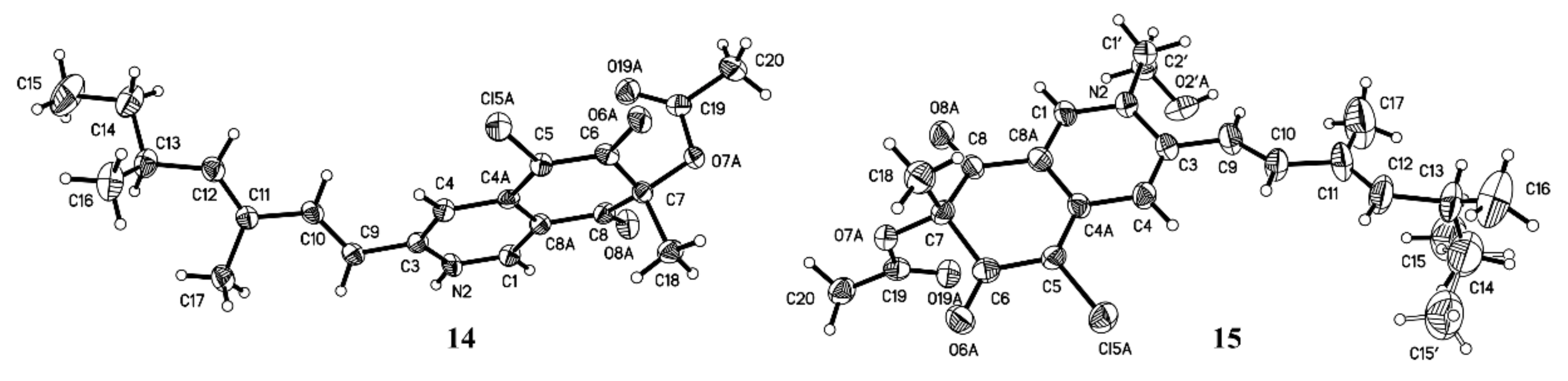
3.9. X-ray Crystal Data for 14 (Mo Kα Radiation)

Red orthorhombic crystal from MeOH; molecular formula C21H25O4NCl; space group P21 with a = 8.6912 (7) Å, b = 7.4026 (6) Å, c = 16.0515 (14) Å, V = 996.39 (14) Å3, absolute structure Flack parameter: 0.01(2). Z = 2, Dcalcd = 1.299 mg/m3, μ = 1.913 mm−1, and F (000) = 412; crystal size 0.35 × 0.12 × 0.07 mm3. T = 293(2) K. A total of 5671 unique reflections (2θ < 140°) were collected on a Bruker Smart CCD area detector diffractometer with graphite monochromated Mo Kα radiation (λ = 1.54178 Å). The structure was solved by direct methods (SHELXS-97) and expanded using Fourier techniques (SHELXL-97). The final cycle of full-matrix least-squares refinement was based on 2612 unique reflections (2θ < 140°) and 249 variable parameters and converged with unweighted and weighted agreement factors of R1 = 0.0426 and wR2 = 0.0952 for I > 2σ (I) data. Crystallographic data for 14 have been deposited in the Cambridge Crystallographic Data Centre as supplementary publication no. CCDC 1055936. Copies of the data can be obtained, free of charge, on application to CCDC, 12 Union Road, Cambridge CB2 1EZ, U.K. (Fax: +44 (0)-1223-336033; e-mail: [email protected]).

3.10. X-ray Crystal Data for 15 (Mo Kα Radiation)

Red orthorhombic crystal from MeOH; molecular formula C23H29O5NCl; space group P212121 with a = 6.7149(3) Å, b = 8.5574 (5) Å, c = 40.085 (2) Å, V = 2303.4 (2) Å3, absolute structure Flack parameter: −0.2(2). Z = 4, Dcalcd = 1.251 mg/m3, μ = 0.774 mm−1, and F (000) = 920; crystal size 0.40 × 0.11 × 0.08 mm3. T = 298(2) K. A total of 10,946 unique reflections (2θ < 50°) were collected on a Bruker Smart CCD area detector diffractometer with graphite monochromated Mo Kα radiation (λ = 0.71073 Å). The structure was solved by direct methods (SHELXS-97) and expanded using Fourier techniques (SHELXL-97). The final cycle of full-matrix least-squares refinement was based on 3965 unique reflections (2θ < 50°) and 288 variable parameters and converged with unweighted and weighted agreement factors of R1 = 0.1023 and wR2 = 0.2473 for I > 2σ(I) data. Crystallographic data for 15 have been deposited in the Cambridge Crystallographic Data Centre as supplementary publication no. CCDC 1056013. Copies of the data can be obtained, free of charge, on application to CCDC, 12 Union Road, Cambridge CB2 1EZ, U.K. (Fax: +44 (0)-1223-336033; e-mail: [email protected])

3.11. Anti-influenza A (H1N1) Virus Bioassay

The antiviral activity against H1N1 was examined by the CPE+MTT assay [38,39]. MDCK cells were cultured in RPMI-1640 medium containing 10% fetal bovine serum in 5% CO2 at 37 °C. When the cells reached 70–80% confluency, they trypsinized into individual cells, and the cell suspension concentration was adjusted to 1 × 10⁵ cells/mL. The cells were seeded in a 96-well plate in 100 μL per well and cultured at 37 °C, 5% CO2, for 18 h. After eliminating the culture medium, the influenza virus (A/Puerto Rico/8/34 (H1N1), PR/8), diluted to 100 TCID50, was added (100 μL per well) to the 96-well plate; an equal amount of virus-free dilution was used as a negative control. The 96-well plate was incubated for 1 h at 37 °C, 5% CO2. The samples to be tested and a positive-control drug were diluted in PBS buffer, and 20 μL of these solutions was added into the wells. PBS buffer was used as a negative control. After incubation for 48 h at 37 °C, the cells were fixed with 100 μL of 4% formaldehyde for 20 min at room temperature, then the formaldehyde was poured out, and 50 μL of 0.1% crystal violet stain was added, staining for 30 min at 37 °C. After the plates were washed and dried, the absorbance (OD) of each well was measured at 570 nm with a microplate reader (Bio-Rad, USA). The IC50, as the compound concentration required to inhibit influenza virus yield at 48 h post-infection by 50%, was calculated. The IC50 value of the positive control, Ribavirin, was 179.8 μM.

3.12. Anti-α-glycosidase Bioassay

The PNPG method [40] used to evaluate the inhibitory activity against α-glycosidase was described in our previous work [41].

4. Conclusions

Four new azaphilones, sclerotiorins A–D (14), along with 12 known analogues (516) were isolated and identified from a fermentation broth of the sponge-derived fungus, P. sclerotiorum OUCMDZ-3839. Compounds 5, 7, 10, 1214, and 16 showed stronger antiviral activity against H1N1 in the MDCK cell line than the positive control Ribavirin. Additionally, compounds 11 and 14 displayed significant inhibitory activity against α-glycosidase, with IC50 values of 17.3 and 166.1 μM, respectively.

Supplementary Materials

The following are available online at https://www.mdpi.com/1660-3397/17/5/260/s1, The ITS rRNA sequences data of Penicillium sp. OUCMDZ-3839; Tables S1–S3: NMR data of known compounds (516); physicochemical data of known compounds 516; specific rotation of synthetic compounds 4, 5 and 1416; Figure S1: HRESIM Spectrum of Compound 1; Figures S2–S9: NMR Spectrum of Compound 1 in DMSO-d6; Figure S10: HRESIM Spectrum of Compound 2; Figures S11–S17: NMR Spectrum of Compound 2 in DMSO-d6; Figure S18: HRESIM Spectrum of Compound 3; Figures S19–S25: NMR Spectrum of Compound 3 in DMSO-d6; Figure S26: HRESIM Spectrum of Compound 4; Figures S27–S32: NMR Spectrum of Compound 4 in CDCl3; Figure S33: HRESIM Spectrum of Compound 5; Figures S34–S36: NMR Spectrum of Compound 5 in CDCl3; Figure S37: Co-HPLC profiles of the synthetic and the natural 4, 5 and 1416; Figure S38: Measured ECD curves of compounds 4, 5, 7 and 1416.

Author Contributions

Y.D. analyzed the data and prepared the draft of the manuscript; Q.J. and C.W. performed most experiments; T.Z. and Y.W. checked the data; W.Z. designed and supervised the research and revised the manuscript.

Funding

This work was financially supported by grants from the National Natural Science Foundation of China (NSFC) (Nos. 41876172, U1501221 & U1606403).

Conflicts of Interest

The authors declare no conflict of interest.

References

  1. Zhao, C.; Zhu, T.; Zhu, W. New marine natural products of microbial origin from 2010 to 2013. Chin. J. Org. Chem. 2013, 33, 1195–1234. [Google Scholar] [CrossRef]
  2. Zhu, T.-H.; Ma, Y.-N.; Wang, W.-L.; Chen, Z.-B.; Qin, S.-D.; Du, Y.-Q.; Wang, D.-Y.; Zhu, W.-M. New marine natural products from the marine-derived fungi other than Penicillium sp. and Aspergillus sp. (1951–2014). Chin. J. Mar. Drugs 2015, 34, 56–108. [Google Scholar] [CrossRef]
  3. Ma, H.; Liu, Q.; Zhu, G.; Liu, H.; Zhu, W. Marine natural products sourced from marine-derived Penicillium fungi. J. Asian Nat. Prod. Res. 2016, 18, 92–115. [Google Scholar] [CrossRef] [PubMed]
  4. Zhao, C.; Liu, H.; Zhu, W. New natural products from the marine-derived Aspergillus fungi-A review. Acta Microbiol. Sin. 2016, 56, 331–362. [Google Scholar] [CrossRef]
  5. Carroll, A.R.; Copp, B.R.; Davis, R.A.; Keyzers, R.A.; Prinsep, M.R. Marine natural products. Nat. Prod. Rep. 2019, 36, 122–173. [Google Scholar] [CrossRef] [PubMed]
  6. Blunt, J.W.; Carroll, A.R.; Copp, B.R.; Davis, R.A.; Keyzers, R.A.; Prinsep, M.R. Marine natural products. Nat. Prod. Rep. 2018, 35, 8–53. [Google Scholar] [CrossRef]
  7. Blunt, J.W.; Copp, B.R.; Keyzers, R.A.; Munro, M.H.G.; Prinsep, M.R. Marine natural products. Nat. Prod. Rep. 2017, 34, 235–294. [Google Scholar] [CrossRef] [PubMed]
  8. Osmanova, N.; Schultze, W.; Ayoub, N. Azaphilones: A class of fungal metabolites with diverse biological activities. Phytochem. Rev. 2010, 9, 315–342. [Google Scholar] [CrossRef]
  9. Gao, J.-M.; Yang, S.-X.; Qin, J.-C. Azaphilones: Chemistry and biology. Chem. Rev. 2013, 113, 4755–4811. [Google Scholar] [CrossRef]
  10. Yamada, T.; Doi, M.; Shigeta, H.; Muroga, Y.; Hosoe, S.; Numata, A.; Tanaka, R. Absolute stereostructures of cytotoxic metabolites, chaetomugilins A–C, produced by a Chaetomium species separated from a marine fish. Tetrahedron Lett. 2008, 49, 4192–4195. [Google Scholar] [CrossRef]
  11. Yasuhide, M.; Yamada, T.; Numata, A.; Tanaka, R. Chaetomugilins, new selectively cytotoxic metabolites, produced by a marine fish-derived Chaetomium species. J. Antibiot. 2008, 61, 615–622. [Google Scholar] [CrossRef] [PubMed]
  12. Yamada, T.; Yasuhide, M.; Shigeta, H.; Numata, A.; Tanaka, R. Absolute stereostructures of chaetomugilins G and H produced by a marine-fish-derived Chaetomium species. J. Antibiot. 2009, 62, 353–357. [Google Scholar] [CrossRef]
  13. Muroga, Y.; Yamada, T.; Numata, A.; Tanaka, R. Chaetomugilins I–O, new potent cytotoxic metabolites from a marine-fish-derived Chaetomium species. Stereochemistry and biological activities. Tetrahedron 2009, 65, 7580–7586. [Google Scholar] [CrossRef]
  14. Luo, X.; Lin, X.; Tao, H.; Wang, J.; Li, J.; Yang, B.; Zhou, X.; Liu, Y. Isochromophilones A–F, cytotoxic chloroazaphilones from the marine mangrove endophytic fungus Diaporthe sp. SCSIO 41011. J. Nat. Prod. 2018, 81, 934–941. [Google Scholar] [CrossRef]
  15. Chen, M.; Shen, N.-X.; Chen, Z.-Q.; Zhang, F.-M.; Chen, Y. Penicilones A–D, anti-MRSA azaphilones from the marine-derived fungus Penicillium janthinellum HK1-6. J. Nat. Prod. 2017, 80, 1081–1086. [Google Scholar] [CrossRef] [PubMed]
  16. Chen, M.; Zheng, Y.-Y.; Chen, Z.-Q.; Shen, N.-X.; Shen, L.; Zhang, F.-M.; Wang, C.-Y. NaBr-induced production of brominated azaphilones and related tricyclic polyketides by the marine-derived fungus Penicillium janthinellum HK1-6. J. Nat. Prod. 2019, 82, 368–374. [Google Scholar] [CrossRef]
  17. Cao, F.; Meng, Z.-H.; Mu, X.; Yue, Y.-F.; Zhu, H.-J. Absolute configuration of bioactive azaphilones from the marine-derived fungus Pleosporales sp. CF09-1. J. Nat. Prod. 2019, 82, 386–392. [Google Scholar] [CrossRef]
  18. Hsu, Y.-W.; Hsu, L.-C.; Liang, Y.-H.; Kuo, Y.-H.; Pan, T.-M. New Bioactive Orange Pigments with Yellow Fluorescence from Monascus-Fermented Dioscorea. J. Agric. Food Chem. 2011, 59, 4512–4518. [Google Scholar] [CrossRef] [PubMed]
  19. Li, J.; Yang, X.; Liu, L.; Lin, Y.C.; Chen, Y.Z.; Lu, Y.J.; He, L.; Li, M.F.; Yuan, J.; He, J.G. Manufacture of azaphilone dimer with marine fungi. CN 103911407, 9 July 2014. [Google Scholar]
  20. Yang, J.; Qi, X.L.; Shao, G.; Pei, Y.H.; Yao, X.S.; Kitanaka, S. Novel antifungal antibiotic, WB produced by the fungus strain 38. Chin. Chem. Lett. 1998, 9, 833–834. [Google Scholar]
  21. Son, S.; Ko, S.K.; Kim, J.W.; Lee, J.K.; Jang, M.; Ryoo, I.J.; Hwang, G.J.; Kwon, M.C.; Shin, K.-S.; Futamura, Y.; et al. Structures and biological activities of azaphilones produced by Penicillium sp. KCB11A109 from a ginseng field. Phytochemistry 2016, 122, 154–164. [Google Scholar] [CrossRef]
  22. Chidananda, C.; Sattur, A.P. Sclerotiorin, a novel inhibitor of lipoxygenase from Penicillium frequentans. J. Agric. Food Chem. 2007, 55, 2879–2883. [Google Scholar] [CrossRef]
  23. Whalley, W.B.; Ferguson, G.; Marsh, W.C.; Restivo, R.J. The chemistry of fungi. Part LXVIII. The absolute configuration of (+)-sclerotiorin and of the azaphilones. J. Chem. Soc. Perkin. Trans. 1 1976, 1366–1369. [Google Scholar] [CrossRef]
  24. Eade, R.A.; Page, H.; Robertson, A.; Turner, K.; Whalley, W.B. The chemistry of fungi. Part XXVIII. Sclerotiorin and its hydrogenation products. J. Chem. Soc. 1957, 4913–4924. [Google Scholar] [CrossRef]
  25. Steyn, P.S.; Vleggaar, R. The structure of dihydrodeoxy-8-epi-austdiol and the absolute configuration of the azaphilones. J. Chem. Soc. Perkin Trans. 1 1976, 204–206. [Google Scholar] [CrossRef]
  26. Matsuzaki, K.; Tanaka, H.; Omura, S. Isochromophilones I and II, novel inhibitors against gpl20-CD4 binding produced by penicillium multicolor FO-2338 II. Structure elucidation. J. Antibiot. 1995, 48, 708–713. [Google Scholar] [CrossRef]
  27. Arai, N.; Shiomi, K.; Tomoda, H.; Tabata, N.; Yang, D.J.; Masuma, R.; Kawakubo, T.; Omura, S. Isochromophilones III-VI, inhibitors of acyl-CoA: Cholesterol acyltransferase produced by Penicillium multicolor FO-3216. J. Antibiot. 1995, 48, 696–702. [Google Scholar] [CrossRef] [PubMed]
  28. Michael, A.; Grace, E.; Kotiw, M.; Barrow, R.A. Isochromophilone IX, a novel GABA-containing metabolite isolated from a cultured fungus, Penicillium sp. Aust. J. Chem. 2003, 56, 13–15. [Google Scholar] [CrossRef]
  29. Yang, D.J.; Tomoda, H.; Tabata, N.; Masuma, R.; Omura, S. New isochromophilones VII and VIII produced by Penicillium sp. FO-4164. J. Antibiot. 1995, 49, 223–229. [Google Scholar] [CrossRef]
  30. Yamazaki, M.; Fujimoto, H.; Matsudo, T.; Yamaguchi, A. Two new fungal azaphilones from Talaromyces luteus, with monoamine oxidase inhibitory effect. Heterocycles 1990, 30, 607–616. [Google Scholar] [CrossRef]
  31. Seto, H.; Tanabe, M. Utilization of 13C-13C coupling in structural and biosynthetic studies. III. Ochrephilone-a new fungal metabolite. Tetrahedron Lett. 1974, 15, 651–654. [Google Scholar] [CrossRef]
  32. Matsuzaki, K.; Tahara, H.; Inokoshi, J.; Tanaka, H.; Masuma, R.; Omura, S. New brominated and halogen-less derivatives and structure-activity relationship of azaphilones inhibiting gp120-CD4 binding. J. Antibiot. 1998, 51, 1004–1011. [Google Scholar] [CrossRef]
  33. Wang, X.; Sena Filho, J.G.; Hoover, A.R.; King, J.B.; Ellis, T.K.; Powell, D.R.; Cichewicz, R.H. Chemical epigenetics alters the secondary metabolite composition of guttate excreted by an Atlantic-forest-soil-derived Penicillium citreonigrum. J. Nat. Prod. 2010, 73, 942–948. [Google Scholar] [CrossRef]
  34. Chen, F.C.; Manchand, P.S.; Whalley, W.B. The chemistry of fungi. Part LXIV. The structure of monascin: The relative stereochemistry of the azaphilones. J. Chem. Soc. C 1971, 3577–3579. [Google Scholar] [CrossRef]
  35. Wang, C.-Y.; Hao, J.-D.; Ning, X.-Y.; Wu, J.-S.; Zhao, D.-L.; Kong, C.-J.; Shao, C.-L.; Wang, C.-Y. Penicilazaphilones D and E: Two new azaphilones from a sponge-derived strain of the fungus Penicillium sclerotiorum. RSC Adv. 2018, 8, 4348–4353. [Google Scholar] [CrossRef]
  36. Yoshida, E.; Fujimoto, H.; Baba, M.; Yamazaki, M. Four new chlorinated azaphilones, helicusins A–D, closely related to 7-epi-sclerotiorin, from an ascomycetous fungus, Talaromyces helices. Chem. Pharm. Bull. 1995, 43, 1307–1310. [Google Scholar] [CrossRef]
  37. Germain, A.R.; Bruggemeyer, D.M.; Zhu, J.; Genet, C.; O’Brien, P.; Porco, J.A. Synthesis of the azaphilones (+)-sclerotiorin and (+)-8-O-methylsclerotiorinamine utilizing (+)-sparteine surrogates in copper-mediated oxidative dearomatization. J. Org. Chem. 2011, 76, 2577–2584. [Google Scholar] [CrossRef]
  38. Hung, H.-C.; Tseng, C.-P.; Yang, J.-M.; Ju, Y.-W.; Tseng, S.-N.; Chen, Y.-F.; Chao, Y.-S.; Hsieh, H.-P.; Shih, S.-R.; Hsu, J.T.-A. Aurintricarboxylic acid inhibits influenza virus neuraminidase. Antivir. Res. 2009, 81, 123–131. [Google Scholar] [CrossRef] [PubMed]
  39. Grassauer, A.; Weinmuellner, R.; Meier, C.; Pretsch, A.; Prieschl-Grassauer, E.; Unger, H. Iota-Carrageenan is a potent inhibitor of rhinovirus infection. Virol. J. 2008, 5, 107. [Google Scholar] [CrossRef]
  40. Nampoothiri, S.V.; Prathapan, A.; Cherian, O.L.; Raghu, K.G.; Venugopalan, V.V.; Sundaresan, A. In vitro antioxidant and inhibitory potential of Terminalia bellerica and Emblica officinalis fruits against LDL oxidation and key enzymes linked to type 2 diabetes. Food Chem. Toxicol. 2011, 49, 125–131. [Google Scholar] [CrossRef] [PubMed]
  41. Chen, Z.; Hao, J.; Wang, L.; Wang, Y.; Kong, F.; Zhu, W. New α-glucosidase inhibitors from marine algae-derived Streptomyces sp. OUCMDZ-3434. Sci. Rep. 2016, 6, 20004. [Google Scholar] [CrossRef]
Figure 1. Structures of compounds 116.
Marinedrugs 17 00260 g001
Figure 2. Key COSY, HMBC, and Nuclear Overhauser Effect (NOE) correlations of compounds 14.
Marinedrugs 17 00260 g002
Figure 3. Measured ECD curves of compounds 13.
Marinedrugs 17 00260 g003
Scheme 1. Chemical transformation of 7 to 4, 5, and 1416. Reagents and conditions: a. NH4OAc, MeOH, THF (tetrahydrofuran), 17h, r.t (room temperature), 40.1% yield; b. 1,4-diiodobutane, K2CO3, acetone, 7d, 50 °C, 3.3% yield; c. aminobutyric acid, DMF (N,N-dimethylformamide), N2, 5h, r.t, 92.7% yield; d. CH3I, K2CO3, acetone, N2, 24h, r.t, 94.1% yield; e. aminoethanol, CH2Cl2, N2, 24h, r.t, 92.4% yield.
Marinedrugs 17 00260 sch001
Figure 4. ORTEP (Oak Ridge Thermal-Ellipsoid Plot) drawing of compounds 14 and 15 (Mo Kα).
Marinedrugs 17 00260 g004
Table 1. 13C (125 MHz) NMR data for compounds 14 (δ in ppm).
Position1 a2 a3 a4 b
δCδCδCδC
1144.6, CH145.4, CH145.4, CH141.2, CH
3157.4, C157.7, C155.4, C148.3, C
4105.0, CH104.9, CH107.7, CH111.7, CH
4a138.9, C139.6, C143.0, C144.9, C
5109.0, C108.4, C106.1, CH102.3, C
6187.7, C187.9, C195.1, C184.5, C
783.5, C84.1, C83.1, C84.8, C
843.4, CH42.7, CH43.2, CH193.9, C
8a116.4, C115.7, C116.4, C115.1, C
9117.6, CH117.5, CH117.4, CH114.6, CH
10140.3, CH140.5, CH139.0, CH145.6, CH
11132.2, C132.2, C132.0, C132.2, C
12146.2, CH146.4, CH145.3, CH148.4, CH
1334.3, CH34.3, CH34.2, CH35.2, CH
1429.6, CH229.6, CH229.6, CH230.0, CH2
1511.8, CH311.8, CH311.9, CH312.1, CH3
1620.2, CH320.2, CH320.3, CH320.3, CH3
1712.3, CH312.3, CH312.3, CH312.6, CH3
1823.9, CH324.4, CH324.3, CH323.4, CH3
19106.0, C105.4, C105.1, C170.3, C
2045.8, CH244.2, CH244.4, CH220.4, CH3
2122.9, CH322.8, CH323.0, CH353.4, CH2
22---25.4, CH2
23---30.2, CH2
24---172.6, C
OCH348.3, CH348.3, CH348.3, CH352.2, CH3
a recorded in DMSO-d6. b recorded in CDCl3.
Table 2. 1H (500 MHz) NMR data for compounds 14 (δ in ppm).
Position1 a2 a3a4 b
δH (J in Hz)δH (J in Hz)δH (J in Hz)δH (J in Hz)
17.65, s7.80, s7.65, s7.76, s
46.67, s6.69, s6.40, s7.05, s
5--5.23, s-
83.23, dd (10.3, 10.3)3.42, dd (12.7, 10.3)3.30, m-
96.44, d (15.8)6.45, d (15.8)6.18, d (15.8)6.31, d (15.3)
106.99, d (15.8)7.01, d (15.8)6.90, d (15.8)6.98, d (15.3)
125.71, d (9.7)5.72, d (9.6)5.65, d (9.7)5.71, d (9.7)
132.46, m2.46, m2.45, m2.48, m
141.39, m; 1.26, m1.39, m; 1.27, m1.37, m; 1.27, m1.44, m; 1.36, m
150.81, t (7.5)0.81, t (7.5)0.81, t (7.4)0.88, t (7.4)
160.96, d (6.6)0.96, d (6.6)0.95, d (6.6)1.02, d (6.6)
171.80, s1.80, s1.77, s1.90, s
181.19, s1.25, s1.20, s1.55, s
202.36, dd (13.0,10.0)
1.89, dd (13.0,10.5)
2.18, dd (12.7,7.3)
1.87, dd (12.7,10.5)
2.15, dd (13.1,10.0)
1.84, m
2.16, s
211.35, s1.25, s1.23, s3.95, t (7.8)
22---2.05, m
23---2.43, t (6.4)
OCH33.03, s3.20, s3.19, s3.70, s
a recorded in DMSO-d6. b recorded in CDCl3.
Table 3. Activity against H1N1 of compounds 5, 7, 10, 1214, and 16. IC50.
Compound571012131416Ribavirin
IC50 (μM)78.6128.7115.0150.591.4133.9156.8179.8

