Article

Structures and Bioactivities of Psolusosides B1, B2, J, K, L, M, N, O, P, and Q from the Sea Cucumber Psolus fabricii. The First Finding of Tetrasulfated Marine Low Molecular Weight Metabolites

by
Alexandra S. Silchenko
,
Anatoly I. Kalinovsky
,
Sergey A. Avilov
,
Vladimir I. Kalinin
*,
Pelageya V. Andrijaschenko
,
Pavel S. Dmitrenok
,
Roman S. Popov
and
Ekaterina A. Chingizova
G.B. Elyakov Pacific Institute of Bioorganic Chemistry, Far Eastern Branch of the Russian Academy of Sciences, Pr. 100-letya Vladivostoka 159, Vladivostok 690022, Russia
*
Author to whom correspondence should be addressed.
Mar. Drugs 2019, 17(11), 631; https://doi.org/10.3390/md17110631
Submission received: 13 October 2019 / Revised: 31 October 2019 / Accepted: 2 November 2019 / Published: 6 November 2019
(This article belongs to the Special Issue Marine Glycoconjugates: Trends and Perspectives)

Abstract

:
Ten new di-, tri- and tetrasulfated triterpene glycosides, psolusosides B1 (1), B2 (2), J (3), K (4), L (5), M (6), N (7), O (8), P (9), and Q (10), were isolated from the sea cucumber Psolus fabricii collected in the Sea of Okhotsk near the Kurile Islands. Structures of these glycosides were established by two-dimensional (2D) NMR spectroscopy and HR-ESI mass-spectrometry. It is particularly interesting that highly polar compounds 9 and 10 contain four sulfate groups in their carbohydrate moieties, including two sulfates in the same terminal glucose residue. Glycoside 2 has an unusual non-holostane aglycone with 18(16)-lactone and a unique 7,8-epoxy fragment. Cytotoxic activities of compounds 110 against several mouse cell lines such as Ehrlich ascites carcinoma cells, neuroblastoma Neuro 2A, normal epithelial JB-6 cells, and erythrocytes were quite different depending both on structural peculiarities of these glycosides and the type of cells subjected to their actions. Psolusoside L (5), pentaoside, with three sulfate groups at C-6 of two glucose and one 3-O-methylglucose residue and holostane aglycone, is the most active compound in the series. The presence of a sulfate group at C-2 of the terminal glucose residue attached to C-4 of the first (xylose) residue significantly decreases activities of the corresponding glycosides. Psolusosides of group B (1, 2, and known psolusoside B) are inactive in all tests due to the presence of non-holostane aglycones and tetrasaccharide-branched sugar chains sulfated by C-2 of Glc4.

1. Introduction

Triterpene glycosides of sea cucumbers are well known by their structural diversity and promising biological effects [1,2,3], including cytotoxicity against cancer cells and antitumor activity [4,5,6]. Therefore, the search for new representatives of this class of marine natural products and studies of their biological activities seem to be relevant. Moreover, structural analysis of diverse glycosides of sea cucumbers allows us to understand the peculiarities of biosynthesis of these complicated and numerous marine metabolites.
As a continuation of our investigation of triterpene glycoside composition of the sea cucumber Psolus fabricii (Psolidae, Dendrochirotida) [7,8,9,10,11,12] we report herein the isolation of ten new glycosides, psolusosides B1 (1), B2 (2), J–Q (310), and their structural elucidation based on the 1H, 13C NMR, one-dimensional (1D) TOCSY and 2D NMR (1H,1H-COSY, HMBC, HSQC, ROESY), and HR-ESI mass spectrometry. The hemolytic activities against mouse erythrocytes and cytotoxic activities of 110 against mouse Ehrlich ascites carcinoma cells, neuroblastoma Neuro 2A, and normal epithelial JB-6 cells have been studied.

2. Results and Discussion

2.1. Structural Elucidation of the Glycosides

The initial stages of isolation of compounds 110 were the same as for other glycosides from P. fabricii and were described earlier [10,11,
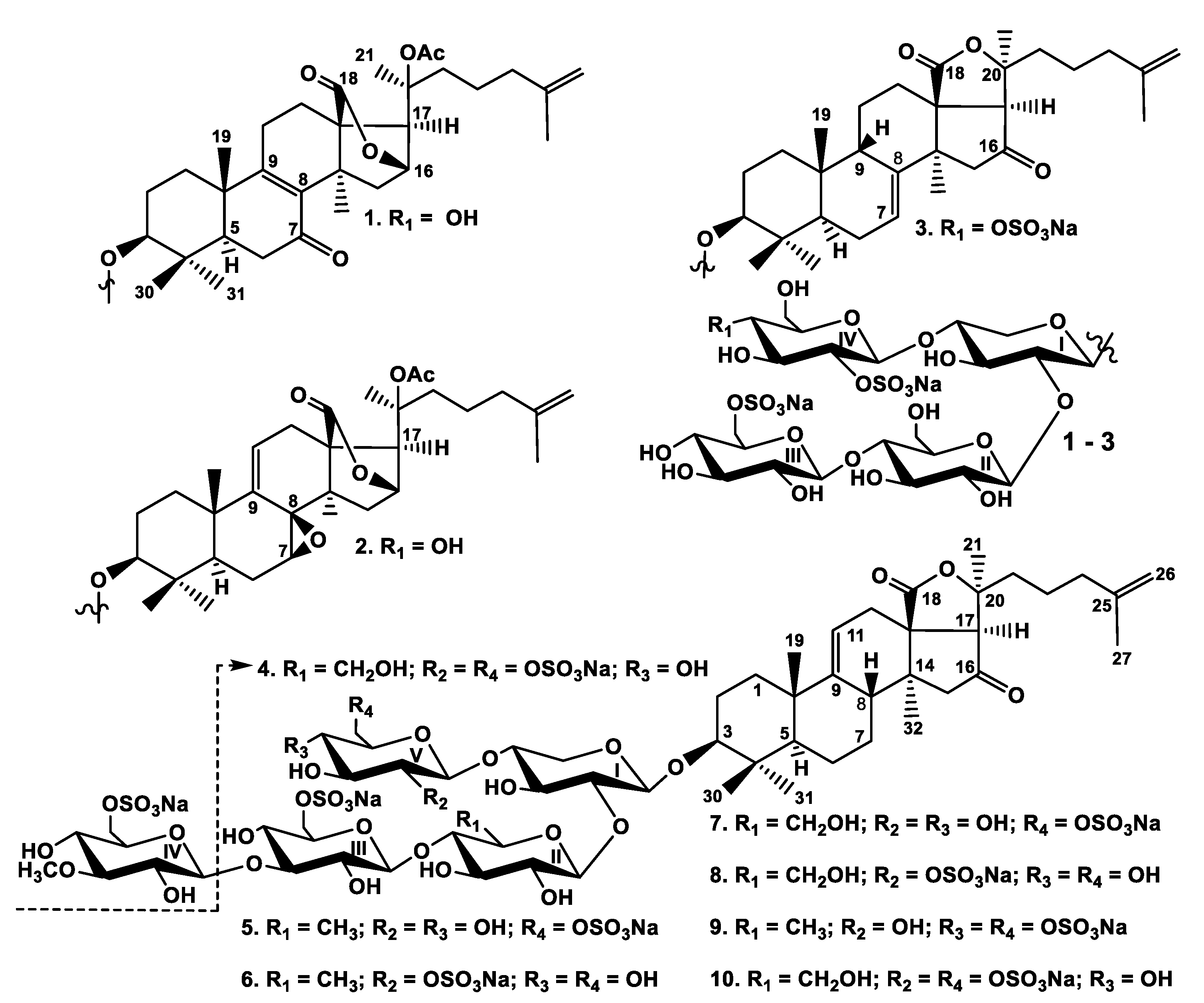
12]. The individual glycosides were isolated by HPLC on reversed-phase columns to give psolusosides: B1 (1) (7,3 mg), B2 (2) (3.4 mg), J (3) (4.8 mg), K (4) (3.4 mg), L (5) (60 mg), M (6) (1.0 mg), N (7) (8.8 mg), O (8) (0.6 mg), P (9) (8.5 mg), and Q (10) (1.4 mg) (Figure 1).
The 1H and 13C NMR spectra corresponding to the carbohydrate chains of psolusosides B1 (1) and B2 (2) were coincident to each other and to those of known psolusoside B [12] showing the identity of their tetrasaccharide carbohydrate moieties branched by C-4 of the xylose unit and having two sulfate groups (Table S1).
The molecular formula of psolusoside B1 (1) was determined to be C55H82O31S2Na2 from the [M2Na − Na] ion peak at m/z 1325.4164 (calc. 1325.4185) and [M2Na − 2Na]2 ion peak at m/z 651.2157 (calc. 651.2146) in the (−)HR-ESI-MS. The signal of H-16 was observed as a broad singlet at δH 4.89 and the signal of H-17 was observed as a singlet at δH 2.97 in the 1H NMR spectrum of 1. These data as well as corresponding signals of carbons at δC 79.9 (C-16) and δC 58.8 (C-17) (Table 1) were indicative for 18(16)-lactone moiety (Table 1). O-acetyl group (δC 170.9 (CH3COO) and 21.6 (CH3COO) in the 13C NMR spectrum), attached to C-20, caused the deshielding of its signal to δC 83.8 in the same manner as in the spectrum of psolusoside B [12]. The side chain of 1 was identical to that of psolusoside B due to the coincidence of those signals in the 1H and 13C NMR spectra. The signal at δC 199.3 corresponded to a keto-group adjacent to a double bond (the signals of olefinic carbons at δC 135.3 (C-8) and 169.0 (C-9)). The position of the keto-group was deduced as C-7 based on the correlations between H2-6 (δH 2.42 and δH 2.29) and C-7 (δC 199.3) in the HMBC spectrum of 1. This was also corroborated by an isolated spin system between the doublet of doublets at δH 1.54 (H-5) and another doublet of doublets at δH 2.42 (H-6a) and the triplet at δH 2.29 (H-6b) observed in the 1H,1H-COSY spectrum. The 8(9)-position of double bond was confirmed by the HMBC correlations H3-32/C-8 and H3-19/C-9. So, the aglycone of psolusoside B1 (1) is characterized by the unique combination of such structural features as 7-keto-8(9)-ene fragment and 18(16)-lactone.
The (−)ESI-MS/MS of 1 demonstrated the fragmentation of [M2Na − Na] ion at m/z 1325.4. The peaks of fragment ions were observed at m/z 1265.4 [M2Na − Na − CH3COOH], 1145.4 [M2Na − Na − CH3COOH − NaHSO4], 1001.4 [M2Na − Na − CH3COOH − C6H10O8SNa (GlcSO3Na) + H], and 839.3 [M2Na − Na − CH3COOH − GlcSO3Na − Glc + H] corroborating the structure of psolusoside B1 (1).
All these data indicate that psolusoside B1 (1) is 3β-O-{6-O-sodium-sulfate-β-d-glucopyranosyl-(1→4)-β-d-glucopyranosyl-(1→2)-[2-O-sodium-sulfate-β-d-glucopyranosyl-(1→4)]-β-d-xylopyranosyl}-7-keto-20(S)-acetoxylanosta-8,25-diene-18(16)-lactone.
The molecular formula of psolusoside B2 (2) (C55H82O31S2Na2) was determined to be the same as of 1 from the [M2Na − Na] ion peak at m/z 1325.4163 (calc. 1325.4185) and [M2Na − 2Na]2− ion peak at m/z 651.2159 (calc. 651.2146) in the (−)HR-ESI-MS. In the 1H and 13C NMR spectra of the aglycone part of 2 the signals characteristic of 18(16)-lactone (δH 4.94 (brs, H-16), δH 3.01 (s, H-17), δC 79.3 (C-16), and δC 60.2 (C-17) as well as O-acetylated C-20 (δH 2.05 (s, CH3COO), δC 21.8 (CH3COO), δC 170.9(CH3COO), and δC 83.9 (C-20)) were observed (Table 2). The side chains in aglycones of 1 and 2 were identical to each other. The signal at δH 3.10 (d, 6.6, H-7) was assigned by the 1H,1H-COSY spectrum where the protons H-5/H-6/H-7 formed an isolated spin system. The corresponding signal of C-7 at δC 56.2 was deduced by the HSQC spectrum of 2. The signal of quaternary C-8 assigned by the HMBC correlations H3-32/C-8, H2-6/C-8, and H-7/C-8 was deshielded to δC 59.6 in the 13C NMR spectrum. These data indicated the presence of an oxygen-bearing substituent at C-7 and C-8, which was supposed to be an 7,8-epoxide [13] that correlated with the MS data. The olefinic broad doublet of doublets at δH 6.00 was assigned to H-11 due to its correlation with H2-12 (δH 2.81 (dd, 5.2; 17.6, H-12a) and 2.60 (brdd, 2.3; 17.6, H-12b)) in the 1H,1H-COSY spectrum. The signal at δC 122.7 corresponded to olefinic C-11 and was deduced by the HSQC spectrum. So, the double bond could occupy the 9(11)-position only. The signal of C-9 at δC 143.2 correlated in the HMBC spectrum with both δH 2.81 (H-12a) and 2.60 (H-12b) and the methyl singlet δH 1.13 (H3-19).
The configuration of C-7 was established as (S) by the ROE-correlation H-7/H3-32 and was confirmed by the coupling pattern of H-7 (δH 3.10 (d, 6.6)), that coincided with the calculated coupling constant based on dihedral angle values in the optimized MM2 model of aglycone of psolusoside B2 (2) having H-7α-orientation and 8(R)-configuration. Thus, the aglycone of psolusoside B2 (2) has unprecedented 7(S),8(R)-epoxy-20(S)-acetoxylanosta-9(11),25-diene-18(16)-lactone structure.
The (−)ESI-MS/MS of 2 showed the fragmentation of [M2Na − Na] ion at m/z 1325.4. The peaks of fragment ions were observed at the same m/z values of 1265.4, 1145.4, 1001.4, and 839.3 as in the spectrum of 1, corroborating the identity of the carbohydrate chains of 1 and 2. Additionally, the fragment ion-peaks at m/z 535.1 [M2Na − Na − C32H45O6 (Agl) − C6H10O8SNa (GlcSO3Na)] and 403 [M2Na − Na − C32H45O6 (Agl) − C6H10O8SNa (GlcSO3Na) − Xyl (C5H8O4)] corresponding to the tri- and disaccharide fragments, were observed in the MS/MS spectrum of 2.
All these data indicate that psolusoside B2 (2) is 3β-O-{6-O-sodium-sulfate-β-d-glucopyranosyl-(1→4)-β-d-glucopyranosyl-(1→2)-[2-O-sodium-sulfate-β-d-glucopyranosyl-(1→4)]-β-d-xylopyranosyl}-7(S),8(R)-epoxy-20(S)-acetoxylanosta-9(11),25-diene-18(16)-lactone.
The molecular formula of psolusoside J (3) (C53H79O32S3Na3) was determined from the [M3Na − Na] ion peak at m/z 1369.3485 (calc. 1369.3517), [M3Na − 2Na]2 ion peak at m/z 673.1812 (calc. 673.1813), and [M3Na − 3Na]3 ion peak at m/z 441.1248 (calc. 441.1244) in the (−)HR-ESI-MS. The 1H and 13C NMR spectra of the aglycone part of psolusoside J (3) coincided with those of psolusoside H isolated earlier from P. farbricii [12] (Table S2) indicating the identity of their holostane-type aglycones having 7(8)- and 25(26)-double bonds and 16-keto-group. This aglycone is common for the glycosides of sea cucumbers belonging to the orders Dendrochirotida and Aspidochirotida [2,12].
In the 1H and 13C NMR spectra of the carbohydrate part of psolusoside J (3) four characteristic doublets at δH 4.60–5.12 (J = 7.3 − 8.1 Hz) and, corresponding to them, signals of anomeric carbons at δC 101.7–105.5 were indicative of a tetrasaccharide chain and β-configurations of glycosidic bonds. The 13C NMR spectra of tetrasaccharide carbohydrate chain of 3 and those of 1 and 2 were quite different, while the 1H,1H-COSY and 1D TOCSY spectra of 3 showed the signals of four isolated spin systems assigned to one xylose and three glucose residues as in psolusosides B [12], B1 (1), and B2 (2). The positions of interglycosidic linkages were elucidated by the ROESY and HMBC spectra of 3 (Table 3), where the correlations between H-1 of the xylose (Xyl1) and H-3 (C-3) of the aglycone, H-1 of the second residue (glucose, Glc2) and H-2 (C-2) of the xylose (Xyl1), H-1 of the third residue (glucose, Glc3) and H-4 (C-4) of the second residue (glucose, Glc2), H-1 of the fourth residue (glucose, Glc4) and H-4 (C-4) of the first residue (xylose, Xyl1) were observed, indicating the same architecture of sugar chains in 3 and 1 and 2. The comparison of the NMR spectra of 1 and 3 showed the coincidence of the signals of three monosaccharide residues corresponding to the linear part of the carbohydrate chain (residues I–III). The signals of terminal monosaccharide unit attached to C-4 of the first (Xyl1) unit, assigned by the 1H,1H-COSY and 1D TOCSY spectra of 3 were indicative of a sulfated by C-2 glucose residue due to characteristic shifting effects observed in the 13C NMR spectrum: the signal of C-2 Glc4 was deshielded to δC 81.2 and the signal of C-1 Glc4 was shielded to δC 101.7 in comparison with the corresponding signals of the same sugar unit in the 13C NMR spectrum of psolusoside I isolated by us earlier [12].
The δC of the signals of C-2 and C-1 of the fourth monosaccharide unit (Glc4) in the 13C NMR spectrum of psolusoside J (3) were very close to those in the 13C NMR spectrum of 1, corroborating the presence of a sulfate group at C-2 of this residue (Glc4). The correlations between H-2/H-3/H-4 in this monosaccharide residue, deduced by the 1H,1H-COSY spectrum of 3, indicated the signal of H-4 Glc4 at δH 4.90. The signal of the corresponding carbon (C-4 Glc4), deduced by the HSQC spectrum, was downshifted to δC 77.3 as compared with the same signal (C-4 Glc4) at δC 70.7 in the 13C NMR spectrum of 1. Actually, the signals at δC ~70.4–70.8 were absent and the signals of C-3 Glc4 and C-5 Glc4 were upshifted to δC 75.6 and 76.6, correspondingly, in the 13C NMR spectrum of 3 due to β-shifting effect of sulfate group, when compared with the corresponding signals in the 13C NMR spectrum of 1. Considering that (−)HR-ESI-MS indicated the presence of three sulfate groups as well as the NMR data, the attachment of the third sulfate group to C-4 of Glc4 was supposed. The signal at δC 62.4 (C-6 Glc4) was characteristic for carbons of non-sulfated hydroxy-methylene groups of glucopyranose residues and excluded the positioning of the third sulfate group at C-6 Glc4 that confirmed our supposition. Hence psolusoside J (3) is a trisulfated tetraoside with two sulfate groups attached to the same glucose residue. To the best our knowledge, this structural feature is first found in the glycosides.
The (−)ESI-MS/MS of 3 demonstrated the fragmentation of [M3Na − Na] ion at m/z 1369.3. The peaks of fragment ions were observed at m/z 1249.4 [M3Na − Na − NaHSO4], 1105.4 [M3Na − Na − C6H9O8SNa (GlcSO3Na)], 1003.4 [M3Na − Na − C6H9O8SNa (GlcSO3Na) − NaSO3 + H], 841.4 [M3Na − Na − NaSO3 − GlcSO3Na − Glc + H], 403.0 [M3Na − Na − C30H43O4 (Agl) − C6H9O11S2Na2 (Glc(SO3Na)2) − Xyl (C5H8O4)], and 241.0 [M3Na − Na − C30H43O4 (Agl) − C6H9O11S2Na2 (Glc(SO3Na)2) − Xyl (C5H8O4) − Glc (C6H10O5)], corroborating the structure of psolusoside J (3).
All these data indicate that psolusoside J (3) is 3β-O-{6-O-sodium-sulfate-β-d-glucopyranosyl-(1→4)-β-d-glucopyranosyl-(1→2)-[2,4-O-sodium-disulfate-β-d-glucopyranosyl-(1→4)]-β-d-xylopyranosyl}-16-ketoholosta-7,25-diene.
The 13C NMR spectra of the aglycone moieties of the glycosides 410 were identical to each other (Table S3) and to those of psolusosides E, F, and G containing 16-ketoholosta-9(11),25-dien-3β-ol as an aglycone, known earlier and frequently occurring in the glycosides of sea cucumbers [12].
The molecular formula of psolusoside K (4) was determined to be C53H79O32S3Na3 from the [M3Na − Na] ion peak at m/z 1369.3485 (calc. 1369.3517), [M3Na − 2Na]2 ion peak at m/z 673.1821 (calc. 673.1813), and [M3Na − 3Na]3 ion peak at m/z 441.1255 (calc. 441.1244) in the (−)HR-ESI-MS and was coincident with the formula of psolusoside J (3). In the 1H and 13C NMR spectra of the carbohydrate moiety of psolusoside K (4) four characteristic doublets at δH 4.61–5.07 (J = 7.2–8.4 Hz) and corresponding signals of anomeric carbons at δC 101.4–104.7 were indicative of a tetrasaccharide chain and β-configurations of glycosidic bonds. The positions of interglycosidic linkages were elucidated by the ROESY and HMBC spectra of 4 (Table 4) as described above indicating the presence of a tetrasaccharide carbohydrate chain branched by C-4 of the xylose residue (Xyl1). The monosaccharide composition of 4, deduced from the 1H,1H-COSY and 1D TOCSY spectra, was the same as in glycosides 13. The comparison of the 13C NMR spectra of trisulfated compounds 3 and 4 showed the coincidence of the signals corresponding to three monosaccharide residues (residues I–III in the formula) forming the linear part of the sugar chain. The signals of C-2 Glc4 at δC 80.3 and C-1 Glc4 at δC 101.4 in the 13C NMR spectrum of 4 were very close to those in the spectrum of 3 that indicated the attachment of a sulfate group to C-2 Glc4 in psolusoside K (4). All of the signals of this monosaccharide residue were assigned using the 1H,1H-COSY and 1D TOCSY spectra. The doublet at δH 5.00 and the doublet of doublets at δH 4.63 corresponded to the protons of the hydroxy-methylene group of the terminal glucose unit (H2-6 Glc4) and were deshielded as compared with the corresponding signals in the 1H NMR spectrum of 3. The signal at δC 67.4 (C-6 Glc4) also indicated the presence of a sulfate group at C-6 of Glc4 in addition to another sulfate group at C-2 of Glc4. So, psolusoside K (4) is an isomer of psolusoside J (3) by the sulfate position and is the second glycoside from sea cucumbers that contains two sulfate groups bonded to the same monosaccharide residue.
The (−)ESI-MS/MS of 4 demonstrated the fragmentation of [M3Na − Na] ion at m/z 1369.3. The peaks of fragment ions were observed at the same m/z: 1249.4 [M3Na − Na − NaHSO4], 1105.4 [M3Na − Na − C6H9O8SNa (GlcSO3Na)], 1003.4 [M3Na − Na − C6H9O8SNa (GlcSO3Na) − NaSO3 + H], 403.0 [M3Na − Na − C30H43O4 (Agl) − C6H9O11S2Na2 (Glc(SO3Na)2) − Xyl (C5H8O4)], and 241.0 [M3Na − Na − C30H43O4 (Agl) − C6H9O11S2Na2 (Glc(SO3Na)2) − Xyl (C5H8O4) − Glc (C6H10O5)] as in the MS/MS of psolusoside J (3) corroborating their isomerism.
All these data indicate that psolusoside K (4) is 3β-O-{6-O-sodium-sulfate-β-d-glucopyranosyl-(1→4)-β-d-glucopyranosyl-(1→2)-[2,6-O-sodium-disulfate-β-d-glucopyranosyl-(1→4)]-β-d-xylopyranosyl}-16-ketoholosta-9,25-diene.
The molecular formula of psolusoside L (5) (C60H91O36S3Na3) was determined from the [M3Na − Na] ion peak at m/z 1529.4222 (calc. 1529.4253), [M3Na − 2Na]2 ion peak at m/z 753.2190 (calc. 753.2180), and [M3Na − 3Na]3 ion peak at m/z 494.4835 (calc. 494.4823) in the (−)HR-ESI-MS, indicating the presence of three sulfate groups. In the 1H and 13C NMR spectra of the carbohydrate part of psolusoside L (5) five characteristic doublets at δH 4.65–5.16 (J = 6.9–8.1 Hz) and, corresponding to them, signals of anomeric carbons at δC 103.4–104.8 were indicative of a pentasaccharide chain and β-configurations of glycosidic bonds (Table 5). Analysis of the 1H,1H-COSY and 1D TOCSY spectra of psolusoside L (5) showed the presence of one xylose, one quinovose, two glucose, and one 3-O-methylglucose residues. The presence of a quinovose residue was confirmed by the 1H and 13C NMR spectra demonstrating the characteristic doublet at δH 1.59 (H-6 Qui2) and the signal at δC 17.7 (C-6 Qui2). The positions of interglycosidic linkages and the consequence of monosaccharides in the chain of 5 were established by analysis of the ROESY and HMBC spectra (Table 5) indicating the presence of branched pentasaccharide moiety with glucose, attached to C-4 Xyl1, and 3-O-methylglucose, attached to C-3 Glc3, as terminal residues. The 13C NMR spectrum of 5 demonstrated three signals at δC 67.0, 67.5, and 67.6, corresponding to sulfated hydroxy-methylene groups of glucopyranose residues that indicated the sulfation of two glucose and 3-O-methylglucose units in the carbohydrate chain of 5.
The comparison of the 13C NMR spectrum of the sugar part of psolusoside L (5) with those of known achlioniceosides A1, A2, and A3, with identical carbohydrate chains, isolated earlier from the sea cucumber Rhipidothuria racowitzai [14] showed the coincidence of the signals of four monosaccharide residues in their spectra. The signals of terminal 3-O-methylglucose residues of the novel and known compounds were different due to the absence of a sulfate group in this residue of known compounds. All these data indicated that psolusoside L (5) is a pentaoside with a new trisulfated carbohydrate chain branched by C-4 Xyl1.
The (−)ESI-MS/MS of 5 demonstrated the fragmentation of [M3Na − Na] ion at m/z 1529.4. The peaks of fragment ions were observed at m/z: 1409.5 [M3Na − Na − NaHSO4], 1265.4 [M3Na − Na − C6H9O8SNa (GlcSO3Na)], 1131.5 [M3Na − Na − C7H12O9SNa (MeGlcSO3Na) − NaSO3], 665.1 [M3Na − Na − C30H43O4 (Agl) − C7H12O9SNa (MeGlcSO3Na) − NaSO3], and 519.0 [M3Na − Na − C30H43O4 (Agl) − C7H12O9SNa (MeGlcSO3Na) − C6H9O7SNa (GlcSO3Na)], confirming the structure of psolusoside L (5).
All these data indicate that psolusoside L (5) is 3β-O-{6-O-sodium-sulfate-3-O-methyl-β-d-glucopyranosyl-(1→3)-6-O-sodium-sulfate-β-d-glucopyranosyl-(1→4)-β-d-quinovopyranosyl-(1→2)-[6-O-sodium-sulfate-β-d-glucopyranosyl-(1→4)]-β-d-xylopyranosyl}-16-ketoholosta-9(11),25-diene.
The molecular formula of psolusoside M (6) was determined to be C60H91O36S3Na3 from the ion peaks at m/z 1529.4273 (calc. 1529.4253) [M3Na − Na], 753.2202 (calc. 753.2180) [M3Na − 2Na]2, and 494.4844 (calc. 494.4823) [M3Na − 3Na]3 in the (−)HR-ESI-MS, indicating this glycoside to be an isomer of psolusoside L (5). In the 1H and 13C NMR spectra of the carbohydrate part of psolusoside M (6) five characteristic doublets at δH 4.58–5.15 (J = 7.1–8.5 Hz) and, corresponding to them, signals of anomeric carbons at δC 100.9–104.8, were indicative of a pentasaccharide chain and β-configurations of glycosidic bonds (Table 6). Analysis of the 1H,1H-COSY and 1D TOCSY, ROESY, and HMBC spectra of psolusoside M (6) showed the same monosaccharide composition and architecture of the carbohydrate chain as in 5. Actually, the comparison of their 13C NMR spectra showed the closeness of the signals corresponding to the monosaccharides from the first to the fourth. The differences of the 13C NMR spectra of compounds 6 and 5 were concerned with the terminal glucose residue (Glc5) connected to C-4 Xyl1. The characteristic signals at δC 100.9 (C-1 Glc5) and at δC 80.6 (C-2 Glc5) in the 13C NMR spectrum of 6 were very close to the corresponding signals in the spectra of the compounds 14 indicating the presence of a sulfate group at C-2 Glc5 in the psolusoside M (6). At the same time, the hydroxy-methylene group of this sugar was free from sulfation, since the signal of C-6 Glc5 was observed at δC 61.8. Two signals of sulfated hydroxy-methylene groups of the glucose (Glc3) and 3-O-methylglucose (MeGlc4) residues were observed at δC 67.5 and 67.0 in the 13C NMR spectrum of 6. Therefore, psolusoside M (6) is an isomer of psolusoside L (5) by the sulfate group position.
The (−)ESI-MS/MS of 6 demonstrated the fragmentation of [M3Na − Na] ion at m/z 1529.4. The peaks of fragment ions were observed at the same m/z: 1409.5 [M3Na − Na − NaHSO4], 1265.4 [M3Na − Na − C6H9O8SNa (GlcSO3Na)], 1131.5 [M3Na − Na − C7H12O9SNa (MeGlcSO3Na) − NaSO3], 665.1 [M3Na − Na − C30H43O4 (Agl) − C7H12O9SNa (MeGlcSO3Na) − NaSO3], and 519.0 [M3Na − Na − C30H43O4 (Agl) − C7H12O9SNa (MeGlcSO3Na) − C6H9O7SNa (GlcSO3Na)] as in the MS/MS spectrum of glycoside 5.
All these data indicate that psolusoside M (6) is 3β-O-{6-O-sodium-sulfate-3-O-methyl-β-d-glucopyranosyl-(1→3)-6-O-sodium-sulfate-β-d-glucopyranosyl-(1→4)-β-d-quinovopyranosyl-(1→2)-[2-O-sodium-sulfate-β-d-glucopyranosyl-(1→4)]-β-d-xylopyranosyl}-16-ketoholosta-9(11),25-diene.
The molecular formula of psolusoside N (7) was determined to be C60H91O37S3Na3 from the [M3Na − Na] ion peak at m/z 1545.4171 (calc. 1545.4202), [M3Na − 2Na]2 ion peak at m/z 761.2164 (calc. 761.2155), and [M3Na − 3Na]3 ion peak at m/z 499.8151 (calc. 499.8139) in the (−)HR-ESI-MS. In the 1H and 13C NMR spectra of the carbohydrate part of psolusoside N (7) five characteristic doublets at δH 4.67–5.12 (J = 6.8–8.3 Hz) and, corresponding to them, signals of anomeric carbons at δC 103.3–104.7 were indicative of a pentasaccharide chain and β-configurations of glycosidic bonds (Table 7). Analysis of the 1H,1H-COSY and 1D TOCSY spectra of psolusoside N (7) showed the presence of one xylose, three glucose, and one 3-O-methylglucose residues. The positions of interglycosidic linkages and the consequence of monosaccharides in the carbohydrate chain of 7 were established in the same manner as for 16 (Table 7) indicating the presence of branched pentasaccharide moiety having the same architecture as in compounds 5 and 6. The comparison of the 13C NMR spectra of 7 and 5 showed the closeness of the signals of all the monosaccharide residues except for the signals assigned to the second sugar units in their chains. Actually, in the 1H and 13C NMR spectra of 7, the signals characteristic of quinovose residue were absent but two doublets of doublets at δH 4.95 (H-6a Glc2) and at δH 4.75 (H-6b Glc2) and the signal at δC 61.0 (C-6 Glc2), assigned to hydroxy-methylene group of glucopyranose moiety, were detected. These data indicated the replacement of quinovose by the glucose residue in the second position of a carbohydrate chain in psolusoside N (7) as compared with psolusoside L (5). Three sulfate groups were supposed to attach the C-6 of two glucose and 3-O-methylglucose residues due to the signals at δC 67.4, 67.5, and 66.9 observed in the spectrum of 7. The carbohydrate chain of psolusoside N (7) is the first found in the glycosides from holothurians.
The (−)ESI-MS/MS of 7 demonstrated the fragmentation of [M3Na − Na] ion at m/z 1545.4. The peaks of fragment ions were observed at m/z: 1425.5 [M3Na − Na − NaHSO4], 1281.4 [M3Na − Na − C6H9O8SNa (GlcSO3Na)], 1147.5 [M3Na − Na − C7H12O9SNa (MeGlcSO3Na) − NaSO3], 1003.4 [M3Na − Na − C6H9O8SNa (GlcSO3Na) − C7H12O8SNa (MeGlcSO3Na) + H], 681.1 [M3Na − Na − C30H43O4 (Agl) − C7H12O9SNa (MeGlcSO3Na) − NaSO3 + H], and 519.0 [M3Na − Na − C30H43O4 (Agl) − C7H12O9SNa (MeGlcSO3Na) − C6H9O7SNa (GlcSO3Na)], corroborating the structure of psolusoside N (7).
All these data indicate that psolusoside N (7) is 3β-O-{6-O-sodium-sulfate-3-O-methyl-β-d-glucopyranosyl-(1→3)-6-O-sodium-sulfate-β-d-glucopyranosyl-(1→4)-β-d-glucopyranosyl-(1→2)-[6-O-sodium-sulfate-β-d-glucopyranosyl-(1→4)]-β-d-xylopyranosyl}-16-ketoholosta-9(11),25-diene.
The molecular formula of psolusoside O (8) was established as the same (C60H91O37S3Na3) as compound 7 from the [M3Na − Na] ion peak at m/z 1545.4197 (calc. 1545.4202), [M3Na − 2Na]2 ion peak at m/z 761.2171 (calc. 761.2155), and [M3Na − 3Na]3 ion peak at m/z 499.8155 (calc. 499.8139) in the (−)HR-ESI-MS.
In the 1H and 13C NMR spectra of the carbohydrate part of psolusoside O (8), five characteristic doublets at δH 4.60–5.12 (J = 7.0–8.6 Hz) and, corresponding to them, signals of anomeric carbons at δC 101.0–104.8 indicated a pentasaccharide carbohydrate chain and β-configurations of glycosidic bonds (Table 8).
Analysis of the 1H,1H-COSY and 1D TOCSY spectra of psolusoside O (8) showed the same monosaccharide composition and positions of interglycosidic linkages as in the carbohydrate chain of compound 7 (Table 8). The coincidence of the molecular formulae of 8 and 7 and the presence of three-charged ions in the (−)HR-ESI-MS of 8 indicated their difference in the position of a sulfate group. Really, the signals of monosaccharide residues from the first to the fourth were almost coincident in their 13C NMR spectra. The characteristic signals at δC 101.0 and δC 80.6 indicated the bonding of a sulfate group to C-2 of a terminal residue which glycosylates C-4 Xyl1. Analysis of the 1H,1H-COSY and 1D TOCSY spectra of 8 showed this unit is a glucose (Glc5). Indeed, the comparison of the 13C NMR spectra of 8 and 6 revealed their difference only in the signals of the second monosaccharide unit and the coincidence of the signals of the remaining ones. All these data indicate psolusoside O (8) has new trisulfated carbohydrate chain with the sulfate groups attached to C-6 of the third (Glc3), to C-6 of the fourth (MeGlc), and to C-2 of the fifth (Glc5) monosaccharide residues.
The (−)ESI-MS/MS of 8 demonstrated the fragmentation of [M3Na − Na] with peaks of fragment ions, observed at m/z 1425.5 [M3Na − Na − NaHSO4], 1281.4 [M3Na − Na − C6H9O8SNa (GlcSO3Na)], 1161.5 [M3Na − Na − C6H9O8SNa (GlcSO3Na) − NaHSO4], 1147.5 [M3Na − Na − C7H12O9SNa (MeGlcSO3Na) − NaSO3], 1003.4 [M3Na − Na − C6H9O8SNa (GlcSO3Na) − C7H12O8SNa (MeGlcSO3Na) + H], and 681.1 [M3Na − Na − C30H43O4 (Agl) − C7H12O9SNa (MeGlcSO3Na) − NaSO3 + H], 519.0 [M3Na − Na − C30H43O4 (Agl) − C7H12O9SNa (MeGlcSO3Na) − C6H9O7SNa (GlcSO3Na)], corroborating the isomerism of psolusosides O (8) and N (7).
All these data indicate that psolusoside O (8) is 3β-O-{6-O-sodium-sulfate-3-O-methyl-β-d-glucopyranosyl-(1→3)-6-O-sodium-sulfate-β-d-glucopyranosyl-(1→4)-β-d-glucopyranosyl-(1→2)-[2-O-sodium-sulfate-β-d-glucopyranosyl-(1→4)]-β-d-xylopyranosyl}-16-ketoholosta-9(11),25-diene.
The molecular formula of psolusoside P (9) was determined to be C60H90O39S4Na4 from the [M4Na − Na] ion peak at m/z 1631.3598 (calc. 1631.3641), [M4Na − 2Na]2 ion peak at m/z 804.1879 (calc. 804.1874), [M4Na − 3Na]3 ion peak at m/z 528.4628 (calc. 528.4619), and [M4Na − 4Na]4 ion peak at m/z 390.6001 (calc. 390.5991) in the (−)HR-ESI-MS indicating the presence of four sulfate groups. In the 1H and 13C NMR spectra of the carbohydrate part of psolusoside P (9), five characteristic doublets at δH 4.66–5.16 (J = 7.2–8.3 Hz) and corresponding signals of anomeric carbons at δC 103.1–104.8 were indicative of a pentasaccharide chain and β-configurations of glycosidic bonds (Table 9). Analysis of the 1H,1H-COSY and 1D TOCSY spectra of psolusoside P (9) showed the presence of one xylose, one quinovose, two glucose, and one 3-O-methylglucose residues. The positions of interglycosidic linkages and the consequence of monosaccharides in the chain of 9 established by the ROESY and HMBC spectra were the same as in the glycosides 5 and 6 (Table 9). The comparison of the 13C NMR spectra of the compounds 9 and 5 showed the coincidence of the signals corresponding to the monosaccharides from the first to the fourth indicating their identity in these glycosides. The signals of the fifth terminal sugar residue assigned by the 1H,1H-COSY and 1D TOCSY spectra corresponded to the glucose residue sulfated by C-6 (the signal at δC 67.9 (C-6 Glc5)). Thus, three sulfate groups were positioned at C-6 of 3-O-methylglucose (MeGlc4) and C-6 of two glucose residues (Glc3 and Glc5) in the carbohydrate chain of psolusoside P (9). The position of the fourth sulfate group at C-4 Glc5 was established by the comparison of the 13C NMR spectra of psolusosides P (9) and L (5). The signal of C-4 Glc5, deduced by the 1H,1H-COSY spectrum of 9, was deshielded to δC 77.1 due to α-shifting effect of the sulfate group, as compared with the corresponding signal in the 13C NMR spectrum of 5 observed at δC 70.7. Oppositely, the signal of C-5 Glc5 was shielded to δC 73.7 in the spectrum of 9 due to the β-shifting effect of the sulfate group as compared with the spectrum of 5C 75.65 (C-5 Glc5)). So, psolusoside P (9) is the first case of triterpene glycoside having four sulfate groups, in that two of them were connected to one monosaccharide residue.
The (−)ESI-MS/MS of 9 demonstrated the fragmentation of [M4Na − Na] ion at m/z 1631.4. The peaks of fragment ions were observed at m/z: 1265.4 [M4Na − Na − C6H8O11S2Na2 (Glc(SO3Na)2)], 1233.4 [M4Na − Na − C7H12O9SNa (MeGlcSO3Na) − NaSO3], 1145.5 [M4Na − Na − C6H9O11S2Na2 (Glc(SO3Na)2 − NaSO4], 1089.4 [M4Na − Na − C7H12O9SNa (MeGlcSO3Na) − C6H8O7SNa (GlcSO3Na)], 969.4 [M4Na − Na − C7H12O9SNa (MeGlcSO3Na) − C6H8O7SNa (GlcSO3Na) − NaHSO4], and 943.3 [M4Na − Na − C7H12O9SNa (MeGlcSO3Na) − C6H8O7SNa (GlcSO3Na) − C6H10O4 (Qui)] corroborating the structure of carbohydrate chain of psolusoside P (9).
All these data indicate that psolusoside P (9) is 3β-O-{6-O-sodium-sulfate-3-O-methyl-β-d-glucopyranosyl-(1→3)-6-O-sodium-sulfate-β-d-glucopyranosyl-(1→4)-β-d-quinovopyranosyl-(1→2)-[4,6-O-sodium-disulfate-β-d-glucopyranosyl-(1→4)]-β-d-xylopyranosyl}-16-ketoholosta-9(11),25-diene.
The molecular formula of psolusoside Q (10) was determined to be C60H90O40S4Na4 from the [M4Na − Na] ion peak at m/z 1647.3544 (calc. 1647.3590), [M4Na − 2Na]2 ion peak at m/z 812.1854 (calc. 812.1849), [M4Na − 3Na]3 ion peak at m/z 533.7944 (calc. 533.7935), and [M4Na − 4Na]4 ion peak at m/z 394.5989 (calc. 394.5978) in the (−)HR-ESI-MS demonstrating the presence of four sulfate groups. In the 1H and 13C NMR spectra of the carbohydrate part of psolusoside Q (10), five characteristic doublets at δH 4.61–5.12 (J = 6.7–8.4 Hz) and, corresponding to them, signals of anomeric carbons at δC 101.5–104.8, were indicative of a pentasaccharide chain and β-configurations of glycosidic bonds (Table 10). The molecular weights of tetrasulfated psolusosides P (9) and Q (10) differed by 16 amu in HR-ESI-MS that along with the absence of the signals corresponding to the quinovose residue in the NMR spectra of 10 indicated the presence of a glucose residue in the second position of its carbohydrate chain. Actually, the coincidence of the signals of monosaccharide residues from the first to the fourth the 13C NMR spectra of psolusosides Q (10), N (7), and O (8) confirmed this supposition. Analysis of the 1H,1H-COSY, 1D TOCSY, ROESY, and HMBC spectra of psolusoside Q (10) showed the same monosaccharide composition and the consequence of monosaccharides in the chain of 10 as in psolusosides N (7) and O (8) (Table 10). The characteristic signals at δC 101.5 (C-1 Glc5) and δC 80.3 (C-2 Glc5) indicated attachment of a sulfate group to C-2 of the fifth residue (Glc5) in the sugar part of 10. The signal of C-6 Glc5 was assigned by the HSQC spectrum of 10, demonstrating the correlation of the both doublet at δH 5.02 (H-6a Glc5) and doublet of doublets at δH 4.64 (H-6b Glc5) with the corresponding resonance at δC 67.5 that indicated the presence of an additional sulfate group at C-6 Glc5 in psolusoside Q (10). All these data show that psolusoside Q (10) has a new carbohydrate chain with four sulfate groups, in that two of them are attached to C-2 and C-6 of the same (Glc5) residue.
The (−)ESI-MS/MS of 10 demonstrated the fragmentation of [M4Na − Na] ion at m/z 1647.4. The peaks of fragment ions were observed at m/z 1527.4 [M4Na − Na − NaHSO4], 1281.4 [M4Na − Na − C6H8O11S2Na2 (Glc(SO3Na)2)], 1161.5 [M4Na − Na − C6H8O11S2Na2 (Glc(SO3Na)2) − NaHSO4], 1003.4 [M4Na − Na − C6H8O11S2Na2 (Glc(SO3Na)2) − C7H11O8SNa (MeGlcSO3Na)], 681.1 [M4Na − Na − C30H43O4 (Agl) − C6H9O11S2Na2 (Glc(SO3Na)2) − C5H8O4 (Xyl)], and 519.0 [M4Na − Na − C30H43O4 (Agl) − C6H9O11S2Na2 (Glc(SO3Na)2) − C5H8O4 (Xyl) − C6H10O5 (Glc)] corroborating the sequence of monosaccharide residues in psolusoside Q (10).
All these data indicate that psolusoside Q (10) is 3β-O-{6-O-sodium-sulfate-3-O-methyl-β-d-glucopyranosyl-(1→3)-6-O-sodium-sulfate-β-d-glucopyranosyl-(1→4)-β-d-glucopyranosyl-(1→2)-[2,6-O-sodium-disulfate-β-d-glucopyranosyl-(1→4)]-β-d-xylopyranosyl}-16-ketoholosta-9(11),25-diene.
Thus, highly polar tetrasulfated glycosides are first discovered in sea cucumbers. Although polysulfated polysaccharides are common biopolymers of marine macrophytes and invertebrates, low molecular weight metabolites, containing several sulfate groups are extremely rare. So far, trisulfated natural compounds such as steroid glycosides were found only in sponges [15,16,17] and trisulfated triterpene glycosides, in some representatives of the class Holothuroidea [18,19].

2.2. Bioactivity of the Glycosides

The cytotoxic activities of the compounds 110 as well as known earlier psolusosides G (used as a positive control) and B [12] against mouse erythrocytes (hemolytic activity), the ascite form of mouse Ehrlich ascites carcinoma cells, neuroblastoma Neuro 2A cells, and normal epithelial JB-6 cells are presented in Table 11. The biological effects of the investigated substances were quite different due to the diverse structures of their aglycones and carbohydrate chains. Moreover, hemolytic effects of these compounds were higher than their cytotoxicity against other cells, especially against the Ehrlich ascites carcinoma cells. For instance, psolusoside P (9) demonstrated high hemolytic action, but moderate cytotoxicity against Neuro 2A and JB-6 cells and was not active against mouse Ehrlich carcinoma cells (ascite form). The analogic dependency was observed for psolusosides M (6) and O (8), which were not cytotoxic against all the cell lines except erythrocytes.
Psolusoside L (5) was shown to be the most active substance in the series. It has a holostane-type aglycone and pentasaccharide chain with three sulfate groups at C-6 of two glucose and 3-O-methylglucose residues. It is very unusual for a glycoside with three sulfate groups to demonstrate high cytotoxic properties, because it is known that sulfate groups attached to the C-6 position of the terminal glucose and 3-O-methylglucose residues greatly decrease the activity of pentaosides branched by the second monosaccharide unit (quinovose) sugar chains [3]. Probably, the peculiarities of architecture of a carbohydrate chain of 5 (the branching at C-4 Xyl1) compensate the negative influence of the three sulfate groups.
The activity of psolusoside N (7) was slightly lower than that of 5, due to the presence of a glucose residue as the second unit in the sugar chain instead of the quinovose (in 5) that is in good accordance with the earlier observations of the glycoside’s SAR [3]. The alteration of the sulfate position attached to the terminal (glucose) residue from C-6 Glc5 to C-2 Glc5 caused the extreme decrease in the activity. This was illustrated by the effects of psolusoside M (6) differing from the compound 5 in this character only and demonstrating much lower hemolytic action than 5 and the absence of the activity against other tested cells. The same relationship was observed for psolusosides N (7) and O (8) differing from each other in the position of the sulfate group in the fifth (Glc5) residue.
The tetrasulfated (at C-6 Glc3, C-6 MeGlc4, C-6 Glc5, and C-4 Glc5) psolusoside P (9) demonstrated high hemolytic and moderate cytotoxic action against Neuro-2A and JB-6 cells and was not active against ascites of Ehrlich carcinoma. However, it was much more active than trisulfated psolusoside M (6) containing sulfate group at C-2 Glc5. The activity of tetrasulfated psolusoside Q (10) was also strongly reduced by the sulfate group attached to C-2 Glc5 as well as by the presence of glucose in the second position of its carbohydrate chain.
Psolusosides B [12], B1 (1), and B2 (2) were not active in all the tests due to the presence of non-holostane aglycones in combination with the tetrasaccharide-branched carbohydrate chain sulfated by C-2 of terminal residue (Glc4) attached to C-4 Xyl1. Moreover, psolusosides J (3) and K (4) with carbohydrate chains with the same architecture and sulfate group at C-2 of the terminal residue (Glc4) were also inactivated despite the presence of holostane aglycones.

3. Materials and Methods

3.1. General Experimental Procedures

Specific rotation, Perkin-Elmer 343 Polarimeter; NMR, Bruker Avance III 500 (Bruker BioSpin GmbH, Rheinstetten, Germany) (500.13/125.77 MHz) or Avance III 700 Bruker FT-NMR (Bruker BioSpin GmbH, Rheinstetten, Germany) (700.13/176.04 MHz) (1H/13C) spectrometers were used with tetramethylsilane as the internal standard. ESI MS (positive and negative ion modes), Agilent 6510 Q-TOF apparatus was used with a sample concentration of 0.01 mg/mL. HPLC, Agilent 1100 apparatus with a differential refractometer was used with columns Supelco Ascentis RP-Amide (10 × 250 mm, 5 μm) and Supelco Discovery HS F5-5 (10 × 250 mm, 5 μm).

3.2. Animals and Cells

Specimens of the sea cucumber Psolus fabricii (family Psolidae; order Dendrochirotida) were collected in the Sea of Okhotsk near Onekotan Island (Kurile Islands). Sampling was performed with a scallop dredge in August–September 1982 at a depth of 100 m during expedition works on fishing seiners “Mekhanik Zhukov” and “Dalarik”. Sea cucumbers were identified by V.S. Levin. Voucher specimens were preserved in the A.V. Zhirmunsky National Scientific Center of Marine Biology, Vladivostok, Russia.
CD-1 mice weighing 18–20 g were purchased from RAMS ‘Stolbovaya’ nursery (Russia) and kept at the animal facility in standard conditions. All experiments were conducted in compliance with all of the rules and international recommendations of the European Convention for the Protection of Vertebrate Animals used for Experimental Studies.
The museum tetraploid strain of murine ascite Ehrlich carcinoma (EAC) cells from the All-Russian Oncology Center (Moscow, Russia) was used. EAC cells were injected into the peritoneal cavity of CD-1 mice. Cells for experimentation were collected 7 days after inoculation. For this purpose, mice were killed by cervical dislocation, and the ascitic fluid containing tumor cells was collected with a syringe. The cells were washed triply by centrifugation at 2000 rpm (450 g) for 10 min in PBS (pH 7.4) followed by resuspension in RPMI-1640 medium containing 8 μg/mL gentamicin (BioloT, Saint Peterburg, Russia). Neuroblastoma Neuro 2A cells were cultured in DMEM medium containing 10% fetal bovine serum (FBS; BioloT, Saint Petersburg, Russia), normal epithelial JB-6 cells were cultured in DMEM medium containing 5% fetal bovine serum (BioloT, Saint Petersburg, Russia), and 1% penicillin/streptomycine (Termo Fisher Scientific (Invitrogen), Waltham, Massachusetts, USA).

3.3. Extraction and Isolation

The sea cucumbers (about 800 specimens, average weight of one specimen is about 100 g) were minced and extracted twice with refluxing 60% EtOH. The extract was evaporated to water residuum and lyophilized followed by extraction with CHCl3/MeOH (1:1). The obtained extract was evaporated and submitted to the subsequent extraction by EtOAc/H2O to remove the lipid fraction. The water layer remaining after this extraction was chromatographed on a Polychrom-1 column (powdered Teflon, Biolar, Olaine, Latvia). The glycosides were eluted with 50% EtOH, evaporated, and subsequently chromatographed on Si gel columns with CHCl3/EtOH/H2O (100:75:10), (100:100:17), and (100:125:25) as the mobile phase to give subfractions III–VIII containing different groups of glycosides. The continued chromatography on Si gel column of glycosidic sum with CHCl3/EtOH/H2O (100:125:25) as the mobile phase also gave subfractions IX (602 mg) and X (405 mg). The total weight of all the glycosidic fractions was about 2 g. HPLC of the subfraction VIII on Supelco Ascentis RP-Amide column with CH3CN/H2O/NH4OAc (35/64/1) as the mobile phase gave psolusoside B [6] and other fractions: Ps-B(2) and Ps-B(3). The pure psolusoside B1 (1) (7.3 mg) was isolated as a result of recromatography of the Ps-B(3) fraction on Discovery HS F5-5 column with MeOH/H2O/NH4OAc (1 M water solution) (50/49/1) as the mobile phase. Psolusoside B2 (2) (3.4 mg) was isolated by HPLC of the Ps-B(2) fraction on the same column with MeOH/H2O/NH4OAc (1 M water solution) (60/38/2) as the mobile phase. The subfraction IX was chromatographed on Supelco Ascentis RP-Amide column with MeOH/H2O/NH4OAc (1 M water solution) (60/39/1) as the mobile phase to give psolusoside L (5) (60 mg) and another subfraction (IXa), that was rechromatographed on Discovery HS F5-5 column with the same solvents in ratio (50/49/1) as the mobile phase to obtain 3.4 mg of psolusoside J (3) and 4.8 mg of psolusoside K (4). The subfraction X was subjected to HPLC on Supelco Discovery HS F5-5 column with MeOH/H2O/NH4OAc (1 M water solution) (55/44/1) as mobile phase to give several subsubfractions, rechromatography of which was carried out using different ratios of MeOH/H2O/NH4OAc (1 M water solution) as mobile phases. The use of the chromatographic system MeOH/H2O/NH4OAc (60/39/1) resulted in psolusosides M (6) (1 mg), N (7) (8.8 mg), and P (9) (8.5 mg) isolation, the system (52/47/1) gave pure psolusoside O (8) (0.6 mg), and the system (50/48.5/1.5) gave 1.4 mg of pure psolusoside Q (10).

3.3.1. Psolusoside B1 (1)

Colorless powder; [α]D20 −23 (c 0.1, 50% MeOH). NMR: See Table 1 and Table S1. (−)HR-ESI-MS m/z: 1325.4164 (calc. 1325.4185) [M2Na − Na], 651.2157 (calc. 651.2146) [M2Na − 2Na]2; (−)ESI-MS/MS m/z: 1265.4 [M2Na − Na − CH3COOH], 1145.4 [M2Na − Na − CH3COOH − NaHSO4], 1001.4 [M2Na − Na − CH3COOH − C6H10O8SNa (GlcSO3Na) + H], 839.3 [M2Na − Na − CH3COOH − GlcSO3Na − Glc + H].

3.3.2. Psolusoside B2 (2)

Colorless powder; [α]D20 −18 (c 0.1, 50% MeOH). NMR: See Table 2 and Table S1. (−)HR-ESI-MS m/z: 1325.4163 (calc. 1325.4185) [MNa − Na], 651.2159 (calc. 651.2146) [M2Na − 2Na]2; (−)ESI-MS/MS m/z: 1265.4 [M2Na − Na − CH3COOH], 1145.4 [M2Na − Na − CH3COOH − NaHSO4], 1001.4 [M2Na − Na − CH3COOH − C6H10O8SNa (GlcSO3Na) + H], 839.3 [M2Na − Na − CH3COOH − GlcSO3Na − Glc + H], 535.1 [M2Na − Na − C32H45O6 (Agl) − C6H10O8SNa (GlcSO3Na)], 403 [M2Na − Na − C32H45O6 (Agl) − C6H10O8SNa (GlcSO3Na) − Xyl (C5H8O4)].

3.3.3. Psolusoside J (3)

Colorless powder; [α]D20 −17 (c 0.1, 50% MeOH). NMR: See Table 3 and Table S2. (−)HR-ESI-MS m/z: 1369.3485 (calc. 1369.3517) [M3Na − Na], 673.1812 (calc. 673.1813) [M3Na − 2Na]2, 441.1248 (calc. 441.1244) [M3Na − 3Na]3; (−)ESI-MS/MS m/z: 1249.4 [M3Na − Na − NaHSO4], 1105.4 [M3Na − Na − C6H9O8SNa (GlcSO3Na)], 1003.4 [M3Na − Na − C6H9O8SNa (GlcSO3Na) − NaSO3 + H], 841.4 [M3Na − Na − NaSO3 − GlcSO3Na − Glc + H], 403.0 [M3Na − Na − C30H43O4 (Agl) − C6H9O11S2Na2 (Glc(SO3Na)2) − Xyl (C5H8O4)] and 241.0 [M3Na − Na − C30H43O4 (Agl) − C6H9O11S2Na2 (Glc(SO3Na)2) − Xyl (C5H8O4) − Glc (C6H10O5)].

3.3.4. Psolusoside K (4)

Colorless powder; [α]D20 −16 (c 0.1, 50% MeOH). NMR: See Table 4 and Table S3. (−)HR-ESI-MS m/z: 1369.3485 (calc. 1369.3517) [M3Na − Na], 673.1821 (calc. 673.1813) [M3Na − 2Na]2, 441.1255 (calc. 441.1244) [M3Na − 3Na]3; (−)ESI-MS/MS m/z: 1249.4 [M3Na − Na − NaHSO4], 1105.4 [M3Na − Na − C6H9O8SNa (GlcSO3Na)], 1003.4 [M3Na − Na − C6H9O8SNa (GlcSO3Na) − NaSO3 + H], 403.0 [M3Na − Na − C30H43O4 (Agl) − C6H9O11S2Na2 (Glc(SO3Na)2) − Xyl (C5H8O4)], 241.0 [M3Na − Na − C30H43O4 (Agl) − C6H9O11S2Na2 (Glc(SO3Na)2) − Xyl (C5H8O4) − Glc (C6H10O5)].

3.3.5. Psolusoside L (5)

Colorless powder; [α]D20 −35 (c 0.1, 50% MeOH). NMR: See Table 5 and Table S3. (−)HR-ESI-MS m/z: 1529.4222 (calc. 1529.4253) [M3Na − Na], 753.2190 (calc. 753.2180) [M3Na − 2Na]2, 494.4835 (calc. 494.4823) [M3Na − 3Na]3; (−)ESI-MS/MS m/z: 1409.5 [M3Na − Na − NaHSO4], 1265.4 [M3Na − Na − C6H9O8SNa (GlcSO3Na)], 1131.5 [M3Na − Na − C7H12O9SNa (MeGlcSO3Na) − NaSO3], 665.1 [M3Na − Na − C30H43O4 (Agl) − C7H12O9SNa (MeGlcSO3Na) − NaSO3], 519.0 [M3Na − Na − C30H43O4 (Agl) − C7H12O9SNa (MeGlcSO3Na) − C6H9O7SNa (GlcSO3Na)].

3.3.6. Psolusoside M (6)

Colorless powder; [α]D20 −20 (c 0.1, 50% MeOH). NMR: See Table 6 and Table S3. (−)HR-ESI-MS m/z: 1529.4273 (calc. 1529.4253) [M3Na − Na], 753.2202 (calc. 753.2180) [M3Na − 2Na]2, 494.4844 (calc. 494.4823) [M3Na − 3Na]3; (−)ESI-MS/MS m/z: 1409.5 [M3Na − Na − NaHSO4], 1265.4 [M3Na − Na − C6H9O8SNa (GlcSO3Na)], 1131.5 [M3Na − Na − C7H12O9SNa (MeGlcSO3Na) − NaSO3], 665.1 [M3Na − Na − C30H43O4 (Agl) − C7H12O9SNa (MeGlcSO3Na) − NaSO3], 519.0 [M3Na − Na − C30H43O4 (Agl) − C7H12O9SNa (MeGlcSO3Na) − C6H9O7SNa (GlcSO3Na)].

3.3.7. Psolusoside N (7)

Colorless powder; [α]D20 −12 (c 0.1, 50% MeOH). NMR: See Table 7 and Table S3. (−)HR-ESI-MS m/z: 1545.4171 (calc. 1545.4202) [M3Na − Na], 761.2164 (calc. 761.2155) [M3Na − 2Na]2, 499.8151 (calc. 499.8139) [M3Na − 3Na]3; (−)ESI-MS/MS m/z: 1425.5 [M3Na − Na − NaHSO4], 1281.4 [M3Na − Na − C6H9O8SNa (GlcSO3Na)], 1147.5 [M3Na − Na − C7H12O9SNa (MeGlcSO3Na) − NaSO3], 1003.4 [M3Na − Na − C6H9O8SNa (GlcSO3Na) − C7H12O8SNa (MeGlcSO3Na) + H], 681.1 [M3Na − Na − C30H43O4 (Agl) − C7H12O9SNa (MeGlcSO3Na) − NaSO3 + H], 519.0 [M3Na − Na − C30H43O4 (Agl) − C7H12O9SNa (MeGlcSO3Na) − C6H9O7SNa (GlcSO3Na)].

3.3.8. Psolusoside O (8)

Colorless powder; [α]D20 −60 (c 0.1, 50% MeOH). NMR: See Table 8 and Table S3. (−)HR-ESI-MS m/z: 1545.4197 (calc. 1545.4202) [M3Na − Na], 761.2171 (calc. 761.2155) [M3Na − 2Na]2, 499.8155 (calc. 499.8139) [M3Na − 3Na]3; (−)ESI-MS/MS m/z: 1425.5 [M3Na − Na − NaHSO4], 1281.4 [M3Na − Na − C6H9O8SNa (GlcSO3Na)], 1161.5 [M3Na − Na − C6H9O8SNa (GlcSO3Na) − NaHSO4], 1147.5 [M3Na − Na − C7H12O9SNa (MeGlcSO3Na) − NaSO3], 1003.4 [M3Na − Na − C6H9O8SNa (GlcSO3Na) − C7H12O8SNa (MeGlcSO3Na) + H], 681.1 [M3Na − Na − C30H43O4 (Agl) − C7H12O9SNa (MeGlcSO3Na) − NaSO3 + H], 519.0 [M3Na − Na − C30H43O4 (Agl) − C7H12O9SNa (MeGlcSO3Na) − C6H9O7SNa (GlcSO3Na)].

3.3.9. Psolusoside P (9)

Colorless powder; [α]D20 −26 (c 0.1, 50% MeOH). NMR: See Table 9 and Table S3. (−)HR-ESI-MS m/z: 1631.3598 (calc. 1631.3641) [M4Na − Na], 804.1879 (calc. 804.1874) [M4Na − 2Na]2, 528.4628 (calc. 528.4619) [M4Na − 3Na]3, 390.6001 (calc. 390.5991) [M4Na − 4Na]4; (−)ESI-MS/MS m/z: 1265.4 [M4Na − Na − C6H8O11S2Na2 (Glc(SO3Na)2)], 1233.4 [M4Na − Na − C7H12O9SNa (MeGlcSO3Na) − NaSO3], 1145.5 [M4Na − Na − C6H9O11S2Na2 (Glc(SO3Na)2 − NaSO4], 1089.4 [M4Na − Na − C7H12O9SNa (MeGlcSO3Na) − C6H8O7SNa (GlcSO3Na)], 969.4 [M4Na − Na − C7H12O9SNa (MeGlcSO3Na) − C6H8O7SNa (GlcSO3Na) − NaHSO4], 943.3 [M4Na − Na − C7H12O9SNa (MeGlcSO3Na) − C6H8O7SNa (GlcSO3Na) − C6H10O4 (Qui)].

3.3.10. Psolusoside Q (10)

Colorless powder; [α]D20 −10 (c 0.1, 50% MeOH). NMR: See Table 10 and Table S3. (−)HR-ESI-MS m/z: 1647.3544 (calc. 1647.3590) [M4Na − Na], 812.1854 (calc. 812.1849) [M4Na − 2Na]2, 533.7944 (calc. 533.7935) [M4Na − 3Na]3, 394.5989 (calc. 394.5978) [M4Na − 4Na]4; (−)ESI-MS/MS m/z: 1527.4 [M4Na − Na − NaHSO4], 1281.4 [M4Na − Na − C6H8O11S2Na2 (Glc(SO3Na)2)], 1161.5 [M4Na − Na − C6H8O11S2Na2 (Glc(SO3Na)2) − NaHSO4], 1003.4 [M4Na − Na − C6H8O11S2Na2 (Glc(SO3Na)2) − C7H11O8SNa (MeGlcSO3Na)], 681.1 [M4Na − Na − C30H43O4 (Agl) − C6H9O11S2Na2 (Glc(SO3Na)2) − C5H8O4 (Xyl)], 519.0 [M4Na − Na − C30H43O4 (Agl) − C6H9O11S2Na2 (Glc(SO3Na)2) − C5H8O4 (Xyl) − C6H10O5 (Glc)].

3.4. Cytotoxic Activity (MTT Assay)

The solutions (20 µL) of tested substances in different concentrations and cell suspension (200 µL) were added in wells of 96-well plates and incubated over night at 37 °C and 5% CO2. After incubation the cells were precipitated by centrifugation, 200 µL of medium from each well were collected and 100 µL of pure medium were added. Then 10 µL of MTT (3-(4,5-dimethylthiazol-2-yl)-2,5-diphenyltetrazolium bromide) solution 5 µg/mL (Sigma, St. Louis, MO, USA) were added in each well. The plate was incubated for 4 h, after that 100 µL SDS-HCl were added to each well and the plate was incubated at 37 °C for 4–18 h. Optical density was measured at 570 nm and 630–690 nm. Cytotoxic activity of the substances was calculated as the concentration that caused 50% metabolic cell activity inhibition (IC50).

3.5. Hemolytic Activity

Blood was taken from CD-1 mice (18–20 g). The mice were anesthetized with diethyl ether, their chests were rapidly opened, and blood was collected in cold (4 °C) 10 mM phosphate-buffered saline, pH 7.4 (PBS) without an anticoagulant. Erythrocytes were washed by centrifugation (2000 rpm) for 5 min, 3 times in PBS using at least 10 vol. of washing solution. Erythrocytes were used at a concentration that provided an optical density of 1.0 at 700 nm for a non-hemolyzed sample. In addition, 20 μL of a water solution of test substance with a fixed concentration was added to a well of a 96-well plate containing 180 μL of the erythrocyte suspension. Erythrocyte suspension was incubated with substances for 24 h at 37 °C. After that, the optical density of the obtained solutions was measured and EC50 for hemolytic activity of each compound was calculated.

4. Conclusions

Ten individual compounds 110, including di-, tri-, and unprecedented tetrasulfated glycosides were isolated from the sea cucumber Psolus fabricii. Psolusosides B1 (1) and B2 (2) have disulfated branched by C-4 Xyl1 tetrasaccharide chains identical to that of psolusoside B [12] and non-holostane aglycones with 18(16)-lactone moiety. They differ from other glycosides by their unique structural feature such as 7,8-epoxy-fragment in 2 or by their combination of unusual features such as 7-keto-8,9-ene fragment and 18(16)-lactone in 1. The compounds 310 contain common for the sea cucumbers glycosides aglycones, but unique carbohydrate chains. Psolusosides J (3) and K (4) are characterized by new trisulfated tetrasaccharide-branched chains with the terminal glucose unit sulfated by two positions: by C-2 and C-4 in the compound 3 and by C-2 and C-6 in the compound 4. Psolusosides L (5), M (6), and P (9) have branched by C-4 Xyl1 pentasaccharide chains with the quinovose as the second sugar unit. These compounds differ from each other by the quantity and positions of sulfate groups in the fifth (Glc5) residue. Psolusoside P (9) is tetrasulfated glycoside, containing two sulfate groups at C-4 and C-6 of the same terminal (Glc5) residue. Psolusosides N (7), O (8), and Q (10) have carbohydrate chains with the same architecture, as the glycosides 5, 6, and 9 and differ from those by the second monosaccharide residue, which is a glucose instead of a quinovose. Psolusosides N (7) and O (8) are the structural analogs of psolusosides L (5) and M (6), correspondingly, having identical sulfate groups positions. Tetrasulfated psolusoside Q (10) differs from psolusoside P (9) by the positions of sulfation—at C-2 and C-6 of terminal (Glc5) residue. Tetrasulfated glycosides have not ever been found in any natural objects.
The present investigation is conclusive in a series of research concerning the glycosides of the sea cucumber Psolus fabricii. Generally, 27 new and 5 known earlier triterpene glycosides have been isolated from this animal. These compounds contain six previously unknown aglycones and 13 novel carbohydrate chains.
The sulfated oligosaccharide moieties predominate in the glycosides of P. fabricii. Monosulfated trisaccharide (psolusosides H and H1) and linear tetrasaccharide (psolusosides E and F) moieties, disulfated branched tetrasaccharide (psolusosides B, B1 (1), B2 (2), and I) or linear tetrasaccharide (psolusosides A and G) carbohydrate chains, trisulfated branched tetrasaccharide (psolusosides J (3) and K (4)) carbohydrate chains, and finally pentasaccharide trisulfated (psolusosides L (5), M (6), N (7), O (8)) and tetrasulfated (psolusosides P (9) and Q (10)) carbohydrate chains were found in glycosides of P. fabricii. The sugar chains also differ from each other by the second monosaccharide unit (quinovose, glucose, or xylose). The most variable structural feature of the carbohydrate chains of the glycosides 110 is the quantity (one or two) and positions of the sulfate groups in terminal glucose unit, attached to C-4 Xyl1. There are three combinations of such positions of sulfate groups in these residues: C-2 and C-4, C-2 and C-6, or C-4 and C-6. Whereas the single sulfate group bonds only to C-2 or C-6 of terminal glucose unit.
It is interesting to note that diverse groups of psolusosides (A–Q) (a certain group of glycosides consists of substances with the same carbohydrate chain and diverse aglycones) characterized by different structural variability of aglycones. All psolusosides belonging to groups C and D (both containing hexasaccharide non-sulfated sugar chains) have the holostane-type aglycones with 9(11)-double bond, 16-keto-group, and different side chains (5 variants). Psolusosides of the group B contain exclusively non-holostane aglycones with 18(16)-lactone and 7(8)-double bond, completely different from the aglycones of the other groups of psolusosides. These could be explained by their special biological functions in the organism-producer. Four holostane aglycones with 7(8)-, or 9(11)-double bond were found in five glycosides having trisaccharide (psolusosides H and H1) or tetrasaccharide-branched carbohydrate chains (psolusosides I, J, K).
Pentaosides (psolusosides L–Q) and tetraosides with linear sugar chains (psolusosides A, E, F, G) contain the same holostane-type aglycone with 9(11)-double bond. It suggests, that linear tetraosides are biosynthetic precursors of pentaosides—psolusosides L (5), M (6) and P (9)—which are biosynthesized via glycosylation and sulfation of psolusosides A, E, and F, correspondingly. Psolusosides N (7), O (8), and Q (10) are formed from psolusoside G through the same processes.
Hence, the biogenetic analysis of the structures of glycosides found in P. fabricii showed that carbohydrate chains and aglycones biosynthesis possesses a mosaic (combinatoric) character, which also has some trends.

Supplementary Materials

The following are available online at https://www.mdpi.com/1660-3397/17/11/631/s1, Table S1: NMR data for the carbohydrate moiety of psolusosides B1 (1) and B2 (2), Table S2: NMR data for the aglycone moiety of psolusoside J (3), Table S3: NMR data for the aglycone moiety of psolusoside K (4), L (5), M (6), N (7), O (8), P (9), Q (10), Figures S1–S56: original 2D and 1D NMR specra of glycosides 1–10, Figures S57–S66: original mass-spectra of glycosides 1–10.

Author Contributions

Conceptualization, A.S.S. and V.I.K.; methodology, A.S.S. and S.A.A.; investigation, A.S.S., A.I.K., S.A.A., V.I.K., P.S.D., R.S.P., E.A.C., and P.V.A.; writing—original draft preparation, A.S.S. and V.I.K.; writing—review and editing, V.I.K.

Funding

The chemical structure and part of the bioassay were carried out with partial financial support of the Grant of the Russian Foundation for Basic Research No. 19-04-000-14.

Acknowledgments

The authors are very appreciative to Valentin A. Stonik (G.B. Elyakov Pacific Institute of Bioorganic Chemistry, Vladivostok, Russia) for the reading and discussion of the manuscript.

Conflicts of Interest

The authors declare no conflicts of interest.

References

  1. Stonik, V.A.; Kalinin, V.I.; Avilov, S.A. Toxins from sea cucumbers (holothuroids): Chemical structures, properties, taxonomic distribution, biosynthesis and evolution. J. Nat. Toxins 1999, 8, 235–248. [Google Scholar] [PubMed]
  2. Mondol, M.A.M.; Shin, H.J.; Rahman, M.A. Sea cucumber glycosides: Chemical structures, producing species and important biological properties. Mar. Drugs 2017, 15, 317. [Google Scholar] [CrossRef] [PubMed]
  3. Kalinin, V.I.; Aminin, D.L.; Avilov, S.A.; Silchenko, A.S.; Stonik, V.A. Triterpene glycosides from sea cucumbers (Holothurioidea, Echinodermata). Biological activities and functions. In Studies in Natural Product Chemistry (Bioactive Natural Products); Atta-ur-Rahman, Ed.; Elsevier Science Publisher: Amsterdam, The Netherlands, 2008; Volume 35, pp. 135–196. [Google Scholar]
  4. Dyshlovoy, S.A.; Menchinskaya, E.S.; Venz, S.; Rast, S.; Amann, K.; Hauschild, J.; Otte, K.; Kalinin, V.I.; Silchenko, A.S.; Avilov, S.A.; et al. The marine triterpene glycoside frondoside A exhibits activity in vitro and in vivo in prostate cancer. Int. J. Cancer 2016, 28, 2450–2465. [Google Scholar] [CrossRef] [PubMed]
  5. Yun, S.H.; Park, E.S.; Shin, S.W.; Na, Y.W.; Han, J.Y.; Jeong, J.S.; Shastina, V.V.; Stonik, V.A.; Park, J.I.; Kwak, J.Y. Stichoposide C induces apoptosis through the generation of ceramide in leukemia and colorectal cancer cells and shows in vivo antitumor activity. Clin. Cancer Res. 2012, 18, 5934–5948. [Google Scholar] [CrossRef] [PubMed]
  6. Aminin, D.L.; Chaykina, E.L.; Agafonova, I.G.; Avilov, S.A.; Kalinin, V.I.; Stonik, V.A. Antitumor activity of the immunomodulatory lead Cumaside. Int. Immunopharmacol. 2010, 10, 648–654. [Google Scholar] [CrossRef] [PubMed]
  7. Kalinin, V.I.; Kalinovskii, A.I.; Stonik, V.A. Psolusoside A—A new triterpene glycoside from the holothurian Psolus fabricii. Chem. Nat. Compd. 1983, 19, 753–754. [Google Scholar] [CrossRef]
  8. Kalinin, V.I.; Kalinovskii, A.I.; Stonik, V.A. Structure of psolusoside A—The main triterpene glycoside from the holothurian Psolus fabricii. Chem. Nat. Compd. 1985, 21, 197–202. [Google Scholar] [CrossRef]
  9. Kalinin, V.I.; Kalinovskii, A.I.; Stonik, V.A.; Dmitrenok, P.S.; El’kin, Y.N. Structure of psolusoside B—A nonholostane triterpene glycoside from the holothurian genus Psolus. Chem. Nat. Compd. 1989, 25, 311–317. [Google Scholar] [CrossRef]
  10. Silchenko, A.S.; Avilov, S.A.; Kalinovsky, A.I.; Kalinin, V.I.; Andrijaschenko, P.V.; Dmitrenok, P.S. Psolusosides C1, C2, and D1, novel triterpene hexaosides from the sea cucumber Psolus fabricii (Psolidae, Dendrochirotida). Nat. Prod. Commun. 2018, 13, 1623–1628. [Google Scholar]
  11. Silchenko, A.S.; Avilov, S.A.; Kalinovsky, A.I.; Kalinin, V.I.; Andrijaschenko, P.V.; Dmitrenok, P.S.; Popov, R.S.; Chingizova, E.A.; Kasakin, M.F. Psolusosides C3 and D2–D5, Five novel triterpene hexaosides from the sea cucumber Psolus fabricii (Psolidae, Dendrochirotida): Chemical structures and bioactivities. Nat. Prod. Commun. 2019, 14. [Google Scholar] [CrossRef]
  12. Silchenko, A.S.; Kalinovsky, A.I.; Avilov, S.A.; Kalinin, V.I.; Andrijaschenko, P.V.; Dmitrenok, P.S.; Popov, R.S.; Chingizova, E.A.; Ermakova, S.P.; Malyarenko, O.S. Structures and bioactivities of six new triterpene glycosides, psolusosides E, F, G, H, H1 and I and the corrected structure of psolusoside B from the sea cucumber Psolus fabricii. Mar. Drugs 2019, 17, 358. [Google Scholar] [CrossRef] [PubMed]
  13. Liu, H.; Heilmann, J.; Rali, T.; Sticher, O. New Tirucallane-Type Triterpenes from Dysoxylum variabile. J. Nat. Prod. 2001, 64, 159–163. [Google Scholar] [CrossRef] [PubMed]
  14. Antonov, A.S.; Avilov, S.A.; Kalinovsky, A.I.; Anastyuk, S.D.; Dmitrenok, P.S.; Kalinin, V.I.; Taboada, S.; Bosh, A.; Avila, C.; Stoni, V.A. Triterpene glycoasides from Antarctic sea cucumbers. 2. Structure of achlioniceosides A1, A2 and A3 from the sea cucumber Achlionice violaescupidata (=Rhipidothuria racowitzai). J. Nat. Prod. 2009, 72, 33–38. [Google Scholar] [CrossRef] [PubMed]
  15. Fusetani, N.; Matsunaga, S.; Konosu, S. Bioactive marine metabolites II. Halistanol sulfate, an antimicrobial novel steroid sulfate from the marine sponge Halichondria cf. moorei Bergquist. Tetrahedron Lett. 1981, 2, 1985–1988. [Google Scholar] [CrossRef]
  16. Makarieva, T.N.; Shubina, L.K.; Kalinovsky, A.I.; Stonik, V.A.; Elyakov, G.B. Steroids in porifera. II. Steroid derivatives from two sponges of the family Halichondriidae. Sokotrasterol sulfate, a marine steroid with a new pattern of side chain alkylation. Steroids 1983, 42, 267–281. [Google Scholar] [CrossRef]
  17. Kanazawa, S.; Fusetani, N.; Matsunaga, S. Halistanol sulfates A-E, new steroid sulfates, from a marine sponge, Epipolasis sp. Tetrahedron 1992, 48, 5467–5472. [Google Scholar] [CrossRef]
  18. Avilov, S.A.; Kalinin, V.I.; Smirnov, A.V. Use of triterpene glycosides for resolving taxonomic problems in the sea cucumber genus Cucumaria (Holothorioidea, Echinodermata). Biochem. Syst. Ecol. 2004, 32, 715–733. [Google Scholar] [CrossRef]
  19. Silchenko, A.S.; Kalinovsky, A.I.; Avilov, S.A.; Andryjaschenko, P.V.; Dmitrenok, P.S.; Kalinin, V.I.; Chingizova, E.A.; Minin, K.V.; Stonik, V.A. Structures and biogenesis of fallaxosides D4, D5, D6 and D7, trisulfated non-holostane triterpene glycosides from the sea cucumber Cucumaria fallax. Molecules 2016, 21, 939. [Google Scholar] [CrossRef] [PubMed]
Figure 1. Chemical structures of glycosides isolated from Psolus fabricii: 1—psolusoside B1; 2—psolusoside B2; 3—psolusoside J; 4—psolusoside K; 5—psolusoside L; 6—psolusoside M; 7—psolusoside N; 8—psolusoside O; 9—psolusoside P; 10—psolusoside Q.
Marinedrugs 17 00631 g001
Table 1. 13C and 1H NMR chemical shifts and HMBC and ROESY correlations of aglycone moiety of psolusoside B1 (1). a Recorded at 176.04 MHz in C5D5N/D2O (4/1). b Recorded at 700.13 MHz in C5D5N/D2O (4/1).
PositionδC Mult. aδH Mult. (J in Hz) bHMBCROESY
134.3 CH21.71 m H-11, H-19
1.24 m H-3, H-5, H-11
226.3 CH22.00 m
1.81 m H-19, H-30
387.7 CH3.08 dd (4.5; 11.6)C: 30, C-1 Xyl1H-5, H-31, H-1 Xyl1
439.4 C
550.8 CH1.54 dd (2.9; 14.5) H-1, H-3, H-31
636.5 CH22.42 dd (2.6; 15.4)C: 7, 10H-31
2.29 t (15.0)C: 5, 7H-19, H-30
7199.3 C
8135.3 C
9169.0 C
1040.0 C
1123.5 CH22.96 m H-19
2.50 dd (9.7; 20.8)
1219.1 CH22.27 m
2.14 m H-32
1354.9 C
1440.1 C
1542.5 CH22.56 d (14.7)C: 8, 14, 16, 17, 32
2.20 d (14.7)C: 14, 32H-17, H-32
1679.9 CH4.89 brsC: 13, 14, 18H-21, H-22, H-23
1758.8 CH2.97 sC: 13, 14, 18, 20, 21, 22H-15, H-21, H-32
18179.2 C
1918.1 CH31.14 sC: 1, 5, 9, 10H-1, H-2, H-6, H-11, H-30
2083.8 C
2123.3 CH31.64 sC: 17, 20, 22H-16, H-17, H-22
2237.6 CH22.23 m H-17, H-21
1.83 m H-16
2321.1 CH21.52 m
1.45 m H-17, H-21
2437.7 CH21.96 brdd (8.9; 16.2)C: 22, 23, 25, 26H-22, H-26, H-27
25145.4 C
26110.7 CH24.74 brsC: 24, 27H-27
2722.1 CH31.66 sC: 24, 25, 26H-26
3015.8 CH30.94 sC: 3, 4, 5, 31H-2, H-6, H-19, H-31
3126.8 CH31.01 sC: 3, 4, 5, 30H-3, H-5, H-6, H-30, H-1 Xyl1
3227.8 CH31.41 sC: 8, 13, 14, 15H-12, H-15, H-17
OAc170.9 C
21.6 CH32.09 sOAc
Table 2. 13C and 1H NMR chemical shifts and HMBC and ROESY correlations of aglycone moiety of psolusoside B2 (2). a Recorded at 176.04 MHz in C5D5N/D2O (4/1). b Recorded at 700.13 MHz in C5D5N/D2O (4/1).
PositionδC Mult. aδH Mult. (J in Hz) bHMBCROESY
137.1 CH21.84 m H-11, H-19
1.32 m H-11
226.3 CH21.99 m
1.78 m H-30
388.5 CH3.04 dd (4.3; 11.8)C: 30, 31, C-1 Xyl1H-1, H-5, H-31, H-1 Xyl1
439.4 C
548.9 CH1.00 brd (12.1)C: 4, 6, 10, 19H-1, H-3, H-31
621.0 CH21.94 m H-31
1.73 mC: 7, 8H-19, H-30
756.2 CH3.10 d (6.6)C: 5, 6, 8, 14H-15, H-32
859.6 C
9143.2 C
1036.7 C
11122.7 CH6.00 brdd (2.6; 5.2)C: 10, 13H-1
1225.2 CH22.81 dd (5.2; 17.6)C: 9, 11, 13, 14, 18H-21
2.60 brdd (2.3; 17.6)C: 9, 11, 13, 18H-21, H-32
1353.3 C
1442.0 C
1535.1 CH21.93 mC: 14, 16, 17, 32
1.50 brdd (2.3; 13.6)C: 32H-17, H-32
1679.3 CH4.94 brsC: 13, 14, 18H-22
1760.2 CH3.01 sC: 13, 14, 18, 20, 21H-12, H-15, H-21, H-32
18177.9 C
1922.4 CH31.13 sC: 1, 5, 9, 10H-2, H-6, H-30
2083.9 C
2123.4 CH31.64 sC: 17, 20, 22H-12, H-17, H-22
2237.6 CH22.24 td (4.6; 13.3)
1.85 dd (4.6; 13.7) H-16
2321.6 CH21.52 mC: 22, 24
1.46 mC: 22, 24
2437.7 CH21.96 mC: 22, 23, 25H-22, H-26
25145.4 C
26110.7 CH24.73 brsC: 24, 25, 27H-24, H-27
2722.1 CH31.65 sC: 25, 26H-26
3015.8 CH30.90 sC: 3, 4, 5, 31H-2, H-6, H-19
3127.4 CH31.09 sC: 3, 4, 5, 30H-3, H-5, H-6, H-30, H-1 Xyl1
3223.9 CH31.33 sC: 8, 13, 14, 15H-7, H-12, H-15, H-17
OAc170.9 C
21.8 CH32.05 sOAc
Table 3. 13C and 1H NMR chemical shifts and HMBC and ROESY correlations of carbohydrate moiety of psolusoside J (3). a Recorded at 176.04 MHz in C5D5N/D2O (4/1). b Bold = interglycosidic positions. c Italic = sulphate position. d Recorded at 700.13 MHz in C5D5N/D2O (4/1). e Recorded at 500.13 MHz in C5D5N/D2O (4/1). Multiplicity by one-dimensional (1D) TOCSY.
AtomδC Mult. a, b, cδH Mult. d (J in Hz)HMBCROESY e
Xyl1 (1→C-3)
1105.5 CH4.60 d (7.3)C: 3H-3; H-3, 5 Xyl1
281.6 CH4.03 t (8.3)C: 1 Glc2; C: 1, 3 Xyl1H-1 Glc2
375.8 CH4.24 t (8.8)C: 2 Xyl1H-1, 5 Xyl1
479.3 CH4.11 mC: 1 Clc4H-1 Glc4
564.3 CH24.50 dd (4.9; 11.9)
3.81 t (11.2)C: 1 Xyl1H-1, 3 Xyl1
Glc2 (1→2Xyl1)
1104.8 CH5.12 d (8.1)C: 2 Xyl1H-2 Xyl1; H-3, 5 Glc2
275.9 CH3.84 t (8.1)C: 1, 3 Glc2
376.0 CH3.98 t (9.5)C: 2, 4 Glc2H-1, 5 Glc2
482.8 CH3.89 t (9.5)C: 1 Glc3; C: 5, 6 Glc2H-1 Glc3
576.7 CH3.71 brd (9.5) H-1, 3 Glc2
662.2 CH24.31 dd (2.2; 12.0)
4.26 dd (7.4; 12.0)
Glc3 (1→4Glc2)
1105.3 CH4.82 d (8.1)C: 4 Glc2H-4 Glc2; H-3, 5 Glc3
274.9 CH3.80 t (8.1)C: 1, 3 Glc3
377.6 CH4.08 t (9.5)C: 2, 4 Glc3
471.4 CH3.92 t (9.5)C: 3, 6 Glc3H-6 Glc3
576.3 CH4.03 dd (4,7; 10.1) H-1, 3 Glc3
668.2 CH25.01 brd (10.1)
4.66 dd (6.1; 10.1)C: 5 Glc3
Glc4 (1→4Xyl1)
1101.7 CH4.99 d (7.4)C: 4 Xyl1H-4 Xyl1; H-3, 5 Glc4
281.2 CH4.87 t (8.8)C: 1, 3 Glc4
375.6 CH4.40 t (8.8)C: 2, 4 Glc4H-1, 5 Glc4
477.3 CH4.90 t (8.8)C: 3, 5, 6 Glc4H-6 Glc4
576.6 CH3.84 t (8.8) H-1, 3 Glc4
662.4 CH24.41 brd (10.2)
4.24 dd (5.4; 12.2)
Table 4. 13C and 1H NMR chemical shifts and HMBC and ROESY correlations of carbohydrate moiety of psolusoside K (4). a Recorded at 176.04 MHz in C5D5N/D2O (4/1). b Bold = interglycosidic positions. c Italic = sulphate position. d Recorded at 700.13 MHz in C5D5N/D2O (4/1). e Recorded at 500.13 MHz in C5D5N/D2O (4/1). Multiplicity by 1D TOCSY.
AtomδC Mult. a, b, cδH Mult. d (J in Hz)HMBCROESY e
Xyl1 (1→C-3)
1104.7 CH4.61 d (7.2)C: 3H-3; H-3, 5 Xyl1
281.0 CH4.00 t (8.7)C: 1 Glc2; C: 1, 3 Xyl1H-1 Glc2
374.9 CH4.20 t (8.7)C: 2, 4 Xyl1H-1, 5 Xyl1
479.7 CH4.01 mC: 1 Clc4H-1 Glc4
563.6 CH24.48 dd (5.9; 11.9)C: 1, 3, 4 Xyl1
3.77 t (10.9) H-1, 3 Xyl1
Glc2 (1→2Xyl1)
1104.1 CH5.07 d (7.9)C: 2 Xyl1H-2 Xyl1; H-3, 5 Glc2
275.2 CH3.84 t (7.9)C: 1, 3 Glc2
374.8 CH4.00 t (8.9)C: 2, 4 Glc2H-5 Glc2
482.1 CH3.90 t (8.9)C: 1 Glc3; C: 3, 5, 6 Glc2H-1 Glc3
575.9 CH3.72 d (9.9) H-1, 3 Glc2
661.2 CH24.29 dd (4.1; 11.6)
4.27 dd (8.4; 11.9)
Glc3 (1→4Glc2)
1104.5 CH4.80 d (8.4)C: 4 Glc2H-4 Glc2; H-3, 5 Glc3
274.1 CH3.80 t (8.4)C: 1, 3 Glc3
376.8 CH4.08 t (9.2)C: 2, 4 Glc3H-1 Glc3
470.8 CH3.88 t (9.2)C: 3, 5, 6 Glc3H-6 Glc3
575.3 CH4.03 dd (5.0; 10.1) H-1 Glc3
667.5 CH25.00 d (10.9)
4.62 dd (7.6; 10.9)C: 5 Glc3
Glc4 (1→4Xyl1)
1101.4 CH4.90 d (7.6)C: 4 Xyl1H-4 Xyl1; H-3, 5 Glc4
280.3 CH4.72 t (8.4)C: 1, 3 Glc4H-4 Glc4
376.4 CH4.26 t (9.2)C: 2, 4 Glc4H-1, 5 Glc4
470.5 CH3.92 t (9.2)C: 3, 5, 6 Glc4H-2, 6 Glc4
575.2 CH4.04 dd (5.0; 10.9) H-1, 3 Glc4
667.4 CH25.00 d (10.1)
4.63 dd (6.7; 11.8)C: 5 Glc4
Table 5. 13C and 1H NMR chemical shifts and HMBC and ROESY correlations of carbohydrate moiety of psolusoside L (5). a Recorded at 176.04 MHz in C5D5N/D2O (4/1). b Bold = interglycosidic positions. c Italic = sulphate position. d Recorded at 700.13 MHz in C5D5N/D2O (4/1). e Recorded at 500.13 MHz in C5D5N/D2O (4/1). Multiplicity by 1D TOCSY.
AtomδC Mult. a, b, cδH Mult. d (J in Hz)HMBCROESY e
Xyl1 (1→C-3)
1104.6 CH4.65 d (7.1)C: 3; C: 5 Xyl1H-3; H-5 Xyl1
282.3 CH3.89 t (7.9)C: 1, 3 Xyl1; 1 Qui2H-1 Qui2; H-4 Xyl1
375.0 CH4.09 t (7.9)C: 2, 4 Xyl1H-1, 5 Xyl1
479.2 CH4.04 mC: 3 Xyl1; 1 Glc5
563.5 CH24.35 dd (5.5; 11.1)C: 1, 3, 4 Xyl1
3.61 dd (9.4; 11.1)C: 1 Xyl1H-1 Xyl1
Qui2 (1→2Xyl1)
1104.7 CH4.89 d (7.5)C: 2 Xyl1H-2 Xyl1; H-5 Qui2
275.4 CH3.84 t (9.0)C: 1, 3 Qui2H-4 Qui2
374.6 CH3.91 t (9.0)C: 2, 4 Qui2H-1, 5 Qui2
487.1 CH3.37 t (8.7)C: 3, 5 Qui2, 1 Glc3H-1 Glc3; H-2 Qui2
571.4 CH3.63 dd (6.4; 9.5) H-1 Qui2
617.7 CH31.59 d (6.4)C: 4, 5 Qui2H-4, 5 Qui2
Glc3 (1→4Qui2)
1104.2 CH4.73 d (8.1)C: 4 Qui2H-4 Qui2; H-3 Glc3
273.4 CH3.84 t (8.1)C: 1, 3 Glc3H-4 Glc3
386.5 CH4.15 t (8.1)C: 2, 4 Glc3; 1 MeGlc4H-1 MeGlc4; H-1 Glc3
469.4 CH3.75 t (9.1)C: 3, 5, 6 Glc3H-6 Glc3
574.6 CH4.12 t (9.1)
667.5 CH24.98 dd (2.0; 11.0)
4.57 dd (7.7; 11.0)C: 5 Glc3H-4 Glc3
MeGlc4 (1→3Glc3)
1104.8 CH5.16 d (6.9)C: 3 Glc3H-3 Glc3; H-3, 5 MeGlc4
274.3 CH3.79 t (8.8)C: 1, 3 MeGlc4H-4 MeGlc4
386.3 CH3.64 t (8.8)C: 2, 4 MeGlc4, OMeH-1, 5 MeGlc4, OMe
469.8 CH4.01 mC: 3, 5 MeGlc4H-2, 6 MeGlc4
575.5 CH4.01 mC: 4, 6 MeGlc4H-1, 3 MeGlc4
667.0 CH24.92 d (10.8)C: 4, 5 MeGlc4
4.75 dd (3.0; 10.8)C: 5 MeGlc4
OMe60.4 CH33.75 sC: 3 MeGlc4
Glc5 (1→4Xyl1)
1103.4 CH4.81 d (7.8)C: 4 Xyl1H-4 Xyl1; H-3 Glc5
273.8 CH3.81 t (7.8)C: 1, 3 Glc5H-4 Glc5
376.8 CH4.10 t (8.8)C: 2, 4 Glc5H-1 Glc5
470.7 CH3.92 t (8.8)C: 3, 5, 6 Glc5H-2, 6 Glc5
575.6 CH4.06 dd (4.9; 9.8) H-1 Glc5
667.6 CH25.02 d (9.8)C: 4 Glc5
4.65 dd (6.9; 11.8)C: 5 Glc5H-4 Glc5
Table 6. 13C and 1H NMR chemical shifts and HMBC and ROESY correlations of carbohydrate moiety of psolusoside M (6). a Recorded at 176.04 MHz in C5D5N/D2O (4/1). b Bold = interglycosidic positions. c Italic = sulphate position. d Recorded at 700.13 MHz in C5D5N/D2O (4/1). e Recorded at 500.13 MHz in C5D5N/D2O (4/1). Multiplicity by 1D TOCSY.
AtomδC Mult. a, b, cδH Mult. d (J in Hz)HMBCROESY e
Xyl1 (1→C-3)
1104.8 CH4.58 d (7.1)C: 3H-3, H-3, 5 Xyl1
282.5 CH3.88 t (7.1)C: 1, 3 Xyl1H-1 Qui2
375.0 CH4.20 t (8.7)C: 2, 4 Xyl1H-1 Xyl1
478.6 CH4.13 m H-1 Glc5
563.6 CH24.48 dd (4.7; 11.9)C: 3 Xyl1
3.77 t (11.9) H-1 Xyl1
Qui2 (1→2Xyl1)
1104.5 CH4.92 d (7.9)C: 2 Xyl1H-2 Xyl1; H-5 Qui2
275.4 CH3.85 t (8.7) H-4 Qui2
374.9 CH3.91 t (8.7) H-1 Qui2
486.9 CH3.39 t (9.6)C: 1 Glc3; 3, 5 Qui2H-1 Glc3
571.3 CH3.59 dd (6.0; 9.6) H-1 Qui2
617.7 CH31.58 d (6.0)
Glc3 (1→4Glc2)
1104.1 CH4.74 d (8.5)C: 4 Qui2H-4 Qui2; H-5 Glc3
273.4 CH3.82 t (8.5)
386.5 CH4.13 t (9.3)C: 4 Glc3; 1 MeGlc4H-1 MeGlc4; H-1 Glc3
469.3 CH3.75 t (9.3)
574.7 CH4.11 t (10.1) H-1 Glc3
667.5 CH24.99 brd (10.1)
4.57 m
MeGlc4 (1→3Glc3)
1104.7 CH5.15 d (7.8)C: 3 Glc3H-3 Glc3; H-3, 5 MeGlc4
274.3 CH3.78 t (8.5)C: 1, 3 MeGlc4H-4 MeGlc4
386.3 CH3.64 mC: 4 MeGlc4H-1, 5 MeGlc4
469.8 CH4.01 mC: 5 MeGlc4H-2, 6 MeGlc4
575.5 CH4.01 m H-1, 3 MeGlc4
667.0 CH24.92 brd (11.6)
4.75 brd (11.6) H-4 MeGlc4
OMe60.5 CH33.76 sC: 3 MeGlc4
Glc5 (1→4Xyl1)
1100.9 CH4.96 d (7.8)C: 4 Xyl1H-4 Xyl1; H-3, 5 Glc5
280.6 CH4.76 t (7.8)C: 1 Glc5H-4 Glc5
376.9 CH4.29 t (8.5)C: 2, 4 Glc5H-1, 5 Glc5
470.8 CH3.90 t (8.5)C: 5 Glc5
577.4 CH3.87 m
661.8 CH24.34 brd (10.1)
4.01 dd (6.2; 12.4)
Table 7. 13C and 1H NMR chemical shifts and HMBC and ROESY correlations of carbohydrate moiety of psolusoside N (7). a Recorded at 176.04 MHz in C5D5N/D2O (4/1). b Bold = interglycosidic positions. c Italic = sulphate position. d Recorded at 700.13 MHz in C5D5N/D2O (4/1). e Recorded at 500.13 MHz in C5D5N/D2O (4/1). Multiplicity by 1D TOCSY.
AtomδC Mult.a, b, cδH Mult.d (J in Hz)HMBCROESY e
Xyl1 (1→C-3)
1104.7 CH4.67 d (6.8)C: 3H-3; H-3, 5 Xyl1
281.3 CH4.01 t (9.0)C: 1 Xyl1; 1 Glc2H-1 Glc2
375.0 CH4.13 t (9.0)C: 4 Xyl1H-1, 5 Xyl1
478.8 CH4.04 dd (4.5; 9.8)C: 1 Glc5H-1 Glc5
563.5 CH24.36 dd (4.5; 10.5)
3.63 dd (9.8; 12.0) H-1 Xyl1
Glc2 (1→2Xyl1)
1104.3 CH5.06 d (8.3)C: 2 Xyl1H-2 Xyl1; H-3, 5 Glc2
275.2 CH3.87 t (9.0)C: 1, 3 Glc2
375.2 CH4.00 t (9.0)C: 4 Glc2H-1 Glc2
481.8 CH3.95 t (9.0)C: 3 Glc2, 1 Glc3H-1 Glc3
575.9 CH3.71 d (9.8) H-1 Glc2
661.0 CH24.31 dd (3.0; 11.3)
4.26 brd (11.3)
Glc3 (1→4Glc2)
1103.8 CH4.84 d (8.3)C: 4 Glc2H-4 Glc2; H-3, 5 Glc3
273.4 CH3.83 t (8.3)C: 1, 3 Glc3
386.3 CH4.10 t (9.0)C: 4 Glc3; 1 MeGlc4H-1 MeGlc4; H-1 Glc3
469.3 CH3.75 t (9.0)C: 5, 6 Glc3
574.8 CH4.04 dd (6.8; 10.0)
667.4 CH24.95 dd (2.3; 10.5)
4.57 dd (7.5; 10.5)
MeGlc4 (1→3Glc3)
1104.6 CH5.12 d (8.3)C: 3 Glc3H-3 Glc3; H-3, 5 MeGlc4
274.3 CH3.78 t (9.0)C: 1, 3 MeGlc4
386.3 CH3.62 t (9.0)C: 2, 4 MeGlc4, OMeH-1 MeGlc4, OMe
469.7 CH4.00 t (9.0)C: 3, 5 MeGlc4
575.5 CH3.98 m H-1 MeGlc4
666.9 CH24.92 dd (2.3; 11.3)
4.75 dd (4.5; 11.3)
OMe60.4 CH33.75 sC: 3 MeGlc4
Glc5 (1→4Xyl1)
1103.3 CH4.81 d (8.3)C: 4 Xyl1H-4 Xyl1; H-3 Glc5
273.8 CH3.82 t (9.0)C: 1, 3 Glc5
376.8 CH4.10 t (9.0)C: 2, 4 Glc5H-1 Glc5
470.7 CH3.94 t (9.0)C: 3, 6 Glc5
575.6 CH4.05 dd (3.8; 9.8)
667.5 CH25.02 d (9.0)
4.67 dd (6.8; 11.3)
Table 8. 13C and 1H NMR chemical shifts and HMBC and ROESY correlations of carbohydrate moiety of psolusoside O (8). a Recorded at 176.04 MHz in C5D5N/D2O (4/1). b Bold = interglycosidic positions. c Italic = sulphate position. d Recorded at 700.13 MHz in C5D5N/D2O (4/1). e Recorded at 500.13 MHz in C5D5N/D2O (4/1). Multiplicity by 1D TOCSY.
AtomδC Mult. a, b, cδH Mult. d (J in Hz)HMBCROESY e
Xyl1 (1→C-3)
1104.8 CH4.60 d (7.1)C: 3H-3, H-3, 5 Xyl1
281.1 CH4.04 t (8.8) H-1 Glc2
375.1 CH4.23 t (8.8)C: 2 Xyl1
478.7 CH4.11 m H-1 Glc5
563.7 CH24.48 m
3.78 t (11.2) H-1 Xyl1
Glc2 (1→2Xyl1)
1104.2 CH5.12 d (8.6)C: 2 Xyl1H-2 Xyl1; H-3, 5 Glc2
275.2 CH3.86 t (8.6)C: 1 Glc2
375.5 CH3.98 t (8.6)
481.9 CH3.93 t (9.4) H-1 Glc3
575.9 CH3.70 brd (11.7)
661.2 CH24.31 brd (11.7)
4.27 dd (5.5; 11.7)
Glc3 (1→4Glc2)
1103.8 CH4.86 d (7.8)C: 4 Glc2H-4 Glc2; H-3 Glc3
273.4 CH3.81 t (8.6)C: 1, 3 Glc3
386.3 CH4.08 t (8.6)C: 2, 4 Glc3; 1 MeGlc4H-1 MeGlc4; H-1 Glc3
469.2 CH3.77 t (9.4)
574.8 CH4.04 m H-1 Glc3
667.3 CH24.97 brd (9.4)
4.58 dd (7.8; 11.7)
MeGlc4 (1→3Glc3)
1104.6 CH5.11 d (7.8)C: 3 Glc3H-3 Glc3; H-3, 5 MeGlc4
274.3 CH3.77 t (8.6)C: 1, 3 MeGlc4
386.4 CH3.63 t (8.6)C: 2, 4 MeGlc4, OMeH-1 MeGlc4
469.7 CH4.02 t (8.6)C: 5 MeGlc4
575.2 CH3.99 m H-1 MeGlc4
666.9 CH24.92 brd (10.1)
4.76 dd (4.7; 12.5)
OMe60.4 CH33.76 sC: 3 MeGlc4
Glc5 (1→4Xyl1)
1101.0 CH4.94 d (7.0)C: 4 Xyl1H-4 Xyl1; H-3, 5 Glc5
280.6 CH4.76 t (8.6)C: 1, 3 Glc5H-4 Glc5
376.9 CH4.30 t (8.6)C: 2, 4 Glc5H-1, 5 Glc5
470.8 CH3.91 t (8.6)C: 5 Glc5
577.4 CH3.87 m H-1 Glc5
661.8 CH24.34 dd (2.3; 12.5)
4.02 dd (7.0; 12.5)
Table 9. 13C and 1H NMR chemical shifts and HMBC and ROESY correlations of carbohydrate moiety of psolusoside P (9). a Recorded at 176.04 MHz in C5D5N/D2O (4/1). b Bold = interglycosidic positions. c Italic = sulphate position. d Recorded at 700.13 MHz in C5D5N/D2O (4/1). e Recorded at 500.13 MHz in C5D5N/D2O (4/1). Multiplicity by 1D TOCSY.
AtomδC Mult. a, b, cδH Mult. d (J in Hz)HMBCROESY e
Xyl1 (1→C-3)
1104.8 CH4.66 d (7.2)C: 3H-3
282.2 CH3.87 t (8.8)C: 1 Xyl1; 1 Qui2H-1 Qui2
375.0 CH4.08 t (8.8)C: 2, 4 Xyl1H-1, 5 Xyl1
479.7 CH4.05 mC: 1 Glc5H-1 Glc5
563.4 CH24.34 dd (5.6; 11.2)C: 3 Xyl1
3.61 dd (9.6; 12.0) H-1, 3 Xyl1
Qui2 (1→2Xyl1)
1104.6 CH4.87 d (7.8)C: 2 Xyl1H-2 Xyl1; H-3, 5 Qui2
275.4 CH3.83 t (7.8)C: 1, 3 Qui2H-4 Qui2
374.6 CH3.90 t (8.6)C: 2, 4 Qui2H-1, 5 Qui2
487.2 CH3.36 t (8.6)C: 3, 5 Qui2, 1 Glc3H-1 Glc3, H-2 Qui2
571.4 CH3.62 dd (6.3; 9.4) H-1 Qui2
617.7 CH31.57 d (5.7)
Glc3 (1→4Qui2)
1104.2 CH4.72 d (8.0)C: 4 Qui2H-4 Qui2; H-5 Glc3
273.7 CH3.83 t (8.8)C: 1, 3 Glc3H-4 Glc3
386.4 CH4.14 t (8.8)C: 2, 4 Glc3; 1 MeGlc4H-1 MeGlc4; H-1 Glc3
469.4 CH3.74 t (9.6)C: 3, 5, 6 Glc3H-6 Glc3
574.5 CH4.11 t (9.6) H-1 Glc3
667.5 CH24.95 dd (2.4; 11.2)
4.55 dd (8.0; 11.2)C: 5 Glc3H-4 Glc3
MeGlc4 (1→3Glc3)
1104.7 CH5.16 d (8.3)C: 3 Glc3H-3 Glc3; H-3, 5 MeGlc4
274.3 CH3.78 t (8.3)C: 1, 3 MeGlc4H-4 MeGlc4
386.5 CH3.63 t (8.3)C: 2, 4 MeGlc4, OMeH-1, 5 MeGlc4, OMe
469.8 CH4.00 mC: 3, 5 MeGlc4H-2, 6 MeGlc4
575.5 CH4.01 mC: 4 MeGlc4H-1, 3 MeGlc4
667.0 CH24.92 d (10.6)C: 4 MeGlc4
4.74 dd (4.5; 11.3) H-4 MeGlc4
OMe60.5 CH33.76 sC: 3 MeGlc4
Glc5 (1→4Xyl1)
1103.1 CH4.79 d (7.7)C: 4 Xyl1H-4 Xyl1; H-3, 5 Glc5
273.4 CH3.84 t (7.7)C: 1, 3 Glc5H-4 Glc5
376.1 CH4.21 t (8.8)C: 2, 4 Glc5H-1 Glc5
477.1 CH4.68 t (8.8)C: 3, 5, 6 Glc5
573.7 CH4.15 dt (9.6; 12.0) H-1 Glc5
667.9 CH25.29 dd (2.2; 11.9)
4.65 dd (8.8; 11.7)C: 5 Glc5
Table 10. 13C and 1H NMR chemical shifts and HMBC and ROESY correlations of carbohydrate moiety of psolusoside Q (10). a Recorded at 176.04 MHz in C5D5N/D2O (4/1). b Bold = interglycosidic positions. c Italic = sulphate position. d Recorded at 700.13 MHz in C5D5N/D2O (4/1). e Recorded at 500.13 MHz in C5D5N/D2O (4/1). Multiplicity by 1D TOCSY.
AtomδC Mult. a, b, cδH Mult. d (J in Hz)HMBCROESY e
Xyl1 (1→C-3)
1104.8 CH4.61 d (6.7)C: 3H-3, H-3, 5 Xyl1
281.2 CH4.01 t (8.9) H-1 Glc2
374.9 CH4.19 t (8.9)C: 2 Xyl1H-1, 5 Xyl1
479.8 CH4.02 mC: 3 Xyl1H-1 Glc5
563.6 CH24.49 dd (5.2; 11.2)
3.77 dd (9.4; 11.2) H-1 Xyl1
Glc2 (1→2Xyl1)
1104.2 CH5.06 d (8.4)C: 2 Xyl1H-2 Xyl1; H-3, 5 Glc2
275.1 CH3.84 t (9.2)C: 3 Glc2
374.6 CH3.99 t (9.2)C: 2, 4 Glc2
481.8 CH3.94 t (8.4)C: 3 Glc2H-1 Glc3, H-2, 6 Glc2
575.9 CH3.71 d (10.2) H-1 Glc2
660.9 CH24.29 m
4.26 m
Glc3 (1→4Glc2)
1103.9 CH4.84 d (7.4)C: 4 Glc2H-4 Glc2; H-3, 5 Glc3
273.4 CH3.82 t (9.2)C: 1, 3 Glc3
386.4 CH4.10 t (9.2)C: 2, 4 Glc3; 1 MeGlc4H-1 MeGlc4; H-1 Glc3
469.3 CH3.76 t (9.2)
574.7 CH4.04 m H-1 Glc3
667.4 CH24.94 d (9.2)
4.58 dd (7.4; 12.0) H-4 Glc3
MeGlc4 (1→3Glc3)
1104.7 CH5.12 d (8.3)C: 3 Glc3H-3 Glc3; H-3, 5 MeGlc4
274.3 CH3.77 t (8.3)C: 1, 3 MeGlc4
386.3 CH3.63 t (8.3)C: 2, 4 MeGlc4, OMeH-1, 5 MeGlc4, OMe
469.7 CH4.03 t (9.2)C: 3, 5, 6 MeGlc4
575.5 CH3.99 m H-1, 3 MeGlc4
666.9 CH24.91 dd (2.1; 11.5)
4.77 dd (4.4; 11.1)
OMe60.4 CH33.75 sC: 3 MeGlc4
Glc5 (1→4Xyl1)
1101.5 CH4.90 d (8.2)C: 4 Xyl1H-4 Xyl1; H-3, 5 Glc5
280.3 CH4.72 t (8.2)C: 1, 3 Glc5
376.5 CH4.27 t (8.2)C: 2, 4 Glc5H-1, 5 Glc5
470.5 CH3.93 t (9.1)C: 6 Glc5
575.2 CH4.05 t (9.1) H-1 Glc5
667.5 CH25.02 d (10.4)
4.64 dd (6.7; 11.4)
Table 11. The cytotoxic activities of glycosides 110 and psolusosides B and G (positive control) against mouse erythrocytes, Ehrlich ascites carcinoma cells, mouse neuroblastoma Neuro 2A cells, and normal epithelial JB-6 cells.
GlycosideCytotoxicity EC50, µM
ErythrocytesEhrlich CarcinomaNeuro-2AJB-6
Psolusoside B>100.0>100.0>100.0>100.0
Psolusoside B1 (1)>100.0>100.0>100.0>100.0
Psolusoside B2 (2)>100.0>100.0>100.0>100.0
Psolusoside J (3)>100.0>100.0>100.0>100.0
Psolusoside K (4)>100.0>100.0>100.0>100.0
Psolusoside L (5)2.429.7310.607.37
Psolusoside M (6)67.83>100.0>100.0>100.0
Psolusoside N (7)12.3757.3213.5219.94
Psolusoside O (8)34.82>100.0>100.0>100.0
Psolusoside P (9)10.92>100.059.9656.40
Psolusoside Q (10)>100.0>100.0>100.0>100.0
Psolusoside G8.8682.1635.14>100.0

