Article

Diphenyl Ethers from a Marine-Derived Aspergillus sydowii

1
School of Life Science and Biopharmaceutics, Shenyang Pharmaceutical University, Shenyang 110016, China
2
State Key Laboratory of Bioactive Substance and Function of Natural Medicines, Institute of Materia Medica, Chinese Academy of Medical Sciences and Peking Union Medical College, Beijing 100050, China
3
State-Province Joint Engineering Laboratory of Marine Bioproducts and Technology, College of Ocean & Earth Sciences, Xiamen University, Xiamen 361102, China
*
Authors to whom correspondence should be addressed.
These authors contributed equally to this work.
Mar. Drugs 2018, 16(11), 451; https://doi.org/10.3390/md16110451
Submission received: 24 October 2018 / Revised: 14 November 2018 / Accepted: 14 November 2018 / Published: 16 November 2018
(This article belongs to the Special Issue Isolation and Structure Elucidation of Marine Secondary Metabolites)

Abstract

:
Six new diphenyl ethers (16) along with eleven known analogs were isolated from the ethyl acetate extract of a marine-derived Aspergillus sydowii guided by LC-UV-MS. Their structures were unambiguously characterized by HRESIMS, NMR, as well as chemical derivatization. Compounds 1 and 2 are rare diphenyl ether glycosides containing d-ribose. The absolute configuration of the sugar moieties in compounds 13 was determined by a LC-MS method. All the compounds were evaluated for their cytotoxicities against eight cancer cell lines, including 4T1, U937, PC3, HL-60, HT-29, A549, NCI-H460, and K562, and compounds 1, 5, 6, and 811 were found to exhibit selective cytotoxicity against different cancer cell lines.

1. Introduction

Marine microorganisms have become an important source of pharmacologically active metabolites [1,2,3,4,5]. In particular, marine-derived fungi have been identified as promising producers of chemically and biologically diverse natural products [6]. The genus Aspergillus, with over 200 species, has attracted considerable attention as a rich source of bioactive compounds including polyketides, peptides, terpenoids [7,8,9] and others. Diphenyl ethers are a group of polyketides with simple structures that are widely distributed in various species of Aspergillus [10,11,12,13] and have been reported to show significant diverse bioactivities, such as antiviral [14], antimicrobial [14,15], β-glucuronidase enzyme inhibitory [16], radical-scavenging [17], cytotoxicity [10,14,18,19,20,21,22,23,24], anti-Aβ 42 aggregation [24], regulating actin function [25] and phytocidal activities [26]. The cytotoxicity of diphenyl ethers against a variety of cancer cell lines has been widely reported. For example, diorcinol, cordyol C, and 3,7-dihydroxy-1,9-dimethyldibenzofuran showed cytotoxicity against HeLa and DU145 cell lines with IC50 values of 1.20–7.12 μM [22]. Sinopestalotiollide D showed cytotoxicity against HeLa, HCT 116 and A549 cell lines with IC50 values of 1.19, 2.66 and 2.14 μM, respectively [19]. In order to further discover novel cytotoxic diphenyl ethers from natural sources, a marine-derived fungus Aspergillus sydowii strain FNA026 was investigated. As a result, seventeen diphenyl ethers 117 (Figure 1), including six new ones (16) were obtained from the ethyl acetate extract of A. sydowii guided by LC-UV-MS. Among them, compounds 13 were identified as diphenyl ether glycosides which are rare in marine secondary metabolites. Herein, we report the isolation, structure elucidation and cytotoxicities of the isolated compounds.

2. Results and Discussion

Compound 1 was obtained as colorless oil. Its IR spectrum suggested the presence of hydroxy (3320 cm−1) and aromatic (1596, 1512, and 1464 cm−1) groups. The molecular formula of 1 was established as C19H22O8 on the basis of HRESIMS, which gave a sodium adduct ion at m/z 401.1206 [M + Na]+. The 1H NMR spectrum (in DMSO-d6) of 1 exhibited signals for two methyl groups at δH 2.15 (3H, s), 2.18 (3H, s), five aromatic protons at δH 6.02 (1H, brs), 6.12 (1H, brs), 6.21 (1H, brs), 6.46 (1H, brs) and 6.81 (1H, brs), as well as two phenolic hydroxy groups at δH 8.41 (1H, s) and 9.26 (1H, s). Analysis of its 13C NMR and HSQC spectra (see Supplementary Information Figures S4 and S6) indicated the presence of five sp2 methines at δC 100.3, 107.5, 109.6, 112.9, and 115.9, seven sp2 quaternary carbons at δC 127.3, 137.3, 139.5, 142.0, 145.7, 158.2, and 159.1 and two methyl carbons at δC 20.6 and 21.2. Careful analysis of the 1H NMR and 13C NMR data (Table 1 and Table 2) of 1 indicated that it was a diphenyl ether derivative with one tetra-substituted phenyl and one trisubstituted phenyl groups, as well as an additional pentose residue. The aglycone of 1 was identified as cordyol C [14] by comparison of the 1D NMR data of 1 with those of cordyol C and was supported by the key HMBC correlations from H3-7 (δH 2.18) to C-4 (δC 112.9), C-5 (δC 127.3), and C-6 (δC 115.9), and from 2-OH (δH 8.41) to C-1 (δC 142.0), C-2 (δC 137.3) and C-3 (δC 145.7), from 3′-OH (δH 9.26) to C-2′ (δC 100.3), C-3′ (δC 158.2), and C-4′ (δC 109.6), as well as from H3-7′ (δH 2.15) to C-4′ (δC 109.6), C-5′ (δC 139.5), and C-6′ (δC 107.5). The pentose residue in 1 showed signals at δC 101.3, 72.3, 69.4, 86.7 and 61.6 in its 13C NMR spectrum and an anomeric proton δH 5.50 (1H, d, J = 4.6 Hz) in its 1H NMR spectrum, which suggested the presence of an α-ribofuranosyl moiety [27]. The key HMBC correlation from H-1″ (δH 5.50) to C-3 (δC 145.7) (Figure 2) established the connection between the ribose and the diphenyl ether moiety. The α-ribose was determined to have a d-configuration by comparison in LC-MS of the retention time of the thiocarbamoyl-thiazolidine derivative prepared after hydrolisis of 1 with those obtained from d-ribose and l-ribose standards (see Supplementary Information Figure S54). Thus, compound 1 was characterized as cordyol C-3-O-α-d-ribofuranoside.
Compound 2 was isolated as colorless oil. Its molecular formula was determined to be C19H22O7, having one oxygen less than that of 1, on the basis of HRESIMS data. The detailed analysis of the 1H NMR and 13C NMR data (Table 1 and Table 2) of 2 indicated that it had a diphenyl ether moiety with two trisubstituted benzene rings, and an additional pentose residue. The two trisubstituted rings were assigned by the HMBC correlations (Figure 2) from H3-7 (δH 2.27) to C-4 (δC 111.8), C-5 (δC 141.6), C-6 (δC 112.1) and from H-2 (δH 6.20) to C-1 (δC 159.6), C-4 (δC 111.8), C-6 (δC 112.1); from H3-7′ (δH 2.22) to C-4′ (δC 114.2), C-5′ (δC 141.7), C-6′ (δC 113.5) and H-2′ (δH 6.57) to C-1’ (δC 159.7), C-4′ (δC 114.2), C-6′ (δC 113.5). The anomeric proton signal at δH 5.57 (1H, d, J = 4.5 Hz) in its 1H NMR spectrum, and carbon signals at δC 102.3, 73.4, 71.2, 87.5 and 63.2 in its 13C NMR spectrum suggested the pentose residue in 2 was an α-ribofuranosyl. The sugar moiety in 2 was identified as d-ribose by using the same method as described for 1 (see Supplementary Information Figure S55). The key HMBC correlation from H-1″ (δH 5.57) to C-3 (δC 159.5) (Figure 2) established the connection between the ribose and the diphenyl ether moiety. As a result, compound 2 was determined as diorcinol-3-O-α-d-ribofuranoside.
The molecular formula C22H26O10 of compound 3 was deduced from positive HRESIMS which gave a sodium adduct ion at m/z 473.1403 [M + Na]+. Its 13C NMR spectrum (Table 2) displayed 22 carbon resonances, including 6 signals for a hexose residue at δC 97.9, 71.5, 73.0, 69.8, 73.8 and 60.6, suggesting 3 to be a glucoside [28]. The 1H and 13C NMR spectra for 3 (Table 1 and Table 2) indicated the aglycone in 3 as 4-methoxycarbonyl diorcinol [8], which was supported by key HMBC correlations from H3-7 (δH 2.26) to C-4 (δC 113.5), C-5 (δC 140.5), C-6 (δC 113.6) and from H-2 (δH 6.57) to C-4 (δC 113.5), C-6 (δC 113.6), from H3-7′ (δH 2.21) to C-4′ (δC 114.9), C-5′ (δC 139.1), C-6′ (δC 110.7), from H-2′ (δH 6.28) to C-4′ (δC 114.9), C-6′ (δC 110.7), and from 3′-OH (δH 10.26) to C-2′ (δC 102.6), C-3′ (δC 157.6), C-4′ (δC 114.9), as well as from H3-9′ (δH 3.78) to C-8′ (δC 168.5). The anomeric signal at δH 5.35 (1H, d, J = 3.6 Hz)/δC 97.9 (C-1″) and the one-bond coupling constant of 172.9 Hz between C-1″ and H-1″ (see Supplementary Information Figure S23) supported an α-configuration of the O-glucoside [29]. The absolute configuration of glucose moiety in 3 was determined as d-glucose based on LC-ESI-MS analysis (see Supplementary Information Figure S56). The key HMBC correlation from H-1″ (δH 5.35) to C-3 (δC 158.3) (Figure 2) established the connection between the glucose and diphenyl ether moiety. Thus, the structure of compound 3 was determined to be 4-methoxycarbonyl diorcinol-3-O-α-d-glucoside.
Compound 4 was obtained as colorless oil. Its molecular formula was determined to be C18H18O7 by HRESIMS, having a fragment of C3H4O2 more than that of 4-carboxydiorcinal (16). The 1H and 13C NMR spectra for 4 (Table 1 and Table 2) and 16 showed very similar signals, with the exception that H-6 at δH 6.17 for 16 was missing for 4, which indicated that the CH at C-6 in 16 was replaced by another substitution in 4. Analysis of the 1H NMR and 13C NMR (Table 1 and Table 2) and 1H-1H COSY correlation data (Figure 2) of 4 indicated the presence of an ester carbonyl (C-8) at δC 172.4 and an oxygenated ethyl group (C-9/C-10) at δC 62.5 and δC 14.5. The presence of an ethyl ester unit was confirmed by the HMBC correlation from H2-9 (δH 4.40) to C-8 (δC 172.4) (Figure 2). Thus, the structure of 4 was elucidated as 2-(ethoxycarbonyl)-4′-carboxydiorcinal.
The HRESIMS of 5 displayed a protonated ion [M + H]+ at m/z 245.1183, corresponding to the molecular formula of C15H16O3, one more carbon and two more hydrogens than that of diorcinol (8). The 1H and 13C NMR spectra for 5 (Table 1 and Table 2) and 8 showed very similar signals, with the exception that a methyl at C-7 (δC 21.5) in 8 was substituted by an ethyl group in 5, which was confirmed by HMBC correlations from the ethyl protons at H2-7 (δH 2.53) to C-4 (δC 111.7), C-5 (δC 148.2), and C-6 (δC 110.8). The full structure of 5 was further confirmed by COSY and HMBC experiments (Figure 2). As a result, 5 was elucidated as 7-ethyldiorcinol.
The molecular formula C14H14O4 of 6 was determined by negative HRESIMS at m/z 245.0808 [M − H], one more oxygen than those of 8. The 1H and 13C NMR spectra for 6 (Table 1 and Table 2) showed very similar signals to those of 8, with the exception that the tertiary carbon signal at δC 111.8 for 8 was replaced by a quaternary carbon signal at δC 140.8 for 6. These data indicated that the hydrogen at C-6 (δC 111.8) in 8 was substituted by a hydroxy group in 6. The two hydroxy groups in the B ring did not display any correlation in the HMBC spectrum, and therefore the position of the ether linkage between the two benzene rings could not be determined by 2D NMR data at this stage. Therefore, full methylation of 6 was completed with CH3I/K2CO3, which afforded its derivative 6a. HMBC correlations of 6a from H3-8 (δH 3.73) to C-3 (δC 162.3), from H3-8′ (δH 3.78) to C-3′ (δC 154.8) and from H3-9′ (δH 3.75) to C-4′ (δC 144.5) (Figure 2) demonstrated that the three free hydroxy groups of 6 were attached to C-3, C-3′ and C-4′, respectively. Therefore, 6 was unambiguously determined as 3-hydroxydiorcinol.
The eleven known compounds (717) were identified as 4-methoxycarbonyl diorcinol (7) [10], diorcinol (8) [11], glyceryl diorcinolic acid (9) [12], cordyol C (10) [14], aspergilol E (11) [13], 4-hydroxy-2-(3′-hydroxy-4-methoxycarbonyl-5′-methylphenoxy)-6-methylbenzoic acid (12) [16], gibellulin B (13) [11], diorcinols F (14) [30], 3,7-dihydroxy-1,9-dimethyldibenzofuran (15) [31], 4-carboxydiorcinal (16) [32] and aspermutarubrol (17) [33] by comparison of their spectroscopic data to those reported in the literature.
The cytotoxicity of all the isolated compounds was tested against a series of cancer cell lines, including 4T1 (Mouse Breast Cancer cell line), U937 (Human Histiocytic Lymphoma cell line), PC3 (Human Prostate Cancer cell line), HL-60 (Human Leukemia cell line), HT-29 (Human Colorectal Adenocarcinoma cell line), A549 (Human Lung Adenocarcinomic cell line), NCI-H460 (Human Large Cell Lung Cancer cell line) and K562 (Human Myelogenous Leukemia cell line), with doxorubicin (DOX) as positive control (Table 3). None of the compounds showed cytotoxicity against the five solid cancer cell lines (4T1, PC3, HT-29 and NCI-H460). Compounds 1, 5, 8 and 9 showed moderate cytotoxicity against A549. These results suggested that glycosylation of the 3-hydroxy group seems to negatively contribute to its cytotoxicity against A549 cell line (2 vs. 8), while substitution at the same 3-OH position by a glycerol group positively compensate for cytotoxicity (16 vs. 9). In addition, compounds 1, 6, 911 showed selective cytotoxicity against different nonsolid cancer cell lines (U937, HL-60, and K562). Interestingly, only compounds 6, 10 and 11, which possess two adjacent hydroxy groups in one of the benzene rings and no substitution at C-2 position in the other ring exhibited varied inhibitory cytotoxicity on K562 cells. Moreover, by comparison of the structures of 9 and 34, 7, 1112 and 16 having a carboxyl group, it was found that when the carboxyl group in the benzene ring is adjacent to a free hydroxy group, the cytotoxicity against HL-60 cells is lost. In summary, we found that when the ortho position of the carboxyl group in the diphenyl ethers is a free phenolic hydroxy group, it will lose all cytotoxicity against cancer cells, and the adjacent phenolic hydroxy groups confer selective cytotoxicity against several cell lines.

3. Materials and Methods

3.1. General Experimental Procedures

Optical rotations were measured with a JASCO P-2000 automatic digital polarimeter (JASCO, Easton, MD, USA). IR spectra were taken on a Nicolet 5700 FT-IR spectrometer (Termo Electron Corporation, Madison, WI, USA). The NMR spectra in CD3OD and DMSO-d6 with TMS as internal reference were obtained on a Bruker AVANCE III HD 600 MHz spectrometer equipped with a 5 mm cryogenic CPDCH probe (Bruker, Fällanden, Switzerland). HRESIMS were recorded on an Agilent Technologies 6520 Accurate Mass Q-TOF LC/MS spectrometer (Agilent Technologies, Santa Clara, CA, USA). Column chromatography (CC) was carried out on Sephadex LH-20 (GE Healthcare, Sweden), silica gel (300–400 mesh, Qingdao Marine Chemical Inc., Qingdao, China) and MCI gel CHP 20P/P120 (Middle Chromatogram Isolated Gel, Mitsubishi Chemical Corporation, Tokyo, Japan). LC-ESI-MS analyses were performed on a Bruker micrOTOF-Q II (Bruker, Billerica, MA, USA) using a COSMOSIL C18 column (5 μm, 4.6 × 250 mm). TLC was performed on GF254 plates (Qingdao Marine Chemical Factory, Qingdao, China). Medium pressure liquid chromatography (MPLC) was carried out on a TELEDYNE ISCO CombiFlash Rf+ [Universal Technology, Hong Kong, China]. HPLC was conducted using a SSI instrument with a Series 1500 photo diode array detector and COSMOSIL C18 column (5 μm, 4.6 × 250 mm). Standards of d/l-ribose and d/l-glucose were purchased from Sigma (St. Louis, MO, USA), and Derivatization reagents L-cysteine and o-tolyl isothiocyanate were purchased from J&K Scientific Ltd. (Beijing, China).

3.2. Fungal Material

The fungal strain FNA026 was isolated from marine water collected in the sea of China, Xiamen. The voucher specimen is deposited in our laboratory at −80 °C. The partial 18S rRNA sequence was compared to sequences in available databases using the Basic Local Alignment Search Tool and strain FNA026 determined to be an Aspergillus sydowii (Supplementary Information Figure S57).

3.3. Fermentation

The fungal strain FNA026 was grown on potato dextrose agar at 28 °C for 5 days. Five pieces (0.5 × 0.5 cm2) of mycelial agar plugs were inoculated into 500 mL Erlenmeyer flasks containing 300 mL of potato dextrose broth, which were then incubated on a rotary shaker at 250 rpm and 28 °C for 3 days. Then the seed liquid was spread in 500 mL Roux flasks (30 flasks) containing rice (100 g per flask) and artificial seawater (120 mL per flask). The flasks were incubated at 28 °C for 4 weeks.

3.4. Extraction and Isolation

The extraction and isolation procedures were guided by LC-MS screening with UV absorption characteristics (207 nm and 270 nm) and molecular weight (m/z 230–280 and m/z 380–480) as search criteria. The fermented rice inoculated with FNA026 (3 kg) was extracted three times with ethyl acetate (500 mL) at room temperature under sonication to give a crude extract (28.86 g), which was then dissolved in MeOH, and extracted three times using petroleum ether to afford MeOH-soluble (22.24 g) and petroleum ether-soluble (5.65 g) fractions. The MeOH-soluble fraction was subjected to MCI gel (Middle Chromatogram Isolated Gel) with a stepped gradient of MeOH–H2O (20:80, 50:50, 90:10, 100:0 v/v) to give 4 fractions (A–D). Fraction C (2.79 g) was separated on a silica gel column eluting with a dichloromethane-methanol gradient (1:0–0:1, v/v) to give 12 fractions (C1–C12). Fraction C4 (0.83 g) was subjected to MPLC eluting with a gradient of increasing MeCN (20–50%) in H2O to give 8 fractions (C4A–C4H), where pure compound 8 (328.6 mg) was obtained from fraction C4D. Fraction C4A (15.6 mg) was further purified by HPLC (1.0 mL/min; 46% MeCN in H2O) to give compound 9 (tR 15.6 min, 3.5 mg). Fraction C4E (5.3 mg) was further purified by HPLC (1.0 mL/min; 64% MeOH in H2O) to give compound 5 (tR 13.2 min, 1.2 mg). Fraction C4H (22.8 mg) was further purified by HPLC (1.0 mL/min; 69% MeOH in H2O) to give compound 13 (tR 22.2 min, 3.3 mg). Fraction C5 (390.4 mg) was subjected to MPLC eluting with a gradient of MeCN (30–60%) in H2O to yield into 6 fractions (C5A–C5F). Fraction C5A (38.3 mg) was further purified by HPLC (1.0 mL/min; 59% MeOH in H2O) to give compound 10 (tR 17.3 min, 5.2 mg). Fraction C6 (58.8 mg) was chromatographed over SephadexLH-20 and eluted with MeOH to yield fractions C6A–C6G. Pure compound 11 (3.7 mg) was obtained directly from fraction C6C. Fraction C7 (88.3 mg) was subjected to HPLC (1.0 mL/min; 32% MeCN in H2O, 0.1%TFA) to yield into 4 fractions (C7A–C7D). Pure compound 14 (3.8 mg) was obtained directly from fraction C7B. Fraction C7D (10.9 mg) was further purified by HPLC (1.0 mL/min; 68% MeOH in H2O) to give compound 17 (tR 26.3 min, 3.1 mg). Fraction C9 (22.1 mg) was purified by HPLC (1.0 mL/min; 43% MeCN in H2O, 0.1%TFA) to give compound 15 (tR 12.1 min, 2.2 mg). Fraction C10 (476.4 mg) was subjected to MPLC eluting with a gradient of acetonitrile (30–53%) in H2O to give 7 fractions (C10A–C10G), and two pure compounds 2 (5.2 mg) and 16 (4.2 mg) were obtained directly from fraction C10C and fraction C10E, respectively. Fraction C10F (165.4 mg) was applied to a Sephadex LH-20 column chromatography eluted with MeOH to give 17 fractions (C10F1–C10F17). Fraction C10F7 (8.2 mg) was further purified by HPLC (1.0 mL/min; 42% MeCN in H2O, 0.1%TFA) to give compound 4 (tR 14.2 min, 1.7 mg). Fraction C10F10 (25.8 mg) was further purified by HPLC (1.0 mL/min; 44% MeCN in H2O) to give compound 6 (tR 13.4 min, 1.6 mg). Fraction C11 (42.5 mg) was purified by HPLC (1.0 mL/min; 33% MeCN in H2O, 0.1%TFA) to give compound 12 (tR 18.5 min, 6.4 mg). Fraction C12 (495.6 mg) was subjected to the Sephadex LH-20 column chromatography eluted with MeOH to give 8 fractions (C12A–C12H). Fraction C12G (28.6 mg) was purified by HPLC (1.0 mL/min; 34% MeCN in H2O, 0.1%TFA) to give compound 1 (tR 24 min, 1.3 mg). Fraction C12H (18.6 mg) was further purified by HPLC (1.0 mL/min; 36% MeCN in H2O, 0.1%TFA) to give compound 3 (tR 20 min, 2.2 mg). Fraction D (1.33 g) was subjected to column chromatography on silica gel and eluted with dichloromethane–methanol gradient (1:0–0:1, v/v), which gave 8 fractions (D1–D8). Fraction D5 (325.2 mg) was further by MPLC eluting with a gradient of increasing MeCN (50–100%) in H2O to give 5 fractions (D5A–D5E), and pure compound 7 (185.6 mg) was obtained directly from fraction D5C.

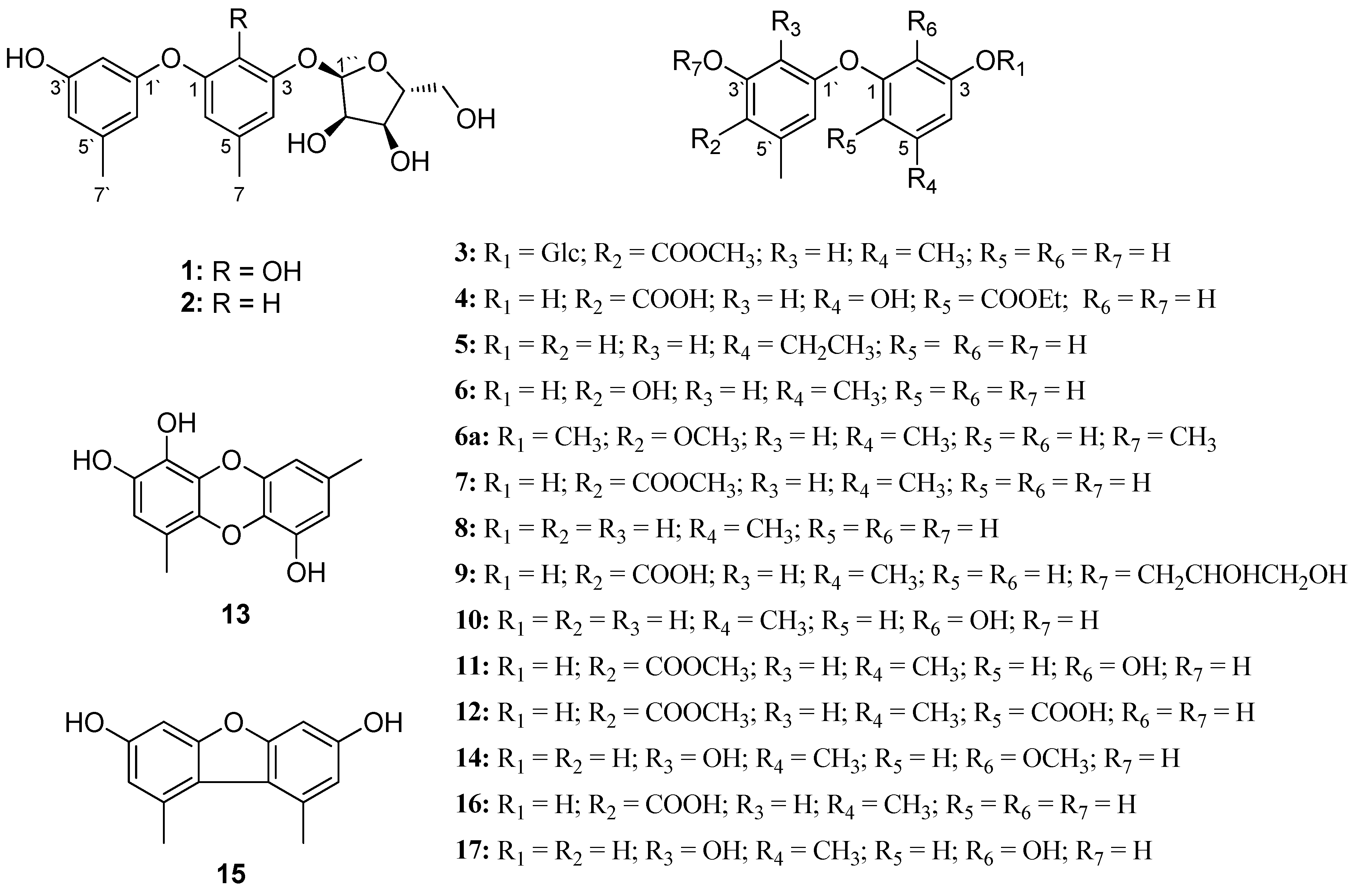
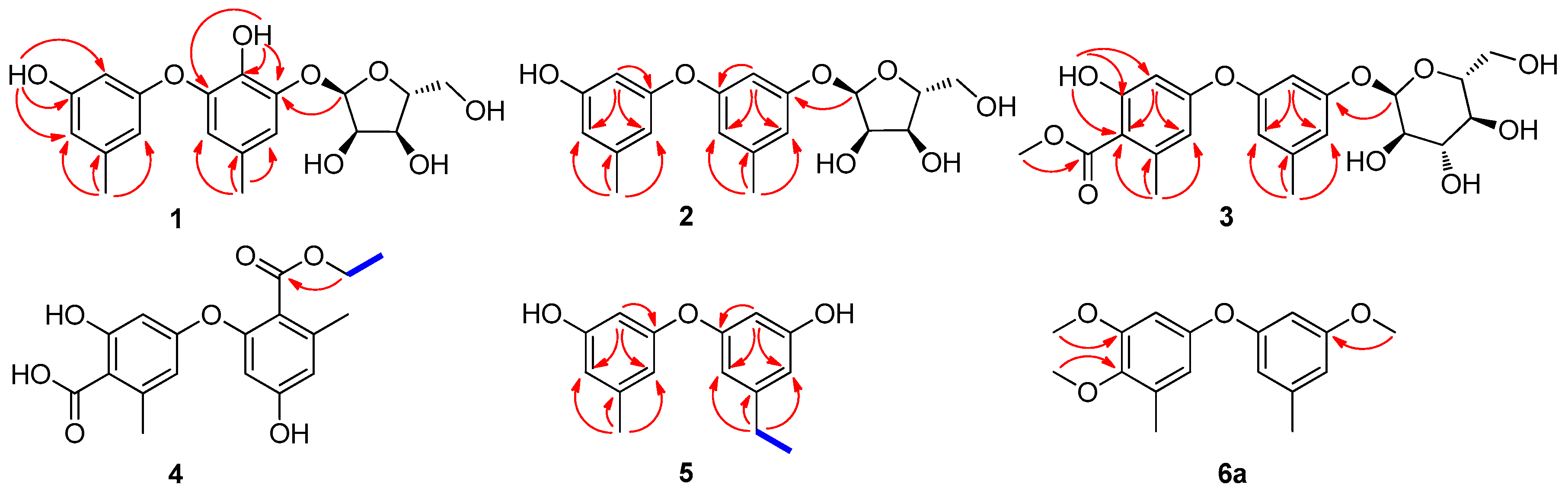
3.4.1. Cordyol C-3-O-α-d-ribofuranoside (1)

Colorless oil; [α] D 25 −8.0 (c 0.20, MeOH), UV(MeOH) λmax (log ε): 204.8 (4.02) nm, 279.4 (3.25) nm; IR νmax: 3320.1, 1677.7, 1596.2, 1512.1, 1464.3, 1322.1, 1210.8, 1140.0, 1046.8, 997.6, 836.2 cm−1; 1H and 13C NMR spectroscopic data see Table 1 and Table 2; HRESIMS m/z 401.1206 [M + Na]+ (calcd. for C19H22O8Na, 401.1207).

3.4.2. Diorcinol-3-O-α-d-ribofuranoside (2)

Colorless oil; [α] D 25 −18.6 (c 0.40, MeOH), UV(MeOH) λmax (log ε): 207.2 (4.05) nm, 273.4 (3.25) nm; IR νmax: 3344.2, 2931.4, 1600.7, 1464.7, 1325.1, 1154.9, 1039.8, 839.2 cm−1; 1H and 13C NMR spectroscopic data see Table 1 and Table 2; HRESIMS m/z 385.1261 [M + Na]+ (calcd. for C19H22O7Na, 385.1258).

3.4.3. 4-Methoxycarbonyl Diorcinol-3-O-α-d-glucoside (3)

Colorless oil; [α] D 25 +9.6 (c 0.20, MeOH), UV(MeOH) λmax (log ε): 214.2 (4.03) nm, 261.5 (3.64) nm, 298.4 (3.28) nm; IR νmax: 3334.5, 1651.3, 1579.3, 1454.2, 1324.9, 1268.4, 1161.2, 1023.9, 847.5 cm−1; 1H and 13C NMR spectroscopic data see Table 1 and Table 2; HRESIMS m/z 473.1403 [M + Na]+ (calcd. for C22H26O10Na, 473.1418).

3.4.4. 2-(Ethoxycarbonyl)-4′-carboxydiorcinal (4)

Colorless oil; UV(MeOH) λmax (log ε): 216.6 (4.04) nm, 259.2 (3.59) nm, 299.6 (3.26) nm; IR νmax: 3251.1, 1654.4, 1613.0, 1460.1, 1317.8, 1260.4, 1167.0, 845.9, 802.3 cm−1; 1H and 13C NMR spectroscopic data see Table 1 and Table 2; HRESIMS m/z 369.0954 [M + Na]+ (calcd. for C18H18O7Na, 369.0945).

3.4.5. 7-Ethyldiorcinol (5)

Colorless oil; UV(MeOH) λmax (log ε): 207.2 (4.03) nm, 280.5 (3.27) nm; IR νmax: 3343.5, 1598.3, 1459.8, 1329.8, 1155.3, 995.4, 841.3 cm−1; 1H and 13C NMR spectroscopic data see Table 1 and Table 2; HRESIMS m/z 245.1183 [M + H]+ (calcd. for C15H17O3, 245.1172).

3.4.6. 3-Hydroxydiorcinol (6)

Colorless oil; UV(MeOH) λmax (log ε): 221.3 (4.03) nm, 280.5 (3.71) nm; IR νmax: 3286.7, 1598.2, 1491.4, 1324.2, 1154.0, 1024.7, 976.7, 836.8 cm−1; 1H and 13C NMR spectroscopic data see Table 1 and Table 2; HRESIMS m/z 245.0808 [M − H] (calcd. for C14H13O4, 245.0819).

3.5. Determination of the Absolute Configuration of Sugar Moieties in 13

To determine the absolute configurations of sugar moieties in 13, a modified method based on LC-ESI-MS analysis was performed, where the retention time of sugar samples obtained after hydrolysis of the parent compounds were compared with those from standard sugars (d/l) [34]. In detail, compounds 1, 2 and 3 (approximately 0.05 mg, each) were hydrolyzed with 2 mol/L HCl (400 μL) in a 2 mL glass vial at 80 °C for 4 h. The reaction mixture was then diluted with H2O (400 μL) and extracted with CHCl3 (400 μL) three times. The aqueous layer containing monosaccharides was concentrated in vacuo to yield a dried sugar mixture. The resulting sugar mixture (not weighed out) and standard sugar samples (d/l-ribose and d/l-glucose, 0.1 mg for each) respectively, were heated with l-cysteine methyl ester (0.1 mg) in pyridine (400 μL) in a 2 mL glass vial at 60 °C for 60 min, then o-tolyl isothiocyanate (200 μL) was added to the reaction mixture and kept at 60 °C for additional 60 min. Then, the reaction mixture was directly analyzed by LC-ESI-MS (COSMOSIL 5 μm, 4.6 × 250 mm, C18 column). Analysis was performed at 30 °C with a flow rate of 1.0 mL/min, and the elution was carried out using a gradient of MeCN (0–30 min, 10–50%, linear gradient) in H2O. Source parameters in the positive ion mode were set as follows: Capillary entrance voltage = −4500 V, end plate offset = −500 V, nebulizer pressure (N2) = 11.6 psi, dry gas (N2) = 6.0 L/min, dry gas temperature = 220 °C. High-purity nitrogen (N2) were used as the nebulizing gas. Ion Peaks were extracted at m/z 447 for ribose and at m/z 471 for glucose identified by comparison of retention time with those of standards. The retention time of d-ribose and d-glucose derivatives were 22.3 and 21.0 min, respectively.

3.6. Cytotoxicity Assay

Cancer cell lines, including 4T1, U937, PC3, HL-60, HT-29, A549, NCI-H460, and K562, were purchased from ATCC. All the cells were maintained in RPMI1640 supplemented with 10% FBS, 100 units/mL Penicillin G and 100 μg/mL streptomycin. All the cancer cells were incubated at 37 °C in humidified air containing 5% CO2. MTT assay was used to determine the cell viability. Cells were seeded in 96-well plates at 1.5–3.0 × 10⁴/mL (100 μL/well). After 24 h incubation, 5 different concentrations (final concentrations were 1.6, 3.1, 6.3, 12.5 and 25.0 μM) of tested compounds were added into the wells in triplicate. Five concentrations of doxorubicin were tested, including 2.0, 1.0, 0.5, 0.25, and 0.125 μM. Cells were incubated for 96 h before MTT was added into the cells at a final concentration of 500 μg/mL, and the plates were incubated for an additional 4 h. The resultant formazan crystals were dissolved in 200 μL of DMSO, then a microplate reader (Synergy HT, Bio-Tek) was used to measure the absorbance of the plates at 570 nm for testing the cell viability of serial concentrations of compounds and the IC50 were estimated.

4. Conclusions

A total of 17 diphenyl ethers, including 6 new compounds, were isolated from a marine-derived Aspergillus sydowii. Compounds 1 and 2 are rare diphenyl ether glycosides containing a d-ribofuranose moiety. Although natural diphenyl ethers have been extensively investigated, their structures still exhibit variability due to the presence of hydroxy groups, and the diversity and location of sugar moieties. Furthermore, a modified method based on LC-MS analysis was used to determine the absolute configuration of sugar moieties. Comparing to conventional method based on LC-UV, which normally requires 0.5–3 mg of sample [34,35,36,37], our method has higher sensitivity due to the use of ESIMS detection, and as low as 0.05 mg of sample was enough to determine the absolute configuration of the sugar units using this procedure. Moreover, all the compounds were evaluated for their cytotoxicity against eight cancer cell lines, and compounds 1, 5, 6, and 811 were found to exhibit highly selective cytotoxicities against different cancer cell lines.

Supplementary Materials

The following are available online at https://www.mdpi.com/1660-3397/16/11/451/s1, Figures S1–S53: HRESIMS, 1D and 2D NMR, IR, and UV spectra of 119 and 6a, Figures S54–S56: LC-ESI-MS analysis of sugar derivatives of 13, Figure S57: the internal transcribed spacers (ITS) sequence of strain FNA026.

Author Contributions

Y.-N.W., Y.D. and Y.-H.M. conducted most of the isolation, acquirement of NMR spectra, structural determination and the bioactivity evaluation of compounds; Y.W., B.-Y.L., J.B., D.-J.Y. and L.Z. assisted the data analyzes. D.-Q.F. isolated the fungal species, Y.-H.P. and Y.-C.H. designed the experiments and wrote the manuscript. All authors reviewed the manuscript.

Funding

This research was funded by the National Natural Sciences Foundation of China (No. 81522043) and the CAMS Innovation Fund for Medical Sciences (CIFMS, No. 2016-I2M-3-010, 2017-I2M-4-004).

Acknowledgments

We are grateful to the Department of Instrumental Analysis in Institute of Materia Medica, Chinese Academy of Medical Sciences & Peking Union Medical College for the spectroscopic measurements and J.-B.L. for the HRMS analysis.

Conflicts of Interest

The authors declare no conflict of interest.

References

  1. Hu, Y.; Potts, M.B.; Colosimo, D.; Herrera-Herrera, M.L.; Legako, A.G.; Yousufuddin, M.; White, M.A.; MacMillan, J.B. Discoipyrroles A–D: Isolation, structure determination, and synthesis of potent migration inhibitors from Bacillus hunanensis. J. Am. Chem. Soc. 2013, 135, 13387–13392. [Google Scholar] [CrossRef] [PubMed]
  2. Hu, Y.; Wang, K.; MacMillan, J.B. Hunanamycin A, an antibiotic from a marine-derived Bacillus hunanensis. Org. Lett. 2013, 15, 390–393. [Google Scholar] [CrossRef] [PubMed]
  3. Song, X.; Xiong, Y.; Qi, X.; Tang, W.; Dai, J.; Gu, Q.; Li, J. Molecular Targets of Active Anticancer Compounds Derived from Marine Sources. Mar. Drugs 2018, 16, 175. [Google Scholar] [CrossRef] [PubMed]
  4. Moghadamtousi, S.; Nikzad, S.; Kadir, H.; Abubakar, S.; Zandi, K. Potential Antiviral Agents from Marine Fungi: An Overview. Mar. Drugs 2015, 13, 4520–4538. [Google Scholar] [CrossRef] [PubMed] [Green Version]
  5. Blunt, J.W.; Carroll, A.R.; Copp, B.R.; Davis, R.A.; Keyzers, R.A.; Prinsep, M.R. Marine natural products. Nat. Prod. Rep. 2018, 35, 8–53. [Google Scholar] [CrossRef] [PubMed] [Green Version]
  6. Bugni, T.S.; Ireland, C.M. Marine-derived fungi: A chemically and biologically diverse group of microorganisms. Nat. Prod. Rep. 2004, 21, 143–163. [Google Scholar] [CrossRef] [PubMed]
  7. Sanchez, J.F.; Somoza, A.D.; Keller, N.P.; Wang, C.C.C. Advances in Aspergillus secondary metabolite research in the post-genomic era. Nat. Prod. Rep. 2012, 29, 351–371. [Google Scholar] [CrossRef] [PubMed]
  8. Peng, J.; Gao, H.; Zhang, X.; Wang, S.; Wu, C.; Gu, Q.; Guo, P.; Zhu, T.; Li, D. Psychrophilins E–H and Versicotide C, Cyclic Peptides from the Marine-Derived Fungus Aspergillus versicolor ZLN-60. J. Nat. Prod. 2014, 77, 2218–2223. [Google Scholar] [CrossRef] [PubMed]
  9. Bladt, T.; Frisvad, J.; Knudsen, P.; Larsen, T. Anticancer and Antifungal Compounds from Aspergillus, Penicillium and Other Filamentous Fungi. Molecules 2013, 18, 11338–11376. [Google Scholar] [CrossRef] [PubMed] [Green Version]
  10. Gong, D.L.; Wang, X.J.; Xiang, Z.D.; Wang, J.D.; Zhang, H.; Liu, C.X.; Zhang, J.; Xiang, W.S. Diphenyl etheric metabolites from Streptomyces sp. neau50. J. Antibiot. 2011, 64, 465–467. [Google Scholar] [CrossRef] [PubMed]
  11. Li, X.; Xia, Z.; Tang, J.; Wu, J.; Tong, J.; Li, M.; Ju, J.; Chen, H.; Wang, L. Identification and Biological Evaluation of Secondary Metabolites from Marine Derived Fungi-Aspergillus sp. SCSIOW3, Cultivated in the Presence of Epigenetic Modifying Agents. Molecules 2017, 22, 1302. [Google Scholar] [CrossRef] [PubMed]
  12. Yurchenko, A.A.; Smetanina, O.F.; Kalinovsky, A.I.; Kirichuk, N.N.; Pivkin, M.V.; Ivanets, E.V.; Yurchenko, E.A.; Afiyatullov, S.S. New Metabolites from a Marine Sediment-Derived Fungus, Aspergillus carneus. Nat. Prod. Commun. 2015, 10, 1247–1250. [Google Scholar] [PubMed]
  13. Wu, Z.; Wang, Y.; Liu, D.; Proksch, P.; Yu, S.; Lin, W. Antioxidative phenolic compounds from a marine-derived fungus Aspergillus versicolor. Tetrahedron 2016, 72, 50–57. [Google Scholar] [CrossRef]
  14. Bunyapaiboonsri, T.; Yoiprommarat, S.; Intereya, K.; Kocharin, K. New diphenyl ethers from the insect pathogenic fungus Cordyceps sp. BCC 1861. Chem. Pharm. Bull. 2007, 55, 304–307. [Google Scholar] [CrossRef] [PubMed]
  15. Shi, T.; Qi, J.; Shao, C.L.; Zhao, D.L.; Hou, X.M.; Wang, C.Y. Bioactive Diphenyl Ethers and Isocoumarin Derivatives from a Gorgonian-Derived Fungus Phoma sp. (TA07-1). Mar. Drugs 2017, 15, 146. [Google Scholar] [CrossRef] [PubMed]
  16. Thadhani, V.M.; Choudhary, M.I.; Andersen, R.J.; Karunaratne, V. Novel entry into 5-decarboxydibenzofurans via Smiles rearrangement of the lichen para-depside, erythrin. J. Chem. Res. 2010, 34, 154–157. [Google Scholar] [CrossRef]
  17. Yang, G.; Yun, K.; Nenkep, V.N.; Choi, H.D.; Kang, J.S.; Son, B.W. Induced production of halogenated diphenyl ethers from the marine-derived fungus Penicillium chrysogenum. Chem. Biodivers. 2010, 7, 2766–2770. [Google Scholar] [CrossRef] [PubMed]
  18. Zhao, D.L.; Shao, C.L.; Zhang, Q.; Wang, K.L.; Guan, F.F.; Shi, T.; Wang, C.Y. Azaphilone and Diphenyl Ether Derivatives from a Gorgonian-Derived Strain of the Fungus Penicillium pinophilum. J. Nat. Prod. 2015, 78, 2310–2314. [Google Scholar] [CrossRef] [PubMed]
  19. Xiao, J.; Hu, J.Y.; Sun, H.D.; Zhao, X.; Zhong, W.T.; Duan, D.Z.; Wang, L.; Wang, X.L. Sinopestalotiollides A–D, cytotoxic diphenyl ether derivatives from plant endophytic fungus Pestalotiopsis palmarum. Bioorg. Med. Chem. Lett. 2018, 28, 515–518. [Google Scholar] [CrossRef] [PubMed]
  20. Wang, J.; Lu, Z.; Liu, P.; Wang, Y.; Li, J.; Hong, K.; Zhu, W. Cytotoxic polyphenols from the fungus Penicillium expansum 091 006 endogenous with the mangrove plant Excoecaria agallocha. Planta Med. 2012, 78, 1861–1866. [Google Scholar] [CrossRef] [PubMed]
  21. Peng, W.; You, F.; Li, X.L.; Jia, M.; Zheng, C.J.; Han, T.; Qin, L.P. A new diphenyl ether from the endophytic fungus Verticillium sp. isolated from Rehmannia glutinosa. Chin. J. Nat. Med. 2013, 11, 673–675. [Google Scholar] [CrossRef] [PubMed]
  22. Tian, Y.; Qin, X.; Lin, X.; Kaliyaperumal, K.; Zhou, X.; Liu, J.; Ju, Z.; Tu, Z.; Liu, Y. Sydoxanthone C and acremolin B produced by deep-sea-derived fungus Aspergillus sp. SCSIO Ind09F01. J. Antibiot. 2015, 68, 703–706. [Google Scholar] [CrossRef] [PubMed]
  23. Lu, Z.; Zhu, H.; Fu, P.; Wang, Y.; Zhang, Z.; Lin, H.; Liu, P.; Zhuang, Y.; Hong, K.; Zhu, W. Cytotoxic polyphenols from the marine-derived fungus Penicillium expansum. J. Nat. Prod. 2010, 73, 911–914. [Google Scholar] [CrossRef] [PubMed]
  24. Zhao, H.; Wang, G.Q.; Tong, X.P.; Chen, G.D.; Huang, Y.F.; Cui, J.Y.; Kong, M.Z.; Guo, L.D.; Zheng, Y.Z.; Yao, X.S.; et al. Diphenyl ethers from Aspergillus sp. and their anti-Aβ42 aggregation activities. Fitoterapia 2014, 98, 77–83. [Google Scholar] [CrossRef] [PubMed]
  25. Asami, Y.; Jang, J.H.; Oh, H.; Sohn, J.H.; Kim, J.W.; Moon, D.O.; Kwon, O.; Kawatani, M.; Osada, H.; Kim, B.Y.; et al. Violaceols Function as Actin Inhibitors Inducing Cell Shape Elongation in Fibroblast Cells. Biosci. Biotechnol. Biochem. 2012, 76, 1431–1437. [Google Scholar] [CrossRef] [PubMed] [Green Version]
  26. Camilleri, P.; Weaver, K.; Clark, M.T.; Bowyer, J.R.; Hallahan, B.J. Some Novel Diphenyl Ether Herbicides with Peroxidizing Activity. J. Agric. Food Chem. 1988, 36, 1061–1063. [Google Scholar] [CrossRef]
  27. Serianni, A.S.; Barker, R. [C-13]-Enriched tetroses and tetrofuranosides—An evaluation of the relationship between NMR parameters and furanosyl ring conformation. J. Org. Chem. 1984, 49, 3292–3300. [Google Scholar] [CrossRef]
  28. Gubica, T.; Szeleszczuk, L.; Pisklak, D.M.; Stepien, D.K.; Cyranski, M.K.; Kanska, M. Reliable evaluation of molecular structure of methyl 3-O-nitro-alpha-d-glucopyranoside and its intermediates by means of solid-state NMR spectroscopy and DFT optimization in the absence of appropriate crystallographic data. Tetrahedron 2014, 70, 1910–1917. [Google Scholar] [CrossRef]
  29. Tvaroska, I.; Taravel, F.R. Carbon-proton coupling constants in the conformational analysis of sugar molecules Adv. Carbohydr. Chem. Biochem. 1995, 51, 15–61. [Google Scholar] [CrossRef]
  30. Li, X.B.; Zhou, Y.H.; Zhu, R.X.; Chang, W.Q.; Yuan, H.Q.; Gao, W.; Zhang, L.L.; Zhao, Z.T.; Lou, H.X. Identification and Biological Evaluation of Secondary Metabolites from the Endolichenic Fungus Aspergillus versicolor. Chem. Biodivers. 2015, 12, 575–592. [Google Scholar] [CrossRef] [PubMed]
  31. Tanahashi, T.; Takenaka, Y.; Nagakura, N.; Hamada, N. Dibenzofurans from the cultured lichen mycobionts of Lecanora cinereocarnea. Phytochemistry 2001, 58, 1129–1134. [Google Scholar] [CrossRef]
  32. Liu, S.; Wang, H.; Su, M.; Hwang, G.J.; Hong, J.; Jung, J.H. New metabolites from the sponge-derived fungus Aspergillus sydowii J05B-7F-4. Nat. Prod. Res. 2017, 31, 1682–1686. [Google Scholar] [CrossRef] [PubMed]
  33. Takenaka, Y.; Tanahashi, T.; Nagakura, N.; Hamada, N. Phenyl ethers from cultured lichen mycobionts of Graphis scripta var. serpentina and G. rikuzensis. Chem. Pharm. Bull. 2003, 51, 794–797. [Google Scholar] [CrossRef]
  34. Tanaka, T.; Nakashima, T.; Ueda, T.; Tomii, K.; Kouno, I. Facile discrimination of aldose enantiomers by reversed-phase HPLC. Chem. Pharm. Bull. 2007, 55, 899–901. [Google Scholar] [CrossRef] [PubMed]
  35. Liu, X.; Fu, J.; Yao, X.J.; Yang, J.; Liu, L.; Xie, T.G.; Jiang, P.C.; Jiang, Z.H.; Zhu, G.Y. Phenolic Constituents Isolated from the Twigs of Cinnamomum cassia and Their Potential Neuroprotective Effects. J. Nat. Prod. 2018, 81, 1333–1342. [Google Scholar] [CrossRef] [PubMed]
  36. Kim, B.; Han, J.W.; Ngo, M.T.; Dang, Q.L.; Kim, J.-C.; Kim, H.; Choi, G.J. Identification of novel compounds, oleanane- and ursane-type triterpene glycosides, from Trevesia palmata: Their biocontrol activity against phytopathogenic fungi. Sci. Rep. 2018, 8, 14522. [Google Scholar] [CrossRef] [PubMed]
  37. Wang, L.L.; Hao, L.J.; Zhou, Z.B.; Zhu, X.L.; Shi, Z.H.; Miyamoto, T.; Pan, K. Lycodine-type alkaloids and their glycosides from Lycopodiastrum casuarinoides. Phytochemistry 2018, 154, 63–72. [Google Scholar] [CrossRef] [PubMed]
Figure 1. Structures of compounds 117 and 6a.
Marinedrugs 16 00451 g001
Figure 2. Key 1H-1H COSY (blue lines) and key HMBC (arrows) correlations of 15 and 6a.
Marinedrugs 16 00451 g002
Table 1. The 1H NMR spectroscopic data for compounds 16 and 6a (600 MHz).
NO.δH (J in Hz)
1 a2 b3 a4 b5 b6 b6a b
2 6.20 t (2.2)6.57 t (2.4)6.24 d (2.3)6.21 brs6.13 t (2.4)6.30 d (2.2)
46.81 brs6.27 brs6.75 brs6.35 d (2.3)6.30 m6.28 brs6.48 brs
66.46 brs6.37 brs6.53 brs 6.39 m6.20 brs6.43 brs
72.18 s2.27 s2.26 s2.47 s2.53 q (7.5)2.19 s2.26 s
8 1.18 t (7.5) 3.73 s
9 4.40 q (7.5)
10 1.40 t (7.5)
2′6.02 brs6.57 t (2.8)6.28 d (2.3)6.28 d (2.7)6.21 brs6.33 d (2.8)6.52 d (2.8)
4′6.21 brs6.45 brs 6.28 brs
6′6.12 brs6.72 brs6.37 d (2.3)6.56 d (2.7)6.36 brs6.26 d (2.8)6.37 d (2.8)
7′2.15 s2.22 s2.21 s2.35 s2.22 s2.17 s2.20 s
8′ 3.78 s
9′ 3.78 s 3.75 s
1″5.50 d (4.6)5.57 d (4.5)5.35 d (3.6)
2″4.07 ddd (10.7, 6.8, 4.1)4.15 dd (6.5, 4.5)3.33 m
3″3.93 ddd (11.7, 5.9, 2.7)4.06 dd (6.5, 3.2)3.58 t (9.2)
4″3.98 q (4.0)4.12 dd (6.9, 3.5)3.17 t (9.2)
5″3.46 brt (5.1)3.63 dd (12.1, 3.9) 3.69 dd (11.7, 3.4)3.42 m
6″ 3.47 dd (11.7, 5.2)
3.55 dd (11.8, 1.8)
2′-OH8.41 s
3″-OH9.26 s 10.26 s
2′-OH5.14 d (6.0)
3″-OH5.16 brs
5″-OH4.81 t (5.6)
a Recorded in DMSO-d6; b Recorded in CD3OD.
Table 2. 13C NMR spectroscopic data for compounds 16 and 6a (150 MHz).
NO.δC, Type
1 a2 b3 a4 b5 b6 b6a b
1142.0 (C)159.6 (C) 156.1 (C) 163.2 (C) 159.5 (C) 161.2 (C) 160.2 (C)
2137.3 (C)106.3 (CH)105.5 (CH)103.9 (C)104.5 (CH)102.8 (CH)102.6 (CH)
3145.7 (C)159.5 (C)158.3 (C)164.9 (C)159.7 (C)159.4 (C)162.3 (C)
4112.9 (CH)111.8 (CH)113.5 (CH)113.3 (CH)111.7 (CH)111.0 (CH)110.3 (CH)
5127.3 (C)141.6 (C)140.5 (C)144.1 (C)148.2 (C)141.4 (C)141.7 (C)
6115.9 (CH)112.1 (CH)113.6 (CH)109.7 (CH)110.8 (CH)110.4 (CH)112.2 (CH)
720.6 (CH3)21.6 (CH3)21.1 (CH3)23.9 (CH3)29.8 (CH2)21.6 (CH3)21.7 (CH3)
8 172.4 (C)15.9 (CH3) 55.7 (CH3)
9 62.5 (CH2)
10 14.5 (CH3)
1′159.1 (C)159.7 (C)158.9 (C)160.8 (C)159.6 (C) 150.1 (C)154.3 (C)
2′100.3 (CH)104.3 (CH)102.6 (CH)106.4 (CH)104.2 (CH)106.1 (CH)103.4 (CH)
3′158.2 (C)159.4 (C)157.6 (C)155.0 (C)159.7 (C)146.8 (C)154.8 (C)
4′109.6 (CH)114.2 (CH)114.9 (C)120.1(C)111.9 (CH)140.8 (C)144.5 (C)
5′139.5 (C)141.7 (C)139.1 (C)140.6 (C)141.6 (C)126.7 (C)133.5 (C)
6′107.5 (CH)113.5 (CH)110.7 (CH)114.9 (CH)110.6 (CH)113.6 (CH)113.7 (CH)
7′21.2 (CH3)21.5 (CH3)20.1 (CH3)20.2 (CH3)21.5 (CH3)16.2 (CH3)16.0 (CH3)
8′ 168.5 (C)170.7 (C) 56.3 (CH3)
9′ 51.9 (CH3) 60.6 (CH3)
1″101.3 (CH)102.3 (CH)97.9 (CH)
2″72.3 (CH)73.4 (CH)71.5 (CH)
3″69.4 (CH)71.2 (CH)73.0 (CH)
4″86.7 (CH)87.5 (CH)69.8 (CH)
5″61.6 (CH2)63.2 (CH2)73.8 (CH)
6″ 60.6 (CH2)
a Recorded in DMSO-d6; b Recorded in CD3OD.
Table 3. Cytotoxicity (IC50 in µM) for compounds 1, 56, 811.
CompoundsA549U937HL-60K562
18.97 ± 0.484.64 ± 0.35//
516.13 ± 1.24///
6//11.98 ± 0.7318.89 ± 1.14
815.51 ± 1.59///
93.36 ± 0.68/21.22 ± 1.25/
10//16.52 ± 0.9920.88 ± 1.60
11//13.33 ± 0.8723.03 ± 1.34
DOX0.19 ± 0.04<0.125<0.1250.49 ± 0.08
“/” no cytotoxicity was detected. Inactive compounds were not shown here.

