Article

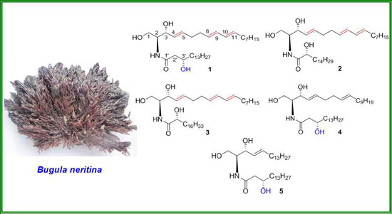
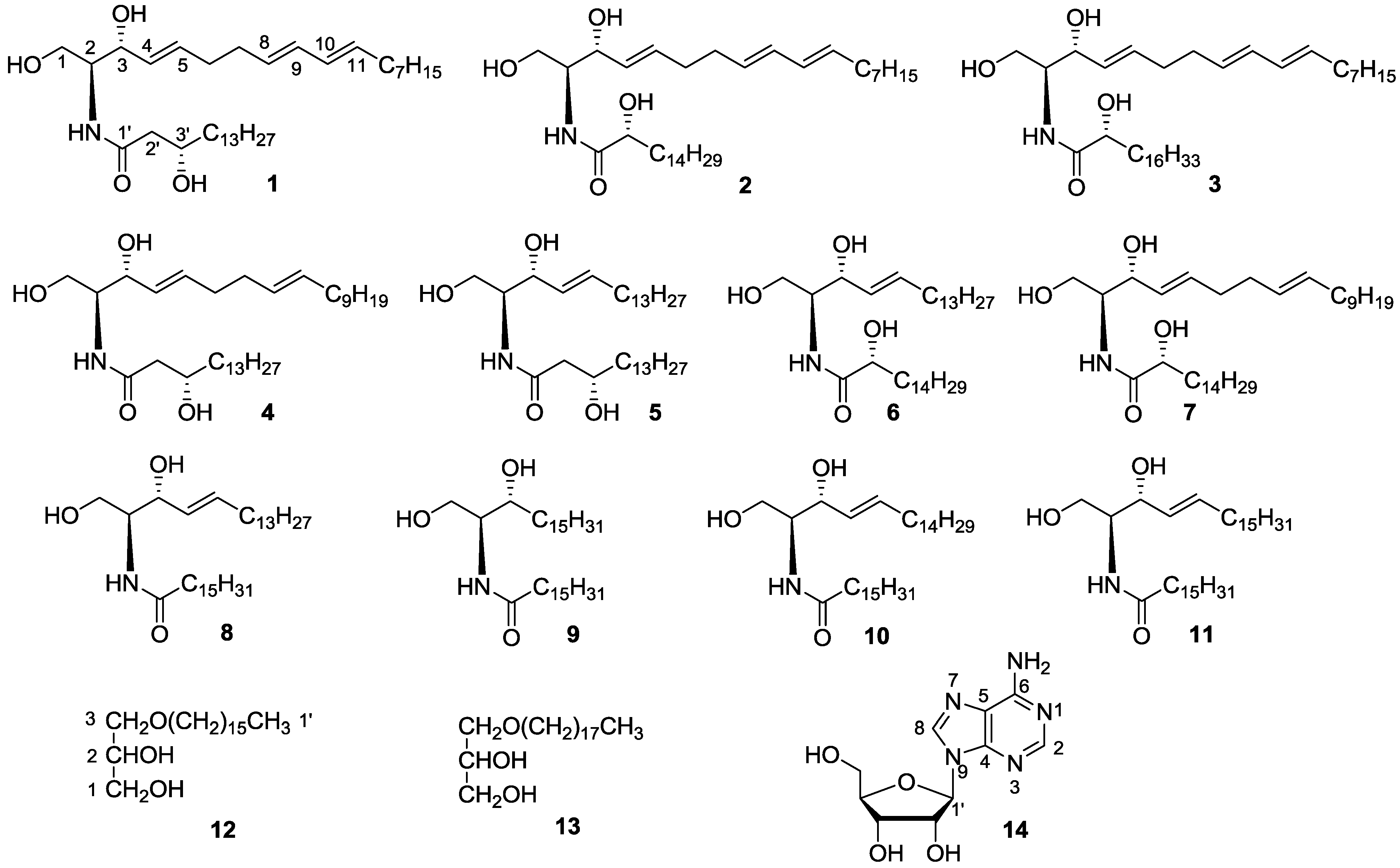
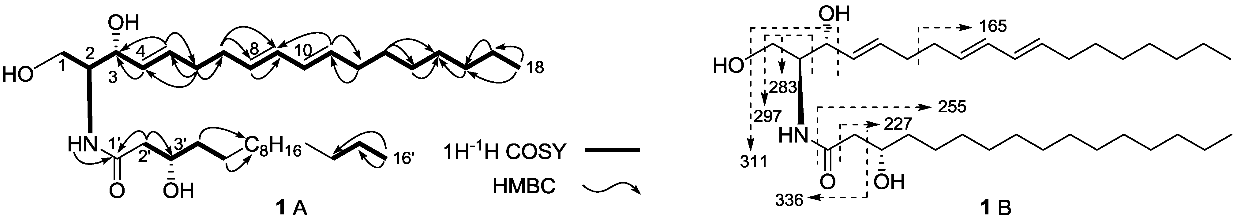
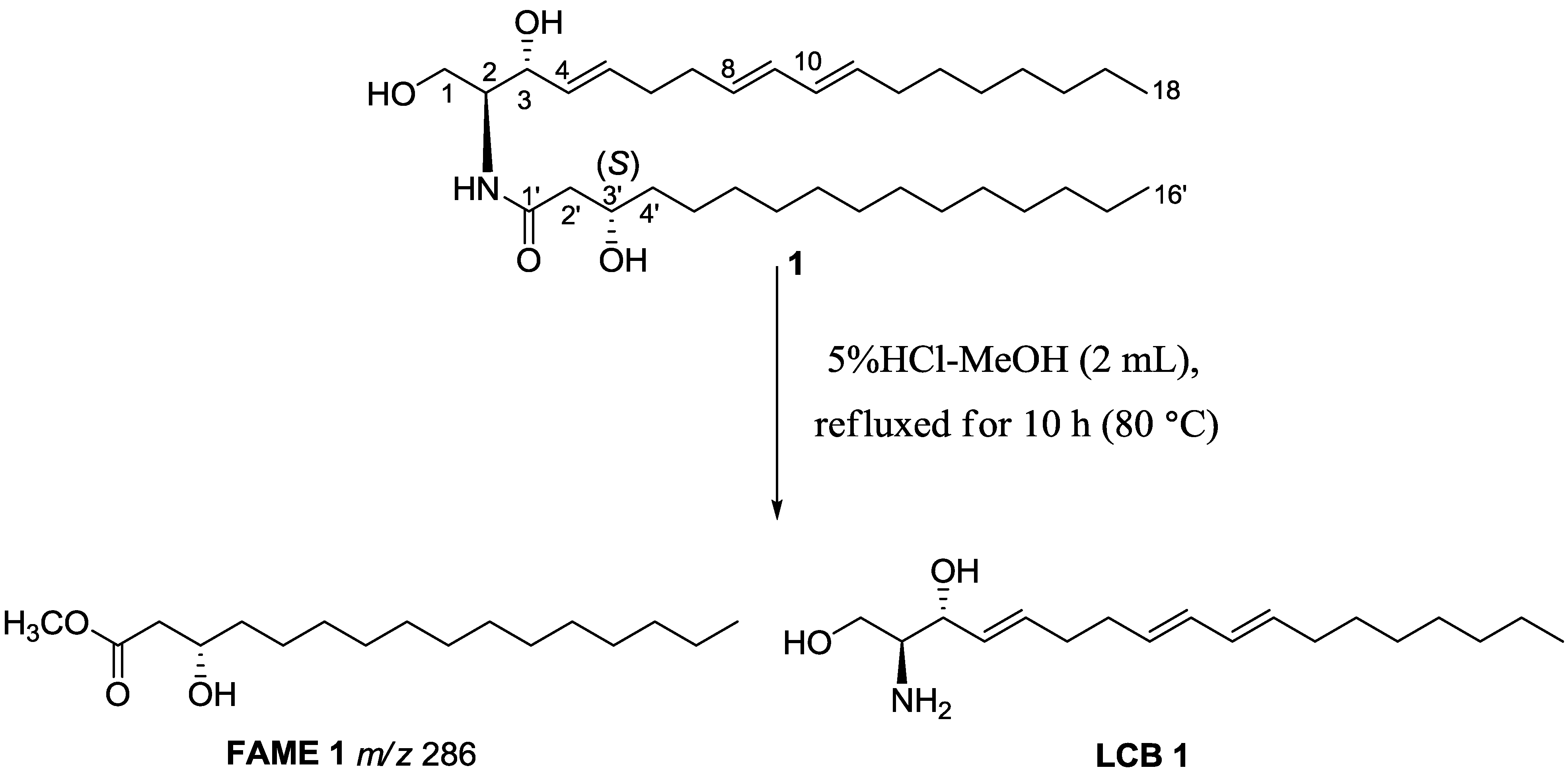
Neritinaceramides A–E, New Ceramides from the Marine Bryozoan Bugula neritina Inhabiting South China Sea and Their Cytotoxicity

1
Research & Development Center of Biorational Pesticide, College of Plant Protection, Northwest A&F University, Yangling 712100, China
2
Institute of Materia Medica, School of Pharmacy, Fourth Military Medical University, Xi'an 710032, China
3
Department of Pharmacy, Xijing Hospital, Fourth Military Medical University, Xi'an 710032, China
4
School of Traditional Chinese Medicines, Shenyang Pharmaceutical University, Shenyang 110016, China
5
Department of Pharmacy, Renji Hospital, Affiliated to School of Medicine, Shanghai Jiao-Tong University, Shanghai 200127, China
*
Authors to whom correspondence should be addressed.
Mar. Drugs 2014, 12(4), 1987-2003; https://doi.org/10.3390/md12041987
Submission received: 23 January 2014 / Revised: 2 March 2014 / Accepted: 13 March 2014 / Published: 2 April 2014
(This article belongs to the Collection Bioactive Compounds from Marine Invertebrates)

Abstract

:
Five new ceramides, neritinaceramides A (1), B (2), C (3), D (4) and E (5), together with six known ceramides (611), two known alkyl glycerylethers (12 and 13) and a known nucleoside (14), were isolated from marine bryozoan Bugula neritina, which inhabits the South China Sea. The structures of the new compounds were elucidated as (2S,3R,3′S,4E,8E,10E)-2-(hexadecanoylamino)-4,8,10-octadecatriene-l,3,3′-triol (1), (2S,3R,2′R,4E,8E,10E)-2-(hexadecanoylamino)-4,8,10-octadecatriene-l,3,2′-triol (2), (2S,3R,2′R,4E,8E,10E)-2-(octadecanoylamino)-4,8,10-octadecatriene-l,3,2′-triol (3), (2S,3R,3′S,4E,8E)-2-(hexadecanoylamino)-4,8-octadecadiene-l,3,3′-triol (4) and (2S,3R,3′S,4E)-2-(hexadecanoylamino)-4-octadecene-l,3,3′-triol (5) on the basis of extensive spectral analysis and chemical evidences. The characteristic C-3′S hydroxyl group in the fatty acid moiety in compounds 1, 4 and 5, was a novel structural feature of ceramides. The rare 4E,8E,10E-triene structure in the sphingoid base of compounds 13, was found from marine bryozoans for the first time. The new ceramides 15 were evaluated for their cytotoxicity against HepG2, NCI-H460 and SGC7901 tumor cell lines, and all of them exhibited selective cytotoxicity against HepG2 and SGC7901 cells with a range of IC50 values from 47.3 μM to 58.1 μM. These chemical and cytotoxic studies on the new neritinaceramides A–E (15) added to the chemical diversity of B. neritina and expanded our knowledge of the chemical modifications and biological activity of ceramides.

Graphical Abstract

1. Introduction

Ceramides, a family of sphingolipids, are important components of a wide variety of tissues and organs in biological systems [1]. Chemically, ceramides are composed of a sphingoid long chain base (LCB) and an amide-linked long-chain fatty acid base (FAB) [2]. Ceramides and their glycosylated-ceramides (cerebrosides), exhibiting a wide variety of biological effects including cytotoxic, antifungal, immunostimulating and immunosuppressive activities [3], have been isolated from a number of marine invertebrates, including starfishes [4], sea anemones [5,6], sponges [7], corals [8], tunicates [9] and bryozoans [10], as well as terrestrial plants [11]. Marine bryozoans are sedentary, colonial invertebrates that are widely distributed throughout the marine environment, but are less common in freshwater. They have been proved to be an important marine drug sources due to their affluent bioactive secondary metabolites, including macrolide, alkaloids, sterols and heteratom-containing compounds, etc., [12]. In particular, a highly oxygenated macrolide, bryostatin 1, isolated previously from marine bryozoan Bugula neritina, has been focused on preclinical and clinical studies [13]. In our previous studies focused on marine bryozoans, a new terminal branched ceramide and its analogues [14], cytotoxic sterols [15,16], and bromized alkaloids [17], have been reported from B. neritina and Cryptosula pallasiana. As part of our ongoing investigations toward the discovery of bioactive secondary metabolites from marine bryozoan B. neritina, five new ceramides, neritinaceramides A–E (15), six known ceramides (611), two known alkyl glycerylethers (12 and 13), and a known nucleoside (14) were isolated. The characteristic C-3′S hydroxyl group in the fatty acid moiety in compounds 1, 4 and 5, was a novel structure feature of ceramides. The rare 4E,8E,10E-triene structure in the sphingoid base of compounds 13, was found from marine bryozoans for the first time. Herein, we report the isolation, structure identification and cytotoxicity evaluation of those new ceramides.

2. Results and Discussion

The marine bryozoan Bugula neritina was extracted with 95% EtOH, condensed, and suspended in water, subsequently, and then was further extracted with EtOAc. The EtOAc extract was subjected to column chromatography (CC) over normal silica gel and Sephadex LH-20, respectively, and then was further purified by reverse semi-preparative HPLC to yield compounds 114 (Figure 1).
Figure 1. Chemical structures of compounds 114 from the marine bryozoan Bugula neritina.
Marinedrugs 12 01987 g001
Neritinaceramide A (1) was isolated as a white amorphous powder. The positive ion mode HR-ESI-MS showed a pseudomolecular ion peak at m/z 572.4658 [M + Na]+ (calcd. for C34H63NO4Na, 572.4655), which, together with the pseudomolecular ion peak atm/z 584 [M + Cl] in the negative mode ESI-MS, enabled the determination of the molecular formula as C34H63NO4, with the help of NMR spectral data. A close scrutiny of the 1H-NMR and 13C-NMR spectra of 1 (Table 1) by DEPT and HSQC experiments revealed the presence of an amide group [δ 173.0 (C-1′), δ 6.65 (1H, d, J = 8.0 Hz, NH)], a nitrogen-linked methine [δ 54.4 (C-2), δ 3.92 (1H, m, H-2)], a hydroxylated methylene [δ 62.0 (C-1), δ 3.72 (1H, br d, J = 8.3 Hz, H-1a), 3.90 (1H, br d, J = 8.3 Hz, H-1b)] and two hydroxylated methines [δ 74.2 (C-3), δ 4.31 (1H, br s, H-3); δ 68.9 (C-3′), δ 4.00 (1H, m, H-3′)], which together with the long aliphatic carbon signals at 29.2–29.7, demonstrated a trihydroxyl ceramide structure of compound 1. The observation of 1H-1H COSY correlations from H2-1 to H-2, and in turn, from H-2 to NH and H-3, from H-3′ to H-2a′ (δ 2.30), H-2b′ (δ 2.43), H-4a′ (δ 1.43) and H-4b′ (δ 1.51), and the HMBC correlations from NH to C-1′, and from H2-2′ to C-1′ and C-3′, indicated the three hydroxyl groups were located at C-1, C-3 an C-3′, respectively. The 1H-, 13C-NMR and HSQC spectra of 1 suggested the presence of three double bonds (C4=C5, C8=C9 and C10=C11) on the basis of six proton signals at δ 5.54 (1H, dd, J = 15.2, 6.2 Hz, H-4), 5.77 (1H, dt, J = 15.2, 6.3 Hz, H-5), 5.59 (1H, m, H-8), 5.99 (1H, dd, J = 15.3, 5.0 Hz, H-9), 6.00 (1H, dd, J = 15.3, 5.0 Hz, H-10) and 5.52 (1H, m, H-11), as well as the corresponding carbons at δ 129.3 (C-4), 133.2 (C-5), 133.3 (C-8), 130.0 (C-9), 131.2 (C-10) and 130.7 (C-11). The positions of the three double bonds were confirmed to be C-4, C-8 and C-10 on the basis of 1H-1H COSY correlations of H-3/H-4, H-4/H-5, H-5/H2-6, H2-6/H2-7, H2-7/H-8, H-8/H-9, H-9/H-10 and H-10/H-11, and HMBC correlations of H-4/C-3, H-5/C-3, H-5/C-6, H2-6/C-4, H2-6/C-5, H2-6/C-7, H2-7/C-8, H2-7/C-9, H-8/C-10, H-11/C-9 and H-11/C-12 (Figure 2A). The lengths of the sphingoid long chain base (LCB) and the amide-linked long-chain fatty acid base (FAB) were determined to be composed of 18 and 16 carbons, respectively, based on the negative ESI-MS fragment ions at m/z 165, 227, 255, 283, 297, 311 and 336 (Figure 2B). Based on the above evidences, the planar structure and the key connectivities of ceramide 1 were established.
Table 1. 1H and 13C-NMR data of compounds 1 and 2 (500MHz for 1H-NMR, 125MHz for 13C-NMR) a.
No.12
δC, mult. bδH (int., mult., J in Hz) bδC, mult. bδC, mult. cδH (int., mult., J in Hz) c
162.0 (t)3.72 (1H, br d, 8.3),
3.90 (1H, br d, 8.3)
62.0 (t)61.9 (t)4.22 (1H, dd, 10.8, 4.2),
4.46 (1H, dd, 10.8, 4.5)
254.4 (d)3.92 (1H, m)54.4 (d)56.1 (d)4.67 (1H, m)
374.2 (d)4.31 (1H, br s)74.2 (d)73.0 (d)4.83 (1H, br t, 6.1)
4129.3 (d)5.54 (1H, dd, 15.2, 6.2)129.1 (d)131.4 (d)6.13 (1H, m)
5133.2 (d)5.77 (1H, dt, 15.2, 6.3)133.3 (d)132.6 (d)6.05 (1H, m)
631.9 (t)2.16 (2H, m)31.9 (t)32.7 (t)2.18 (1H, m)
732.1 (t)2.04 (2H, dt, 14.1, 6.9)32.1 (t)32.9 (t)2.04 (1H, m)
8133.3 (d)5.59 (1H, m)133.5 (d)132.8 (d)5.64 (1H, m)
9130.0 (d)5.99 (1H, dd, 15.3, 5.0)130.0 (d)131.0 (d)6.11 (1H, m)
10131.2 (d)6.00 (1H, dd, 15.3, 5.0)131.2 (d)131.6 (d)5.65 (1H, m)
11130.7 (d)5.52 (1H, m)130.7 (d)131.5 (d)5.99 (1H, m)
1232.1 (t)2.16 (2H, m)32.1 (t)32.7 (t)2.18 (2H, br s)
1332.6 (t)2.04 (2H, dt, 14.1, 6.9)32.6 (t)32.1 (t)1.23 (2H, br s) d
1429.4 (t)1.36 (2H, m) d29.4 (t)29.4 (t)1.33 (2H, m) d
1529.2 (t)1.36 (2H, m) d29.2 (t)29.3 (t)1.33 (2H, m) d
1631.9 (t)1.26 (2H, br s) d31.9 (t)32.1 (t)1.23 (2H, br s) d
1722.7 (t)1.26 (2H, br s) d22.7 (t)22.9 (t)1.23 (2H, br s) d
1814.1 (q)0.88 (3H, t, 7.1)14.1 (q)14.2 (q)0.84 (3H, t, 7.1)
1′173.0 (s)-175.0 (s)175.4 (s)-
2′43.1 (t)a 2.30 (1H, m), b 2.43 (1H, d, 2.2) 72.4 (d)72.5 (d)4.59 (1H, m)
3′68.9 (d)4.00 (1H, m)34.8 (t)35.8 (t)2.22 (1H, m), 2.03 (1H, m)
4′37.1 (t)a 1.43 (1H, m), b 1.51 (1H, m)31.8 (t)32.0 (t)1.23 (2H, br s) d
5′25.6 (t)1.37 (2H, m)25.1 (t)25.9 (t)1.64 (2H, m)
6′~13′29.2–29.7 (t)1.26 (16H, br s) d29.2–29.7 (t)29.3–30.0 (t)1.23 (16H, br s) d
14′31.9 (t)1.26 (2H, br s) d31.9 (t)32.1 (t)1.23 (2H, br s) d
15′22.7 (t)1.26 (2H, br s) d22.7 (t)22.9 (t)1.23 (2H, br s) d
16′14.1 (q)0.88 (3H, t, 7.1)14.1 (q)14.2 (q)0.84 (3H, t, 7.1)
NH-6.65 (1H, d, 8.0)--8.34 (1H, d, 8.7)
a Assignments aided by the DEPT, COSY, HSQC, HMBC, and NOESY experiments; b data were obtained in CDCl3; c data were obtained in C5D5N; d overlapped with other signals.
Figure 2. Key 1H–1H COSY and HMBC correlations (A), together with negative ESI-MS fragments (B) of compound 1.
Marinedrugs 12 01987 g002
The geomertry of the C-4/C-5, C-8/C-9 and C-10/C-11 alkenyl bonds was confirmed to be trans based on the vicinal coupling constants of J4,5 (15.2 Hz), J8,9 (15.3 Hz) and J9,10 (15.3 Hz). Moreover, the trans configuration (E) for those alkenyl bonds was further confirmed by the chemical shifts of the allylic carbons C-6 (δ 31.9), C-7 (δ 32.1) and C-12 (δ 32.1), since allylic carbon signals of trans- and cis-isomers were observed at δ 32–33 and 27–28, respectively [18]. The relative stereochemistry at C-2 and C-3 in LCB of 1 were found to be same as that of d-sphingosine (d-erythro), as evidenced by 13C-NMR chemical shifts of C-2 (δ 54.4) and C-3 (δ 74.2), which are consistent with those reported for natural product N-palmitoyl-d-erythro-(2S,3R)-octadecasphinga-4(E)-ene (δ 54.5 and 74.7) [19]. Furthermore, the specific rotation ([α]D = −2.8°) of LCB 1 obtained by methanolysis of ceramide 1, was in good accordance with d-erythro-sphingosine, (2S,3R)-(E)-2-aminooctadec-4-ene-1,3-diol ([α]D = −3.0°) [20], while different from l-erythro-sphingosine, (2R,3S)-(+)-erythro-N-lauroyldocosasphinga-4,8-dienine ([α]D = +2.94°) [21]. This further confirmed the 2S,3R configuration for ceramide 1. Similarly, the 3′S-configuration was proved by the methanolysis of 1 and comparison of the specific rotation of the resulting methyl ester of the fatty acid with known compounds. In details, methanolysis of 1 by hydrochloric acid/methanol gave fatty acid methyl ester (FAME 1) as the major product (Figure 3). The specific rotation of FAME 1 ([α]D +12.5°) was in good agreement with the synthetic (S)-methyl 3-hydroxyhexadecanoate ([α]D = +13.8°), while the specific rotation [α]D for (R)-methyl 3-hydroxyhexadecanoate was −16.6° [22]. Thus, the structure of neritinaceramide A was unambiguously assigned as (2S,3R,3′S,4E,8E,10E)-2-(hexadecanoylamino)-4,8,10-octadecatriene-l,3,3′-triol.
Neritinaceramide B (2) was isolated as an isomer of 1, due to the same molecular formula of C34H63NO4 from ESI-MS at m/z 572 [M + Na]+ (positive mode) and 584 [M + Cl] (negative mode), and the HR-ESI-MS pseudomolecular ion peak at m/z 572.4652 [M + Na]+ (calcd. for C34H63NO4Na, 572.4655). By comparison of the 13C-NMR data of 2 with those of 1, it could be confirmed that 2 and 1 shared the same LCB structural skeleton, while the hydroxyl group at C-3′ in ceramide 1 was replaced to be at C-2′ in 2, as 2 showed different chemical shifts at C-1′ (δ 175.4), C-2′ (δ 72.5) and C-3′ (δ 35.8) in its FAB. The methene protons H2-3′ (δ 2.03 and 2.22) showed coupling relationships with H2-4′ (δ 1.23) and H-2′ (δ 4.59) in the 1H-1H COSY spectrum, which together with HMBC correlations from NH (δ 8.34, d, J = 8.7 Hz) to C-1′, from H-2′ to C-1′ and from H2-3′ to C-2′, confirmed that the hydroxyl group was attached at C-2′ (Figure 4A). Fragment peaks at m/z 166, 227, 255, 283 and 311 indicated that the number of carbons in the sphingoid long chain base was 18, and in the fatty acid base was 16 (Figure 4B). The stereochemistry at C-2, C-3 and C-2′ were proposed as 2S, 3R and 2′R on the basis of the chemical shifts of C-2, C-3, and C-2′ in CDCl3 (δ 54.4, 74.2 and 72.4, respectively) and C5D5N (δ 56.1, 73.0 and 72.5, respectively), which were close to analogue (2S,3R,4E,2′R)-2-(2-hydroxyhexadecanoylamino)-16-methyl-4-octadecene-1,3-diol [δ 54.6 (C-2), 74.3 (C-3) and 72.5 (C-2′) in CDCl3, and δ 56.1 (C-2), 73.1(C-3) and 72.5 (C-2′) in C5D5N.] [23]. Moreover, the configurations were confirmed by the specific rotation of ceramide 2 ([α]D = +9.8°) which was consistent with that of the synthetic ceramide with the 2S,3R,2′R-configuration ([α]D = +7.4°) [24], and this method has been applied to determine the stereochemistry for ceramide with the core structure of 2,3,2′ chiral centre [25,26]. Besides, methanolysis of 2 with the same method of 1 gave fatty acid methyl ester (FAME 2), its specific rotation ([α]D = –6.7°) was similar to (R)-methyl 2-hydroxyhexadecanoate ([α]D = –5.7°) [26], while was different with (S)-methyl 2-hydroxyhexadecanoate ([α]D = +1.15°) [27], which also confirmed the 2′R-configuration for ceramide 2. Accordingly, the structure of neritinaceramide B (2) was deduced as (2S,3R,2′R,4E,8E,10E)-2-(hexadecanoylamino)-4,8,10-octadecatriene-l,3,2′-triol.
Figure 3. Methanolysis of compound 1.
Marinedrugs 12 01987 g003
Figure 4. Key 1H-1H COSY and HMBC correlations (A), together with ESI-MS fragments (B) of compound 2 (a positive fragment ions; b negative fragment ions).
Marinedrugs 12 01987 g004
Neritinaceramide C (3) was found to have a molecular formula C36H67NO4 according to the positive ESI-MS pseudomolecular ion peak at m/z 600 [M + Na]+, and negative ESI-MS pseudomolecular ion peak at m/z 576 [M − H], with the help of NMR spectral data (Table 2). The NMR spectra of 3 showed a close resemblance to those of ceramide 2, except for the carbon number in the FAB. The negative ESI-MS fragment ions at m/z 324, 298, 283, 255 and 225 indicated that the numbers of carbons for both of the FAB and LCB were 18 (Figure 5). The carbon number of the FAB was further validated through EI-MS molecular ion at m/z 314 (M+) for the methyl ester obtained after methanolysis of 3 with 5% mehtanolic HCl. Therefore, neritinaceramide C (3) was determined to be (2S,3R,2′R,4E,8E,10E)-2-(octadecanoylamino)-4,8,10-octadecatriene-l,3,2′-triol.
Table 2. 1H and 13C-NMR data of compounds 35 (CDCl3, 500MHz for 1H-NMR, 125MHz for 13C-NMR) a.
No.345
δC, mult.δH (int., mult., J in Hz)δC, mult.δH (int., mult., J in Hz)δC, mult.δH (int., mult., J in Hz)
162.2 (t)3.75 (1H, m),
3.89 (1H, br d, 10.0)
61.8 (t)3.71 (1H, d, 11.0),
3.86 (1H, d, 11.2)
61.9 (t)3.74 (1H, m),
3.87 (1H, m)
254.4 (d)3.91 (1H, br d, 10.0)54.5 (d)3.92 (1H, m)56.1 (d)3.93 (1H, m)
374.2 (d)4.28 (1H, m)74.1 (d)4.28 (1H, br s)74.1 (d)4.28 (1H, m)
4129.2 (d)5.54 (1H, m)128.9 (d)5.51 (1H, dd, 15.5, 6.0)128.6 (d)5.53 (1H, br d, 14.0)
5133.3 (d)5.77 (1H, m)133.5 (d)5.77 (1H, m)134.3 (d)5.65 (1H, dt, 14.3, 6.5)
631.9 (t)2.16 (2H, t, 2.9)32.6 (t)1.96 (2H, q-like, 6.2)32.3 (t)2.05 (2H, m)
732.1 (t)2.04 (2H, q, 7.1)32.4 (t)2.11 (2H, m)29.2–29.7 (t)1.26 (2H, br s) b
8133.5 (d)5.59 (1H, dt, 15.0, 6.0)131.3 (d)5.41 (1H, m)29.2–29.7 (t)1.26 (2H, br s) b
9130.0 (d)5.98 (1H, m)129.0 (d)5.38 (1H, m)29.2–29.7 (t)1.26 (2H, br s) b
10131.2 (d)6.00 (1H, m)32.2 (t)2.07 (2H, m)29.2–29.7 (t)1.26 (2H, br s) b
11130.7 (d)5.52 (1H, m)29.3–29.7 (t)1.26 (2H, br s) b29.2–29.7 (t)1.26 (2H, br s) b
1232.1 (t)2.16 (2H, t, 2.9)29.3–29.7 (t)1.26 (2H, br s) b29.2–29.7 (t)1.26 (2H, br s) b
1332.6 (t)2.04 (2H, q, 7.1)29.3–29.7 (t)1.26 (2H, br s) b29.2–29.7 (t)1.26 (2H, br s) b
1429.4 (t)1.36 (2H, m)29.3–29.7 (t)1.26 (2H, br s) b29.2–29.7 (t)1.26 (2H, br s) b
1529.2 (t)1.36 (2H, m)29.3–29.7 (t)1.26 (2H, br s) b29.2–29.7 (t)1.26 (2H, br s) b
1631.9 (t)1.26 (2H, br s) b32.0 (t)1.26 (2H, br s) b32.0 (t)1.26 (2H, br s) b
1722.7 (t)1.26 (2H, br s) b22.7 (t)1.26 (2H, br s) b22.7 (t)1.26 (2H, br s) b
1814.1 (q)0.87 (3H, t, 7.1)14.1 (q)0.88(3H, t, 7.0)14.1 (q)0.88(3H, t, 7.1)
1′175.1 (s)-173.0 (s)-173.0 (s)-
2′72.4 (d)4.12 (1H, dd, 7.8, 3.4)43.4 (t)a 2.29 (1H, dd, 14.5, 9.5),43.3 (t)a 2.31 (1H, m),
b 2.41 (1H, m)
b 2.41 (1H, d, 14.3)
3′34.8 (t)2.16 (2H, t, 2.9)68.9 (d)4.00 (1H, m)68.9 (d)4.00 (1H, m)
4′31.8 (t)1.26 (2H, br s) b37.2 (t)a 1.43 (1H, m),
b 1.51 (1H, m)
37.1 (t)a 1.43 (1H, m),
b 1.51 (1H, m)
5′25.1 (t)1.62 (2H, m)25.6 (t)a 1.26 (1H, br s) b,
b 1.43 (1H, m)
25.6 (t)a 1.30 (1H, m),
b 1.43 (1H, m)
6′~13′29.1–29.7 (t)1.26 (16H, br s) b29.3–29.7 (t)1.26 (16H, br s) b29.2–29.7 (t)1.26 (16H, br s) b
14′29.1–29.7 (t)1.26 (2H, br s) b32.0 (t)1.26 (2H, br s) b32.0 (t)1.26 (2H, br s) b
15′29.1–29.7 (t)1.26 (2H, br s) b22.7 (t)1.26 (2H, br s) b22.7 (t)1.26 (2H, br s) b
16′31.9 (t)1.26 (2H, br s) b14.1 (q)0.88 (3H, t, 7.0)14.1 (q)0.88 (3H, t, 7.1)
17′22.7 (t)1.26 (2H, br s) b----
18′14.1 (q)0.87 (3H, t, 7.1)----
NH-7.20 (1H, d, 7.9)-6.76 (1H, d, 7.8)-6.77 (1H, d, 7.8)
a Assignments aided by DEPT, 1H-1H COSY, HSQC, HMBC and NOESY experiments; b overlapped with other signals.
Figure 5. Key ESI-MS fragments of compound 3 (a positive fragment ions; b negative fragment ions).
Marinedrugs 12 01987 g005
The molecular formula of neritinaceramide D (4) was assigned as C34H65NO4 on the basis of HR-ESI-MS pseudomolecular ion peak at m/z 574.4815 [M + Na]+ (calcd. for C34H65NO4Na, 574.4811) and the results of 1H and 13C NMR spectral interpretations (Table 2). Again, the 1H- and 13C-NMR spectra of 4 were found to be nearly identical to those of 1 except for the missing of two olefinic carbons at C-10 and C-11 in the LCB. The positions of the four reminder olefinic carbons δ 133.5 (C-5), 131.3 (C-8), 129.0 (C-9) and 128.9 (C-4) were determined to be C-4 and C-8 mainly by 1H-1H COSY correlations from H-3 (δ 4.28) to H-4 (δ 5.51), and in turn, from H-4 to H-5 (δ 5.77), H-5 to H2-6 (δ 1.96), H2-6 to H2-7 (δ 2.11), H2-7 to H-8 (δ 5.41), H-8 to H-9 (δ 5.38) and H-9 to H2-10 (δ 2.07), and further confirmed by HMBC correlations from H-3 to C-4 and C-5, H-5 to C-6 (δ 32.6), H2-6 to C-4 and C-5, H-8 to C-6, C-7 (δ 32.4) and C-9, and H-9 to C-10 (δ 32.2) (Figure 6A). Ceramide 4 possessed the similar negative ESI-MS fragment ions as compared with 1, while the fragment ion m/z 338 of 4 replaced the fragment ion m/z 336 of 1 (Figure 6B). This also confirmed the missing of two olefinic carbons in 4, and further deduced that 1 and 4 possessed the same carbon number in their long aliphatic chain. The stereochemistry of 4 was determined by the same method with those of 1. Finally, neritinaceramide D (4) was elucidated as (2S,3R,3′S,4E,8E)-2-(hexadecanoylamino)-4,8-octadecadiene-l,3,3′-triol.
Neritinaceramide E (5) was obtained as a white amorphous powder with a molecular formula of C34H67NO3, determined by positive HR-ESI-MS m/z 576.4967 [M + Na]+ (calcd. for C34H67NO3Na, 576.4968), and the negative ESI-MS m/z 552 [M − H], with the help of NMR data. Comparison of the NMR spectra of 5 with 4 indicated that they possessed the same ceramide skeleton, except for the absence of a double bond between C-8 and C-9 of 5. The fragment ions of 5 were almost identical with those of 4 in the negative ESI-MS, but the fragmet ion m/z 338 in 4 was replaced by fragmet ion m/z 340 in 5 (Figure 7). This indicated that the numbers of carbons of the FAB and LCB were also 16 and 18, respectively, and allowed the assignment of structure 5 to be (2S,3R,3′S,4E)-2-(hexadecanoylamino)-4-octadecene-l,3,3′-triol.
Figure 6. Key 1H–1H COSY and HMBC correlations (A), and negative ESI-MS fragments (B) of compound 4.
Marinedrugs 12 01987 g006
Figure 7. Key negative ESI-MS fragments of compound 5.
Marinedrugs 12 01987 g007
The remainder six known ceramides were established as (2S,3R,2′R,4E)-2-(hexadecanoylamino)-4-octadecene-l,3,2′-triol (6) [28], (2S,3R,2′R,4E,8E)-2-(hexadecanoylamino)-4,8-octadecadiene-l,3,2′-triol (7) [29], (2S,3R,4E)-2-(hexadecanoylamino)-4-octadecene-l,3-diol (8) [30], (2S,3R)-2-(hexadecanoylamino)-4-octadecane-l,3-diol (9) [14], (2S,3R,4E)-2-(hexadecanoylamino)-4-nonadecene-l,3-diol (10) [14], and (2S,3R,4E)-2-(hexadecanoylamino)-4-eicosene-l,3-diol (11) [14], and a known nucleoside was elucidated as adenine riboside (14) [31], by comparing their spectroscopic data with those published in the literatures. The 1H- and 13C-NMR spectra of 12 and 13 showed a resemblance to those identified ceramides, since they shared the similar long chain and oxygen-bearing signals. However, the structural type of 12 and 13 was determined as alkyl glycerylether, since they lack a carbonyl carbon signal at δ 173.0–175.1 in the 13C-NMR spectra, and the loss of NH signal at δ 6.65–8.34 in the 1H-NMR spectra. The 13C-NMR and DEPT spectra of 12 showed an oxygenated methine at δ 64.5 (C-1) and three oxygenated methylenes at δ 64.5 (C-1), 72.0 (C-3) and 72.7 (C-16′), indicating the terminal substituted alkyl glycerylether for 12. The pseudomolecular ion peak at m/z 339 [M + Na]+ in the positive ESI-MS, along with the analysis of NMR spectral data enabled the determination of the molecular formula of 12 as C19H40O3, and 16 carbons for the alkyl long chain accordingly. Thus, the structure of 12 was determined as 1-O-palmityl glycerin ether [32]. Similarly, alkyl glycerylether 13, also known as batiolum, was elucidated as 1-O-octadecyl glycerin ether based on the pseudomolecular ion peak at m/z 367 [M + Na]+ in the positive ESI-MS, with the help of NMR spectral data [33]. Ceramides 911, alkyl glycerylethers 12 and 13, and nucleoside (14) were isolated from marine bryozoans for the first time. Besides, batiolum (13) has been developed as medicine to increase the level of leukocyte [34].
The new ceramides 15 were evaluated for their cytotoxicity against human tumor cell lines HepG2, NCI-H460 and SGC7901 in vitro with a method of MTT assay. The results of their cytotoxicity were shown in Table 3. Ceramides 15 exhibited selective cytotoxicity against HepG2 and SGC7901 cells with IC50 values range from 47.3 μM to 58.1 μM, while showed relative weak activity to NCI-H460 cell line with IC50 values more than 84.4 μM. The above information suggested a good selectivity of those ceramides against different tumor cell lines. Besides, ceramides 2 and 3 characterized at a 2′R hydroxyl group, showed more activity than ceramides 1, 4 and 5 with a 3′S hydroxyl group. This indicated that the 2′R hydroxyl group in FAB is important for their cytotoxicity [35]. However, all of the test ceramides appeared lower cytotoxicity compared with corresponding glycosylated-ceramides, cerebrosides, which suggested that the sugar moiety is critical to cytotoxic activity [36]. In fact, multiple related studies indicated that the structure-activity relationships (SAR) about the antitumor activity of ceramides and cerebrosides were rendered by the presence of the trans double bond between C-4 and C-5 in the vicinity of their polar head [37,38], the category of the sugar moieties at C-1 in the LCB [36,39], and the additional hydroxyl group at position C-2′ or C-4 [3,35]. The SAR studies of those ceramides provided potential chemical modification values to search for approach to expand their biological properties.
Table 3. Cytotoxicities of compounds 15 against three tumor cell lines in vitro (IC50, μM) a.
CompoundHepG2NCI-H460SGC7901
152.1>10057.7
247.384.452.2
347.885.152.9
453.7>10057.9
554.6>10058.1
Adriamycin b0.140.130.17
a IC50 values are means from three independent experiments in which each compound concentration was tested in three replicate wells; b Adriamycin as positive control.
Among the marine invertebrates, the primary ceramides and cerebrosides were often found as those derived from the doubly unsaturated LCB with 4E-sphinganine and 4E,8E-sphingadiene [19], as well as from the saturated phytosphingosine, 4-hydroxysphingosine [40]. To date, the 4E,8E,10E-sphingatriene LCB in ceramides and cerebrosides were rare encountered in natural, and just isolated from the star fish Ophidiaster ophidiamus [2], ascidian Phallusia fumigate [41] and gorgonian Acabaria undulate [19]. Ceramides (13) characterized at a 4E,8E,10E-sphingatriene LCB were isolated from marine bryozoans for the first time. Interestingly, to the best of our knowledge, naturally occurring FAB in ceramides included no hydroxyl substituted form [19] and 2′(R)-hydroxyl substituted form [3]. The 3′(S)-hydroxyl substituted FAB in compounds 1, 4 and 5 was a novel structural feature in ceramides. Thus, the discovery of those five new ceramides, neritinaceramides A−E (15), from Bugula neritina was a typical example to illustrate the chemical diversity of the title species.

3. Experimental Section

3.1. General Experimental Procedures

Melting points were determined on an XT5-XMT apparatus and uncorrected. Specific rotation [α]D was obtained on a Perkin-Elmer 343 polarimeter. 1D and 2D NMR spectral experiments were measured in CDCl3 or pyridine-d5 on a Bruker AVANCE-500 spectrometer with TMS as an internal standard. Chemical shifts (δ) were expressed in ppm and coupling constants (J) in Hz. EI-MS spectra were obtained on a Finnigan MAT 212 mass spectrometer. ESI-MS and HR-ESI-MS spectra were carried out on a Micromass Quattro Micro mass spectrometer. Column chromatography (CC) was performed on silica gel H (10−40 μm, Qingdao Marine Chemical Inc., Qingdao, China) and Sephadex LH-20 (GE-Healthcare, Sweden). HPLC was carried out on a Dionex P680 liquid chromatograph equipped with a UV 170 UV/Vis detector at 206 nm using a YMC-Pack R & D ODS-A column (250 × 20 mm i.d., 5 μm, YMC, Kyoto, Japan) for semi-preparation and a Thermo ODS-2 column (250 × 4.6 mm i.d., 5 μm, Thermo Hypersi-Keystone Inc., Bellefonte, USA) for analysis. TLC detection was achieved by spraying the silica gel plates (Qingdao Marine Chemical Inc., Qingdao, China) with 10% H2SO4 in EtOH, followed by heating for 3 min.

3.2. Animal Material

Marine bryozoan Bugula neritina was collected in March 2008 in Daya Bay, Shenzhen, China. The specimen identification was conducted by Prof. H.-W. Lin. A voucher specimen numbered 20080312, was deposited in the Herbarium of Pharmacy, Changzheng Hospital, Second Military Medical University, Shanghai, China.

3.3. Extraction and Isolation

The marine bryozoan B. neritina (about 100 kg, dry weight) was extracted with 95% EtOH (4 × 500 L) at room temperature with each extraction taking one week. The concentrated EtOH extract (2.0 kg) was suspended in water, and then partitioned successively with EtOAc to obtained EtOAc extract (600 g). One third of the EtOAc extract was subjected to CC over silica gel (1200 g, 10−40 μm) eluting with the CHCl3-MeOH (100:1, 50:1, 20:1, 10:1, 1:1, 0:1) gradient to afford nine major fractions (A−I) based on TLC analysis. Fr. E (15.0 g) was subject to CC on silica gel (150 g, 10−40 μm) eluting with petroleum ether-EtOAc (10:1 to 1:1) gradient to give four major fractions (Fr. E1−E4). The Fr. E3 (2.5 g) was recrystallisated with CH3COCH3 to obtained a crude white powder (900 mg), and then further purified by semi-preparative reverse HPLC with MeOH-H2O (97:3) as the mobile phase at a flow rate of 8.0 mL/min to afford five new ceramides 1 (3.7 mg, tR = 32.9 min), 2 (11.5 mg, tR = 39.9 min), 3 (19.7 mg, tR = 38.3 min), 4 (50.3 mg, tR = 45.5 min) and 5 (8.5 mg, tR = 57.9 min). Fr. E4 (1.3 g) was eluted with CHCl3-MeOH (1:1) on Sephadex LH-20 to remove pigments, and then further purified by semi-preparative reverse HPLC using MeOH-H2O (97:3) as mobile phase at a flow rate of 8.0 mL/min to afford ceramides 6 (11.4 mg, tR = 41.5 min) and 7 (8.7 mg, tR = 43.7 min). Similarly, the remainder four known ceramides 8 (34.0 mg, tR = 65.4 min), 9 (14.4 mg, tR = 75.3 min), 10 (10.0 mg, tR = 63.2 min) and 11 (12.6 mg, tR = 94.4 min) were obtained from Fr. E2 (176.5 mg) by the same method with Fr. E4. The two known alkyl glycerylethers 12 (13.0 mg, tR = 37.2 min) and 13 (18.7 mg, tR = 39.5 min) were isolated from Fr. D via CC on silica gel column with 5:1 ether-EtOAc as eluting solvent, and finally further purified by semi-preparative reverse HPLC using MeOH-H2O (97:3) as mobile phase at a flow rate of 8.0 mL/min. The known nucleoside 14 (9.0 mg, tR = 24.7 min) was purfied from Fr. H via CC over repeated sephadex LH-20 with CHCl3-MeOH (1:1) as eluting solvent, and then further purified by semi-preparative HPLC with MeOH-H2O (90:10) as mobile phase at a flow rate of 7.0 mL/min.

3.3.1. Neritinaceramide A (1)

White amorphous powder, mp 109−110 °C; [α]20D −4.5° (c 0.05, CHCl3); 1H-NMR (500 MHz, CDCl3) and 13C-NMR (125 MHz, CDCl3) data see Table 1; Positive ESI-MS m/z 572 [M + Na]+, 437, 409, 330, 312, 303, 284, 275, 256; Negative ESI-MS m/z 548 [M − H], 584 [M + Cl], 336, 325, 311, 297, 283, 255, 227, 165, 113; Positive HR-ESI-MS m/z 572.4658 [M + Na]+ (calcd. for C34H63NO4Na, 572.4655).

3.3.2. Neritinaceramide B (2)

White amorphous powder, mp 111−112 °C; [α]20D +9.8° (c 0.14, CHCl3); 1H-NMR (500 MHz, C5D5N) and 13C-NMR (125 MHz, C5D5N) data see Table 1; Positive ESI-MS m/z 572 [M + Na]+, 437, 409, 312, 284, 274, 256, 166; Negative ESI-MS m/z 548 [M − H], 584 [M + Cl], 283, 255, 227, 113; Positive HR-ESI-MS m/z 572.4652 [M + Na]+ (calcd. for C34H63NO4Na, 572.4655).

3.3.3. Neritinaceramide C (3)

White amorphous powder, mp 105−106 °C; [α]20D +10.1° (c 0.10, CHCl3); 1H-NMR (500 MHz, CDCl3) and 13C-NMR (125 MHz, CDCl3) data see Table 2; Positive ESI-MS m/z 600 [M + Na]+, 437, 409, 330, 311, 283, 274, 264, 256, 219, 166; Negative ESI-MS (−) m/z 576 [M − H], 340, 324, 312, 298, 283, 271, 255, 253, 237, 225, 113.

3.3.4. Neritinaceramide D (4)

White amorphous powder, mp 104−105 °C; [α]20D −4.3° (c 0.15, CHCl3); 1H-NMR (500 MHz, CDCl3) and 13C-NMR (125 MHz, CDCl3) data see Table 2; Positive ESI-MS m/z 574 [M + Na]+, 1125 [2M + Na]+, 437, 409, 274, 262, 256; Negative ESI-MS m/z 550 [M − H], 586 [M + Cl], 338, 283, 255, 228, 113; Positive HR-ESI-MS m/z 574.4815 [M + Na]+ (calcd. for C34H65NO4Na, 574.4811).

3.3.5. Neritinaceramide E (5)

White amorphous powder, mp 101−102 °C; [α]20D −4.1° (c 0.10, CHCl3); 1H-NMR (500 MHz, CDCl3) and 13C-NMR (125 MHz, CDCl3) data see Table 2; Positive ESI-MS m/z 576 [M + Na]+, 437, 409, 311, 300, 283, 274, 264, 256, 228; Negative ESI-MS m/z 552 [M − H], 588 [M + Cl], 340, 283, 255, 227, 113; Positive HR-ESI-MS m/z 576.4967 [M + Na]+ (calcd. for C34H67NO3Na, 576.4968).
The copies of the original spectra of the new compounds 15 are available in Supplementary Information.

3.3.6. 1-O-Palmityl Glycerin Ether (12)

White amorphous powder, mp 60–62 °C. 1H-NMR (500 MHz, CDCl3) δ 0.88 (3H, t, J = 6.5 Hz, H3-1′), 1.26 (26H, br s, H2-2′ to H2-14′), 1.58 (2H, t, J = 7.0 Hz, H2-15′), 3.52 (4H, m, H2-3 and H2-16′), 3.65 (1H, dd, J = 11.0, 5.0 Hz, Ha-1), 3.72 (1H, dd, J = 11.5, 3.5 Hz, Hb-1), 3.86 (1H, m, H-2); 13C-NMR (125 MHz, CDCl3) δ 64.5 (C-1), 70.6 (C-2), 72.0 (C-3), 14.3 (C-1′), 23.1 (C-2′), 32.1 (C-3′), 29.1-29.8 (C-4′~13′), 26.2 (C-14′), 29.8 (C-15′), 72.7 (C-16′); Positive ESI-MS m/z 339 [M + Na]+, 655 [2M + Na]+; Negative ESI-MS m/z 631 [2M − H], 351 [M + Cl], 361 [M + COOH].

3.3.7. 1-O-Octadecyl Glycerin Ether (13)

White amorphous powder, mp 62–63 °C. 1H-NMR (500 MHz, CDCl3) δ 0.86 (3H, t, J = 7.0 Hz, H3-1′), 1.27 (30H, br s, H2-2′ to H2-16′), 1.57 (2H, t, J = 7.0 Hz, H2-17′), 3.49 (4H, m, H2-3 and H2-18′), 3.63 (1H, dd, J = 11.5, 5.5 Hz, Ha-1), 3.72 (1H, dd, J = 11.5, 4.0 Hz, Hb-1), 3.86 (1H, m, H-2); 13C-NMR (125 MHz, CDCl3) δ 64.4 (C-1), 70.6 (C-2), 72.0 (C-3), 14.3 (C-1′), 22.8 (C-2′), 32.1 (C-3′), 29.5-29.9 (C-4′~15′), 26.2 (C-16′), 29.9 (C-17′), 72.6 (C-18′); Positive ESI-MS m/z 367 [M + Na]+, 711 [2M + Na]+.

3.4. Methanolysis of Ceramides 1−5

Each ceramide (about 2 mg) was dissolved in 5% HCl-MeOH (2 mL) and refluxed for 10 h at 80 °C. The reaction mixture was extracted with n-hexane for three times. The n-hexane layer was washed with H2O and concentrated in vacuo to yield the corresponding fatty acid methyl ester (FAME 15) [14]. The remainder H2O layer was neutralized with NH4OH, and then further concentrated in vacuo to yield the corresponding long chain base (LCB 15) with specific rotation [α]20D about −2.8° (c 0.10, CHCl3).
(S)-Methyl 3-hydroxylhexadecanoate (FAME 1, 4 and 5): [α]20D +12.5° (c 0.11, CHCl3); EI-MS m/z286 [M]+ (4), 268 [M H2O]+ (9), 236 (6), 211 (4), 194 (7), 111 (9), 103 (100), 83 (10), 74 (22), 55 (19).
(R)-Methyl 2-hydroxylhexadecanoate (FAME 2): [α]20D 6.7° (c 0.15, CHCl3); EI-MS m/z 286 [M]+ (9), 254 [M OCH3 H]+ (4), 227 (67), 208 (6), 145 (13), 111 (50), 103 (21), 97 (96), 83 (100).
(R)-Methyl 2-hydroxyloctadecanoate (FAME 3): [α]20D 6.5° (c 0.10, CHCl3); EI-MS m/z 314 [M]+ (26), 296 [M H2O]+ (4), 282 [M OCH3 H]+ (6), 269 (5), 255 (76), 236 (6), 145 (14), 111 (52), 103 (19), 97 (93), 83 (85), 57 (100).

3.5. MTT Cytotoxicity Assays

Neritinaceramides A−E (1−5) were evaluated for their cytotoxic activity against human hepatoma HepG2, pulmonary carcinoma NCI-H460 and gastric carcinoma SGC7901 tumor cells by microculture tetrazolium (MTT) assay. The cells were obtained from American Type Culture Collection (ATCC), and were seeded to RPMI-1640 medium with 10% fetal bovine serum and 100 U/mL benzyl penicillin-streptomycin solution at 37 °C in a humidified atmosphere containing in 5% CO2/air for 24 h. Then the test samples were added and incubated at 37 °C for another 72 h. The detailed experiments procedure has been described in our previous published literature [16]. The inhibition was expressed as IC50 value with adriamycin as positive control.

4. Conclusions

The present chemical study of the marine bryozoan B. neritina resulted in the isolation and characterization of five new ceramides, neritinaceramides A−E (15), six known ceramides (611), two known alkyl glycerylethers (12 and 13) and a known nucleoside (14). The characteristic C-3′S hydroxyl group in the fatty acid moiety in compounds 1, 4 and 5, was a novel structure feature in ceramides. The rare 4E,8E,10E-triene structure in the sphingoid base of compounds 13, was found from marine bryozoans for the first time. Bioacitve studies indicated that those new ceramides (15) exhibited selective cytotoxicity against HepG2 and SGC7901 cells with IC50 values range from 47.3 μM to 58.1 μM. These chemical and cytotoxic studies on the new neritinaceramides A–E (15) added to the chemical diversity of B. neritina and expanded our knowledge of the chemical modifications and biological activity of ceramides.

Acknowledgments

This work was financially supported by National Natural Science Foundation of China (No. 31201551) and National High-Tech Research and Development Project (863 Project, No. 2007AA09Z401). The authors are grateful to Hui-Min Wang, Mass Measurement Center, Shanghai Institute of Pharmaceutical Industry, for the MS measurements, Min-Chang Wang, Nuclear Magnetic Resonance Center, Xi’an Modern Chemistry Research Institute, for the NMR measurements, and Ping-Hu Zhang, Jiangsu Center for Drug Screening & National Drug Screening Laboratory, China Pharmaceutical University, for the evaluation of cytotoxicity.

Author Contributions

The listed authors contributed to this work as described in the following. Xiang-Rong Tian, Jun-Tao Feng and Xiao-Pei Fan carried out the extraction and isolation. Xiang-Rong Tian also carried out the methanolysis of ceramides 15 and participated in the structural elucidation. Jun-Tao Feng also conducted the MTT cytotoxicity assays and helped interpreting the results. Yu-Shan Li participated in the structural elucidation. Hou-Wen Lin collected and identified the samples of Bugula neritina, and participated in the structural elucidation. As corresponding authors Hai-Feng Tang and Xing Zhang organized the study and participated in the structural elucidation. All authors helped preparing the manuscript and approved the final version.

Conflicts of Interest

The authors declare no conflict of interest.

References

  1. Hammad, S.M. Blood sphingolipids in homeostasis and pathobiology. Adv. Exp. Med. Biol. 2011, 721, 57–66. [Google Scholar] [CrossRef]
  2. Jin, W.; Rinehart, K.L.; Jares-Erijman, E.A. Ophidiacerebrosides: Cytotoxic glycosphingolipids containing a novel sphingosine from a sea star. J. Org. Chem. 1994, 59, 144–147. [Google Scholar] [CrossRef]
  3. Tan, R.X.; Chen, J.H. The cerebrosides. Nat. Prod. Rep. 2003, 20, 509–534. [Google Scholar] [CrossRef]
  4. Farokhi, F.; Wielgosz-Collin, G.; Clement, M.; Kornprobst, J.-M.; Barnathan, G. Cytotoxicity on human cancer cells of ophidiacerebrosides isolated from the African starfish Narcissia canariensis. Mar. Drugs 2010, 8, 2988–2998. [Google Scholar] [CrossRef]
  5. Chebaane, K.; Guyot, M. Occurrence of erythro-docosasphinga-4,8-dienine, as an ester, in Anemonia sulcata. Tetrahedron Lett. 1986, 27, 1495–1496. [Google Scholar]
  6. Almeida, J.G.L.; Maia, A.I.V.; Wilke, D.V.; Silveira, E.R.; Braz-Filho, R.; La Clair, J.J.; Costa-Lotufo, L.V.; Pesspa, O.D.L. Palyosulfonoceramides A and B: Unique sulfonylated ceramides from the Brazilian zoanthids Palythoa caribaeorum and Protopalyhtoa variabilis. Mar. Drugs 2012, 10, 2846–2860. [Google Scholar] [CrossRef]
  7. Hattori, T.; Adachi, K.; Shizuri, Y. New ceramides from marine sponge Haliclona koremella and related compounds as antifouling substances against macroalgae. J. Nat. Prod. 1998, 61, 823–826. [Google Scholar]
  8. Su, J.Y.; Kuang, Y.Y.; Zeng, L.M.; Li, H. New tetracyclic diterpenoid and new ceramides from the soft coral Sinularia conferta. J. Asian Nat. Prod. Res. 2005, 7, 107–113. [Google Scholar] [CrossRef]
  9. Loukaci, A.; Bultel-Ponce, V.; Longeon, A.; Guyot, M. New lipids from the tunicate Cystodytes cf. dellechiajei, as PLA2 inhibitors. J. Nat. Prod. 2000, 63, 799–802. [Google Scholar] [CrossRef]
  10. Ojika, M.; Yoshino, G.; Sakagami, Y. Novel ceramide 1-sulfates, potent DNA topoisomerase I inhibitors isolated from the bryozoa Watersipora cucullata. Tetrahddron Lett. 1997, 38, 4235–4238. [Google Scholar] [CrossRef]
  11. Cateni, F.; Zilic, J.; Zacchigna, M.; Procida, G. Cerebrosides with antiproliferative activity from Euphorbia peplis L. Fitoterapia 2010, 81, 97–103. [Google Scholar] [CrossRef]
  12. Blackman, A.J.; Walls, J.T. Bryozoan secondary metabolites and their chemical ecology. In Studies in Natural Products Chemistry; Atta-ur-Rahman, Ed.; Elsevier: Amsterdam, The Netherlands, 1995; pp. 73–112. [Google Scholar]
  13. Cragg, G.M.; Grothaus, P.G.; Newman, D.J. Impact of natural products on developing new anti-cancer agents. Chem. Rev. 2009, 109, 3012–3043. [Google Scholar] [CrossRef]
  14. Tian, X.R.; Tang, H.F.; Li, Y.S.; Lin, H.W.; Ma, N.; Zhang, W.; Yao, M.N. Ceramides and cerebrosides from the marine bryozoan Bugula neritina inbating South China Sea. J. Asian Nat. Prod. Res. 2009, 11, 1005–1012. [Google Scholar] [CrossRef]
  15. Tian, X.R.; Tang, H.F.; Li, Y.S.; Lin, H.W.; Ma, N.; Zhang, W. Sterols from marine bryozoan Bugula neritina. Biochem. Syst. Ecol. 2010, 38, 435–437. [Google Scholar] [CrossRef]
  16. Tian, X.R.; Tang, H.F.; Li, Y.S.; Lin, H.W.; Chen, X.L.; Ma, N.; Yao, M.N.; Zhang, P.H. New cytotoxic oxygenated sterols from the marine bryozoan Cryptosula pallasiana. Mar. Drugs 2011, 9, 162–183. [Google Scholar] [CrossRef]
  17. Tian, X.R.; Tang, H.F.; Li, Y.S.; Lin, H.W.; Tong, X.Y.; Ma, N. Alkaloids from marine bryozoan Cryptosula pallasiana. Biochem. Syst. Ecol. 2010, 38, 1250–1252. [Google Scholar] [CrossRef]
  18. De Hann, J.W.; Van de Ven, L.J.M. Configurations and conformations in acyclic unsaturated hydrocarbons. A 13C-NMR study. Org. Magn. Reson. 1973, 5, 147–153. [Google Scholar] [CrossRef]
  19. Shi, J.; Seo, Y. Isolation of new ceramides from the gorgonian Acabaria undulate. J. Nat. Prod. 1995, 58, 948–953. [Google Scholar] [CrossRef]
  20. Kim, S.; Lee, S.; Lee, T.; Ko, H.; Kim, D. Efficient synthesis of d-erythro-sphingosine and d-erythro-azidosphingosine from d-ribo-phytosphingosine via a cyclic sulfate intermediate. J. Org. Chem. 2006, 71, 8661–8664. [Google Scholar] [CrossRef]
  21. Nakagawa, M.; Tsuruoka, A.; Yoshida, J.; Hino, T. Total synthesis of (+)-erythro-N-lauroyldocosasphinga-4,8-dienine from Anemonia sulcata and determination of the absolute configuration. J. Chem. Soc. Chem. Commun. 1990, 603–605. [Google Scholar] [CrossRef]
  22. Blanc, D.; Madec, J.; Popowyck, F.; Ayad, T.; Phansavath, P.; Ratovelomanana-Vidal, V.; Genet, J.P. General enantioselective synthesis of butyrolactone natural products as ruthenium-SYNPHOS-catalyzed hydrogenation reactions. Adv. Synth. Catal. 2007, 349, 943–950. [Google Scholar] [CrossRef]
  23. Inagaki, M.; Ikeda, Y.; Kawatake, S.; Nakamura, K.; Tanaka, M.; Misawa, E.; Yamada, M.; Higuchi, R. Isolation and structure of four new ceramides from the starfish Luidia maculate. Chem. Pharm. Bull. 2006, 54, 1647–1649. [Google Scholar] [CrossRef]
  24. Mori, K.; Funaki, Y. Synthesis of (4E,8E,2S,3R,2′R)-N-2′-hydroxyhexadecanoyl-1-O-β-d-gluco pyranosyl-9-methyl-4,8-sphingadienine, the fruiting inducing cerebroside in a basidiomycete Schizophyllum commune. Tetrahedron 1985, 41, 2379–2386. [Google Scholar] [CrossRef]
  25. Ishii, T.; Okino, T.; Mino, Y. A ceramide and cerebroside from the starfish Asterias amurensis Lutken and their plant-growth promotion activities. J. Nat. Prod. 2006, 69, 1080–1082. [Google Scholar] [CrossRef]
  26. Liu, J.K.; Hu, L.; Dong, Z.J. A glucosylceramide with a novel ceramide and three novel ceramides from the basidiomycete Cortinarius umidicola. Lipids 2003, 38, 669–675. [Google Scholar] [CrossRef]
  27. Dokolina, E.V.; Bushnev, A.S.; Zvonkova, E.N.; Evstigneeva, R.P. Resolution of racemic 2-hydroxypalmitic acid into antipodes. Bioorg. Khim. 1981, 7, 277–279. [Google Scholar]
  28. Yan, Z.; Lu, J.; Lu, D.; Xue, J.; Chen, S.; Li, P. Chemical constituents from the bile of Anser anser and their anti-MMP activity. Nat. Prod. Res. Dev. 2008, 20, 960–963. [Google Scholar]
  29. Shibuya, H.; Kawashima, K.; Sakagami, M.; Kawanishi, H.; Shimomura, M.; Ohashi, K.; Kitagawa, I. Sphingolipids and Glycerolipids. 1. Chemical structures and Ionophoretic activites of soyacerebrosides I and II from Soybean. Chem. Pharm. Bull. 1990, 38, 2933–2938. [Google Scholar] [CrossRef]
  30. Chakrabarty, M.; Batabyal, A.; Barua, A.K.; Patra, A. New ceramides from the hypotensive extract of a sea anemone, Paracondylactis indicus. J. Nat. Prod. 1994, 57, 393–395. [Google Scholar] [CrossRef]
  31. Zhang, F.; Luo, S.; Gao, B.; Ding, L. Chemical constituents from the sprout of Pteridium aquilinum var. latiusculum. Nat. Prod. Res. Dev. 2004, 16, 121–123. [Google Scholar]
  32. Wang, G.; Liu, Q.; Zeng, L. The chemical constituents of soft coral Cladiella densa from South China Sea. Acta Sci. Nat. Univ. Sunyatseni 1995, 34, 110–113. [Google Scholar]
  33. Bayer, E.; Fischer, H.O.L. Studies on acetone-glyceraldehydes, and optically active glycerides. J. Bio. Chem. 1941, 140, 397–410. [Google Scholar]
  34. Linman, J.M.; Long, M.J.; Korst, D.R.; Bethell, F.H. Studies on the stimulation of hemopoiesis by batyl alcohol. J. Lab. Clin. Med. 1959, 54, 335–343. [Google Scholar]
  35. Szulc, Z.M.; Bai, A.; Bielawski, J.; Mayroo, N.; Miller, D.E.; Gracz, H.; Hannun, Y.A.; Bielawska, A. Synthesis, NMR characterization and divergent biological actions of 2′-hydroxy-ceramide/dihydroceramide stereoisomers in MCF7 cells. Bioorg. Med. Chem. 2010, 18, 7565–7579. [Google Scholar]
  36. Pettit, G.R.; Tang, Y.; Knight, J.C. Antineoplastic agents. 545. Isolation and structure of turbostatins 1-4 from the Asian marine mollusk Turbo stenogyrus. J. Nat. Prod. 2005, 68, 974–978. [Google Scholar] [CrossRef]
  37. Sultan, I.; Senkal, C.E.; Ponnusamy, S.; Bielawski, J.; Szulc, Z.; Bielawska, A.; Hannun, Y.A.; Ogretmen, B. Regulation of the sphingosine-recycling pathway for ceramide generation by oxidative stressm and its role in controlling c-Myc/Max function. Biochem. J. 2006, 393, 513–521. [Google Scholar] [CrossRef]
  38. Ogretmen, B.; Pettus, B.J.; Rossi, M.J.; Wood, R.; Usta, J.; Szulc, Z.; Bielawska, A.; Obeid, L.M.; Hannun, Y.A. Biochemical mechanisms of the generation of endogenous long chain ceramide in response to exogenous short chain ceramide in the A549 human lung adenocarcinoma cell line. Role for endogenous ceramide in mediating the action of exogenous ceramide. J. Biol. Chem. 2002, 277, 12960–12969. [Google Scholar] [CrossRef]
  39. Zeng, X.; Xiang, L.; Li, C.-Y.; Wang, Y.; Qiu, G.; Zhang, Z.-X.; He, X. Cytotoxic ceramides and glycerides from the roots of Livistona chinensis. Fitoterapia 2012, 83, 609–616. [Google Scholar] [CrossRef]
  40. Li, H.Y.; Matsunaga, S.; Fusetani, N. Halicylindrosides, antifungaland cytotoxic cerebrosides from the marine sponge Halichondria cylindrata. Tetrahedron 1995, 51, 2273–2280. [Google Scholar] [CrossRef]
  41. Duran, R.; Zubia, E.; Ortega, M.J.; Naranjo, S.; Salva, J. Phallusides, new glucosphingolipids from the ascidian Phallusia fumigata. Tetrahedron 1998, 54, 14597–14602. [Google Scholar] [CrossRef]

