Review

Marine Low Molecular Weight Natural Products as Potential Cancer Preventive Compounds

by
Valentin A. Stonik
1,2,* and
Sergey N. Fedorov
1
1
Elyakov Pacific Institute of Bioorganic Chemistry, Far-Eastern Branch of the Russian Academy of Science, Prospect 100 let Vladivostoku, 159, Vladivostok 690950, Russia
2
School of Natural Sciences, Far East Federal University, Vladivostok 690950, Russia
*
Author to whom correspondence should be addressed.
Mar. Drugs 2014, 12(2), 636-671; https://doi.org/10.3390/md12020636
Submission received: 3 December 2013 / Revised: 14 January 2014 / Accepted: 15 January 2014 / Published: 27 January 2014
(This article belongs to the Collection Marine Compounds and Cancer)

Abstract

:
Due to taxonomic positions and special living environments, marine organisms produce secondary metabolites that possess unique structures and biological activities. This review is devoted to recently isolated and/or earlier described marine compounds with potential or established cancer preventive activities, their biological sources, molecular mechanisms of their action, and their associations with human health and nutrition. The review covers literature published in 2003–2013 years and focuses on findings of the last 2 years.

1. Introduction

The outstanding biological/biochemical diversity of marine environment serves as a source of inspiration and attracts great interest of chemists and pharmacologists. Some new secondary metabolites of marine origin, produced by diverse marine organisms are important leads for the development of anti-cancer pharmaceuticals. Recent research has also included previously known physiologically active marine natural products, which themselves or their synthetic derivatives are applicable in cancer therapy or cancer prevention [1,2,3,4,5,6,7,8].
The latter involves the employment of natural or synthetic compounds for prevention, suppression or reversion of the process of carcinogenesis [9]. This area is a fast growing scientific field, because cancer prevention is suggested to be a promising way to decrease the number of human cancer cases. Increasing attention is also being paid to the possibility of application of natural products with cancer preventive properties for individuals at the high risk of neoplastic development [10].
The carcinogenic process consists of three phases: initiation, promotion, and progression [11,12]. Initiation and progression are relatively fulminant and irreversible, so promotion is the most appropriate phase for chemoprevention [13,14]. Anti-promotional agents selectively inhibit the process of preneoplastic changes. It often requires the continuous presence of chemopreventive agents [15]. Showing, as a rule, low cytotoxic activities against tumor cells, cancer preventive compounds inhibit the malignant transformation of normal cells into tumor cells, or influence other stages of carcinogenesis [15,16,17,18]. Some of them are known antagonists of cancer promoters and oncogenes, others stimulate anticancer immunity, induce apoptosis, or arrest the cell cycle, inhibit tumor invasion, angiogenesis, or inflammation [19,20,21,22].
Recent progress in the research of marine anticancer compounds resulted in identification of various molecular targets for chemopreventive drugs, such as indoleamine 2,3-dioxygenase, histone deacetylases and methyl-transferases, matrix metalloproteinases (MMPs), hypoxia-inducible factor-1 (HIF-1), retinoic acid receptor, peroxisome proliferator-activated receptor (PPAR) isoforms, ubiquitin-proteasome pathway, oncogenic nuclear factors activator protein-1 (AP-1) and nuclear factor-κB (NF-κB), tumor suppressor protein p53-mouse double minute 2 protein (Mdm2) interaction, different tumor-related kinases, etc. [23,24,25,26,27,28,29,30,31,32].
Natural products are particularly useful as cancer preventive or anticarcinogenic agents if they show good availability, low toxicity, suitability for oral application, and a vast variety of mechanisms of their action. It is believed more and more that cancer may be prevented or at least delayed and inhibited through the use of natural compounds [20]. In fact, there are clear links between human cancer and diet, and seafood is considered to be exclusively useful with respect to cancer prevention.
Herein, we review the studies, mainly published in recent years, on several groups of the marine naturally occurring compounds, which are potentially useful for cancer prevention as can be judged from in vitro and/or in vivo results. Mostly, these compounds are noncytotoxic, or at least show their anticancer properties at nontoxic concentrations. Our review highlights biological sources, structures and mechanisms of action of the marine lipids, carotenoids, glycosides, terpenoids, alkaloids, and other marine natural products that are currently undergoing evaluation as cancer preventive agents either in laboratories or in clinical trials.

2. Marine Lipids

Many marine edible organisms contain lipids enriched by polyunsaturated fatty acids (PUFAs). Marine ω-3 fatty acids, mainly consisting of eicosapentaenoic (EPA) (1, Figure 1) and docosahexaenoic acids (DHA) (2, Figure 1), compete in various enzymatic processes with ω-6 polyunsaturated acids such as arachidonic acid. The role played by ω-3 (DHA and EPA) and the ratio of ω-3/ω-6 PUFAs needed to optimally suppress the development of most cancers, including breast, colon, prostate, liver, and pancreatic tumors, were established in many experimental studies [33,34,35]. The mechanisms by which ω-3 PUFAs are thought to possess antineoplastic activity, as well as preclinical and current clinical trials, investigating the potential therapeutic roles of ω-3 PUFAs at different stages of colorectal carcinogenesis, have been reported [36].
Figure 1. Structures of the compounds 1 and 2.
Marinedrugs 12 00636 g001
Recently, in a large colonoscopy-based case-control study that involved 5307 Western individuals, the association of dietary PUFA intake and the risk of colorectal polyps were evaluated. It was found that the dietary intake of the marine-derived ω-3 PUFAs was associated with a decreased risk of adenomatous polyps in women, but not in men. For women, higher intake of the marine-derived ω-3 PUFAs was associated with lower levels of prostaglandin E2, which may suggest that the alteration of eicosanoid production is an important mechanism that underlies the chemopreventive effects of the marine- derived ω-3 PUFAs [37].
Another recent study showed that marine ω-3 PUFA ameliorated inflammation, fibrosis, and vascular abnormalities in fat tissue through a decrease in adipose tissue macrophages, an increase in adipose capillaries, and a decrease in macrophage chemoattractant protein 1 (MCP-1) levels [38].
Numerous experiments on animals confirmed the cancer preventive properties of fish oils and ω-3 fatty acids from the marine sources. The chemopreventive effect of ω3 PUFA was evaluated in rats with colorectal cancer, induced via the carcinogen 1,2-dimethylhydrazine (DMH). Lower levels of aberrant crypt foci were found in rats fed with ω3 PUFA. In addition, this group of carcinogen-treated rats showed greater expression of transforming growth factor β and lower interleukin-8 expression, resulting in a protective effect on the colonic precancerous mucosa and a beneficial effect on inflammatory modulation [39]. Using the Fat-1 mice, a genetic model that synthesizes long-chain ω-3 PUFAs de novo, it was shown that ω-3 PUFAs can modulate the colonic mucosal microenvironment to suppress Th17 cell accumulation and inflammatory damage following the induction of chronic colitis [40]. The anti-inflammatory properties of ω-6 docosapentaenoic acid derivatives in vitro and in vivo were demonstrated using murine macrophage RAW 264.7 cells and mice with dextran sodium sulfate (DSS)-induced colitis. Intake of the compounds modulated macrophage function and alleviated the experimental colitis [41]. The regulation of the cellular anti-apoptotic glucose related protein of 78 kDa (GRP78) expression and location have been demonstrated to be a possible route through which DHA can exert pro-apoptotic and antitumoral effects in colon cancer cells [42].
Some other marine lipids also showed potential cancer preventive properties. Monogalactosyldiacylglycerols (MGDGs) 3 and 4 (Figure 2) isolated from the marine microalgae Tetraselmis chui were tested for their nitric oxide (NO) inhibitory activity on lipopolysaccharide-induced NO production in RAW264.7 macrophage cells. The compounds showed strong NO inhibitory activity compared to NG-methyl-l-arginine acetate salt, a well known NO inhibitor used as a positive control. Isolated MGDGs suppressed NO production through down-regulation of inducible NO synthase protein [43].
Figure 2. Structures of the compounds 3 and 4.
Marinedrugs 12 00636 g002
Leucettamol A (5, Figure 3), a bipolar lipid that inhibits the formation of the complex composed of the ubiquitin-conjugated E2 enzyme (Ubc13) and ubiquitin-conjugated enzyme variant 1A (Uev1A), was isolated from the marine sponge Leucetta aff. microrhaphis. Its inhibition of Ubc13-Uev1A interaction was tested by the enzyme-linked immunosorbent assay (ELISA), revealing IC50 value of 50 μg/mL. Such inhibitors are presumed to be leads for anti-cancer agents that upregulate activity of the tumor suppressor p53 protein [44]. They may therefore be interesting as the cancer preventive agents.
Figure 3. Structure of the compound 5.
Marinedrugs 12 00636 g003
Chemical investigation of polar lipids from the marine eustigmatophyte microalga Nannochloropsis granulata led to the isolation of six betaine lipid diacylglyceryltrimethylhomoserines (611, Figure 4). The isolated betaine lipids showed dose-dependent nitric oxide (NO) inhibitory activity against lipopolysaccharide-induced nitric oxide production in RAW264.7 macrophage cells. Further study suggested that this activity is exerted by the compounds through downregulation of inducible nitric oxide synthase expression, indicating a possible value as anti-inflammatory agents [45].
Figure 4. Structures of the compounds 611.
Marinedrugs 12 00636 g004

3. Marine Carotenoids

Marine carotenoids are fat-soluble pigments that provide bright coloration to animals and seaweeds. The most common marine carotenoids are: Astaxanthin (12, Figure 5), fucoxanthin (13, Figure 5), canthaxanthin and related carotenoids (xanthophylls) from salmon, shrimp, mollusks, β-carotene from microalgae and some other marine organisms. All these carotenoids showed anticarcinogenic activities in vitro and in vivo by interrupting several stages of carcinogenesis including initiation, promotion, progression, and metastasis [46,47,48,49,50,51,52,53]. Astaxanthin demonstrated strong antioxidant properties in the variety of in vitro and in vivo studies and has a great potential for reducing the burden of human diseases related to oxidative damage. Being the effective radical quencher [54], astaxanthin forms radical cations, which are converted into the stable compound very easily via electron transfer from vitamin E [55]. Peroxynitrite (ONOO-), the reactive nitrogen species, was found to induce various forms of cell oxidative damage such as low-density lipoprotein oxidation, lipid peroxidation, deoxyribonucleic acid (DNA) strand breakage, and nitration of protein tyrosine residues that may lead to tumor-promotion [56]. It was reported that astaxanthin and lutein, a major carotenoid in green algae, could take up peroxynitrite through the formation of 15-nitroastaxanthin and 15-nitrolutein, respectively, thus inhibiting oxidative damage to cells, eventually leading to carcinogenesis [57]. The antioxidant activities of fucoxanthin and its two metabolites, fucoxanthinol and halocynthiaxanthin, were assessed in vitro with respect to radical scavenging and singlet oxygen quenching abilities. The 1,1-diphenyl-2-picrylhydrazyl radical scavenging activity of fucoxanthin and fucoxanthinol was higher than that of halocynthiaxanthin, with the effective concentration for 50% scavenging (EC50) being 164, 153, and 826 μM, respectively. Hydroxyl radical scavenging activity as measured by the chemiluminescence technique showed that the scavenging activity of fucoxanthin was 7.9 times higher than that of fucoxanthinol and 16.3 times higher than that of halocynthiaxanthin [58]. Fucoxanthin protection against oxidative stress induced by ultraviolet B (UVB) radiation in human fibroblasts might be applicable in the cosmetic industry [59]. The role of the both major marine carotenoids as dietary antioxidants has been suggested to be one of the main mechanisms underlying their preventive effect against cancer and inflammatory diseases. In addition, it has been demonstrated that fucoxanthin improves insulin resistance and decreases blood glucose level, at least in part, through the downregulation of tumor necrosis factor-α (TNF-α) in white adipose tissue (WAT) of animals [60].
Figure 5. Structures of the compounds 12 and 13.
Marinedrugs 12 00636 g005
All xanthophylls studied so far are active as inhibitors of cancer cell proliferation in vitro and in vivo. The inhibitory effect of astaxanthin against inflammation-related mouse colon carcinogenesis and dextran sulfate sodium (DSS)-induced colitis in male imprinting control region (ICR) mice was investigated. Dietary astaxanthin suppressed colitis and colitis-related colon carcinogenesis, partly through inhibition of NF-κB, and downregulation of the messenger ribonucleic acid (mRNA) expression of inflammatory cytokines, including interleukin-1β (IL-1β), interleukin-6 (IL)-6, and cyclooxygenase (COX)-2 [61]. Astaxanthin from the alga Haematococcus pluvialis inhibited growth of HCT-116 and HT-29 human colon cancer cells in a dose- and time-dependent manner by arresting cell cycle progression and promoting apoptosis [62]. Fucoxanthin induced apoptosis and cell cycle arrest in human leukemia HL-60, gastric adenocarcinoma MGC-803, LNCap prostate cancer cells, and primary effusion lymphomas [63,64,65,66]. Another marine carotenoid, siphonaxanthin from the green algae Codium fragile potently induced apoptosis in HL-60 cells. The effective apoptotic activity of siphonaxanthin was observed by increased numbers of terminal deoxynucleotidyl transferase dUTP nick end labeling (TUNEL)-positive cells, and by increased chromatin condensation. This induction of apoptosis was associated with a decreased expression of B-cell lymphoma-2 (Bcl-2) protein and a subsequently increased activation of cysteine-aspartic acid protease-3 (caspase-3) [67].
All the properties of astaxanthin and the related marine carotenoids confirm the potential role for these dietary constituents in cancer prevention or inhibition of carcinogenesis and make astaxanthin and fucoxanthin candidates for further investigation as anticancer agents in humans.

4. Glycosides

Various marine invertebrates such as echinoderms, octocorals and sponges contain steroid and triterpene glycosides. These compounds demonstrate a wide spectrum of biological activities including antitumor and cancer preventive effects both in vitro and in vivo [68,69,70]. All the studied sea cucumbers, including edible species, contain physiologically active triterpene glycosides. These substances are ingested as a part of traditional seafood diet all over the world, especially in Asian countries. Although triterpene glycosides are cytotoxic in vitro, uptake via the peroral application reduces their toxicity and induces a rather stimulatory action, mainly on the immune system. Immunostimulatory effects of these natural products are observed when very small noncytotoxic doses of the glycosides are used in in vitro and in vivo experiments. That is why these compounds are considered to be both anticancer and cancer preventive agents, depending on both the dose and the route of administration. Glycosides from distinctive biological sources differ from each other in their structures and activities, although some similarity in their action has also been indicated [69,70,71].
The patented pharmaceutical lead, named cumaside, was created on the basis of monosulfated cucumariosides from the edible sea cucumber Cucumaria japonica. This preparation demonstrates potent immunostimulatory properties and activates the cellular immunity, in particular lysosomal, phagocytic and bactericide activities of macrophages. Being less toxic than glycosides themselves, it retains immunostimulatory activity and showed inhibition of Ehrlich carcinoma cells in vivo [72,73]. When cumaside was injected intraperitoneally in mice at a dose of 200 ng/mouse (days—4 and —1 before tumor inoculation), about 40% of animals were without tumor on day 15 after inoculation. Prophylactic treatment with cumaside (using peroral administration) and subsequent application of 5-fluorouracil suppressed tumor growth by 43% [74]. Recently, it was shown that cucumarioside A2-2 (14, Figure 6), one of the main components of cumaside, exhibited a cytostatic effect against Ehrlich carcinoma cells at a subcytotoxic range of concentrations by blocking cell proliferation and DNA biosynthesis in the S phase. It also induced apoptosis in tumor cells in a caspase-dependent way, by-passing the activation of the p53-dependent pathway [75].
Figure 6. Structures of the compounds 14 and 15.
Marinedrugs 12 00636 g006
Cucumariosides I2, B2, and A5 isolated from the sea cucumber Eupentacta fraudatrix (Djakonov et Baranova) stimulated an increase of the lysosomal activity of mouse peritoneal macrophages of 15%–16% at doses of 1–5 μg/mL. It was demonstrated that this immunostimulatory activity depended on structures of both the aglycone and carbohydrate chains and was not directly correlated to cytotoxic activities of the glycosides [76]. The structures, antitumor and cytotoxic activities against mouse Ehrlich carcinoma cells and mouse splenic lymphocytes, along with hemolytic activities against mouse erythrocytes, and antifungal activities of twenty eight new triterpene glycosides from three species of holothurians, namely Eupentacta fraudatrix, Cladolabes schmeltzii, and Actinocucumis typica1, were recently investigated. The structure-activity relationships of these compounds were also studied [77,78,79,80,81,82].
As it was recently demonstrated, frondoside А (15, Figure 6) from the sea cucumber Cucumaria okhotensis and cucumarioside А2-2 from C. japonica, as well as their complexes with cholesterol can be considered as potential inhibitors of multidrug resistance of tumor cells. These substances in non-cytotoxic concentrations blocked the activity of the transmembrane transporter P-glycoprotein of mouse Ehrlich carcinoma cells and prevented an efflux of the fluorescent probe Calcein from the cells [83].
Stichoposide C (STC) isolated from Thelenota anax induced apoptosis in human leukemia and colorectal cancer cells in a dose-dependent manner and led to the activation of Fas and caspase-8, cleavage of BH3 interacting-domain death agonist (Bid), mitochondrial damage, and activation of caspase-3. Furthermore, STC activated acid sphingomyelinase (SMase) and neutral SMase, that resulted in the generation of ceramide. Specific inhibition of acidic SMase or neutral SMase and siRNA knockdown experiments partially blocked STC-induced apoptosis. Moreover, STC markedly reduced tumor growth of HL-60 xenograft and mouse colon adenocarcinoma CT-26 subcutaneous tumors and increased ceramide generation in vivo. It was therefore concluded that STC may have therapeutic relevance for human leukemia and colorectal cancer [84].

5. Terpenoids

Cembrane diterpenoids and their semisynthetic derivatives attracted attention as the potential anticarcinogenic agents. The most intensively investigated cembrane diterpenoid sarcophytol A (Sarc A) (16, Figure 7) with the 14-membered ring from the soft coral Sarcophyton glaucum inhibited tumor promotion induced by okadaic acid [85], teleocidin [86], and 12-O-tetradecanoylphorbol-13-acetate (TPA) [87]. Sarc A inhibited TPA-induced invasion of neutrophils, their levels of myeloperoxidase, and DNA oxidation in the epidermis of sensitive to carcinogenesis (SENCAR) mice exposed to TPA [88]. Inhibition of oxidative stress induced by TPA in human cervix adenocarcinoma HeLa cells led to a 50% decrease in H2O2 levels when Sarc A was used at a concentration of 75 μM [89]. Nude mice with transplanted human pancreatic cancer cells were fed with a diet containing 0.01% Sarc A. On day 29 after transplantation, tumor volume was significantly smaller in the Sarc A group than in the control group [90]. Sarc A also inhibited methylnitrosourea-induced large bowel-cancer in rats and the development of spontaneous hepatomas in mice [91,92].
Recently, a chemical investigation of an ethyl acetate extract of the Red Sea soft coral Sarcophyton glaucum has led to the isolation of five cembranoids 1721 (Figure 7). These compounds were found to be the inhibitors of cytochrome P450 1A activity as well as the inducers of glutathione S-transferases (GST), quinone reductase (QR), and epoxide hydrolase (mEH), establishing potential modes of action with regards to cancerpreventive activity of these agents [93].
Figure 7. Structures of the compounds 1621.
Marinedrugs 12 00636 g007
Sesterterpenoid luffariellolide (22, Figure 8) isolated earlier from the marine sponge Luffariella sp. [94], was uncovered as a novel agonist for retinoic acid receptors by inducing co-activator binding to these receptors in vitro, further inhibiting cell growth and regulating RAR target genes in various cancer cells [95]. Triterpenoid stellettin A (23, Figure 8), obtained from the marine sponge Geodia japonica, inhibited the growth of B16 murine melanoma cells by the induction of endoplasmic reticulum stress, abnormal protein glycosylation and autophagy [96]. Cadinane-type sesquiterpene 24 (Figure 8), isolated from the marine-derived fungus Hypocreales sp. strain HLS-104, associated with the sponge Gelliodes carnosa, showed moderate anti-inflammatory activity in lipopolysaccharide (LPS)-treated RAW264.7 cells with an average maximum inhibition (Emax) value of 10.22% at the concentration of 1 μM [97].
Figure 8. Structures of the compounds 2224.
Marinedrugs 12 00636 g008
A chemical study on the alga Cystoseira usneoides has led to the isolation of twelve meroterpenoids. In antioxidant assays, meroterpenes 2532 (Figure 9) exhibited radical-scavenging activity. In anti-inflammatory assays, usneoidone Z (31) and its 6E isomer (32) showed significant activities as inhibitors of the production of the proinflammatory cytokine tumor necrosis factor-α (TNF-α) in LPS-stimulated THP-1 human macrophages [98].
Figure 9. Structures of the compounds 2534.
Marinedrugs 12 00636 g009
Ansellone B (35) and phorbasone A acetate (36) (Figure 10), isolated from the Korean marine sponge Phorbas sp., exhibited potent inhibitory activity on nitric oxide production in RAW 264.7 LPS-activated mouse macrophage cells with IC50 values of 4.5 and 2.8 μM, respectively. In particular, ansellone B showed a favorable selectivity index (SI) of 3.8, which is indicative of its inducible isoform nitric oxide synthase (iNOS) inhibitory activity without significant cytotoxicity [99].
Figure 10. Structures of the compounds 35 and 36.
Marinedrugs 12 00636 g010
Metachromins are a series of sesquiterpenoid quinones with an amino acid residue isolated from Okinawan marine sponges. Metachromins L–Q (3742, Figure 11) showed inhibitory activities against receptor tyrosine kinases HER2 with an IC50 in the range from 18 to 190 μg/mL [100].
Figure 11. Structures of the compounds 3742.
Marinedrugs 12 00636 g011
Nine unusual isomalabaricane-type triterpenoids with new skeletons in respect to the unprecedented side chains, namely globostelletins J–R (4351, Figure 12) were isolated from the marine sponge Rhabdastrella globostellata. All compounds were tested for their inhibitory activities against a profile of human tumor-related protein kinases and showed moderate inhibition against the protein kinases ALK (anaplastic lymphoma kinase), FAK (focal adhesion kinase), Aurora-B (serine/threonine kinase), IGF-1R (insulin-like growth factor receptor-1), SRC (proto-oncogene tyrosine-protein kinase), and VEGF-R2 (vascular endothelial growth factor receptor 2) [101].
Figure 12. Structures of the compounds 4351.
Marinedrugs 12 00636 g012

6. Alkaloids

The known marine alkaloid aaptamine (52, Figure 13) and related compounds 5360 (Figure 13) from the sponge Aaptos sp. were shown to inhibit epidermal growth factor (EGF)-induced malignant transformation of mouse epidermal JB6 P+Cl41 cells, to possess potent antioxidant properties, and to induce apoptosis in human cancer cells. It was therefore suggested that these agents possess cancer preventive properties [102,103,104]. Recently, the mechanisms of anticancer action of the aaptamine derivatives have become somewhat clearer. Dyshlovoy et al. analyzed the effects of aaptamine and its derivatives on the proliferation and protein expression of the pluripotent human embryonal carcinoma cell line NT2. Effects on cell cycle and induction of apoptosis were also analyzed. At lower concentrations, including the IC50 of 50 μM, aaptamine treatment resulted in a G2/M arrest, whereas at higher concentrations, induction of apoptosis was observed. Proteomic screening revealed that aaptamine treatment of NT2 cells at the IC50 for 48 h resulted in an alteration of 10 proteins. Interestingly, these studies identified the posttranslational hypusine modification of the eukaryotic translation initiation factor 5A-1 (eIF5A) as a prominent target of aaptamine action [105]. It was also shown that aaptamine and its derivatives 9-demethyl(oxy)aaptamine (56, Figure 13) and isoaaptamine (53, Figure 13) were equally effective as anti-cancer agents in cisplatin-sensitive and -resistant germ cell tumour cells [106]. Again, proteomic profiling was performed, and identified several altered proteins when cisplatin-resistant embryonal carcinoma cells NT2-R were treated with compounds 52, 53, and 56 at the corresponding IC50.
Figure 13. Structures of the compounds 5260.
Marinedrugs 12 00636 g013
Treatment of lymphoma U937 cells with pentacyclic alkaloids, papuamine and haliclonadiamine (61 and 62, Figure 14), isolated from an Indonesian marine sponge Haliclona sp., resulted in accumulation of cells in the sub-G1 phase, and induced a condensation of chromatin and fragmentation of nucleus [107]. Streptocarbazole A (63, Figure 14), isolated from the marine-derived actinomycete strain Streptomyces sp., arrested the cell cycle of HeLa cells in the G2/M phase at the noncytotoxic concentration of 10 μM [108].
Figure 14. Structures of the compounds 6163.
Marinedrugs 12 00636 g014
Bengamides 6469 (Figure 15), isolated from two disparate sources, Myxococcus virescens (bacterium) and Jaspis coriacea (sponge) was shown to be a new class of immune modulators exerting their activity through the inhibition of NF-κB without exerting cytotoxicity in RAW264.7 macrophage immune cells. Western blot and qPCR analysis indicated that bengamides A and B reduce the phosphorylation of nuclear factor of κ light polypeptide gene enhancer in B-cells inhibitor α (IκBα) and the LPS-induced expression of the proinflammatory cytokines/chemokines TNFα, IL-6 and monocyte chemoattractant protein-1 (MCP-1), but do not affect NO production or the expression of iNOS [109].
Figure 15. Structures of bengamides 6469 and their effects on the NF-κB activity.
Marinedrugs 12 00636 g015
New macrocyclic pyrrole alkaloids densanins A and B (70 and 71, Figure 16) were isolated from the sponge Haliclona densaspicula. The compounds showed relatively potent inhibitory effects on lipopolysaccharide-induced nitric oxide production in BV2 microglial cells, with IC50 values of 1.05 and 2.14 μM, respectively [110]. β-Сarboline alkaloid, variabine B (72, Figure 16), was isolated from the Indonesian marine sponge Luffariella variabilis. The compound inhibited chymotrypsin-like activity of the proteasome and Ubc13 (E2)–Uev1A interaction with IC50 values of 4 and 5 μg/mL, respectively [111].
The components of the ubiquitin–proteasome system like ubiquitin-specific-processing protease 7 (USP7) have become attractive structures for the development of anticancer agents. USP7, a deubiquitylating enzyme hydrolyzing the isopeptide bond at the C-terminus of ubiquitin, is an emerging cancer target. USP7 inhibitors stabilize p53 in cells through degradation of Hdm2 (also known as MDM2), which subsequently results in the suppression of cancer. Spongiacidin C (73, Figure 16) isolated from the marine sponge Stylissa massa is the first USP7 inhibitor obtained from a natural source. This compound inhibited USP7 with an IC50 of 3.8 μM [112].
Figure 16. Structures of the compounds 7073.
Marinedrugs 12 00636 g016

7. Other Low Molecular Weight Marine Natural Compounds

Some other marine natural products, isolated from invertebrates, algae and microorganisms showed cancer preventive activities, mainly in vitro. For example, C11 cyclopentenone, 5-hydroxy-7-prop-2-en-(E)-ylidene-7,7a-dihydro-2H-cyclopenta[b]-pyran-6-one (74, Figure 17), isolated from a sponge and ascidians, inhibited EGF-induced neoplastic JB6 Cl41 P+ cell transformation in soft agar, and induced apoptosis of HL-60 and THP-1 human leukemia cells [113].
Figure 17. Structures of the compounds 7476.
Marinedrugs 12 00636 g017
The highly hydroxylated polyketide nahuoic acid A (75, Figure 17), produced in culture by a Streptomyces sp. obtained from a marine sediment, is a selective S-adenosylmethionine (SAM) competitive inhibitor of the histone methyltransferase SETD8 in vitro [114]. SETD8 is overexpressed in various types of cancer, and aberrant monomethylation by SETD8 may lead to human carcinogenesis [115]. A new oxepin-containing diketopiperazine-type compound protuboxepin A (76, Figure 17), binds to α, β-tubulin heterodimers, and accelerates tubulin polymerization in vitro, resulting in chromosome misalignment and metaphase arrest which leads to apoptosis in tumor cells [116].
Mycalamide A (77, Figure 18), isolated from sponges and known as a protein synthesis inhibitor at nanomolar concentrations, inhibits EGF-induced neoplastic transformation and induces caspase-3-dependent apoptosis of mouse JB6 Cl41 P+ cells. The compound also inhibits transcriptional activity of oncogenic AP-1 and NF-κB nuclear factors [117].
Figure 18. Structures of the compounds 7780.
Marinedrugs 12 00636 g018
Two enantiomeric C-20 bisacetylenic alcohols 78; 79 (Figure 18) were isolated from a marine sponge Callyspongia sp. as a result of screening of antilymphangiogenic agents from marine invertebrates. Both compounds showed inhibition of the capillary-like tube formation of temperature-sensitive rat lymphatic endothelial from thoracic duct (TR-LE) cells [118].
Toluquinol (80, Figure 18), a methylhydroquinone produced by a marine fungus, was selected in the course of an unselected screening for new potential inhibitors of angiogenesis. The compound demonstrated antiangiogenic effects both in vitro and in vivo that were exerted partly by suppression of the VEGF and FGF-induced cytosolic protein kinase Akt activation of endothelial cells [119].
The tricyclic peptides neopetrosiamides A and B, isolated from the marine sponge Neopetrosia sp., differ in the stereochemistry of the methionine sulfoxide. They are potential antimetastatic agents that prevent tumour cell invasion by inhibition of both amoeboid and mesenchymal migration pathways [120].
Pterocidin (81, Figure 19), a linear polyketide with a δ-lactone terminus, was rediscovered from a Streptomyces strain of a marine sediment-origin and was found to exhibit potent anti-invasive activity at non-cytotoxic concentrations. The invasion of murine colon 26-L5 carcinoma cells across a matrigel-fibronectin membrane was inhibited by pterocidin at an IC50 value of 0.25 μM, whereas the cytotoxicity was not apparent upon treatment with concentrations up to 7 μM [121].
Sungsanpin (82, Figure 19) from a Streptomyces species, isolated from deep-sea sediment, displayed inhibitory activity in a cell invasion assay using the human lung cancer cell line A549 [122]. Agelasine B (83, Figure 19), a compound purified from the marine sponge Agelas clathrodes, induced fragmentation of DNA, reduced the expression of Bcl-2, and was able to activate caspase 8 in MCF-7 human breast cancer cells [123]. Alterporriol L (84, Figure 19), a new bianthraquinone derivative, was isolated from the marine fungus Alternaria sp. The reactive oxygen species, mitochondrial membrane potential, and cytosolic free calcium level in MCF-7 breast cancer cells were changed after treatment with alterporriol L, suggesting that alterporriol L plays a vital role in cancer cells through destroying the mitochondrial potential and inducing apoptosis [124].
Figure 19. Structures of the compounds 8184.
Marinedrugs 12 00636 g019
Marine macrolides aplyronine A and mycalolide B (85 and 86, Figure 20) were shown to induce apoptosis in human leukemia HL60 cells and human epithelial carcinoma HeLa S3 cells [125].
Figure 20. Structures of the compounds 85 and 86.
Marinedrugs 12 00636 g020
Biselyngbyaside (87), biselyngbyolide A (88), and biselyngbyaside B (89) (Figure 21), isolated from the marine cyanobacterium Lyngbya sp., induced apoptosis and increased cytosolic Ca2+ concentration in human cancer HeLa S3 and HL60 cells [126].
Dieckol (90, Figure 21), a nutrient polyphenol compound from the brown alga Ecklonia cava, inhibited migration and invasion of human fibrosarcoma HT1080 cells by scavenging intracellular reactive oxygen species (ROS). Dieckol treatment also decreases complex formation of focal adhesion kinase (FAK)-proto-oncogene tyrosine-protein kinase Src-p130 Crk-associated substrate (p130Cas) and expression of MMP2, 9, and 13 [127].
Figure 21. Structures of the compounds 8790.
Marinedrugs 12 00636 g021
Palmadorin M (91, Figure 22), isolated from the Antarctic nudibranch Austrodoris kerguelenensis, inhibited Janus kinase 2 (Jak2), signal transducer and activator of transcription 5 (STAT5), and extracellular signal-regulated kinase 1/2 (Erk1/2) activation in human erythroleukemia HEL cells and caused apoptosis at a concentration of 5 μM [128].
Thienodolin (92, Figure 22), isolated from a Streptomyces sp. derived from Chilean marine sediment, inhibited nitric oxide production in LPS-stimulated RAW 264.7 cells (IC50 = 17.2 ± 1.2 μM). At both the mRNA and protein levels, inducible nitric oxide synthase (iNOS) was suppressed in a dose dependent manner. The compound blocked the degradation of IκBα, resulting in an inhibition of NF-κB p65 nuclear translocation, and inhibited the phosphorylation of the signal transducer and activator of transcription (STAT1) at Tyr701 [129].
Figure 22. Structures of the compounds 9194.
Marinedrugs 12 00636 g022
Two new merohexaprenoids, halicloic acids A and B (93 and 94, Figure 22), have been isolated from the marine sponge Haliclona (Halichoclona) sp., collected in the Philippine waters. The compounds showed inhibition of indoleamine 2,3-dioxygenase (IDO) at 10 and 11 μM, respectively [130].
Polyketides coibacins 9598 (Figure 23), isolated from a Panamanian marine cyanobacterium cf. Oscillatoria sp., were tested for anti-inflammatory activity in a cell-based nitric oxide (NO) inhibition assay. In this assay, coibacin B (96) was determined to be the most active of these natural compounds. Coibacin A (95) significantly reduced gene transcription of four cytokines (TNF-R, IL-6, IL-1β, and iNOS), with especially notable effects on IL-1β and iNOS, at a concentration of 10 μg/mL. Using ELISA, changes in protein expression for some of these inflammatory cytokines were measured in murine RAW264.7 cells stimulated with lipopolysaccharide (LPS) in the absence or presence of the coibacins. Coibacin A (95) at 10 μg/mL was found to significantly reduce TNF-R and IL-6 secretion. Coibacins B-D (9698) also affected protein expression of TNF-R and IL-6, albeit to a lesser extend [131].
Figure 23. Structures of the compounds 9598.
Marinedrugs 12 00636 g023
Five new cyclopeptides, perthamides G-K (99103, Figure 24), were isolated from the polar extract of the marine sponge Theonella swinhoei. Pharmacological analysis demonstrated that these natural cyclopeptides are endowed with anti-inflammatory potential, as assessed by their ability to reduce carrageenan-induced mouse paw oedema [132].
Figure 24. Structures of the compounds 99103.
Marinedrugs 12 00636 g024
New elastase inhibitors symplostatins 5–9 (104109, Figure 25) were isolated from red cyanobacterium collected from Cetti Bay, Guam. Symplostatin 5 (104) was shown to attenuate the downstream cellular effects of elastase in an epithelial lung airway model system, alleviating clinical hallmarks of chronic pulmonary diseases such as cell death, cell detachment, and inflammation. This compound attenuated the effects of elastase on receptor activation, proteolytic processing of the inter-cellular adhesion molecule-1 (ICAM-1), NF-κB activation, and transcriptomic changes, including the expression of pro-inflammatory cytokines IL1A, IL1B, and IL8 [133].
Figure 25. Structures of the compounds 104109.
Marinedrugs 12 00636 g025
Flexibilisquinone (110, Figure 26), isolated from the cultured soft coral Sinularia flexibilis, originally distributed in the waters of Taiwan, was found to inhibit the accumulation of the pro-inflammatory iNOS and cyclooxygenase-2 (COX-2) proteins of LPS-stimulated RAW264.7 macrophage cells [134]. Compound 111 (Figure 26), isolated from the marine-derived fungus Hypocreales sp. strain HLS-104, isolated from a sponge Gelliodes carnosa, was effective against the nitric oxide (NO) production in lipopolysaccharide (LPS)-treated RAW264.7 cells and showed moderate inhibition with Emax values of 26.5% at a concentration of 1 μM [97].
Mycoepoxydiene (MED) (112, Figure 26) is a polyketide isolated from the marine fungal strain Diaporthe sp. HLY-1. MED induced DNA damage through the production of reactive oxygen species (ROS), which resulted in the phosphorylation of H2A histone family memeber X (H2AX) and the activation of the Ataxia telangiectasia mutated kinase (ATM) and p53 signaling pathways. In addition, MED increased the accumulation of IkBα and enhanced the association between IκB kinase γ (IKKγ) and heat shock protein 27 (Hsp27) via the activation of Hsp27, which eventually resulted in the inhibition of TNF-α-induced NF-κB transactivation [135].
Figure 26. Structures of the compounds 110113.
Marinedrugs 12 00636 g026
Nocapyrone H (113, Figure 26) isolated from the marine actinomycete Nocardiopsis sp. KMF-001 reduced the pro-inflammatory factors such as nitric oxide (NO), prostaglandin E-2 (PGE(2)) and interleukin-1 β (IL-1 β). Moreover, nocapyrone H showed a 5.8% stronger inhibitory effect on NO production than chrysin at a concentration of 10 μM in lipopolysaccharide (LPS)-stimulated BV-2 microglial cells [136].
Bis-N-norgliovictin (114, Figure 27), a small molecule, isolated during the screening of natural products against inflammation from the culture broth of a marine derived fungus named S3-1-c, significantly inhibited LPS (ligand of toll-like receptor 4 (TLR4))-induced TNF-α production in RAW264.7 cells. In this cell line and in mouse peritoneal macrophages, bis-N-norgliovictin inhibited LPS-induced production of TNF-α, IL-6, interferon-β (IFN-β) and monocyte chemoattractant protein (MCP-1) in a dose-dependent manner, without suppressing cell viability. The anti-inflammatory effect was attributed to the down-regulation of the TLR4-triggered myeloid differentiation primary response protein 88 (MyD88) and TIR-containing adapter inducing interferon-β (TRIF) signaling pathways, including p38 and c-Jun N-terminal kinase (JNK) of mitogen-activated protein kinases (MAPKs), NF-κB, and interferon regulatory factor 3 (IRF3) cascades. Importantly, bis-N-norgliovictin also protected mice against LPS-induced endotoxic shock [137].
Biochemical characterization of a library of 13 oxygenated polyketides isolated from the marine sponge Plakinastrella mamillaris led to the discovery of gracilioethers B and C, and plakilactone C (115, 116, and 117, Figure 27) as selective PPARγ ligands in transactivation assays [138]. PPAR ligands activated PPAR signaling and exerted cancer-preventive and cytotoxic effects in vitro and/or in vivo [31].
Figure 27. Structures of the compounds 114117.
Marinedrugs 12 00636 g027
Isoindole pseudoalkaloid conioimide (118, Figure 28) and the polyketide cereoanhydride (119, Figure 28) were isolated from the fungus Coniothyrium cereale isolated from the Baltic Sea alga Enteromorpha sp. Conioimide has prominent and selective inhibitory activity towards the protease human leukocyte elastase (HLE), an enzyme involved in many inflammatory diseases, with an IC50 value of 0.2 μg/mL, whereas cereoanhydride showed weaker inhibition (IC50 = 16 μg/mL) [139].
Figure 28. Structures of the compounds 118121.
Marinedrugs 12 00636 g028
A ubiquinone derivative, pseudoalteromone A (120, Figure 28), possessing a 9C nor-monoterpenoid moiety, and a 15C compound, pseudoalteromone B (121, Figure 28), were obtained from the marine bacterium Pseudoalteromonas sp. CGH2XX, originally isolated from a cultured-type octocoral Lobophytum crassum. Pseudoalteromones A and B exhibited anti-inflammatory activity through inhibitory effects (inhibition rates 45.1% and 20.7%, correspondingly) on the release of elastase by human neutrophils at a concentration of 10 μg/mL [140,141].
Two dimeric sterols, manadosterols A and B (122 and 123, Figure 29), were isolated from the marine sponge Lissodendryx fibrosa collected in Indonesia. The compounds inhibited the Ubc13-Uev1A interaction with IC50 values of 0.09 and 0.13 μM, respectively [142] They are the second and third natural compounds showing inhibitory activities against the Ubc13–Uev1A interaction and are more potent than leucettamol A (IC50, 106 μM), the first such inhibitor, isolated from another marine sponge [44].
Figure 29. Structures of the compounds 122 and 123.
Marinedrugs 12 00636 g029
The symmetrical disulfide psammaplin A (124, Figure 30) was isolated from the marine sponge Pseudoceratina sp. and could be interesting for treatment approaches targeting epigenetic alterations, since it showed potent inhibition of histone deacetylases (HDAC, IC50 4.2 ± 2.4 nM) [143]. The psammaplin-derived thiol (125, Figure 30) exhibited potent activity against histone deacetylases in a low nanomolar range, but showed low cytotoxicity [144]. Five HDAC1 inhibitors, trichostatin A (126, Figure 30) and its analogues trichostatic acid, JBIR-109, JBIR-110, and JBIR-111 (127130, Figure 30) were isolated from the culture of the marine sponge-derived Streptomyces sp. strain RM72. The IC50 values against HDAC1 of 126130 were 0.012, 73, 48, 74, and 57 μM, respectively [145].
Figure 30. Structures of the compounds 124130.
Marinedrugs 12 00636 g030
Ten extracts from various marine sponges were identified as containing inhibitors of the transcription factor HIF-2α which has been shown to play a distinct role in tumorigenesis. Chemical exploration of these sponge extracts led to isolation of seven specific HIF inhibitors, compounds 131136 (Figure 31) and haliclonadiamine (62) [146].
Figure 31. Structures of the compounds 131136.
Marinedrugs 12 00636 g031
The organic extract of a marine sponge, Petrosia alfiani, selectively inhibited iron chelator-induced hypoxia-inducible factor-1 (HIF-1) activation in a human breast tumor T47D cell-based reporter assay. Bioassay-guided fractionation yielded seven xestoquinones 137143 (Figure 32). Among them, compounds 141 and 142, which possess a 3,4-dihydro-2H-1,4-thiazine-1,1-dioxide moiety, potently and selectively inhibited HIF-1 activation in T47D cells, each with an IC50 value of 0.2 μM, whereas other compounds showed IC50 in the range of 1.2 to 30 μM [147].
Figure 32. Structures of the compounds 137143.
Marinedrugs 12 00636 g032
Gliotoxin-related compounds 144147 (Figure 33), containing a disulfide or tetrasulfide bond, were isolated from the fungus Penicillium sp. strain JMF034, obtained from deep sea sediments of Suruga Bay, Japan. They showed inhibitory activity against histone methyltransferase (HMT) G9a [148].
Figure 33. Structures of the compounds 144147.
Marinedrugs 12 00636 g033
A new proline-rich cyclic octapeptide named stylissamide X 148 (Figure 34) was isolated from an Indonesian marine sponge Stylissa sp. as an inhibitor of cell migration using a wound-healing assay. Compound 148 showed inhibitory activity against migration of HeLa cells in the concentration range of 0.1–10 μM in both a wound-healing assay and a chemotaxicell chamber assay, while cell viability was maintained at more than 75% at even the highest concentration of 10 μM [149].
The oligopeptide 149 (Figure 34) was isolated from the digests of abalone Haliotis discus hannai intestine. This marine gastropod is an important fishery and food industrial resource that is massively maricultured in Asia, Africa, Australia, and America. The purified abalone oligopeptide 149 (AOP) exhibited a specific inhibitory effect against MMP-2/-9 activity and attenuated protein expression of p50 and p65 in human fibrosarcoma (HT1080) cells via the nuclear factor-κB (NF-κB) pathway. This data suggest that AOP may possess therapeutic and preventive potential for the treatment of MMPs-related disorders such as angiogenesis and metastasis formation [150].
Figure 34. Structures of the compounds 148 and 149.
Marinedrugs 12 00636 g034
Targeting Mdm2/Hdm2 is a promising way to reactivate p53, inducing apoptosis in transformed human cells. New sulfonated serinol derivatives, siladenoserinols A–L (150161, Figure 35) were isolated from a tunicate of the family Didemnidae as inhibitors of p53-Hdm2 interaction. The compounds inhibited p53-Hdm2 interaction with IC50 values ranging from 2.0 to 55 μM. Among them, siladenoserinol A and B (150, 151) exhibited the strongest inhibition with an IC50 value of 2.0 μM [151]. Reactivation of p53 via this approach is also considered a potential way to cancer-prevention, although this needs further study.
Figure 35. Structures of the compounds 150162.
Marinedrugs 12 00636 g035
Bioassay-guided fractionation of two cyanobacterial extracts from Papua New Guinea has yielded hoiamide D (162, Figure 35), a polyketide synthase (PKS)/non-ribosomal peptide synthetase (NRPS)-derived natural product that features two consecutive thiazolines and a thiazole, as well as a modified isoleucine residue. Hoiamide D (162) displayed inhibitory activity against p53/Mdm2 interaction (EC50 = 4.5 μM) [152].
Two rare bromoditerpenes, parguerenes I and II (163, 164, Figure 36), were isolated as P-gp inhibitors from a southern Australian collection of the red alga Laurencia filiformis. It was determined that the parguerenes were non-cytotoxic, dose-dependent inhibitors of P-gp mediated drug efflux, by modifying the extracellular antibody binding epitope of P-gp in a manner that differs markedly from that of the other known P-gp inhibitors verapamil and cyclosporine A [153]. Their cancer preventive activities, however, have not been studied yet.
Figure 36. Structures of the compounds 163170.
Marinedrugs 12 00636 g036
Naturally occurring trimeric hemibastadin congeners, sesquibastadin 1 (165, Figure 36), and bastadins 3, 6, 7, 11, and 16 (166170, Figure 36) were isolated from the marine sponge Ianthella basta, collected in Indonesia. The compounds showed inhibition of several protein kinases with IC50 values between 0.1 and 10.2 μM [154]. They may well be interesting for their potential anticancer and chemo-preventive properties.

8. Conclusions

Many secondary metabolites, isolated from marine organisms in recent years, were shown to be potential anticarcinogenic and chemo-preventive agents using mainly in vitro and sometimes in vivo experiments. Among these agents are compounds with such different activities as inhibition of transformation of normal cells into cancer cells, abrogation of tumor cell growth and formation of microtumors, and induction of apoptosis. Some compounds showing mainly antioxidative, immunostimulatory, and anti-inflammatory activity also may be promising agents for cancer prevention. Notably, the availability of ever more precise research tools allows the dissection of the molecular mode of action of many natural compounds, which will lead to a more differentiated therapeutic use of these agents in the era of “targeted therapy”. Numerous reports cited in this review clearly indicate that marine organisms are an irreplaceable source of bioactive and often low toxic compounds, which may play an important role in prevention and inhibition of cancer development in humans in the near future. Particularly, natural products isolated from edible species seem an attractive source of cancer-preventive agents. Interdisciplinary studies on marine natural products and close cooperation of bioorganic chemists with the molecular biologists, pharmacologists, and clinicians should help to find new and effective ways of cancer prevention.

Acknowledgments

The study was supported by the Program of the Presidium of RAS “Molecular and Cell Biology” (grant 12-IP6-11), Grant No. 13-03-00986 from the RFBR, and Grant of President of Russia No. 148.2014.4 supporting leading Russian scientific schools.

Conflicts of Interest

The authors declare no conflict of interest.

References

  1. Fujiki, H.; Suganuma, M.; Yatsunami, J.; Komori, A.; Okabe, S.; Nishiwaki-Matsushima, R.; Ohta, T. Significant marine natural products in cancer research. Gazz. Chim. Ital. 1993, 123, 309–316. [Google Scholar]
  2. Simmons, T.L.; Andrianasolo, E.; McPhail, K.; Flatt, P.; Gerwick, W.H. Marine natural products as anticancer drugs. Mol. Cancer Ther. 2005, 4, 333–342. [Google Scholar]
  3. Bhatnagar, I.; Kim, S.-K. Marine antitumor drugs: Status, shortfalls and strategies. Mar. Drugs 2010, 8, 2702–2720. [Google Scholar] [CrossRef]
  4. Schumacher, M.; Kelkel, M.; Dicato, M.; Diederich, M. Gold from the sea: Marine compounds as inhibitors of the hallmarks of cancer. Biotechnol. Adv. 2011, 29, 531–547. [Google Scholar] [CrossRef]
  5. Stonik, V.A.; Fedorov, S.N. Cancer preventive marine natural product. In Cellular and Genetic Practices for Translational Medicine; Kwak, J.-Y., Han, J.-Y., Eds.; Research Signpost: Karalla, India, 2011; pp. 1–36. [Google Scholar]
  6. Gerwick, W.H.; Moore, B.S. Lessons from the past and charting the future of marine natural products drug discovery and chemical biology. Chem. Biol. 2012, 19, 85–98. [Google Scholar] [CrossRef]
  7. Vinothkumar, S.; Parameswaran, P.S. Recent advances in marine drug research. Biotechnol. Adv. 2013, 31, 1826–1845. [Google Scholar] [CrossRef]
  8. Sawadogo, W.R.; Schumacher, M.; Teiten, M.-H.; Cerella, C.; Dicato, M.; Diederich, M. A survey of marine natural compounds and their derivatives with anti-cancer activity reported in 2011. Molecules 2013, 18, 3641–3673. [Google Scholar] [CrossRef]
  9. Azmi, A.S.; Ahmad, A.; Banerjee, S.; Rangnekar, V.M.; Mohammad, R.M.; Sarkar, F.H. Chemoprevention of pancreatic cancer: characterization of Par-4 and its modulation by 3,3′-diindolylmethane (DIM). Pharm. Res. 2008, 25, 2117–2124. [Google Scholar] [CrossRef]
  10. Tsuda, H.; Ohshima, Y.; Nomoto, H.; Fujita, K.; Matsuda, E.; Iigo, M.; Takasuka, N.; Moore, M.A. Cancer prevention by natural compounds. Drug Metab. Pharmacokin. 2004, 19, 245–263. [Google Scholar] [CrossRef]
  11. Berenblum, I.; Armuth, V. Two independent aspects of tumor promotion. Biochim. Biophys. Acta 1981, 651, 51–63. [Google Scholar]
  12. Heidelberger, C.; Freeman, A.E.; Pienta, R.J.; Sivak, A.; Bertram, J.S.; Casto, B.C.; Dunkel, V.C.; Francis, M.W.; Kakunaga, T.; Little, J.B.; et al. Cell transformation by chemical agents—a review and analysis of the literature. A report of the U.S. Environmental Protection Agency Gene-Tox Program. Mutat. Res. 1983, 114, 283–385. [Google Scholar] [CrossRef]
  13. Sporn, M.B. Approaches to prevention of epithelial cancer during the preneoplastic period. Cancer Res. 1976, 36, 2699–2702. [Google Scholar]
  14. Umar, A.; Viner, J.L.; Hawk, E.T. The future of colon cancer prevention. Ann. N. Y. Acad. Sci. 2001, 952, 88–108. [Google Scholar] [CrossRef]
  15. Mehta, R.G.; Pezzuto, J.M. Discovery of cancer preventive agents from natural products: From plants to prevention. Curr. Oncol. Rep. 2002, 4, 478–486. [Google Scholar] [CrossRef]
  16. Hong, W.K.; Sporn, M.B. Recent advances in chemoprevention of cancer. Science 1997, 278, 1073–1077. [Google Scholar] [CrossRef]
  17. Hanahan, D.; Weinberg, R.A. The hallmarks of cancer. Cell 2000, 100, 57–70. [Google Scholar] [CrossRef]
  18. Nobili, S.; Lippi, D.; Witort, E.; Donnini, M.; Bausi, L.; Mini, E.; Capaccioli, S. Natural compounds for cancer treatment and prevention. Pharm. Res. 2009, 59, 365–378. [Google Scholar] [CrossRef]
  19. Surh, Y.-J. Molecular mechanisms of chemopreventive effects of selected dietary and medicinal phenolic compounds. Mutat. Res. 1999, 428, 305–327. [Google Scholar] [CrossRef]
  20. Pan, M.-H.; Ho, C.-T. Chemopreventive effects of natural dietary compounds on cancer development. Chem. Soc. Rev. 2008, 37, 2558–2574. [Google Scholar] [CrossRef]
  21. Cerella, C.; Sobolewski, C.; Dicato, M.; Diederich, M. Targeting COX-2 expression by natural compounds: A promising alternative strategy to synthetic COX-2 inhibitors for cancer chemoprevention and therapy. Biochem. Pharmacol. 2010, 80, 1801–1815. [Google Scholar] [CrossRef]
  22. von Schwarzenberg, K.; Vollmar, A.M. Targeting apoptosis pathways by natural compounds in cancer: Marine compounds as lead structures and chemical tools for cancer therapy. Cancer Lett. 2013, 332, 295–303. [Google Scholar] [CrossRef]
  23. Gupta, S.C.; Sundaram, C.; Reuter, S.; Aggarwal, B.B. Inhibiting NF-κB activation by small molecules as a therapeutic strategy. Biochim. Biophys. Acta 1799, 775–787. [Google Scholar]
  24. Bharate, S.B.; Sawant, S.D.; Pal Singh, P.; Vishwakarma, R.A. Kinase inhibitors of marine origin. Chem. Rev. 2013, 113, 6761–6815. [Google Scholar] [CrossRef]
  25. Muller, A.J.; DuHadaway, J.B.; Chang, M.Y.; Ramalingam, A.; Sutanto-Ward, E.; Boulden, J.; Soler, A.P.; Mandik-Nayak, L.; Gilmour, S.K.; Prendergast, G.C. Nonhematopoietic expression of IDO is integrally required for inflammatory tumor promotion. Cancer Immunol. Immunother. 2010, 59, 1655–1663. [Google Scholar] [CrossRef]
  26. Marks, P.A.; Richon, V.M.; Rifkind, R.A. Histone deacetylase inhibitors: Inducers of differentiation or apoptosis of transformed cells. J. Natl. Cancer Inst. 2000, 92, 1210–1216. [Google Scholar] [CrossRef]
  27. Simeone, A.-M.; Tari, A.M. How retinoids regulate breast cancer cell proliferation and apoptosis. Cell. Mol. Life Sci. 2004, 61, 1475–1484. [Google Scholar]
  28. Xia, Y.; Choi, H.-K.; Lee, K. Recent advances in hypoxia-inducible factor (HIF)-1 inhibitors. Eur. J. Med. Chem. 2012, 49, 24–40. [Google Scholar] [CrossRef]
  29. Nencioni, A.; Grunebach, F.; Patrone, F.; Ballestrero, A.; Brossart, P. Proteasome inhibitors: Antitumor effects and beyond. Leukemia 2007, 21, 30–36. [Google Scholar] [CrossRef]
  30. Zhang, C.; Kim, S.K. Matrix metalloproteinase inhibitors (MMPs) from marine natural products: The current situation and future prospects. Mar. Drugs 2009, 7, 71–84. [Google Scholar] [CrossRef]
  31. Yasui, Y.; Kim, M.; Tanaka, T. PPAR ligands for cancer chemoprevention. PPAR Res. 2008, 2008. [Google Scholar] [CrossRef]
  32. Fedorov, S.N.; Krasokhin, V.B.; Shubina, L.K.; Dyshlovoy, S.A.; Nam, N.H.; Minh, C.V. The extracts of some marine invertebrates and algae collected off the coast of Vietnam induce the inhibitory effects on the Activator Protein-1 transcriptional activity in JB6 Cl41 cells. J. Chem. 2013, 2013. [Google Scholar] [CrossRef]
  33. Candela, C.G.; Lopez, L.; Kohen, V. Importance of a balanced omega 6/omega 3 ratio for the maintenance of health. Nutritional recommendations. Nutr. Hosp. 2011, 26, 323–329. [Google Scholar]
  34. Wendel, M.; Heller, A.R. Anticancer actions of omega-3 fatty acids-Current state and future perspectives. Anticancer Agents Med. Chem. 2009, 9, 457–470. [Google Scholar] [CrossRef]
  35. Hall, M.N.; Chavarro, J.E.; Lee, I.-M.; Willett, W.C.; Ma, J. A 22-year prospective study of fish, n-3 fatty acid intake, and colorectal cancer risk in men. Cancer Epidemiol. Biomarkers Prev. 2008, 17, 1136–1143. [Google Scholar] [CrossRef]
  36. Cockbain, A.J.; Toogood, G.J.; Hull, M.A. Omega-3 polyunsaturated fatty acids for the treatment and prevention of colorectal cancer. Gut 2012, 61, 135–149. [Google Scholar] [CrossRef]
  37. Murff, H.J.; Shrubsole, M.J.; Cai, Q.; Smalley, W.E.; Dai, Q.; Milne, G.L.; Ness, R.M.; Zheng, W. Dietary intake of PUFAs and colorectal polyp risk. Am. J. Clin. Nutr. 2012, 95, 703–712. [Google Scholar] [CrossRef]
  38. Spencer, M.; Finlin, B.S.; Unal, R.; Zhu, B.; Morris, A.J.; Shipp, L.R.; Lee, J.; Walton, R.G.; Adu, A.; Erfani, R.; et al. Omega-3 fatty acids reduce adipose tissue macrophages in human subjects with insulin resistance. Diabetes 2013, 62, 1709–1717. [Google Scholar] [CrossRef]
  39. Rosa, D.D.; Lourenco, F.C.; da Fonseca, A.C.M.; de Sales, R.L.; Ribeiro, S.M.R.; Neves, C.A.; Peluzio, M.C.G. Fish oil improves the lipid profile and reduces inflammatory cytokines in Wistar rats with precancerous colon lesions. Nutr. Cancer 2012, 64, 569–579. [Google Scholar] [CrossRef]
  40. Monk, J.M.; Jia, Q.; Callaway, E.; Weeks, B.; Alaniz, R.C.; McMurray, D.N.; Chapkin, R.S. Th17 cell accumulation is decreased during chronic experimental colitis by (n-3) PUFA in Fat-1 mice. J. Nutr. 2012, 142, 117–124. [Google Scholar] [CrossRef]
  41. Chiu, C.-Y.; Gomolka, B.; Dierkes, C.; Huang, N.R.; Schroeder, M.; Purschke, M.; Manstein, D.; Dangi, B.; Weylandt, K.H. Omega-6 docosapentaenoic acid-derived resolvins and 17-hydroxydocosahexaenoic acid modulate macrophage function and alleviate experimental colitis. Inflamm. Res. 2012, 61, 967–976. [Google Scholar] [CrossRef]
  42. Fasano, E.; Serini, S.; Piccioni, E.; Toesca, A.; Monego, G.; Cittadini, A.R.; Ranelletti, F.O.; Calviello, G. DHA induces apoptosis by altering the expression and cellular location of GRP78 in colon cancer cell lines. Biochim. Biophys. Acta 2012, 1822, 1762–1772. [Google Scholar] [CrossRef]
  43. Banskota, A.H.; Gallant, P.; Stefanova, R.; Melanson, R.; O’Leary, S.J.B. Monogalactosyldiacylglycerols, potent nitric oxide inhibitors from the marine microalga Tetraselmis chui. Nat. Prod. Res. 2013, 27, 1084–1090. [Google Scholar] [CrossRef]
  44. Tsukamoto, S.; Takeuchi, T.; Rotinsulu, H.; Mangindaan, R.E.P.; van Soest, R.W.M.; Ukai, K.; Kobayashi, H.; Namikoshi, M.; Ohta, T.; Yokosawa, H. Leucettamol A: A new inhibitor of Ubc13-Uev1A interaction isolated from a marine sponge, Leucetta aff. Microrhaphis. Bioorg.Med. Chem. Lett. 2008, 18, 6319–6320. [Google Scholar] [CrossRef]
  45. Banskota, A.H.; Stefanova, R.; Sperker, S.; McGinn, P.J. New diacylglyceryltrimethyl-homoserines from the marine microalga Nannochloropsis granulata and their nitric oxide inhibitory activity. J. Appl. Phycol. 2013, 25, 1513–1521. [Google Scholar] [CrossRef]
  46. Tang, F.-Y. The silver bullet for cancer prevention: Chemopreventive effects of carotenoids. Bio. Med. 2012, 2, 117–121. [Google Scholar]
  47. Tanaka, T.; Shnimizu, M.; Moriwaki, H. Cancer chemoprevention by carotenoids. Molecules 2012, 17, 3202–3242. [Google Scholar] [CrossRef]
  48. Nishino, H.; Tokuda, H.; Satomi, Y.; Masuda, M.; Bu, P.; Onozuka, M.; Yamaguchi, S.; Okuda, Y.; Takayasu, J.; Tsuruta, J.; et al. Cancer prevention by carotenoids. Pure Appl. Chem. 1999, 71, 2273–2278. [Google Scholar] [CrossRef]
  49. Nishino, H.; Murakoshi, M.; Ii, T.; Takemura, M.; Kuchide, M.; Kanazawa, M.; Mou, X.Y.; Wada, S.; Masuda, M.; Ohsaka, Y.; et al. Carotenoids in cancer chemoprevention. Cancer Metastasis Rev. 2002, 21, 257–264. [Google Scholar] [CrossRef]
  50. Amaro, H.M.; Barros, R.; Guedes, A.C.; Sousa-Pinto, I.; Malcata, F.X. Microalgal compounds modulate carcinogenesis in the gastrointestinal tract. Trends Biotechnol. 2013, 31, 92–98. [Google Scholar] [CrossRef]
  51. Mikami, K.; Hosokawa, M. Biosynthetic pathway and health benefits of fucoxanthin, an algae-specific xanthophyll in brown seaweeds. Int. J. Mol. Sci. 2013, 14, 13763–13781. [Google Scholar] [CrossRef]
  52. Abd El Baky, H.H.; El-Baroty, G.S. Healthy Benefit of Microalgal Bioactive Substances. J. Aquat. Sci. 2013, 1, 11–23. [Google Scholar]
  53. D’Orazio, N.; Gemello, E.; Gammone, M.A.; de Girolamo, M.; Ficoneri, C.; Riccioni, G. Fucoxantin: A treasure from the sea. Mar. Drugs 2012, 10, 604–616. [Google Scholar] [CrossRef]
  54. Cantrell, A.; McGarvey, D.J.; Truscott, T.G., Rancan; Böhm, F. Singlet oxygen quenching by dietary carotenoids in a model membrane environment. Arch. Biochem. Biophys. 2003, 412, 47–54. [Google Scholar] [CrossRef]
  55. Edge, R.; Land, E.J.; McGarvey, D.; Mulroy, L.; Truscott, T.G. Relative one-electron reduction potentials of carotenoid radical cations and the interactions of carotenoids with the vitamin E radical cation. J. Am. Chem. Soc. 1998, 120, 4087–4090. [Google Scholar]
  56. Halliwell, B. Free radicals, antioxidants, and human disease: Curiosity, cause or consequence? Lancet 1994, 344, 721–724. [Google Scholar] [CrossRef]
  57. Maoka, T.; Tokuda, H.; Suzuki, N.; Kato, H.; Etoh, H. Anti-oxidative, anti-tumor-promoting, and anti-carcinogensis activities of nitroastaxanthin and nitrolutein, the reaction products of astaxanthin and lutein with peroxynitrite. Mar. Drugs 2012, 10, 1391–1399. [Google Scholar] [CrossRef]
  58. Sachindra, N.M.; Sato, E.; Maeda, H.; Hosokawa, M.; Niwano, Y.; Kohno, M.; Miyashita, K. Radical scavenging and singlet oxygen quenching activity of marine carotenoid fucoxanthin and its metabolites. J. Agric. Food Chem. 2007, 55, 8516–8522. [Google Scholar] [CrossRef]
  59. Heo, S.-J.; Jeon, Y.-J. Protective effect of fucoxanthin isolated from Sargassum siliquastrum on UV-B induced cell damage. J. Photochem. Photobiol. B 2009, 95, 101–107. [Google Scholar]
  60. Miyashita, K. Function of marine carotenoids. Forum Nutr. 2009, 61, 136–146. [Google Scholar] [CrossRef]
  61. Yasui, Y.; Hosokawa, M.; Mikami, N.; Miyashita, K.; Tanaka, T. Dietary astaxanthin inhibits colitis and colitis-associated colon carcinogenesis in mice via modulation of the inflammatory cytokines. Chem. Biol. Interact. 2011, 193, 79–87. [Google Scholar]
  62. Palozza, P.; Torelli, C.; Boninsegna, A.; Simone, R.; Catalano, A.; Mele, M.C.; Picci, N. Growth-inhibitory effects of the astaxanthin-rich alga Haematococcus pluvialis in human colon cancer cells. Cancer Lett. 2009, 283, 108–117. [Google Scholar] [CrossRef]
  63. Kim, K.N.; Heo, S.J.; Kang, S.M.; Ahn, G.; Jeon, Y.J. Fucoxanthin induces apoptosis in human leukemia HL-60 cells through a ROS-mediated Bcl-xL pathway. Toxicol. in Vitro 2010, 24, 1648–1654. [Google Scholar] [CrossRef]
  64. Yu, R.X.; Hu, X.M.; Xu, S.Q.; Jiang, Z.J.; Yang, W. Effects of fucoxanthin on proliferation and apoptosis in human gastric adenocarcinoma MGC-803 cells via JAK/STAT signal pathway. Eur. J. Pharmacol. 2011, 657, 10–19. [Google Scholar] [CrossRef]
  65. Satomi, Y. Fucoxanthin induces GADD45A expression and G1 arrest with SAPK/JNK activation in LNCap human prostate cancer cells. Anticancer Res. 2012, 32, 807–813. [Google Scholar]
  66. Yamamoto, K.; Ishikawa, C.; Katano, H.; Yasumoto, T.; Mori, N. Fucoxanthin and its deacetylated product, fucoxanthinol, induce apoptosis of primary effusion lymphomas. Cancer Lett. 2011, 300, 225–234. [Google Scholar] [CrossRef]
  67. Ganesan, P.; Noda, K.; Manabe, Y.; Ohkubo, T.; Tanaka, Y.; Maoka, T.; Sugawara, T.; Hirata, T. Siphonaxanthin, a marine carotenoid from green algae, effectively induces apoptosis in human leukemia (HL-60) cells. Biochim. Biophys. Acta 2011, 1810, 497–503. [Google Scholar] [CrossRef]
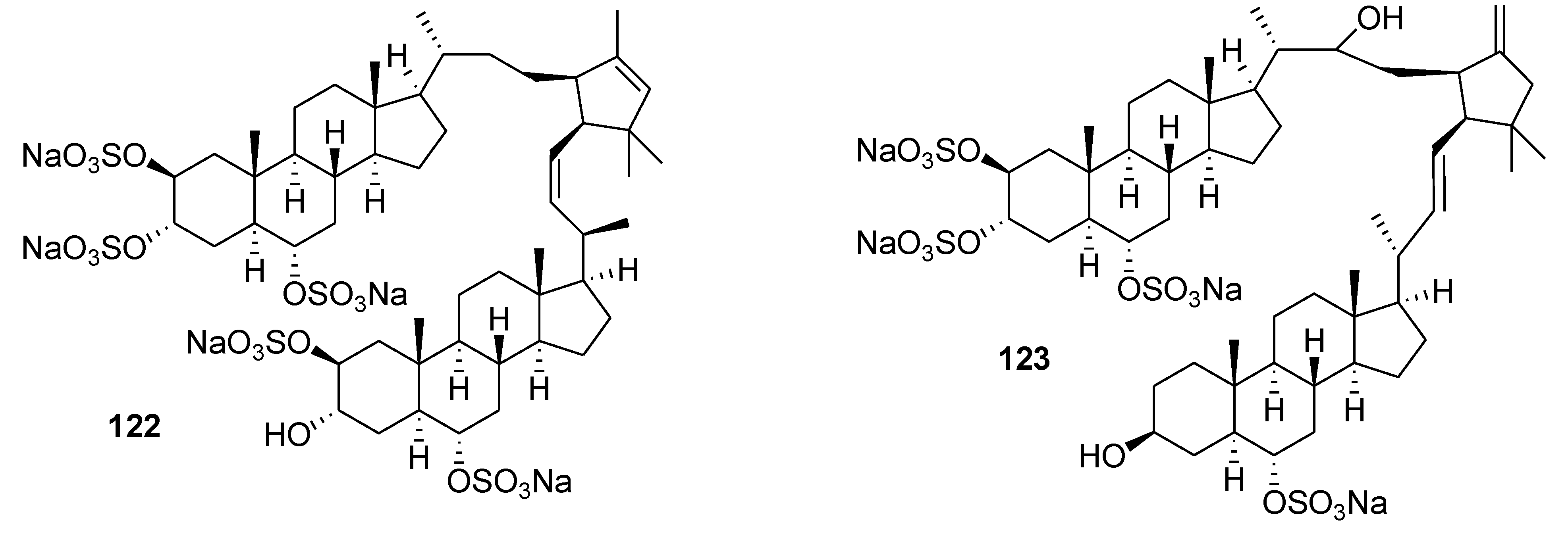
  68. Ivanchina, N.V.; Kicha, A.A.; Stonik, V.A. Steroid glycosides from marine organisms. Steroids 2011, 76, 425–454. [Google Scholar] [CrossRef]
  69. Stonik, V.A.; Kalinin, V.I.; Avilov, S.A. Toxins from sea cucumbers (holothuroids): Chemical structures, properties, taxonomic distribution, biosynthesis, and evolution. J. Nat. Toxins 1999, 8, 235–248. [Google Scholar]
  70. Kalinin, V.I.; Aminin, D.L.; Avilov, S.A.; Silchenko, A.S.; Stonik, V.A. Triterpene glycosides from sea cucumbers (Holothurioidea, Echinodermata), biological activities and functions. In Studies in Natural Product Chemistry; ur Rahman, A., Ed.; Elsevier: Amsterdam, The Netherlands, 2008; pp. 135–196. [Google Scholar]
  71. Aminin, D.L.; Pislyagin, E.A.; Menchinskaya, E.S.; Silchenko, A.S.; Avilov, S.A.; Kalinin, V.I. Immunomodulatory and Anticancer Activity of Sea Cucumber Triterpene Glycosides. In Studies in Natural Products Chemistry; ur Rahman, A., Ed.; Elsevier: Amsterdam, The Netherlands, 2014; pp. 73–91. [Google Scholar]
  72. Aminin, D.L.; Agafonova, I.G.; Berdyshev, E.V.; Isachenko, E.G.; Avilov, S.A.; Stonik, V.A. Immunomodulatory properties of cucumariosides from the edible Far-Eastern holothurian Cucumaria japonica. J. Med. Food 2001, 4, 127–135. [Google Scholar] [CrossRef]
  73. Aminin, D.L.; Pinegin, B.V.; Pichugina, L.V.; Zaporozhets, T.S.; Agafonova, I.G.; Boguslavski, V.M.; Silchenko, A.S.; Avilov, S.A.; Stonik, V.A. Immunomodulatory properties of cumaside. Int. Immunopharm. 2006, 6, 1070–1082. [Google Scholar] [CrossRef]
  74. Aminin, D.L.; Chaikina, E.L.; Agafonova, I.G.; Avilov, S.A.; Kalinin, V.I.; Stonik, V.A. Antitumor activity of the immunomodulatory lead cumaside. Int. Immunopharm. 2010, 10, 648–654. [Google Scholar] [CrossRef]
  75. Menchinskaya, E.S.; Pislyagin, E.A.; Kovalchyk, S.N.; Davydova, V.N.; Silchenko, A.S.; Avilov, S.A.; Kalinin, V.I.; Aminin, D.L. Antitumor Activity of Cucumarioside A2-2. Chemotherapy 2013, 59, 181–191. [Google Scholar] [CrossRef]
  76. Silchenko, A.S.; Kalinovsky, A.I.; Avilov, S.A.; Andryjaschenko, P.V.; Dmitrenok, P.S.; Menchinskaya, E.S.; Aminin, D.L.; Kalinin, V.I. Structure of cucumarioside I2 from the sea cucumber Eupentacta fraudatrix (Djakonov et Baranova) and cytotoxic and immunostimulatory activities of this saponin and relative compounds. Nat. Prod. Res. 2013, 27, 1776–1783. [Google Scholar] [CrossRef]
  77. Silchenko, A.S.; Kalinovsky, A.I.; Avilov, S.A.; Andryjaschenko, P.V.; Dmitrenok, P.S.; Martyyas, E.A.; Kalinin, V.I. Triterpene glycosides from the sea cucumber Eupentacta fraudatrix. Structure and biological action of cucumariosides A1, A3, A4, A5, A6, A12 and A15, seven new minor non-sulfated tetraosides and unprecedented 25-keto, 27-norholostane aglycone. Nat. Prod. Commun. 2012, 7, 517–525. [Google Scholar]
  78. Silchenko, A.S.; Kalinovsky, A.I.; Avilov, S.A.; Andryjaschenko, P.V.; Dmitrenok, P.S.; Martyyas, E.A.; Kalinin, V.I. Triterpene glycosides from the sea cucumber Eupentacta fraudatrix. Structure and cytotoxic action of cucumariosides A2, A7, A9, A10, A11, A13 and A14, seven new minor non-sulfated tetraosides and an aglycone with an uncommon 18-hydroxy group. Nat. Prod. Commun. 2012, 7, 845–852. [Google Scholar]
  79. Silchenko, A.S.; Kalinovsky, A.I.; Avilov, S.A.; Andryjaschenko, P.V.; Dmitrenok, P.S.; Martyyas, E.A.; Kalinin, V.I. Triterpene glycosides from the sea cucumber Eupentacta fraudatrix. Structure and biological activity of cucumariosides B1 and B2, two new minor non-sulfated unprecedented triosides. Nat. Prod. Commun. 2012, 7, 1157–1162. [Google Scholar]
  80. Silchenko, A.S.; Kalinovsky, A.I.; Avilov, S.A.; Andryjaschenko, P.V.; Dmitrenok, P.S.; Martyyas, E.A.; Kalinin, V.I.; Jayasandhya, P.; Rajan, G.C.; Padmakumar, K.P. Structures and biological activities of typicosides A1, A2, B1, C1 and C2, triterpene glycosides from the sea cucumber Actinocucumis typical. Nat. Prod. Commun. 2013, 8, 301–310. [Google Scholar]
  81. Silchenko, A.S.; Kalinovsky, A.I.; Avilov, S.A.; Andryjaschenko, P.V.; Dmitrenok, P.S.; Martyyas, E.A.; Kalinin, V.I. Triterpene glycosides from the sea cucumber Eupentacta fraudatrix. Structure and biological action of cucumariosides I1, I3, I4, three new minor disulfated pentaosides. Nat. Prod. Commun. 2013, 8, 1053–1058. [Google Scholar]
  82. Silchenko, A.S.; Kalinovsky, A.I.; Avilov, S.A.; Andryjaschenko, P.V.; Dmitrenok, P.S.; Yurchenko, E.A.; Dolmatov, I.Yu.; Kalinin, V.I.; Stonik, V.A. Structure and biological action of cladolosides B1, B2, C, C1, C2 and D, six new triterpene glycosides from the sea cucumber Cladolabes schmeltzii. Nat. Prod. Commun. 2013, 8, 1527–1534. [Google Scholar]
  83. Menchinskaya, E.S.; Aminin, D.L.; Avilov, S.A.; Silchenko, A.S.; Andryjashchenko, P.V.; Kalinin, V.I.; Stonik, V.A. Inhibition of tumor cells multidrug resistance by cucumarioside A2-2, frondoside A and their complexes with cholesterol. Nat. Prod. Commun. 2013, 8, 1377–1380. [Google Scholar]
  84. Yun, S.-H.; Park, E.-S.; Shin, S.-W.; Na, Y.-W.; Han, J.-Y.; Jeong, J.-S.; Shastina, V.V.; Stonik, V.A.; Park, J.-I.; Kwak, J.-Y. Stichoposide C induces apoptosis through the generation of ceramide in leukemia and colorectal cancer cells and shows in vivo antitumor activity. Clin. Cancer Res. 2012, 18, 5934–5948. [Google Scholar] [CrossRef]
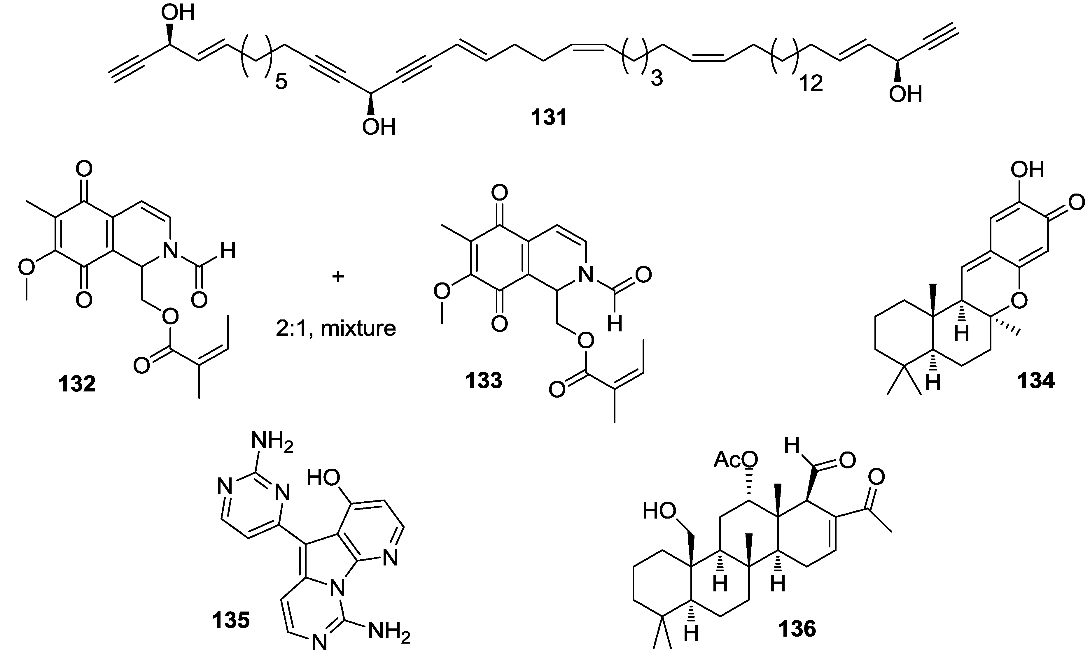
  85. Fujiki, H.; Cope, F.O.; Issinger, O.G. Sarcophytol-A, a potent tumor promoter inhibitor, prevents phosphorylation of a proteolytic fragment of C23 induced by the non-TPA type tumor promoter, okadaic acid. Proc. Am. Assoc. Cancer Res. 1988, 29, 155–155. [Google Scholar]
  86. Fujiki, H.; Suganuma, M.; Suguri, H.; Yoshizawa, S.; Takagi, K.; Kobayashi, M. Sarcophytol-A and sarcophytol-B inhibit tumor promotion by teleocidin in 2-stage carcinogenesis in mouse skin. J. Cancer Res. Clin. Oncol. 1989, 115, 25–28. [Google Scholar] [CrossRef]
  87. Wei, H.; Frenkel, K. Suppression of tumor promoter-induced oxidative events and DNA damage in vivo by sarcophytol A: A possible mechanism of antipromotion. Cancer Res. 1992, 52, 2298–2303. [Google Scholar]
  88. Fujiki, H.; Suganuma, M.; Komori, A.; Yatsunami, J.; Okabe, S.; Ohta, T.; Sueoka, E. A new tumor promotion pathway and its inhibitors. Cancer Det. Prev. 1994, 18, 1–7. [Google Scholar]
  89. Bhimani, R.S.; Troll, W.; Grunberger, D.; Frenkel, K. Inhibition of oxidative stress in HeLa cells by chermopreventive agents. Cancer Res. 1993, 53, 4528–4533. [Google Scholar]
  90. Yokomatsu, H.; Satake, K.; Hiura, A.; Tsutsumi, M.; Suganuma, M. Sarcophytol-A—a new chemotherapeutic and chemopreventive agent for pancreatic-cancer. Pancreas 1994, 9, 526–530. [Google Scholar] [CrossRef]
  91. Narisawa, T.; Takahashi, M.; Niwa, M.; Fukaura, Y.; Fujiki, H. Inhibition of methylnitrosourea-induced large bowel-cancer development in rats by sarcophytol A, a product from a marine soft coral Sarcophyton glaucum. Cancer Res. 1989, 49, 3287–3289. [Google Scholar]
  92. Yamauchi, O.; Omori, M.; Ninomiya, M.; Okuno, M.; Moriwaki, H.; Suganuma, M.; Fujiki, H.; Muto, Y. Inhibitory effect of sarcophytol-A on development of spontaneous hepatomas in mice. Jpn. J. Cancer Res. 1992, 82, 1234–1238. [Google Scholar]
  93. Hegazy, M.-E.F.; Gamal Eldeen, A.M.; Shahat, A.A.; Abdel-Latif, F.F.; Mohamed, T.A.; Whittlesey, B.R.; Paré, P.W. Bioactive hydroperoxyl cembranoids from the Red Sea soft coral Sarcophyton glaucum. Mar. Drugs 2012, 10, 209–222. [Google Scholar] [CrossRef]
  94. Albizat, K.F.; Holman, T.; Faulkner, D.J.; Glaser, K.B.; Jacobs, R.S. Luffariellolide, an anti-inflammatory sesterterpene from the marine sponge Luffariella sp. Experientia 1987, 45, 388–390. [Google Scholar]
  95. Wang, S.; Wang, Z.; Lin, S.; Zheng, W.; Wang, R.; Jin, S.; Chen, J.; Jin, L.; Li, Y. Revealing a natural marine product as a novel agonist for retinoic acid receptors with a unique binding mode and inhibitory effects on cancer cells. Biochem. J. 2012, 446, 79–87. [Google Scholar] [CrossRef]
  96. Liu, W.K.; Ling, Y.H.; Cheung, F.W.K.; Che, C.-T. Stellettin A induces endoplasmic reticulum stress in murine B16 melanoma cells. J. Nat. Prod. 2012, 75, 586–590. [Google Scholar] [CrossRef]
  97. Zhu, H.; Hua, X.; Gong, T.; Pang, J.; Hou, Q.; Zhu, P. Hypocreaterpenes A and B, cadinane-type sesquiterpenes from a marine-derived fungus, Hypocreales sp. Phytochem. Lett. 2013, 6, 392–396. [Google Scholar] [CrossRef]
  98. de los Reyes, C.; Zbakh, H.; Motilva, V.; Zubía, E. Antioxidant and anti-inflammatory meroterpenoids from the brown alga Cystoseira usneoides. J. Nat. Prod. 2013, 76, 621–629. [Google Scholar] [CrossRef]
  99. Wang, W.; Lee, Y.; Lee, T.G.; Mun, B.; Giri, A.G.; Lee, J.; Kim, H.; Hahn, D.; Yang, I.; Chin, J.; et al. Phorone A and isophorbasone A, sesterterpenoids isolated from the marine sponge Phorbas sp. Org. Lett. 2012, 14, 4486–4489. [Google Scholar] [CrossRef]
  100. Takahashi, Y.; Kubota, T.; Yamamoto, S.; Kobayashi, J. Inhibitory effects of metachromins L–Q and its related analogs against receptor tyrosine kinases EGFR and HER2. Bioorg. Med. Chem. Lett. 2013, 23, 117–118. [Google Scholar] [CrossRef]
  101. Li, J.; Zhu, H.; Ren, J.; Deng, Z.; de Voogd, N.J.; Proksch, P.; Lin, W. Globostelletins J–S, isomalabaricanes with unusual cyclopentane sidechains from the marine sponge Rhabdastrella globostellata. Tetrahedron 2012, 68, 559–565. [Google Scholar] [CrossRef]
  102. Shubina, L.K.; Kalinovsky, A.I.; Fedorov, S.N.; Radchenko, O.S.; Denisenko, V.A.; Dmitrenok, P.S.; Dyshlovoy, S.A.; Krasokhin, V.B.; Stonik, V.A. Aaptamine alkaloids from the Vietnamese sponge Aaptos sp. Nat. Prod. Commun. 2009, 4, 1085–1088. [Google Scholar]
  103. Shubina, L.K.; Makarieva, T.N.; Dyshlovoy, S.A.; Fedorov, S.N.; Dmitenok, P.S.; Stonik, V.A. Three new aaptamines from the marine sponge Aaptos sp. and their proapoptotic properties. Nat. Prod. Commun. 2010, 5, 1881–1884. [Google Scholar]
  104. Utkina, N.K. Antioxidant activity of aromatic alkaloids from the marine sponges Aaptos aaptos and Hyrtios sp. Chem. Nat. Comp. 2009, 45, 849–853. [Google Scholar] [CrossRef]
  105. Dyshlovoy, S.A.; Naeth, I.; Venz, S.; Preukschas, M.; Sievert, H.; Jacobsen, C.; Shubina, L.K.; Salazar, M.G.; Scharf, C.; Walther, R.; et al. Proteomic profiling of germ cell cancer cells treated with aaptamine, a marine alkaloid with antiproliferative activity. J. Proteome Res. 2012, 11, 2316–2330. [Google Scholar] [CrossRef]
  106. Dyshlovoy, S.A.; Venz, S.; Shubina, L.K.; Fedorov, S.N.; Walther, R.; Jacobsen, C.; Stonik, V.A.; Bokemeyer, C.; Balabanov, S.; Honecker, F. Activity of aaptamine and two derivatives, demethyloxyaaptamine and isoaaptamine, in cisplatin-resistant germ cell cancer. J. Proteomics 2014, 96, 223–239. [Google Scholar] [CrossRef]
  107. Yamazaki, H.; Wewengkang, D.S.; Kanno, S.; Ishikawa, M.; Rotinsulu, H.; Mangindaan, R.E.P.; Namikoshi, M. Papuamine and haliclonadiamine, obtained from an Indonesian sponge Haliclona sp., inhibited cell proliferation of human cancer cell lines. Nat. Prod. Res. 2013, 27, 1012–1015. [Google Scholar] [CrossRef]
  108. Fu, P.; Yang, C.; Wang, Y.; Liu, P.; Ma, Y.; Xu, L.; Su, M.; Hong, K.; Zhu, W. Streptocarbazoles A and B, two novel indolocarbazoles from the marine-derived actinomycete strain Streptomyces sp. FMA. Org. Lett. 2012, 14, 2422–2425. [Google Scholar] [CrossRef]
  109. Johnson, T.A.; Sohn, J.; Vaske, Y.M.; White, K.N.; Cohen, T.L.; Vervoort, H.C.; Tenney, K.; Valeriote, F.A.; Bjeldanes, L.F.; Crews, P. Myxobacteria versus sponge-derived alkaloids: The bengamide family identified as potent immune modulating agents by scrutiny of LC–MS/ELSD libraries. Bioorg. Med. Chem. 2012, 20, 4348–4355. [Google Scholar] [CrossRef]
  110. Hwang, B.S.; Oh, J.S.; Jeong, E.J.; Sim, C.J.; Rho, J.-R. Densanins A and B, new macrocyclic pyrrole alkaloids isolated from the marine sponge Haliclona densaspicula. Org. Lett. 2012, 14, 6154–6157. [Google Scholar] [CrossRef]
  111. Sakai, E.; Kato, H.; Rotinsulu, H.; Losung, F.; Mangindaan, R.E.P.; de Voogd, N.J.; Yokosawa, H.; Tsukamoto, S. Variabines A and B: New β-carboline alkaloids from the marine sponge Luffariella variabilis. J. Nat. Med. 2014, 68, 215–219. [Google Scholar] [CrossRef]
  112. Yamaguchi, M.; Miyazaki, M.; Kodrasov, M.P.; Rotinsulu, H.; Losung, F.; Mangindaan, R.E.P.; de Voogd, N.J.; Yokosawa, H.; Nicholson, B.; Tsukamoto, S. Spongiacidin C, a pyrrole alkaloid from the marine sponge Stylissa massa, functions as a USP7 inhibitor. Bioorg. Med. Chem. Lett. 2013, 23, 3884–3886. [Google Scholar] [CrossRef]
  113. Fedorov, S.N.; Dyshlovoy, S.А.; Shubina, L.K.; Guzii, A.G.; Kuzmich, A.S.; Makarieva, T.N. C11 cyclopentenone from the ascidian Diplosoma sp. prevents epidermal growth factor-induced transformation of JB6 cells. Drugs Ther. Stud. 2012, 2, e4. [Google Scholar]
  114. Williams, D.E.; Dalisay, D.S.; Li, F.; Amphlett, J.; Maneerat, W.; Chavez, M.A.G.; Wang, Y.A.; Matainaho, T.; Yu, W.; Brown, P.J.; et al. Nahuoic acid A produced by a Streptomyces sp. isolated from a marine sediment is a selective SAM-competitive inhibitor of the histone metyltransferase SETD8. Org. Lett. 2013, 15, 414–417. [Google Scholar] [CrossRef]
  115. Takawa, M.; Cho, H.S.; Hayami, S.; Toyokawa, G.; Kogure, M.; Yamane, Y.; Iwai, Y.; Maejima, K.; Ueda, K.; Masuda, A.; et al. Histone lysine methyltransferase SETD8 promotes carcinogenesis by deregulating PCNA expression. Cancer Res. 2012, 72, 3217–3227. [Google Scholar] [CrossRef]
  116. Asami, Y.; Jang, J.-H.; Soung, N.-K.; He, L.; Moon, D.O.; Kim, J.W.; Oh, H.; Muroi, M.; Osada, H.; Kim, B.Y.; et al. Protuboxepin A, a marine fungal metabolite, inducing metaphase arrest and chromosomal misalignment in tumor cells. Bioorg. Med. Chem. 2012, 20, 3799–3806. [Google Scholar] [CrossRef]
  117. Dyshlovoy, S.A.; Fedorov, S.N.; Kalinovsky, A.I.; Shubina, L.K.; Bokemeyer, C.; Stonik, V.A.; Honecker, F. Mycalamide A shows cytotoxic properties and prevents EGF-induced neoplastic transformation through inhibition of nuclear factors. Mar. Drugs 2012, 10, 1212–1224. [Google Scholar] [CrossRef]
  118. Shirouzu, T.; Watari, K.; Ono, M.; Koizumi, K.; Saiki, I.; Tanaka, C.; van Soest, R.W.M.; Miyamoto, T. Structure, synthesis, and biological activity of a C-20 bisacetylenic alcohol from a marine sponge Callyspongia sp. J. Nat. Prod. 2013, 76, 1337–1342. [Google Scholar] [CrossRef]
  119. Garcia-Caballero, M.; Mari-Beffa, M.; Canedo, L.; Medina, M.A.; Quesada, A.R. Toluquinol, a marine fungus metabolite, is a new angiosuppresor that interferes the Akt pathway. Biochem. Pharmacol. 2013, 85, 1727–1740. [Google Scholar] [CrossRef]
  120. Towle, K.M.; Chaytor, J.L.; Liu, H.; Austin, P.; Roberge, M.; Roskelley, C.D.; Vederas, J.C. Synthesis and biological studies of neopetrosiamides as inhibitors of cancer cell invasion. Org. Biomol. Chem. 2013, 11, 1476–1481. [Google Scholar] [CrossRef]
  121. Igarashi, Y.; Asano, D.; Furihata, K.; Oku, N.; Miyanaga, S.; Sakurai, H.; Saiki, I. Absolute configuration of pterocidin, a potent inhibitor of tumor cell invasion from a marine-derived Streptomyces. Tetrahedron Lett. 2012, 53, 654–656. [Google Scholar] [CrossRef]
  122. Um, S.; Kim, Y.-J.; Kwon, H.; Wen, H.; Kim, S.-H.; Kwon, H.C.; Park, S.; Shin, J.; Oh, D.-C. Sungsanpin, a Lasso Peptide from a Deep-Sea Streptomycete. J. Nat. Prod. 2013, 76, 873–879. [Google Scholar] [CrossRef]
  123. Pimentel, A.A.; Felibertt, P.; Sojo, F.; Colman, L.; Mayora, A.; Silva, M.L.; Rojas, H.; Dipolo, R.; Suarez, A.I.; Compagnone, R.S.; et al. The marine sponge toxin agelasine B increases the intracellular Ca2+ concentration and induces apoptosis in human breast cancer cells (MCF-7). Cancer Chemother. Pharmacol. 2012, 69, 71–83. [Google Scholar] [CrossRef]
  124. Huang, C.; Jin, H.; Song, B.; Zhu, X.; Zhao, H.; Cai, J.; Lu, Y.; Chen, B.; Lin, Y. The cytotoxicity and anticancer mechanisms of alterporriol L, a marine bianthraquinone, against MCF-7 human breast cancer cells. Appl. Microbiol. Biotechnol. 2012, 93, 777–785. [Google Scholar] [CrossRef]
  125. Ohno, O.; Morita, M.; Kitamura, K.; Teruya, T.; Yoneda, K.; Kita, M.; Kigoshi, H.; Suenaga, K. Apoptosis-inducing activity of the actin-depolymerizing agent aplyronine A and its side-chain derivatives. Bioorg. Med. Chem. Lett. 2013, 23, 1467–1471. [Google Scholar] [CrossRef]
  126. Morita, M.; Ohno, O.; Teruya, T.; Yamori, T.; Inuzuka, T.; Suenaga, K. Isolation and structures of biselyngbyasides B, C, and D from the marine cyanobacterium Lyngbya sp., and the biological activities of biselyngbyasides. Tetrahedron 2012, 68, 5984–5990. [Google Scholar]
  127. Park, S.J.; Jeon, Y.J. Dieckol from Ecklonia cava suppresses the migration and invasion of HT1080 cells by inhibiting the focal adhesion kinase pathway downstream of Rac1-ROS signaling. Mol. Cells 2012, 33, 141–149. [Google Scholar] [CrossRef]
  128. Maschek, J.A.; Mevers, E.; Diyabalanage, T.; Chen, L.; Ren, Y.; McClintock, J.B.; Amsler, C.D.; Wu, J.; Baker, B.J. Palmadorin chemodiversity from the antarctic nudibranch Austrodoris kerguelenensis and inhibition of Jak2/STAT5-dependent HEL leukemia cells. Tetrahedron 2012, 68, 9095–9104. [Google Scholar] [CrossRef]
  129. Park, E.-J.; Pezzuto, J.M.; Jang, K.H.; Nam, S.-J.; Bucarey, S.A.; Fenical, W. Suppression of nitric oxide synthase by thienodolin in lipopolysaccharide-stimulated RAW 264.7 murine macrophage cells. Nat. Prod. Commun. 2012, 7, 789–794. [Google Scholar]
  130. Williams, D.E.; Steino, A.; de Voogd, N.J.; Mauk, A.G.; Andersen, R.J. Halicloic acids A and B isolated from the marine sponge Haliclona sp. collected in the Philippines inhibit indoleamine 2,3-dioxygenase. J. Nat. Prod. 2012, 75, 1451–1458. [Google Scholar] [CrossRef]
  131. Balunas, M.J.; Grosso, M.F.; Villa, F.A.; Engene, N.; McPhail, K.L.; Tidgewell, K.; Pineda, L.M.; Gerwick, L.; Spadafora, C.; Kyle, D.E.; et al. Coibacins A–D, antileishmanial marine cyanobacterial polyketides with intriguing biosynthetic origins. Org. Lett. 2012, 14, 3878–3881. [Google Scholar] [CrossRef]
  132. Festa, C.; De Marino, S.; D’Auria, M.V.; Monti, M.C.; Bucci, M.; Vellecco, V.; Debitus, C.; Zampella, A. Anti-inflammatory cyclopeptides from the marine sponge Theonella swinhoei. Tetrahedron 2012, 68, 2851–2857. [Google Scholar] [CrossRef]
  133. Salvador, L.A.; Taori, K.; Biggs, J.S.; Jakoncic, J.; Ostrov, D.A.; Paul, V.J.; Luesch, H. Potent elastase inhibitors from cyanobacteria: Structural basis and mechanisms mediating cytoprotective and anti-inflammatory effects in bronchial epithelial cells. J. Med. Chem. 2013, 56, 1276–1290. [Google Scholar] [CrossRef]
  134. Lin, Y.-F.; Kuo, C.-Y.; Wen, Z.-H.; Lin, Y.-Y.; Wang, W.-H.; Su, J.-H.; Sheu, J.-H.; Sung, P.-J. Flexibilisquinone, a new anti-inflammatory quinone from the cultured soft coral Sinularia flexibilis. Molecules 2013, 18, 8160–8167. [Google Scholar] [CrossRef]
  135. Wang, J.; Zhao, B.; Yi, Y.; Zhang, W.; Wu, X.; Zhang, L.; Shen, Y. Mycoepoxydiene, a fungal polyketide inhibits MCF-7 cells through simultaneously targeting p53 and NF-κB pathways. Biochem. Pharmacol. 2012, 84, 891–899. [Google Scholar] [CrossRef]
  136. Kim, M.C.; Kwon, O.-W.; Park, J.-S.; Kim, S.Y.; Kwon, H.C. Nocapyrones H-J, 3,6-disubstituted α-pyrones from the marine actinomycete Nocardiopsis sp. KMF-001. Chem. Pharm. Bull. 2013, 61, 511–515. [Google Scholar] [CrossRef]
  137. Song, Y.; Dou, H.; Gong, W.; Liu, X.; Yu, Z.; Li, E.; Tan, R.; Hou, Y. Bis-N-norgliovictin, a small-molecule compound from marine fungus, inhibits LPS-induced inflammation in macrophages and improves survival in sepsis. Eur. J. Pharm. 2013, 705, 49–60. [Google Scholar] [CrossRef]
  138. Festa, C.; Lauro, G.; De Marino, S.; D’Auria, M.V.; Monti, M.C.; Casapullo, A.; D’Amore, C.; Renga, B.; Mencarelli, A.; Petek, S.; et al. Plakilactones from the marine sponge Plakinastrella mamillaris. Discovery of a new class of marine ligands of peroxisome proliferator-activated receptor γ. J. Med. Chem. 2012, 55, 8303–8317. [Google Scholar] [CrossRef]
  139. Elsebai, M.F.; Nazir, M.; Kehraus, S.; Egereva, E.; Ioset, K.N.; Marcourt, L.; Jeannerat, D.; Gütschow, M.; Wolfender, J.-L.; König, G.M. Polyketide skeletons from the marine alga-derived fungus Coniothyrium cereale. Eur. J. Org. Chem. 2012, 2012, 6197–6203. [Google Scholar]
  140. Chen, Y.-H.; Lu, M.-C.; Chang, Y.-C.; Hwang, T.-L.; Wang, W.-H.; Weng, C.-F.; Kuo, J.; Sung, P.-J. Pseudoalteromone A: A novel bioactive ubiquinone from a marine bacterium Pseudoalteromonas sp. CGH2XX (Pseudoalteromonadaceae). Tetrahedron Lett. 2012, 53, 1675–1677. [Google Scholar]
  141. Chen, Y.-H.; Kuo, J.; Su, J.-H.; Hwang, T.-L.; Chen, Y.-H.; Lee, C.-H.; Weng, C.-F.; Sung, P.-J. Pseudoalteromone B: A novel 15C compound from a marine bacterium Pseudoalteromonas sp CGH2XX. Mar. Drugs 2012, 10, 1566–1571. [Google Scholar] [CrossRef]
  142. Ushiyama, S.; Umaoka, H.; Kato, H.; Suwa, Y.; Morioka, H.; Rotinsulu, H.; Losung, F.; Mangindaan, R.E.P.; de Voogd, N.J.; Yokosawa, H.; et al. Manadosterols A and B, sulfonated sterol dimers inhibiting the Ubc13–Uev1A interaction, isolated from the marine sponge Lissodendryx fibrosa. J. Nat. Prod. 2012, 75, 1495–1499. [Google Scholar] [CrossRef]
  143. Pina, I.C.; Gautschi, J.T.; Wang, G.-Y.-S.; Sanders, M.L.; Schmitz, F.J.; France, D.; Cornell-Kennon, S.; Sambucetti, L.C.; Remiszewski, S.W.; Perez, L.B.; et al. Psammaplins from the sponge Pseudoceratina purpurea:Inhibition of both histone deacetylase and DNA methyltransferase. J. Org. Chem. 2003, 68, 3866–3873. [Google Scholar] [CrossRef]
  144. Hentschel, F.; Sasseb, F.; Lindel, T. Fluorescent analogs of the marine natural product psammaplin A: Synthesis and biological activity. Org. Biomol. Chem. 2012, 10, 7120–7133. [Google Scholar] [CrossRef]
  145. Hosoya, T.; Hirokawa, T.; Takagi, M.; Shin-ya, K. Trichostatin analogues JBIR-109, JBIR-110, and JBIR-111 from the marine sponge-derived Streptomyces sp. RM72. J. Nat. Prod. 2012, 75, 285–289. [Google Scholar] [CrossRef]
  146. McKee, T.C.; Rabe, D.; Bokesch, H.R.; Grkovic, T.; Whitson, E.L.; Diyabalanage, T.; Van Wyk, A.W.W.; Marcum, S.R.; Gardella, R.S.; Gustafson, K.R.; et al. Inhibition of Hypoxia Inducible Factor-2 transcription: Isolation of active modulators from marine sponges. J. Nat. Prod. 2012, 75, 1632–1636. [Google Scholar] [CrossRef]
  147. Du, L.; Mahdi, F.; Datta, S.; Jekabsons, M.B.; Zhou, Y.-D.; Nagle, D.G. Structures and mechanisms of antitumor agents: Xestoquinones uncouple cellular respiration and disrupt HIF signaling in human breast tumor cells. J. Nat. Prod. 2012, 75, 1553–1559. [Google Scholar] [CrossRef]
  148. Sun, Y.; Takada, K.; Takemoto, Y.; Yoshida, M.; Nogi, Y.; Okada, S.; Matsunaga, S. Gliotoxin analogues from a marine-derived fungus, Penicillium sp., and their cytotoxic and histone methyltransferase inhibitory activities. J. Nat. Prod. 2012, 75, 111–114. [Google Scholar] [CrossRef]
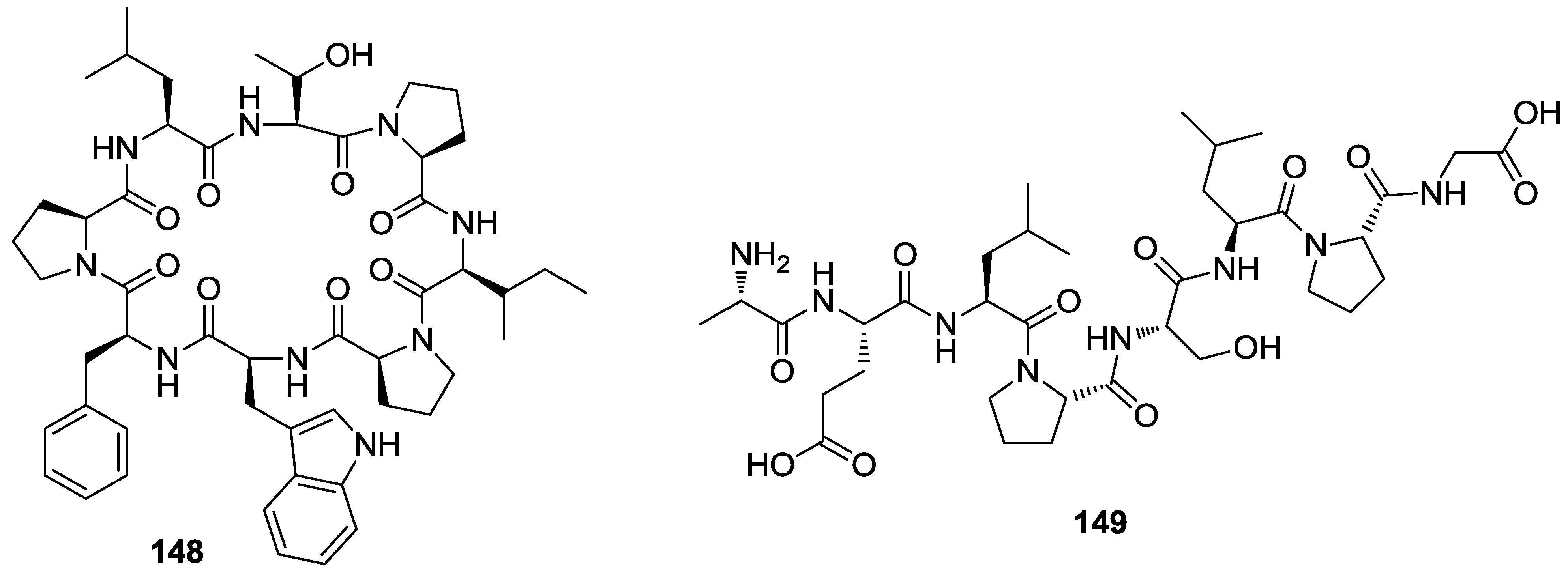
  149. Arai, M.; Yamano, Y.; Fujita, M.; Setiawan, A.; Kobayashi, M. Stylissamide X, a new proline-rich cyclic octapeptide as an inhibitor of cell migration, from an Indonesian marine sponge of Stylissa sp. Bioorg. Med. Chem. Lett. 2012, 22, 1818–1821. [Google Scholar] [CrossRef]
  150. Nguyen, V.-T.; Qian, Z.-J.; Ryu, B.M.; Kim, K.-N.; Kim, D.; Kim, Y.-M.; Jeon, Y.-J.; Park, W.S.; Choi, I.-W.; Kim, G.H.; et al. Matrix metalloproteinases (MMPs) inhibitory effects of an octameric oligopeptide isolated from abalone Haliotis discus hannai. Food Chem. 2013, 141, 503–509. [Google Scholar] [CrossRef]
  151. Nakamura, Y.; Kato, H.; Nishikawa, T.; Iwasaki, N.; Suwa, Y.; Rotinsulu, H.; Losung, F.; Maarisit, W.; Mangindaan, R.E.P.; Morioka, H.; et al. Siladenoserinols A–L: New sulfonated serinol derivatives from a tunicate as inhibitors of p53-Hdm2 interaction. Org. Lett. 2013, 15, 322–325. [Google Scholar] [CrossRef]
  152. Malloy, K.L.; Choi, H.; Fiorilla, C.; Valeriote, F.A.; Matainaho, T.; Gerwick, W.H. Hoiamide D, a marine cyanobacteria-derived inhibitor of p53/MDM2 interaction. Bioorg. Med. Chem. Lett. 2012, 22, 683–688. [Google Scholar] [CrossRef]
  153. Huang, X.; Sun, Y.-L.; Salim, A.A.; Chen, Z.-S.; Capon, R.J. Parguerenes: Marine red alga bromoditerpenes as inhibitors of P-glycoprotein (ABCB1) in multidrug resistant human cancer cells. Biochem. Pharmacol. 2013, 85, 1257–1268. [Google Scholar]
  154. Niemann, H.; Lin, W.; Müller, W.E.G.; Kubbutat, M.; Lai, D.; Proksch, P. Trimeric hemibastadin congener from the marine sponge Ianthella basta. J. Nat. Prod. 2013, 76, 121–125. [Google Scholar]

