Neurogenic Rosacea and Facial Dysesthesia: Neurovascular–Immune Mechanisms and Translational Therapeutic Perspectives


Abstract
1. Introduction
2. Materials and Methods
3. Findings of the Narrative Synthesis: Clinical Presentation and Proposed Diagnostic Framework
4. Hierarchy of Evidence for Proposed Neurovascular–Immune Pathways
4.1. Direct and Indirect Human Evidence
4.2. Animal and Cellular Evidence
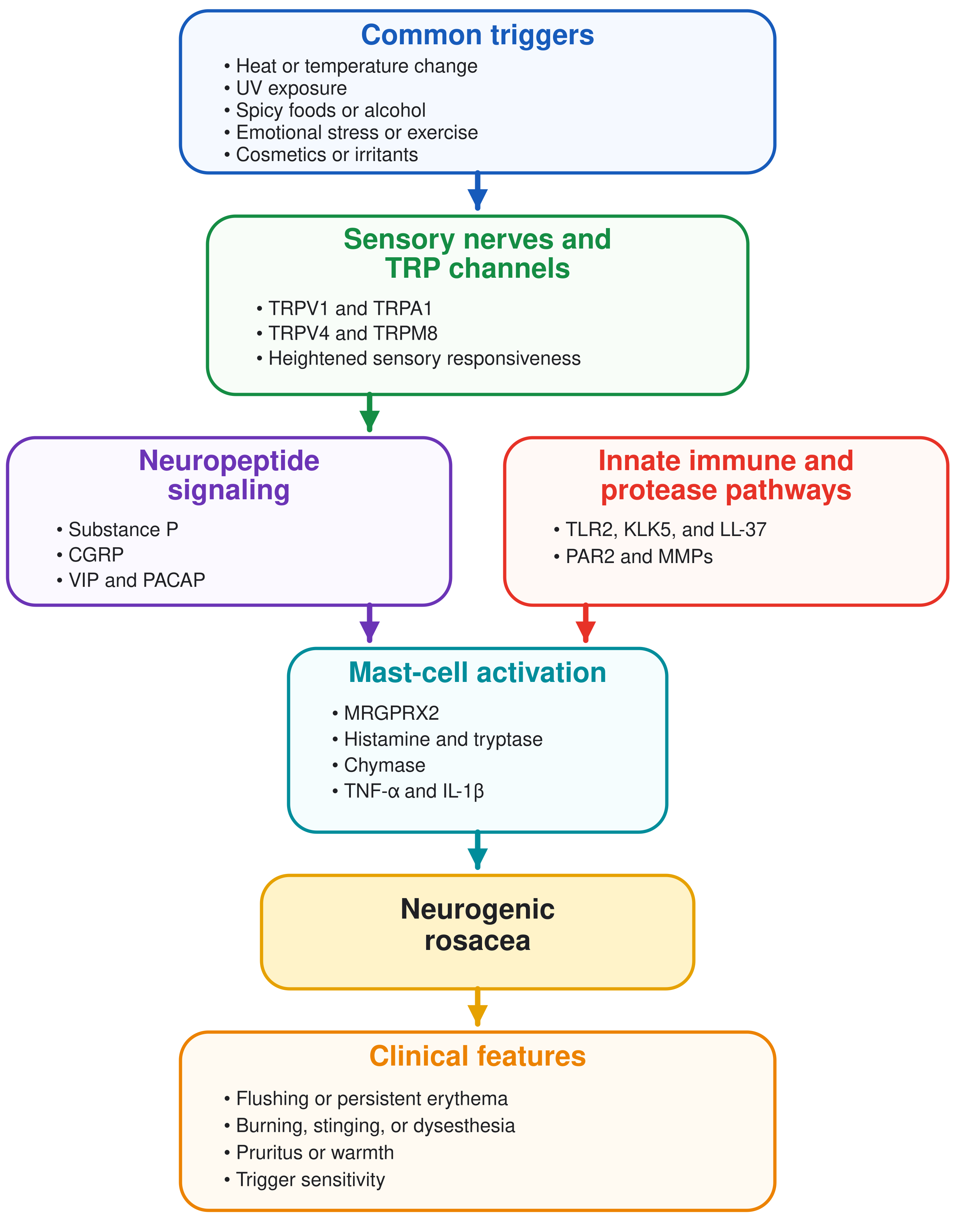
4.3. TRP Channels and Trigger Sensitivity
4.4. Neuropeptides, Vasodilation, and Sensory Symptoms
4.5. Protease-Dependent Innate Immune Amplification
5. Differential Diagnosis
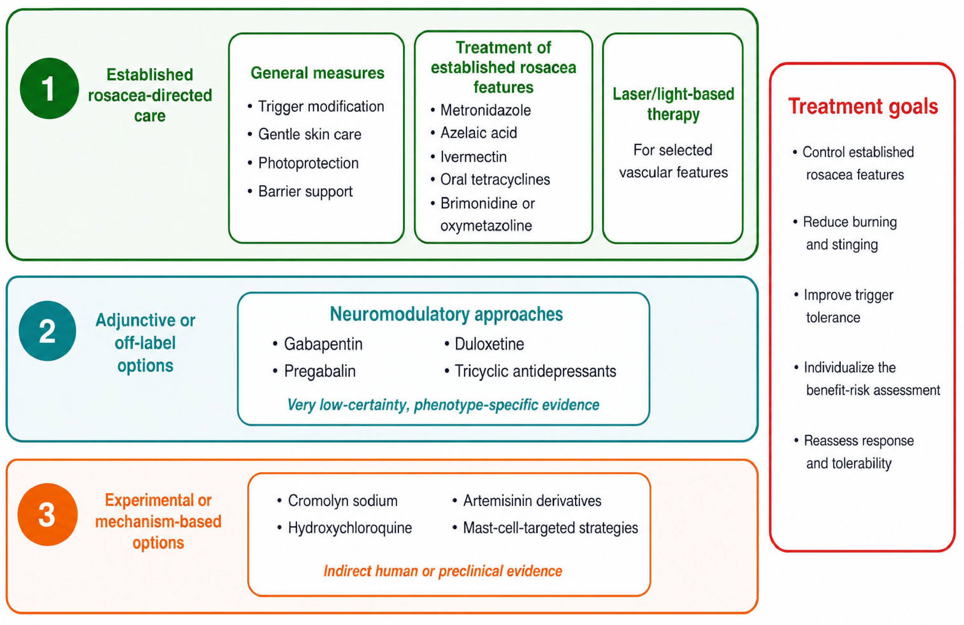
6. Management
7. Discussion
8. Limitations and Future Directions
9. Conclusions
Funding
Institutional Review Board Statement
Informed Consent Statement
Data Availability Statement
Conflicts of Interest
Abbreviations
| CCL | C-C motif chemokine ligand |
| CGRP | calcitonin gene-related peptide |
| CXCL | C-X-C motif chemokine ligand |
| IL | interleukin |
| IgE | immunoglobulin E |
| IPL | intense pulsed light |
| KLK | kallikrein |
| KLK5 | kallikrein 5 |
| KLK7 | kallikrein 7 |
| LL-37 | cathelicidin antimicrobial peptide LL-37 |
| MMPs | matrix metalloproteinases |
| MMP-9 | matrix metalloproteinase 9 |
| MRGPRX2 | Mas-related G protein-coupled receptor X2 |
| MrgprB2 | Mas-related G protein-coupled receptor B2 |
| NK1 | neurokinin 1 |
| PACAP | pituitary adenylate cyclase-activating polypeptide |
| PAR2 | protease-activated receptor 2 |
| SP | substance P |
| TLR2 | Toll-like receptor 2 |
| TNF-α | tumor necrosis factor alpha |
| TRP | transient receptor potential |
| TRPA1 | transient receptor potential ankyrin 1 |
| TRPM8 | transient receptor potential melastatin 8 |
| TRPV1 | transient receptor potential vanilloid 1 |
| TRPV2 | transient receptor potential vanilloid 2 |
| TRPV3 | transient receptor potential vanilloid 3 |
| TRPV4 | transient receptor potential vanilloid 4 |
| UV | ultraviolet |
| VIP | vasoactive intestinal peptide |
References
- Tan, J.; Almeida, L.M.C.; Bewley, A.; Cribier, B.; Dlova, N.C.; Gallo, R.; Kautz, G.; Mannis, M.; Oon, H.; Rajagopalan, M.; et al. Updating the diagnosis, classification and assessment of rosacea: Recommendations from the global ROSacea COnsensus (ROSCO) panel. Br. J. Dermatol. 2017, 176, 431–438. [Google Scholar] [CrossRef] [Scilit] [PubMed]
- Schaller, M.; Almeida, L.M.C.; Bewley, A.; Cribier, B.; Del Rosso, J.Q.; Dlova, N.C.; Gallo, R.L.; Granstein, R.D.; Kautz, G.; Mannis, M.J.; et al. Recommendations for rosacea diagnosis, classification and management: Update from the global ROSaceaCOnsensus 2019 panel. Br. J. Dermatol. 2020, 182, 1269–1276. [Google Scholar] [CrossRef] [Scilit] [PubMed]
- van Zuuren, E.J.; Arents, B.W.M.; van der Linden, M.M.D.; Vermeulen, S.; Fedorowicz, Z.; Tan, J. Rosacea: New concepts in classification and treatment. Am. J. Clin. Dermatol. 2021, 22, 457–465. [Google Scholar] [CrossRef] [Scilit] [PubMed]
- Thiboutot, D.; Anderson, R.; Cook-Bolden, F.; Draelos, Z.; Gallo, R.L.; Granstein, R.D.; Kang, S.; Macsai, M.; Gold, L.S.; Tan, J. Standard management options for rosacea: The 2019 update by the National Rosacea Society Expert Committee. J. Am. Acad. Dermatol. 2020, 82, 1501–1510. [Google Scholar] [CrossRef] [Scilit] [PubMed]
- Schaller, M.; Dirschka, T.; Lonne-Rahm, S.B.; Micali, G.; Stein Gold, L.F.; Tan, J.; Del Rosso, J. The importance of assessing burning and stinging when managing rosacea: A review. Acta Derm.-Venereol. 2021, 101, adv00584. [Google Scholar] [CrossRef] [Scilit] [PubMed]
- Gallo, R.L.; Granstein, R.D.; Kang, S.; Mannis, M.; Steinhoff, M.; Tan, J.; Thiboutot, D. Standard classification and pathophysiology of rosacea: The 2017 update by the National Rosacea Society Expert Committee. J. Am. Acad. Dermatol. 2018, 78, 148–155. [Google Scholar] [CrossRef] [Scilit] [PubMed]
- Geng, R.S.Q.; Bourkas, A.N.; Mufti, A.; Sibbald, R.G. Rosacea: Pathogenesis and therapeutic correlates. J. Cutan. Med. Surg. 2024, 28, 178–189. [Google Scholar] [CrossRef] [Scilit] [PubMed]
- Tu, K.Y.; Jung, C.J.; Shih, Y.H.; Chang, A.L.S. Therapeutic strategies focusing on immune dysregulation and neuroinflammation in rosacea. Front. Immunol. 2024, 15, 1403798. [Google Scholar] [CrossRef] [Scilit] [PubMed]
- Galluccio, G.; D’Onghia, M.; Malvaso, D.; Lazzeri, L.; Cinotti, E.; Rubegni, G.; Rubegni, P.; Calabrese, L. Advances in the pathogenesis and treatment of rosacea: A phenotype-based therapeutic approach. Cosmetics 2024, 11, 11. [Google Scholar] [CrossRef] [Scilit]
- Chen, L.-X.-Y.; Hao, P.-S. The role of skin barrier and immune abnormalities in the pathogenesis of rosacea. Clin. Exp. Med. 2025, 25, 324. [Google Scholar] [CrossRef] [Scilit] [PubMed]
- Li, D.; Yang, W.; Chen, N.; Xu, Y. Updates in the treatment of rosacea with γ-aminobutyric acid derivatives. Clin. Cosmet. Investig. Dermatol. 2025, 18, 2207–2214. [Google Scholar] [CrossRef] [Scilit] [PubMed]
- Scharschmidt, T.C.; Yost, J.M.; Truong, S.V.; Steinhoff, M.; Wang, K.C.; Berger, T.G. Neurogenic rosacea: A distinct clinical subtype requiring a modified approach to treatment. Arch. Dermatol. 2011, 147, 123–126. [Google Scholar] [CrossRef] [Scilit] [PubMed]
- Aedo, G.; Chahuán, M.; Gatica, E.; Herrera, I.; Parada, L.F.; Seguel, A.; Murray, N.P.; Aedo, S.; Aragón-Caqueo, D. Managing a burning face: Clinical manifestations and therapeutic approaches for neurogenic rosacea. Int. J. Mol. Sci. 2025, 26, 2366. [Google Scholar] [CrossRef] [Scilit] [PubMed]
- Li, M.; Tao, M.; Zhang, Y.; Pan, R.; Gu, D.; Xu, Y. Neurogenic rosacea could be a small fiber neuropathy. Front. Pain Res. 2023, 4, 1122134. [Google Scholar] [CrossRef] [Scilit] [PubMed]
- Kim, H.O.; Kang, S.Y.; Kim, K.E.; Cho, S.Y.; Kim, K.H.; Kim, I.H. Neurogenic rosacea in Korea. J. Dermatol. 2021, 48, 49–55. [Google Scholar] [CrossRef] [Scilit] [PubMed]
- Wang, L.; Wang, Y.J.; Hao, D.; Wen, X.; Du, D.; He, G.; Jiang, X. The theranostic role of mast cells in the pathophysiology of rosacea. Front. Med. 2020, 6, 324. [Google Scholar] [CrossRef] [Scilit] [PubMed]
- Chen, M.; Yang, L.; Zhou, P.; Jin, S.; Wu, Z.; Tan, Z.; Xiao, W.; Xu, S.; Zhu, Y.; Wang, M.; et al. Single-cell transcriptomics reveals aberrant skin-resident cell populations and identifies fibroblasts as a determinant in rosacea. Nat. Commun. 2024, 15, 8737. [Google Scholar] [CrossRef] [Scilit] [PubMed]
- Choi, J.E.; Di Nardo, A. Skin neurogenic inflammation. Semin. Immunopathol. 2018, 40, 249–259. [Google Scholar] [CrossRef] [Scilit] [PubMed]
- Luo, D.Q.; Zhao, Y.K.; Xu, Q.F.; He, X.Q.; Wu, L.C. Recurrent facial erythema with burning sensation and increased temperature: A variant of erythromelalgia or a new entity? Pain Med. 2014, 15, 1007–1010. [Google Scholar] [CrossRef] [Scilit] [PubMed]
- Wienholtz, N.K.F.; Christensen, C.E.; Coskun, H.; Zhang, D.G.; Ghanizada, H.; Egeberg, A.; Thyssen, J.P.; Ashina, M. Infusion of pituitary adenylate cyclase-activating polypeptide-38 in patients with rosacea induces flushing and facial edema that can be attenuated by sumatriptan. J. Investig. Dermatol. 2021, 141, 1687–1698. [Google Scholar] [CrossRef] [Scilit] [PubMed]
- Wienholtz, N.K.F.; Christensen, C.E.; Ashina, H.; Jørgensen, N.R.; Egeberg, A.; Thyssen, J.P.; Ashina, M. Elevated plasma levels of calcitonin gene-related peptide in individuals with rosacea: A cross-sectional case-control study. J. Eur. Acad. Dermatol. Venereol. 2025, 39, 181–188. [Google Scholar] [CrossRef] [Scilit] [PubMed]
- Holmes, A.D.; Steinhoff, M. Integrative concepts of rosacea pathophysiology, clinical presentation and new therapeutics. Exp. Dermatol. 2017, 26, 659–667. [Google Scholar] [CrossRef] [Scilit] [PubMed]
- Sulk, M.; Seeliger, S.; Aubert, J.; Schwab, V.D.; Cevikbas, F.; Rivier, M.; Nowak, P.; Voegel, J.J.; Buddenkotte, J.; Steinhoff, M. Distribution and expression of non-neuronal transient receptor potential ion channels in rosacea. J. Investig. Dermatol. 2012, 132, 1253–1262. [Google Scholar] [CrossRef] [Scilit] [PubMed]
- Zhang, Y.; Li, T.; Zhao, H.; Xiao, X.; Hu, X.; Wang, B.; Huang, Y.; Yin, Z.; Zhong, Y.; Li, Y.; et al. High-sensitive sensory neurons exacerbate rosacea-like dermatitis in mice by activating γδ T cells directly. Nat. Commun. 2024, 15, 7265. [Google Scholar] [CrossRef] [Scilit] [PubMed]
- Mascarenhas, N.L.; Wang, Z.; Chang, Y.L.; Di Nardo, A. TRPV4 mediates mast cell activation in cathelicidin-induced rosacea inflammation. J. Investig. Dermatol. 2017, 137, 972–975. [Google Scholar] [CrossRef] [Scilit] [PubMed]
- Yamasaki, K.; Kanada, K.; Macleod, D.T.; Borkowski, A.W.; Morizane, S.; Nakatsuji, T.; Cogen, A.L.; Gallo, R.L. TLR2 expression is increased in rosacea and stimulates enhanced serine protease production by keratinocytes. J. Investig. Dermatol. 2011, 131, 688–697. [Google Scholar] [CrossRef] [Scilit] [PubMed]
- Subramanian, H.; Gupta, K.; Guo, Q.; Price, R.; Ali, H. Mas-related gene X2 (MrgX2) is a novel G protein-coupled receptor for the antimicrobial peptide LL-37 in human mast cells: Resistance to receptor phosphorylation, desensitization, and internalization. J. Biol. Chem. 2011, 286, 44739–44749. [Google Scholar] [CrossRef] [Scilit] [PubMed]
- Subramanian, H.; Gupta, K.; Ali, H. Roles of Mas-related G protein-coupled receptor X2 on mast cell-mediated host defense, pseudoallergic drug reactions, and chronic inflammatory diseases. J. Allergy Clin. Immunol. 2016, 138, 700–710. [Google Scholar] [CrossRef] [Scilit] [PubMed]
- Yu, Y.; Zhang, Y.; Zhang, Y.; Lai, Y.; Chen, W.; Xiao, Z.; Zhang, W.; Jin, M.; Yu, B. LL-37-induced human mast cell activation through G protein-coupled receptor MrgX2. Int. Immunopharmacol. 2017, 49, 6–12. [Google Scholar] [CrossRef] [Scilit] [PubMed]
- Muto, Y.; Wang, Z.; Vanderberghe, M.; Two, A.; Gallo, R.L.; Di Nardo, A. Mast cells are key mediators of cathelicidin-initiated skin inflammation in rosacea. J. Investig. Dermatol. 2014, 134, 2728–2736. [Google Scholar] [CrossRef] [Scilit] [PubMed]
- Roy, S.; Alkanfari, I.; Chaki, S.; Ali, H. Role of MrgprB2 in rosacea-like inflammation in mice: Modulation by β-arrestin 2. J. Investig. Dermatol. 2022, 142, 2988–2997.e3. [Google Scholar] [CrossRef] [Scilit] [PubMed]
- Chow, B.K.C.; Choi, Y.G.; Wong, T.K.; Nawabjan, S.A.; Li, K.; Kumar, M. Therapeutic effect of an MRGPRX2/MRGPRB2 antagonist on LL-37-induced rosacea-like inflammation in mice. Inflamm. Res. 2025, 74, 174. [Google Scholar] [CrossRef] [Scilit] [PubMed]
- Fan, H.; Sun, R.; Ma, Q.; Li, X.; Liu, J.; Zhang, M.; Xu, C.; Zhang, D.; Ma, W. LL-37-driven mast cell degranulation and inflammation in rosacea via the TLR2/JAK2/STAT3 axis. Front. Immunol. 2025, 16, 1672021. [Google Scholar] [CrossRef] [Scilit] [PubMed]
- Zhang, M.; Ma, Y.; Ye, X.; Zhang, N.; Pan, L.; Wang, B. TRP (transient receptor potential) ion channel family: Structures, biological functions and therapeutic interventions for diseases. Signal Transduct. Target. Ther. 2023, 8, 261. [Google Scholar] [CrossRef] [Scilit] [PubMed]
- Parenti, A.; De Logu, F.; Geppetti, P.; Benemei, S. What is the evidence for the role of TRP channels in inflammatory and immune cells? Br. J. Pharmacol. 2016, 173, 953–969. [Google Scholar] [CrossRef] [Scilit] [PubMed]
- Gouin, O.; L’Herondelle, K.; Lebonvallet, N.; Le Gall-Ianotto, C.; Sakka, M.; Buhé, V.; Plée-Gautier, E.; Carré, J.-L.; Lefeuvre, L.; Misery, L.; et al. TRPV1 and TRPA1 in cutaneous neurogenic and chronic inflammation: Pro-inflammatory response induced by their activation and their sensitization. Protein Cell 2017, 8, 644–661. [Google Scholar] [CrossRef] [Scilit] [PubMed]
- Schwab, V.D.; Sulk, M.; Seeliger, S.; Nowak, P.; Aubert, J.; Mess, C.; Rivier, M.; Carlavan, I.; Rossio, P.; Metze, D.; et al. Neurovascular and neuroimmune aspects in the pathophysiology of rosacea. J. Investig. Dermatol. Symp. Proc. 2011, 15, 53–62. [Google Scholar] [CrossRef] [Scilit] [PubMed]
- Shuba, Y.M. Beyond neuronal heat sensing: Diversity of TRPV1 heat-capsaicin receptor-channel functions. Front. Cell. Neurosci. 2021, 14, 612480. [Google Scholar] [CrossRef] [Scilit] [PubMed]
- Vetter, I.; Wyse, B.D.; Roberts-Thomson, S.J.; Monteith, G.R.; Cabot, P.J. Mechanisms involved in potentiation of transient receptor potential vanilloid 1 responses by ethanol. Eur. J. Pain 2008, 12, 441–454. [Google Scholar] [CrossRef] [Scilit] [PubMed]
- Kwan, K.Y.; Allchorne, A.J.; Vollrath, M.A.; Christensen, A.P.; Zhang, D.S.; Woolf, C.J.; Corey, D.P. TRPA1 contributes to cold, mechanical, and chemical nociception but is not essential for hair-cell transduction. Neuron 2006, 50, 277–289. [Google Scholar] [CrossRef] [Scilit] [PubMed]
- Chuquilin, M.; Alghalith, Y.; Fernandez, K.H. Neurocutaneous disease: Cutaneous neuroanatomy and mechanisms of itch and pain. J. Am. Acad. Dermatol. 2016, 74, 197–212. [Google Scholar] [CrossRef] [Scilit] [PubMed]
- Searle, T.; Ali, F.R.; Carolides, S.; Al-Niaimi, F. Rosacea and diet: What is new in 2021? J. Clin. Aesthet. Dermatol. 2021, 14, 49–54. [Google Scholar] [PubMed]
- Nguyen, C.; Kuceki, G.; Birdsall, M.; Sahni, D.R.; Sahni, V.N.; Hull, C.M. Rosacea: Practical guidance and challenges for clinical management. Clin. Cosmet. Investig. Dermatol. 2024, 17, 175–190. [Google Scholar] [CrossRef] [Scilit] [PubMed]
- McNamara, C.R.; Mandel-Brehm, J.; Bautista, D.M.; Siemens, J.; Deranian, K.L.; Zhao, M.; Hayward, N.J.; Chong, J.A.; Julius, D.; Moran, M.M.; et al. TRPA1 mediates formalin-induced pain. Proc. Natl. Acad. Sci. USA 2007, 104, 13525–13530. [Google Scholar] [CrossRef] [Scilit] [PubMed]
- Meseguer, V.; Alpizar, Y.A.; Luis, E.; Tajada, S.; Denlinger, B.; Fajardo, O.; Manenschijn, J.-A.; Fernández-Pena, C.; Talavera, A.; Kichko, T.; et al. TRPA1 channels mediate acute neurogenic inflammation and pain produced by bacterial endotoxins. Nat. Commun. 2014, 5, 3125. [Google Scholar] [CrossRef] [Scilit] [PubMed]
- Mikkelsen, C.S.; Holmgren, H.R.; Kjellman, P.; Heidenheim, M.; Karppinnen, A.; Bjerring, P.; Huldt-Nystrøm, T. Rosacea: A clinical review. Dermatol. Rep. 2016, 8, 6387. [Google Scholar] [CrossRef] [Scilit] [PubMed]
- Brain, S.D.; Grant, A.D. Vascular actions of calcitonin gene-related peptide and adrenomedullin. Physiol. Rev. 2004, 84, 903–934. [Google Scholar] [CrossRef] [Scilit] [PubMed]
- Huang, S.M.; Lee, H.; Chung, M.K.; Park, U.; Yu, Y.Y.; Bradshaw, H.B.; Coulombe, P.A.; Walker, J.M.; Caterina, M.J. Overexpressed transient receptor potential vanilloid 3 ion channels in skin keratinocytes modulate pain sensitivity via prostaglandin E2. J. Neurosci. 2008, 28, 13727–13737. [Google Scholar] [CrossRef] [Scilit] [PubMed]
- Moriyama, T.; Higashi, T.; Togashi, K.; Iida, T.; Segi, E.; Sugimoto, Y.; Tominaga, T.; Narumiya, S.; Tominaga, M. Sensitization of TRPV1 by EP1 and IP reveals peripheral nociceptive mechanism of prostaglandins. Mol. Pain 2005, 1, 3. [Google Scholar] [CrossRef] [Scilit] [PubMed]
- Ahn, C.S.; Huang, W.W. Rosacea pathogenesis. Dermatol. Clin. 2018, 36, 81–86. [Google Scholar] [CrossRef] [Scilit] [PubMed]
- Aubdool, A.A.; Brain, S.D. Neurovascular aspects of skin neurogenic inflammation. J. Investig. Dermatol. Symp. Proc. 2011, 15, 33–39. [Google Scholar] [CrossRef] [Scilit] [PubMed]
- Seeliger, S.; Buddenkotte, J.; Schmidt-Choudhury, A.; Rosignoli, C.; Shpacovitch, V.; von Arnim, U.; Metze, D.; Rukwied, R.; Schmelz, M.; Paus, R.; et al. Pituitary adenylate cyclase-activating polypeptide: An important vascular regulator in human skin in vivo. Am. J. Pathol. 2010, 177, 2563–2575. [Google Scholar] [CrossRef] [Scilit] [PubMed]
- Wienholtz, N.K.F.; Christensen, C.E.; Do, T.P.; Frifelt, L.E.W.; Snellman, J.; Lopez-Lopez, C.L.; Egeberg, A.; Thyssen, J.P.; Ashina, M. Erenumab for treatment of persistent erythema and flushing in rosacea: A nonrandomized controlled trial. JAMA Dermatol. 2024, 160, 612–620. [Google Scholar] [CrossRef] [Scilit] [PubMed]
- Kulka, M.; Sheen, C.H.; Tancowny, B.P.; Grammer, L.C.; Schleimer, R.P. Neuropeptides activate human mast cell degranulation and chemokine production. Immunology 2008, 123, 398–410. [Google Scholar] [CrossRef] [Scilit] [PubMed]
- Gerber, P.A.; Buhren, B.A.; Steinhoff, M.; Homey, B. Rosacea: The cytokine and chemokine network. J. Investig. Dermatol. Symp. Proc. 2011, 15, 40–47. [Google Scholar] [CrossRef] [Scilit] [PubMed]
- Shi, X.; Wang, L.; Li, X.; Sahbaie, P.; Kingery, W.S.; Clark, J.D. Neuropeptides contribute to peripheral nociceptive sensitization by regulating interleukin-1β production in keratinocytes. Anesth. Analg. 2011, 113, 175–183. [Google Scholar] [CrossRef] [Scilit] [PubMed]
- Lindsey, K.Q.; Caughman, S.W.; Olerud, J.E.; Bunnett, N.W.; Armstrong, C.A.; Ansel, J.C. Neural regulation of endothelial cell-mediated inflammation. J. Investig. Dermatol. Symp. Proc. 2000, 5, 74–78. [Google Scholar] [CrossRef] [Scilit] [PubMed]
- Kim, Y.J.; Granstein, R.D. Roles of calcitonin gene-related peptide in the skin, and other physiological and pathophysiological functions. Brain Behav. Immun. Health 2021, 18, 100361. [Google Scholar] [CrossRef] [Scilit] [PubMed]
- Lowy, D.B.; Makker, P.G.S.; Moalem-Taylor, G. Cutaneous neuroimmune interactions in peripheral neuropathic pain states. Front. Immunol. 2021, 12, 660203. [Google Scholar] [CrossRef] [Scilit] [PubMed]
- Liu, T.; Ji, R.R. Toll-like receptors and itch. In Itch: Mechanisms and Treatment; Carstens, E., Akiyama, T., Eds.; CRC Press: Boca Raton, FL, USA; Taylor & Francis: Boca Raton, FL, USA, 2014; pp. 257–269. [Google Scholar] [CrossRef] [Scilit]
- List, K.; Szabo, R.; Wertz, P.W.; Segre, J.; Haudenschild, C.C.; Kim, S.Y.; Bugge, T.H. Loss of proteolytically processed filaggrin caused by epidermal deletion of matriptase/MT-SP1. J. Cell Biol. 2003, 163, 901–910. [Google Scholar] [CrossRef] [Scilit] [PubMed]
- Hoste, E.; Kemperman, P.; Devos, M.; Denecker, G.; Kezic, S.; Yau, N.; Gilbert, B.; Lippens, S.; De Groote, P.; Roelandt, R.; et al. Caspase-14 is required for filaggrin degradation to natural moisturizing factors in the skin. J. Investig. Dermatol. 2011, 131, 2233–2241. [Google Scholar] [CrossRef] [Scilit] [PubMed]
- Two, A.M.; Del Rosso, J.Q. Kallikrein 5-mediated inflammation in rosacea: Clinically relevant correlations with acute and chronic manifestations in rosacea and how individual treatments may provide therapeutic benefit. J. Clin. Aesthet. Dermatol. 2014, 7, 20–25. [Google Scholar] [PubMed]
- Amadesi, S.; Cottrell, G.S.; Divino, L.; Chapman, K.; Grady, E.F.; Bautista, F.; Karanjia, R.; Barajas-Lopez, C.; Vanner, S.; Vergnolle, N.; et al. Protease-activated receptor 2 sensitizes TRPV1 by protein kinase Cε- and A-dependent mechanisms in rats and mice. J. Physiol. 2006, 575, 555–571. [Google Scholar] [CrossRef] [Scilit] [PubMed]
- Dai, Y.; Wang, S.; Tominaga, M.; Yamamoto, S.; Fukuoka, T.; Higashi, T.; Kobayashi, K.; Obata, K.; Yamanaka, H.; Noguchi, K. Sensitization of TRPA1 by PAR2 contributes to the sensation of inflammatory pain. J. Clin. Investig. 2007, 117, 1979–1987. [Google Scholar] [CrossRef] [Scilit] [PubMed]
- Meyer-Hoffert, U.; Schröder, J.M. Epidermal proteases in the pathogenesis of rosacea. J. Investig. Dermatol. Symp. Proc. 2011, 15, 16–23. [Google Scholar] [CrossRef] [Scilit] [PubMed]
- Patel, S. A critical review on serine protease: Key immune manipulator and pathology mediator. Allergol. Immunopathol. 2017, 45, 579–591. [Google Scholar] [CrossRef] [Scilit] [PubMed]
- Akiyama, T.; Lerner, E.A.; Carstens, E. Protease-activated receptors and itch. In Pharmacology of Itch; Cowan, A., Yosipovitch, G., Eds.; Springer: Berlin/Heidelberg, Germany, 2015; Volume 226, pp. 219–235. [Google Scholar] [CrossRef] [Scilit] [PubMed]
- Aroni, K.; Tsagroni, E.; Kavantzas, N.; Patsouris, E.; Ioannidis, E. A study of the pathogenesis of rosacea: How angiogenesis and mast cells may participate in a complex multifactorial process. Arch. Dermatol. Res. 2008, 300, 125–131. [Google Scholar] [CrossRef] [Scilit] [PubMed]
- Pesqué, D.; Aerts, O.; Bizjak, M.; Gonçalo, M.; Dugonik, A.; Simon, D.; Ljubojević-Hadzavdić, S.; Malinauskiene, L.; Wilkinson, M.; Czarnecka-Operacz, M.; et al. Differential diagnosis of contact dermatitis: A practical-approach review by the EADV Task Force on Contact Dermatitis. J. Eur. Acad. Dermatol. Venereol. 2024, 38, 1704–1722. [Google Scholar] [CrossRef] [Scilit] [PubMed]
- Misery, L.; Ständer, S.; Szepietowski, J.C.; Reich, A.; Wallengren, J.; Evers, A.W.M.; Takamori, K.; Brenaut, E.; Le Gall-Ianotto, C.; Fluhr, J.; et al. Definition of sensitive skin: An expert position paper from the Special Interest Group on Sensitive Skin of the International Forum for the Study of Itch. Acta Derm. Venereol. 2017, 97, 4–6. [Google Scholar] [CrossRef] [Scilit] [PubMed]
- Allam, A.K.; Sharma, H.; Larkin, M.B.; Viswanathan, A. Trigeminal neuralgia: Diagnosis and treatment. Neurol. Clin. 2023, 41, 107–121. [Google Scholar] [CrossRef] [Scilit] [PubMed]
- Hannah-Shmouni, F.; Stratakis, C.A.; Koch, C.A. Flushing in (neuro)endocrinology. Rev. Endocr. Metab. Disord. 2016, 17, 373–380. [Google Scholar] [CrossRef] [Scilit] [PubMed]
- Nguyen, L.; Sorbe, C.; Seeber, N.; Schneider, S.W.; Herberger, K. Laser and energy-based devices for treating rosacea: A systematic review and network meta-analysis. J. Dtsch. Dermatol. Ges. 2026, 24, 24–32. [Google Scholar] [CrossRef] [Scilit] [PubMed]
- Jóźwiak, W.; Pietrusiewicz, M.; Piechota-Urbańska, M.; Markowicz-Piasecka, M. Brimonidine beyond a single specialty: Pharmacological profile, dermatologic applications, and advances in drug delivery systems. Int. J. Mol. Sci. 2026, 27, 1281. [Google Scholar] [CrossRef] [Scilit] [PubMed]
- Marchitto, M.C.; Chien, A.L. Mast cell stabilizers in the treatment of rosacea: A review of existing and emerging therapies. Dermatol. Ther. 2021, 11, 1541–1549. [Google Scholar] [CrossRef] [Scilit] [PubMed]
- Li, J.; Yuan, X.; Tang, Y.; Wang, B.; Deng, Z.; Huang, Y.; Liu, F.; Zhao, Z.; Zhang, Y. Hydroxychloroquine is a novel therapeutic approach for rosacea. Int. Immunopharmacol. 2020, 79, 106178. [Google Scholar] [CrossRef] [Scilit] [PubMed]
- Wang, B.; Yuan, X.; Huang, X.; Tang, Y.; Zhao, Z.; Yang, B.; Yang, B.; Zheng, Y.; Yuan, C.; Xie, H.; et al. Efficacy and safety of hydroxychloroquine for treatment of patients with rosacea: A multicenter, randomized, double-blind, double-dummy, pilot study. J. Am. Acad. Dermatol. 2021, 84, 543–545. [Google Scholar] [CrossRef] [Scilit] [PubMed]
- Yuan, X.; Li, J.; Li, Y.; Deng, Z.; Zhou, L.; Long, J.; Tang, Y.; Zuo, Z.; Zhang, Y.; Xie, H. Artemisinin, a potential option to inhibit inflammation and angiogenesis in rosacea. Biomed. Pharmacother. 2019, 117, 109181. [Google Scholar] [CrossRef] [Scilit] [PubMed]
- Wang, G.-J.; Gao, X.-Y.; Wu, Y.; He, H.-Q.; Yu, Y.; Qin, H.-H.; Shen, W.-T. Evaluation of the efficacy and tolerance of artemether emulsion for the treatment of papulopustular rosacea: A randomized pilot study. J. Dermatol. Treat. 2019, 30, 809–812. [Google Scholar] [CrossRef] [Scilit] [PubMed]
- Choi, J.E.; Werbel, T.; Wang, Z.; Wu, C.C.; Yaksh, T.L.; Di Nardo, A. Botulinum toxin blocks mast cells and prevents rosacea-like inflammation. J. Dermatol. Sci. 2019, 93, 58–64. [Google Scholar] [CrossRef] [Scilit] [PubMed]
- Yoon, D.; Lee, J.O.; Jang, Y.N.; Yoo, K.H.; Kim, B.J.; Choi, S.Y. Effects of botulinum toxin A on rosacea-like inflammation in an LL-37-induced rosacea mouse model. Ann. Dermatol. 2026, 38, 226–236. [Google Scholar] [CrossRef] [Scilit] [PubMed]
- Geng, R.S.Q.; Bourkas, A.N.; Sibbald, R.G.; Sibbald, C. Biomarkers in rosacea: A systematic review. J. Eur. Acad. Dermatol. Venereol. 2024, 38, 1048–1057. [Google Scholar] [CrossRef] [Scilit] [PubMed]
- Themstrup, L.; Ciardo, S.; Manfredi, M.; Ulrich, M.; Pellacani, G.; Welzel, J.; Jemec, G.B.E. In vivo, micro-morphological vascular changes induced by topical brimonidine studied by dynamic optical coherence tomography. J. Eur. Acad. Dermatol. Venereol. 2016, 30, 974–979. [Google Scholar] [CrossRef] [Scilit] [PubMed]
- Guarino, L.; Cannarozzo, G.; Gargano, L.; Zappia, E.; Clementi, A.; Sannino, M.; Pellacani, G.; Nisticò, S.P. Dynamic optical coherence tomography monitoring of keloid laser treatment. Optics 2026, 7, 13. [Google Scholar] [CrossRef] [Scilit]



| Clinical Feature | Suggested Features | Qualification |
|---|---|---|
| Rosacea context | Centrofacial flushing, persistent erythema, telangiectasia, and/or other compatible rosacea manifestations. | Sensory symptoms alone are insufficient to establish an association with rosacea. |
| Prominent sensory burden | Persistent or recurrent burning, stinging, dysesthesia, warmth, pruritus, or pain that is clinically important and may be disproportionate to visible inflammation. | No validated symptom threshold or phenotype-specific diagnostic criterion is available. |
| Supportive observations | Reproducible thermal, environmental, dietary, emotional, or cosmetic triggers; persistent sensory symptoms despite improvement of visible signs; treatment intolerance. | These observations are supportive but nonspecific, and overlap with erythematotelangiectatic rosacea is expected. |
| Features warranting consideration of alternative diagnoses | Markedly unilateral or dermatomal symptoms; electric-shock-like attacks; generalized or sweating-associated flushing; systemic, neurologic, or eczematous features. | Targeted evaluation should be guided by the clinical history and examination; routine extensive testing is not required in a typical presentation. |
| Condition | Distinguishing Clinical Features | Suggested Clinical Evaluation |
|---|---|---|
| Erythematotelangiectatic rosacea | Erythema/telangiectasia dominates; burning or stinging is generally less dominant. | Phenotype-based rosacea assessment; recognize overlap [1,5,6]. |
| Facial erythromelalgia | Severe episodic burning, warmth, and erythema; frequent heat exacerbation and reported relief with cooling. | History during attacks; examine extremities; evaluate secondary causes when indicated [19]. |
| Small-fiber neuropathy | Neuropathic pain, allodynia, altered pinprick/temperature sensation, distal symptoms, or autonomic complaints. | Neurologic examination; skin biopsy or autonomic testing in selected cases; neurogenic rosacea itself is not proven to be SFN [14]. |
| Allergic/irritant contact dermatitis | Pruritus, scaling, fissuring, vesiculation, a distribution corresponding to exposure, new cosmetics/topicals, occupational exposure. | Exposure withdrawal; patch testing for suspected allergy [70]. |
| Sensitive skin syndrome | Stinging/burning triggered by products or environment, often with minimal objective signs and without clear accompanying features of rosacea. | Structured exposure and skincare history; exclude dermatoses [71]. |
| Trigeminal neuropathic pain | Unilateral or trigeminally distributed pain; trigeminal neuralgia is characterized by recurrent, brief, triggerable electric-shock-like attacks. | Focused neurologic examination; consider imaging or specialist referral when atypical neurologic features are present [72]. |
| Menopausal flushing | Episodic hot flashes with sweating, upper-body spread, sleep disturbance, and perimenopausal timing. | Review menstrual and menopausal history; investigate further only when clinically indicated [73]. |
| Endocrine/systemic flushing | Generalized or wet/dry episodic flushing with diarrhea, wheeze, palpitations, labile blood pressure, weight loss, fever, or medication temporal relationship. | Medication review and targeted testing/referral; avoid indiscriminate screening [73]. |
| Intervention | Reported Regimen/Target | Reported Outcome | Key Safety Considerations | Evidence Level |
|---|---|---|---|---|
| Gentle skincare, photoprotection, trigger modification | Individualized; no NR-specific regimen | May reduce irritant burden and supports standard rosacea care | Generally low risk; avoid over-restriction | Consensus-based rosacea care; no NR-specific trial [4] |
| Conventional topical/oral rosacea therapy | Label- or guideline-directed regimen for papules/pustules or erythema | Treats visible rosacea features; sensory response uncertain | Agent-specific irritation; antimicrobial stewardship and pregnancy-related precautions | Established for rosacea; indirect for NR [4] |
| Gabapentin | Variable off-label titration in reports; no standardized NR dose | Partial improvement in burning/flushing in small series | Somnolence, dizziness, edema; renal dose adjustment; taper gradually | Case series/observational reports [12,13,15] |
| Pregabalin ± duloxetine | Pregabalin 150 mg/day plus duloxetine 90 mg/day for 6 months in one case report | Marked reduction in pain and burning in the reported patient | Sedation/dizziness/edema; renal adjustment; duloxetine interactions and withdrawal precautions | Case-based evidence summarized in a clinical review; very low certainty [13] |
| Tricyclic antidepressant or duloxetine | Variable off-label dosing; no standardized NR regimen | Reported benefit in subsets of small series | Anticholinergic/cardiac effects for TCAs; nausea, blood pressure and interaction concerns for duloxetine | Case series/case reports [12,13] |
| Brimonidine 0.33% gel | Once-daily topical use for persistent erythema | Reduces erythema; effect on dysesthesia unproven | Paradoxical or rebound erythema; irritation | Established rosacea evidence; indirect for sensory symptoms [4,75]. |
| Topical cromolyn sodium | 4% topical formulation reported | Preliminary reduction in erythema | Local irritation; limited formulation and efficacy data | Small preliminary human evidence plus experimental data [30,76] |
| Hydroxychloroquine | Regimens evaluated in clinical rosacea studies; not standardized for NR | Pilot rosacea study suggested clinical improvement | Retinal, cardiac, hematologic and drug-interaction risks; monitoring required | Pilot randomized evidence in rosacea populations; indirect for NR [77,78] |
| Artemether/artemisinin derivatives | Topical artemether emulsion studied in papulopustular rosacea | Pilot improvement in inflammatory rosacea | Product-specific safety; insufficient long-term and NR data | Pilot study in rosacea populations plus experimental evidence [79,80] |
| Botulinum toxin A | Injection techniques and doses vary across reports | Possible improvement in refractory flushing/erythema | Pain, bruising, asymmetry, unwanted muscle weakness; procedural expertise required | Case-based clinical observations summarized in a review, together with predominantly experimental mechanistic evidence [13,81,82] |
Disclaimer/Publisher’s Note: The statements, opinions and data contained in all publications are solely those of the individual author(s) and contributor(s) and not of MDPI and/or the editor(s). MDPI and/or the editor(s) disclaim responsibility for any injury to people or property resulting from any ideas, methods, instructions or products referred to in the content. |
© 2026 by the author. Published by MDPI on behalf of the Lithuanian University of Health Sciences. Licensee MDPI, Basel, Switzerland. This article is an open access article distributed under the terms and conditions of the Creative Commons Attribution (CC BY) license.
Share and Cite
Maden, S. Neurogenic Rosacea and Facial Dysesthesia: Neurovascular–Immune Mechanisms and Translational Therapeutic Perspectives. Medicina 2026, 62, 1689. https://doi.org/10.3390/medicina62091689
Maden S. Neurogenic Rosacea and Facial Dysesthesia: Neurovascular–Immune Mechanisms and Translational Therapeutic Perspectives. Medicina. 2026; 62(9):1689. https://doi.org/10.3390/medicina62091689
Chicago/Turabian StyleMaden, Serap. 2026. "Neurogenic Rosacea and Facial Dysesthesia: Neurovascular–Immune Mechanisms and Translational Therapeutic Perspectives" Medicina 62, no. 9: 1689. https://doi.org/10.3390/medicina62091689
APA StyleMaden, S. (2026). Neurogenic Rosacea and Facial Dysesthesia: Neurovascular–Immune Mechanisms and Translational Therapeutic Perspectives. Medicina, 62(9), 1689. https://doi.org/10.3390/medicina62091689