Quantity Does Not Matter: Number, Ratio, or Grouping of Hodgkin/Reed–Sternberg Cells Does Not Affect Prognosis in Patients with Classic Hodgkin Lymphoma
 ,
,
Abstract
1. Introduction
2. Materials and Methods
2.1. Case Selection
2.2. Evaluation of HRS Cells
2.3. Statistical Analysis
3. Results
3.1. Clinicopathological Characteristics
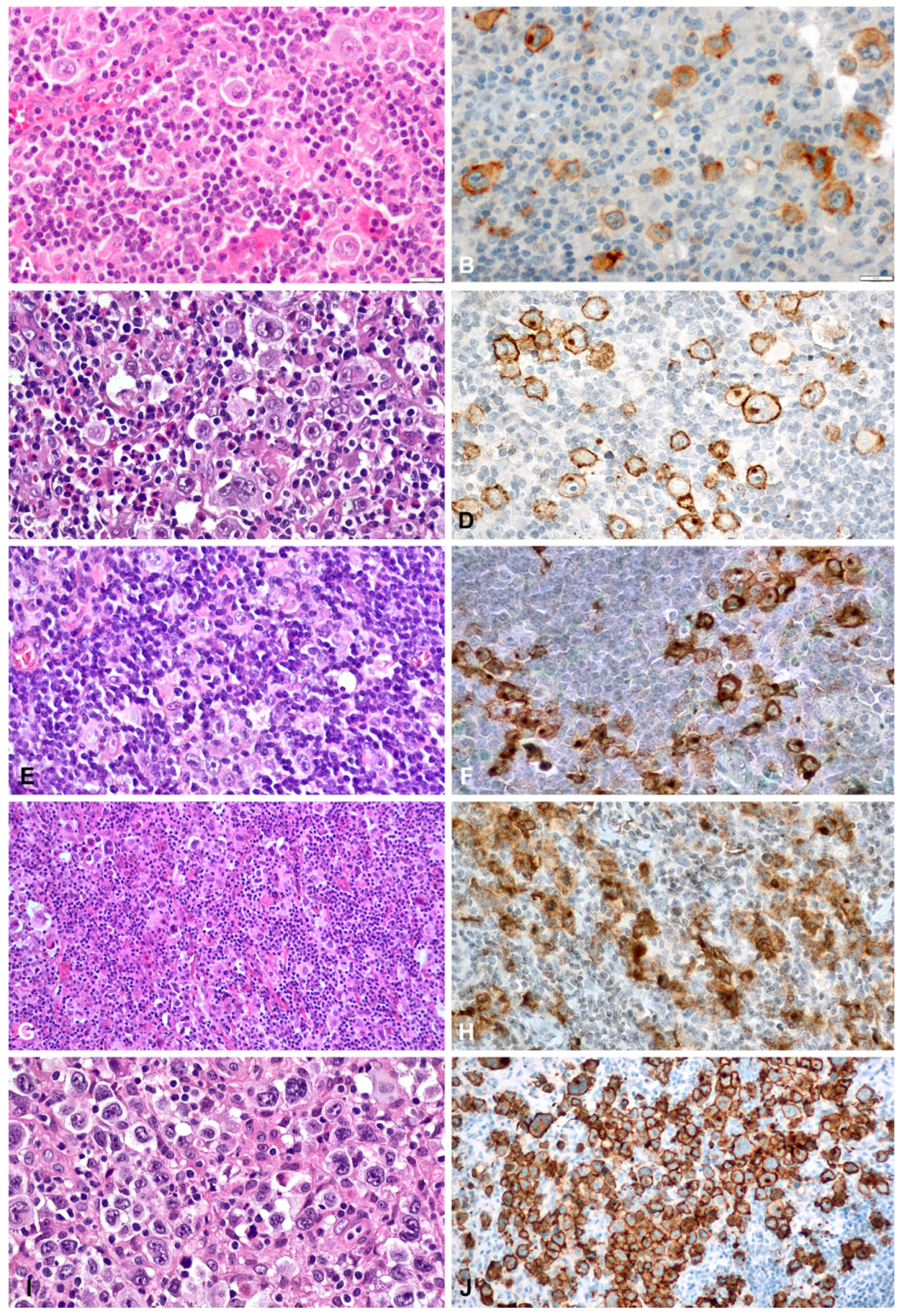
3.2. Results of Evaluation of HRS Cells
3.3. Survival Analysis Results
4. Discussion
5. Conclusions
Author Contributions
Funding
Institutional Review Board Statement
Informed Consent Statement
Data Availability Statement
Conflicts of Interest
References
- Borges, A.M.; Delabie, J.; Vielh, P.; d’Amore, E.S.G.; Hebeda, K.M.; Diepstra, A.; Naresh, K.N.; Garcia, J.F.; Tamaru, J.; Laskar, S.; et al. Classic Hodgkin lymphoma. In WHO Classification of Tumours Online. Haematolymphoid Tumours, 5th ed.; Gujral, S., Siebert, R., de Jong, D., Eds.; IARC: Lyon, France, 2022. [Google Scholar]
- Mani, H.; Jaffe, E.S. Hodgkin lymphoma: An update on its biology with new insights into classification. Clin. Lymphoma Myeloma 2009, 9, 206–216. [Google Scholar] [CrossRef] [Scilit] [PubMed]
- MacLennan, K.A.; Bennett, M.H.; Tu, A.; Hudson, B.V.; Easterling, M.J.; Hudson, G.V.; Jelliffe, A.M. Relationship of histopathologic features to survival and relapse in nodular sclerosing Hodgkin’s disease. A study of 1659 patients. Cancer 1989, 64, 1686–1693. [Google Scholar] [CrossRef] [Scilit] [PubMed]
- Wijlhuizen, T.J.; Vrints, L.W.; Jairam, R.; Breed, W.P.; Wijnen, J.T.; Bosch, L.J.; Crommelin, M.A.; van Dam, F.E.; de Koning, J.; Verhagen-Teulings, M. Grades of nodular sclerosis (NSI-NSII) in Hodgkin’s disease. Are they of independent prognostic value? Cancer 1989, 63, 1150–1153. [Google Scholar] [CrossRef] [Scilit]
- Ferry, J.A.; Linggood, R.M.; Convery, K.M.; Efird, J.T.; Eliseo, R.; Harris, N.L. Hodgkin disease, nodular sclerosis type. Implications of histologic subclassification. Cancer 1993, 71, 457–463. [Google Scholar] [CrossRef] [Scilit]
- D’Amore, E.S.; Lee, C.K.; Aeppli, D.M.; Levitt, S.H.; Frizzera, G. Lack of prognostic value of histopathologic parameters in Hodgkin’s disease, nodular sclerosis type. A study of 123 patients with limited stage disease who had undergone laparotomy and were treated with radiation therapy. Arch. Pathol. Lab. Med. 1992, 116, 856–861. [Google Scholar] [PubMed]
- Hess, J.L.; Bodis, S.; Pinkus, G.; Silver, B.; Mauch, P. Histopathologic grading of nodular sclerosis Hodgkin’s disease. Lack of prognostic significance in 254 surgically staged patients. Cancer 1994, 74, 708–714. [Google Scholar] [CrossRef] [Scilit] [PubMed]
- Zhang, Q.; Kim, D.H.; Xu, Y.; Wang, W.; Medeiros, L.J. Clinicopathological features of syncytial variant nodular sclerosis Hodgkin lymphoma. Hum. Pathol. 2022, 119, 105–113. [Google Scholar] [CrossRef] [Scilit] [PubMed]
- Sethi, T.; Nguyen, V.; Li, S.; Morgan, D.; Greer, J.; Reddy, N. Differences in outcome of patients with syncytial variant Hodgkin lymphoma compared with typical nodular sclerosis Hodgkin lymphoma. Ther. Adv. Hematol. 2017, 8, 13–20. [Google Scholar] [PubMed]
- Darabi, K.; Tester, W.; Daskal, I.; Cohn, J. Syncytial Variant of Nodular Sclerosing Hodgkin’s Disease. Blood 2004, 104, 4533. [Google Scholar] [CrossRef] [Scilit]
- Ben-Yehuda-Salz, D.; Ben-Yehuda, A.; Polliack, A.; Ron, N.; Okon, E. Syncytial variant of nodular sclerosing Hodgkin’s disease. A new clinicopathologic entity. Cancer 1990, 65, 1167–1172. [Google Scholar] [CrossRef] [Scilit] [PubMed]
- Lamaison, C.; Ferrant, J.; Gravelle, P.; Traverse-Glehen, A.; Ghesquières, H.; Tosolini, M.; Rossi, C.; Ysebaert, L.; Brousset, P.; Laurent, C.; et al. Histological Subtypes Drive Distinct Prognostic Immune Signatures in Classical Hodgkin Lymphoma. Cancers 2022, 14, 4893. [Google Scholar] [CrossRef] [Scilit] [PubMed]
- Gobbi, P.G.; Broglia, C.; Di Giulio, G.; Mantelli, M.; Anselmo, P.; Merli, F.; Zinzani, P.L.; Rossi, G.; Callea, V.; Iannitto, E.; et al. The clinical value of tumor burden at diagnosis in Hodgkin lymphoma. Cancer 2004, 101, 1824–1834. [Google Scholar] [CrossRef] [Scilit]
- Miljak, A.; Pavlović, A.; Benzon, B.; Čutura, L.V.; Galušić, D.; Vujčić, M.; Blaslov, V.; Durdov, M.G. High PD-L1 Expression in HRS Cells and Macrophages in Tumor Immune Microenvironment Is Associated with Adverse Outcome and EBV Positivity in Classical Hodgkin Lymphoma. Int. J. Mol. Sci. 2025, 26, 5592. [Google Scholar] [CrossRef] [Scilit] [PubMed]
- Zargari, A.; Cummins, K.D.; Hosking, P.; Pham, A.; Hawkes, E.; Ting, S.B. Increased STAT expression in Reed-Sternberg cells as a potential positive prognostication biomarker in Hodgkin lymphoma. Pathology 2023, 55, 650–655. [Google Scholar] [CrossRef] [Scilit] [PubMed]
- Steidl, C.; Diepstra, A.; Lee, T.; Chan, F.C.; Farinha, P.; Tan, K.; Telenius, A.; Barclay, L.; Shah, S.P.; Connors, J.M.; et al. Gene expression profiling of microdissected Hodgkin Reed-Sternberg cells correlates with treatment outcome in classical Hodgkin lymphoma. Blood 2012, 120, 3530–3540. [Google Scholar] [CrossRef] [Scilit] [PubMed]
- Rengstl, B.; Kim, S.; Döring, C.; Weiser, C.; Bein, J.; Bankov, K.; Herling, M.; Newrzela, S.; Hansmann, M.-L.; Hartmann, S. Small and big Hodgkin-Reed-Sternberg cells of Hodgkin lymphoma cell lines L-428 and L-1236 lack consistent differences in gene expression profiles and are capable to reconstitute each other. PLoS ONE 2017, 12, e0177378. [Google Scholar] [CrossRef] [Scilit] [PubMed]
- Santisteban-Espejo, A.; Benavides-De la Fuente, C.; Mangas-Rojas, A.; Montero-Pavon, P.; Bernal-Florindo, I.; Aldaco-Puntas, E.; Prieto-Conde, I.; Perez-Requena, J.; Atienza-Cuevas, L.; Fernández-Valle, M.d.C.; et al. Computational pathology identifies a low B-cell content in the tumour microenvironment as a predictor of adverse outcome in patients with classic Hodgkin lymphoma treated with ABVD. J. Clin. Pathol. 2025, 78, 381–389. [Google Scholar] [CrossRef] [Scilit] [PubMed]
- Aoki, T.; Jiang, A.; Xu, A.; Yin, Y.; Gamboa, A.; Milne, K.; Takata, K.; Miyata-Takata, T.; Chung, S.; Rai, S.; et al. Spatially Resolved Tumor Microenvironment Predicts Treatment Outcomes in Relapsed/Refractory Hodgkin Lymphoma. J. Clin. Oncol. 2024, 42, 1077–1087. [Google Scholar] [CrossRef] [Scilit] [PubMed]
- Grund, J.; Iben, K.; Reinke, S.; Bühnen, I.; Plütschow, A.; Müller-Meinhard, B.; Marquez, M.A.G.; Schlößer, H.A.; von Tresckow, B.; Kellermeier, F.; et al. Low B-cell content is associated with a CD73-low tumour microenvironment and unfavourable prognosis in classic Hodgkin lymphoma. Br. J. Haematol. 2023, 201, 1097–1102. [Google Scholar] [CrossRef] [Scilit] [PubMed]
- Granot, N.; Ben-Barak, A.; Fisher, Y.; Golan, H.; Weyl Ben-Arush, M. Syncytial variant of nodular sclerosing Hodgkin lymphoma in children: A prognostic factor? Pediatr. Hematol. Oncol. 2018, 35, 33–36. [Google Scholar] [CrossRef] [Scilit] [PubMed]
- Hasenclever, D.; Diehl, V. A prognostic score for advanced Hodgkin’s disease. International Prognostic Factors Project on Advanced Hodgkin’s Disease. N. Engl. J. Med. 1998, 339, 1506–1514. [Google Scholar] [PubMed]
- Shenoy, P.; Maggioncalda, A.; Malik, N.; Flowers, C.R. Incidence patterns and outcomes for hodgkin lymphoma patients in the United States. Adv. Hematol. 2011, 2011, 725219. [Google Scholar] [CrossRef] [Scilit] [PubMed]
- Berkman, A.M.; Andersen, C.R.; Puthenpura, V.; Livingston, J.A.; Ahmed, S.; Cuglievan, B.; Hildebrandt, M.A.; Roth, M.E. Impact of Race, Ethnicity, and Socioeconomic Status over Time on the Long-term Survival of Adolescent and Young Adult Hodgkin Lymphoma Survivors. Cancer Epidemiol. Biomark. Prev. 2021, 30, 1717–1725. [Google Scholar] [CrossRef] [Scilit] [PubMed]
- Klimm, B.; Reineke, T.; Haverkamp, H.; Eich, H.T.; Josting, A.; Pfistner, B.; Diehl, V.; Engert, A. The Superior Prognosis of Female Patients with Hodgkin’s Lymphoma Is Related to More Treatment-Induced Leucopenia and Warrants a More Individualized Therapy. Blood 2004, 104, 1313. [Google Scholar] [CrossRef] [Scilit]
- Roemer, K.; Pfreundschuh, M. How do estrogens control lymphoma? Blood 2014, 123, 1980–1981. [Google Scholar] [CrossRef] [Scilit] [PubMed]
- Pierdominici, M.; Maselli, A.; Locatelli, S.L.; Ciarlo, L.; Careddu, G.; Patrizio, M.; Ascione, B.; Tinari, A.; Carlo-Stella, C.; Malorni, W.; et al. Estrogen receptor β ligation inhibits Hodgkin lymphoma growth by inducing autophagy. Oncotarget 2017, 8, 8522–8535. [Google Scholar] [CrossRef] [Scilit] [PubMed]
- Maynadier, M.; Chambon, M.; Basile, I.; Gleizes, M.; Nirde, P.; Gary-Bobo, M.; Garcia, M. Estrogens promote cell-cell adhesion of normal and malignant mammary cells through increased desmosome formation. Mol. Cell Endocrinol. 2012, 364, 126–133. [Google Scholar] [CrossRef] [Scilit] [PubMed]
- Hu, J.; Zhang, X.; Tao, H.; Jia, Y. The prognostic value of Epstein-Barr virus infection in Hodgkin lymphoma: A systematic review and meta-analysis. Front. Oncol. 2022, 12, 1034398. [Google Scholar] [CrossRef] [Scilit] [PubMed]
- Sandvej, K.B.; Hamilton-Dutoit, S.J.; Pallesen, G. Influence of Epstein-Barr virus encoded latent membrane protein 1 on the expression of CD23 antigen, ICAM-1 and LFA-3 in Hodgkin and Reed-Sternberg cells. A morphometric analysis. Leuk. Lymphoma 1993, 9, 95–101. [Google Scholar] [CrossRef] [Scilit] [PubMed]
- Kim, S.H.; Shin, Y.K.; Lee, I.S.; Bae, Y.M.; Sohn, H.W.; Suh, Y.H.; Ree, H.J.; Rowe, M.; Park, S.H. Viral latent membrane protein 1 (LMP-1)-induced CD99 down-regulation in B cells leads to the generation of cells with Hodgkin’s and Reed-Sternberg phenotype. Blood 2000, 95, 294–300. [Google Scholar] [CrossRef] [Scilit]
- Lee, I.S.; Shin, Y.K.; Chung, D.H.; Park, S.H. LMP1-induced downregulation of CD99 molecules in Hodgkin and Reed-Sternberg cells. Leuk. Lymphoma 2001, 42, 587–594. [Google Scholar] [CrossRef] [Scilit] [PubMed]
- Lee, I.; Kim, M.K.; Choi, E.Y.; Mehl, A.; Jung, K.C.; Gil, M.C.; Rowe, M.; Park, S.H. CD99 expression is positively regulated by Sp1 and is negatively regulated by Epstein-Barr virus latent membrane protein 1 through nuclear factor-kappaB. Blood 2001, 97, 3596–3604. [Google Scholar] [CrossRef] [Scilit] [PubMed]
- Roy, P.; Vaughan Hudson, G.; Vaughan Hudson, B.; Esteve, J.; Swerdlow, A.J. Long-term survival in Hodgkin’s disease patients. A comparison of relative survival in patients in trials and those recorded in population-based cancer registries. Eur. J. Cancer 2000, 36, 384–389. [Google Scholar] [PubMed]
- Engert, A.; Ballova, V.; Haverkamp, H.; Pfistner, B.; Josting, A.; Dühmke, E.; Müller-Hermelink, K.; Diehl, V. Hodgkin’s lymphoma in elderly patients: A comprehensive retrospective analysis from the German Hodgkin’s Study Group. J. Clin. Oncol. 2005, 23, 5052–5060. [Google Scholar] [CrossRef] [Scilit] [PubMed]
- Evens, A.M.; Helenowski, I.; Ramsdale, E.; Nabhan, C.; Karmali, R.; Hanson, B.; Parsons, B.; Smith, S.; Larsen, A.; McKoy, J.M.; et al. A retrospective multicenter analysis of elderly Hodgkin lymphoma: Outcomes and prognostic factors in the modern era. Blood 2012, 119, 692–695. [Google Scholar] [CrossRef] [Scilit] [PubMed]


| Mean # of HRS Cells | ||||
|---|---|---|---|---|
| HE-Stained Slides | p | CD30-Stained Slides | p | |
| Gender | ||||
| Male (n = 74) | 34.2 ± 21.1 (Range 7–105) | 0.87 | 43.7 ± 29 (Range 9–146) | 0.29 |
| Female (n = 61) | 34.7 ± 15 (Range 10–77) | 48.7 ± 26.1 (Range 9–116) | ||
| Age | ||||
| <40 (n = 84) | 35.3 ± 16.4 (Range 8–91) | 0.45 | 47.5 ± 25.4 (Range 13–144) | 0.41 |
| ≥40 (n = 51) | 32.9 ± 21.7 (Range 7–105) | 43.4 ± 31.4 (Range 9–146) | ||
| Subtype | ||||
| Nodular sclerosis (n = 93) | 37.7 ± 17.6 (Range 11–91) | <0.001 | 50.4 ± 27.5 (Range 14–144) | <0.001 |
| Mixed cellularity (n = 31) | 26.6 ± 15.2 (Range 7–82) | 35 ± 20.3 (Range 9–106) | ||
| Lymphocyte-rich (n = 5) | 15.2 ± 7.8 (Range 8–28) | 18.2 ± 9.2 (Range 9–30) | ||
| Lymphocyte-depleted (n = 1) | 105 | 146 | ||
| SV-NSCHL (n = 46) | 43.7 ± 17.3 (Range 12–79) | 60.2 ± 24.5 (Range 14–116) | ||
| Non-SV-NSCHL (n = 84) | 29.8 ± 17.7 (Range 7–105) | <0.001 | 38.5 ± 27.1 (Range 9–146) | <0.001 |
| EBER status | ||||
| Positive (n = 48) | 31 ± 21.2 (Range 7–105) | 0.07 | 43.1± 30.4 (Range 9–146) | 0.28 |
| Negative (n = 83) | 37.1 ± 16.6 (Range 12–91) | 48.6 ± 26.2 (Range 13–144) | ||
| Stage | ||||
| Stages I–II | 32.9 ± 16.8 (Range 7–91) | 0.49 | 44.9 ± 26.8 (Range 9–144) | 0.72 |
| Stages III–IV | 35.2 ± 19.9 (Range 8–105) | 46.6 ± 28.9 (Range 9–146) | ||
| Subtype | p | SV Status | p | ||||||
|---|---|---|---|---|---|---|---|---|---|
| Nodular Sclerosis | Mixed Cellularity | Lymphocyte-Rich | Lymphocyte Depleted | Non-SV-NSCHL | SV-NSCHL | ||||
| HE | 1 (n = 24) | 9 (39.1%) | 9 (39.1%) | 5 (21.7%) | 0 (0.0%) | <0.001 | 21 (91.3%) | 2 (8.7%) | <0.001 |
| 2 (n = 76) | 55 (74.3%) | 19 (25.7%) | 0 (0.0%) | 0 (0.0%) | 48 (64.9%) | 26 (35.1%) | |||
| 3 (n = 29) | 23 (85.2%) | 3 (11.1%) | 0 (0.0%) | 1 (3.7%) | 15 (55.6%) | 12 (44.4%) | |||
| 4 (n = 5) | 5 (100.0%) | 0 (0.0%) | 0 (0.0%) | 0 (0.0%) | 0 (0.0%) | 5 (100.0%) | |||
| 5 (n = 1) | 1 (100.0%) | 0 (0.0%) | 0 (0.0%) | 0 (0.0%) | 0(0.0%) | 1 (100.0%) | |||
| CD30 | 1 (n = 27) | 12 (46.2%) | 9 (34.6%) | 5 (19.2%) | 0 (0.0%) | <0.001 | 24 (92.3%) | 2 (7.7%) | <0.001 |
| 2 (n = 72) | 49 (71.0%) | 20 (29%) | 0 (0.0%) | 0 (0.0%) | 48 (69.6%) | 21 (30.4%) | |||
| 3 (n = 29) | 26 (92.9%) | 2 (7.1%) | 0 (0.0%) | 0 (0.0%) | 11 (39.3%) | 17 (60.7%) | |||
| 4 (n = 3) | 3 (100.0%) | 0 (0.0%) | 0 (0.0%) | 0 (0.0%) | 0 (0.0%) | 3 (100.0%) | |||
| 5 (n = 4) | 3 (75.0%) | 0 (0.0%) | 0 (0.0%) | 1 (25.0%) | 1 (25.0%) | 3 (75.0%) | |||
| Group | # of the Cases | |
|---|---|---|
| HE-stained slides | 0 | 52 |
| 1 | 18 | |
| 2 | 8 | |
| 3 | 8 | |
| 4 | 49 | |
| CD30-stained slides | 0 | 41 |
| 1 | 14 | |
| 2 | 8 | |
| 3 | 7 | |
| 4 | 64 |
Disclaimer/Publisher’s Note: The statements, opinions and data contained in all publications are solely those of the individual author(s) and contributor(s) and not of MDPI and/or the editor(s). MDPI and/or the editor(s) disclaim responsibility for any injury to people or property resulting from any ideas, methods, instructions or products referred to in the content. |
© 2026 by the authors. Published by MDPI on behalf of the Lithuanian University of Health Sciences. Licensee MDPI, Basel, Switzerland. This article is an open access article distributed under the terms and conditions of the Creative Commons Attribution (CC BY) license.
Share and Cite
Pehlivanoglu, B.; Tezel, N.; Ozturk, O.C.; Emecen, S.B.; Ozcan, M.A.; Ozkal, S. Quantity Does Not Matter: Number, Ratio, or Grouping of Hodgkin/Reed–Sternberg Cells Does Not Affect Prognosis in Patients with Classic Hodgkin Lymphoma. Medicina 2026, 62, 1569. https://doi.org/10.3390/medicina62081569
Pehlivanoglu B, Tezel N, Ozturk OC, Emecen SB, Ozcan MA, Ozkal S. Quantity Does Not Matter: Number, Ratio, or Grouping of Hodgkin/Reed–Sternberg Cells Does Not Affect Prognosis in Patients with Classic Hodgkin Lymphoma. Medicina. 2026; 62(8):1569. https://doi.org/10.3390/medicina62081569
Chicago/Turabian StylePehlivanoglu, Burcin, Nazimcan Tezel, Osman Can Ozturk, Serra Begum Emecen, Mehmet Ali Ozcan, and Sermin Ozkal. 2026. "Quantity Does Not Matter: Number, Ratio, or Grouping of Hodgkin/Reed–Sternberg Cells Does Not Affect Prognosis in Patients with Classic Hodgkin Lymphoma" Medicina 62, no. 8: 1569. https://doi.org/10.3390/medicina62081569
APA StylePehlivanoglu, B., Tezel, N., Ozturk, O. C., Emecen, S. B., Ozcan, M. A., & Ozkal, S. (2026). Quantity Does Not Matter: Number, Ratio, or Grouping of Hodgkin/Reed–Sternberg Cells Does Not Affect Prognosis in Patients with Classic Hodgkin Lymphoma. Medicina, 62(8), 1569. https://doi.org/10.3390/medicina62081569