Calculation of Ejection Fraction Using Cardiac Computed Tomography: Clinical Evolution, Reliability, and Technological Challenges—A Narrative Review

 , ,
, ,  ,
,  and
and
Abstract
1. Introduction
1.1. Definition and Clinical Relevance of the Ejection Fraction
1.2. Points of Strength and Weaknesses of the Use of the Ejection Fraction in Clinical Practice
1.3. CCT as a Versatile Alternative and Aim of the Review

2. Evolution of CCT
3. Aim and Review Methodology
4. Clinical Use of Ejection Fraction Measured by CCT
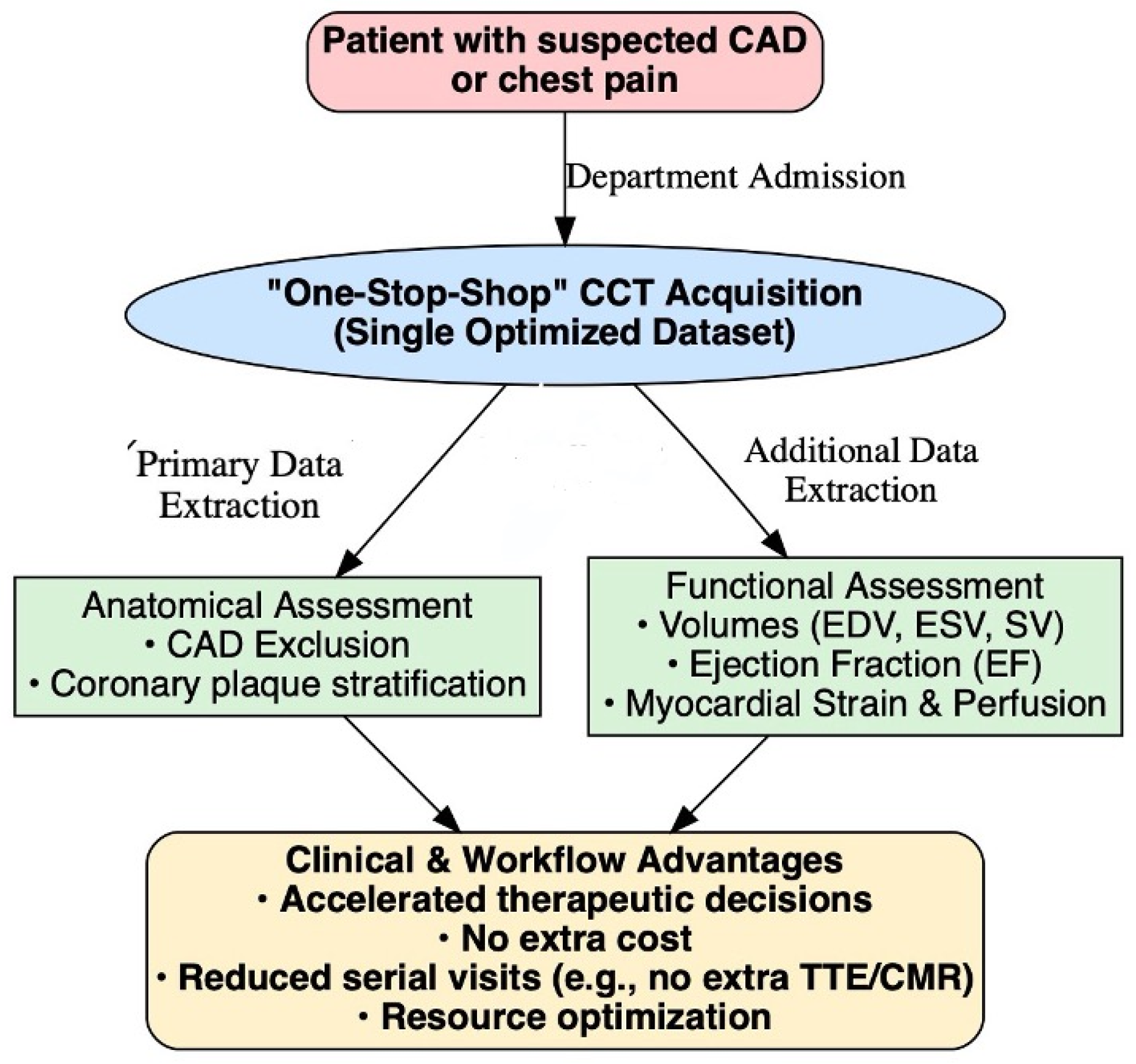
4.1. “One-Stop-Shop” Assessment
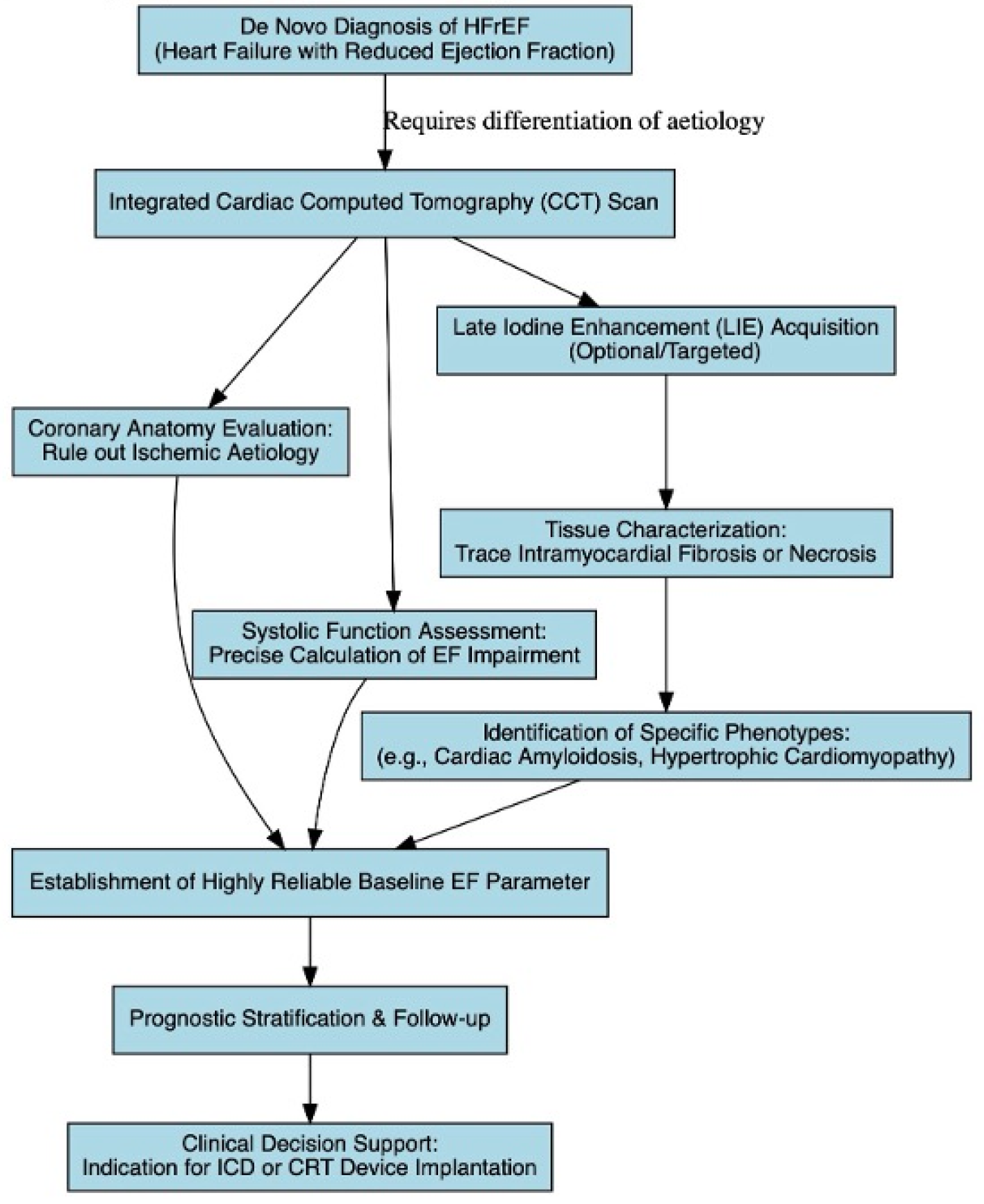
4.2. Aetiological Diagnosis of New-Onset Heart Failure
4.3. Diagnosis of Intraventricular Thrombosis in HFrEF
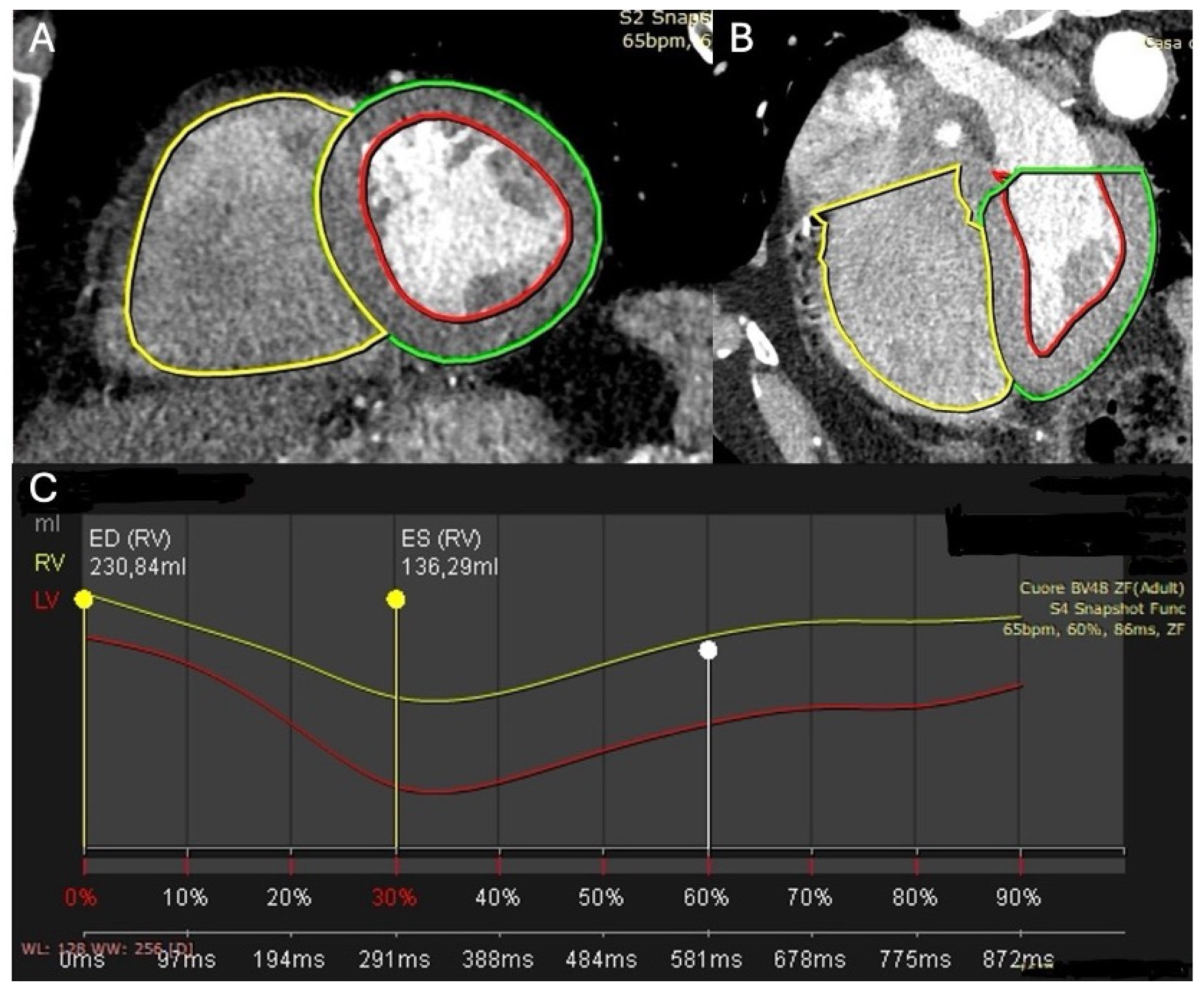
4.4. CCT to Assess of Right Ventricular Ejection Fraction (RVEF)
4.5. CCT for Advanced Pre-Procedural Planning
4.6. The Emerging Role in Opportunistic Screening and Cardio-Oncology

| Clinical Scenario | Primary Indication for CCT | Added Value of Functional Assessment (Volumes and EF) |
|---|---|---|
| Suspected CAD (“One-stop-shop”) | Ruling out coronary artery disease or stratifying the atheromatous plaque [22,65]. | Simultaneous derivation of biventricular systolic function (EDV, ESV, SV, EF) optimizing workflow and accelerating therapeutic decisions at no extra cost [24,33,51,69]. |
| New-onset HFrEF | Differentiating ischemic from non-ischemic aetiology and tracing patterns of intramyocardial fibrosis/necrosis via LIE [22,27,70,71]. | Establishes a highly reliable baseline EF for prognostic stratification and to guide ICD or CRT device implantation [2,7,36,37]. |
| Intraventricular thrombosis (HFrEF) | Complementary imaging when standard TTE sensitivity is limited by suboptimal acoustic windows [23,42]. | High spatial resolution enables detailed visualization and accurate differentiation between thrombus, trabeculations, and slow-flow artifacts [20,24] |
| Right ventricular assessment | Assessing RV size and function when traditional TTE parameters are limited by load dependence and complex geometry [9,51] | Provides reliable volumetric quantification of RV volumes and RVEF, correlating with clinical outcomes and RV remodelling [9,51] |
| Advanced pre-procedural planning | Evaluating vascular accesses, aortic root, valvular calcium, and prosthesis sizing for TAVI or mitral interventions [21,23,26]. | Extracts EF, EDV, and ESV to quantify cardiac damage, which is strongly predictive of post-operative outcomes [26,59]. |
| Opportunistic screening and Cardio-Oncology | Routine chest, abdomen, and pelvis CT for tumour staging and therapeutic response evaluation [31,32] | Calculation of biventricular EF from non-cardiac scans (via AI or modified ECG-gated protocols) avoiding extra TTE/CMR to monitor cardiotoxicity [6,22,31]. |
5. Technological Challenges: Acquisition, Wide-Detector Scanners, and Artificial Intelligence
5.1. Hardware Evolution: The Impact of 16 cm Scanners (Wide-Detector CT)
5.2. The Breakthrough of Artificial Intelligence and Deep Learning
5.3. Analysis of Diastolic Function and Atrial Volumes
6. Reliability: Comparison with Clinical Gold Standards (Echocardiography and CMR)
6.1. Comparison with Transthoracic Echocardiography
6.2. Comparison with Cardiac Magnetic Resonance
7. Limitations and Issues of the Method
7.1. Exposure to Ionizing Radiation and Contrast Media Toxicity
7.2. Artifact Management and Heart Rhythm
7.3. Temporal Resolution
8. Current Guideline-Based Clinical Positioning of CCT-Derived Functional Assessment
9. Conclusions
Author Contributions
Funding
Data Availability Statement
Conflicts of Interest
Abbreviations
| AI | Artificial Intelligence |
| CAD | Coronary Artery Disease |
| CCT | Cardiac Computed Tomography |
| CRT | Cardiac Resynchronization Therapy |
| CTP | Computed Tomography Perfusion |
| CTRCD | Cancer Therapy-Related Cardiac Dysfunction |
| CT | Computed Tomography |
| CMR | Cardiac Magnetic Resonance |
| ECV | Extracellular Volume |
| EDV | End-Diastolic Volume |
| ESV | End-Systolic Volume |
| EF | Ejection Fraction |
| GLS | Global Longitudinal Strain |
| HF | Heart Failure |
| HFmrEF | Heart Failure with mildly reduced Ejection Fraction |
| HFpEF | Heart Failure with preserved Ejection Fraction |
| HFrEF | Heart Failure with reduced Ejection Fraction |
| ICD | Implantable Cardiac Device |
| LA | Left Atrial |
| LIE | Late Iodine Enhancement |
| LV | Left Ventricle |
| LVEF | Left Ventricle Ejection Fraction |
| MACE | Major Adverse Cardiovascular Events |
| RV | Right Ventricle |
| RVEF | Right Ventricle Ejection Fraction |
| SV | Stroke Volume |
| TAVI | Transcatheter Aortic Valve Implantation |
| TTE | Transthoracic Echocardiography |
| ACC | America College of Cardiology |
| AHA | American Heart Association |
| ASE | American Society of Echocardiography |
| CCTA | Coronary Computed Tomography Angiography |
| CI-AKI | Contrast-Induced Acute Kidney Injury |
| CNNs | Convolutional Neural Networks |
| ECG | Electrocardiogram |
| ESC | European Society of Cardiology |
| HER2 | Human Epidermal Growth Factor Receptor 2 |
| LOE | Level of Evidence |
| MDCT | Multidetector Computed Tomography |
| MUGA | Multiple Gated Acquisition (scan)/Radionuclide ventriculography |
| Nt-proBNP | N-terminal pro-b-type natriuretic peptide |
| PACS | Picture Archiving and Communication System |
| SPECT | Single-Photon Emission Computed Tomography |
| SSFP | Steady-State Free Precession |
References
- Kusunose, K.; Zheng, R.; Yamada, H.; Sata, M. How to standardize the measurement of left ventricular ejection fraction. J. Med. Ultrason. 2022, 49, 35–43. [Google Scholar] [CrossRef] [PubMed] [PubMed Central]
- Clark, J.; Ionescu, A.; Chahal, C.A.A.; Bhattacharyya, S.; Lloyd, G.; Galanti, K.; Gallina, S.; Chong, J.H.; Petersen, S.E.; Ricci, F.; et al. Interchangeability in Left Ventricular Ejection Fraction Measured by Echocardiography and cardiovascular Magnetic Resonance: Not a Perfect Match in the Real World. Curr. Probl. Cardiol. 2023, 48, 101721. [Google Scholar] [CrossRef] [PubMed]
- Gać, P.; Wysocki, A.; Beck, E.; Poręba, M.; Poręba, R. Multimodal assessment of the left ventricular ejection fraction by echocardiography, cardiac computed tomography and cardiac magnetic resonance in patients after SARS-CoV2 infection. Front. Physiol. 2025, 16, 1629065. [Google Scholar] [CrossRef] [PubMed] [PubMed Central]
- Zhang, Z.; Zhu, Y.; Liu, M.; Zhang, Z.; Zhao, Y.; Yang, X.; Xie, M.; Zhang, L. Artificial Intelligence-Enhanced Echocardiography for Systolic Function Assessment. J. Clin. Med. 2022, 11, 2893. [Google Scholar] [CrossRef] [PubMed] [PubMed Central]
- Mahmood, A.; Dhall, E.; Primus, C.P.; Gallagher, A.; Zakeri, R.; Mohammed, S.F.; Chahal, A.A.; Ricci, F.; Aung, N.; Khanji, M.Y. Heart failure with preserved ejection fraction management: A systematic review of clinical practice guidelines and recommendations. Eur. Heart J. Qual. Care Clin. Outcomes 2024, 10, 571–589. [Google Scholar] [CrossRef] [PubMed] [PubMed Central]
- Tu, C.; Shen, H.; Liu, R.; Wang, X.; Li, X.; Yuan, X.; Chen, Q.; Wang, Y.; Ran, Z.; Lan, X.; et al. Myocardial extracellular volume derived from contrast-enhanced chest computed tomography for longitudinal evaluation of cardiotoxicity in patients with breast cancer treated with anthracyclines. Insights Imaging 2022, 13, 85. [Google Scholar] [CrossRef] [PubMed] [PubMed Central]
- Vancheri, F.; Longo, G.; Henein, M.Y. Left ventricular ejection fraction: Clinical, pathophysiological, and technical limitations. Front. Cardiovasc. Med. 2024, 11, 1340708. [Google Scholar] [CrossRef] [PubMed] [PubMed Central]
- Johannesen, J.; Fukuda, R.; Zhang, D.T.; Tak, K.; Meier, R.; Agoglia, H.; Horn, E.; Devereux, R.B.; Weinsaft, J.W.; Kim, J. Direct comparison of echocardiography speckle tracking and cardiac magnetic resonance feature tracking for quantification of right ventricular strain: A prospective intermodality study in functional mitral regurgitation. Echo Res. Pract. 2022, 9, 11. [Google Scholar] [CrossRef] [PubMed] [PubMed Central]
- Ahmad, A.; Li, H.; Zhang, Y.; Liu, J.; Gao, Y.; Qian, M.; Lin, Y.; Yi , L.; Zhang, L.; Li, Y.; et al. Three-Dimensional Echocardiography Assessment of Right Ventricular Volumes and Function: Technological Perspective and Clinical Application. Diagnostics 2022, 12, 806. [Google Scholar] [CrossRef]
- Nazir, M.S.; Okafor, J.; Murphy, T.; Andres, M.S.; Ramalingham, S.; Rosen, S.D.; Chiribiri, A.; Plein, S.; Prasad, S.; Mohiaddin, R.; et al. Echocardiography versus Cardiac MRI for Measurement of Left Ventricular Ejection Fraction in Individuals with Cancer and Suspected Cardiotoxicity. Radiol. Cardiothorac. Imaging 2024, 6, e230048. [Google Scholar] [CrossRef] [PubMed] [PubMed Central]
- Jollans, L.; Bustamante, M.; Henriksson, L.; Persson, A.; Ebbers, T. Accurate fully automated assessment of left ventricle, left atrium, and left atrial appendage function from computed tomography using deep learning. Eur. Heart J. Imaging Methods Pract. 2025, 2, qyaf011. [Google Scholar] [CrossRef] [PubMed] [PubMed Central]
- Kadoya, Y.; Omaygenc, M.O.; Abtahi, S.S.; Sritharan, S.; Nehmeh, A.; Yam, Y.; Small, G.R.; Chow, B. Prognostic value of systolic left ventricular ejection fraction using prospective ECG-triggered cardiac CT. J. Cardiovasc. Comput. Tomogr. 2025, 19, 58–63. [Google Scholar] [CrossRef] [PubMed]
- Chao, M.-F.; Jacob, A.J.; Sinha, A.; Hallam, K.; Kragholm, K.H.; Sharma, P.; Rapaka, S.; Ramirez-Giraldo, J.C.; Chang, S.-M. Artificial intelligence-based bi-ventricular systolic and diastolic volume, ejection fraction using non-contrast ECG-gated cardiac computed tomography. Eur. Heart J. Imaging Methods Pract. 2025, 3, qyaf121. [Google Scholar] [CrossRef] [PubMed] [PubMed Central]
- Luke, P.; Alkhalil, M.; Eggett, C. Current and novel echocardiographic assessment of left ventricular systolic function in aortic stenosis-A comprehensive review. Echocardiography 2022, 39, 1470–1480. [Google Scholar] [CrossRef] [PubMed] [PubMed Central]
- Chaudhari, M.; Ashwath, M.L. Cardiac Imaging in the Diagnosis and Management of Heart Failure. J. Clin. Med. 2025, 14, 5002. [Google Scholar] [CrossRef] [PubMed] [PubMed Central]
- Mahmod, M.; Bull, S.; Kailayanathan, T.; Davis, T.A.; Borlotti, A.; Popescu, I.A.; Das, I.; Wamil, M.; Brears, H.T.; Banerjee, R.; et al. Left atrial volume quantification by transthoracic echocardiography versus cardiovascular magnetic resonance: A systematic review and meta-analysis. Int. J. Cardiovasc. Imaging 2025, 41, 1657–1669. [Google Scholar] [CrossRef] [PubMed] [PubMed Central]
- Kinoshita, M.; Tanabe, Y.; Yoshida, K.; Kurata, A.; Kobayashi, Y.; Uetani, T.; Inoue, K.; Nishimura, K.; Ikeda, S.; Mochizuki, T.; et al. Left ventricular longitudinal strain is a major determinant of CT-derived three-dimensional maximum principal strain: Comparison with two-dimensional speckle tracking echocardiography. Heart Vessel. 2022, 37, 31–39. [Google Scholar] [CrossRef] [PubMed]
- Zwijnen, A.W.; Watzema, L.; Ridwan, Y.; van Der Pluijm, I.; Smal, I.; Essers, J. Self-Adaptive Deep Learning-Based Segmentation for Universal and Functional Clinical and Preclinical CT Image Analysis. Comput. Biol. Med. 2024, 179, 108853. [Google Scholar]
- Scatteia, A.; Silverio, A.; Padalino, R.; De Stefano, F.; America, R.; Cappelletti, A.M.; Dalla Vecchia, L.A.; Guarini, P.; Donatelli, F.; Caiazza, F.; et al. Non-Invasive Assessment of Left Ventricle Ejection Fraction: Where Do We Stand? J. Pers. Med. 2021, 11, 1153. [Google Scholar] [CrossRef] [PubMed] [PubMed Central]
- Ibrahim, R.; Abdelnabi, M.; Pathangey, G.; Farina, J.; Lester, S.J.; Ayoub, C.; Alsidawi, S.; Tamarappoo, B.K.; Jokerst, C.; Arsanjani, R. Utility of Cardiac CT for Cardiomyopathy Phenotyping. Tomography 2025, 11, 39. [Google Scholar] [CrossRef] [PubMed]
- Mitsis, A.; Kyriakou, M.; Fouseki, A.; Myrianthopoulos, K.; Hadjicosti, M.; Christodoulou, E.; Kadoglou, N.P.; Eftychiou, C. Cardiac Computed Tomography in Structural Heart Interventions: From Preprocedural Planning to Procedural Strategy. Rev. Cardiovasc. Med. 2025, 26, 46998. [Google Scholar] [CrossRef] [PubMed] [PubMed Central]
- Cau, R.; Gatti, M.; Suri, J.S.; D’Angelo, T.; Saba, L. Clinical application of cardiac computed tomography in cardiomyopathy. Int. J. Cardiovasc. Imaging 2026, 42, 71–85. [Google Scholar] [CrossRef] [PubMed] [PubMed Central]
- Krawczyk-Ożóg, A.; Batko, J.; Koziej, M.; Tokarek, T.; Sorysz, D.; Siudak, Z.; Dudek, D.; Bartuś, S.; Surdacki, A.; Hołda, M.K. Computed tomography and transthoracic echocardiography for assessment of left ventricle geometry in patients with aortic valve stenosis. Postep. Kardiol. Interwencyjnej 2023, 19, 47–55. [Google Scholar] [CrossRef] [PubMed] [PubMed Central]
- Toia, P.; La Grutta, L.; Sollami, G.; Clemente, A.; Gagliardo, C.; Galia, M.; Maffei, E.; Midiri, M.; Cademartiri, F. Technical Development in Cardiac CT: Current Standards and Future Improvements—A Narrative Review. Cardiovas. Diagn. Ther. 2020, 10, 2018–2035. [Google Scholar]
- Bernhard, B.; Schütze, J.; Leib, Z.L.; Spano, G.; Berto, M.B.; Bakula, A.; Tomii, D.; Shiri, I.; Brugger, N.; Marchi, S.D.; et al. Myocardial analysis from routine 4D cardiac-CT to predict reverse remodeling and clinical outcomes after transcatheter aortic valve implantation. Eur. J. Radiol. 2024, 175, 111425. [Google Scholar] [CrossRef] [PubMed]
- Vignale, D.; Palmisano, A.; Gnasso, C.; Margonato, D.; Romagnolo, D.; Barbieri, S.; Ingallina, G.; Stella, S.; Ancona, M.B.; Montorfano, M.; et al. Extracellular volume fraction (ECV) derived from pre-operative computed tomography predicts prognosis in patients undergoing transcatheter aortic valve implantation (TAVI). Eur. Heart J. Cardiovasc. Imaging 2023, 24, 887–896. [Google Scholar] [CrossRef] [PubMed] [PubMed Central]
- Hayashi, H.; Oda, S.; Kidoh, M.; Yamaguchi, S.; Yoshimura, F.; Takashio, S.; Usuku, H.; Nagayama, Y.; Nakaura, T.; Ueda, M.; et al. Myocardial extracellular volume quantification in cardiac amyloidosis: A comparative study between cardiac computed tomography and magnetic resonance imaging. Eur. Radiol. 2024, 34, 1016–1025. [Google Scholar] [CrossRef] [PubMed]
- Li, H.; Chen, Z.; Kahn, A.M.; Kligerman, S.; Narayan, H.K.; Contijoch, F.J. Deep learning automates detection of wall motion abnormalities via measurement of longitudinal strain from ECG-gated CT images. Front. Cardiovasc. Med. 2022, 9, 1009445. [Google Scholar] [CrossRef] [PubMed] [PubMed Central]
- Wu, P.; Kim, K.; Severance, L.; McVeigh, E.; Pack, J.D. Low dose threshold for measuring cardiac functional metrics using four-dimensional CT with deep learning. J. Appl. Clin. Med. Phys. 2024, 26, e14593. [Google Scholar] [CrossRef] [PubMed] [PubMed Central]
- Chen, Z.; Rigolli, M.; Vigneault, D.M.; Kligerman, S.; Hahn, L.; Narezkina, A.; Craine, A.; Lowe, K.; Contijoch, F. Automated cardiac volume assessment and cardiac long- and short-axis imaging plane prediction from electrocardiogram-gated computed tomography volumes enabled by deep learning. Eur. Heart J. Digit. Health 2021, 2, 311–322. [Google Scholar] [CrossRef] [PubMed] [PubMed Central]
- Kusk, M.W.; Hess, S.; Gerke, O.; Kristensen, L.D.; Oxlund, C.S.; Ormstrup, T.E.; Christiansen, J.M.; Foley, S.J. Minimal dose CT for left ventricular ejection fraction and combination with chest-abdomen-pelvis CT. Eur. J. Radiol. Open 2024, 13, 100583. [Google Scholar] [CrossRef] [PubMed] [PubMed Central]
- Canan, A.; Ghandour, A.A.-H.; Saboo, S.S.; Rajiah, P.S. Opportunistic screening at chest computed tomography: Literature review of cardiovascular significance of incidental findings. Cardiovasc. Diagn. Ther. 2023, 13, 743–761. [Google Scholar] [CrossRef] [PubMed] [PubMed Central]
- Manohar, A.; Colvert, G.M.; Ortuño, J.E.; Chen, Z.; Yang, J.; Colvert, B.T.; Bandettini, W.P.; Chen, M.Y.; Ledesma-Carbayo, M.J.; McVeigh, E.R. Regional left ventricular endocardial strains estimated from low-dose 4DCT: Comparison with cardiac magnetic resonance feature tracking. Med. Phys. 2022, 49, 5841–5854. [Google Scholar] [CrossRef] [PubMed] [PubMed Central]
- Hu, Y.; Peng, W.; Liu, K.; Xu, X.; Hu, S.; Lyu, X.; Liu, X.; Qin, M.; Sun, R.; Ren, W.; et al. Application of low-dose “one-stop” myocardial computed tomography perfusion imaging in coronary artery disease. Quant. Imaging Med. Surg. 2026, 16, 167. [Google Scholar] [CrossRef] [PubMed] [PubMed Central]
- Piscione, M.; Pala, B.; Cribari, F.; Mitri, S.D.; La Placa, G.; Gaudio, D.; Gualtieri, P.; Di Renzo, L. Multimodal Cardiac Imaging in Systemic Lupus Erythematosus: From Clinical Suspicion to Diagnosis in Clinical Practice. Diagnostics 2026, 16, 988. [Google Scholar] [CrossRef]
- Schiau, C.; Leucuța, D.-C.; Dudea, S.M.; Manole, S. Magnetic Resonance Assessment of Ejection Fraction Versus Echocardiography for Cardioverter-Defibrillator Implantation Eligibility. Biology 2021, 10, 1108. [Google Scholar] [CrossRef] [PubMed] [PubMed Central]
- Khatun, N.; Sanchez-Nadales, A.; Francois, J.; Hussein, M.; Atere, M.; Rodriguez, Y.; Baez-Escudero, J.; Budzikowski, A. The Role of Cardiac Magnetic Resonance to Predict Response to Cardiac Resynchronization Therapy: A Systematic Review and Meta-analysis. J. Innov. Card. Rhythm. Manag. 2024, 15, 6095–6106. [Google Scholar] [CrossRef] [PubMed] [PubMed Central]
- Manohar, A.; Yang, J.; Pack, J.D.; Ho, G.; McVeigh, E.R. Motion correction of wide-detector 4DCT images for cardiac resynchronization therapy planning. J. Cardiovasc. Comput. Tomogr. 2024, 18, 170–178. [Google Scholar] [CrossRef] [PubMed] [PubMed Central]
- Massussi, M.; Scotti, A.; Lip, G.Y.H.; Proietti, R. Left ventricular thrombosis: New perspectives on an old problem. Eur. Heart J. Cardiovasc. Pharmacother. 2021, 7, 158–167. [Google Scholar] [CrossRef] [PubMed]
- Levine, G.N.; McEvoy, J.W.; Fang, J.C.; Ibeh, C.; McCarthy, C.P.; Misra, A.; Shah, Z.I.; Shenoy, C.; Spinler, S.A.; Vallurupalli, S.; et al. Management of Patients at Risk for and With Left Ventricular Thrombus: A Scientific Statement From the American Heart Association. Circulation 2022, 146, e205–e223. [Google Scholar] [CrossRef] [PubMed]
- Becher, H.; Alhumaid, W.; Windram, J.; Choy, J. Contrast Echocardiography in Heart Failure: Update 2023. Curr. Heart Fail. Rep. 2024, 21, 63–72. [Google Scholar] [CrossRef] [PubMed]
- Aimo, A.; Kollia, E.; Ntritsos, G.; Barison, A.; Masci, P.-G.; Figliozzi, S.; Klettas, D.; Stamatelopoulos, K.; Delialis, D.; Emdin, M.; et al. Echocardiography versus computed tomography and cardiac magnetic resonance for the detection of left heart thrombosis: A systematic review and meta-analysis. Clin. Res. Cardiol. 2021, 110, 1697–1703. [Google Scholar] [CrossRef] [PubMed]
- Catalani, F.; Sarzilla, S.; Will, M.; Pedrazzini, G.; Demarchi, A. Left Ventricular Thrombosis in Ischemic and Non-Ischemic Cardiomyopathies: Focus on Evidence-Based Treatment. J. Clin. Med. 2025, 14, 1615. [Google Scholar] [CrossRef] [PubMed]
- Tops, L.F.; Aimo, A.; Adamopoulos, S.; Bonios, M.; Çelik, A.; Chioncel, O.; Cikes, M.; Crespo-Leiro, M.G.; Damman, K.; Di Nora, C.; et al. Multimodality imaging in advanced heart failure. A clinical consensus statement of the Heart Failure Association of the ESC. Part 2: Mechanical circulatory support and heart transplantation. Eur. J. Heart Fail. 2025, 27, 2557–2570. [Google Scholar] [CrossRef] [PubMed]
- Saric, M.; Armour, A.C.; Arnaout, M.S.; Chaudhry, F.A.; Grimm, R.A.; Kronzon, I.; Landeck, B.F.; Maganti, K.; Michelena, H.I.; Tolstrup, K. Guidelines for the Use of Echocardiography in the Evaluation of a Cardiac Source of Embolism. J. Am. Soc. Echocardiogr. 2016, 29, 1–42. [Google Scholar] [CrossRef] [PubMed]
- Rizvi, A.; Deaño, R.C.; Bachman, D.P.; Xiong, G.; Min, J.K.; Truong, Q.A. Analysis of Ventricular Function by Computed Tomography. J. Cardiovasc. Comput. Tomogr. 2015, 9, 1–12. [Google Scholar] [CrossRef] [PubMed] [PubMed Central]
- Printezi, M.I.; Yousif, L.I.E.; Kamphuis, J.A.M.; van Laake, L.W.; Cramer, M.J.; Hobbelink, M.G.G.; Asselbergs, F.W.; Teske, A.J. LVEF by Multigated Acquisition Scan Compared to Other Imaging Modalities in Cardio-Oncology: A Systematic Review. Curr. Heart Fail. Rep. 2022, 19, 136–145. [Google Scholar] [CrossRef] [PubMed] [PubMed Central]
- Bombace , S.; Fortuni, F.; Viggiani, G.; Meucci, M.C.; Condorelli, G.; Carluccio, E.; von Roeder, M.; Jobs, A.; Thiele, H.; Esposito, G.; et al. Right Heart Remodeling and Outcomes in Patients With Tricuspid Regurgitation: A Literature Review and Meta-Analysis. JACC Cardiovasc. Imaging 2024, 17, 595-606. [Google Scholar]
- Ahn, Y.; Koo, H.J.; Kang, J.-W.; Yang, D.H. Tricuspid Valve Imaging and Right Ventricular Function Analysis Using Cardiac CT and MRI. Korean J. Radiol. 2021, 22, 1946–1963. [Google Scholar] [CrossRef] [PubMed] [PubMed Central]
- Piscione, M.; Pala, B.; Gaudio, D.; Gualtieri, P.; Laudazi, M.; Steffani, S.; Chiocchi, M.; Iellamo, F.; Garaci, F.G.; Perrone, M.A.; et al. Tricuspid Regurgitation: Pathophysiology, Risk Stratification, and Implications for Intervention. J. Clin. Med. 2026, 15, 3622. [Google Scholar] [CrossRef]
- Kirchner, J.; Gerçek, M.; Gesch, J.; Omran, H.; Friedrichs, K.; Rudolph, F.; Ivannikova, M.; Rossnagel, T.; Piran, M.; Pfister, R.; et al. Artificial intelligence-analyzed computed tomography in patients undergoing transcatheter tricuspid valve repair. Int. J. Cardiol. 2024, 411, 132233. [Google Scholar] [CrossRef] [PubMed]
- Kovács, A.; Lakatos, B.; Tokodi, M.; Merkely, B. Right ventricular mechanical pattern in health and disease: Beyond longitudinal shortening. Heart Fail. Rev. 2019, 24, 511–520. [Google Scholar] [CrossRef] [PubMed] [PubMed Central]
- Defoe, M.; Lam, W.; Becher, H.; Lydell, C.; Hong, Y.; Sidhu, S. Right Ventricular Ejection Fraction Derived from Intraoperative Three-Dimensional Transesophageal Echocardiography versus Cardiac Magnetic Resonance Imaging. Can. J. Anaesth. 2023. [Google Scholar]
- Hinojar, R.; Zamorano, J.L.; González Gómez, A.; García-Martin, A.; Monteagudo, J.M.; García Lunar, I.; Sanchez Recalde, A.; Fernández-Golfín, C. Prognostic Impact of Right Ventricular Strain in Isolated Severe Tricuspid Regurgitation. J. Am. Soc. Echocardiogr. 2023, 36, 615–623. [Google Scholar] [CrossRef] [PubMed]
- Gaudio, D.; Piscione, M.; Pala, B.; De Mitri, S.; Romaniello, A. TAPSE/sPAP Ratio in Systemic Sclerosis: Evidence From Pulmonary Hypertension and Implications for Early Risk Stratification—A Systematic Review. Echocardiography 2026, 43, e70440. [Google Scholar] [CrossRef] [PubMed]
- Lebeau, R.; Page, M.; Serri, K.; Pichette, M.; Di Lorenzo, M.; Sauve, C.; Vinet, A.; Poulin, F. Right Ventricular Ejection Fraction with Cardiac Magnetic Resonance Using a Wall Motion Score. Arch. Cardiovasc. Dis. 2022, 115, 126–133. [Google Scholar]
- Brener, M.I.; Lurz, P.; Hausleiter, J.; Rodés-Cabau, J.; Fam, N.; Kodali, S.K.; Rommel, K.-P.; Muntané-Carol, G.; Gavazzoni, M.; Nazif, T.M.; et al. Right Ventricular-Pulmonary Arterial Coupling and Afterload Reserve in Patients Undergoing Transcatheter Tricuspid Valve Repair. J. Am. Coll. Cardiol. 2022, 79, 448–461. [Google Scholar] [CrossRef]
- Obermeier, L.; Vellguth, K.; Schlief, A.; Tautz, L.; Bruening, J.; Knosalla, C.; Kuehne, T.; Solowjowa, N.; Goubergrits, L. CT-Based Simulation of Left Ventricular Hemodynamics: A Pilot Study in Mitral Regurgitation and Left Ventricle Aneurysm Patients. Front. Cardiovasc. Med. 2022, 9, 828556. [Google Scholar] [CrossRef] [PubMed] [PubMed Central]
- Reddy, P.; Anand, V.; Rajiah, P.; Larson, N.B.; Bird, J.; Williams, J.M.; Williamson, E.E.; Nishimura, R.A.; Crestanello, J.A.; Arghami, A.; et al. Predicting postoperative systolic dysfunction in mitral regurgitation: CT vs. echocardiography. Front. Cardiovasc. Med. 2024, 11, 1297304. [Google Scholar] [CrossRef] [PubMed] [PubMed Central]
- Jin, C.-L.; Shi, X.-G.; Wang, T.-T.; Li, H.-W.; Zhang, D.-X.; Sheng, Z.; Xiao, J.; Yu, Y.-Q. Value of Echocardiography and Cardiac Magnetic resonance in assessing left ventricular function in breast and gastric cancer patients after Anthracycline Chemotherapy. BMC Cardiovasc. Disord. 2023, 23, 465. [Google Scholar] [CrossRef] [PubMed] [PubMed Central]
- Kusk, M.W.; Hess, S.; Gerke, O.; Foley, S.J. Potential for Dose Reduction in CT-Derived Left Ventricular Ejection Fraction: A Simulation Study. Tomography 2023, 9, 2089–2102. [Google Scholar] [CrossRef] [PubMed] [PubMed Central]
- Bao, J.; Wang, C.; Zhang, Y.; Su, Z.; Du, X.; Lu, J. Evaluating cardiac function with chest computed tomography in acute ischemic stroke: Feasibility and correlation with short-term outcome. Front. Neurol. 2023, 14, 1173276. [Google Scholar] [CrossRef] [PubMed] [PubMed Central]
- Aquino, G.J.; Chamberlin, J.; Mercer, M.; Kocher, M.; Kabakus, I.; Akkaya, S.; Fiegel, M.; Brady, S.; Leaphart, N.; Dippre, A.; et al. Deep learning model to quantify left atrium volume on routine non-contrast chest CT and predict adverse outcomes. J. Cardiovasc. Comput. Tomogr. 2022, 16, 245–253. [Google Scholar] [CrossRef] [PubMed]
- Naghavi, M.; Reeves, A.; Budoff, M.; Li, D.; Atlas, K.; Zhang, C.; Atlas, T.; Roy, S.K.; Henschke, C.I.; Wong, N.D.; et al. AI-enabled cardiac chambers volumetry in coronary artery calcium scans (AI-CACTM) predicts heart failure and outperforms NT-proBNP: The multi-ethnic study of Atherosclerosis. J. Cardiovasc. Comput. Tomogr. 2024, 18, 392–400. [Google Scholar] [CrossRef] [PubMed] [PubMed Central]
- Mastrodicasa, D.; van Assen, M.; Huisman, M.; Leiner, T.; Williamson, E.E.; Nicol, E.D.; Allen, B.D.; Saba, L.; Vliegenthart, R.; Hanneman, K. Use of AI in Cardiac CT and MRI: A Scientific Statement from the ESCR, EuSoMII, NASCI, SCCT, SCMR, SIIM, and RSNA. Radiology 2025, 314, e240516. [Google Scholar] [CrossRef]
- Zhang, J.; Yang, L.; Hu, Y.; Leng, X.; Huang, W.; Liu, Y.; Liu, X.; Wang, L.; Zhang, J.; Li, D.; et al. Calculation of left ventricular ejection fraction using an 8-layer residual U-Net with deep supervision based on cardiac CT angiography images versus echocardiography: A comparative study. Quant. Imaging Med. Surg. 2023, 13, 5852–5862. [Google Scholar] [CrossRef] [PubMed] [PubMed Central]
- Rosenfeld, R.; Riondino, S.; Cerocchi, M.; Luciano, A.; Idone, G.; Lecis, D.; Illuminato, F.; Tolomei, A.; Torino, F.; Chiocchi, M.; et al. Extracellular volume measured by whole body CT scans predicts chronic cardiotoxicity in breast cancer patients treated with neoadjuvant therapies based on anthracyclines: A retrospective study. Breast 2024, 76, 103755. [Google Scholar] [CrossRef] [PubMed] [PubMed Central]
- He, B.; Kwan, A.C.; Cho, J.H.; Yuan, N.; Pollick, C.; Shiota, T.; Ebinger, J.; Bello, N.A.; Wei, J.; Josan, K.; et al. Blinded, randomized trial of sonographer versus AI cardiac function assessment. Nature 2023, 616, 520–524. [Google Scholar] [CrossRef] [PubMed] [PubMed Central]
- Wang, S.; Patel, H.; Miller, T.; Ameyaw, K.; Miller, P.; Narang, A.; Kawaji, K.; Singh, A.; Landeras, L.; Liu, X.-P.; et al. Relation of Myocardial Perfusion Reserve and Left Ventricular Ejection Fraction in Ischemic and Nonischemic Cardiomyopathy. Am. J. Cardiol. 2022, 174, 143–150. [Google Scholar] [CrossRef] [PubMed] [PubMed Central]
- Palmisano, A.; Vignale, D.; Tadic, M.; Moroni, F.; De Stefano, D.; Gatti, M.; Boccia, E.; Faletti, R.; Oppizzi, M.; Peretto, G.; et al. Myocardial Late Contrast Enhancement CT in Troponin-Positive Acute Chest Pain Syndrome. Radiology 2022, 302, 545–553. [Google Scholar]
- Palmisano, A.; Vignale, D.; Bendetti, G.; Del Maschio, A.; De Cobelli, F.; Esposito, A. Late Iodine Enhancement Cardiac Computed Tomography for Detection of Myocardial Scars: Impact of Experience in the Clinical Practice. Radiol. Med. 2020, 125, 128–136. [Google Scholar]
- Pellikka, P.A.; She, L.; Holly, T.A.; Lin, G.; Varadarajan, P.; Pai, R.G.; Bonow, R.O.; Pohost, G.M.; Panza, J.A.; Berman, D.S.; et al. Variability in Ejection Fraction Measured By Echocardiography, Gated Single-Photon Emission Computed Tomography, and Cardiac Magnetic Resonance in Patients With Coronary Artery Disease and Left Ventricular Dysfunction. JAMA Netw. Open 2018, 1, e181456. [Google Scholar] [CrossRef] [PubMed] [PubMed Central]
- Klok, F.A.; Barco, S.; Bertoletti, L.; Bhalla, S.; Dubois, S.; Le Gal, G.; Haramati, L.B.; van Hooidonk, E.; Humbert, M.; Konstantinides, S.V.; et al. Optimal Approach to Performing and Reporting Computed Tomography Angiography for Suspected Acute Pulmonary Embolism: A Clinical Consensus Statement of the ESC Working Group on Pulmonary Circulation & Right Ventricular Function, the Fleischner Society, the Association for Acute Cardiovascular Care (ACVC) and the European Association of Cardiovascular Imaging (EACVI) of the ESC, Endorsed by European Respiratory Society (ERS), Asian Society of Thoracic Radiology (ASTR), European Society of Thoracic Imaging (ESTI), and Society of Thoracic Radiology (STR). Radiology 2025, 315, e243833. [Google Scholar] [CrossRef]
- Liu, Y.; Pan, T.; Tian, X.; Li, C.-Y. Evaluation of the volume and function of the right atrial appendage using computed tomography. BMC Cardiovasc. Disord. 2025, 25, 476. [Google Scholar] [CrossRef] [PubMed] [PubMed Central]
- Feher, A.; Bednarski, B.; Miller, R.J.; Shanbhag, A.; Lemley, M.; Miras, L.; Sinusas, A.J.; Miller, E.J.; Slomka, P.J. Artificial Intelligence Predicts Hospitalization for Acute Heart Failure Exacerbation in Patients Undergoing Myocardial Perfusion Imaging. J. Nucl. Med. 2024, 65, 768–774. [Google Scholar] [CrossRef] [PubMed] [PubMed Central]
- Yuan, V.; Lee, K.; Ambrosy, A.P.; Ouyang, D.; Ieki, H. Current State of Artificial Intelligence in Assessing Cardiac Function. Curr. Cardiol. Rep. 2025, 27, 158. [Google Scholar] [CrossRef] [PubMed]
- Borlaug, B.A.; Redfield, M.M. Diastolic and systolic heart failure are distinct phenotypes within the heart failure spectrum. Circulation 2011, 123, 2006–2013; discussion 2014. [Google Scholar] [CrossRef] [PubMed] [PubMed Central]
- Otani, T.; Ide, S.; Mashitani, Y.; Sakata, Y.; Wada, S. Four-Dimensional Flow Magnetic Resonance Imaging Assessment of Left Ventricular Hemodynamics in Transplanted Hearts with Good Postoperative Course. Med. Eng. Phys. 2025, 142, 104373. [Google Scholar]
- Rigolli, M.; Anandabaskaran, S.; Christiansen, J.P.; Whalley, G.A. Bias associated with left ventricular quantification by multimodality imaging: A systematic review and meta-analysis. Open Heart 2016, 3, e000388. [Google Scholar] [CrossRef] [PubMed] [PubMed Central]
- De Bosscher, R.; Claeys, M.; Dausin, C.; Goetschalckx, K.; Claus, P.; Herbots, L.; Ghekiere, O.; Van De Heyning, C.; Paelinck, B.P.; Janssens, K.; et al. Three-dimensional echocardiography of the athlete’s heart: A comparison with cardiac magnetic resonance imaging. Int. J. Cardiovasc. Imaging 2023, 39, 295–306. [Google Scholar] [CrossRef] [PubMed]
- Wang, H.; Zhou, Q.; Lu, W.; Dong, L.; Sun, Y.; Jiang, J.; Leng, X.; Liu, Y.; Xiang, J.; Li, C. Agreement of ejection fraction measured by coronary computed tomography (CT) and cardiac ultrasound in evaluating patients with chronic heart failure: An observational comparative study. Quant. Imaging Med. Surg. 2024, 14, 3619–3627. [Google Scholar] [CrossRef] [PubMed] [PubMed Central]
- Guo, F.-Q.; Wu, B.-L.; Liu, X.-W.; Pan, T.; Gao, B.-L.; Li, C.-Y. Three-Tesla magnetic resonance imaging of left ventricular volume and function in comparison with computed tomography and echocardiography. Medicine 2023, 102, e33549. [Google Scholar] [CrossRef] [PubMed] [PubMed Central]
- Vrints, C.; Andreotti, F.; Koskinas, K.C.; Rossello, X.; Adamo, M.; Ainslie, J.; Banning, A.P.; Budaj, A.; Buechel, R.R.; Chiariello, G.A.; et al. 2024 ESC Guidelines for the management of chronic coronary syndromes. Eur. Heart J. 2024, 45, 3415–3537. [Google Scholar] [CrossRef] [PubMed]
- Virani, S.S.; Newby, L.K.; Arnold, S.V.; Bittner, V.; Brewer, L.C.; Demeter, S.H.; Dixon, D.L.; Fearon, W.F.; Hess, B.; Johnson, H.M.; et al. 2023 AHA/ACC/ACCP/ASPC/NLA/PCNA Guideline for the Management of Patients With Chronic Coronary Disease: A Report of the American Heart Association/American College of Cardiology Joint Committee on Clinical Practice Guidelines. Circulation 2023, 148, e9–e119. [Google Scholar] [CrossRef] [PubMed]
- McDonagh, T.A.; Metra, M.; Adamo, M.; Gardner, R.S.; Baumbach, A.; Böhm, M.; Burri, H.; Butler, J.; Čelutkienė, J.; Chioncel, O.; et al. 2021 ESC Guidelines for the diagnosis and treatment of acute and chronic heart failure: Developed by the Task Force for the diagnosis and treatment of acute and chronic heart failure of the European Society of Cardiology (ESC) With the special contribution of the Heart Failure Association (HFA) of the ESC. Eur. Heart J. 2021, 42, 3599–3726. [Google Scholar] [CrossRef] [PubMed]


| Parameter | Historical CT Scanners (e.g., 64-slice) | Modern Wide-Detector CT Scanners (e.g., 256/320-slice, 16 cm) |
|---|---|---|
| Acquisition strategy & Z-axis coverage | Requires multiple rotations of the X-ray tube and data acquisition over several cardiac cycles to cover the entire longitudinal extension of the heart (approximately 12–14 cm) [1,24,38,72]. | Z-axis coverage of up to 16 cm allows for whole-heart volumetric imaging in a single gantry rotation and within a single heartbeat [24,28,30,38]. |
| ECG synchronization protocol | Retrospective protocols are necessary to reconstruct the entire R-R cycle in 5–10% intervals [3,23,25,30,46,74] | Allows for prospective acquisitions limited only to the systolic and diastolic phases [12,13]. |
| Radiation exposure | Exposes the patient to considerable radiation doses [24,31,46,61]. | Effective dose can be reduced below 1–2 mSv [24,31,33,61]. |
| Susceptibility to artifacts | Prone to stair-step artifacts in cases of heart rate variability or arrhythmias [38,46]. | Absence of spatial misregistration and mitigation of stair-step artifacts. However, extreme variations in the R-R cycle length can still require complex motion-correction algorithms [33,38] |
| Impact on volumetric and EF Accuracy | Artifacts can irreparably invalidate the calculation of EDV and ESV [29,38,46]. | Guarantees volumetric measurements of the left ventricular chamber of unprecedented precision, returning highly accurate SV and EF values [23,24,25,33,46]. |
| Parameter | Two-Dimensional Transthoracic Echocardiography | Cardiac Magnetic Resonance | Cardiac Computed Tomography |
|---|---|---|---|
| Clinical Role | Most accessible and widely adopted first-line modality [9,19,46]. | Absolute reference gold standard for functional analysis [2,16,19,49,79]. | Diagnostic tool when functional analysis is integrated with primary anatomical indications [12,19,22,33]. |
| Temporal Resolution | Extremely high: 10–11 ms (equivalent to 90–100 fps) [8,17,19]. | High: 20–40 ms (acquiring between 30 and 50 phases per cardiac cycle) [3,31,36,69]. | Lower: 66–140 ms, physically constrained by the gantry rotation speed [17,24,33]. |
| Volumetric Assessment (EDV/ESV) | Systematic underestimation of true volumes due to geometric assumptions and apical foreshortening artifacts [14,19,46,79]. | High precision due to intrinsic contrast between hyperintense blood pool and medium-signal intensity myocardium [2,19,49]. | Overcomes geometric assumptions, generating consistently higher EDV and ESV values than 2D-TTE. May slightly overestimate ESV and potentially underestimate EDV compared to CMR [19,23,61,82]. |
| Ejection Fraction | Based on the biplane method of disks (modified Simpson’s rule) [1,19]. | Reference method characterized by high reproducibility [2,15,19]. | Slight systematic underestimation (on average by 2–4%) compared to CMR [19,65,79,82]. |
| Primary Limitations | Operator dependence, assumption of standard geometric shape, and suboptimal acoustic windows (e.g., obese patients, severe emphysema, or mechanical ventilation) [1,14,19,46]. | Precluded in patients with non-MRI-conditional implantable electronic devices, severe claustrophobia, or marked dyspnoea preventing prolonged supine positioning [16,19,22]. | Exposure to ionizing radiation, toxicity of iodinated contrast media, and reliance on heart rhythm stability (severe arrhythmias invalidate ECG-gating) [19,21,22,46]. |
Disclaimer/Publisher’s Note: The statements, opinions and data contained in all publications are solely those of the individual author(s) and contributor(s) and not of MDPI and/or the editor(s). MDPI and/or the editor(s) disclaim responsibility for any injury to people or property resulting from any ideas, methods, instructions or products referred to in the content. |
© 2026 by the authors. Published by MDPI on behalf of the Lithuanian University of Health Sciences. Licensee MDPI, Basel, Switzerland. This article is an open access article distributed under the terms and conditions of the Creative Commons Attribution (CC BY) license.
Share and Cite
Steffani, S.; Piscione, M.; Gaudio, D.; Meghnagi, G.; Crignola, G.G.; Asmundo, L.; Tagliati, C.; Laudazi, M.; Chiocchi, M. Calculation of Ejection Fraction Using Cardiac Computed Tomography: Clinical Evolution, Reliability, and Technological Challenges—A Narrative Review. Medicina 2026, 62, 1084. https://doi.org/10.3390/medicina62061084
Steffani S, Piscione M, Gaudio D, Meghnagi G, Crignola GG, Asmundo L, Tagliati C, Laudazi M, Chiocchi M. Calculation of Ejection Fraction Using Cardiac Computed Tomography: Clinical Evolution, Reliability, and Technological Challenges—A Narrative Review. Medicina. 2026; 62(6):1084. https://doi.org/10.3390/medicina62061084
Chicago/Turabian StyleSteffani, Simone, Mariagrazia Piscione, Dario Gaudio, Giorgia Meghnagi, Gianluca Guelfand Crignola, Luigi Asmundo, Corrado Tagliati, Mario Laudazi, and Marcello Chiocchi. 2026. "Calculation of Ejection Fraction Using Cardiac Computed Tomography: Clinical Evolution, Reliability, and Technological Challenges—A Narrative Review" Medicina 62, no. 6: 1084. https://doi.org/10.3390/medicina62061084
APA StyleSteffani, S., Piscione, M., Gaudio, D., Meghnagi, G., Crignola, G. G., Asmundo, L., Tagliati, C., Laudazi, M., & Chiocchi, M. (2026). Calculation of Ejection Fraction Using Cardiac Computed Tomography: Clinical Evolution, Reliability, and Technological Challenges—A Narrative Review. Medicina, 62(6), 1084. https://doi.org/10.3390/medicina62061084