Coronary Flow Reserve in Adults: Pathophysiology, Assessment Modalities, Clinical Applications, and Prognostic Significance

 ,
,  ,
,  ,
,  and
and 
Abstract
1. Introduction
2. Demographics and Epidemiology of Reduced Coronary Flow Reserve
2.1. Introduction
2.2. Epidemiologic Risk Factors and Comorbid Conditions
2.2.1. Age-Related Decline in CFR
2.2.2. Sex Differences
2.2.3. Ethnic and Racial Variability
2.3. Epidemiologic Limitations
3. Pathophysiology and Assessment Modalities of CFR
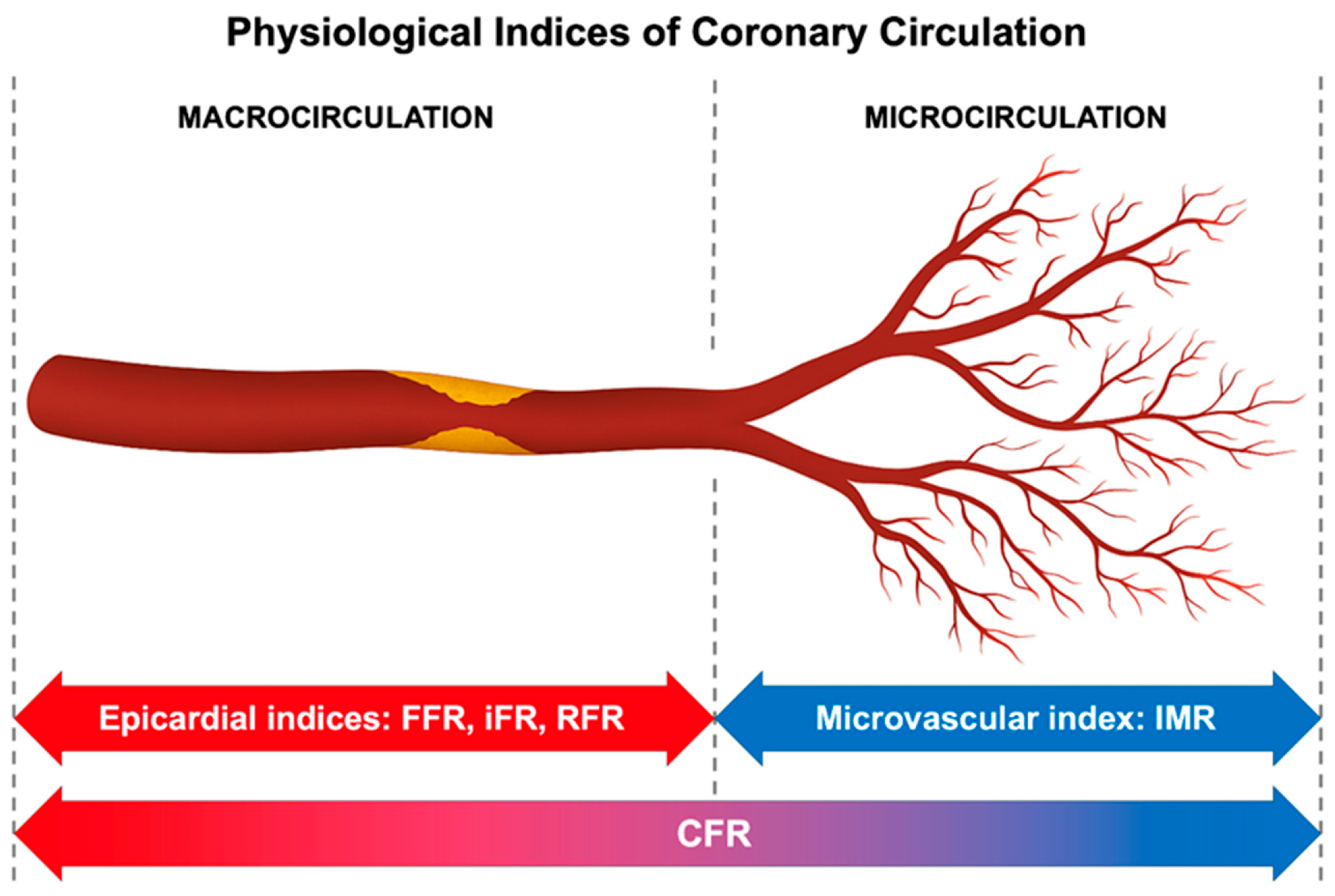
3.1. The Physiological Basis of Coronary Flow Reserve (Figure 1)

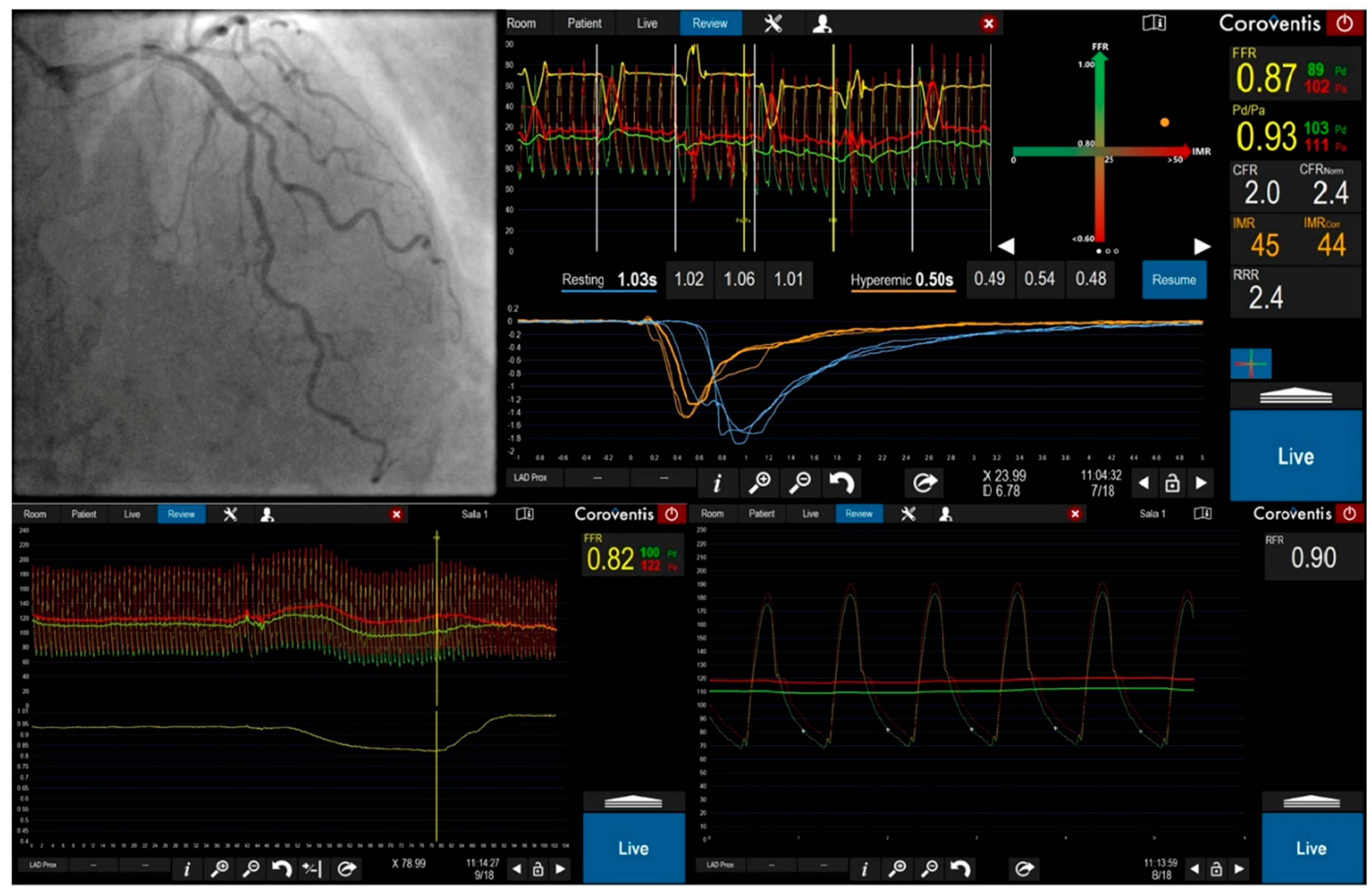
3.2. Invasive Assessment of Coronary Flow Reserve: Principles and Techniques (Figure 2)
3.2.1. Pharmacologic Induction of Hyperemia

3.2.2. Doppler Guidewire-Based Technique (CFVR)
3.2.3. Thermodilution-Based Technique (CFRthermo)
3.3. A Comparative Framework: CFR, FFR, IMR, and Non-Hyperemic Indices
3.4. Integrated Assessment and CFR-IMR Discordance
3.5. Safety, Economic, and Guideline Considerations
4. Non-Invasive Imaging Modalities for CFR Assessment
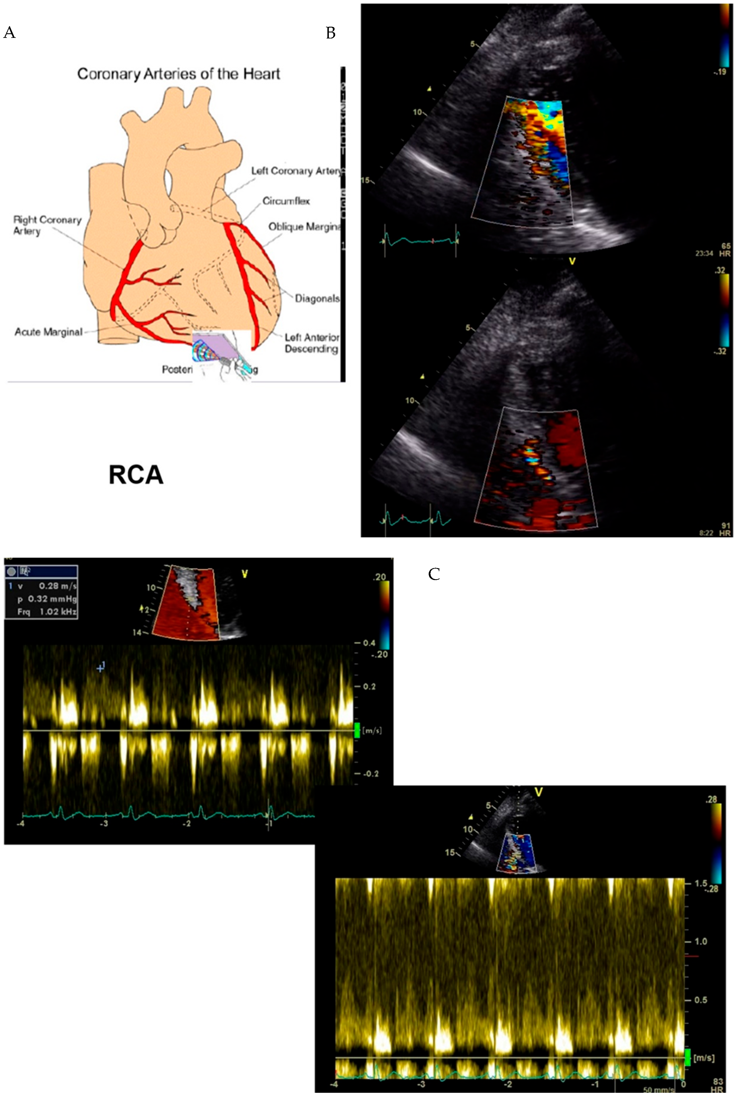
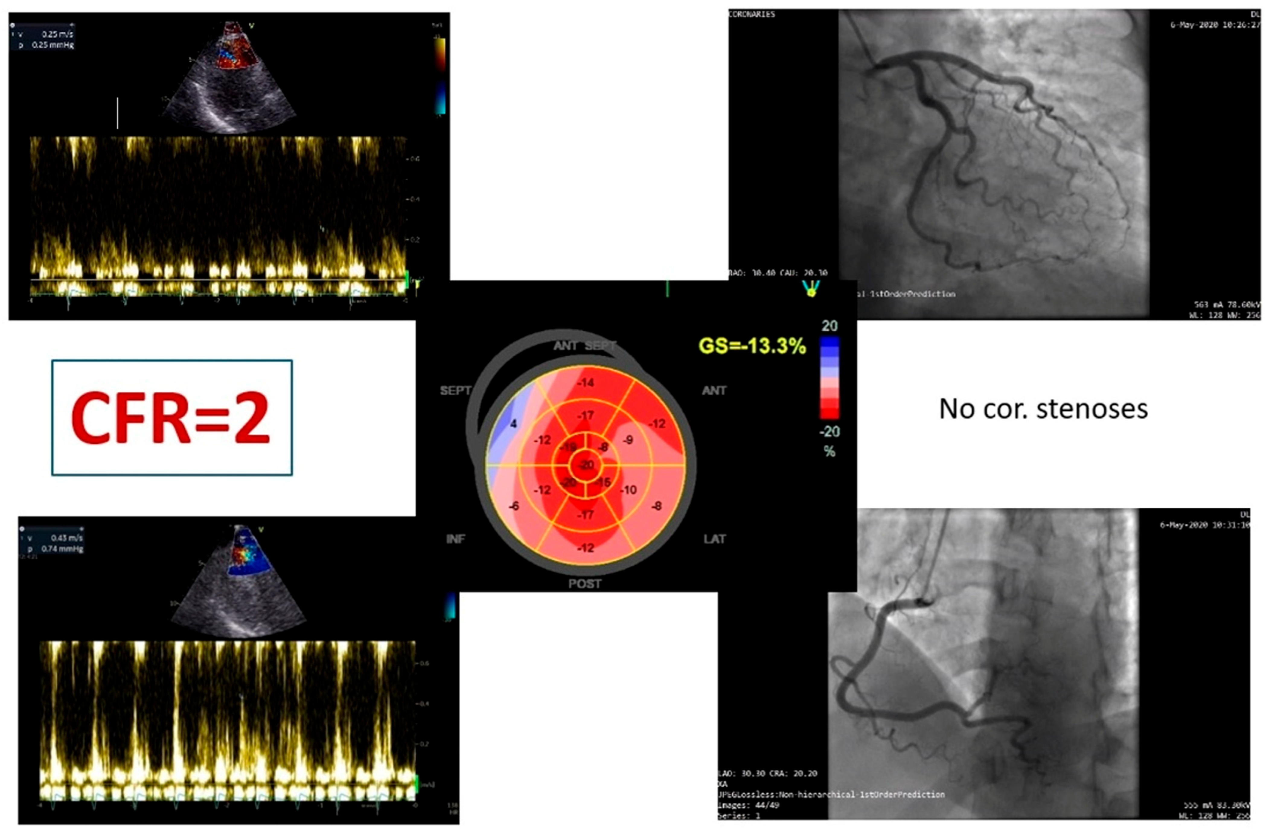
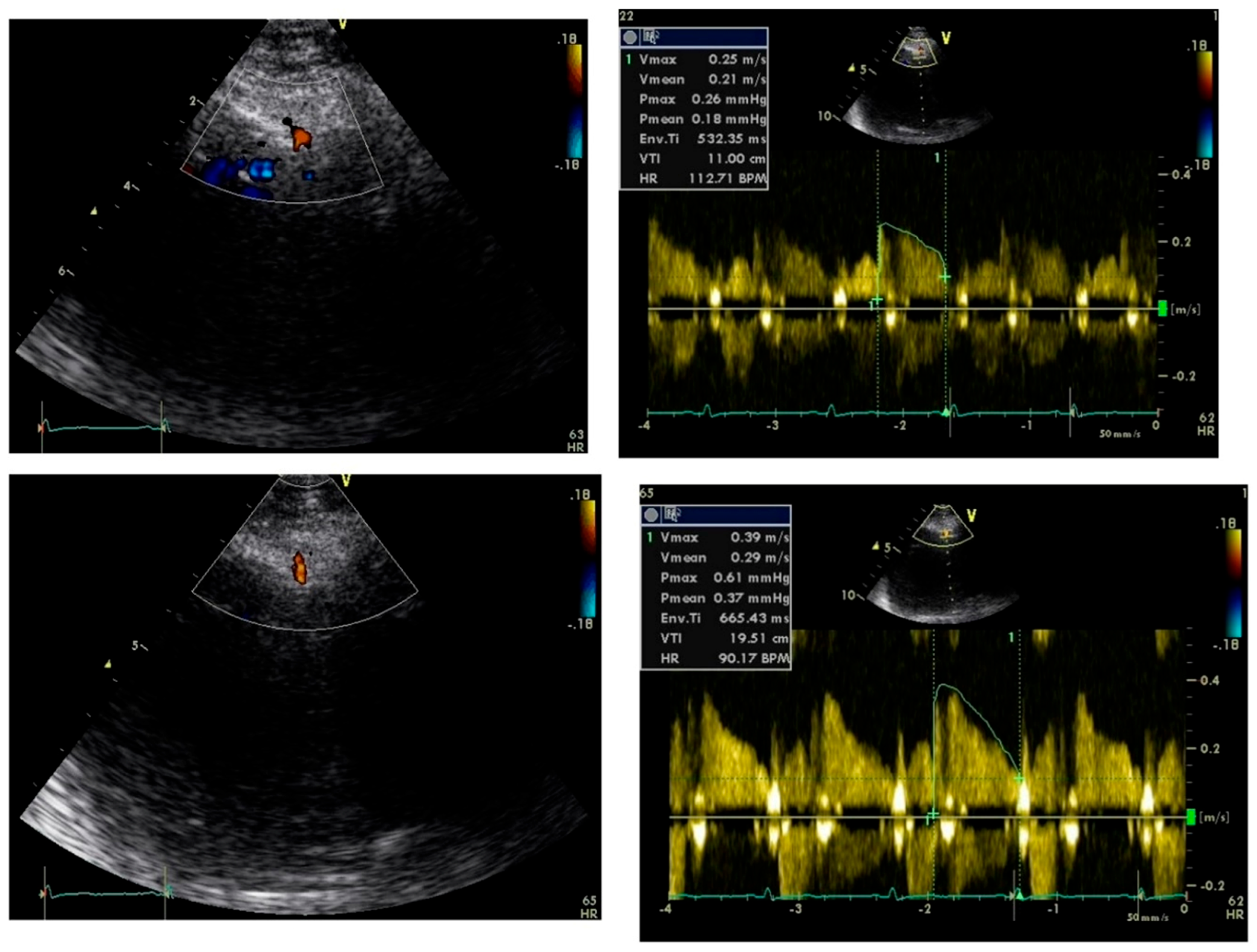
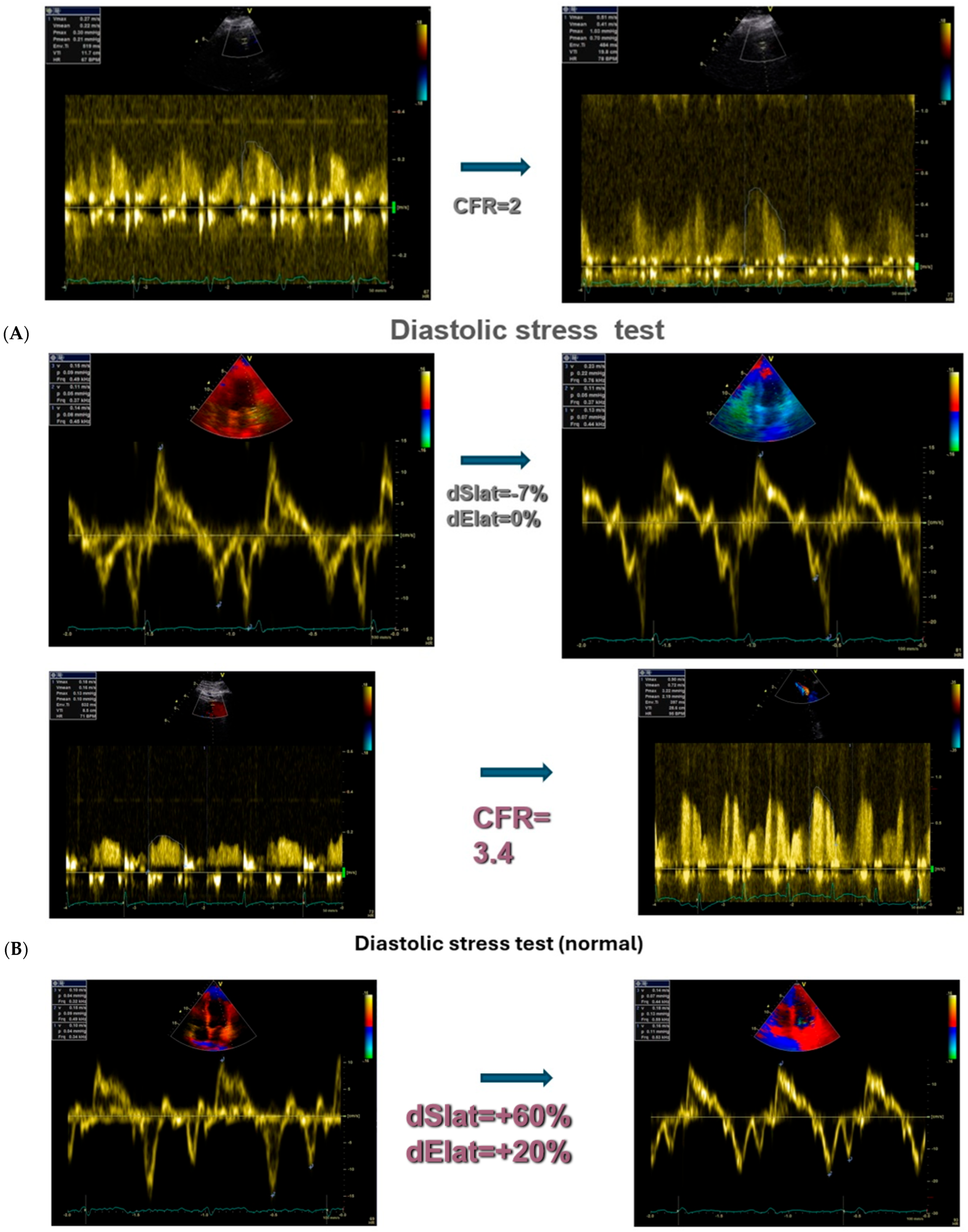
4.1. Transthoracic Doppler Echocardiography (TTDE)
4.2. Positron Emission Tomography (PET)
4.3. Cardiac Magnetic Resonance Imaging (CMR)
4.4. CT Perfusion and Dynamic SPECT
5. Prognostic Significance of Coronary Flow Reserve
5.1. CFR in Stable Coronary Artery Disease (Table 3)

| Study/Year | Population | Method of CFR Assessment | Key Findings | Prognostic Outcome |
|---|---|---|---|---|
| Gould et al., 2007 | 2783 pts, suspected CAD | PET | CFR < 2 predicted mortality independent of stenosis | ↑ CV death, MI |
| Johnson et al., 2012 | 1218 pts, stable CAD | Invasive Doppler/thermodilution | CFR discordant with FFR identified high-risk patients | ↑ MACEs |
| Cortigiani et al., 2014 | 1280 pts, stress echo | Doppler echo | CFR < 2 doubled cardiac death risk | ↑ Cardiac death |
| D’Antonio et al., 2025 | Systematic review, PET | PET | Reduced CFR predicted mortality across studies | ↑ All-cause death |
| Lee et al., 2015 | 1192 pts, revascularization decision | Invasive | CFR impaired → more benefit from PCI | ↓ MACEs after PCI |
5.2. CFR in Non-Obstructive CAD and INOCA (Table 4)
| Study/Year | Population | Sex Distribution | CFR Technique | Main Findings |
|---|---|---|---|---|
| Pepine et al., 2010 | 189 women, suspected ischemia | 100% women | Invasive | Reduced CFR predicted higher MACEs |
| Murthy et al., 2014 | 1218 pts, mixed | ~60% women | PET | CFR < 2 predicted death independent of CAD |
| Jensen et al., 2023 | Meta-analysis, non-obstructive CAD | Mixed | Various (PET, CMR, Echo) | Impaired CFR strongly prognostic, stronger in women |
| Taqueti et al., 2018 | 329 women | 100% women | PET | CFR < 2 → ↑ risk of HFpEF |
| Szolc et al., 2025 | 325 pts with INOCA | 72% women | Invasive | CFR-based tailored therapy improved QoL |
5.3. CFR in Heart Failure and Cardiomyopathies (Table 5)
| Study/Year | Population | Condition | CFR Assessment | Main Findings |
|---|---|---|---|---|
| Shah et al., 2016 | 244 pts with HFpEF | HFpEF | PET | Impaired CFR linked to diastolic dysfunction |
| Taqueti et al., 2018 | 329 women | HFpEF risk | PET | Reduced CFR predicted HFpEF development |
| Neglia et al., 2015 | 201 pts | Dilated cardiomyopathy | PET | CFR < 2 predicted mortality & hospitalization |
| Sciagrà et al., 2016 | 178 pts | Ischemic & non-ischemic cardiomyopathy | PET | Impaired CFR predicted MACEs |
| Toya et al., 2025 | 412 pts with HF | HFpEF & HFrEF | Invasive | CFR + high resistance → worse prognosis |
5.4. CFR in Systemic Diseases (Table 6)
| Disease | Key Study | Method | Main Findings | Prognostic Outcomes |
|---|---|---|---|---|
| Diabetes | Murthy et al., 2012 | PET | CFR < 2 predicted CV death independent of CAD | ↑ Mortality |
| Diabetes | Zhou et al., 2020 | PET | CFR predicted outcomes independent of HbA1c | ↑ CV events |
| CKD | Charytan et al., 2013 | PET | CFR predicted CV mortality | ↑ CV death |
| CKD | Shah et al., 2019 | PET | CFR superior to eGFR in predicting outcomes | ↑ HF hospitalization |
| SLE | Schindler et al., 2020 | PET | Reduced CFR → ↑ CV risk in lupus | ↑ MACEs |
| RA | Baniaamam et al., 2021 | Echo Doppler | Impaired CFR predicted CV and all-cause death | ↑ Mortality |
5.5. CFR in Guiding Therapy (Table 7)
| Strategy | Key Study | Population | CFR Finding | Clinical Implication |
|---|---|---|---|---|
| Revascularization | Lee et al., 2015 | 1192 pts, stable CAD | Impaired CFR → benefit from PCI | CFR can stratify who benefits most |
| Medical therapy | Bairey Merz et al., 2019 | 189 women with microvascular angina | Ranolazine improved CFR & angina | Tailored pharmacotherapy |
| Risk factor control | Takx et al., 2016 | Mixed CAD cohorts | Statins/ACEi improved CFR | Preventive therapy enhances CFR |
| Lifestyle interventions | Mehta et al., 2021 | Women with CMD | Exercise & lifestyle improved CFR | Non-pharmacologic therapy |
| Clinical trials | Al-Gully et al., 2025 | INOCA & CAD pts | Discordant CFR & resistance predicted outcomes | CFR as a trial endpoint |
5.6. Emerging Role of CFR in Arrhythmogenic Conditions and Epicardial Disease
6. Future Directions and Emerging Technologies
6.1. Advancements in Non-Invasive and Hybrid Imaging
6.2. Artificial Intelligence and Advanced Computational Approaches
6.3. Closing the Diagnosis–Treatment Gap
7. Conclusions
Author Contributions
Funding
Data Availability Statement
Conflicts of Interest
Abbreviations
References
- Hozumi, T.; Yoshida, K.; Ogata, Y.; Akasaka, T.; Asami, Y.; Takagi, T.; Morioka, S. Noninvasive assessment of significant left anterior descending coronary artery stenosis by coronary flow velocity reserve with transthoracic color Doppler echocardiography. Circulation 1998, 97, 1557–1562. [Google Scholar] [CrossRef] [Scilit]
- Homuzi, T.; Yoshida, K.; Akasaka, T.; Asami, Y.; Ogata, Y.; Takagi, T.; Kaji, S.; Kawamoto, T.; Ueda, Y.; Morioka, S. Noninvasive assessment of coronary flow velocity and coronary flow velocity reserve in the left anterior descending coronary artery by Doppler echocardiography: Comparison with invasive technique. J. Am. Coll. Cardiol. 1998, 32, 1251–1259. [Google Scholar]
- Gould, K.L.; Johnson, N.P.; Bateman, T.M.; Beanlands, R.S.; Bengel, F.M.; Bober, R.; Camici, P.G.; Cerqueira, M.D.; Chow, B.J.; Di Carli, M.F.; et al. Anatomic versus physiologic assessment of coronary artery disease. J. Am. Coll. Cardiol. 2013, 62, 1639–1653. [Google Scholar] [CrossRef] [Scilit] [PubMed]
- Youn, H.-J.; Foster, E. Demonstration of coronary artery flow using transthoracic Doppler echocardiography. J. Am. Soc. Echocardiogr. 2004, 17, 178–185. [Google Scholar] [CrossRef] [Scilit]
- Voci, P.; Pizzuto, F.; Romeo, F. Coronary flow: A new asset for the echo lab? Eur. Heart J. 2004, 25, 1867–1879. [Google Scholar] [CrossRef] [Scilit]
- Fu, B.; Wei, X.; Lin, Y.; Chen, J.; Yu, D. Pathophysiologic Basis and Diagnostic Approaches for Ischemia with Non-obstructive Coronary Arteries. Front. Cardiovasc. Med. 2022, 9, 731059. [Google Scholar] [CrossRef] [Scilit] [PubMed]
- Jensen, S.M.; Prescott, E.I.B.; Abdulla, J. The prognostic value of coronary flow reserve in patients with non-obstructive coronary artery disease and microvascular dysfunction: A systematic review and meta-analysis with focus on imaging modality and sex difference. Int. J. Cardiovasc. Imaging 2023, 39, 2545–2556. [Google Scholar] [CrossRef] [Scilit]
- Galiuto, L.; Sestito, A.; Barchetta, S.; Sgueglia, G.A.; Infusino, F.; La Rosa, C.; Lanza, G.; Crea, F. Noninvasive evaluation of flow reserve in the left anterior descending coronary artery in patients with cardiac syndrome X. Am. J. Cardiol. 2007, 99, 1378–1383. [Google Scholar] [CrossRef] [Scilit]
- Egashira, K.; Inou, T.; Hirooka, Y.; Yamada, A.; Urabe, Y.; Takeshita, A. Evidence of impaired endothelium-dependent coronary vasodilatation in patients with angina pectoris and normal coronary angiograms. N. Engl. J. Med. 1993, 328, 1659–1664. [Google Scholar] [CrossRef] [Scilit]
- Camici, P.G.; Crea, F. Coronary microvascular dysfunction. N. Engl. J. Med. 2007, 356, 830–840. [Google Scholar] [CrossRef] [Scilit]
- Takeuchi, M.; Lodato, J.A.; Furlong, K.T.; Lang, R.M.; Yoshikawa, J. Feasibility of measuring coronary flow velocity and reserve in the left anterior descending coronary artery by transthoracic Doppler echocardiography in the relatively obese American population. Echocardiography 2005, 22, 225–232. [Google Scholar] [CrossRef] [Scilit]
- Meimoun, P.; Clerc, J.; Ardourel, D.; Djou, U.; Martis, S.; Botoro, T.; Elmkies, F.; Zemir, H.; Luycx-Bore, A.; Boulanger, J. Assessment of left anterior descending artery stenosis of intermediate severity by fractional flow reserve, instantaneous wave-free ratio, and non-invasive coronary flow reserve. Int. J. Cardiovasc. Imaging 2017, 33, 999–1007. [Google Scholar] [CrossRef] [Scilit]
- Kasprzak, J.D.; Wejner-Mik, P.; Nouri, A.; Szymczyk, E.; Krzemińska-Pakuła, M.; Lipiec, P. Transthoracic measurement of left coronary artery flow reserve improves the diagnostic value of routine dipyridamole-atropine stress echocardiogram. Arch. Med. Sci. 2013, 9, 802–807. [Google Scholar] [CrossRef] [Scilit] [PubMed]
- Kelshiker, M.; Seligman, H.; Howard, J.; Haseeb Rahman, H.; Foley, M.; Nowbar, A.; Rajkumar, C.A.; Shun-Shin, M.J.; Ahmad, Y.; Sen, S.; et al. Coronary flow reserve and cardiovascular outcomes: A systematic review and meta-analysis. Eur. Heart J. 2022, 43, 1582–1593. [Google Scholar] [CrossRef] [Scilit]
- Tjoe, B.; Barsky, L.; Wei, C.; Samuels, B.; Azarbal, B.; Merz, C.; Shufelt, C. Coronary microvascular dysfunction: Considerations for diagnosis and treatment. Clevel. Clin. J. Med. 2022, 88, 561–571. [Google Scholar] [CrossRef] [Scilit]
- Bruning, R.; Sturek, M. Benefits of exercise training on coronary blood flow in coronary artery disease patients. Prog. Cardiovasc. Dis. 2015, 57, 443–453. [Google Scholar] [CrossRef] [Scilit]
- Ghodeshwar, G.; Dube, A.; Khobragade, D. Impact of lifestyle modifications on cardiovascular health: A narrative review. Cureus 2023, 15, e42616. [Google Scholar] [CrossRef] [Scilit] [PubMed]
- Bove, K.; Michelsen, M.; Schroder, J.; Suhrs, H.; Bechsgaard, D.; Mygind, N.; Aziz, A.; Kastrup, J.; Gustafsson, I.; Prescott, E. Impaired coronary flow velocity reserve is associated with cardiovascular risk factors but not with angina symptoms. Open Heart 2021, 8, e001486. [Google Scholar] [CrossRef] [Scilit]
- Padro, T.; Manfrini, O.; Bugiardini, R.; Canty, J.; Cenko, E.; De Luca, G.; Duncker, D.J.; Eringa, E.C.; Koller, A.; Tousoulis, D.; et al. ESC Working Group on Coronary Pathophysiology and Microcirculation position paper on ‘coronary microvascular dysfunction in cardiovascular disease. Cardiovasc. Res. 2020, 116, 741–755. [Google Scholar] [CrossRef] [Scilit]
- Shah, S.; Lam, C.; Svedlund, S.; Saraste, A.; Hage, C.; Tan, R.; Beussink-Nelson, L.; Ljung Faxén, U.; Fermer, M.L.; Broberg, M.A.; et al. Prevalence and correlates of coronary microvascular dysfunction in heart failure with preserved ejection fraction: PROMIS-HFpE. Eur. Heart J. 2018, 39, 3439–3450. [Google Scholar] [CrossRef] [Scilit] [PubMed]
- Shah, S.; Borlaug, B.; Kitzman, D.; McCulloch, A.; Blaxall, B.; Agarwal, R.; Chirinos, J.A.; Collins, S.; Deo, R.C.; Gladwin, M.T.; et al. Research priorities for heart failure with preserved ejection fraction. Circulation 2020, 141, 1001–1026. [Google Scholar] [CrossRef] [Scilit]
- Donato, A.; Machin, D.; Lesniewski, L. Mechanisms of Dysfunction in the Aging Vasculature and Role in Age-Related Disease. Circ. Res. 2018, 123, 825–848. [Google Scholar] [CrossRef] [Scilit]
- Mátyás, B.B.; Benedek, I.; Rat, N.; Gerculy, R.; Benedek, T. Coronary Plaque Vulnerability and Pericoronary Adipose Tissue Inflammation: Emerging Insights from Advanced CT Imaging. Medicina 2026, 62, 630. [Google Scholar] [CrossRef] [Scilit] [PubMed]
- Sperry, B.; Metzinger, M.; Ibrahim, A.; Thompson, R.; Cho, Y.; Jones, P.; McGhie, A.; Bateman, T. Age-sex-specific myocardial blood flow values in patients without coronary atherosclerosis on Rb-82 PET myocardial perfusion imaging. Circ. Cardiovasc. Imaging 2024, 17, e016577. [Google Scholar] [CrossRef] [Scilit] [PubMed]
- Faria, D.; Mejia-Renteria, H.; Lee, J.; Lee, S.; Travieso, A.; Jung, J.; Doh, J.; Nam, C.; Shin, E.; Hoshino, M.; et al. Age-related changes in the coronary microcirculation influencing the diagnostic performance of invasive pressure-based indices and long-term patient prognosis. Catheter. Cardiovasc. Interv. 2022, 100, 1195–1205. [Google Scholar] [CrossRef] [Scilit]
- Galderisi, M.; Rigo, F.; Gherardi, S.; Cortigiani, L.; Santoro, C.; Sicari, R.; Picano, E. The impact of aging and atherosclerotic risk factors on transthoracic coronary flow reserve in subjects with normal coronary angiography. Cardiovasc. Ultrasound 2012, 10, 20. [Google Scholar] [CrossRef] [Scilit]
- Hoef, T.; Echavarria-Pinto, M.; Meuwissen, M.; Stegehuis, V.; Escaned, J.; Piek, J. Contribution of age-related microvascular dysfunction to abnormal coronary: Hemodynamics in patients with ischemic heart disease. J. Am. Coll. Cardiol. Cardiovasc. Interv. 2020, 13, 20–29. [Google Scholar]
- Taqueti, V.; Di Carli, M. Coronary microvascular disease pathogenic mechanisms and therapeutic options: State-of-the-art review. J. Am. Coll. Cardiol. 2018, 72, 2625–2641. [Google Scholar] [CrossRef] [Scilit]
- Crea, F.; Battipaglia, I.; Andreotti, F. Sex differences in mechanisms, presentation and management of ischaemic heart disease. Atherosclerosis 2015, 241, 157–168. [Google Scholar] [CrossRef] [Scilit] [PubMed]
- Murthy, V.; Naya, M.; Taqueti, V.; Foster, C.; Gaber, M.; Hainer, J.; Dorbala, S.; Blankstein, R.; Rimoldi, O.; Camici, P.G.; et al. Effects of sex on coronary microvascular dysfunction and cardiac outcomes. Circulation 2014, 129, 2518–2527. [Google Scholar] [CrossRef] [Scilit]
- Pepine, C.; Anderson, R.; Sharaf, B.; Reis, S.; Smith, K.; Handberg, E.; Delia Johnson, B.; Sopko, G.; Noel Bairey Merz, C. Coronary microvascular reactivity to adenosine predicts adverse outcome in women evaluated for suspected ischemia: Results from the NHLBI Women’s Ischemia Syndrome Evaluation (WISE). J. Am. Coll. Cardiol. 2010, 55, 2825–2832. [Google Scholar] [CrossRef] [Scilit] [PubMed]
- Taqueti, V. Sex Differences in the Coronary System. Adv. Exp. Med. Biol. 2018, 1065, 257–278. [Google Scholar]
- Wei, J.; Mehta, P.; Johnson, B.; Samuels, B.; Kar, S.; Anderson, R.; Azarbal, B.; Petersen, J.; Sharaf, B.; Handberg, E.; et al. Safety of coronary reactivity testing in women with no obstructive coronary artery disease: Results from the NHLBI-sponsored WISE (Women’s Ischemia Syndrome Evaluation) study. JACC Cardiovasc. Interv. 2012, 5, 646–653. [Google Scholar] [CrossRef] [Scilit]
- Marinescu, M.; Löffler, A.; Ouellette, M.; Smith, L.; Bourque, J. Coronary Microvascular Dysfunction, Microvascular Angina, and Treatment Strategies. JACC Cardiovasc. Interv. 2015, 8, 210–220. [Google Scholar] [CrossRef] [Scilit]
- Taqueti, V.; Shaw, L.; Cook, N.; Murthy, V.; Shah, N.; Foster, C.; Hainer, J.; Blankstein, R.; Dorbala, S.; Di Carli, M.F. Excess cardiovascular risk in women relative to men referred for coronary angiography is associated with severely impaired coronary flow reserve, not obstructive disease. Circulation 2017, 135, 566–577. [Google Scholar] [CrossRef] [Scilit] [PubMed]
- Yu, Z.; Jiao, Y.; Zhao, Y.; Gu, W. Level of estrogen in females—The different impacts at different life stages. J. Pers. Med. 2022, 12, 1995. [Google Scholar] [CrossRef] [Scilit] [PubMed]
- Corban, M.; Prasad, A.; Gulati, R.; Lerman, L.; Lerman, A. Sex-specific differences in coronary blood flow and flow velocity reserve in symptomatic patients with non-obstructive disease. EuroIntervention 2021, 16, 1079–1084. [Google Scholar] [CrossRef] [Scilit]
- Jalnapurkar, S.; Landes, S.; Wei, J.; Mehta, P.; Shufelt, C.; Minissian, M.; Pepine, C.J.; Handberg, E.; Cook-Wiens, G.; Sopko, G.; et al. Coronary endothelial dysfunction appears to be a manifestation of a systemic process: A report from the Women’s Ischemia Syndrome Evaluation—Coronary Vascular Dysfunction (WISE-CVD) study. PLoS ONE 2021, 16, e0257184. [Google Scholar] [CrossRef] [Scilit]
- Patel, K.; Shaw, L.; Spertus, J.; Sperry, B.; McGhie, A.; Kennedy, K.; Thompson, R.C.; Chan, P.S.; Bateman, T.M. Association of sex, reduced myocardial flow reserve, and long-term mortality across spectrum of atherosclerotic disease. JACC Cardiovasc. Interv. 2022, 15, 1635–1644. [Google Scholar] [CrossRef] [Scilit]
- Houghton, J.; Prisant, M.; Carr, A.; Flowers, N.; Frank, M. Racial differences in myocardial ischemia and coronary flow reserve in hypertension. J. Am. Coll. Cardiol. 1994, 23, 1113–1119. [Google Scholar] [CrossRef] [Scilit]
- Zhang, L.; Olalere, D.; Mayrhofer, T. Difference in cardiovascular risk, coronary artery disease, and cardiac events between black and white individuals enrolled in the PROMISE Trial. JAMA Cardiol. 2022, 3, 259–267. [Google Scholar] [CrossRef] [Scilit]
- Breathett, K.; Sims, M.; Gross, M.; Jackson, E.; Jones, E.; Navas-Acien, A.; Taylor, H.; Thomas, K.L.; Howard, B.V.; On behalf of the American Heart Association Council on Epidemiology; et al. Cardiovascular health in American Indians & Alaska Natives: A scientific statement from the American Heart Association. Circulation 2020, 141, e948–e959. [Google Scholar]
- Gupta, M.; Singh, N.; Verma, S. South Asians and cardiovascular risk: What clinicians should know. Circulation 2006, 113, e924–e929. [Google Scholar] [CrossRef] [Scilit]
- Ihdayhid, A.; Thakur, U.; Yap, G.; Goeller, M.; Nerlekar, N.; Daniel Adams, D.; Isa, M.; Joshi, M.; Cameron, J.; Seneviratne, S.; et al. Ethnic differences in coronary anatomy, left ventricular mass and CT-derived fractional flow reserve. J. Cardiovasc. Comput. Tomogr. 2021, 15, 249–257. [Google Scholar] [CrossRef] [Scilit]
- Miller, M.; Cappuccio, F. Ethnicity and Inflammatory Pathways—Implications for Vascular Disease, Vascular Risk and Therapeutic Intervention. Curr. Med. Chem. 2007, 14, 1409–1425. [Google Scholar] [CrossRef] [Scilit]
- Tanus-Santos, J.E.; Desai, M.; Flockhart, D.A. Effects of ethnicity on the distribution of clinically relevant endothelial nitric oxide variants. Pharmacogenetics 2001, 11, 719–725. [Google Scholar] [CrossRef] [Scilit] [PubMed]
- Shantsila, E.; Wrigley, B.; Shantsila, A.; Tapp, L.; Blann, A.; Gill, P.; Lip, G. Ethnic differences in macrovascular and microvascular function in systolic heart failure. Circ. Heart Fail. 2011, 4, 754–762. [Google Scholar] [CrossRef] [Scilit] [PubMed]
- Young, A.; Sullivan, S.; Moazzami, K.; Lima, B.; Shah, A.; Lewis, T.; Elon, L.; Li, L.; Quyyumi, A.; Vaccarino, V. Abstract P137: Impaired microvascular and endothelial function among African-Americans with coronary artery disease. Circulation 2019, 139, AP137. [Google Scholar] [CrossRef] [Scilit]
- AlJaroudi, W.; Hage, F. Review of cardiovascular imaging in the Journal of Nuclear Cardiology 2017. Part 1 of 2: Positron emission tomography, computed tomography, and magnetic resonance. J. Nucl. Cardiol. 2018, 25, 320–330. [Google Scholar]
- Kaufmann, P.; Camici, P. Myocardial blood flow measurement by PET: Technical aspects and clinical applications. J. Nucl. Med. 2005, 46, 75–88. [Google Scholar] [PubMed]
- Murthy, V.; Di Carli, M. Non-invasive quantification of coronary vascular dysfunction for diagnosis and management of coronary artery disease. J. Nucl. Cardiol. 2012, 19, 1060–1075. [Google Scholar] [CrossRef] [Scilit]
- Nesterov, S.; Deshayes, E.; Sciagrà, R.; Settimo, L.; Declerck, J.; Pan, X.; Yoshinaga, K.; Katoh, C.; Slomka, P.J.; Germano, G.; et al. Quantification of myocardial blood flow in absolute terms using 82Rb PET imaging: The RUBY-10 Study. JACC Cardiovasc. Interv. 2015, 8, 1119–1127. [Google Scholar]
- Fearon, W.F.; Kobayashi, Y. Invasive Assessment of the Coronary Microvasculature: The Index of Microcirculatory Resistance. Circ. Cardiovasc. Interv. 2017, 10, e005361. [Google Scholar] [CrossRef] [Scilit] [PubMed]
- Mangiacapra, F.; Viscusi, M.M.; Verolino, G.; Paolucci, L.; Nusca, A.; Melfi, R.; Ussia, G.P.; Grigioni, F. Invasive Assessment of Coronary Microvascular Function. J. Clin. Med. 2021, 11, 228. [Google Scholar] [CrossRef] [Scilit]
- Belmonte, M.; Gallinoro, E.; Pijls, N.H.J.; Bertolone, D.T.; Keulards, D.C.J.; Viscusi, M.M.; Storozhenko, T.; Mizukami, T.; Mahendiran, T.; Seki, R.; et al. Measuring Absolute Coronary Flow and Microvascular Resistance by Thermodilution: JACC Review Topic of the Week. J. Am. Coll. Cardiol. 2024, 83, 699–709. [Google Scholar] [CrossRef] [Scilit]
- Cokkinos, D.V.; Manginas, A.; Voudris, V. Coronary flow: Clinical considerations. Heart 2003, 89, 361–363. [Google Scholar] [CrossRef] [Scilit]
- Ciaramella, L.; Di Serafino, L.; Mitrano, L.; De Rosa, M.L.; Carbone, C.; Rea, F.S.; Monaco, S.; Scalamogna, M.; Cirillo, P.; Esposito, G. Invasive Assessment of Coronary Microcirculation: A State-of-the-Art Review. Diagnostics 2023, 14, 86. [Google Scholar] [CrossRef] [Scilit]
- Everaars, H.; de Waard, G.A.; Driessen, R.S.; Danad, I.; van de Ven, P.M.; Raijmakers, P.G.; Lammertsma, A.A.; van Rossum, A.C.; Knaapen, P.; van Royen, N. Doppler Flow Velocity and Thermodilution to Assess Coronary Flow Reserve: A Head-to-Head Comparison with [15O]H2O PET. JACC Cardiovasc. Interv. 2018, 11, 2044–2054. [Google Scholar] [CrossRef] [Scilit]
- Gurav, A.; Revaiah, P.C.; Tsai, T.Y.; Miyashita, K.; Tobe, A.; Oshima, A.; Sevestre, E.; Garg, S.; Aben, J.P.; Reiber, J.H.C.; et al. Coronary angiography: A review of the state of the art and the evolution of angiography in cardio therapeutics. Front. Cardiovasc. Med. 2024, 11, 1468888. [Google Scholar] [CrossRef] [Scilit]
- Layland, J.; Carrick, D.; Lee, M.; Oldroyd, K.; Berry, C. Adenosine: Physiology, pharmacology, and clinical applications. JACC Cardiovasc. Interv. 2014, 7, 581–591. [Google Scholar] [CrossRef] [Scilit] [PubMed]
- Sandhu, P.S.; Kaul, U.; Gupta, R.K.; Ghose, T. Fractional flow reserve: Intracoronary versus intravenous adenosine induced maximal coronary hyperemia. Indian Heart J. 2013, 65, 147–151. [Google Scholar] [CrossRef] [Scilit]
- Legutko, J.; Kleczyński, P.; Dziewierz, A.; Rzeszutko, Ł.; Dudek, D. Adenosine intracoronary bolus dose escalation versus intravenous infusion to induce maximum coronary hyperemia for fractional flow reserve assessment. Kardiol. Pol. 2019, 77, 610–617. [Google Scholar] [PubMed]
- Zdzierak, B.; Zasada, W.; Krawczyk-Ożóg, A.; Rakowski, T.; Bartuś, S.; Surdacki, A.; Dziewierz, A. Influence of sex on the functional assessment of myocardial ischemia. Kardiol. Pol. 2023, 81, 895–902. [Google Scholar] [CrossRef] [Scilit]
- Jeremias, A.; Filardo, S.D.; Whitbourn, R.J.; Kernoff, R.S.; Yeung, A.C.; Fitzgerald, P.J.; Yock, P.G. Effects of intravenous and intracoronary adenosine 5′-triphosphate as compared with adenosine on coronary flow and pressure dynamics. Circulation 2000, 101, 318–323. [Google Scholar] [CrossRef] [Scilit]
- Akasaka, T.; Yamamuro, A.; Kamiyama, N.; Koyama, Y.; Akiyama, M.; Watanabe, N.; Neishi, Y.; Takagi, T.; Shalman, E.; Barak, C.; et al. Assessment of coronary flow reserve by coronary pressure measurement: Comparison with flow- or velocity-derived coronary flow reserve. J. Am. Coll. Cardiol. 2003, 41, 1554–1560. [Google Scholar] [CrossRef] [Scilit]
- Petretta, M.; Acampa, W.; Zampella, E.; Assante, R.; Petretta, M.P.; Cuocolo, R.; Fabiani, I.; Della Rattal, G.L.; Perrone-Filardi, P.; Cuocolo, A. Imaging techniques for assessment of coronary flow reserve. Monaldi Arch. Chest Dis. 2011, 76, 192–197. [Google Scholar] [CrossRef] [Scilit]
- Barbato, E.; Aarnoudse, W.; Aengevaeren, W.R.; Werner, G.; Klauss, V.; Bojara, W.; Herzfeld, I.; Oldroyd, K.G.; Pijls, N.H.; De Bruyne, B. Validation of coronary flow reserve measurements by thermodilution in clinical practice. Eur. Heart J. 2004, 25, 219–223. [Google Scholar]
- Candreva, A.; Gallinoro, E.; van’t Veer, M.; Sonck, J.; Collet, C.; Di Gioia, G.; Kodeboina, M.; Mizukami, T.; Nagumo, S.; Keulards, D.; et al. Basics of Coronary Thermodilution. JACC Cardiovasc. Interv. 2021, 14, 595–605. [Google Scholar]
- De Bruyne, B.; Pijls, N.H.; Smith, L.; Wievegg, M.; Heyndrickx, G.R. Coronary thermodilution to assess flow reserve: Experimental validation. Circulation 2001, 104, 2003–2006. [Google Scholar] [CrossRef] [Scilit] [PubMed]
- Gallinoro, E.; Bertolone, D.T.; Mizukami, T.; Paolisso, P.; Bermpeis, K.; Munhoz, D.; Sakai, K.; Seki, R.; Ohashi, H.; Esposito, G.; et al. Continuous vs Bolus Thermodilution to Assess Microvascular Resistance Reserve. JACC Cardiovasc. Interv. 2023, 16, 2767–2777. [Google Scholar] [CrossRef] [Scilit] [PubMed]
- Gallinoro, E.; Bertolone, D.T.; Fernandez-Peregrina, E.; Paolisso, P.; Bermpeis, K.; Esposito, G.; Gomez-Lopez, A.; Candreva, A.; Mileva, N.; Belmonte, M.; et al. Reproducibility of bolus versus continuous thermodilution for assessment of coronary microvascular function in patients with ANOCA. EuroIntervention 2023, 19, e155–e166. [Google Scholar] [CrossRef] [Scilit] [PubMed]
- Gutiérrez-Barrios, A.; Izaga-Torralba, E.; Rivero Crespo, F.; Gheorghe, L.; Cañadas-Pruaño, D.; Gómez-Lara, J.; Silva, E.; Noval-Morillas, I.; Zayas Rueda, R.; Calle-Pérez, G.; et al. Continuous Thermodilution Method to Assess Coronary Flow Reserve. Am. J. Cardiol. 2021, 141, 31–37. [Google Scholar] [CrossRef] [Scilit]
- van’t Veer, M.; Geven, M.C.; Rutten, M.C.; van der Horst, A.; Aarnoudse, W.H.; Pijls, N.H.; van de Vosse, F.N. Continuous infusion thermodilution for assessment of coronary flow: Theoretical background and in vitro validation. Med. Eng. Phys. 2009, 31, 688–694. [Google Scholar] [PubMed]
- Gallinoro, E.; Candreva, A.; Colaiori, I.; Kodeboina, M.; Fournier, S.; Nelis, O.; Di Gioia, G.; Sonck, J.; van’t Veer, M.; Pijls, N.H.J.; et al. Thermodilution-derived volumetric resting coronary blood flow measurement in humans. EuroIntervention 2021, 17, e672–e679. [Google Scholar] [CrossRef] [Scilit] [PubMed]
- Jeremias, A.; Kirtane, A.J.; Stone, G.W. A Test in Context: Fractional Flow Reserve: Accuracy, Prognostic Implications, and Limitations. J. Am. Coll. Cardiol. 2017, 69, 2748–2758. [Google Scholar] [CrossRef] [Scilit]
- De Bruyne, B.; Pijls, N.H.; Kalesan, B.; Barbato, E.; Tonino, P.A.; Piroth, Z.; Jagic, N.; Möbius-Winkler, S.; Rioufol, G.; Witt, N.; et al. Fractional flow reserve-guided PCI versus medical therapy in stable coronary disease. N. Engl. J. Med. 2012, 367, 991–1001. [Google Scholar]
- Fournier, S.; Keulards, D.C.J.; van’t Veer, M.; Colaiori, I.; Di Gioia, G.; Zimmermann, F.M.; Mizukami, T.; Nagumo, S.; Kodeboina, M.; El Farissi, M.; et al. Normal values of thermodilution-derived absolute coronary blood flow and microvascular resistance in humans. EuroIntervention 2021, 17, e309–e316. [Google Scholar] [CrossRef] [Scilit]
- Wang, B.; Gao, Y.; Zhao, Y.; Xu, C.; Zhao, S.; Li, H.; Zhang, Y.; Xu, Y. The spectrum of angiography-derived IMR according to morphological and physiological coronary stenosis in patients with suspected myocardial ischemia. Clin. Cardiol. 2023, 46, 502–511. [Google Scholar] [CrossRef] [Scilit]
- Lee, J.M.; Jung, J.H.; Hwang, D.; Park, J.; Fan, Y.; Na, S.H.; Doh, J.H.; Nam, C.W.; Shin, E.S.; Koo, B.K. Coronary Flow Reserve and Microcirculatory Resistance in Patients with Intermediate Coronary Stenosis. J. Am. Coll. Cardiol. 2016, 67, 1158–1169. [Google Scholar] [CrossRef] [Scilit]
- Smilowitz, N.R.; Toleva, O.; Chieffo, A.; Perera, D.; Berry, C. Coronary Microvascular Disease in Contemporary Clinical Practice. Circ. Cardiovasc. Interv. 2023, 16, e012568. [Google Scholar] [CrossRef] [Scilit]
- Januszek, R.; Kołtowski, Ł.; Tomaniak, M.; Wańha, W.; Wojakowski, W.; Grygier, M.; Siłka, W.; Jan Horszczaruk, G.; Czarniak, B.; Kręcki, R.; et al. Implementation of Microcirculation Examination in Clinical Practice-Insights from the Nationwide POL-MKW Registry. Medicina 2024, 60, 277. [Google Scholar] [CrossRef] [Scilit]
- Fawaz, S.; Khan, S.; Simpson, R.; Clesham, G.; Cook, C.M.; Davies, J.R.; Karamasis, G.V.; Keeble, T.R. Invasive Detection of Coronary Microvascular Dysfunction: How It Began, and Where We Are Now. Interv. Cardiol. 2023, 18, e07. [Google Scholar] [CrossRef] [Scilit]
- Zdzierak, B.; Zasada, W.; Dziewierz, A.; Pawlik, A.; Rzeszutko, Ł.; Bartuś, S. Pullback pressure gradient: A novel dimension in coronary physiology. Kardiol. Pol. 2025; in press.
- Al-Gully, J.; Oliveri, F.; Forouzanfar, J.P.; Montero-Cabezas, J.M.; Jukema, J.W.; den Haan, M.C.; Al Amri, I.; Bingen, B.O. Prognostic role of con-/discordant coronary flow reserve and microvascular resistance in coronary microvascular disease: A systematic review and network meta-analysis. Open Heart 2025, 12, e003055. [Google Scholar] [CrossRef] [Scilit]
- Dimitriadis, K.; Pyrpyris, N.; Sakalidis, A.; Beneki, E.; Chrysohoou, C.; Aznaouridis, K.; Tsioufis, K. The prognostic role of microvascular resistance reserve: A systematic review and meta-analysis. Cardiovasc. Revasc. Med. 2025; in press.
- Zdzierak, B.; Zasada, W.; Rakowski, T.; Krawczyk-Ożóg, A.; Bartuś, S.; Surdacki, A.; Dziewierz, A. Influence of diabetes mellitus on the invasive assessment of myocardial ischemia in patients with coronary artery disease. Pol. Arch. Intern. Med. 2023, 133, 16502. [Google Scholar] [CrossRef] [Scilit]
- Zasada, W.; Zdzierak, B.; Rakowski, T.; Bobrowska, B.; Krawczyk-Ożóg, A.; Surowiec, S.; Bartuś, S.; Surdacki, A.; Dziewierz, A. The Impact of Age on the Physiological Assessment of Borderline Coronary Stenoses. Medicina 2023, 59, 1863. [Google Scholar] [CrossRef] [Scilit] [PubMed]
- Vrints, C.; Andreotti, F.; Koskinas, K.C.; Rossello, X.; Adamo, M.; Ainslie, J.; Banning, A.P.; Budaj, A.; Buechel, R.R.; Chiariello, G.A.; et al. 2024 ESC Guidelines for the management of chronic coronary syndromes. Eur. Heart J. 2024, 45, 3415–3537. [Google Scholar] [CrossRef] [Scilit]
- Simova, I. Coronary Flow Velocity Reserve Assessment with Transthoracic Doppler Echocardiography. Eur. Cardiol. 2015, 10, 12–18. [Google Scholar] [CrossRef] [Scilit]
- Dimitrow, P.P. Transthoracic Doppler echocardiography–noninvasive diagnostic window for coronary flow reserve assessment. Cardiovasc. Ultrasound 2003, 1, 4. [Google Scholar] [CrossRef] [Scilit]
- Tesic, M.; Beleslin, B.; Giga, V.; Jovanovic, I.; Marinkovic, J.; Trifunovic, D.; Petrovic, O.; Dobric, M.; Aleksandric, S.; Juricic, S.; et al. Prognostic Value of Transthoracic Doppler Echocardiography Coronary Flow Velocity Reserve in Patients with Asymmetric Hypertrophic Cardiomyopathy. J. Am. Heart Assoc. 2021, 10, e021936. [Google Scholar] [CrossRef] [Scilit] [PubMed]
- Meimoun, P.; Benali, T.; Sayah, S.; Luycx-Bore, A.; Boulanger, J.; Zemir, H.; Tribouilloy, C. Evaluation of left anterior descending coronary artery stenosis of intermediate severity using transthoracic coronary flow reserve and dobutamine stress echocardiography. J. Am. Soc. Echocardiogr. 2005, 18, 1233–1240. [Google Scholar] [CrossRef] [Scilit]
- Carbone, A.; D’Andrea, A.; Sperlongano, S.; Tagliamonte, E.; Mandoli, G.E.; Santoro, C.; Evola, V.; Bandera, F.; Morrone, D.; Malagoli, A.; et al. Echocardiographic assessment of coronary microvascular dysfunction: Basic concepts, technical aspects, and clinical settings. Echocardiography 2021, 38, 993–1001. [Google Scholar] [CrossRef] [Scilit]
- Bailly, M.; Thibault, F.; Metrard, G.; Courtehoux, M.; Angoulvant, D.; Ribeiro, M.J. Precision of Myocardial Blood Flow and Flow Reserve Measurement During CZT SPECT Perfusion Imaging Processing: Intra- and Interobserver Variability. J. Nucl. Med. 2023, 64, 260–265. [Google Scholar] [PubMed]
- D’Antonio, A.; Assante, R.; Zampella, E.; Cantoni, V.; Green, R.; Gaudieri, V.; Mannarino, T.; Falzarano, M.; Volpicelli, F.; Cutillo, P.; et al. Prognostic value of myocardial flow reserve by PET imaging in patients with suspected coronary artery disease: A systematic review and meta-analysis. Int. J. Cardiol. Heart Vasc. 2025, 58, 101677. [Google Scholar] [CrossRef] [Scilit] [PubMed]
- Pan, C.; Yin, R.; Tang, X.; Wang, T.; Hu, C. Prognostic Significance of Myocardial Blood Flow Quantification for Major Adverse Cardiac Events: A Systematic Review and Meta-analysis. Cardiol. Rev. 2023, 31, 162–167. [Google Scholar] [CrossRef] [Scilit]
- Kramer, C.M.; Chandrashekhar, Y. Quantitative Myocardial Perfusion CMR: Is the Game Worth the Candle? JACC Cardiovasc. Imaging 2018, 11, 709–712. [Google Scholar]
- Catania, R.; Quinn, S.; Rahsepar, A.A.; Trabzonlu, T.A.; Bisen, J.B.; Chow, K.; Lee, D.C.; Avery, R.; Kellman, P.; Allen, B.D. Quantitative Stress First-Pass Perfusion Cardiac MRI: State of the Art. RadioGraphics 2025, 45, e240115. [Google Scholar] [CrossRef] [Scilit]
- Pons-Lladó, G.; Kellman, P. State-of-the-Art of Myocardial Perfusion by CMR: A Practical View. Rev. Cardiovasc. Med. 2022, 23, 325. [Google Scholar] [CrossRef] [Scilit]
- Borodzicz-Jazdzyk, S.; Vink, C.E.M.; Demirkiran, A.; Hoek, R.; de Mooij, G.W.; Hofman, M.B.M.; Wilgenhof, A.; Appelman, Y.; Benovoy, M. Clinical implementation of a fully automated quantitative perfusion cardiovascular magnetic resonance imaging workflow with a simplified dual-bolus contrast administration scheme. Sci. Rep. 2024, 14, 9665. [Google Scholar] [CrossRef] [Scilit] [PubMed]
- Williams, M.C.; Newby, D.E. CT myocardial perfusion imaging: Current status and future directions. Clin. Radiol. 2016, 71, 739–749. [Google Scholar] [CrossRef] [Scilit] [PubMed]
- Rossi, A.; Wragg, A.; Klotz, E.; Pirro, F.; Moon, J.C.; Nieman, K.; Pugliese, F. Dynamic Computed Tomography Myocardial Perfusion Imaging: Comparison of Clinical Analysis Methods for the Detection of Vessel-Specific Ischemia. Circ. Cardiovasc. Imaging 2017, 10, e005505. [Google Scholar] [CrossRef] [Scilit]
- Nieman, K.; Balla, S. Dynamic CT myocardial perfusion imaging. J. Cardiovasc. Comput Tomogr. 2020, 14, 303–306. [Google Scholar] [CrossRef] [Scilit]
- Otaki, Y.; Manabe, O.; Miller, R.J.H.; Manrique, A.; Nganoa, C.; Roth, N.; Berman, D.S.; Germano, G.; Slomka, P.J.; Agostini, D. Quantification of myocardial blood flow by CZT-SPECT with motion correction and comparison with 15O-water PET. J. Nucl. Cardiol. 2021, 28, 1477–1486. [Google Scholar] [CrossRef] [Scilit]
- Mallet, F.; Poitrasson-Rivière, A.; Mariano-Goulart, D.; Agostini, D.; Manrique, A. Measuring myocardial blood flow using dynamic myocardial perfusion SPECT: Artifacts and pitfalls. J. Nucl. Cardiol. 2023, 30, 2006–2017. [Google Scholar] [CrossRef] [Scilit] [PubMed]
- Slomka, P.J.; Patton, J.A.; Berman, D.S.; Germano, G. Advances in technical aspects of myocardial perfusion SPECT imaging. J. Nucl. Cardiol. 2009, 16, 255–276. [Google Scholar] [CrossRef] [Scilit]
- Gould, K.L.; Kitkungvan, D.; Johnson, N.P.; Nguyen, T.; Kirkeeide, R.; Bui, L.; Patel, M.B.; Roby, A.E.; Madjid, M.; Zhu, H.; et al. Mortality Prediction by Quantitative PET Perfusion Expressed as Coronary Flow Capacity with and Without Revascularization. JACC Cardiovasc. Imaging 2021, 14, 1020–1034. [Google Scholar] [CrossRef] [Scilit]
- Johnson, N.P.; Kirkeeide, R.L.; Gould, K.L. Is discordance of coronary flow reserve and fractional flow reserve due to methodology or clinically relevant coronary pathophysiology? JACC Cardiovasc. Imaging 2012, 5, 193–202. [Google Scholar] [CrossRef] [Scilit]
- Maron, D.J.; Hochman, J.S.; Reynolds, H.R.; Bangalore, S.; O’Brien, S.M.; Boden, W.E.; Chaitman, B.R.; Senior, R.; López-Sendón, J.; Alexander, K.P.; et al. Initial Invasive or Conservative Strategy for Stable Coronary Disease. N. Engl. J. Med. 2020, 382, 1395–1407. [Google Scholar] [CrossRef] [Scilit]
- Choi, K.H.; Lee, J.M.; Koo, B.K.; Nam, C.W.; Shin, E.S.; Doh, J.H.; Rhee, T.M.; Hwang, D.; Park, J.; Zhang, J.; et al. Prognostic Implication of Functional Incomplete Revascularization and Residual Functional SYNTAX Score in Patients with Coronary Artery Disease. JACC Cardiovasc. Interv. 2018, 11, 237–245. [Google Scholar] [CrossRef] [Scilit]
- Jespersen, L.; Hvelplund, A.; Abildstrøm, S.Z.; Pedersen, F.; Galatius, S.; Madsen, J.K.; Jørgensen, E.; Kelbæk, H.; Prescott, E. Stable angina pectoris with no obstructive coronary artery disease is associated with increased risks of major adverse cardiovascular events. Eur. Heart J. 2012, 33, 734–744. [Google Scholar] [CrossRef] [Scilit]
- Bairey Merz, C.N.; Pepine, C.J.; Walsh, M.N.; Fleg, J.L. Ischemia and No Obstructive Coronary Artery Disease (INOCA): Developing Evidence-Based Therapies and Research Agenda for the Next Decade. Circulation 2017, 135, 1075–1092. [Google Scholar] [CrossRef] [Scilit] [PubMed]
- Taqueti, V.R.; Solomon, S.D.; Shah, A.M.; Desai, A.S.; Groarke, J.D.; Osborne, M.T.; Hainer, J.; Bibbo, C.F.; Dorbala, S.; Blankstein, R.; et al. Coronary microvascular dysfunction and future risk of heart failure with preserved ejection fraction. Eur. Heart J. 2018, 39, 840–849. [Google Scholar] [CrossRef] [Scilit]
- Heggie, R.; Briggs, A.; Stanley, B.; Good, R.; Rocchiccioli, P.; McEntegart, M.; Watkins, S.; Eteiba, H.; Shaukat, A.; Lindsay, M.; et al. Stratified medicine using invasive coronary function testing in angina: A cost-effectiveness analysis of the British Heart Foundation CorMicA trial. Int. J. Cardiol. 2021, 337, 44–51. [Google Scholar] [CrossRef] [Scilit] [PubMed]
- Waheed, N.; Elias-Smale, S.; Malas, W.; Maas, A.H.; Sedlak, T.L.; Tremmel, J.; Mehta, P.K. Sex differences in non-obstructive coronary artery disease. Cardiovasc. Res. 2020, 116, 829–840. [Google Scholar] [CrossRef] [Scilit]
- Mehta, P.K.; Quesada, O.; Al-Badri, A.; Fleg, J.L.; Volgman, A.S.; Pepine, C.J.; Merz, C.N.B.; Shaw, L.J. Ischemia and no obstructive coronary arteries in patients with stable ischemic heart disease. Int. J. Cardiol. 2022, 348, 1–8. [Google Scholar] [CrossRef] [Scilit]
- Sinha, A.; Rahman, H.; Perera, D. Coronary microvascular disease: Current concepts of pathophysiology, diagnosis and management. Cardiovasc. Endocrinol. Metab. 2020, 10, 22–30. [Google Scholar] [CrossRef] [Scilit]
- Boerhout, C.K.M.; Namba, H.F.; Liu, T.; Beijk, M.A.M.; Damman, P.; Meuwissen, M.; Ong, P.; Sechtem, U.; Appelman, Y.; Berry, C.; et al. Rationale and design of the ILIAS ANOCA clinical trial: A blinded-arm controlled trial for routine ad-hoc coronary function testing. Am. Heart J. 2025, 286, 1–13. [Google Scholar] [CrossRef] [Scilit]
- Szolc, P.; Guzik, B.; Niewiara, Ł.; Kleczyński, P.; Bernacik, A.; Diachyshyn, M.; Stąpór, M.; Żmudka, K.; Legutko, J. Tailored treatment of specific diagnosis improves symptoms and quality of life in patients with myocardial Ischemia and Non-obstructive Coronary Arteries. Sci. Rep. 2025, 15, 18968. [Google Scholar] [CrossRef] [Scilit]
- Neglia, D.; Michelassi, C.; Trivieri, M.G.; Sambuceti, G.; Giorgetti, A.; Pratali, L.; Gallopin, M.; Salvadori, P.; Sorace, O.; Carpeggiani, C.; et al. Prognostic role of myocardial blood flow impairment in idiopathic left ventricular dysfunction. Circulation 2002, 105, 186–193. [Google Scholar] [CrossRef] [Scilit]
- Dryer, K.; Gajjar, M.; Narang, N.; Lee, M.; Paul, J.; Shah, A.P.; Nathan, S.; Butler, J.; Davidson, C.J.; Fearon, W.F.; et al. Coronary microvascular dysfunction in patients with heart failure with preserved ejection fraction. Am. J. Physiol. Heart Circ. Physiol. 2018, 314, H1033–H1042. [Google Scholar] [CrossRef] [Scilit]
- Sharka, I.; Panichella, G.; Grigoratos, C.; Muca, M.; De Gori, C.; Keilberg, P.; Novani, G.; Barra, V.; Hlavata, H.; Bianchi, M.; et al. Myocardial Perfusion Imaging with Cardiovascular Magnetic Resonance in Nonischemic Cardiomyopathies: An In-Depth Review of Techniques and Clinical Applications. Medicina 2025, 61, 875. [Google Scholar] [CrossRef] [Scilit]
- Toya, T.; Corban, M.T.; Park, J.Y.; Ahmad, A.; Özcan, I.; Sebaali, F.; Sara, J.D.S.; Gulati, R.; Lerman, L.O.; Lerman, A. Prognostic impact and clinical outcomes of coronary flow reserve and hyperaemic microvascular resistance. EuroIntervention 2021, 17, 569–575. [Google Scholar] [CrossRef] [Scilit]
- Sadeghi, R.; Adnani, N.; Sohrabi, M.R.; Alipour Parsa, S. Risk of sudden cardiac death. ARYA Atheroscler. 2013, 9, 274–279. [Google Scholar]
- Niewiara, Ł.; Kleczyński, P.; Guzik, B.; Szolc, P.; Baran, J.; Podolec, J.; Diachyshyn, M.; Żmudka, K.; Legutko, J. Impaired coronary flow reserve in patients with poor type 2 diabetes control: Preliminary results from prospective microvascular dysfunction registry. Cardiol. J. 2024, 31, 185–192. [Google Scholar] [CrossRef] [Scilit]
- Kawata, T.; Daimon, M.; Hasegawa, R.; Toyoda, T.; Sekine, T.; Himi, T.; Uchida, D.; Miyazaki, S.; Hirose, K.; Ichikawa, R.; et al. Prognostic value of coronary flow reserve assessed by transthoracic Doppler echocardiography on long-term outcome in asymptomatic patients with type 2 diabetes without overt coronary artery disease. Cardiovasc. Diabetol. 2013, 12, 121. [Google Scholar] [CrossRef] [Scilit]
- Dahl, J.N.; Nielsen, M.B.; Birn, H.; Rasmussen, L.D.; Ivarsen, P.; Svensson, M.; Bangalore, S.; Bøttcher, M.; Winther, S. Prognostic value of computed tomography derived fractional flow reserve for predicting cardiac events and mortality in kidney transplant candidates. J. Cardiovasc. Comput Tomogr. 2022, 16, 442–451. [Google Scholar] [CrossRef] [Scilit]
- Itakura, R.; Kuramitsu, S.; Kikuchi, J.; Kawase, Y.; Mizukami, T.; Shinozaki, T.; Horie, K.; Takashima, H.; Terai, H.; Kikuta, Y.; et al. Prognostic Impact of Renal Function on 5-Year Outcomes After Fractional Flow Reserve-Guided Deferral of Revascularization. J. Am. Heart Assoc. 2023, 12, e030886. [Google Scholar] [CrossRef] [Scilit]
- Li, Z.; Zhu, S.; Tang, W.; Zhang, H.; Le, W.; Luo, S.; Zhou, C.; Wang, Y.; Xu, S.; Hu, W.; et al. Clinical features and two-year outcomes in systemic lupus erythematosus patients with heart failure and reduced, mid-range and preserved ejection fractions. Lupus 2025, 34, 88–101. [Google Scholar] [CrossRef] [Scilit]
- Liao, K.P.; Huang, J.; He, Z.; Cremone, G.; Lam, E.; Hainer, J.M.; Morgan, V.; Bibbo, C.; Di Carli, M. Coronary Microvascular Dysfunction in Rheumatoid Arthritis Compared to Diabetes Mellitus and Association with All-Cause Mortality. Arthritis Care Res. 2021, 73, 159–165. [Google Scholar] [CrossRef] [Scilit]
- Ikonomidis, I.; Iliodromitis, E.K.; Tzortzis, S.; Antoniadis, A.; Paraskevaidis, I.; Andreadou, I.; Fountoulaki, K.; Farmakis, D.; Kremastinos, D.T.; Anastasiou-Nana, M. Staccato reperfusion improves myocardial microcirculatory function and long-term left ventricular remodelling: A randomised contrast echocardiography study. Heart 2010, 96, 1898–1903. [Google Scholar] [CrossRef] [Scilit]
- Paraskevaidis, I.A.; Iliodromitis, E.K.; Ikonomidis, I.; Rallidis, L.; Hamodraka, E.; Parissis, J.; Andoniadis, A.; Tzortzis, S.; Anastasiou-Nana, M. The effect of acute administration of statins on coronary microcirculation during the pre-revascularization period in patients with myocardial infraction. Atherosclerosis 2012, 223, 184–189. [Google Scholar] [CrossRef] [Scilit]
- Tzortzis, S.; Ikonomidis, I.; Triantafyllidi, H.; Trivilou, P.; Pavlidis, G.; Katsanos, S.; Katogiannis, K.; Birba, D.; Thymis, J.; Makavos, G.; et al. Optimal Blood Pressure Control Improves Left Ventricular Torsional Deformation and Vascular Function in Newly Diagnosed Hypertensives: A 3-Year Follow-up Study. J. Cardiovasc. Transl. Res. 2020, 13, 814–825. [Google Scholar] [CrossRef] [Scilit]
- Ling, H.; Fu, S.; Xu, M.; Wang, B.; Li, B.; Li, Y.; Liu, X.; Zhang, X.; Wang, Q.; Li, A.; et al. Ranolazine for improving coronary microvascular function in patients with nonobstructive coronary artery disease: A systematic review and meta-analysis with a trial sequential analysis of randomized controlled trials. Quant. Imaging Med. Surg. 2024, 14, 1451–1465. [Google Scholar] [CrossRef] [Scilit]
- Yong, J.; Tian, J.; Yang, X.; Xing, H.; He, Y.; Song, X. Effects of Oral Drugs on Coronary Microvascular Function in Patients Without Significant Stenosis of Epicardial Coronary Arteries: A Systematic Review and Meta-Analysis of Coronary Flow Reserve. Front. Cardiovasc. Med. 2020, 7, 580419. [Google Scholar] [CrossRef] [Scilit]
- Vervaat, F.E.; de Vos, A.; Schenk, J.; Tonino, P.A.L.; Wijnbergen, I.F. Treatment Modalities for Angina with Non-Obstructive Coronary Arteries (ANOCA): A Systematic Review and Meta-Analysis. J. Clin. Med. 2025, 14, 4069. [Google Scholar] [CrossRef] [Scilit] [PubMed]
- Ikonomidis, I.; Papadavid, E.; Makavos, G.; Andreadou, I.; Varoudi, M.; Gravanis, K.; Theodoropoulos, K.; Pavlidis, G.; Triantafyllidi, H.; Moutsatsou, P.; et al. Lowering Interleukin-12 Activity Improves Myocardial and Vascular Function Compared with Tumor Necrosis Factor-a Antagonism or Cyclosporine in Psoriasis. Circ. Cardiovasc. Imaging 2017, 10, e006283. [Google Scholar] [CrossRef] [Scilit] [PubMed]
- Ikonomidis, I.; Lekakis, J.P.; Nikolaou, M.; Paraskevaidis, I.; Andreadou, I.; Kaplanoglou, T.; Katsimbri, P.; Skarantavos, G.; Soucacos, P.N.; Kremastinos, D.T. Inhibition of interleukin-1 by anakinra improves vascular and left ventricular function in patients with rheumatoid arthritis. Circulation 2008, 117, 2662–2669. [Google Scholar] [CrossRef] [Scilit] [PubMed]
- Matteucci, A.; Mariani, M.V.; Sgarra, L.; Bonanni, M.; Frazzetto, M.; La Fazia, V.M.; Pierucci, N.; Lavalle, C.; Pandozi, C.; Nardi, F.; et al. Epicardial Ablation for Arrhythmogenic Disorders in Patients with Brugada Syndrome. Biomedicines 2024, 13, 27. [Google Scholar] [CrossRef] [Scilit]
- Matteucci, A.; Massaro, G.; Mamas, M.A.; Biondi-Zoccai, G. Expanding the role of fractional flow reserve derived from computed tomography (FFRCT) for the non-invasive imaging of patients with coronary stents: Rise of the machines? Eur. Radiol. 2021, 31, 6589–6591. [Google Scholar] [CrossRef] [Scilit]
- Cipollone, P.; Pierucci, N.; Matteucci, A.; Palombi, M.; Laviola, D.; Bruti, R.; Vinciullo, S.; Bernardi, M.; Spadafora, L.; Cersosimo, A.; et al. Artificial Intelligence in Cardiac Electrophysiology: A Comprehensive Review. J. Pers. Med. 2025, 15, 532. [Google Scholar] [CrossRef] [Scilit]
- Andersen, B.K.; Sejr-Hansen, M.; Maillard, L.; Campo, G.; Råmunddal, T.; Stähli, B.E.; Guiducci, V.; Serafino, L.D.; Escaned, J.; Santos, I.A.; et al. Quantitative flow ratio versus fractional flow reserve for coronary revascularisation guidance (FAVOR III Europe): A multicentre, randomised, non-inferiority trial. Lancet 2024, 404, 1835–1846. [Google Scholar] [CrossRef] [Scilit]


| Agent | Route | Dosage | Half-Life | Time to Maximal Hyperemia | Advantages | Disadvantages |
|---|---|---|---|---|---|---|
| Adenosine | IV | 140 μg/kg/min | 1–2 min | <1–2 min | Gold standard; produces stable, steady-state hyperemia | Systemic side effects (hypotension, chest discomfort, dyspnea); requires central access; higher cost; time-consuming |
| Adenosine | IC | 60–100 μg (LCA) 20–30 μg (RCA) | 30–60 s | 5–10 s | Rapid onset; brief duration; fewer systemic effects; lower cost | Transient hyperemia may be suboptimal; risk of transient AV block |
| Regadenoson | IV | 0.4 mg bolus | 2–4 min (functional effect ≤ 30 min) | 1–4 min | Single bolus administration; A2a receptor selective | Prolonged action delays subsequent measurements; tachycardia; higher cost |
| ATP | IC | 40 μg | Similar to adenosine | Similar to adenosine | Equivalent to adenosine for CFR/FFR measurements | Similar side effect profile to adenosine |
| Papaverine | IC | 10–15 mg | 2 min | 20–60 s | Effective; short-acting | Risk of ventricular arrhythmias (torsades de pointes); hypotension |
| Feature | CFR | FFR | iFR/RFR/dPR | IMR | HMR | HSR | RRR | PPG |
|---|---|---|---|---|---|---|---|---|
| Physiological principle | Ratio of hyperemic to resting flow | Ratio of distal to aortic pressure during hyperemia | Ratio of distal to aortic pressure at rest | Product of distal pressure and transit time during hyperemia | Ratio of distal pressure to flow velocity during hyperemia | Ratio of pressure gradient to flow velocity during hyperemia | Ratio of resting to hyperemic microvascular resistance | Longitudinal distribution of pressure loss |
| Primary application | Global vascular function; CMD diagnosis | Epicardial stenosis assessment | Epicardial stenosis assessment | Microvascular resistance (thermodilution-based) | Microvascular resistance (Doppler-based) | Epicardial stenosis assessment | Microvascular vasodilatory capacity | Focal versus diffuse disease pattern |
| Measurement technique | Doppler or thermodilution | Pressure wire | Pressure wire | Thermodilution wire | Doppler wire | Doppler wire | Doppler or thermodilution | Pressure wire pullback |
| Hyperemia required | Yes | Yes | No | Yes | Yes | Yes | Yes | Optional |
| Anatomical specificity | Global (epicardial + microvascular) | Epicardial | Epicardial | Microvascular | Microvascular | Epicardial | Microvascular | Epicardial pattern |
| Abnormal threshold | <2.0 (<2.5 depending on modality) | ≤0.80 | ≤0.89 | ≥25 units | ≥2.5 mmHg·cm−1·s | ≥0.8 mmHg·cm−1·s | <2.62 | 0 (diffuse) to 1 (focal) |
Disclaimer/Publisher’s Note: The statements, opinions and data contained in all publications are solely those of the individual author(s) and contributor(s) and not of MDPI and/or the editor(s). MDPI and/or the editor(s) disclaim responsibility for any injury to people or property resulting from any ideas, methods, instructions or products referred to in the content. |
© 2026 by the authors. Published by MDPI on behalf of the Lithuanian University of Health Sciences. Licensee MDPI, Basel, Switzerland. This article is an open access article distributed under the terms and conditions of the Creative Commons Attribution (CC BY) license.
Share and Cite
Katogiannis, K.; Efird, J.T.; Dziewierz, A.; Epelde, F.; Ikonomidis, I. Coronary Flow Reserve in Adults: Pathophysiology, Assessment Modalities, Clinical Applications, and Prognostic Significance. Medicina 2026, 62, 1035. https://doi.org/10.3390/medicina62061035
Katogiannis K, Efird JT, Dziewierz A, Epelde F, Ikonomidis I. Coronary Flow Reserve in Adults: Pathophysiology, Assessment Modalities, Clinical Applications, and Prognostic Significance. Medicina. 2026; 62(6):1035. https://doi.org/10.3390/medicina62061035
Chicago/Turabian StyleKatogiannis, Konstantinos, Jimmy T. Efird, Artur Dziewierz, Francisco Epelde, and Ignatios Ikonomidis. 2026. "Coronary Flow Reserve in Adults: Pathophysiology, Assessment Modalities, Clinical Applications, and Prognostic Significance" Medicina 62, no. 6: 1035. https://doi.org/10.3390/medicina62061035
APA StyleKatogiannis, K., Efird, J. T., Dziewierz, A., Epelde, F., & Ikonomidis, I. (2026). Coronary Flow Reserve in Adults: Pathophysiology, Assessment Modalities, Clinical Applications, and Prognostic Significance. Medicina, 62(6), 1035. https://doi.org/10.3390/medicina62061035