Can FoCUS Speed Up the Management of Acute Coronary Syndrome in the Emergency Department?

 , , and
Abstract
1. Introduction
2. What Are the Major Applications of FoCUS in Chest Pain Evaluation?
3. What Are the Major Applications of FoCUS in ACS?
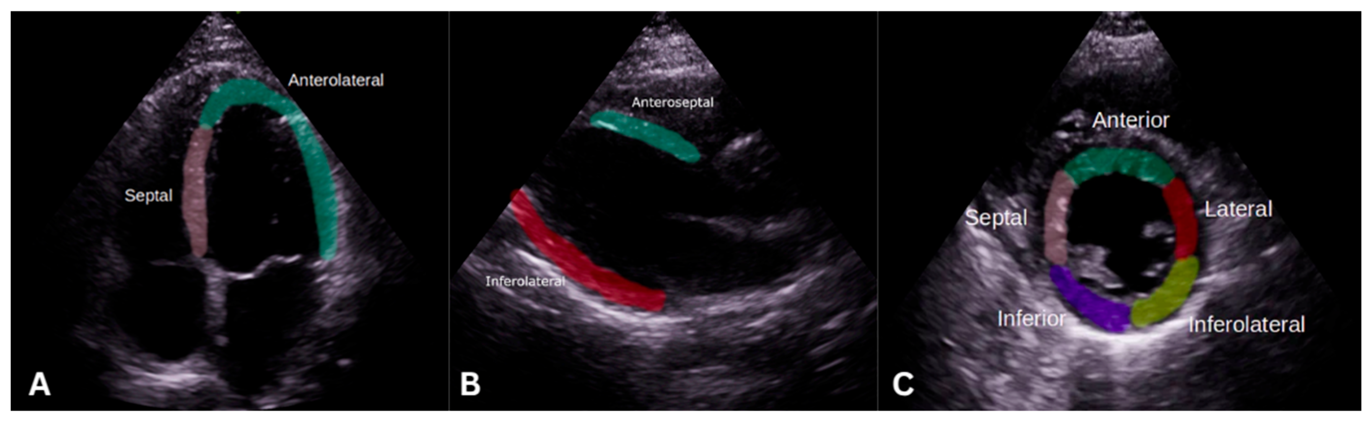
3.1. Regional Wall Motion Abnormalities
3.2. Assessment of Global Cardiac Function
4. What Are the Major Applications of FoCUS in ACS Complicated by HF and Cardiogenic Shock?
4.1. Severe Left-Ventricular Dysfunction
4.2. Right-Ventricular Failure
4.3. Mechanical Complications
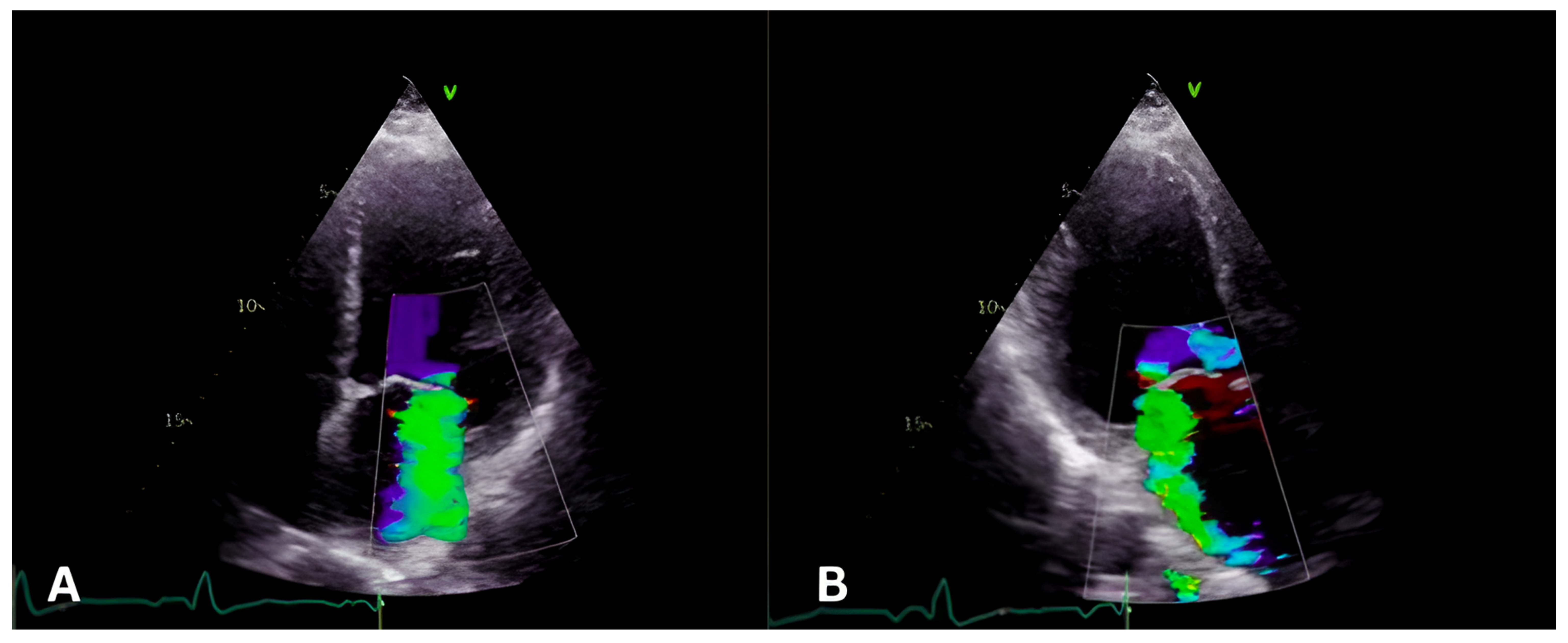
4.3.1. Papillary Muscle Rupture
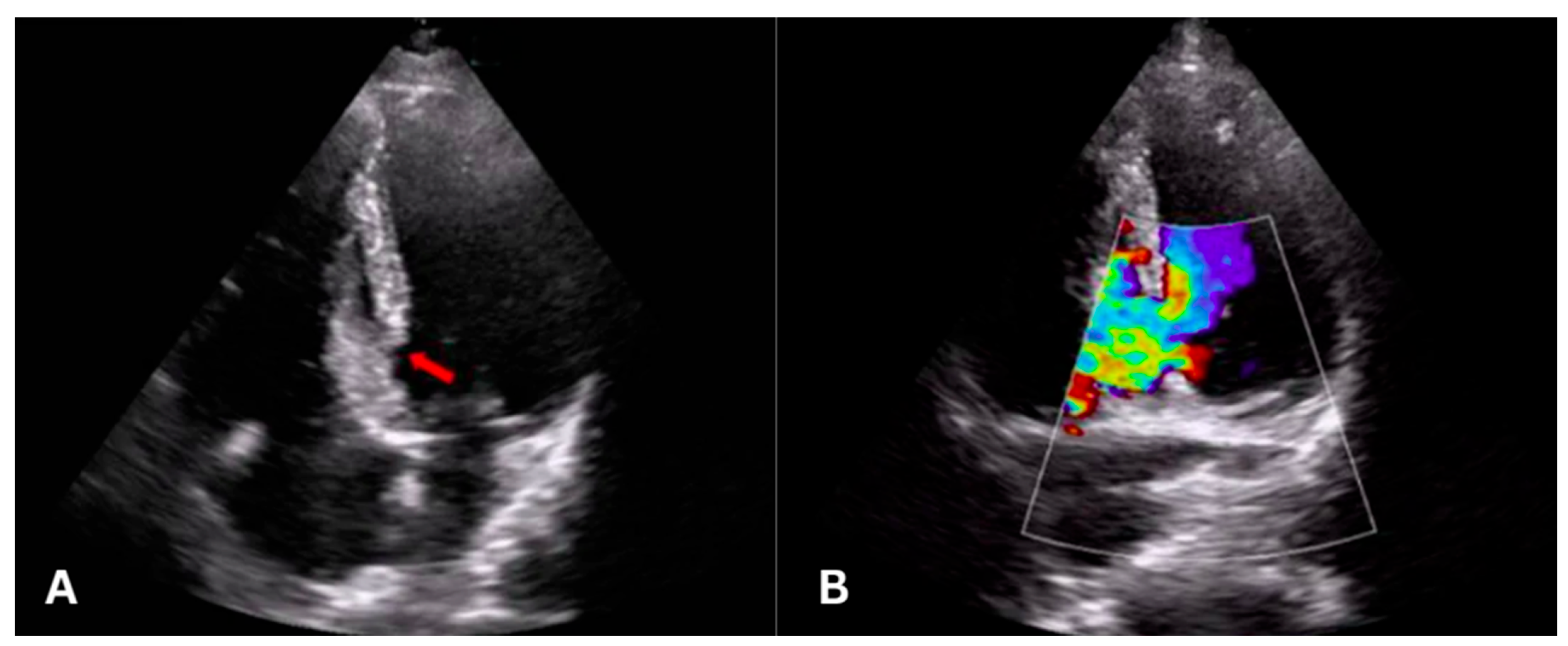
4.3.2. Ventricular Septal Rupture
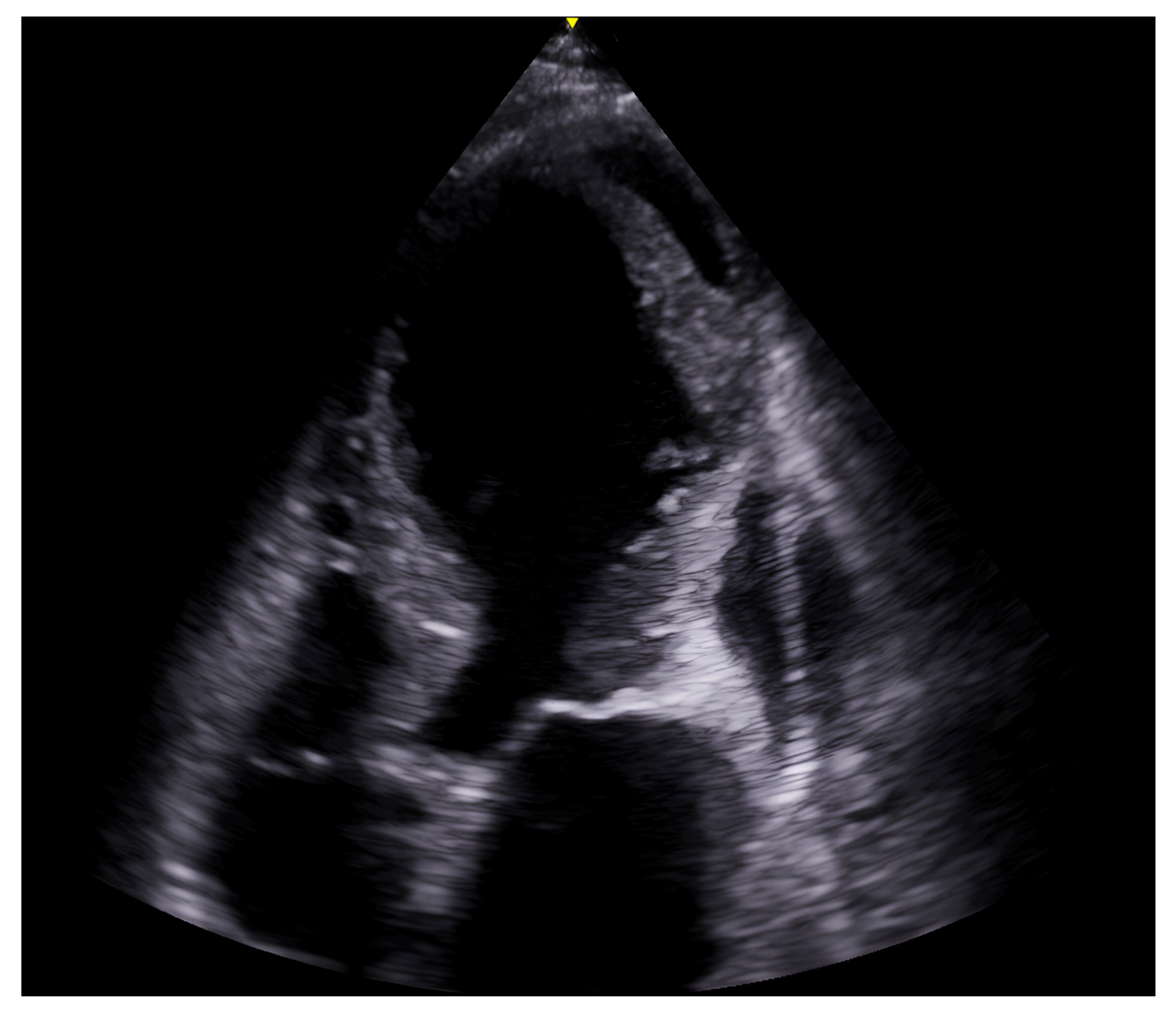
4.3.3. Free LV Rupture
5. What Are the Major Applications of FoCUS in ACS Complicated by Cardiac Arrest?
6. Can FoCUS Differentiate Other Mimickers of Chest Pain?
6.1. Takotsubo Cardiomyopathy
6.2. Acute Pericarditis
6.3. Acute Pulmonary Embolism
6.4. Cardiac Tamponade
6.5. Acute Aortic Dissection
7. Limitations and Special Considerations
8. Conclusions
Supplementary Materials
Author Contributions
Funding
Institutional Review Board Statement
Informed Consent Statement
Data Availability Statement
Conflicts of Interest
References
- Gulati, M.; Levy, P.D.; Mukherjee, D.; Amsterdam, E.; Bhatt, D.L.; Birtcher, K.K.; Blankstein, R.; Boyd, J.; Bullock-Palmer, R.P.; Conejo, T.; et al. 2021 AHA/ACC/ASE/CHEST/SAEM/SCCT/SCMR Guideline for the Evaluation and Diagnosis of Chest Pain: A Report of the American College of Cardiology/American Heart Association Joint Committee on Clinical Practice Guidelines. Circulation 2021, 144, e187–e285. [Google Scholar] [CrossRef]
- Rao, S.V.; O’Donoghue, M.L.; Ruel, M.; Rab, T.; Tamis-Holland, J.E.; Alexander, J.H.; Baber, U.; Baker, H.; Cohen, M.G.; Cruz-Ruiz, M.; et al. 2025 ACC/AHA/ACEP/NAEMSP/SCAI Guideline for the Management of Patients with Acute Coronary Syndromes: A Report of the American College of Cardiology/American Heart Association Joint Committee on Clinical Practice Guidelines. Circulation 2025, 151, e771–e862. [Google Scholar] [CrossRef]
- Harnish, P.; Nesheiwat, Z.; Mahmood, S.; Soni, R.; Eltahawy, E. Echocardiography in Detecting Mechanical Complications in Acute Coronary Syndrome. CASE 2020, 4, 393–398. [Google Scholar] [CrossRef]
- Thygesen, K.; Alpert, J.S.; Jaffe, A.S.; Chaitman, B.R.; Bax, J.J.; Morrow, D.A.; White, H.D. Executive Group on behalf of the Joint European Society of Cardiology (ESC)/American College of Cardiology (ACC)/American Heart Association (AHA)/World Heart Federation (WHF) Task Force for the Universal Definition of Myocardial Infarction. Fourth Universal Definition of Myocardial Infarction (2018). J. Am. Coll. Cardiol. 2018, 72, 2231–2264. [Google Scholar] [CrossRef]
- Verras, C.; Ventoulis, I.; Bezati, S.; Matsiras, D.; Parissis, J.; Polyzogopoulou, E. Point of Care Ultrasonography for the Septic Patient in the Emergency Department: A Literature Review. J. Clin. Med. 2023, 12, 1105. [Google Scholar] [CrossRef]
- Neskovic, A.N.; Skinner, H.; Price, S.; Via, G.; De Hert, S.; Stankovic, I.; Galderisi, M.; Donal, E.; Muraru, D.; Sloth, E.; et al. Focus cardiac ultrasound core curriculum and core syllabus of the European Association of Cardiovascular Imaging. Eur. Heart J.-Cardiovasc. Imaging 2018, 19, 475–481. [Google Scholar] [CrossRef]
- Neskovic, A.N.; Edvardsen, T.; Galderisi, M.; Garbi, M.; Gullace, G.; Jurcut, R.; Dalen, H.; Hagendorff, A.; Lancellotti, P.; for the European Association of Cardiovascular Imaging Document Reviewers; et al. Focus cardiac ultrasound: The European Association of Cardiovascular Imaging viewpoint. Eur. Heart J.-Cardiovasc. Imaging 2014, 15, 956–960. [Google Scholar] [CrossRef]
- Kirkpatrick, J.N.; Panebianco, N.; Díaz-Gómez, J.L.; Adhikari, S.; Bremer, M.L.; Bronshteyn, Y.S.; Damewood, S.; Jankowski, M.; Johri, A.; Kaplan, J.R.H.; et al. Recommendations for Cardiac Point-of-Care Ultrasound Nomenclature. J. Am. Soc. Echocardiogr. 2024, 37, 809–819. [Google Scholar] [CrossRef] [PubMed]
- Kontos, M.C. Role of Echocardiography in the Emergency Department for Identifying Patients with Myocardial Infarction and Ischemia. Echocardiography 1999, 16, 193–205. [Google Scholar] [CrossRef]
- Labovitz, A.J.; Noble, V.E.; Bierig, M.; Goldstein, S.A.; Jones, R.; Kort, S.; Porter, T.R.; Spencer, K.T.; Tayal, V.S.; Wei, K. Focused Cardiac Ultrasound in the Emergent Setting: A Consensus Statement of the American Society of Echocardiography and American College of Emergency Physicians. J. Am. Soc. Echocardiogr. 2010, 23, 1225–1230. [Google Scholar] [CrossRef] [PubMed]
- Zarama, V.; Arango-Granados, M.C.; Manzano-Nunez, R.; Sheppard, J.P.; Roberts, N.; Plüddemann, A. The diagnostic accuracy of cardiac ultrasound for acute myocardial ischemia in the emergency department: A systematic review and meta-analysis. Scand. J. Trauma. Resusc. Emerg. Med. 2024, 32, 19. [Google Scholar] [CrossRef]
- Regional Myocardial Contractile Function: Wall Motion Abnormalities. The Cardiovascular. Available online: https://ecgwaves.com/topic/regional-myocardial-contractile-function-wall-motion-abnormality/ (accessed on 15 February 2026).
- Hauser, A.M.; Gangadharan, V.; Ramos, R.G.; Gordon, S.; Timmis, G.C.; Dudlets, P. Sequence of mechanical, electrocardiographic and clinical effects of repeated coronary artery occlusion in human beings: Echocardiographic observations during coronary angioplasty. J. Am. Coll. Cardiol. 1985, 5, 193–197. [Google Scholar] [CrossRef] [PubMed]
- Yavagal, S.T.; Baliga, V.B. Non-Ischemic Regional Wall Motion Abnormality. J. Indian Acad. Echocardiogr. Cardiovasc. Imaging 2019, 3, 7–11. [Google Scholar] [CrossRef]
- Lang, R.M.; Badano, L.P.; Mor-Avi, V.; Afilalo, J.; Armstrong, A.; Ernande, L.; Flachskampf, F.A.; Foster, E.; Goldstein, S.A.; Kuznetsova, T.; et al. Recommendations for Cardiac Chamber Quantification by Echocardiography in Adults: An Update from the American Society of Echocardiography and the European Association of Cardiovascular Imaging. J. Am. Soc. Echocardiogr. 2015, 28, 1–39.e14. [Google Scholar] [CrossRef]
- Bracey, A.; Meyers, H.P.; Watkins, C.; Shroff, G.R.; Lee, D.; Singer, A.J.; Smith, S.W. Comparison of regional wall motion abnormalities in patients with occlusion myocardial infarction with and without ST-elevation myocardial infarction (STEMI). Clin. Exp. Emerg. Med. 2025, 12, 342–349. [Google Scholar] [CrossRef]
- Croft, P.E.; Strout, T.D.; Kring, R.M.; Director, L.; Vasaiwala, S.C.; Mackenzie, D.C. WAMAMI: Emergency physicians can accurately identify wall motion abnormalities in acute myocardial infarction. Am. J. Emerg. Med. 2019, 37, 2224–2228. [Google Scholar] [CrossRef]
- Sağlam, C.; Ünlüer, E.E.; Yamanoğlu, N.G.Ç.; Kara, P.H.; Ediboğlu, E.; Bektaşlı, R.; Tandon, S.; Gönüllü, H. Accuracy of Emergency Physicians for Detection of Regional Wall Motion Abnormalities in Patients with Chest Pain Without ST-Elevation Myocardial Infarction. J. Ultrasound Med. 2021, 40, 1335–1342. [Google Scholar] [CrossRef]
- Bracey, A.; Massey, L.; Pellet, A.C.; Thode, H.C.; Holman, T.R.; Singer, A.J.; McClure, M.; Secko, M.A. FOCUS may detect wall motion abnormalities in patients with ACS. Am. J. Emerg. Med. 2023, 69, 17–22. [Google Scholar] [CrossRef] [PubMed]
- Rossetto, A.; Fabiani, G.; Bima, P.; Morello, F.; Mueller, C.; Nazerian, P.; WINFOCUS Study Group. Integration of focused cardiac ultrasound in the HEAR and HEART scores in emergency department patients with chest pain. Intern. Emerg. Med. 2026. [Google Scholar] [CrossRef] [PubMed]
- Bima, P.; Agishev, I.; Fucile, I.; De Stefano, G.; Morello, F.; Mueller, C.; Nazerian, P. on behalf of the WINFOCUS Study Group. Diagnostic accuracy of point-of-care cardiac ultrasound for acute coronary syndromes in patients presenting with chest pain to the emergency department: A single-center prospective study. Eur. J. Emerg. Med. 2025, 32, 38–45. [Google Scholar] [CrossRef]
- Spencer, K.T.; Kimura, B.J.; Korcarz, C.E.; Pellikka, P.A.; Rahko, P.S.; Siegel, R.J. Focused Cardiac Ultrasound: Recommendations from the American Society of Echocardiography. J. Am. Soc. Echocardiogr. 2013, 26, 567–581. [Google Scholar] [CrossRef] [PubMed]
- Xu, C.; Melendez, A.; Nguyen, T.; Ellenberg, J.; Anand, A.; Delgado, J.; Herbst, M.K. Point-of-care ultrasound may expedite diagnosis and revascularization of occult occlusive myocardial infarction. Am. J. Emerg. Med. 2022, 58, 186–191. [Google Scholar] [CrossRef]
- Roggel, A.; Jehn, S.; Dykun, I.; Balcer, B.; Al-Rashid, F.; Totzeck, M.; Risse, J.; Kill, C.; Rassaf, T.; Mahabadi, A. Regional wall motion abnormalities on focused transthoracic echocardiography in patients presenting with acute chest pain: A predefined post hoc analysis of the prospective single-centre observational EPIC-ACS study. BMJ Open 2024, 14, e085677. [Google Scholar] [CrossRef]
- Global Ultrasound Institute. Left Ventricular Function. Available online: https://globalultrasoundinstitute.com/pocuspedia/lv-function/ (accessed on 1 May 2026).
- Marbach, J.A.; Almufleh, A.; Di Santo, P.; Jung, R.; Simard, T.; McInnes, M.; Salameh, J.-P.; McGrath, T.A.; Millington, S.J.; Diemer, G.; et al. Comparative Accuracy of Focused Cardiac Ultrasonography and Clinical Examination for Left Ventricular Dysfunction and Valvular Heart Disease: A Systematic Review and Meta-analysis. Ann. Intern. Med. 2019, 171, 264–272. [Google Scholar] [CrossRef]
- Ünlüer, E.; Karagöz, A.; Akoğlu, H.; Bayata, S. Visual Estimation of Bedside Echocardiographic Ejection Fraction by Emergency Physicians. West. J. Emerg. Med. 2014, 15, 221–226. [Google Scholar] [CrossRef] [PubMed]
- Kadloor, P.; Hidayathullah, M.; Golla, A. Reduced left ventricular ejection fraction in patients with acute coronary syndrome as a risk factor for mortality. Int. J. Res. Med. Sci. 2021, 10, 80–85. [Google Scholar] [CrossRef]
- Kusunose, K.; Zheng, R.; Yamada, H.; Sata, M. How to standardize the measurement of left ventricular ejection fraction. J. Med. Ultrason. 2022, 49, 35–43. [Google Scholar] [CrossRef]
- Gohar, E.; Herling, A.; Mazuz, M.; Tsaban, G.; Gat, T.; Kobal, S.; Fuchs, L. Artificial Intelligence (AI) versus POCUS Expert: A Validation Study of Three Automatic AI-Based, Real-Time, Hemodynamic Echocardiographic Assessment Tools. J. Clin. Med. 2023, 12, 1352. [Google Scholar] [CrossRef]
- Lin, X.; Yang, F.; Chen, Y.; Chen, X.; Wang, W.; Chen, X.; Wang, Q.; Zhang, L.; Guo, H.; Liu, B.; et al. Echocardiography-based AI detection of regional wall motion abnormalities and quantification of cardiac function in myocardial infarction. Front. Cardiovasc. Med. 2022, 9, 903660. [Google Scholar] [CrossRef]
- Kim, J.; Maranna, S.; Watson, C.; Parange, N. A scoping review on the integration of artificial intelligence in point-of-care ultrasound: Current clinical applications. Am. J. Emerg. Med. 2025, 92, 172–181. [Google Scholar] [CrossRef]
- Zako, J.; Wang, S.S.; Pintilei, T.; Burey, J.; Hemmerling, T.M.; Morisson, L.; Denault, A.; Laferrière-Langlois, P. Artificial Intelligence–Assisted Point-of-Care Ultrasound for Evaluating Left Ventricular Ejection Fraction: A Systematic Review of Prospective Observational Studies. J. Cardiothorac. Vasc. Anesth. 2026; in press. [CrossRef]
- Byrne, R.A.; Rossello, X.; Coughlan, J.J.; Barbato, E.; Berry, C.; Chieffo, A.; Claeys, M.J.; Dan, G.-A.; Dweck, M.R.; Galbraith, M.; et al. 2023 ESC Guidelines for the management of acute coronary syndromes. Eur. Heart J. 2023, 44, 3720–3826. [Google Scholar] [CrossRef]
- Osawa, T.; Nakayama, N.; Ishizu, T.; Kondo, T.; Nakashima, T.; Yamamoto, T.; Hanada, H.; Hashiba, K.; Kirigaya, J.; Hosoya, Y.; et al. Diagnostic Accuracy of Point-of-Care Ultrasound for Patients with Cardiogenic Shock—A Meta-Analysis and Systematic Review. Circ. Rep. 2025, 7, 727–734. [Google Scholar] [CrossRef] [PubMed]
- Wainstein, M.V.; Machado, G.P.; Telo, G.H.; Silveira, A.D.D.; Nasi, L.A.; Araujo, G.N.D. Correction: Point-of-care ultrasound in the evaluation of STEMI patients. Front. Cardiovasc. Med. 2025, 12, 1698664. [Google Scholar] [CrossRef] [PubMed]
- Soliman Aboumarie, H.; Tavazzi, G.; Via, G.; Guarracino, F.; Stankovic, I.; Hagendorff, A.; Gargani, L.; Cardim, N.; Flachskampf, F.A.; Bax, J.; et al. Cardiac ultrasound in cardiovascular emergency and critical care: A clinical consensus statement of the European Association of Cardiovascular Imaging, the Acute CardioVascular Care Association of the European Society of Cardiology, and the European Association of Cardiothoracic Anaesthesia and Intensive Care. Eur. Heart J.-Cardiovasc. Imaging 2025, 26, 1699–1724. [Google Scholar] [CrossRef]
- Parker, C.W.; Kolimas, A.M.; Kotini-Shah, P. Velocity-Time Integral: A Bedside Echocardiography Technique Finding a Place in the Emergency Department. J. Emerg. Med. 2022, 63, 382–388. [Google Scholar] [CrossRef]
- Fellows’ Section: Diastology at the Bedside. Available online: https://www.acep.org/emultrasound/newsroom/mar2020/fellows-section-diastology-at-the-bedside (accessed on 14 April 2026).
- Arnone, M.I.; Sforza, A.; Carlino, M.V.; Guarino, M.; Candido, R.; Bertolone, D.; Fucile, I.; De Luca, N.; Mancusi, C. Assessment of E/A ratio helps emergency clinicians in the management of patients with acute dyspnea. Intern. Emerg. Med. 2023, 18, 1823–1830. [Google Scholar] [CrossRef]
- Søholm, H.; Lønborg, J.; Andersen, M.J.; Vejlstrup, N.; Engstrøm, T.; Hassager, C.; Møller, J.E. Association diastolic function by echo and infarct size by magnetic resonance imaging after STEMI. Scand. Cardiovasc. J. 2016, 50, 172–179. [Google Scholar] [CrossRef]
- Mitchell, C.; Rahko, P.S.; Blauwet, L.A.; Canaday, B.; Finstuen, J.A.; Foster, M.C.; Horton, K.; Ogunyankin, K.O.; Palma, R.A.; Velazquez, E.J. Guidelines for Performing a Comprehensive Transthoracic Echocardiographic Examination in Adults: Recommendations from the American Society of Echocardiography. J. Am. Soc. Echocardiogr. 2019, 32, 1–64. [Google Scholar] [CrossRef]
- Spencer, K.T.; Flachskampf, F.A. Focused Cardiac Ultrasonography. JACC Cardiovasc. Imaging 2019, 12, 1243–1253. [Google Scholar] [CrossRef] [PubMed]
- Betcher, J.; Majkrzak, A.; Cranford, J.; Kessler, R.; Theyyunni, N.; Huang, R. Feasibility study of advanced focused cardiac measurements within the emergency department. Crit. Ultrasound J. 2018, 10, 10. [Google Scholar] [CrossRef]
- Nägele, M.P.; Flammer, A.J. Heart Failure After Right Ventricular Myocardial Infarction. Curr. Heart Fail. Rep. 2022, 19, 375–385. [Google Scholar] [CrossRef]
- Femia, G.; French, J.K.; Juergens, C.; Leung, D.; Lo, S. Right ventricular myocardial infarction: Pathophysiology, clinical implications and management. Rev. Cardiovasc. Med. 2021, 22, 1229–1240. [Google Scholar] [CrossRef]
- Brenner, D.S.; Ilonze, O.J.; Bell, S.; Kaneshiro, K.; Harrison, N.E. Emergency department management of patients with right heart failure. Emerg. Med. Pract. 2024, 26, 1–32. [Google Scholar] [PubMed]
- POCUS 101. Right Ventricular Ultrasound-Qualitative and Quantitative Assessments. Available online: https://www.pocus101.com/right-ventricular-ultrasound-qualitative-and-quantitative-assessments/ (accessed on 17 January 2026).
- Daley, J.; Grotberg, J.; Pare, J.; Medoro, A.; Liu, R.; Hall, M.K.; Taylor, A.; Moore, C.L. Emergency physician performed tricuspid annular plane systolic excursion in the evaluation of suspected pulmonary embolism. Am. J. Emerg. Med. 2017, 35, 106–111. [Google Scholar] [CrossRef]
- Gong, F.F.; Vaitenas, I.; Malaisrie, S.C.; Maganti, K. Mechanical Complications of Acute Myocardial Infarction: A Review. JAMA Cardiol. 2021, 6, 341–349. [Google Scholar] [CrossRef] [PubMed]
- Murphy, A.; Goldberg, S. Mechanical Complications of Myocardial Infarction. Am. J. Med. 2022, 135, 1401–1409. [Google Scholar] [CrossRef] [PubMed]
- Watanabe, N. Acute mitral regurgitation. Heart 2019, 105, 671–677. [Google Scholar] [CrossRef]
- Pino, P.G.; Parrini, I.; Moreo, A.; Terranova, A.; Nardi, F. Echocardiographic Management of papillary muscle rupture during acute myocardial infarction. Explor. Cardiol. 2025, 3, 101242. [Google Scholar] [CrossRef]
- Ternus, B.W.; Mankad, S.; Edwards, W.D.; Mankad, R. Clinical presentation and echocardiographic diagnosis of postinfarction papillary muscle rupture: A review of 22 cases. Echocardiography 2017, 34, 973–977. [Google Scholar] [CrossRef]
- Stenberg, R.; Bowling, J.; Jacquet, J.; Watkins, K.; Eggleston, J.; Hill, A.; Krizo, J. Point-of-care ultrasound diagnosis of acute valvular emergencies. Am. J. Emerg. Med. 2025, 89, 36–50. [Google Scholar] [CrossRef]
- Viana-Tejedor, A.; Ferrera, C.; Estévez-Loureiro, R.; Barreiro-Pérez, M.; Jiménez Quevedo, P.; Nombela-Franco, L.; Jorge-Pérez, P.; Pascual, I.; Martínez Monzonís, A.; Cid Álvarez, A.B. Edge-to-edge therapy in acute mitral regurgitation. Proposal for a management protocol of the Ischemic Heart Disease and Acute Cardiac Care, Interventional Cardiology, and Cardiovascular Imaging Associations of the Spanish Society of Cardiology. REC Interv. Cardiol. 2024, 6, 313–320. [Google Scholar] [CrossRef]
- Li, S.; Fang, L.; Wu, W.; Zhang, Z.; Ji, L.; Sun, Z.; He, L.; Wang, Z.; Fu, W.; Li, F.; et al. Echocardiographic diagnosis of rupture of mitral valve papillary muscle. Int. J. Cardiol. 2023, 391, 131273. [Google Scholar] [CrossRef]
- Colombo, P.C.; Wu, R.H.; Weiner, S.; Marinaccio, M.; Brofferio, A.; Banchs, J.; Malla, S.; Frater, R.; Shirani, J.; Nanna, M. Value of quantitative analysis of mitral regurgitation jet eccentricity by color flow Doppler for identification of flail leaflet. Am. J. Cardiol. 2001, 88, 534–540. [Google Scholar] [CrossRef] [PubMed]
- Damluji, A.A.; Van Diepen, S.; Katz, J.N.; Menon, V.; Tamis-Holland, J.E.; Bakitas, M.; Cohen, M.G.; Balsam, L.B.; Chikwe, J.; on behalf of the American Heart Association Council on Clinical Cardiology; et al. Mechanical Complications of Acute Myocardial Infarction: A Scientific Statement from the American Heart Association. Circulation 2021, 144, e16–e35. [Google Scholar] [CrossRef] [PubMed]
- Birnbaum, Y.; Wagner, G.S.; Gates, K.B.; Thompson, T.D.; Barbash, G.I.; Siegel, R.J.; Granger, C.B.; Fishbein, M.C.; Crenshaw, B.S.; Califf, R.M. Clinical and electrocardiographic variables associated with increased risk of ventricular septal defect in acute anterior myocardial infarction. Am. J. Cardiol. 2000, 86, 830–834. [Google Scholar] [CrossRef] [PubMed]
- Cubeddu, R.J.; Lorusso, R.; Ronco, D.; Matteucci, M.; Axline, M.S.; Moreno, P.R. Ventricular Septal Rupture After Myocardial Infarction: JACC Focus Seminar 3/5. J. Am. Coll. Cardiol. 2024, 83, 1886–1901. [Google Scholar] [CrossRef]
- Skehan, J.D.; Carey, C.; Norrell, M.S.; de Belder, M.; Balcon, R.; Mills, P.G. Patterns of coronary artery disease in post-infarction ventricular septal rupture. Br. Heart J. 1989, 62, 268–272. [Google Scholar] [CrossRef]
- Vargas-Barrón, J.; Molina-Carrión, M.; Romero-Cárdenas, Á.; Roldán, F.-J.; Medrano, G.A.; Ávila-Casado, C.; Martínez-Ríos, M.A.; Lupi-Herrera, E.; Zabalgoitia, M. Risk Factors, Echocardiographic Patterns, and Outcomes in Patients with Acute Ventricular Septal Rupture During Myocardial Infarction. Am. J. Cardiol. 2005, 95, 1153–1158. [Google Scholar] [CrossRef]
- Birnbaum, Y.; Fishbein, M.C.; Blanche, C.; Siegel, R.J. Ventricular septal rupture after acute myocardial infarction. N. Engl. J. Med. 2002, 347, 1426–1432. [Google Scholar] [CrossRef]
- Bishop, H.L.; Gibson, R.S.; Stamm, R.B.; Beller, G.A.; Martin, R.P. Role of two-dimensional echocardiography in the evaluation of patients with ventricular septal rupture postmyocardial infarction. Am. Heart J. 1981, 102, 965–971. [Google Scholar] [CrossRef]
- Smyllie, J.H.; Sutherland, G.R.; Geuskens, R.; Dawkins, K.; Conway, N.; Roelandt, J.R. Doppler color flow mapping in the diagnosis of ventricular septal rupture and acute mitral regurgitation after myocardial infarction. J. Am. Coll. Cardiol. 1990, 15, 1449–1455. [Google Scholar] [CrossRef]
- Lee, B.W.; Cha, Y.S.; Hwang, S.O.; Kim, Y.-S.; Kim, S.J. Echocardiographic features of myocardial rupture after acute myocardial infarction on emergency echocardiography. Clin. Exp. Emerg. Med. 2023, 10, 393–399. [Google Scholar] [CrossRef]
- Bansal, R.C.; Eng, A.K.; Shakudo, M. Role of two-dimensional echocardiography, pulsed, continuous wave and color flow Doppler techniques in the assessment of ventricular septal rupture after myocardial infarction. Am. J. Cardiol. 1990, 65, 852–860. [Google Scholar] [CrossRef]
- Fortin, D.F.; Sheikh, K.H.; Kisslo, J. The utility of echocardiography in the diagnostic strategy of postinfarction ventricular septal rupture: A comparison of two-dimensional echocardiography versus Doppler color flow imaging. Am. Heart J. 1991, 121, 25–32. [Google Scholar] [CrossRef] [PubMed]
- Formica, F.; Mariani, S.; Singh, G.; D’Alessandro, S.; Messina, L.A.; Jones, N.; Bamodu, O.A.; Sangalli, F.; Paolini, G. Postinfarction left ventricular free wall rupture: A 17-year single-centre experience. Eur. J. Cardiothorac. Surg. 2018, 53, 150–156. [Google Scholar] [CrossRef]
- Plummer, D.; Dick, C.; Ruiz, E.; Clinton, J.; Brunette, D. Emergency department two-dimensional echocardiography in the diagnosis of nontraumatic cardiac rupture. Ann. Emerg. Med. 1994, 23, 1333–1342. [Google Scholar] [CrossRef]
- Pollak, H.; Diez, W.; Spiel, R.; Enenkel, W.; Mlczoch, J. Early diagnosis of subacute free wall rupture complicating acute myocardial infarction. Eur. Heart J. 1993, 14, 640–648. [Google Scholar] [CrossRef] [PubMed]
- López-Sendón, J.; González, A.; López de Sá, E.; Coma-Canella, I.; Roldán, I.; Domínguez, F.; Maqueda, I.; Martín Jadraque, L. Diagnosis of subacute ventricular wall rupture after acute myocardial infarction: Sensitivity and specificity of clinical, hemodynamic and echocardiographic criteria. J. Am. Coll. Cardiol. 1992, 19, 1145–1153. [Google Scholar] [CrossRef] [PubMed]
- Long, B.; Alerhand, S.; Maliel, K.; Koyfman, A. Echocardiography in cardiac arrest: An emergency medicine review. Am. J. Emerg. Med. 2018, 36, 488–493. [Google Scholar] [CrossRef]
- Long, B.; Adrian, R.J.; Gottlieb, M. Emergency medicine updates: Point-of-care ultrasound in cardiac arrest. Am. J. Emerg. Med. 2026, 99, 176–187. [Google Scholar] [CrossRef]
- Andrus, P.; Dean, A. Focused cardiac ultrasound. Glob. Heart 2013, 8, 299–303. [Google Scholar] [CrossRef]
- Zaki, H.A.; Iftikhar, H.; Shaban, E.E.; Najam, M.; Alkahlout, B.H.; Shallik, N.; Elnabawy, W.; Basharat, K.; Azad, A.M. The role of point-of-care ultrasound (POCUS) imaging in clinical outcomes during cardiac arrest: A systematic review. Ultrasound J. 2024, 16, 4. [Google Scholar] [CrossRef]
- Roshanzamir, S.; Showkathali, R. Takotsubo cardiomyopathy a short review. Curr. Cardiol. Rev. 2013, 9, 191–196. [Google Scholar] [CrossRef] [PubMed]
- Madhavan, M.; Prasad, A. Proposed Mayo Clinic criteria for the diagnosis of Tako-Tsubo cardiomyopathy and long-term prognosis. Herz 2010, 35, 240–243. [Google Scholar] [CrossRef]
- Meigh, K.; Caja, M.; Sharon, M.; Tadros, A.; Dragan, S.; Henkel, D.; Minardi, J. Takotsubo Cardiomyopathy in the Emergency Department: A FOCUS Heart Breaker. Clin. Pract. Cases Emerg. Med. 2018, 2, 158–162. [Google Scholar] [CrossRef] [PubMed][Green Version]
- Citro, R.; Okura, H.; Ghadri, J.R.; Izumi, C.; Meimoun, P.; Izumo, M.; Dawson, D.; Kaji, S.; Eitel, I.; Kagiyama, N.; et al. Multimodality imaging in takotsubo syndrome: A joint consensus document of the European Association of Cardiovascular Imaging (EACVI) and the Japanese Society of Echocardiography (JSE). Eur. Heart J.-Cardiovasc. Imaging 2020, 21, 1184–1207. [Google Scholar] [CrossRef] [PubMed]
- Wang, T.K.M.; Klein, A.L.; Cremer, P.C.; Imazio, M.; Kohnstamm, S.; Luis, S.A.; Mardigyan, V.; Mukherjee, M.; Ordovas, K.; Vakamudi, S.; et al. 2025 Concise Clinical Guidance: An ACC Expert Consensus Statement on the Diagnosis and Management of Pericarditis. J. Am. Coll. Cardiol. 2025, 86, 2691–2719. [Google Scholar] [CrossRef]
- Chiabrando, J.G.; Bonaventura, A.; Vecchié, A.; Wohlford, G.F.; Mauro, A.G.; Jordan, J.H.; Grizzard, J.D.; Montecucco, F.; Berrocal, D.H.; Brucato, A.; et al. Management of Acute and Recurrent Pericarditis. J. Am. Coll. Cardiol. 2020, 75, 76–92. [Google Scholar] [CrossRef]
- Mandavia, D.P.; Hoffner, R.J.; Mahaney, K.; Henderson, S.O. Bedside echocardiography by emergency physicians. Ann. Emerg. Med. 2001, 38, 377–382. [Google Scholar] [CrossRef]
- Balderston, J.R.; Gertz, Z.M.; Brooks, S.; Joyce, J.M.; Evans, D.P. Diagnostic Yield and Accuracy of Bedside Echocardiography in the Emergency Department in Hemodynamically Stable Patients. J. Ultrasound Med. 2019, 38, 2845–2851. [Google Scholar] [CrossRef]
- Bustam, A.; Noor Azhar, M.; Singh Veriah, R.; Arumugam, K.; Loch, A. Performance of emergency physicians in point-of-care echocardiography following limited training. Emerg. Med. J. 2014, 31, 369–373. [Google Scholar] [CrossRef]
- Barcellos Amon, A.; Pinheiro Machado, G.; Heiden Telo, G.; Petersen Saadi, M.; Azeredo Azevedo, W.T.; Martins, A.; Nassif, M.; Neves de Araújo, G.; Scolari, F.L.; Donelli da Silveira, A.; et al. Clinical impact of a cardiothoracic ultrasound protocol in patients with ST-segment elevation myocardial infarction: The focused assessment in STEMI (FASTEMI) protocol. Intern. Emerg. Med. 2025. [Google Scholar] [CrossRef]
- Kukla, P.; Długopolski, R.; Krupa, E.; Furtak, R.; Mirek-Bryniarska, E.; Szełemej, R.; Jastrzębski, M.; Nowak, J.; Kulak, L.; Hybel, J.; et al. How often pulmonary embolism mimics acute coronary syndrome? Kardiol. Pol. 2011, 69, 235–240. [Google Scholar]
- Zaki, H.A.; Ponappan, B.; Shaban, E.E.; Shaban, A.; Shaban, A.; Khan, A.; Basharat, K.; Abosamak, M.F. Enhancing rapid diagnosis of pulmonary embolism in the emergency department: The critical role of point-of-care ultrasound (POCUS)—A systematic review and meta-analysis. Egypt. J. Radiol. Nucl. Med. 2025, 56, 35. [Google Scholar] [CrossRef]
- Ginghina, C.; Caloianu, G.-A.; Serban, M.; Dragomir, D. Right ventricular myocardial infarction and pulmonary embolism differential diagnosis—A challenge for the clinician. J. Med. Life 2010, 3, 242–253. [Google Scholar] [PubMed]
- Rafie, N.; Foley, D.A.; Ripoll, J.G.; Booth-Kowalczyk, M.L.; Arghami, A.; Pochettino, A.; Michelena, H.I. McConnell’s Sign Is Not Always Pulmonary Embolism: The Importance of Right Ventricular Ischemia. JACC Case Rep. 2022, 4, 802–807. [Google Scholar] [CrossRef]
- Alerhand, S.; Adrian, R.J.; Long, B.; Avila, J. Pericardial tamponade: A comprehensive emergency medicine and echocardiography review. Am. J. Emerg. Med. 2022, 58, 159–174. [Google Scholar] [CrossRef] [PubMed]
- Stolz, L.; Valenzuela, J.; Situ-LaCasse, E.; Stolz, U.; Hawbaker, N.; Thompson, M.; Adhikari, S. Clinical and historical features of emergency department patients with pericardial effusions. World J. Emerg. Med. 2017, 8, 29–33. [Google Scholar] [CrossRef]
- Ienghong, K.; Cheung, L.W.; Tiamkao, S.; Bhudhisawasdi, V.; Apiratwarakul, K. The diagnostic capabilities of the combined cardiac and lung point of care ultrasound in shocked patients at the emergency department—Resourced limited country. Eur. J. Radiol. Open 2022, 9, 100446. [Google Scholar] [CrossRef]
- Alerhand, S.; Carter, J.M. What echocardiographic findings suggest a pericardial effusion is causing tamponade? Am. J. Emerg. Med. 2019, 37, 321–326. [Google Scholar] [CrossRef] [PubMed]
- Hoch, V.C.; Abdel-Hamid, M.; Liu, J.; Hall, A.E.; Theyyunni, N.; Fung, C.M. ED point-of-care ultrasonography is associated with earlier drainage of pericardial effusion: A retrospective cohort study. Am. J. Emerg. Med. 2022, 60, 156–163. [Google Scholar] [CrossRef]
- Wang, J.-L.; Chen, C.-C.; Wang, C.-Y.W.; Hsieh, M.-J.; Chang, S.-H.; Lee, C.-H.; Chen, D.-Y.; Hsieh, I.-C. Acute Type A Aortic Dissection Presenting as ST-Segment Elevation Myocardial Infarction Referred for Primary Percutaneous Coronary Intervention. Acta Cardiol. Sin. 2016, 32, 265–272. [Google Scholar] [CrossRef] [PubMed]
- Cannesson, M.; Burckard, E.; Lefèvre, M.; Bastien, O.; Lehot, J.-J. Predictors of in-hospital mortality in the surgical management of acute type A aortic dissections: Impact of anticoagulant therapies. Ann. Fr. Anesth. Reanim. 2004, 23, 568–574. [Google Scholar] [CrossRef]
- Nishigami, K. Use of aortic point-of-care ultrasound in conventional and emergent echocardiography. J. Med. Ultrasound 2022, 49, 655–661. [Google Scholar] [CrossRef]
- Nazerian, P.; Vanni, S.; Castelli, M.; Morello, F.; Tozzetti, C.; Zagli, G.; Giannazzo, G.; Vergara, R.; Grifoni, S. Diagnostic performance of emergency transthoracic focus cardiac ultrasound in suspected acute type A aortic dissection. Intern. Emerg. Med. 2014, 9, 665–670. [Google Scholar] [CrossRef]
- Mani, N.; Cherian, N.; Burkert, J.; Jarman, R.D. Bedside-focused transthoracic echocardiography in acute atraumatic thoracic aortic syndrome: A systematic review and meta-analysis of diagnostic accuracy. Eur. J. Emerg. Med. 2024, 31, 398–412. [Google Scholar] [CrossRef]
- Pare, J.R.; Liu, R.; Moore, C.L.; Sherban, T.; Kelleher, M.S.; Thomas, S.; Taylor, R.A. Emergency physician focused cardiac ultrasound improves diagnosis of ascending aortic dissection. Am. J. Emerg. Med. 2016, 34, 486–492. [Google Scholar] [CrossRef]
- Marbach, J.A.; Almufleh, A.; Di Santo, P.; Simard, T.; Jung, R.; Diemer, G.; West, F.M.; Millington, S.J.; Mathew, R.; Le May, M.R.; et al. A Shifting Paradigm: The Role of Focused Cardiac Ultrasound in Bedside Patient Assessment. Chest 2020, 158, 2107–2118. [Google Scholar] [CrossRef] [PubMed]
- Gibson, L.E.; White-Dzuro, G.A.; Lindsay, P.J.; Berg, S.M.; Bittner, E.A.; Chang, M.G. Ensuring competency in focused cardiac ultrasound: A systematic review of training programs. J. Intensive Care 2020, 8, 93. [Google Scholar] [CrossRef]
- Yamada, H.; Ito, H.; Fujiwara, M. Cardiac and vascular point-of-care ultrasound: Current situation, problems, and future prospects. J. Med. Ultrasound 2022, 49, 601–608. [Google Scholar] [CrossRef] [PubMed]
- Lenk, T.; Whittle, J.; Miller, T.E.; Williams, D.G.A.; Bronshteyn, Y.S. Focused cardiac ultrasound in preoperative assessment: The perioperative provider’s new stethoscope? Perioper. Med. 2019, 8, 16. [Google Scholar] [CrossRef]
- Ultrasound Guidelines: Emergency, Point-of-Care, and Clinical Ultrasound Guidelines in Medicine. Ann. Emerg. Med. 2023, 82, e115–e155. [CrossRef] [PubMed]
- Eppel, F.; Hunstig, F.; Bélard, S.; Kreuels, B. Concepts for point-of-care ultrasound training in low resource settings: A scoping review. Ultrasound J. 2025, 17, 24. [Google Scholar] [CrossRef]
- Saadi, M.P.; Telo, G.H.; Rasmeehirun, P.; Donal, E. The rise of point-of-care ultrasound in cardiopulmonary diagnostics. Eur. Heart J. Imaging Methods Pract. 2026, 4, qyaf147. [Google Scholar] [CrossRef] [PubMed]



| ACS | Key Findings | Limitations |
| Regional Wall Motion Abnormalities | Segmental impaired contractility | Previous MI Non-ischemic cause (focal myocarditis, Takotsubo cardiomyopathy) Cannot be used to rule out ACS |
| Reduced LV Ejection Fraction | “Eye ball” method Global LV impaired contractility | Chronic left-ventricular heart failure |
| Reduced RV Ejection Fraction | Impaired RV contractility RV dilation TAPSE < 17 mm Tricuspid valve regurgitation | Chronic right-ventricular heart failure |
| ACS with Mechanical Complications | Key Findings | Limitations |
| Papillary Muscle Rupture | Severe MR findings (color Doppler) Flail leaflet LV wall motion abnormalities | Chronic MR of non-ischemic cause |
| LV Septal Rupture | Septal wall defect (color Doppler) | B-mode alone—poor accuracy |
| LV Free Rupture | Pericardial effusion/Tamponade LV wall defect |
Disclaimer/Publisher’s Note: The statements, opinions and data contained in all publications are solely those of the individual author(s) and contributor(s) and not of MDPI and/or the editor(s). MDPI and/or the editor(s) disclaim responsibility for any injury to people or property resulting from any ideas, methods, instructions or products referred to in the content. |
© 2026 by the authors. Published by MDPI on behalf of the Lithuanian University of Health Sciences. Licensee MDPI, Basel, Switzerland. This article is an open access article distributed under the terms and conditions of the Creative Commons Attribution (CC BY) license.
Share and Cite
Karaolia, M.; Bezati, S.; Papasolomou, K.; Kiouri, E.; Verras, C.; Polyzogopoulou, E. Can FoCUS Speed Up the Management of Acute Coronary Syndrome in the Emergency Department? Medicina 2026, 62, 1013. https://doi.org/10.3390/medicina62061013
Karaolia M, Bezati S, Papasolomou K, Kiouri E, Verras C, Polyzogopoulou E. Can FoCUS Speed Up the Management of Acute Coronary Syndrome in the Emergency Department? Medicina. 2026; 62(6):1013. https://doi.org/10.3390/medicina62061013
Chicago/Turabian StyleKaraolia, Melina, Sofia Bezati, Katerina Papasolomou, Estela Kiouri, Christos Verras, and Effie Polyzogopoulou. 2026. "Can FoCUS Speed Up the Management of Acute Coronary Syndrome in the Emergency Department?" Medicina 62, no. 6: 1013. https://doi.org/10.3390/medicina62061013
APA StyleKaraolia, M., Bezati, S., Papasolomou, K., Kiouri, E., Verras, C., & Polyzogopoulou, E. (2026). Can FoCUS Speed Up the Management of Acute Coronary Syndrome in the Emergency Department? Medicina, 62(6), 1013. https://doi.org/10.3390/medicina62061013