The Overlap Between Crohn’s Disease and Intestinal Tuberculosis: A Never-Ending Story

 ,
, 
 ,
,
Abstract
1. Introduction
2. Epidemiological Trend and Misdiagnosis Rates Between CD and ITB
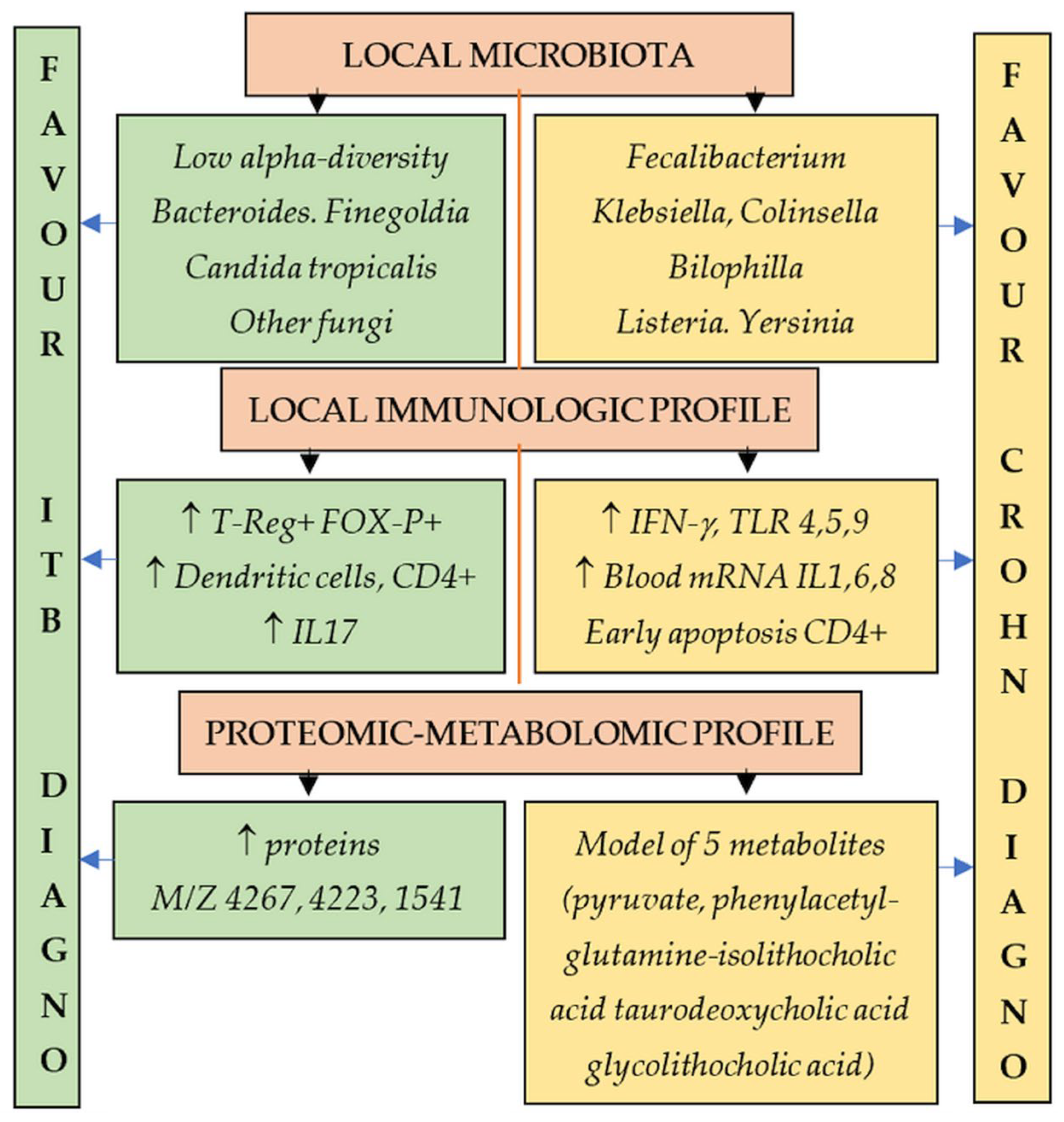
3. Microbiome, Immunology, and Proteomic Particularities
4. Clinical Pattern in CD and ITB
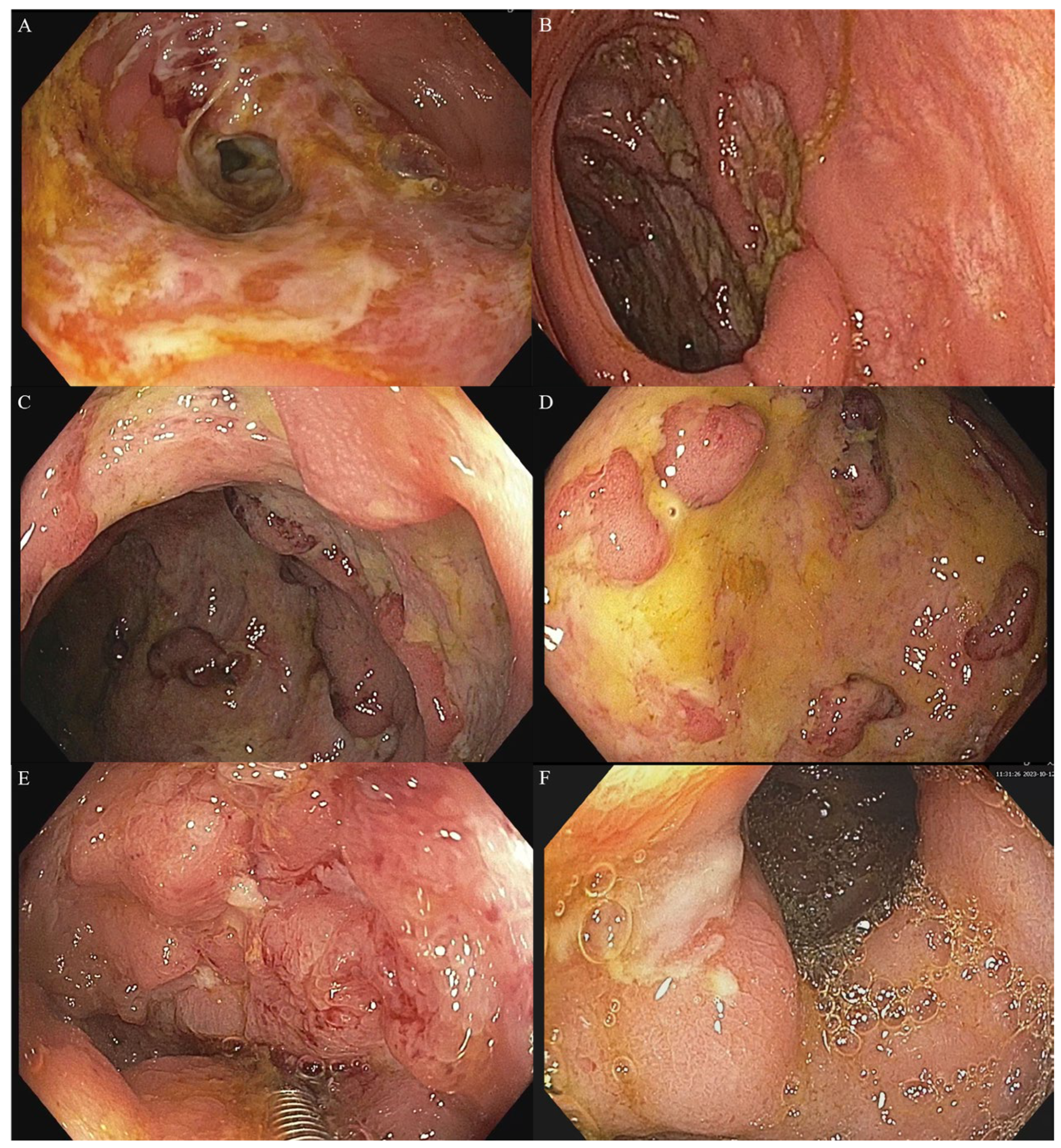
5. Endoscopy
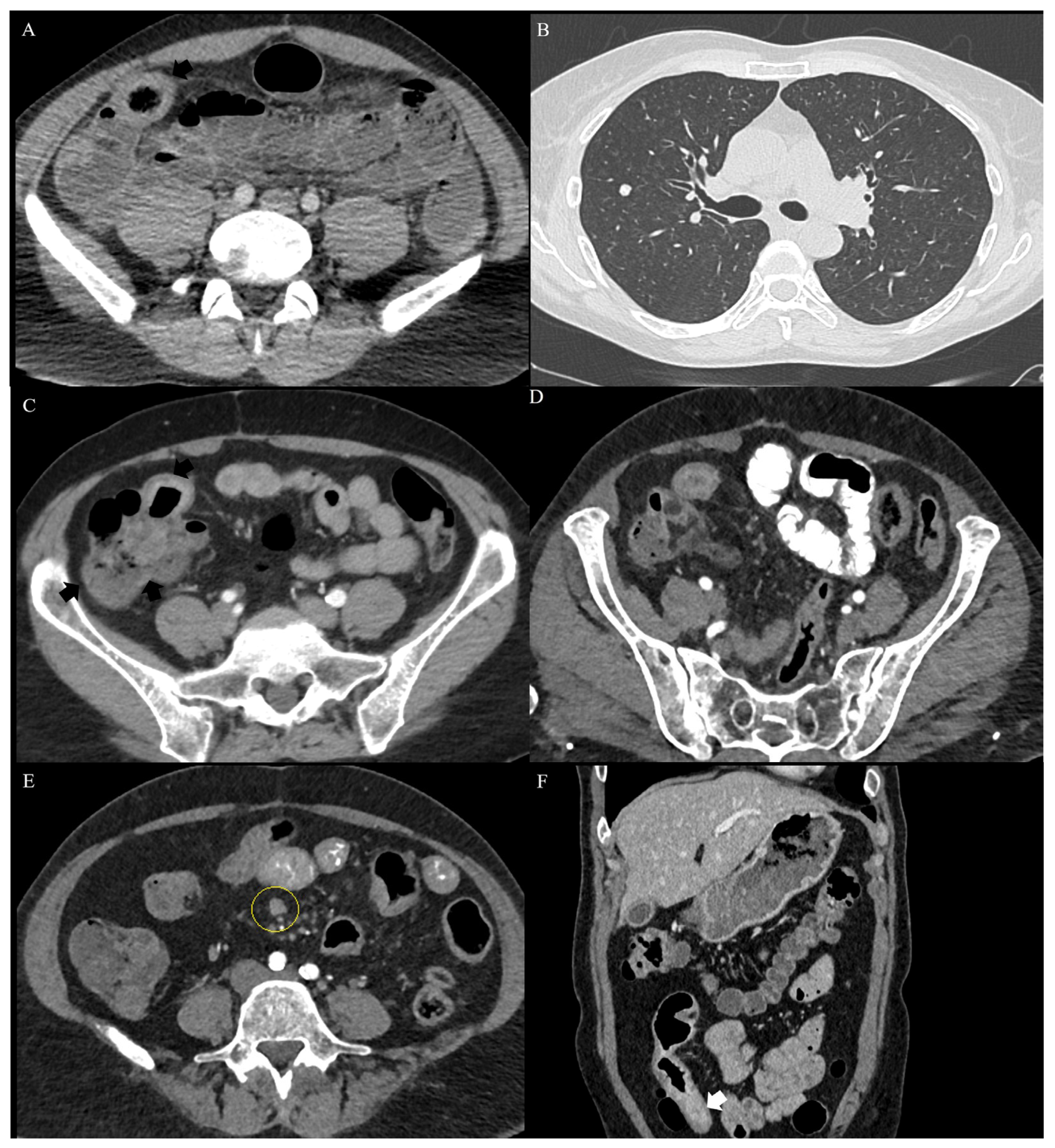
6. Cross-Sectional Imaging
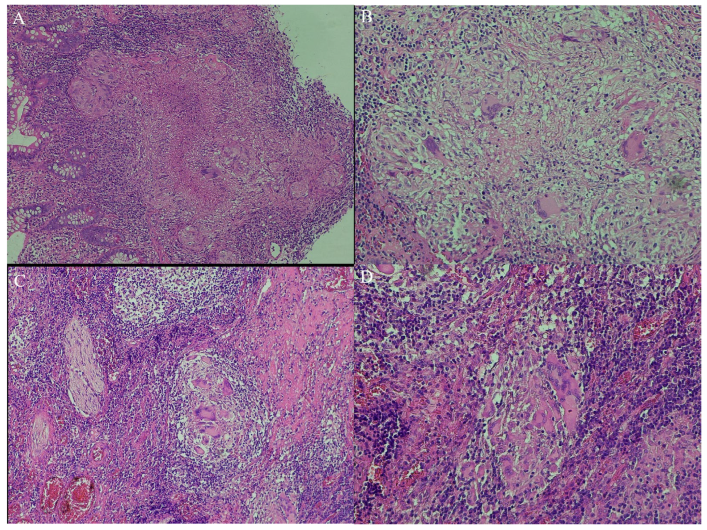
7. Pathology
8. Gamma-IFN Assays and Serology
9. Microbiology and Nuclear Testing
10. Therapeutic Response
11. Computerized Models and Artificial Intelligence

| Author, Year | Country | Patients | Parameters | Model | Performance | Validation Set |
|---|---|---|---|---|---|---|
| Watermeyer, 2018 [27] | South Africa | 68 ITB-48 CD | HIV positivity, isolated colitis, and absence of extraintestinal manifestations | Multivariate analysis | NO DATA | NO |
| Zhang, 2015 [30] | China | 31 ITB-92 CD | Clinical (diarrhea, night sweat, perianal disease), colonoscopic (transverse, longitudinal, or rodent-like ulcers and patulous ileocecal valve), CT enterography (skip lesions, asymmetric involvement, ileocecal valve), T-SPOT | Multivariate analysis | Sn 97.8% Sp 96.8% ACC 97.6% PPV 98.9% NPV 93.7% | NO |
| Huang, 2015 [49] | China | 40 ITB-25 CD | 12 features (longitudinal ulcers, ring ulcers, ulcer scars, nodular hyperplasia, cobblestone-like mucosa, intestinal diseases, intestinal fistula, target sign, the comb sign, night sweats, tuberculin skin test, T-SPOT.TB). | Simple score (features supporting CD +1, supporting ITB scored −1) | AUC 0.997 | NO |
| Limsrivilai, 2017 [54] | Multicentric | 38 studies 2117 CD-1589 ITB | CD: male, hematochezia, perianal disease, intestinal obstruction, extraintestinal manifestations; longitudinal ulcers, cobblestone, luminal stricture, mucosal bridge, rectal involvement; focally enhanced colitis; CT enterography (asymmetrical wall thickening, wall stratification, comb sign, fibrofatty proliferation ITB: fever, night sweats, lung involvement, ascites; transverse ulcers, patulous ileocecal valve, cecal involvement; confluent or submucosal granulomas, lymphocyte cuffing, ulcers lined by histiocytes; CTE short segmental | Bayesian meta-analysis | Sn 90.9% Sp 92.6% Acc 91.8% | Not appliable |
| Bae, 2017 [65] | South Korea | 40 ITB-40 CD | Colonoscopic, laboratory, and radiologic factors | Multivariate analysis | Combined score increased accuracy from 81.2% to 96.3%. AUC 0.990–0.981 | YES |
| Cheng 2024 [173] | China | 85 ITB-245CD | Radiomics features | Deep learning CNN followed by LASSO regression | The arterial-venous combined deep learning radiomics model had AUCs of 0.885, 0.877, and 0.800 | YES (2 datasets) |
| Li 2022, [174] | China | 105 ITB-227 CD | 13 parameters in the LASSO model (abdominal pain; perianal abscess; ileus; hepatobiliary disease; tuberculosis history; CRP; T-SPOT; segmental lesions; longitudinal ulcer; jejunum involvement; ascending colon involvement; rectum involvement; perianal fistula) | LASSO regression vs. classical regression | Sn 84.4 vs. 65.9% Sp 80.6 vs. 89.9% PPV 82.3 vs. 88.5% NPV 82.9 vs. 68.9% AUC 0.887 vs. 0.811, | YES |
| Shu, 2024 [175] | China | 123 ITB-118 CD | 51 demographic, clinical, laboratory, colonoscopy, and pathology parameters. Best performance: T-spot, pulmonary tuberculosis, and age at onset | Six machine-learning methods tested (XGBoost best) | Sn 87.1% Sp 83.3% Acc 86% AUC 0.946 | NO |
| Weng, 2022 [176] | China | 40 ITB-160 CD | Nine variables (intestinal surgery, abdominal, bloody stool, PPD, knot, ESAT-6, CFP-10, intestinal dilatation, and comb sign) | 10 Machine-learning methods (XBoost best) | Sn 75.2–81.3% Sp 95.3–96.9% AUC 0.853–0.891 | YES |
| Zeng 2022 [177] | China | 43 ITB-90 CD | T-SPOT positive, cobblestone appearance, comb sign, and granuloma | Multivariate analysis LogitP = 0.340 − 5.457 × T-SPOT positive + 3.353 × cobblestone appearance + 4.436 × comb sign − 2.967 × granuloma | Sn 94.4% Sp 93% Acc 94% AUC 0.988 | NO |
| Zhu, 2021 [178] | China | 93 CD-67 ITB | 2 clinical and 9 radiomics features for the region of interest (ROI) of the ileocecal region (CT enterography), filtered by the gradient boosting decision tree (GBDT). The radiomics score was calculated using the radiomics signature-based formula | Multivariate analysis | AUC 0.96 (95% CI: 0.93–0.99) in the training cohort, 0.93 (95%CI: 0.86–1.00) in validation cohort. | YES |
| Lee, 2006 [179] | South Korea | 44 ITB-44 CD | Four parameters were more common in CD (anorectal lesions, longitudinal or aphthous ulcers, and cobblestone appearance), and four other parameters (involvement of fewer than four segments, a patulous ileocecal valve, transverse ulcers, and scars or pseudopolyps) were more common in ITB | Decision Tree Analysis | Correct diagnosis 87.5% | NO |
| Kedia [180] | India | 50 ITB-54 CD | Colonoscopic and CTE (ileocecal involvement, long-segment involvement, lymph node ≥1 cm) | Simple score (1 p each) | 0–1p: Sp 100–87%, 100–76% PPV (ITB). 2–3p: Sp 68–90%, PPV 63–80% (CD) | YES |
| Jung 2016 [182] | South Korea | 99 ITB-162 CD | Age, gender, diarrhea, ring-shaped or longitudinal ulcer, sigmoid involvement, suspicious radiological pulmonary TB | Multivariate logistic regression | Sn 98%, Sp 92.4% PPV 88.9% NPV 98.6% | YES |
| Makharia 2009 [183] | India | 53 ITB-53 CD | Blood in stool, weight loss, sigmoid involvement, and focally enhanced colitis | Multivariate analysis | Sn 83–90% Sp 79.2–60% Acc 81.1% AUC 0.9089–0.892 | YES |
| Hilmi, 2022 [184] | Malaysia | 30 ITB-134 CD | Close contact TB, AFB/PCR/culture positive, multiple/large/caseating granulomas, ascites, lymph nodes, pulmonary findings, TST/IGRA positive + ATT | Decision tree algorithm | Sn 96.7% Sp 88.1% Acc 89.6% NPV 99.1% PPV 64.4% | NO |
| Kim, 2021 [186] | South Korea | 217 ITB-211 DC | Colonoscopy images | Convolution Neural Network Analysis | AUC 0.7846–0.8586 For typical images, AUC ranged from 0.8211 to 0.9360 | NO |
| Gong, 2023 [187] | China | 105 ITB-CD | Clinical–radiomic (one radiomic signature from the intestinal wall, one radiomic signature from the LN, involved bowel segments on CTE, and a longitudinal ulcer on endoscopy) | Multivariate analysis | AUC 0.975 in the training cohort and 0.958 in the validation cohort. | YES |
| Zhao, 2014 [188] | China | 47 ITB-141 CD | Clinical (hematochezia, perianal disease, ascites, night sweats, pulmonary TB), CTE (left colon, asymmetric involvement, abscess, comb sign, lymph nodes along the right colic artery, contracture of the ileocecal valve, fixed patulous ileocecal valve, and lymph nodes with central necrosis), and TST | Multivariate analysis | Clinical/CTE features Sn 94.3–96.5% Sp 80.4–93.6% Acc 91.0–95.7% PPV 93.7–97.8% NPV 82.6–89.8% | NO |
| He, 2019 [189] | China, Israel | 69 ITB-143 CD | Age, transverse ulcer, rectum involvement, skipped involvement of the small bowel, target sign, comb sign, and IRGA | Multivariate analysis | Sn 86.8% Sp 90.9% PPV 97.1% NPV 66.7% Acc 87.8% | NO |
| Yu, 2012 [190] | China | 43 ITB-53 CD | Night sweats, longitudinal ulcers, and granulomas | Multivariate analysis | AUC 0.8642 | NO |
| Lu 2021 [193] | China | 106 ITB-106 CD | IGRA, ≥4 segments involved, longitudinal, circular, or aphthous ulcer | CART | Sn 90.91% Sp 86.36% NPV 90.48% PPV 86.96% Acc 88.64% | YES |
| Wu 2018 [194] | China | 86 ITB-153 CD | Perianal disease, pulmonary involvement, longitudinal ulcer, left colon, and ratio of tuberculosis-specific antigen to phytohemagglutinin | Multivariate analysis | AUC, Sn, Sp, and accuracy were 0.975–0.950, 96.7–88.5%, 90.7–93.5%, and 92.8–91.7% in the training and validation sets. | YES |
12. Conclusions
Supplementary Materials
Author Contributions
Funding
Institutional Review Board Statement
Informed Consent Statement
Data Availability Statement
Conflicts of Interest
Abbreviations
| AFB | Acid-fast bacilli |
| ASCA | Anti-Saccharomyces cerevisiae antibodies |
| ATT | Antitubercular therapy |
| AUC | Area under the ROC curve |
| BMI | Body mass index |
| CD | Crohn’s disease |
| CEUS | Contrast-enhanced ultrasound |
| CNN | Convolutional neural network |
| CFP-10 | Culture filtrate protein-10 |
| ESAT-6 | Early secreted antigenic target 6 kDa |
| EUS | Endoscopic ultrasound |
| G-CSF | Granulocyte-colony stimulating factor |
| HE | Hematoxylin and eosin stain |
| IBD | Inflammatory bowel disease |
| IFN | Interferon |
| IGRA | Interferon-gamma release assay |
| IL | Interleukin |
| ITB | Intestinal tuberculosis |
| LASSO | Least absolute shrinkage and selection operator |
| NAAT | Nucleic acid amplification test |
| NPV | Negative predictive value |
| PPAR | Peroxisome proliferator-activated receptor |
| PPD | Negative predictive value |
| PPV | Positive predictive value |
| TB | Tuberculosis |
| TNF | Tumoral necrosis factor |
References
- Kedia, S.; Ahuja, V. Intestinal tuberculosis or Crohn’s disease: Illusion or delusion or allusion. JGH Open 2021, 5, 177–179. [Google Scholar] [CrossRef] [PubMed]
- Mehta, V.; Desai, D.; Abraham, P.; Rodrigues, C. Making a Positive Diagnosis of Intestinal Tuberculosis with the Aid of New Biologic and Histologic Features: How Far Have We Reached? Inflamm. Intest. Dis. 2019, 3, 155–160. [Google Scholar] [CrossRef]
- Choudhury, A.; Dhillon, J.; Sekar, A.; Gupta, P.; Singh, H.; Sharma, V. Differentiating gastrointestinal tuberculosis and Crohn’s disease- a comprehensive review. BMC Gastroenterol. 2023, 23, 246. [Google Scholar] [CrossRef] [PubMed]
- Jin, X.J.; Kim, J.M.; Kim, H.K.; Kim, L.; Choi, S.J.; Park, I.S.; Han, J.Y.; Chu, Y.C.; Song, J.Y.; Kwon, K.S.; et al. Histopathology and TB-PCR kit analysis in differentiating the diagnosis of intestinal tuberculosis and Crohn’s disease. World J. Gastroenterol. 2010, 16, 2496–2503. [Google Scholar] [CrossRef]
- Kedia, S.; Das, P.; Madhusudhan, K.S.; Dattagupta, S.; Sharma, R.; Sahni, P.; Makharia, G.; Ahuja, V. Differentiating Crohn’s disease from intestinal tuberculosis. World J. Gastroenterol. 2019, 25, 418–432. [Google Scholar] [CrossRef]
- Jha, D.K.; Pathiyil, M.M.; Sharma, V. Evidence-based approach to diagnosis and management of abdominal tuberculosis. Indian J. Gastroenterol. 2023, 42, 17–31. [Google Scholar] [CrossRef]
- Jena, A.; Mohindra, R.; Rana, K.; Neelam, P.B.; Thakur, D.C.; Singh, H.; Gupta, P.; Suri, V.; Sharma, V. Frequency, outcomes, and need for intervention in stricturing gastrointestinal tuberculosis: A systematic review and meta-analysis. BMC Gastroenterol. 2023, 23, 46. [Google Scholar] [CrossRef] [PubMed]
- Merino Gallego, E.; Gallardo Sánchez, F.; Gallego Rojo, F.J. Intestinal tuberculosis and Crohn’s disease: The importance and difficulty of a differential diagnosis. Rev. Esp. Enfermedades Dig. 2018, 110, 650–657. [Google Scholar] [CrossRef]
- Dilauro, S.; Crum-Cianflone, N.F. Ileitis: When it is not Crohn’s disease. Curr. Gastroenterol. Rep. 2010, 12, 249–258. [Google Scholar] [CrossRef]
- Shi, X.C.; Zhang, L.F.; Zhang, Y.Q.; Liu, X.Q.; Fei, G.J. Clinical and Laboratory Diagnosis of Intestinal Tuberculosis. Chin. Med. J. 2016, 129, 1330–1333. [Google Scholar] [CrossRef]
- Sandgren, A.; Hollo, V.; van der Werf, M.J. Extrapulmonary tuberculosis in the European Union and European Economic Area, 2002 to 2011. Eurosurveillance 2013, 18, 20431. [Google Scholar] [CrossRef] [PubMed]
- Seo, H.; Lee, S.; So, H.; Kim, D.; Kim, S.O.; Soh, J.S.; Bae, J.H.; Lee, S.H.; Hwang, S.W.; Park, S.H.; et al. Temporal trends in the misdiagnosis rates between Crohn’s disease and intestinal tuberculosis. World J. Gastroenterol. 2017, 23, 6306–6314. [Google Scholar] [CrossRef] [PubMed]
- Venkatesh, P.G.; Navaneethan, U. Mimickers of intestinal tuberculosis: Could this be Crohn’s disease? An unsolved enigma. Saudi J. Gastroenterol. 2011, 17, 95–96. [Google Scholar] [CrossRef]
- Pulimood, A.B.; Amarapurkar, D.N.; Ghoshal, U.; Phillip, M.; Pai, C.G.; Reddy, D.N.; Nagi, B.; Ramakrishna, B.S. Differentiation of Crohn’s disease from intestinal tuberculosis in India in 2010. World J. Gastroenterol. 2011, 17, 433–443. [Google Scholar] [CrossRef]
- Banerjee, R.; Pal, P.; Patel, R.; Godbole, S.; Komawar, A.; Mudigonda, S.; Akki, Y.; Gaddam, A.; Pasula, N.P.; Joseph, S.; et al. Inflammatory bowel disease (IBD) in rural and urban India: Results from community colonoscopic evaluation of more than 30,000 symptomatic patients. Lancet Reg. Health-Southeast Asia 2023, 19, 100259. [Google Scholar] [CrossRef]
- Dhoble, P.; Desai, D.; Abraham, P. Is the rise in Crohn’s disease in India accompanied by a fall in intestinal tuberculosis? A single-center experience. Indian J. Tuberc. 2021, 68, 210–214. [Google Scholar] [CrossRef]
- Chung, K.M.; Kim, H.S.; Park, S.Y.; Lim, S.R.; Ryang, D.Y.; Jeong, H.K.; Lee, W.S.; Park, C.H.; Lee, J.H.; Choi, S.K.; et al. The changes in incidence of Crohn’s disease and intestinal tuberculosis in Korea. Korean J. Gastroenterol. 2008, 52, 351–358. [Google Scholar] [PubMed]
- Navaneethan, U.; Cherian, J.V.; Prabhu, R.; Venkataraman, J. Distinguishing tuberculosis and Crohn’s disease in developing countries: How certain can you be of the diagnosis? Saudi J. Gastroenterol. 2009, 15, 142–144. [Google Scholar] [CrossRef]
- Almadi, M.A.; Ghosh, S.; Aljebreen, A.M. Differentiating intestinal tuberculosis from Crohn’s disease: A diagnostic challenge. Am. J. Gastroenterol. 2009, 104, 1003–1012. [Google Scholar] [CrossRef]
- Rolo, R.; Campainha, S.; Duarte, R. Crohn’s disease and intestinal tuberculosis: A clinical challenge. Rev. Port. Pneumol. 2012, 18, 205–206. [Google Scholar] [CrossRef] [PubMed]
- Sato, R.; Nagai, H.; Matsui, H.; Yamane, A.; Kawashima, M.; Higa, K.; Nakamura, S.; Ohshima, N.; Tamura, A.; Hebisawa, A. Ten Cases of Intestinal Tuberculosis Which Were Initially Misdiagnosed as Inflammatory Bowel Disease. Intern. Med. 2019, 58, 2003–2008. [Google Scholar] [CrossRef]
- Weinberg, S.E.; Mughal, A.M. A Case of Intestinal Tuberculosis Mimicking Crohn’s Disease: A Clinical and Diagnostic Dilemma. Eur. J. Case Rep. Intern. Med. 2021, 8, 002699. [Google Scholar] [CrossRef]
- Limsrivilai, J.; Pausawasdi, N. Intestinal tuberculosis or Crohn’s disease: A review of the diagnostic models designed to differentiate between these two gastrointestinal diseases. Intest. Res. 2021, 19, 21–32. [Google Scholar] [CrossRef] [PubMed]
- Kedia, S.; Sharma, R.; Makharia, G.; Ahuja, V.; Desai, D.; Kandasamy, D.; Eapen, A.; Ganesan, K.; Ghoshal, U.C.; Kalra, N.; et al. Indian guidelines on imaging of the small intestine in Crohn’s disease: A joint Indian Society of Gastroenterology and Indian Radiology and Imaging Association consensus statement. Indian J. Radiol. Imaging 2019, 29, 111–132. [Google Scholar] [CrossRef]
- Sharma, R.; Madhusudhan, K.S.; Ahuja, V. Intestinal tuberculosis versus Crohn’s disease: Clinical and radiological recommendations. Indian J. Radiol. Imaging 2016, 26, 161–172. [Google Scholar] [CrossRef]
- Narang, H.; Kedia, S.; Ahuja, V. New diagnostic strategies to distinguish Crohn’s disease and gastrointestinal tuberculosis. Curr. Opin. Infect. Dis. 2024, 37, 392–401. [Google Scholar] [CrossRef]
- Watermeyer, G.; Thomson, S. Differentiating Crohn’s disease from intestinal tuberculosis at presentation in patients with tissue granulomas. S. Afr. Med. J. 2018, 108, 399–402. [Google Scholar] [CrossRef] [PubMed]
- Agranoff, D.; Fernandez-Reyes, D.; Papadopoulos, M.C.; Rojas, S.A.; Herbster, M.; Loosemore, A.; Tarelli, E.; Sheldon, J.; Schwenk, A.; Pollok, R.; et al. Identification of diagnostic markers for tuberculosis by proteomic fingerprinting of serum. Lancet 2006, 368, 1012–1021. [Google Scholar] [CrossRef]
- Ning, L.; Shan, G.; Sun, Z.; Lou, X.; Zhang, F.; Li, S.; Du, H.; Yu, J.; Chen, H.; Xu, G. Serum proteome profiles to differentiate Crohn disease from intestinal tuberculosis and primary intestinal lymphoma: A pilot study. Medicine 2019, 98, e18304. [Google Scholar] [CrossRef] [PubMed]
- Zhang, T.; Fan, R.; Wang, Z.; Hu, S.; Zhang, M.; Lin, Y.; Tang, Y.; Zhong, J. Differential diagnosis between Crohn’s disease and intestinal tuberculosis using integrated parameters including clinical manifestations, T-SPOT, endoscopy and CT enterography. Int. J. Clin. Exp. Med. 2015, 8, 17578–17589. [Google Scholar] [PubMed] [PubMed Central]
- Zhang, F.; Xu, C.; Ning, L.; Hu, F.; Shan, G.; Chen, H.; Yang, M.; Chen, W.; Yu, J.; Xu, G. Exploration of Serum Proteomic Profiling and Diagnostic Model That Differentiate Crohn’s Disease and Intestinal Tuberculosis. PLoS ONE 2016, 11, e0167109, Erratum in PLoS ONE 2019, 14, e0212300. https://doi.org/10.1371/journal.pone.0212300. [Google Scholar] [CrossRef] [PubMed]
- Rukmangadachar, L.A.; Makharia, G.K.; Mishra, A.; Das, P.; Hariprasad, G.; Srinivasan, A.; Gupta, S.D.; Ahuja, V.; Acharya, S.K. Proteome analysis of the macroscopically affected colonic mucosa of Crohn’s disease and intestinal tuberculosis. Sci. Rep. 2016, 6, 23162. [Google Scholar] [CrossRef]
- Ma, R.; Zhu, Y.; Li, X.; Hu, S.; Zheng, D.; Xiong, S.; Xu, S.; Xiang, L.; Zhao, M.; Tang, C.; et al. A Novel Serum Metabolomic Panel for the Diagnosis of Crohn’s Disease. Inflamm. Bowel Dis. 2023, 29, 1524–1535. [Google Scholar] [CrossRef]
- Ahuja, V.; Subodh, S.; Tuteja, A.; Mishra, V.; Garg, S.K.; Gupta, N.; Makharia, G.; Acharya, S.K. Genome-wide gene expression analysis for target genes to differentiate patients with intestinal tuberculosis and Crohn’s disease and discriminative value of FOXP3 mRNA expression. Gastroenterol. Rep. 2016, 4, 59–67. [Google Scholar] [CrossRef][Green Version]
- Rampal, R.; Kedia, S.; Wari, M.N.; Madhu, D.; Singh, A.K.; Tiwari, V.; Mouli, V.P.; Mohta, S.; Makharia, G.; Ahuja, V. Prospective validation of CD4+CD25+FOXP3+ T-regulatory cells as an immunological marker to differentiate intestinal tuberculosis from Crohn’s disease. Intest. Res. 2021, 19, 232–238. [Google Scholar] [CrossRef]
- Roy, S.; Ghosh, S.; Banerjee, M.; Laha, S.; Bhattacharjee, D.; Sarkar, R.; Ray, S.; Banerjee, A.; Ghosh, R.; Halder, A.; et al. A combination of circulating microRNA-375-3p and chemokines CCL11, CXCL12, and G-CSF differentiate Crohn’s disease and intestinal tuberculosis. Sci. Rep. 2021, 11, 23303. [Google Scholar] [CrossRef]
- Tiwari, V.; Kedia, S.; Garg, S.K.; Rampal, R.; Mouli, V.P.; Purwar, A.; Mitra, D.K.; Das, P.; Dattagupta, S.; Makharia, G.; et al. CD4+ CD25+ FOXP3+ T cell frequency in the peripheral blood is a biomarker that distinguishes intestinal tuberculosis from Crohn’s disease. PLoS ONE 2018, 13, e0193433. [Google Scholar] [CrossRef]
- Yoo, J.W.; Jo, S.I.; Shin, D.W.; Park, J.W.; Kim, S.E.; Lim, H.; Kang, H.S.; Moon, S.H.; Kim, M.K.; Kim, S.Y.; et al. Clinical Usefulness of Immune Profiling for Differential Diagnosis between Crohn’s Disease, Intestinal Tuberculosis, and Behcet’s Disease. Diagnostics 2023, 13, 2904. [Google Scholar] [CrossRef] [PubMed]
- Yu, Z.Q.; Wang, W.F.; Dai, Y.C.; Chen, X.C.; Chen, J.Y. Interleukin-22 receptor 1 is expressed in multinucleated giant cells: A study on intestinal tuberculosis and Crohn’s disease. World J. Gastroenterol. 2019, 25, 2473–2488. [Google Scholar] [CrossRef] [PubMed]
- Gupta, A.; Sharma, K.; Sharma, V.; Singh, J.; Nada, R.; Saikia, B.; Minz, R.W.; Anand, S.; Kumar, M. Comparative evaluation of interleukin-10, transforming growth factor-β, and interleukin-17 in gastrointestinal tuberculosis and crohn’s disease. Int. J. Mycobacteriol. 2022, 11, 384–388. [Google Scholar] [CrossRef]
- Nayak, S.S.; Shetty, M.V.; Pai, C.G.; Guruprasad, K.P.; Satyamoorthy, K. Apoptosis in peripheral blood lymphocytes in intestinal tuberculosis and Crohn’s disease: Implications to diagnostic differentiation. Indian J. Gastroenterol. 2020, 39, 338–345. [Google Scholar] [CrossRef]
- Jiang, M.; Zeng, Z.; Chen, K.; Dang, Y.; Li, L.; Ma, C.; Cheng, R.; Hu, K.; Li, X.; Zhang, H. Enterogenous Microbiotic Markers in the Differential Diagnosis of Crohn’s Disease and Intestinal Tuberculosis. Front. Immunol. 2022, 13, 820891. [Google Scholar] [CrossRef] [PubMed]
- Pugazhendhi, S.; Jayakanthan, K.; Pulimood, A.B.; Ramakrishna, B.S. Cytokine gene expression in intestinal tuberculosis and Crohn’s disease. Int. J. Tuberc. Lung Dis. 2013, 17, 662–668. [Google Scholar] [CrossRef] [PubMed]
- Ma, J.Y.; Tong, J.L.; Ran, Z.H. Intestinal tuberculosis and Crohn’s disease: Challenging differential diagnosis. J. Dig. Dis. 2016, 17, 155–161. [Google Scholar] [CrossRef]
- He, C.; Wang, H.; Yu, C.; Peng, C.; Shu, X.; Liao, W.; Zhu, Z. Alterations of Gut Microbiota in Patients with Intestinal Tuberculosis That Different From Crohn’s Disease. Front. Bioeng. Biotechnol. 2021, 9, 673691. [Google Scholar] [CrossRef]
- Bajaj, A.; Markandey, M.; Samal, A.; Goswami, S.; Vuyyuru, S.K.; Mohta, S.; Kante, B.; Kumar, P.; Makharia, G.; Kedia, S.; et al. Depletion of core microbiome forms the shared background against diverging dysbiosis patterns in Crohn’s disease and intestinal tuberculosis: Insights from an integrated multi-cohort analysis. Gut Pathog. 2024, 16, 65. [Google Scholar] [CrossRef] [PubMed]
- Khan, I.A.; Nayak, B.; Markandey, M.; Bajaj, A.; Verma, M.; Kumar, S.; Singh, M.K.; Kedia, S.; Ahuja, V. Differential prevalence of pathobionts and host gene polymorphisms in chronic inflammatory intestinal diseases: Crohn’s disease and intestinal tuberculosis. PLoS ONE 2021, 16, e0256098. [Google Scholar] [CrossRef]
- Zeng, J.; Zhou, G.; Pan, F. Clinical Analysis of Intestinal Tuberculosis: A Retrospective Study. J. Clin. Med. 2023, 12, 445. [Google Scholar] [CrossRef]
- Huang, X.; Liao, W.D.; Yu, C.; Tu, Y.; Pan, X.L.; Chen, Y.X.; Lv, N.H.; Zhu, X. Differences in clinical features of Crohn’s disease and intestinal tuberculosis. World J. Gastroenterol. 2015, 21, 3650–3656. [Google Scholar] [CrossRef]
- Lu, S.; Fu, J.; Guo, Y.; Huang, J. Clinical diagnosis and endoscopic analysis of 10 cases of intestinal tuberculosis. Medicine 2020, 99, e21175. [Google Scholar] [CrossRef]
- Amarapurkar, D.N.; Patel, N.D.; Rane, P.S. Diagnosis of Crohn’s disease in India where tuberculosis is widely prevalent. World J. Gastroenterol. 2008, 14, 741–746. [Google Scholar] [CrossRef]
- Ionescu, S.; Nicolescu, A.C.; Madge, O.L.; Marincas, M.; Radu, M.; Simion, L. Differential Diagnosis of Abdominal Tuberculosis in the Adult-Literature Review. Diagnostics 2021, 11, 2362. [Google Scholar] [CrossRef]
- Gompertz, M.; Carreño, L.; Gil La Rotta, L.C. Rectal tuberculosis: An uncommon clinical presentation and differential diagnosis with Crohn’s disease. Rev. Gastroenterol. Mex. (Engl. Ed.) 2019, 84, 524–526. (In English) [Google Scholar] [CrossRef]
- Limsrivilai, J.; Shreiner, A.B.; Pongpaibul, A.; Laohapand, C.; Boonanuwat, R.; Pausawasdi, N.; Pongprasobchai, S.; Manatsathit, S.; Higgins, P.D. Meta-Analytic Bayesian Model For Differentiating Intestinal Tuberculosis from Crohn’s Disease. Am. J. Gastroenterol. 2017, 112, 415–427. [Google Scholar] [CrossRef] [PubMed]
- Liu, Y.Y.; Chen, M.K.; Cao, Z.; Liu, S.Z.; Ding, B.J. Differential diagnosis of intestinal tuberculosis from Crohn’s disease and primary intestinal lymphoma in China. Saudi J. Gastroenterol. 2014, 20, 241–247. [Google Scholar] [CrossRef] [PubMed]
- Larsson, G.; Shenoy, T.; Ramasubramanian, R.; Balakumaran, L.K.; Småstuen, M.C.; Bjune, G.A.; Moum, B.A. Routine diagnosis of intestinal tuberculosis and Crohn’s disease in Southern India. World J. Gastroenterol. 2014, 20, 5017–5024. [Google Scholar] [CrossRef]
- Nițu, F.M.; Olteanu, M.; Streba, C.T.; Jimborean, G.; Postolache, P.; Man, M.A.; Trofor, A.C.; Nemeş, R.M.; Dragonu, L.; Olteanu, M. Tuberculosis and its particularities in Romania and worldwide. Rom. J. Morphol. Embryol. 2017, 58, 385–392. [Google Scholar] [PubMed]
- Singh, S.K.; Srivastava, A.; Kumari, N.; Poddar, U.; Yachha, S.K.; Pandey, C.M. Differentiation Between Crohn Disease and Intestinal Tuberculosis in Children. J. Pediatr. Gastroenterol. Nutr. 2018, 66, e6–e11. [Google Scholar] [CrossRef]
- Wen, J.; He, J.Q. Clinical characteristics and pregnancy outcomes in pregnant women with TB: A retrospective cohort study. Ann. Med. 2024, 56, 2401108. [Google Scholar] [CrossRef] [PubMed] [PubMed Central]
- de Figueiredo, I.R.; Ferrão, J.B.; Dias, S.; Alves, R.V.; Borges, D.D.; Torres, M.; Castro, S.G.; Lourenço, F.; Antunes, A.M.; Gruner, H.; et al. Tuberculosis infection in the elderly versus in the young adult. J. Gerontol. Geriatr. 2022, 70, 187–191. [Google Scholar] [CrossRef]
- Weng, M.T.; Wei, S.C.; Lin, C.C.; Tsang, Y.M.; Shun, C.T.; Wang, J.Y.; Shieh, M.J.; Wang, C.Y.; Wong, J.M. Seminar Report From the 2014 Taiwan Society of Inflammatory Bowel Disease (TSIBD) Spring Forum (May 24th, 2014): Crohn’s Disease Versus Intestinal Tuberculosis Infection. Intest. Res. 2015, 13, 6–10. [Google Scholar] [CrossRef]
- Lee, J.M.; Lee, K.M. Endoscopic Diagnosis and Differentiation of Inflammatory Bowel Disease. Clin. Endosc. 2016, 49, 370–375. [Google Scholar] [CrossRef]
- Pulimood, A.B.; Ramakrishna, B.S.; Kurian, G.; Peter, S.; Patra, S.; Mathan, V.I.; Mathan, M.M. Endoscopic mucosal biopsies are useful in distinguishing granulomatous colitis due to Crohn’s disease from tuberculosis. Gut 1999, 45, 537–541. [Google Scholar] [CrossRef]
- Cheng, L.; Huang, M.F.; Mei, P.F.; Bo, W.H.; Deng, C.S. The clinical, endoscopic and pathologic features of Crohn’s disease in the differentiation from intestinal tuberculosis. Zhonghua Nei Ke Za Zhi 2013, 52, 940–944. [Google Scholar] [PubMed]
- Bae, J.H.; Park, S.H.; Ye, B.D.; Kim, S.O.; Cho, Y.K.; Youn, E.J.; Lee, H.S.; Hwang, S.W.; Yang, D.H.; Kim, K.J.; et al. Development and Validation of a Novel Prediction Model for Differential Diagnosis Between Crohn’s Disease and Intestinal Tuberculosis. Inflamm. Bowel Dis. 2017, 23, 1614–1623. [Google Scholar] [CrossRef]
- Artru, P.; Lavergne-Slove, A.; Joly, F.; Bitoun, A.; Rambaud, J.C.; Bouhnik, Y. Tuberculose jéjunale isolée simulant une maladie de Crohn. Diagnostic par vidéo-entéroscopie poussée [Isolated jejunal tuberculosis mimicking Crohn disease. Diagnosis by push video-enteroscopy]. Gastroenterol. Clin. Biol. 1999, 23, 1086–1089. [Google Scholar] [PubMed]
- Pennazio, M.; Rondonotti, E.; Despott, E.J.; Dray, X.; Keuchel, M.; Moreels, T.; Sanders, D.S.; Spada, C.; Carretero, C.; Cortegoso Valdivia, P.; et al. Small-bowel capsule endoscopy and device-assisted enteroscopy for diagnosis and treatment of small-bowel disorders: European Society of Gastrointestinal Endoscopy (ESGE) Guideline—Update 2022. Endoscopy 2023, 55, 58–95. [Google Scholar] [CrossRef]
- Vere, C.C.; Foarfă, C.; Streba, C.T.; Cazacu, S.; Pârvu, D.; Ciurea, T. Videocapsule endoscopy and single balloon enteroscopy: Novel diagnostic techniques in small bowel pathology. Rom. J. Morphol. Embryol. 2009, 50, 467–474. [Google Scholar] [PubMed]
- Cazacu, S.; Vere, C.C.; Comănescu, V.; Dumitrescu, C.; Enache, S.D.; Ciurea, T. Crohn disease or intestinal fistulising tuberculosis? Diagnosis difficulties in a case treated with infliximab and corticosteroids. Rom. J. Morphol. Embryol. 2009, 50, 263–268. [Google Scholar] [PubMed]
- Kim, Y.G.; Kim, K.J.; Min, Y.K. Comparison of small bowel findings using capsule endoscopy between Crohn’s disease and intestinal tuberculosis in Korea. Yeungnam Univ. J. Med. 2020, 37, 98–105. [Google Scholar] [CrossRef] [PubMed]
- Ramchandani, M.; Reddy, D.N.; Gupta, R.; Lakhtakia, S.; Tandan, M.; Rao, G.V.; Darisetty, S. Diagnostic yield and therapeutic impact of single-balloon enteroscopy: Series of 106 cases. J. Gastroenterol. Hepatol. 2009, 24, 1631–1638. [Google Scholar] [CrossRef]
- Goyal, P.; Shah, J.; Gupta, S.; Gupta, P.; Sharma, V. Imaging in discriminating intestinal tuberculosis and Crohn’s disease: Past, present and the future. Expert Rev. Gastroenterol. Hepatol. 2019, 13, 995–1007. [Google Scholar] [CrossRef]
- Park, Y.H.; Chung, W.S.; Lim, J.S.; Park, S.J.; Cheon, J.H.; Kim, T.I.; Kim, W.H.; Hong, S.P. Diagnostic role of computed tomographic enterography differentiating crohn disease from intestinal tuberculosis. J. Comput. Assist. Tomogr. 2013, 37, 834–839. [Google Scholar] [CrossRef]
- Singh, H.; Mandavdhare, H.; Sharma, V. All that fistulises is not Crohn’s disease: Multiple entero-enteric fistulae in intestinal tuberculosis. Pol. J. Surg. 2019, 91, 35–37. [Google Scholar] [CrossRef] [PubMed]
- Choi, Y.S.; Kim, D.S.; Lee, J.B.; Kim, J.K.; Jung, H.J.; Lee, S.D.; Song, K.H.; Lee, D.H.; Kim, M.J. Clinical Features of Tuberculous Versus Crohn’s Anal Fistulas, in Korea. J. Crohn’s Colitis 2015, 9, 1132–1137. [Google Scholar] [CrossRef] [PubMed]
- Cheon, J.H.; Moon, W.; Park, S.J.; Park, M.I.; Kim, S.E.; Choi, Y.J.; Kim, J.B.; Kwon, H.J. A case of intestinal tuberculosis with perianal fistula diagnosed after 30 years. Korean J. Gastroenterol. 2013, 62, 370–374. [Google Scholar] [CrossRef] [PubMed][Green Version]
- Takakazu Ribas Asato, I.; Souza Novaes Asato, N.; Amariz Yamamoto, F.; Ferreira Siqueira, A.C.; Pereira da Silva, A.M. Intestinal Tuberculosis with Recurrent Gluteal Abscesses and Perianal Fistulas: A Case Report and Literature Review. Cureus 2024, 16, e76456. [Google Scholar] [CrossRef]
- Kakkar, C.; Bonaffini, P.A.; Singh, A.; Narang, V.; Mahajan, R.; Verma, S.; Singla, S.; Sood, A. MR enterography in Crohns disease and beyond: A pictorial review: Beyond Crohn’s: A pictorial guide to MR enterography in small bowel disease. Abdom. Radiol. 2025, 51, 1129–1142. [Google Scholar] [CrossRef]
- Israrahmed, A.; Yadav, R.R.; Yadav, G.; Alpana Helavar, R.V.; Rai, P.; Jain, M.K.; Gupta, A. Systematic reporting of computed tomography enterography/enteroclysis as an aid to reduce diagnostic dilemma when differentiating between intestinal tuberculosis and Crohn’s disease: A prospective study at a tertiary care hospital. JGH Open 2020, 5, 180–189. [Google Scholar] [CrossRef]
- Kim, S.H.; Kim, J.W.; Jeong, J.B.; Lee, K.L.; Kim, B.G.; Choi, Y.H. Differential diagnosis of Crohn’s disease and intestinal tuberculosis in patients with spontaneous small-bowel perforation. Dig. Surg. 2014, 31, 151–156. [Google Scholar] [CrossRef]
- Huang, M.; Tu, L.; Li, J.; Yue, X.; Wu, L.; Yang, M.; Chen, Y.; Han, P.; Li, X.; Zhu, L. Differentiation of Crohn’s disease, ulcerative colitis, and intestinal tuberculosis by dual-layer spectral detector CT enterography. Clin. Radiol. 2024, 79, e482–e489. [Google Scholar] [CrossRef]
- Makanjuola, D. Is it Crohn’s disease or intestinal tuberculosis? CT analysis. Eur. J. Radiol. 1998, 28, 55–61. [Google Scholar] [CrossRef]
- Kedia, S.; Sharma, R.; Vuyyuru, S.K.; Madhu, D.; Sahu, P.; Kante, B.; Das, P.; Goyal, A.; Madan, K.; Makharia, G.; et al. Addition of computed tomography chest increases the diagnosis rate in patients with suspected intestinal tuberculosis. Intest. Res. 2022, 20, 184–191. [Google Scholar] [CrossRef] [PubMed]
- Park, S.; Lee, T.; Lim, W.; Park, S.; Park, H.; Yun, J.; Kim, D.; Choi, S.; Oh, H.; Song, C. Case of Crohn’s Disease Initially Misdiagnosed as Intestinal Tuberculosis Due to Active Pulmonary Tuberculosis. Korean J. Gastroenterol. 2021, 77, 30–34. [Google Scholar] [CrossRef] [PubMed]
- Zhou, D.; Ouyang, Q.; Xiong, M.; Zhang, Y. Crohn’s disease with positive Ziehl-Neelsen stain: Three case reports. Niger. J. Clin. Pract. 2018, 21, 1387–1390. [Google Scholar] [CrossRef] [PubMed]
- Kedia, S.; Sharma, R.; Sreenivas, V.; Madhusudhan, K.S.; Sharma, V.; Bopanna, S.; Pratap Mouli, V.; Dhingra, R.; Yadav, D.P.; Makharia, G.; et al. Accuracy of computed tomographic features in differentiating intestinal tuberculosis from Crohn’s disease: A systematic review with meta-analysis. Intest. Res. 2017, 15, 149–159. [Google Scholar] [CrossRef]
- Qiu, E.Q.; Guo, W.; Cheng, T.M.; Yao, Y.L.; Zhu, W.; Liu, S.D.; Zhi, F.C. Diagnostic classification of endosonography for differentiating colorectal ulcerative diseases: A new statistical method. World J. Gastroenterol. 2017, 23, 8207–8216. [Google Scholar] [CrossRef]
- Seth, R.; Gupta, P.; Debi, U.; Prasad, K.K.; Singh, H.; Sharma, V. Perfusion Computed Tomography May Help in Discriminating Gastrointestinal Tuberculosis and Crohn’s Disease. Diagnostics 2023, 13, 1255. [Google Scholar] [CrossRef]
- Ma, L.; Zhu, Q.; Li, Y.; Li, W.; Wang, X.; Liu, W.; Li, J.; Jiang, Y. The potential role of CT enterography and gastrointestinal ultrasound in the evaluation of anti-tubercular therapy response of intestinal tuberculosis: A retrospective study. BMC Gastroenterol. 2019, 19, 106. [Google Scholar] [CrossRef]
- Ko, J.K.; Lee, H.L.; Kim, J.O.; Song, S.Y.; Lee, K.N.; Jun, D.W.; Lee, O.Y.; Han, D.S.; Yoon, B.C.; Choi, H.S.; et al. Visceral fat as a useful parameter in the differential diagnosis of Crohn’s disease and intestinal tuberculosis. Intest. Res. 2014, 12, 42–47. [Google Scholar] [CrossRef]
- Yadav, D.P.; Madhusudhan, K.S.; Kedia, S.; Sharma, R.; Pratap Mouli, V.; Bopanna, S.; Dhingra, R.; Pradhan, R.; Goyal, S.; Sreenivas, V.; et al. Development and validation of visceral fat quantification as a surrogate marker for differentiation of Crohn’s disease and intestinal tuberculosis. J. Gastroenterol. Hepatol. 2017, 32, 420–426. [Google Scholar] [CrossRef]
- Kedia, S.; Madhusudhan, K.S.; Sharma, R.; Bopanna, S.; Yadav, D.P.; Goyal, S.; Jain, S.; Das, P.; Dattagupta, S.; Makharia, G.; et al. Combination of increased visceral fat and long segment involvement: Development and validation of an updated imaging marker for differentiating Crohn’s disease from intestinal tuberculosis. J. Gastroenterol. Hepatol. 2018, 33, 1234–1241. [Google Scholar] [CrossRef]
- Seetharaman, J.; Srivastava, A.; Yadav, R.R.; Singh, S.K.; Mishra, P.; Sen Sarma, M.; Poddar, U. Visceral Fat Indices: Do They Help Differentiate Crohn’s Disease and Intestinal Tuberculosis in Children? J. Crohn’s Colitis. 2023, 17, 2026–2032. [Google Scholar] [CrossRef] [PubMed]
- Hong, S.M.; Baek, D.H. Diagnostic Procedures for Inflammatory Bowel Disease: Laboratory, Endoscopy, Pathology, Imaging, and Beyond. Diagnostics 2024, 14, 1384. [Google Scholar] [CrossRef] [PubMed]
- Maulahela, H.; Simadibrata, M.; Nelwan, E.J.; Rahadiani, N.; Renesteen, E.; Suwarti, S.W.T.; Anggraini, Y.W. Recent advances in the diagnosis of intestinal tuberculosis. BMC Gastroenterol. 2022, 22, 89. [Google Scholar] [CrossRef] [PubMed]
- Ye, Z.; Lin, Y.; Cao, Q.; He, Y.; Xue, L. Granulomas as the Most Useful Histopathological Feature in Distinguishing between Crohn’s Disease and Intestinal Tuberculosis in Endoscopic Biopsy Specimens. Medicine 2015, 94, e2157. [Google Scholar] [CrossRef]
- Pulimood, A.B.; Peter, S.; Ramakrishna, B.; Chacko, A.; Jeyamani, R.; Jeyaseelan, L.; Kurian, G. Segmental colonoscopic biopsies in the differentiation of ileocolic tuberculosis from Crohn’s disease. J. Gastroenterol. Hepatol. 2005, 20, 688–696. [Google Scholar] [CrossRef]
- Tao, S.; Chen, Y.; Hu, W.; Shen, K.; Xu, J. Is the identification of a caseating granuloma in the intestine indicative of tuberculosis? a rare case of Crohn’s disease. Diagn. Pathol. 2024, 19, 139. [Google Scholar] [CrossRef]
- Han, Y.N.; Gu, Q.R.; Wu, Y.C.; Topatana, W.; Jiang, Z.N. Granulomas without foreign body giant cells in perianal fistula tissue suggest Crohn’s disease. J. Dig. Dis. 2024, 25, 484–489. [Google Scholar] [CrossRef]
- Montes-Arcón, P.S. The pathologist’s perspective in the differential diagnosis between Crohn’s disease and intestinal tuberculosis. Rev. Gastroenterol. Mex. (Engl. Ed.) 2022, 87, 398–399. [Google Scholar] [CrossRef]
- Du, J.; Ma, Y.Y.; Xiang, H.; Li, Y.M. Confluent granulomas and ulcers lined by epithelioid histiocytes: New ideal method for differentiation of ITB and CD? A meta-analysis. PLoS ONE 2014, 9, e103303. [Google Scholar] [CrossRef]
- Das, P.; Akhtar, S.; Bhowmik, S.; Mehra, L.; Sakhuja, P.; Vaiphei, K.; Pulimood, A.; Tiwari, A.; Kumari, N.; Amarapurkar, A.D.; et al. A Pan-Indian survey on the interpretation of intestinal biopsies in inflammatory bowel disease, and differentiating intestinal tuberculosis and Crohn’s disease from a pathologist’s perspective. Indian J. Pathol. Microbiol. 2025, 68, 692–702. [Google Scholar] [CrossRef]
- Mehta, V.; Desai, D.; Abraham, P.; Gupta, T.; Rodrigues, C.; Joshi, A.; Deshpande, R.; Sawant, P.; Ingle, M.; Rathi, P.; et al. Do additional colonoscopic biopsies increase the yield of Mycobacterium tuberculosis culture in suspected ileo-colonic tuberculosis? Indian J. Gastroenterol. 2018, 37, 226–230. [Google Scholar] [CrossRef]
- Kirsch, R.; Pentecost, M.; Hall Pde, M.; Epstein, D.P.; Watermeyer, G.; Friederich, P.W. Role of colonoscopic biopsy in distinguishing between Crohn’s disease and intestinal tuberculosis. J. Clin. Pathol. 2006, 59, 840–844. [Google Scholar] [CrossRef]
- Watermeyer, G.A.; Locketz, M. CD73 expression in tissue granulomas in distinguishing intestinal tuberculosis from Crohn’s disease in a South African cohort. Scand. J. Gastroenterol. 2018, 53, 1217–1221. [Google Scholar] [CrossRef]
- Banerjee, R.; Balaji, M.; Sasikala, M.; Anuradha, S.; Rao, G.V.; Nageshwar Reddy, D. Granulomas of intestinal tuberculosis and Crohn’s disease can be differentiated by CD73 cell surface marker expression: A pilot study. Dig. Dis. Sci. 2013, 58, 2301–2307. [Google Scholar] [CrossRef]
- Das, P.; Rampal, R.; Udinia, S.; Kumar, T.; Pilli, S.; Wari, N.; Ahmed, I.K.; Kedia, S.; Gupta, S.D.; Kumar, D.; et al. Selective M1 macrophage polarization in granuloma-positive and granuloma-negative Crohn’s disease, in comparison to intestinal tuberculosis. Intest. Res. 2018, 16, 426–435. [Google Scholar] [CrossRef] [PubMed]
- Mao, H.; Su, P.; Qiu, W.; Huang, L.; Yu, H.; Wang, Y. The use of Masson’s trichrome staining, second harmonic imaging and two-photon excited fluorescence of collagen in distinguishing intestinal tuberculosis from Crohn’s disease. Color. Dis. 2016, 18, 1172–1178. [Google Scholar] [CrossRef] [PubMed]
- Ince, A.T.; Güneş, P.; Senateş, E.; Sezikli, M.; Tiftikçi, A.; Ovünç, O. Can an immunohistochemistry method differentiate intestinal tuberculosis from Crohn’s disease in biopsy specimens? Dig. Dis. Sci. 2011, 56, 1165–1170. [Google Scholar] [CrossRef] [PubMed]
- Zhang, S.; Qing, Q.; Wang, Q.; Xu, J.; Zhi, F.; Park, P.W.; Zhang, Y.; Chen, Y. Syndecan-1 and heparanase: Potential markers for activity evaluation and differential diagnosis of Crohn’s disease. Inflamm. Bowel Dis. 2013, 19, 1025–1033. [Google Scholar] [CrossRef]
- Kim, B.J.; Choi, Y.S.; Jang, B.I.; Park, Y.S.; Kim, W.H.; Kim, Y.S.; Jung, S.A.; Han, D.S.; Kim, J.S.; Choi, J.H.; et al. Prospective evaluation of the clinical utility of interferon-γ assay in the differential diagnosis of intestinal tuberculosis and Crohn’s disease. Inflamm. Bowel Dis. 2011, 17, 1308–1313. [Google Scholar] [CrossRef]
- Lei, Y.; Yi, F.M.; Zhao, J.; Luckheeram, R.V.; Huang, S.; Chen, M.; Huang, M.F.; Li, J.; Zhou, R.; Yang, G.F.; et al. Utility of in vitro interferon-γ release assay in differential diagnosis between intestinal tuberculosis and Crohn’s disease. J. Dig. Dis. 2013, 14, 68–75. [Google Scholar] [CrossRef]
- Zhao, Y.; Xu, M.; Chen, L.; Liu, Z.; Sun, X. Levels of TB-IGRA may help to differentiate between intestinal tuberculosis and Crohn’s disease in patients with positive results. Ther. Adv. Gastroenterol. 2020, 13, 1756284820922003. [Google Scholar] [CrossRef]
- Sachdeva, K.; Kumar, P.; Kante, B.; Vuyyuru, S.K.; Mohta, S.; Ranjan, M.K.; Singh, M.K.; Verma, M.; Makharia, G.; Kedia, S.; et al. Interferon-gamma release assay has poor diagnostic accuracy in differentiating intestinal tuberculosis from Crohn’s disease in tuberculosis endemic areas. Intest. Res. 2023, 21, 226–234. [Google Scholar] [CrossRef] [PubMed]
- Chen, W.; Fan, J.H.; Luo, W.; Peng, P.; Su, S.B. Effectiveness of interferon-gamma release assays for differentiating intestinal tuberculosis from Crohn’s disease: A meta-analysis. World J. Gastroenterol. 2013, 19, 8133–8140. [Google Scholar] [CrossRef] [PubMed]
- Ng, S.C.; Hirai, H.W.; Tsoi, K.K.; Wong, S.H.; Chan, F.K.; Sung, J.J.; Wu, J.C. Systematic review with meta-analysis: Accuracy of interferon-gamma releasing assay and anti-Saccharomyces cerevisiae antibody in differentiating intestinal tuberculosis from Crohn’s disease in Asians. J. Gastroenterol. Hepatol. 2014, 29, 1664–1670. [Google Scholar] [CrossRef] [PubMed]
- Xu, H.; Li, Y.; Qian, J.M. A Meta-analysis of the accuracy of interferon-γ release assays in differentiating intestinal tuberculosis from Crohn’s disease in Asia. Zhonghua Nei Ke Za Zhi 2016, 55, 535–540. [Google Scholar] [CrossRef]
- Li, Y.; Zhang, L.F.; Liu, X.Q.; Wang, L.; Wang, X.; Wang, J.; Qian, J.M. The role of in vitro interferonγ-release assay in differentiating intestinal tuberculosis from Crohn’s disease in China. J. Crohn’s Colitis. 2012, 6, 317–323. [Google Scholar] [CrossRef][Green Version]
- Man, M.A.; Arghir, O.C.; Man, S.; Streba, C.T.; Olteanu, M.; Nitu, M. Fatal paradoxical cryptic miliary tuberculosis and immune reconstitution disease in a young non-HIV immunocompromised male patient: Case report with autopsy findings. Rom. J. Morphol. Embryol. 2014, 55, 453–457. [Google Scholar] [PubMed]
- Fee, M.J.; Oo, M.M.; Gabayan, A.E.; Radin, D.R.; Barnes, P.F. Abdominal tuberculosis in patients infected with the human immunodeficiency virus. Clin. Infect. Dis. 1995, 20, 938–944. [Google Scholar] [CrossRef] [PubMed]
- Ghoshal, U.C.; Ghoshal, U.; Singh, H.; Tiwari, S. Anti-Saccharomyces cerevisiae antibody is not useful to differentiate between Crohn’s disease and intestinal tuberculosis in India. J. Postgrad. Med. 2007, 53, 166–170. [Google Scholar] [CrossRef]
- Makharia, G.K.; Sachdev, V.; Gupta, R.; Lal, S.; Pandey, R.M. Anti-Saccharomyces cerevisiae antibody does not differentiate between Crohn’s disease and intestinal tuberculosis. Dig. Dis. Sci. 2007, 52, 33–39. [Google Scholar] [CrossRef]
- Zhang, S.; Luo, J.; Wu, Z.; Roggenbuck, D.; Schierack, P.; Reinhold, D.; Li, J.; Zeng, X.; Zhang, F.; Qian, J.; et al. Antibodies against glycoprotein 2 display diagnostic advantages over ASCA in distinguishing CD from intestinal tuberculosis and intestinal Behçet’s disease. Clin. Transl. Gastroenterol. 2018, 9, e133. [Google Scholar] [CrossRef]
- Kim, Y.S.; Kim, Y.H.; Kim, W.H.; Kim, J.S.; Park, Y.S.; Yang, S.K.; Ye, B.D.; Jang, B.I.; Jung, S.A.; Jeen, Y.T.; et al. Diagnostic utility of anti-Saccharomyces cerevisiae antibody (ASCA) and Interferon-γ assay in the differential diagnosis of Crohn’s disease and intestinal tuberculosis. Clin. Chim. Acta 2011, 412, 1527–1532. [Google Scholar] [CrossRef] [PubMed]
- Kashima, K.; Oka, S.; Tabata, A.; Yasuda, K.; Kitano, A.; Kobayashi, K.; Yano, I. Detection of anti-cord factor antibodies in intestinal tuberculosis for its differential diagnosis from Crohn’s disease and ulcerative colitis. Dig. Dis. Sci. 1995, 40, 2630–2634. [Google Scholar] [CrossRef] [PubMed]
- Patel, B.; Yagnik, V.D. Clinical and laboratory features of intestinal tuberculosis. Clin. Exp. Gastroenterol. 2018, 11, 97–103. [Google Scholar] [CrossRef]
- Kurnick, A.; Bar, N.; Maharshak, N. Intestinal Tuberculosis and Crohn’s Disease is Always a Diagnostic Challenge: A Case Report and Review of the Literature on the Importance of Fecal Mycobacterial Cultures and the Limitations of Latent Infection Testing. Cureus 2019, 11, e5689. [Google Scholar] [CrossRef]
- Gan, H.; Ouyang, Q.; Bu, H.; Li, S.; Chen, D.; Li, G.; Yang, X. Value of polymerase chain reaction assay in diagnosis of intestinal tuberculosis and differentiation from Crohn’s disease. Chin. Med. J. 1995, 108, 215–220. [Google Scholar] [PubMed]
- Babafemi, E.O.; Cherian, B.P.; Banting, L.; Mills, G.A.; Ngianga, K., 2nd. Effectiveness of real-time polymerase chain reaction assay for the detection of Mycobacterium tuberculosis in pathological samples: A systematic review and meta-analysis. Syst. Rev. 2017, 6, 215. [Google Scholar] [CrossRef] [PubMed]
- Bellam, B.L.; Mandavdhare, H.S.; Sharma, K.; Shukla, S.; Soni, H.; Kumar-M, P.; Singh, H.; Prasad, K.K.; Dutta, U.; Sharma, V. Utility of tissue Xpert-Mtb/Rif for the diagnosis of intestinal tuberculosis in patients with ileocolonic ulcers. Ther. Adv. Infect. Dis. 2019, 6, 2049936119863939. [Google Scholar] [CrossRef]
- Fei, B.; Zhou, L.; Zhang, Y.; Luo, L.; Chen, Y. Application value of tissue tuberculosis antigen combined with Xpert MTB/RIF detection in differential diagnoses of intestinal tuberculosis and Crohn’s disease. BMC Infect. Dis. 2021, 21, 498. [Google Scholar] [CrossRef]
- Kumar, S.; Bopanna, S.; Kedia, S.; Mouli, P.; Dhingra, R.; Padhan, R.; Kohli, M.; Chaubey, J.; Sharma, R.; Das, P.; et al. Evaluation of Xpert MTB/RIF assay performance in the diagnosis of abdominal tuberculosis. Intest. Res. 2017, 15, 187–194. [Google Scholar] [CrossRef]
- Mor, P.; Dahiya, B.; Parshad, S.; Gulati, P.; Mehta, P.K. Recent updates in diagnosis of abdominal tuberculosis with emphasis on nucleic acid amplification tests. Expert Rev. Gastroenterol. Hepatol. 2022, 16, 33–49. [Google Scholar] [CrossRef] [PubMed]
- Jin, T.; Fei, B.; Zhang, Y.; He, X. The diagnostic value of polymerase chain reaction for Mycobacterium tuberculosis to distinguish intestinal tuberculosis from crohn’s disease: A meta-analysis. Saudi J. Gastroenterol. 2017, 23, 3–10. [Google Scholar] [CrossRef] [PubMed]
- Paulose, R.R.; Kumar, V.A.; Sharma, A.; Damle, A.; Saikumar, D.; Sudhakar, A.; Koshy, A.K.; Damle, R.P.V.P. An outcome-based composite approach for the diagnosis of intestinal tuberculosis: A pilot study from a tertiary care centre in South India. J. R. Coll. Physicians Edinb. 2021, 51, 344–350. [Google Scholar] [CrossRef] [PubMed]
- Fei, B.Y.; Lv, H.X.; Zheng, W.H. Fluorescent quantitative PCR of Mycobacterium tuberculosis for differentiating intestinal tuberculosis from Crohn’s disease. Braz. J. Med. Biol. Res. 2014, 47, 166–170. [Google Scholar] [CrossRef][Green Version]
- Ramadass, B.; Chittaranjan, S.; Subramanian, V.; Ramakrishna, B.S. Fecal polymerase chain reaction for Mycobacterium tuberculosis IS6110 to distinguish Crohn’s disease from intestinal tuberculosis. Indian J. Gastroenterol. 2010, 29, 152–156. [Google Scholar] [CrossRef]
- Sharma, V.; Soni, H.; Kumar-M, P.; Dawra, S.; Mishra, S.; Mandavdhare, H.S.; Singh, H.; Dutta, U. Diagnostic accuracy of the Xpert MTB/RIF assay for abdominal tuberculosis: A systematic review and meta-analysis. Expert Rev. Anti-Infect. Ther. 2021, 19, 253–265. [Google Scholar] [CrossRef]
- Sharma, K.; Sharma, M.; Sharma, V.; Sharma, M.; Samanta, J.; Sharma, A.; Kochhar, R.; Sinha, S.K. Evaluating diagnostic performance of Truenat MTB Plus for gastrointestinal tuberculosis. J. Gastroenterol. Hepatol. 2022, 37, 1571–1578. [Google Scholar] [CrossRef]
- Chen, Y.; Fan, L.; Ren, Z.; Yu, Y.; Sun, J.; Wang, M.; Liu, C.; Zhang, Y.; Lu, S.; Liu, X.; et al. Sensitive diagnosis of paucibacillary tuberculosis with targeted next-generation sequencing: A molecular diagnostic study. BMC Med. 2025, 23, 178. [Google Scholar] [CrossRef]
- Gao, W.; Yang, C.; Wang, T.; Guo, Y.; Zeng, Y. Nanopore-based targeted next-generation sequencing of tissue samples for tuberculosis diagnosis. Front. Microbiol. 2024, 15, 1403619. [Google Scholar] [CrossRef]
- Ye, L.; Cao, Y.; Fu, Y.; Tian, C.; Cao, Q. Crohn’s Disease with Latent Tuberculosis Infection or Intestinal Tuberculosis: Rapid Discrimination by Targeted Next-Generation Sequencing. Aliment. Pharmacol. Ther. 2025, 61, 1218–1225. [Google Scholar] [CrossRef]
- Dahiya, B.; Prasad, T.; Rais, A.; Sheoran, A.; Kamra, E.; Mor, P.; Soni, A.; Sharma, S.; Mehta, P.K. Quantification of mycobacterial proteins in extrapulmonary tuberculosis cases by nano-based real-time immuno-PCR. Future Microbiol. 2023, 18, 771–783. [Google Scholar] [CrossRef] [PubMed]
- Liu, F.; Tang, J.; Ye, L.; Tan, J.; Qiu, Y.; Hu, F.; He, J.; Chen, B.; He, Y.; Zeng, Z.; et al. Prophylactic Antitubercular Therapy Is Associated with Accelerated Disease Progression in Patients with Crohn’s Disease Receiving Anti-TNF Therapy: A Retrospective Multicenter Study. Clin. Transl. Gastroenterol. 2022, 13, e00493. [Google Scholar] [CrossRef]
- Banerjee, R.; Pal, P.; Girish, B.G.; Reddy, D.N. Risk factors for diagnostic delay in Crohn’s disease and their impact on long-term complications: How do they differ in a tuberculosis endemic region? Aliment. Pharmacol. Ther. 2018, 47, 1367–1374. [Google Scholar] [CrossRef]
- Gupta, A.; Pratap Mouli, V.; Mohta, S.; Kante, B.; Kalaivani, M.; Madhu, D.; Sahu, P.; Kumar, S.; Sharma, R.; Sahni, P.; et al. Antitubercular Therapy Given to Differentiate Crohn’s Disease From Intestinal Tuberculosis Predisposes to Stricture Formation. J. Crohn’s Colitis. 2020, 14, 1611–1618. [Google Scholar] [CrossRef]
- Sharma, S.; Sherpa, P.; Giri, G. Crohn’s Disease Previously Mistreated as Intestinal Tuberculosis Complicated with Ileocecal Lump: A Case Report. JNMA J. Nepal Med. Assoc. 2024, 62, 275–278. [Google Scholar] [CrossRef] [PubMed]
- Wei, J.P.; Wu, X.Y.; Gao, S.Y.; Chen, Q.Y.; Liu, T.; Liu, G. Misdiagnosis and Mistherapy of Crohn’s Disease as Intestinal Tuberculosis: Case Report and Literature Review. Medicine 2016, 95, e2436. [Google Scholar] [CrossRef] [PubMed]
- Stephens, M.; Keane, K.; Roizes, S.; Defaye, M.; Altier, C.; von der Weid, P.Y. Uncovering the therapeutic potential of anti-tuberculoid agent Isoniazid in a model of microbial-driven Crohn’s disease. J. Crohn’s Colitis. 2025, 19, jjaf032. [Google Scholar] [CrossRef]
- Abadir, A.P.; Han, J.Y.; Youssef, F.A. Intestinal Tuberculosis Masquerading as Crohn’s Disease? A Case of Disseminated Tuberculosis after Anti-TNF Therapy for Suspected Crohn’s Disease. Case Rep. Gastrointest. Med. 2019, 2019, 6053503. [Google Scholar] [CrossRef]
- Castellotti, P.; Riccardi, N.; Ferrarese, M.; Canetti, D.; Fanti, D.; Forti, E.; Reda, M.; Alexiadis, S.; Bonoldi, E.; Saporiti, M.; et al. Intestinal tuberculosis versus Inflammatory Bowel Diseases: A never-ending challenge. New Microbiol. 2020, 43, 139–143. [Google Scholar] [PubMed]
- Costa-Barney, V.; Delgado-Villarreal, A.F.; Leguízamo, A.M.; Vargas, R.D. Diagnostic challenge between Crohn’s disease and intestinal tuberculosis in chronic diarrhea with ulcerated jejunal stenosis: A case report. Rev. Gastroenterol. Perú 2020, 40, 346–350. [Google Scholar] [CrossRef] [PubMed]
- Demory, D.; Forel, J.M.; Michel, F.; Guillin-Poujol, A.; Consentino, B.; Gainnier, M.; Papazian, L.; Sainty, J.M. Maladie de Crohn ou tuberculose digestive: Complications liées à une erreur diagnostique [Crohn disease and abdominal tuberculosis: Complications related to a diagnostic error]. Presse Medicale 2006, 35, 51–54. [Google Scholar] [CrossRef] [PubMed]
- Dones, P.; Di Gangi, M.; Failla, M.C.; Genova, S.; Giannitto, C.; Corsello, G.; Principi, N.; Esposito, S. Intestinal tuberculosis in a child living in a country with a low incidence of tuberculosis: A case report. BMC Res. Notes 2014, 7, 762. [Google Scholar] [CrossRef] [PubMed][Green Version]
- Feenstra, E.; Driesen, Y.; Moes, N.; Jouret, N.; Vanden Driessche, K. Disseminated Tuberculosis Mimicking as Crohn’s Disease in a Paediatric Patient. Case Rep. Infect. Dis. 2023, 2023, 7312630. [Google Scholar] [CrossRef]
- Gargouri, L.; Boudabous, M.; Safi, F.; Maalej, B.; Mnif, H.; Chtourou, L.; Mejdoub, I.; Mnif, L.; Amouri, A.; Boudawara, T.; et al. Tuberculose intestinale ou maladie de Crohn: Un défi diagnostique [Crohn’s disease or intestinal tuberculosis: A diagnostic challenge]. Arch. Pediatr. 2014, 21, 1123–1126. [Google Scholar] [CrossRef]
- Razafindrazoto, C.I.; Randriamifidy, N.H.; Rakotomalala, J.A.; Ralaizanaka, B.M.; Rakotomaharo, M.; Hasina Laingonirina, D.H.; Maherison, S.; Rasolonjatovo, A.S.; Rakotozafindrabe, A.L.R.; Rabenjanahary, T.H.; et al. Inappropriate prescription of corticosteroid therapy during inflammatory ileo-colitis revealing disseminated tuberculosis with digestive involvement: Two case reports. Clin. Case Rep. 2021, 9, e04215. [Google Scholar] [CrossRef]
- Fernández Sarabia, M.T.; Cardenal Escarcena, A.; Roldán Lora, F.A.; Rivero Cruz, M.A.; Encinas Tobajas, V.M. Perforación intestinal: Complicación inusual de la tuberculosis entérica que simulaba enfermedad de Crohn [Intestinal perforation: An unusual complication of the intestinal tuberculosis that simulated Crohn’s disease]. Rev. Esp. Enfermedades Dig. 2005, 97, 536–537. [Google Scholar] [CrossRef][Green Version]
- Wu, Y.F.; Ho, C.M.; Yuan, C.T.; Chen, C.N. Intestinal tuberculosis previously mistreated as Crohn’s disease and complicated with perforation: A case report and literature review. SpringerPlus 2015, 4, 326. [Google Scholar] [CrossRef]
- Foster, B.D.; Buchberg, B.; Parekh, N.K.; Mills’, S. Case of intestinal tuberculosis mimicking Crohn’s disease. Am. J. Case Rep. 2012, 13, 58–61. [Google Scholar] [CrossRef]
- Praveen, S.; Razman, J. The TB—Crohn’s affair—A never ending dilemma. Med. J. Malays. 2008, 63, 259–260. [Google Scholar] [PubMed]
- Arhan, M.; Köksal, A.S.; Ozin, Y.; Kiliç, Z.M.; Tunç, B.; Ulker, A. Colonic tuberculosis or Crohn’s disease? An important differential diagnosis. Acta Gastro-Enterol. Belg. 2013, 76, 59–61. [Google Scholar] [PubMed]
- González-Puga, C.; Palomeque-Jiménez, A.; García-Saura, P.L.; Pérez-Cabrera, B.; Jiménez-Ríos, J.A. Colonic tuberculosis mimicking Crohn’s disease: An exceptional cause of massive surgical rectal bleeding. Med. Mal. Infect. 2015, 45, 44–46. [Google Scholar] [CrossRef] [PubMed]
- Niriella, M.A.; Kodisinghe, S.K.; De Silva, A.P.; Hewavisenthi, J.; de Silva, H.J. Intestinal tuberculosis masquerading as difficult to treat Crohn disease: A case report. BMC Res. Notes 2016, 9, 417. [Google Scholar] [CrossRef][Green Version]
- Papis, D.; Branchi, V.; Gomez, L.; Herrerias, F.; Vilardell, F.; Gonzalez, M.; Olsina, J.J. Abdominal tuberculosis mimicking Crohn’s disease’s exacerbation: A clinical, diagnostic and surgical dilemma. A case report. Int. J. Surg. Case Rep. 2015, 6, 122–125. [Google Scholar] [CrossRef][Green Version]
- Gurzu, S.; Molnar, C.; Contac, A.O.; Fetyko, A.; Jung, I. Tuberculosis terminal ileitis: A forgotten entity mimicking Crohn’s disease. World J. Clin. Cases 2016, 4, 273–280. [Google Scholar] [CrossRef]
- Sharma, V.; Verma, S.; Kumar-M, P.; Mandavdhare, H.S.; Singh, H.; Shah, J.; Kalsi, D.; Dutta, A.; Mishra, S.; Prasad, K.K.; et al. Serial measurements of faecal calprotectin may discriminate intestinal tuberculosis and Crohn’s disease in patients started on antitubercular therapy. Eur. J. Gastroenterol. Hepatol. 2021, 33, 334–338. [Google Scholar] [CrossRef]
- Sharma, V.; Mandavdhare, H.S.; Lamoria, S.; Singh, H.; Kumar, A. Serial C-reactive protein measurements in patients treated for suspected abdominal tuberculosis. Dig. Liver Dis. 2018, 50, 559–562. [Google Scholar] [CrossRef] [PubMed]
- Sharma, V.; Singh, H.; Mandavdhare, H.S. Defining ‘Satisfactory Response’ to Therapy in Abdominal Tuberculosis: A Work in Progress. Infect. Disord.-Drug Targets 2020, 20, 111–114. [Google Scholar] [CrossRef] [PubMed]
- Pratap Mouli, V.; Munot, K.; Ananthakrishnan, A.; Kedia, S.; Addagalla, S.; Garg, S.K.; Benjamin, J.; Singla, V.; Dhingra, R.; Tiwari, V.; et al. Endoscopic and clinical responses to anti-tubercular therapy can differentiate intestinal tuberculosis from Crohn’s disease. Aliment. Pharmacol. Ther. 2017, 45, 27–36. [Google Scholar] [CrossRef]
- Deka, U.J.; Sarkar, R.; Dasgupta, J.K.; Bhattacharyya, A.; Ray, S.; Basu, K.; Dhali, G.K.; Das, K. Endoscopic Monitoring of Treatment of Indeterminate Intestinal Lesions in a Prospective “Real-Life” Cohort in Eastern India Where Tuberculosis Remains Endemic: Distinguishing Intestinal Tuberculosis From Crohn’s Disease. Cureus 2024, 16, e75663. [Google Scholar] [CrossRef]
- Aggarwal, P.; Kedia, S.; Sharma, R.; Bopanna, S.; Madhusudhan, K.S.; Yadav, D.P.; Goyal, S.; Jain, S.; Mouli, V.P.; Das, P.; et al. Tubercular Intestinal Strictures Show a Poor Response to Anti-Tuberculous Therapy. Dig. Dis. Sci. 2017, 62, 2847–2856. [Google Scholar] [CrossRef]
- Cheng, M.; Zhang, H.; Huang, W.; Li, F.; Gao, J. Deep Learning Radiomics Analysis of CT Imaging for Differentiating Between Crohn’s Disease and Intestinal Tuberculosis. J. Imaging Inform. Med. 2024, 37, 1516–1528. [Google Scholar] [CrossRef] [PubMed]
- Li, Y.; Lu, F.; Yin, Y. Applying logistic LASSO regression for the diagnosis of atypical Crohn’s disease. Sci. Rep. 2022, 12, 11340. [Google Scholar] [CrossRef] [PubMed]
- Shu, Y.; Chen, Z.; Chi, J.; Cheng, S.; Li, H.; Liu, P.; Luo, J. A Machine Learning Method for Differentiation Crohn’s Disease and Intestinal Tuberculosis. J. Multidiscip. Healthc. 2024, 17, 3835–3847. [Google Scholar] [CrossRef]
- Weng, F.; Meng, Y.; Lu, F.; Wang, Y.; Wang, W.; Xu, L.; Cheng, D.; Zhu, J. Differentiation of intestinal tuberculosis and Crohn’s disease through an explainable machine learning method. Sci. Rep. 2022, 12, 1714. [Google Scholar] [CrossRef] [PubMed]
- Zeng, S.; Lin, Y.; Guo, J.; Chen, X.; Liang, Q.; Zhai, X.; Tao, J. Differential diagnosis of Crohn’s disease and intestinal tuberculosis: Development and assessment of a nomogram prediction model. BMC Gastroenterol. 2022, 22, 461. [Google Scholar] [CrossRef]
- Zhu, C.; Yu, Y.; Wang, S.; Wang, X.; Gao, Y.; Li, C.; Li, J.; Ge, Y.; Wu, X. A Novel Clinical Radiomics Nomogram to Identify Crohn’s Disease from Intestinal Tuberculosis. J. Inflamm. Res. 2021, 14, 6511–6521. [Google Scholar] [CrossRef]
- Lee, Y.J.; Yang, S.K.; Byeon, J.S.; Myung, S.J.; Chang, H.S.; Hong, S.S.; Kim, K.J.; Lee, G.H.; Jung, H.Y.; Hong, W.S.; et al. Analysis of colonoscopic findings in the differential diagnosis between intestinal tuberculosis and Crohn’s disease. Endoscopy 2006, 38, 592–597. [Google Scholar] [CrossRef]
- Kedia, S.; Sharma, R.; Nagi, B.; Mouli, V.P.; Aananthakrishnan, A.; Dhingra, R.; Srivastava, S.; Kurrey, L.; Ahuja, V. Computerized tomography-based predictive model for differentiation of Crohn’s disease from intestinal tuberculosis. Indian J. Gastroenterol. 2015, 34, 135–143. [Google Scholar] [CrossRef]
- Mao, R.; Liao, W.D.; He, Y.; Ouyang, C.H.; Zhu, Z.H.; Yu, C.; Long, S.H.; Chen, Y.J.; Li, Z.P.; Wu, X.P.; et al. Computed tomographic enterography adds value to colonoscopy in differentiating Crohn’s disease from intestinal tuberculosis: A potential diagnostic algorithm. Endoscopy 2015, 47, 322–329. [Google Scholar] [CrossRef]
- Jung, Y.; Hwangbo, Y.; Yoon, S.M.; Koo, H.S.; Shin, H.D.; Shin, J.E.; Moon, H.S.; Kang, S.B.; Lee, J.R.; Huh, K.C. Predictive Factors for Differentiating Between Crohn’s Disease and Intestinal Tuberculosis in Koreans. Am. J. Gastroenterol. 2016, 111, 1156–1164. [Google Scholar] [CrossRef]
- Makharia, G.K.; Srivastava, S.; Das, P.; Goswami, P.; Singh, U.; Tripathi, M.; Deo, V.; Aggarwal, A.; Tiwari, R.P.; Sreenivas, V.; et al. Clinical, endoscopic, and histological differentiations between Crohn’s disease and intestinal tuberculosis. Am. J. Gastroenterol. 2010, 105, 642–651. [Google Scholar] [CrossRef]
- Hilmi, I.N.; Nik Muhamad Affendi, N.A.; Shahrani, S.; Thalha, A.M.; Leow, A.H.; Khoo, X.H. High Accuracy of a Simplified, Practical Algorithm in Differentiating Crohn’s Disease from Intestinal Tuberculosis. Dig. Dis. 2023, 41, 581–588. [Google Scholar] [CrossRef]
- Li, X.; Liu, X.; Zou, Y.; Ouyang, C.; Wu, X.; Zhou, M.; Chen, L.; Ye, L.; Lu, F. Predictors of clinical and endoscopic findings in differentiating Crohn’s disease from intestinal tuberculosis. Dig. Dis. Sci. 2011, 56, 188–196, Erratum in Dig. Dis. Sci. 2011, 56, 920. [Google Scholar] [CrossRef] [PubMed]
- Kim, J.M.; Kang, J.G.; Kim, S.; Cheon, J.H. Deep-learning system for real-time differentiation between Crohn’s disease, intestinal Behçet’s disease, and intestinal tuberculosis. J. Gastroenterol. Hepatol. 2021, 36, 2141–2148. [Google Scholar] [CrossRef] [PubMed]
- Gong, T.; Li, M.; Pu, H.; Yin, L.L.; Peng, S.K.; Zhou, Z.; Zhou, M.; Li, H. Computed tomography enterography-based multiregional radiomics model for differential diagnosis of Crohn’s disease from intestinal tuberculosis. Abdom. Radiol. 2023, 48, 1900–1910. [Google Scholar] [CrossRef] [PubMed]
- Zhao, X.S.; Wang, Z.T.; Wu, Z.Y.; Yin, Q.H.; Zhong, J.; Miao, F.; Yan, F.H. Differentiation of Crohn’s disease from intestinal tuberculosis by clinical and CT enterographic models. Inflamm. Bowel Dis. 2014, 20, 916–925. [Google Scholar] [CrossRef]
- He, Y.; Zhu, Z.; Chen, Y.; Chen, F.; Wang, Y.; Ouyang, C.; Yang, H.; Huang, M.; Zhuang, X.; Mao, R.; et al. Development and Validation of a Novel Diagnostic Nomogram to Differentiate Between Intestinal Tuberculosis and Crohn’s Disease: A 6-year Prospective Multicenter Study. Am. J. Gastroenterol. 2019, 114, 490–499. [Google Scholar] [CrossRef]
- Yu, H.; Liu, Y.; Wang, Y.; Peng, L.; Li, A.; Zhang, Y. Clinical, endoscopic and histological differentiations between Crohn’s disease and intestinal tuberculosis. Digestion 2012, 85, 202–209. [Google Scholar] [CrossRef]
- Sachan, A.; Kakadiya, R.; Mishra, S.; Kumar-M, P.; Jena, A.; Gupta, P.; Sebastian, S.; Deepak, P.; Sharma, V. Artificial intelligence for discrimination of Crohn’s disease and gastrointestinal tuberculosis: A systematic review. J. Gastroenterol. Hepatol. 2024, 39, 422–430. [Google Scholar] [CrossRef] [PubMed]
- Limsrivilai, J.; Lee, C.K.; Prueksapanich, P.; Harinwan, K.; Sudcharoen, A.; Cheewasereechon, N.; Aniwan, S.; Sripongpan, P.; Wetwittayakhlang, P.; Pongpaibul, A.; et al. Validation of models using basic parameters to differentiate intestinal tuberculosis from Crohn’s disease: A multicenter study from Asia. PLoS ONE 2020, 15, e0242879. [Google Scholar] [CrossRef]
- Lu, K.; Tong, Y.; Yu, S.; Lin, Y.; Yang, Y.; Xu, H.; Li, Y.; Yu, S. Building a trustworthy AI differential diagnosis application for Crohn’s disease and intestinal tuberculosis. BMC Med. Inform. Decis. Mak. 2023, 23, 160. [Google Scholar] [CrossRef] [PubMed]
- Wu, X.; Huang, H.; Hou, H.; Shen, G.; Yu, J.; Zhou, Y.; Bosco, M.J.; Mao, L.; Wang, F.; Sun, Z. Diagnostic Performance of a 5-Marker Predictive Model for Differential Diagnosis Between Intestinal Tuberculosis and Crohn’s Disease. Inflamm. Bowel Dis. 2018, 24, 2452–2460. [Google Scholar] [CrossRef] [PubMed]


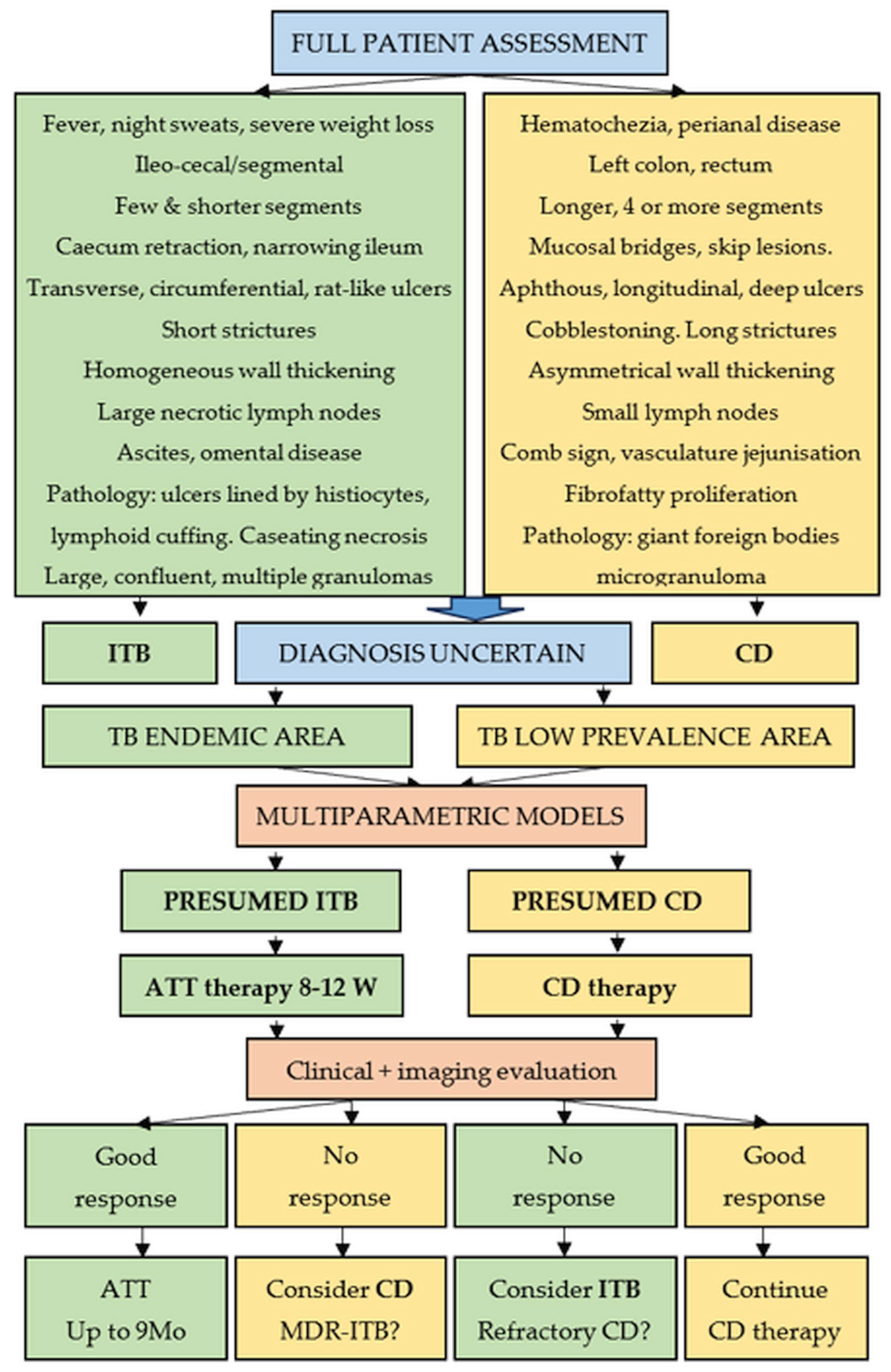
| Findings | ITB | CD |
|---|---|---|
| Site | Segmental colitis Isolated ileocecal involvement | Left colon Rectal (rarely) |
| Segments involved | Fewer | 4 or more |
| Ulcers | Transverse, circumferential Rat-like | Longitudinal, serpiginous Aphthous, segmental |
| Skip lesions | Absent | Present |
| Cobblestoning | Absent | Present |
| Strictures | Short | Long |
| Particular aspects | Scar diverticula Patulous ileocecal valve Narrowed terminal ileum | Mucosal bridges |
| Findings | ITB | CD |
|---|---|---|
| Site particularities | Caecum retraction Patulous ileocecal valve Narrowed terminal ileum | Left colon |
| Segments involved | Shorter, fewer than 4 | Longer, multiple |
| Ulcers | Transverse | Aphthous, longitudinal, deep |
| Skip lesions | Absent | Present |
| Cobblestone appearance | Absent | Present |
| Strictures | Short | Long |
| Wall thickening | Homogeneous | Asymmetrical |
| Stratification | Lost | Retained, intervening fat layer |
| Mucosal enhancement | Homogeneous | May be present |
| Vasculature abnormalities | - | Comb sign |
| Ileal vasculature “jejunization” | ||
| Lymph nodes | Necrotic, conglomerated, large, peripheral enhanced | Smaller |
| Extraintestinal signs | Ascites, omental disease | Fibrofatty proliferation |
| Pulmonary abnormalities | Up to 25% | No |
Disclaimer/Publisher’s Note: The statements, opinions and data contained in all publications are solely those of the individual author(s) and contributor(s) and not of MDPI and/or the editor(s). MDPI and/or the editor(s) disclaim responsibility for any injury to people or property resulting from any ideas, methods, instructions or products referred to in the content. |
© 2026 by the authors. Published by MDPI on behalf of the Lithuanian University of Health Sciences. Licensee MDPI, Basel, Switzerland. This article is an open access article distributed under the terms and conditions of the Creative Commons Attribution (CC BY) license.
Share and Cite
Cazacu, S.M.; Streba, C.T.; Constantin, C.; Ionele, C.M.; Rogoveanu, I.; Popescu, A.V.; Florescu, M.-M. The Overlap Between Crohn’s Disease and Intestinal Tuberculosis: A Never-Ending Story. Medicina 2026, 62, 794. https://doi.org/10.3390/medicina62040794
Cazacu SM, Streba CT, Constantin C, Ionele CM, Rogoveanu I, Popescu AV, Florescu M-M. The Overlap Between Crohn’s Disease and Intestinal Tuberculosis: A Never-Ending Story. Medicina. 2026; 62(4):794. https://doi.org/10.3390/medicina62040794
Chicago/Turabian StyleCazacu, Sergiu Marian, Costin Teodor Streba, Cristian Constantin, Claudiu Marinel Ionele, Ion Rogoveanu, Alexandru Valentin Popescu, and Mirela-Marinela Florescu. 2026. "The Overlap Between Crohn’s Disease and Intestinal Tuberculosis: A Never-Ending Story" Medicina 62, no. 4: 794. https://doi.org/10.3390/medicina62040794
APA StyleCazacu, S. M., Streba, C. T., Constantin, C., Ionele, C. M., Rogoveanu, I., Popescu, A. V., & Florescu, M.-M. (2026). The Overlap Between Crohn’s Disease and Intestinal Tuberculosis: A Never-Ending Story. Medicina, 62(4), 794. https://doi.org/10.3390/medicina62040794