Comprehensive Overview of Gastric Cancer Immunohistochemistry: Key Biomarkers, Advanced Detection Methods, and Perspectives

Abstract
1. Introduction
2. Materials and Methods
3. Results
3.1. Current Paradigm of Gastric Neoplasia Diagnosis
3.1.1. Methodological Foundations and IHC Technical Mechanisms
3.1.2. Morphological Foundations and Oncogenic Cascades
3.1.3. Epidemiological Dynamics and Redefinition of Risk Factors in 2026
3.1.4. Differential Diagnosis of Poorly Differentiated Tumors
3.2. Progress in the DAB Application and Advanced Reagents in Gastric Cancer IHC
3.2.1. DAB Evolution in the Context of Gastric Pathology
3.2.2. Optimization and Modern DAB Generations
4. Discussion
4.1. Precision Biomarkers in Gastric Cancer: The Role of IHC and DAB
4.1.1. Human Epidermal Growth Factor Receptor 2 (HER2)
Challenges of HER2 Assessment: From Heterogeneity to Score Discrepancies
Current Approaches
4.1.2. Immune Checkpoints: Programmed Death-Ligand 1 (PD-L1) and CPS Scoring
Interpretive Pitfalls
Interobserver Variability and Inter-Assay Discordance
4.1.3. Mismatch Repair and Microsatellite Instability (MMR/MSI)
4.1.4. Claudin 18.2: A New Frontier in Gastric Pathology (CLDN18.2)
Specificity and Rigor in Scoring CLDN18.2
4.1.5. Fibroblast Growth Factor Receptor 2b (FGFR2b)
4.2. Intestinal and Gastrointestinal Differentiation Markers: CDX2 and SATB2
4.3. Emerging and Aggressive Histological Subtypes: GAED and GA-FG
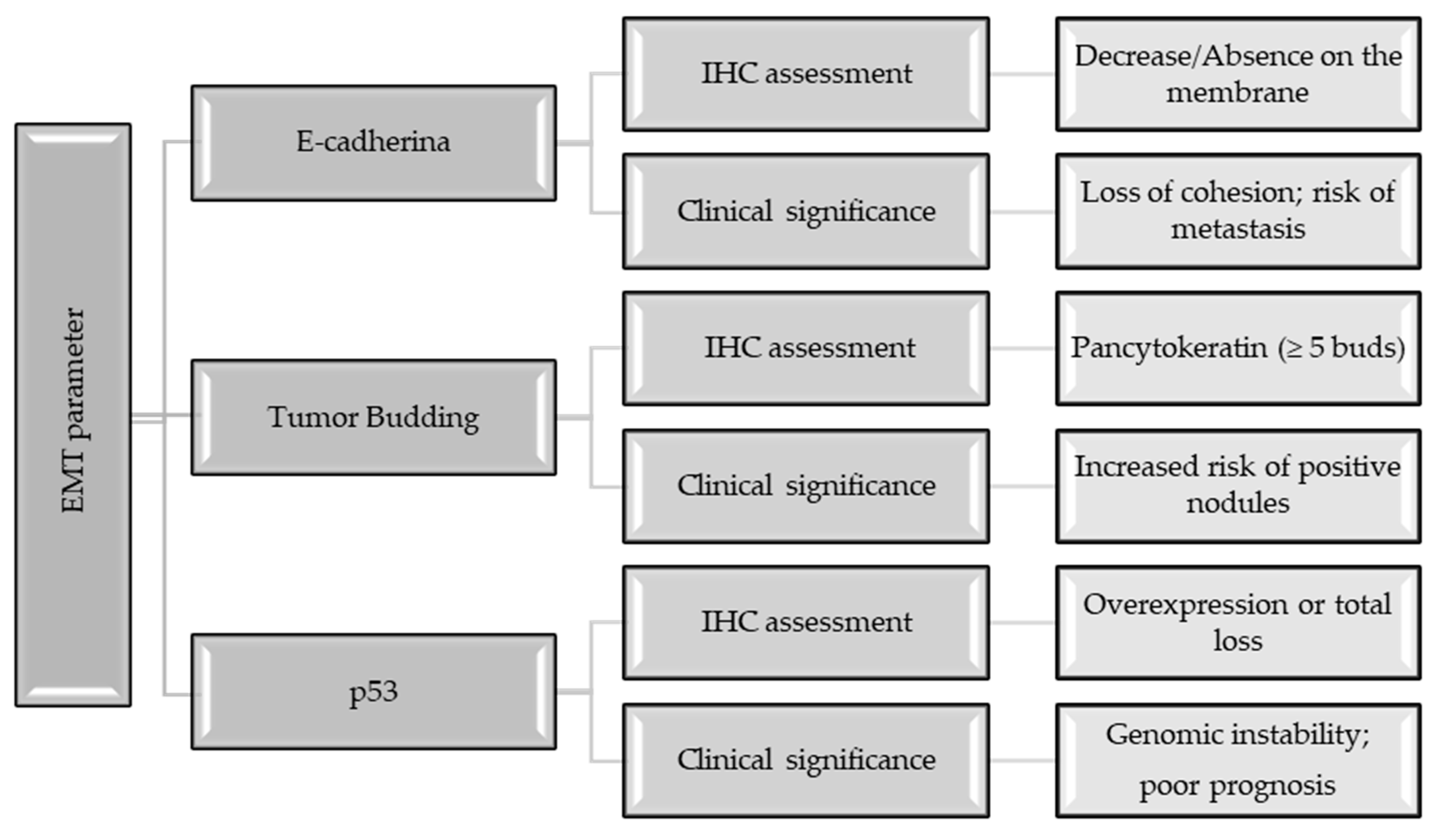
4.4. Epithelial–Mesenchymal Transition, E-Cadherin, and the Process of Tumor Budding
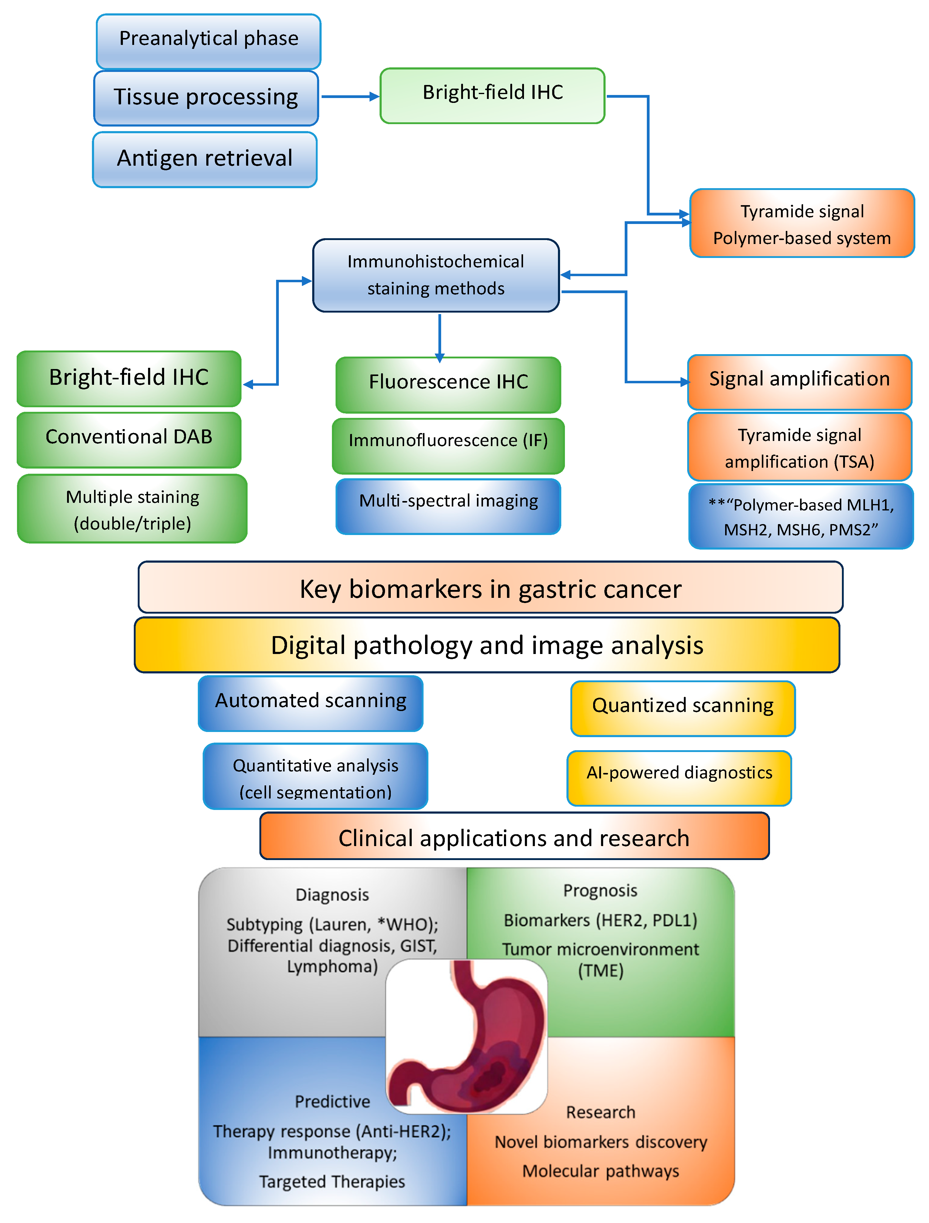
4.5. Implementation of Innovative Procedures in Gastric Cancer Immunohistochemistry
4.5.1. Synthetic Conclusions Regarding the IHC Limits
4.5.2. Advanced Methods in Gastric Cancer Immunohistochemistry
Advanced Signal Amplification Strategies: TSA and HCR
mIHC and TME Characterization
Multiplexing Reagents and Translucent Chromogens
Digital Pathology and Quantitative Image Analysis
4.5.3. Artificial Intelligence (AI) and Deep Learning Architectures
4.5.4. AI-Assisted Interpretation of PD-L1 and HER2
4.6. Automation and Quality Control in the Modern Laboratory
4.7. Sustainability and Green Chemistry in the Pathology Laboratory
5. Perspectives
- Standardization of “Reflex Testing”: Implementation of automated protocols where each gastric biopsy is immediately tested for HER2, MSI, PD-L1, and CLDN18.2, shortening the time until personalized treatment.
- Transition to all-digital diagnostics: Laboratories can adopt routine scanning of DAB-stained slides, using AI algorithms to identify focal heterogeneity that might be missed during manual examination.
- Development of environmentally friendly reagents: Systematic replacement of toxic components in IHC kits with biodegradable alternatives, without compromising diagnostic sensitivity.
- Brightfield multiplexing: Widespread adoption of translucent chromogens to replace costly immunofluorescence in routine diagnostics.
- The future of gastric diagnostics may be defined by:
- Digitalized quality control: Replacing classic tissue controls with “virtual controls” and automatically monitoring staining intensity through DIA algorithms to ensure global diagnostic uniformity [241].
6. Conclusions
Funding
Institutional Review Board Statement
Informed Consent Statement
Data Availability Statement
Conflicts of Interest
Abbreviations
| IHC | Immunohistochemistry |
| GC | Gastric cancer |
| HER2 | Human epidermal growth factor receptor 2 |
| PD-L1 | Programmed death-ligand 1 |
| MMR | Mismatch repair |
| dMMR | Mismatch repair deficiency |
| MSI | Microsatellite instability |
| AI | Artificial intelligence |
| CLDN 18.2 | Claudin 18.2 |
| TCGA | The Cancer Genome Atlas |
| ACRG | Asian Cancer Research Group |
| DAB | 3,3′-diaminobenzidine |
| CPS | Combined positive score |
| PCR | Polymerase chain reaction |
| mAbs | Monoclonal antibodies |
| pAbs | Polyclonal antibodies |
| RT-PCR | Reverse transcription-polymerase chain reaction |
| EBV | Epstein–Barr virus |
| CIN | Chromosomal instability |
| EOGC | Early-onset gastric cancer |
| FAP | Familial adenomatous polyposis |
| WLE | White light endoscopy |
| GIST | Gastrointestinal Stromal Tumor |
| PGHL | Primary Gastric Hodgkin Lymphoma |
| SPY | Synaptophysin |
| CgA | Acidic protein |
| HRP | Horseradish peroxidase |
| PCNA | Proliferating cell nuclear antigen |
| VEGF | Vascular endothelial growth factor |
| ToGA | Trastuzumab for Gastric Cancer |
| ADCs | Antibody–drug conjugates |
| FISH | Fluorescence in situ hybridization |
| ISH | In situ hybridization |
| CISH | Chromogenic in situ hybridization |
| IICs | Infiltrating immune cells |
| ICIs | Immune checkpoint inhibitors |
| LELGC | Lymphoepithelioma-like gastric carcinoma |
| NGS | Next-generation sequencing |
| ASCO/CAP | American Society of Clinical Oncology/College of American Pathologists |
| FGFR2b | Fibroblast growth factor receptor 2b |
| GEJ | Gastroesophageal junction |
| CDX2 | Caudal-type homeobox 2 |
| SATB2 | Special AT-rich sequence-binding protein 2 |
| GAED | Gastric Adenocarcinoma with enteroblastic differentiation |
| GPC3 | Glypican-3 |
| SALL4 | Spalt-like family of 4 C2H2 zinc finger transcription factors |
| AFP | Alpha-fetoprotein |
| GA-FG | Gastric adenocarcinoma of the fundic gland |
| EMT | Epithelial–mesenchymal transition |
| TSA | Tyramide signal amplification |
| HRC | Hybridization chain reaction |
| mIHC | Multiplex immunohistochemistry |
| mIF | Multiplex immunofluorescence |
| TME | Tumor microenvironment |
| CD8+ | T lymphocytes or cytotoxic T cells |
| CTCs | Circulating tumor cells |
| TLS | Tertiary lymphoid structures |
| NK | Natural killer cell function |
| LC3B | Microtubule-associated protein 1 light chain 3B |
| Cav-1 | Caveolin-1 |
| COX-2/PGE2 | Cyclooxygenase-2/prostaglandin E2 |
| QDs | Quantum dots |
| DIA | Digital image analysis |
| WSI | Whole-slide imaging |
| CNN | Convolutional neural network |
References
- Agrawal, R.; Jurel, P.; Garg, A.; Prajapati, B.G.; Ashique, S. Advances in Gastric Cancer Management: Signaling Pathways, Emerging Diagnostic and Therapeutic Strategies. Cancer Biother. Radiopharm. 2025, 10849785251408598. [Google Scholar] [CrossRef] [PubMed]
- Mamun, T.I.; Younus, S.; Rahman, H. Gastric cancer—Epidemiology, modifiable and non-modifiable risk factors, challenges and opportunities: An updated review. Cancer Treat. Res. Commun. 2024, 41, 100845. [Google Scholar] [CrossRef] [PubMed]
- Iwu, C.D.; Iwu-Jaja, C.J. Gastric Cancer Epidemiology: Current Trend and Future Direction. Hygiene 2023, 3, 256–268. [Google Scholar] [CrossRef]
- Lei, Z.-N.; Teng, Q.-X.; Tian, Q.; Chen, W.; Xie, Y.; Wu, K.; Zeng, Q.; Zeng, L.; Pan, Y.; Chen, Z.-S.; et al. Signaling pathways and therapeutic interventions in gastric cancer. Signal Transduct. Target. Ther. 2022, 7, 358. [Google Scholar] [CrossRef]
- Abengozar, R.; Sharma, A.; Sharma, R. Gastric cancer: Lessons learned from high-incidence geographic regions. J. Gastrointest. Oncol. 2021, 12, S350–S360. [Google Scholar] [CrossRef]
- Park, Y.S.; Kook, M.C.; Kim, B.H.; Lee, H.S.; Kang, D.W.; Gu, M.J.; Shin, O.R.; Choi, Y.; Lee, W.; Kim, H.; et al. The Gastrointestinal Pathology Study Group of the Korean Society of Pathologists. A standardized pathology report for gastric cancer: 2nd edition. J. Pathol. Transl. Med. 2023, 57, 1–27. [Google Scholar] [CrossRef]
- Magaki, S.; Hojat, S.A.; Wei, B.; So, A.; Yong, W.H. An Introduction to the Performance of Immunohistochemistry. In Biobanking; Yong, W., Ed.; Methods in Molecular Biology; Humana Press: New York, NY, USA, 2019; Volume 1897. [Google Scholar] [CrossRef]
- Bellizzi, A.M. An Algorithmic Immunohistochemical, Approach to Define Tumor Type and Assign Site of Origin. Adv. Anat. Pathol. 2020, 27, 114–163. [Google Scholar] [CrossRef]
- Lastraioli, E.; Romoli, M.R.; Arcangeli, A. Immunohistochemical biomarkers in gastric cancer research and management. Int. J. Surg. Oncol. 2012, 2012, 868645. [Google Scholar] [CrossRef]
- Idikio, H.A. Immunohistochemistry in diagnostic surgical pathology: Contributions of protein life-cycle, use of evidence-based methods and data normalization on interpretation of immunohistochemical stains. Int. J. Clin. Exp. Pathol. 2009, 3, 169–176. [Google Scholar]
- Rosenbaum, M.W.; Gonzalez, R.S. Immunohistochemistry as predictive and prognostic markers for gastrointestinal malignancies. Semin. Diagn. Pathol. 2022, 39, 48–57. [Google Scholar] [CrossRef]
- Angerilli, A.; Ghelardi, F.; Nappo, F.; Grillo, F.; Parente, P.; Lonardi, S.; Luchini, C.; Pietrantonio, F.; Ugolini, C.; Vanoli, A.; et al. Claudin-18.2 testing and its impact in the therapeutic management of patients with gastric and gastroesophageal adenocarcinomas: A literature review with expert opinion. Pathol. Res. Pract. 2024, 254, 155145. [Google Scholar] [CrossRef] [PubMed]
- Prodan-Bărbulescu, C.; Faur, F.I.; Varga, N.I.; Hajjar, R.; Pașca, P.; Ghenciu, L.A.; Feier, C.I.V.; Dema, A.; Fărcuț, N.; Bolintineanu, S.; et al. A Histopathological and Surgical Analysis of Gastric Cancer: A Two-Year Experience in a Single Center. Cancers 2025, 17, 2219. [Google Scholar] [CrossRef] [PubMed]
- Shi, D.; Yang, Z.; Cai, Y.; Li, H.; Lin, L.; Wu, D.; Zhang, S.; Guo, Q. Research advances in the molecular classification of gastric cancer. Cell. Oncol. 2024, 47, 1523–1536. [Google Scholar] [CrossRef] [PubMed]
- Kabir, I.M.; Idris, A.T.; Abubakar, S.D.; Isah, M.M.; Usman, A.; Yusuf, L.; Bello, Z.M.; Mohammed, I. Immunohistochemistry as an Indispensable Tool in Oncology. Indian. J. Gynecol. Oncolog. 2024, 22, 106. [Google Scholar] [CrossRef]
- Scheck, M.K.; Hofheinz, R.D.; Lorenzen, S. HER2-Positive Gastric Cancer and Antibody Treatment: State of the Art and Future Developments. Cancers 2024, 16, 1336. [Google Scholar] [CrossRef]
- Wang, Y.; Zhang, H.; Liu, C.; Wang, Z.; Wu, W.; Zhang, N.; Zhang, L.; Hu, J.; Luo, P.; Zhang, J.; et al. Immune checkpoint modulators in cancer immunotherapy: Recent advances and emerging concepts. J. Hematol. Oncol. 2022, 15, 111. [Google Scholar] [CrossRef]
- Chen, Z.; Song, Z.; Den, S.; Zhang, W.; Han, M.; Lan, T.; Du, X.; Ning, J.; XinHui Chen, X.H.; Lin, H.; et al. Application of Immune Checkpoint Inhibitors in Cancer. MedComm 2025, 6, e70176. [Google Scholar] [CrossRef]
- Starling, N.; Zhang, L.; Dunton, K.; Strübing, A.; Xiong, Y.; Livings, C.; Brannman, L.; Beykloo, M.Y.; Mohamed, H.; Trankov, N.; et al. Real-world treatment patterns and outcomes in advanced/metastatic gastric cancer or gastroesophageal junction adenocarcinoma treated with first-line anti-HER2 therapy in England. ESMO Gastrointest. Oncol. 2025, 10, 100242. [Google Scholar] [CrossRef]
- Baretton, G.B.; Lordick, F.; Gaiser, T.; Hofheinz, R.; Horst, D.; Lorenzen, S.; Moehler, M.; Röcken, C.; Schirmacher, P.; Stahl, M.; et al. Standardized and quality-assured predictive PD-L1 testing in the upper gastrointestinal tract. J. Cancer Res. Clin. Oncol. 2023, 149, 16231–16238. [Google Scholar] [CrossRef]
- Lordick, F.; Rha, S.Y.; Muro, K.; Yong, W.P.; Obermannová, R.L. Systemic Therapy of Gastric Cancer-State of the Art and Future Perspectives. Cancers 2024, 16, 3337. [Google Scholar] [CrossRef]
- Kim, S.W.; Roh, J.; Park, C.S. Immunohistochemistry for Pathologists: Protocols, Pitfalls, and Tips. J. Pathol. Transl. Med. 2016, 50, 411–418. [Google Scholar] [CrossRef]
- Alsina Maqueda, M.; Teijo Quintáns, A.; Cuatrecasas, M.; Fernández Aceñero, M.J.; Fernández Montes, A.; Gómez Martín, C.; Jiménez Fonseca, P.; Martínez Ciarpaglini, C.; Rivera Herrero, F.; Iglesias Coma, M. Biomarkers in gastroesophageal cancer 2025: An updated consensus statement by the Spanish Society of Medical Oncology (SEOM) and the Spanish Society of Pathology (SEAP). Clin. Transl. Oncol. 2025, 27, 3580–3594. [Google Scholar] [CrossRef] [PubMed]
- Li, S.; Liang, H.; Li, G. Predictive biomarkers for immunotherapy in gastric cancer. J. Cancer Metastasis Treat. 2025, 11, 8. [Google Scholar] [CrossRef]
- Caputo, A.; Angerilli, A.; Gambella, A.; L’Imperio, L.; Perrone, G.; Taffon, C.; Milione, M.; Grillo, F.; Mastracci, L.; Vanoli, A.; et al. Immunohistochemical biomarker scoring in gastroesophageal cancers: Can computers help us? Pathol. Res. Pract. 2025, 272, 156068. [Google Scholar] [CrossRef] [PubMed]
- Wang, X.T.; Kong, F.B.; Mai, W.; Li, L.; Pang, L.M. MUC1 Immunohistochemical Expression as a Prognostic Factor in Gastric Cancer: Meta-Analysis. Dis. Markers 2016, 2016, 9421571. [Google Scholar] [CrossRef]
- Mihaila, R.I.; Gheorghe, A.S.; Zob, D.L.; Stanculeanu, D.L. The Importance of Predictive Biomarkers and Their Correlation with the Response to Immunotherapy in Solid Tumors-Impact on Clinical Practice. Biomedicines 2024, 12, 2146. [Google Scholar] [CrossRef]
- Sanjari Moghaddam, A.; Lu, K.Y.; Nasrollahi, E.; Oji, L.; Homeniuk, A.; Amosu, O.; Chelysheva, D.; Brufsky, A.M. Biomarker prediction of immunotherapy response in breast cancer: From single markers to multi-omics integration. npj Breast Cancer 2026, 12, 10. [Google Scholar] [CrossRef]
- Kang, B.W.; Chau, I. Current status and future potential of predictive biomarkers for immune checkpoint inhibitors in gastric cancer. ESMO Open 2020, 5, e000791. [Google Scholar] [CrossRef]
- AlDoughaim, M.; AlSuhebany, N.; AlZahrani, M.; AlQahtani, T.; AlGhamdi, S.; Badreldin, H.; Al Alshaykh, H. Cancer Biomarkers and Precision Oncology: A Review of Recent Trends and Innovations. Clin. Med. Insights Oncol. 2024, 18, 11795549241298541. [Google Scholar] [CrossRef]
- Das, S.; Dey, M.K.; Devireddy, R.; Gartia, M.R. Biomarkers in Cancer Detection, Diagnosis, and Prognosis. Sensors 2024, 24, 37. [Google Scholar] [CrossRef]
- Zafar, S.; Hafeez, A.; Shah, H.; Mutiullah, I.; Ali, A.; Khan, K.; Figueroa-González, G.; Reyes-Hernández, O.D.; Quintas-Granados, L.I.; Peña-Corona, S.I.; et al. Emerging biomarkers for early cancer detection and diagnosis: Challenges, innovations, and clinical perspectives. Eur. J. Med. Res. 2025, 30, 760. [Google Scholar] [CrossRef] [PubMed]
- Passaro, A.; Al Bakir, M.; Hamilton, E.G.; Diehn, M.; André, F.; Roy-Chowdhuri, S.; Mountzios, G.; Wistuba, I.I.; Swanton, C.; Peters, S. Cancer biomarkers: Emerging trends and clinical implications for personalized treatment. Cell 2024, 187, 1617–1635. [Google Scholar] [CrossRef] [PubMed]
- Li, F.; Deng, H.; Hu, Z.; Chen, Z.; Zhang, H.; He, J.; Wang, X.; Liu, Y. Immunohistochemical-Based Molecular Typing of ACRG Combined with Immune-Associated PD-L1 Expression Can Predict the Prognosis of Gastric Cancer. Cancer Med. 2025, 14, e70863. [Google Scholar] [CrossRef] [PubMed]
- Nshizirungu, J.P.; Bennis, S.; Mellouki, I.; Sekal, M.; Benajah, D.A.; Lahmidani, N.; El Bouhaddouti, H.; Ibn Majdoub, K.; Ibrahimi, S.A.; Celeiro, S.P.; et al. Reproduction of the Cancer Genome Atlas (TCGA) and Asian Cancer Research Group (ACRG) Gastric Cancer Molecular Classifications and Their Association with Clinicopathological Characteristics and Overall Survival in Moroccan Patients. Dis. Markers 2021, 9980410. [Google Scholar] [CrossRef]
- Yoon, J.-Y.; Sy, K.; Brezden-Masley, C.; Streutker, C.J. Histo- and immunohistochemistry-based estimation of the TCGA and ACRG molecular subtypes for gastric carcinoma and their prognostic significance: A single-institution study. PLoS ONE 2019, 14, e0224812. [Google Scholar] [CrossRef]
- Raj, N.; Verma, D.; Kumar, A.; Rai, P.; Rao, R.N. HER2 Oncogene Amplification and Immunohistochemical Profiling in Gastric Adenocarcinoma. Discoveries 2018, 6, e83. [Google Scholar] [CrossRef]
- Migliore, C.; Fenocchio, E.; Giordano, S.; Corso, S. Precision oncology in gastric cancer: Shaping the future of personalized treatment. Cancer Treat. Rev. 2025, 141, 103038. [Google Scholar] [CrossRef]
- Airò, G.; Agnetti, V.; Pratticò, F.; Peroni, M.; Mura, G.; Urbanowicz-Nijaki, M.; Lai, E.; Bui, S.; Puzzoni, M.; Contu, F.; et al. Tissue Biomarkers in Gastric Cancer Treatment: Present and Future. Int. J. Transl. Med. 2024, 4, 640–660. [Google Scholar] [CrossRef]
- Zhou, J.; Li, J.; Chen, J.; Lan, X.; Ai, Y.; Liu, P.; Peng, J.; Pan, X.; Zhang, Y.; Zhang, H.; et al. Decoding inflammatory mediators in the Correa’s cascade: From chronic gastritis to carcinogennesis and targeted therapies. Int. Immunopharmacol. 2025, 162, 115191. [Google Scholar] [CrossRef]
- Miao, L.; Sun, Y.; Guo, M.; Yang, H.; Du, X.; Li, J.; Shen, J.; Wang, X.; Lei, R. Unique immunohistochemical profiles of MUC5AC, MUC6, P53, and Ki67 in gastric atypical hyperplasia and dysplasia. Int. J. Clin. Exp. Pathol. 2024, 17, 63–71. [Google Scholar] [CrossRef]
- Kim, M.; Seo, A.N. Molecular Pathology of Gastric Cancer. J. Gastric Cancer. 2022, 22, 273–305. [Google Scholar] [CrossRef] [PubMed]
- Lv, M.; Chen, F.; Li, Q.; Xue, M.; Wang, J. Comparative diagnostic accuracy of different artificial intelligence models for early gastric cancer: A systematic review and meta-analysis. Front. Oncol. 2025, 15, 1670843. [Google Scholar] [CrossRef] [PubMed]
- de Haan, K.; Zhang, Y.; Zuckerman, J.E.; Liu, T.; Sisk, A.E.; Diaz, M.F.P.; Jen, K.-Y.; Nobori, A.; Liou, S.; Zhang, S.; et al. Deep learning-based transformation of H&E stained tissues into special stains. Nat. Commun. 2021, 12, 4884. [Google Scholar] [PubMed]
- Pati, P.; Karkampouna, S.; Bonollo, F.; Compérat, E.; Radić, M.; Spahn, M.; Martinelli, A.; Wartenberg, M.; Kruithof-de Julio, M.; Rapsomaniki, M. Accelerating histopathology workflows with generative AI-based virtually multiplexed tumour profiling. Nat. Mach. Intell. 2024, 6, 1077–1093. [Google Scholar] [CrossRef]
- Chang, J.; Hatfield, B. Advances in Cancer Research, Chapter Ten—Advancements in computer vision and pathology: Unraveling the potential of artificial intelligence for precision diagnosis and beyond. Adv. Cancer Res. 2024, 161, 431–478. [Google Scholar]
- Zhou, Z.; Xie, Y.; Feng, X.; Li, Y.; Shen, L.; Chen, Y. Artificial intelligence in gastrointestinal cancer research: Image learning advances and applications. Cancer Lett. 2025, 614, 217555. [Google Scholar] [CrossRef]
- Liatsou, E.; Driva, T.S.; Vergadis, C.; Sakellariou, S.; Lykoudis, P.; Apostolou, K.G.; Tsapralis, D.; Schizas, D. Current Role of Artificial Intelligence in the Management of Gastric Cancer. Biomedicines 2025, 13, 2939. [Google Scholar] [CrossRef]
- Suri, C.; Ratre, Y.K.; Pande, B.; Bhaskar, L.; Verma, H.K. Artificial intelligence and machine learning-driven advancements in gastrointestinal cancer: Paving the way for precision medicine. World J. Gastroenterol. 2026, 32, 111428. [Google Scholar] [CrossRef]
- Ajani, J.A.; D’Amico, T.A.; Bentrem, D.J.; Corvera, C.U.; Das, P.; Enzinger, P.C.; Enzler, T.; Gerdes, H.; Gibson, M.K.; Grierson, P.; et al. Gastric Cancer, Version 2.2025. J. Natl. Compr. Canc. Netw. 2025, 23, 169–191. [Google Scholar] [CrossRef]
- Health Commission of The People’s Republic of China. National guidelines for diagnosis and treatment of gastric cancer 2022 in China (English version). Chin. J. Cancer Res. 2022, 34, 207–237. [Google Scholar]
- Zheng, C.; Jiang, Q.; Wang, K.; Li, T.; Zheng, W.; Cheng, Y.; Ning, Q.; Cui, D. Nanozyme enhanced magnetic immunoassay for dual-mode detection of gastrin-17. Analyst 2022, 147, 1678–1687. [Google Scholar] [CrossRef]
- Amemiya, K.; Hirotsu, Y.; Nagakubo, Y.; Watanabe, S.; Amemiya, S.; Mochizuki, H.; Oyama, T.; Kondo, T.; Omata, M. Simple IHC reveals complex MMR alternations than PCR assays: Validation by LCM and next-generation sequencing. Cancer Med. 2022, 11, 4479–4490. [Google Scholar] [CrossRef] [PubMed]
- Naseri, S.; Shukla, S.; Vagha, S. To study the utility of HER2 and Ki-67 as immunohistochemical prognostic markers in comparison to histopathological parameters and tumour, node and metastasis staging in colorectal carcinoma. Pan. Afr. Med. J. 2024, 48, 39. [Google Scholar] [CrossRef] [PubMed]
- Selvan, T.G.; Gollapalli, P.; Kumar, S.H.; Ghate, S.D. Early diagnostic and prognostic biomarkers for gastric cancer: Systems-level molecular basis of subsequent alterations in gastric mucosa from chronic atrophic gastritis to gastric cancer. J. Genet. Eng. Biotechnol. 2023, 21, 86. [Google Scholar] [CrossRef] [PubMed]
- Wu, X.; Wang, F.; Dai, W.; Ni, C.; Sun, L.; Gong, Y.; Dong, N.; Wang, Z.; Li, L.; Xu, Q.; et al. Mucin phenotype-based deep learning framework for intestinal metaplasia-carcinogenesis progression prediction. npj Precis. Onc. 2026, 10, 40. [Google Scholar] [CrossRef]
- Varon, V.; Mégraud, F.; Herrero, R.; Meng, W.; Qiao, L. Stomach cancer: Still one of the main cancer types worldwide. In World Cancer Report; Wild, C.P., Weiderpass, E., Stewart, B.W., Eds.; Cancer research for cancer prevention; International Agency for Research on Cancer: Lyon, France, 2020. Available online: https://www.ncbi.nlm.nih.gov/books/NBK606497/ (accessed on 25 February 2026).
- Weiqing, L.; Tai, Z. Precancerous pathways to gastric cancer: A review of experimental animal models recapitulating the correa cascade. Front. Cell Dev. Biol. 2025, 13, 1620756. [Google Scholar] [CrossRef]
- Zhang, W.; Zhang, Y.; Ning, J.; Fu, W.; Ding, S. Helicobacter pylori infection status and evolution of gastric cancer. Chin. Med. J. 2025, 138, 3083–3096. [Google Scholar] [CrossRef]
- Liliac, I.M.; Ungureanu, B.S.; Margaritescu, C.; Sacerdotianu, V.M.; Saftoiu, A.; Mogoanta, L.; Moraru, E.; Pirici, D. E-Cadherin Modulation and Inter-Cellular Trafficking in Tubular Gastric Adenocarcinoma: A High-Resolution Microscopy Pilot Study. Biomedicines 2022, 10, 349. [Google Scholar] [CrossRef]
- Stojanovic, B.; Jovanovic, I.; Dimitrijevic Stojanovic, M.; Milosevic, B.; Spasic, M.; Stojanovic, B.S.; Jakovljevic, S.; Zornic, N.; Jovanovic, D.; Nesic, J.; et al. Galectins as Master Regulators of Gastric Cancer Progression. Cells 2025, 14, 1090. [Google Scholar] [CrossRef]
- Lee, J.-S. Evolving Molecular Subtypes of Gastric Cancer: From Past Classifications to Present Consensus and Future Directions for Precision Therapy. J. Gastric Cancer 2026, 26, 16–30. [Google Scholar] [CrossRef]
- Das, A.; Chetta, P.M.; Zhang, L. Molecular Advances in Gastrointestinal Pathology. Semin. Diagn. Pathol. 2026, 43, 150990. [Google Scholar] [CrossRef] [PubMed]
- Bos, J.; Groen-van Schooten, T.S.; Brugman, C.P.; Jamaludin, F.S.; van Laarhoven, H.W.M.; Derks, S. The tumor immune composition of mismatch repair deficient and Epstein-Barr virus-positive gastric cancer: A systematic review. Cancer Treat. Rev. 2024, 127, 102737. [Google Scholar] [PubMed]
- Park, J.Y.; Georges, D.; Alberts, C.J.; Bray, F.; Gary Clifford, G.; Baussano, I. Global lifetime estimates of expected and preventable gastric cancers across 185 countries. Nat. Med. 2025, 31, 3020–3027. [Google Scholar] [CrossRef] [PubMed]
- Sebesta, C.; Sebesta, C.G.; Sebesta, M.C.; Köcher, M.; Müllner-Ammer, K.; Zottl, J. How the Fight against Stomach Cancer can be won. J. Cancer Sci. Clin. Ther. 2024, 8, 295–309. [Google Scholar] [CrossRef]
- Ashman, J.B.; Hallemeier, C.L.; Beamer, S.E.; Tepper, J.E. 56—Esophagus-Gastric Cancer. In Gunderson & Tepper’s Clinical Radiation Oncology, 6th ed.; Elsevier: Amsterdam, The Netherlands, 2026; pp. 936–972.e16. Available online: https://www.sciencedirect.com/science/chapter/edited-volume/abs/pii/B9780443114762000568 (accessed on 25 February 2026).
- Oh, S.E.; Park, S.; Ahn, S.; An, J.Y.; Lee, J.H.; Sohn, T.S.; Bae, J.M.; Choi, M.-G. Prognostic Significance of Esophagogastric Junction Invasion in Patients with Adenocarcinoma of the Cardia or Subcardia. Cancers 2023, 15, 1656. [Google Scholar] [CrossRef]
- Wang, X.; Gao, X.; Yu, J.; Zhang, X.; Nie, Y. Emerging trends in early-onset gastric cancer. Chin. Med. J. 2024, 137, 2146–2156. [Google Scholar]
- Heidary, M.; Akrami, S.; Madanipour, T.; Shakib, N.H.; Ari, M.M.; Bei, M.; Khoshnood, S.; Ghanavati, R.; Bazdar, M. Effect of Helicobacter pylori–induced gastric cancer on gastrointestinal microbiota: A narrative review. Front. Oncol. 2025, 14, 495596. [Google Scholar] [CrossRef]
- Yee, E.J.; Gilbert, D.; Kaplan, J.; van Dyk, L.; Kim, S.S.; Berg, L.; Clambey, E.; Wani, S.; McCarter, M.D.; Stewart, C.L. Immune Landscape of Epstein-Barr Virus Associated Gastric Cancer: Analysis from a Western Academic Institution. J. Surg. Res. 2024, 296, 742–750. [Google Scholar]
- Malfertheiner, P.; Camargo, M.C.; El-Omar, E.; Liou, J.M.; Peek, R.; Schulz, C.; Suerbaum, S.I.S. Helicobacter pylori infection. Nat. Rev. Dis. Primers 2023, 9, 19. [Google Scholar]
- Naseem, M.; Barzi, A.; Brezden-Masley, C.; Puccini, A.; Berger, M.D.; Tokunaga, R.; Battaglin, F.; Soni, S.; McSkane, M.; Zhang, W.; et al. Outlooks on Epstein-Barr virus associated gastric cancer. Cancer Treat. Rev. 2018, 66, 15–22. [Google Scholar] [CrossRef]
- Leoz, M.L.; Sánchez, A.; Carballal, S.; Ruano, L.; Ocana, T.; Pellisé, M.; Castells, A.; Balaguer, F.; Moreira, L. Hereditary gastric and pancreatic cancer predisposition syndromes. Gastroenterol. Hepatol. Engl. Ed. 2016, 39, 481–493. [Google Scholar] [CrossRef] [PubMed]
- Ko, K.-P. Risk Factors of Gastric Cancer and Lifestyle Modification for Prevention. J. Gastric Cancer 2024, 24, 99–107. [Google Scholar] [CrossRef] [PubMed]
- Panozzo, M.P.; Antico, A.; Bizzaro, N. Monitoring the follow-up of autoimmune chronic atrophic gastritis using parietal cell antibodies and markers of gastric function. J. Transl. Autoimmun. 2025, 10, 100273. [Google Scholar] [CrossRef] [PubMed]
- Castellana, C.; Eusebi, L.H.; Dajti, E.; Iascone, V.; Vestito, A.; Fusaroli, P.; Fuccio, L.; D’Errico, A.; Zagari, R.M. Autoimmune Atrophic Gastritis: A Clinical Review. Cancers 2024, 16, 1310. [Google Scholar] [CrossRef]
- Martins, B.C.; Moura, R.N.; Kum, A.S.T.; Matsubayashi, C.O.; Marques, S.B.; Vaz Safatle-Ribeiro, A. Endoscopic Imaging for the Diagnosis of Neoplastic and Pre-Neoplastic Conditions of the Stomach. Cancers 2023, 15, 2445. [Google Scholar] [CrossRef]
- Pradnyani, P.I.; Willy Sandhika, W. Tissue Biomarker in Colorectal Carcinoma. GSC Biol. Pharm. Sci. 2025, 33, 308–315. [Google Scholar] [CrossRef]
- Zhang, Y.J.; Chen, J.W.; He, X.S.; Zhang, H.Z.; Ling, Y.H.; Wen, J.H.; Deng, W.H.; Li, P.; Yun, J.P.; Xie, D.; et al. SATB2 is a Promising Biomarker for Identifying a Colorectal Origin for Liver Metastatic Adenocarcinomas. eBioMedicine 2018, 28, 62–69. [Google Scholar] [CrossRef]
- Murphy, B.; Dowling, G.; O’Toole, G.; Molloy, A. Metastatic gastrointestinal adenocarcinoma masquerading as a primary malignant bone tumour in the humerus: A case report and review of the literature. J. Orthop. Rep. 2026, 100832. [Google Scholar] [CrossRef]
- Çoban, F.; Doğan, M. CK7-negative and CK20-Positive intestinal- type gastric adenocarcinoma metastasis to the bladder: A case report of an unprecedented phenotype. Urol. Case Rep. 2025, 63, 103184. [Google Scholar] [CrossRef]
- Dhannoon, A.N.; Khalid, Z.A.; Mirza, S.A. Immunohistochemical Expression of CDx2, CK20, Ck7 in Carcinoma of Ampulla of Vater: Clinicopathological Study. Eur. J. Med. Health Res. 2025, 3, 47–53. [Google Scholar] [CrossRef]
- Carbone, A.; Alibrahim, M.N.; Gloghini, A. What Is Still Unclear or Unresolved in Classic Hodgkin Lymphoma Pathobiology, Diagnosis, and Treatment. Hemato 2025, 6, 20. [Google Scholar] [CrossRef]
- Parente, P.; Zanelli, M.; Sanguedolce, F.; Mastracci, L.; Graziano, P. Hodgkin Reed–Sternberg-Like Cells in Non-Hodgkin Lymphoma. Diagnostics 2020, 10, 1019. [Google Scholar]
- Parab, T.M.; DeRogatis, M.J.; Boaz, A.M.; Grasso, S.A.; Issack, P.S.; Duarte, D.A.; Urayeneza, O.; Vahdat, S.; Qiao, J.H.; Hinika, G.S. Gastrointestinal stromal tumors: A comprehensive review. J. Gastrointest. Oncol. 2019, 10, 144–154. [Google Scholar] [PubMed]
- Usama, F.; Rasikh, R.; Hassam, K.; Rahman, M.; Khalil Ur Rehman, F.; Khan, I.W.; Lau, D.T. An update on gastrointestinal stromal tumors (GISTs) with a focus on extragastrointestinal stromal tumors (EGISTs). Gastroenterol. Rep. 2025, 13, goaf068. [Google Scholar] [CrossRef]
- Mukherjee, S.; Vagha, S.; Mukherjee, M. Various Markers of Neuroendocrine Tumor: A Narrative Review. Cureus 2024, 16, e67493. [Google Scholar] [CrossRef]
- Marinoni, I.; Avanthay, S.; Alcala, N. Novel concepts of cell-of origin in neuroendocrine neoplasms. Virchows Arch. 2026, 448, 21–32. [Google Scholar]
- La Rosa, S. Challenges in High-grade Neuroendocrine Neoplasms and Mixed Neuroendocrine/Non-neuroendocrine Neoplasms. Endocr. Pathol. 2021, 32, 245–257. [Google Scholar]
- Sun, Y.; Puspanathan, P.; Lim, T.; Lin, D. Advances and challenges in gastric cancer testing: The role of biomarkers. Cancer. Biol. Med. 2025, 22, 212–230. [Google Scholar]
- Yao, J.; Sun, Q.; Wu, H.; Zhao, X.; Yang, P.; Wang, X.; Wang, X.; Gu, M.; Li, J.; Zheng, Y.; et al. Decoding the molecular landscape: HER2 and PD-L1 in advanced gastric cancer. Front. Immunol. 2025, 16, 1567308. [Google Scholar] [CrossRef]
- Chen, S.; Ding, P.; Guo, H.; Meng, L.; Zhao, Q.; Li, C. Applications of artificial intelligence in digital pathology for gastric cancer. Front. Oncol. 2024, 14, 1437252. [Google Scholar] [CrossRef]
- Gupta, B.; Yang, G.; Petrauskene, O.; Key, M. Recent Advances in Chromogens for Immunohistochemistry. In Signal Transduction Immunohistochemistry; Kalyuzhny, A.E., Ed.; Methods in Molecular Biology; Humana: New York, NY, USA, 2023; Volume 2593. [Google Scholar] [CrossRef]
- Natera, J.E.; Walter, A.; Massad, A.; Amat-Guerri, F.; García, N.A. Elementary processes in the eosin-sensitized photooxidation of 3,3′-diaminobenzidine for correlative fluorescence and electron microscopy. J. Photochem. Photobiol. A-Chem. 2011, 220, 25–30. [Google Scholar] [CrossRef]
- Rodig, S.J. Detecting Horseradish Peroxidase-Labeled Cells. Cold Spring Harb. Protoc. 2019, 2019, pdb.prot099713. [Google Scholar] [CrossRef] [PubMed]
- Dölle, C.; Laurence, A.; Bindoff, L.A.; Charalampos Tzoulis, C. 3,3′-Diaminobenzidine staining interferes with PCR-based DNA analysis. Sci. Rep. 2018, 8, 1272. [Google Scholar] [CrossRef] [PubMed]
- Salvatori, S.; Marafini, I.; Laudisi, F.; Monteleone, G.; Stolfi, C. Helicobacter pylori and Gastric Cancer: Pathogenetic Mechanisms. Int. J. Mol. Sci. 2023, 24, 2895. [Google Scholar] [CrossRef] [PubMed]
- Hu, L.; Li, H.L.; Li, W.F.; Chen, J.M.; Yang, J.T.; Gu, J.J.; Xin, L. Clinical significance of expression of proliferating cell nuclear antigen and E-cadherin in gastric carcinoma. World J. Gastroenterol. 2017, 23, 3721–3729. [Google Scholar] [CrossRef]
- Ionescu, C.; Oprea, B.; Ciobanu, G.; Georgescu, M.; Bica, R.; Mateescu, G.-O.; Huseynova, F.; Veronique Barragan-Montero, V. The Angiogenic Balance and Its Implications in Cancer and Cardiovascular Diseases: An Overview. Medicina 2022, 58, 903. [Google Scholar] [CrossRef]
- Ghalehbandi, S.; Yuzugulen, J.; Pranjol, Z.I.; Pourgholami, M.H. The role of VEGF in cancer-induced angiogenesis and research progress of drugs targeting VEGF. Eur. J. Pharmacol. 2023, 949, 175586. [Google Scholar] [CrossRef]
- He, M.-Q.; He, M.-Q.; Wang, J.F.; Zhu, B.L.; Sun, N.; Zhou, X.H.; Yao, R.X. Vascular Endothelial Growth Factor and Cluster of Differentiation 34 for Assessment of Perioperative Bleeding Risk in Gastric Cancer Patients. Chin. Med. J. 2016, 129, 1950–1954. [Google Scholar] [CrossRef]
- Liu, X.; Chu, K.M. E-cadherin and gastric cancer: Cause, consequence, and applications. BioMed. Res. Int. 2014, 2014, 637308. [Google Scholar] [CrossRef]
- Betazoid DAB Chromogen Kit—Biocare Medical. Available online: https://biocare.net/product/betazoid-dab-chromogen-kit/#:~:text=$195.00%20%E2%80%93%20$3%2C416.00,manually%20or%20on%20automated%20stainers (accessed on 20 February 2026).
- Detection and Amplification Systems. Available online: https://www.abcam.com/en-us/technical-resources/guides/ihc-guide/detection-and-amplification-systems?hl=en-US (accessed on 20 February 2026).
- Ultrasensitive IHC Detection with HRP-Polymer Conjugates. 2017. Available online: https://www.novusbio.com/antibody-news/antibodies/ultrasensitive-ihc-detection-with-hrp-polymer-conjugates?hl=en-US (accessed on 20 February 2026).
- Mendler, C.T.; Friedrich, L.; Laitinen, I.; Schlapschy, M.; Schwaiger, M.; Wester, H.J.; Skerra, A. High contrast tumor imaging with radio-labeled antibody Fab fragments tailored for optimized pharmacokinetics via PASylation. MAbs 2015, 7, 96–109. [Google Scholar] [CrossRef]
- Al Ojaimi, Y.; Blin, T.; Lamamy, J.; Gracia, M.; Pitiot, A.; Denevault-Sabourin, C.; Joubert, N.; Pouget, J.-P.; Gouilleux-Gruart, V.G.; Heuzé-Vourc’h, N.; et al. Therapeutic antibodies—Natural and pathological barriers and strategies to overcome them. Pharmacol. Ther. 2022, 233, 108022. [Google Scholar] [CrossRef] [PubMed]
- Choi, S.; Park, S.; Kim, H.; Kang, S.Y.; Ahn, S.; Kim, K.M. Gastric Cancer: Mechanisms, Biomarkers, and Therapeutic Approaches. Biomedicines 2022, 10, 543. [Google Scholar] [CrossRef] [PubMed]
- Sato, Y.; Okamoto, K.; Kawano, Y.; Kasai, A.; Kawaguchi, T.; Sagawa, T.; Sogabe, M.; Miyamoto, H.; Takayama, T. Novel Biomarkers of Gastric Cancer: Current Research and Future Perspectives. J. Clin. Med. 2023, 12, 4646. [Google Scholar] [CrossRef] [PubMed]
- Angerilli, V.; Parente, P.; Campora, M.; Ugolini, C.; Battista, S.; Cassoni, P.; Gambella, A.; Cavallin, F.; De Lisi, G.; Vanoli, A.; et al. HER2-low in gastro-oesophageal adenocarcinoma: A real-world pathological perspective. J. Clin. Pathol. 2023, 76, 815–821. [Google Scholar] [CrossRef]
- Ricci, A.D.; Rizzo, A.; Rojas Llimpe, F.L.; Di Fabio, F.; De Biase, D.; Rihawi, K. Novel HER2-Directed Treatments in Advanced Gastric Carcinoma: AnotHER Paradigm Shift? Cancers 2021, 13, 1664. [Google Scholar] [CrossRef]
- Bonomi, M.; Spada, D.; Baiocchi, G.L.; Celotti, A.; Brighenti, M.; Grizzi, G. Targeting HER2 in Gastroesophageal Adenocarcinoma: Molecular Features and Updates in Clinical Practice. Int. J. Mol. Sci. 2024, 25, 3876. [Google Scholar] [CrossRef]
- Nida, I.; Naveed, I. Human Epidermal Growth Factor Receptor 2 (HER2) in Cancers: Overexpression and Therapeutic Implications. Mol. Biol. Int. 2014, 2014, 852748. [Google Scholar] [CrossRef]
- Dhakras, P.; Uboha, N.; Horner, V.; Reinig, E.; Matkow, K.A. Gastrointestinal cancers: Current biomarkers in esophageal and gastric adenocarcinoma. Transl. Gastroenterol. Hepatol. 2020, 5, 55. [Google Scholar] [CrossRef]
- Shimozaki, K.; Fukuoka, S.; Ooki, A.; Yamaguchi, K. HER2-low gastric cancer: Is the subgroup targetable? ESMO Open 2024, 9, 103679. [Google Scholar] [CrossRef]
- Ma, C.; Wang, X.; Guo, J.; Yang, B.; Li, Y. Challenges and future of HER2-positive gastric cancer therapy. Front. Oncol. 2023, 13, 1080990. [Google Scholar] [CrossRef]
- Abrahao-Machado, L.F.; Scapulatempo-Neto, C. HER2 testing in gastric cancer: An update. World J. Gastroenterol. 2016, 22, 4619–4625. [Google Scholar] [CrossRef] [PubMed]
- Zhou, R.; Yuan, P.; Zhang, L.; Shen, S.; Li, Z.; Wang, Y. Using digital PCR to detect HER2 amplification in breast and gastric cancer patients. Front. Lab. Med. 2018, 2, 102–108. [Google Scholar] [CrossRef]
- Symeonidis, D.; Tepetes, K. Techniques and Current Role of Sentinel Lymph Node (SLN) Concept in Gastric Cancer Surgery. Front. Surg. 2019, 5, 77. [Google Scholar] [CrossRef] [PubMed]
- Ahn, S.; Ahn, S.; Van Vrancken, M.; Lee, M.; Ha, S.Y.; Lee, H.; Min, B.H.; Lee, J.H.; Kim, J.J.; Choi, S.; et al. Ideal number of biopsy tumor fragments for predicting HER2 status in gastric carcinoma resection specimens. Oncotarget 2015, 6, 38372–38380. [Google Scholar] [CrossRef]
- Huang, S.C.; Ng, K.F.; Lee, S.E.; Chen, K.H.; Yeh, T.S.; Chen, T.C. HER2 testing in paired biopsy and excision specimens of gastric cancer: The reliability of the scoring system and the clinicopathological factors relevant to discordance. Gastric Cancer 2016, 19, 176–182. [Google Scholar] [CrossRef]
- Cho, E.Y.; Srivastava, A.; Park, K.; Kim, J.; Lee, M.H.; Do, I.; Lee, J.; Kim, K.M.; Sohn, T.S.; Kang, W.K.; et al. Comparison of four immunohistochemical tests and FISH for measuring HER2 expression in gastric carcinomas. Pathology 2012, 44, 216–220. [Google Scholar] [CrossRef]
- Park, Y.; Koh, J.; Na, H.Y.; Kwak, Y.; Lee, K.-W.; Ahn, S.-H.; Park, D.J.; Kim, H.-H.; Lee, H.S. PD-L1 Testing in Gastric Cancer by the Combined Positive Score of the 22C3 PharmDx and SP263 Assay with Clinically Relevant Cut-offs. Cancer Res. Treat. 2020, 52, 661–670. [Google Scholar] [CrossRef]
- Woo, C.G.; Ho, W.J.; Park, Y.S.; Park, S.R.; Ryu, M.H.; Jung, H.Y.; Kang, Y.K. A potential pitfall in evaluating HER2 immunohistochemistry for gastric signet ring cell carcinomas. Pathology 2017, 49, 38–43. [Google Scholar] [CrossRef]
- Onguru, O.; Zhang, P.J. The relation between percentage of immunostained cells and amplification status in breast cancers with equivocal result for Her2 immunohistochemistry. Pathol. Res. Pract. 2016, 212, 381–384. [Google Scholar] [CrossRef]
- Yu, M.; Liang, Y.; Li, L.; Zhao, L.; Kong, F. Research progress of antibody-drug conjugates therapy for HER2-low expressing gastric cancer. Transl. Oncol. 2023, 29, 101624. [Google Scholar] [CrossRef]
- Guliyev, M.; Safarov, S.; Günaltılı, M.; Fidan, M.C.; Çerme, E.; Alkan Şen, G.; Emin Öztürk, A.; Kepil, N.; Alan, Ö.; Demirci, N.S. The impact of low HER2 expression on clinicopathological features and clinical outcomes in patients with metastatic gastric cancer. Clin. Res. Hepatol. Gastroenterol. 2025, 49, 102646. [Google Scholar] [CrossRef]
- Huang, D.; Sun, F.; Li, S.; Ke, L. Efficacy and safety of antibody-drug conjugates for HER2-expressing advanced gastric and gastroesophageal junction adenocarcinoma: A systematic review and meta-analysis. Front. Pharmacol. 2025, 16, 1668511. [Google Scholar] [CrossRef]
- Abrahão-Machado, L.F.; Jácome, A.A.; Wohnrath, D.R.; dos Santos, J.S.; Carneseca, E.C.; Fregnani, J.H.; Scapulatempo-Neto, C. HER2 in gastric cancer: Comparative analysis of three different antibodies using whole-tissue sections and tissue microarrays. World J. Gastroenterol. 2013, 19, 6438–6446. [Google Scholar] [CrossRef] [PubMed]
- Werner, D.; Battmann, A.; Steinmetz, K.; Jones, T.; Lamb, T.; Martinez, M.; Altmannsberger, H.M.; Al-Batran, S.E. The validation of a novel method combining both HER2 immunohistochemistry and HER2 dual-colour silver in situ hybridization on one slide for gastric carcinoma testing. J. Transl. Med. 2014, 12, 160. [Google Scholar] [CrossRef] [PubMed]
- Gülten, G.; Yilmaz, Y.; Çalli Demirkan, N.Ç. Comparing human epidermal growth factor receptor 2 amplification and expression using immunohistochemistry and silver in situ hybridisation in gastric carcinoma and lymph node metastasis. Oncol. Lett. 2020, 20, 1897–1905. [Google Scholar] [CrossRef] [PubMed]
- Davidson, M.; Starling, N. Trastuzumab in the management of gastroesophageal cancer: Patient selection and perspectives. Onco. Targets Ther. 2016, 9, 7235–7245. [Google Scholar] [CrossRef]
- Das, A.; Tomita, N.; Syme, K.J.; Ma, W.; O’Connor, P.; Corbett, K.N.; Ren, B.; Liu, X.; Hassanpour, S. Cross-Modality Learning for Predicting Immunohistochemistry Biomarkers from Hematoxylin and Eosin-Stained Whole Slide Images. Am. J. Pathol. 2025, 195, 2400–2410. [Google Scholar] [CrossRef]
- de Ruiter, E.J.; Mulder, F.J.; Koomen, B.M.; Speel, E.-J.; van den Hout, M.F.C.M.; de Roest, R.H.; Bloemena, E.; Bloemena, E.; Devriese, L.A.; Willems, S.M. Comparison of three PD-L1 immunohistochemical assays in head and neck squamous cell carcinoma (HNSCC). Mod. Pathol. 2021, 34, 1125–1132. [Google Scholar] [CrossRef]
- Ye, M.; Huang, D.; Zhang, Q.; Weng, W.; Tan, C.; Qin, G.; Jiang, W.; Sheng, W.; Wang, L. Heterogeneous programmed death-ligand 1 expression in gastric cancer: Comparison of tissue microarrays and whole sections. Cancer Cell Int. 2020, 20, 186. [Google Scholar] [CrossRef]
- Moehler, M.; Yoon, H.H.; Wagner, D.-C.; Yang, S.; Shi, J.; Yun Zhang, Y.; Hu, H.; La Placa, C.; Peng, Y.; Du, W.; et al. Concordance Between the PD-L1 Tumor Area Positivity Score and Combined Positive Score for Gastric or Esophageal Cancers Treated with Tislelizumab. Mod. Pathol. 2025, 38, 100793. [Google Scholar] [CrossRef]
- Nakano, H.; Saito, M.; Nakajima, S.; Saito, K.; Nakayama, Y.; Kase, K.; Yamada, L.; Kanke, Y.; Hanayama, H.; Onozawa, H.; et al. PD-L1 overexpression in EBV-positive gastric cancer is caused by unique genomic or epigenomic mechanisms. Sci. Rep. 2021, 11, 1982. [Google Scholar] [CrossRef] [PubMed]
- Lima, Á.; Sousa, H.; Medeiros, R.; Nobre, A.; Machado, M. PD-L1 expression in EBV associated gastric cancer: A systematic review and meta-analysis. Discov. Oncol. 2022, 13, 19. [Google Scholar] [CrossRef] [PubMed]
- Svensson, M.; Borg, D.; Zhang, C.; Hedner, C.; Nodin, B.; Uhlen, M.; Mardinoglu, A.; Leandersson, K.; Jirstrom, K. Associations of PD-1 and PD-L1 expression with mismatch repair status and prognosis in chemoradiotherapy-naïve esophageal and gastric adenocarcinoma. J. Clin. Oncol. 2018, 36, 9. [Google Scholar] [CrossRef]
- Noori, M.; Mahjoubfar, A.; Azizi, S.; Fayyaz, F.; Rezaei, N. Immune checkpoint inhibitors plus chemotherapy versus chemotherapy alone as first-line therapy for advanced gastric and esophageal cancers: A systematic review and meta-analysis. Int. Immunopharmacol. 2022, 113, 109317. [Google Scholar] [CrossRef]
- Wang, H.L.; Tang, L.H.; Troncone, G.; Rojo, F.; Van Treeck, B.J.; Pratt, J.; Shnitsa, I.; Kumar, G.; Karasarides, M.; Anders, R.A. High Interobserver Variability Among Pathologists Using Combined Positive Score to Evaluate PD-L1 Expression in Gastric, Gastroesophageal Junction, and Esophageal Adenocarcinoma. Mod. Pathol. 2023, 36, 100154. [Google Scholar]
- Ahn, S.; Kim, K.-M. PD-L1 expression in gastric cancer: Interchangeability of 22C3 and 28-8 pharmDx assays for responses to immunotherapy. Mod. Pathol. 2021, 34, 1719–1727. [Google Scholar] [CrossRef]
- Shigeta, N.; Murakami, S.; Yokose, T.; Isaka, T.; Shinada, K.; Nagashima, T.; Adachi, H.; Shigefuku, S.; Murakami, K.; Miura, J.; et al. Comparison of SP263 and 22C3 pharmDx assays to test programmed death ligand-1 (PD-L1) expression in surgically resected non-small cell lung cancer. Thorac. Cancer 2024, 15, 1343–1349. [Google Scholar]
- Abrha, A.; Shukla, N.D.; Hodan, R.; Longacre, T.; Raghavan, S.; Pritchard, C.C.; Fisher, G.; Ford, J.; Haraldsdottir, S. Universal Screening of Gastrointestinal Malignancies for Mismatch Repair Deficiency at Stanford. JNCI Cancer Spectr. 2020, 4, pkaa054. [Google Scholar] [CrossRef]
- Kim, S.M.; An, J.Y.; Byeon, S.J.; Lee, J.; Kim, K.M.; Choi, M.G.; Lee, J.H.; Sohn, T.S.; Bae, J.M.; Kim, S. Prognostic value of mismatch repair deficiency in patients with advanced gastric cancer, treated by surgery and adjuvant 5-fluorouracil and leucovorin chemoradiotherapy. Eur. J. Surg. Oncol. 2020, 46, 189–194. [Google Scholar]
- Maja, L.; Nádorvári, G.L.; Kulka, J.; Kiss, A.; Tímár, J. Microsatellite instability and mismatch repair protein deficiency: Equal predictive markers. Pathol. Oncol. Res. 2024, 30, 1611719. [Google Scholar] [CrossRef]
- Wang, C.; Zhang, L.; Vakiani, E.; Shia, J. Detecting mismatch repair deficiency in solid neoplasms: Immunohistochemistry, microsatellite instability, or both? Mod. Pathol. 2022, 35, 1515–1528. [Google Scholar] [CrossRef]
- Svrcek, M.; Lascols, O.; Cohen, R.; Collura, A.; Jonchère, V.; Fléjou, J.-F.; Buhard, O.; Alex Duval, A. MSI/MMR-deficient tumor diagnosis: Which standard for screening and for diagnosis? Diagnostic modalities for the colon and other sites: Differences between tumors. Bull. Cancer 2019, 106, 119–128. [Google Scholar] [CrossRef] [PubMed]
- Reitsam, N.G.; Märkl, B.; Dintner, S.; Waidhauser, J.; Vlasenko, D.; Grosser, B. Concurrent loss of MLH1, PMS2 and MSH6 immunoexpression in digestive system cancers indicating a widespread dysregulation in DNA repair processes. Front. Oncol. 2022, 12, 1019798. [Google Scholar] [CrossRef] [PubMed]
- Zhou, K.I.; Hanks, B.A.; Strickler, J.H. Management of Microsatellite Instability High (MSI-H) Gastroesophageal Adenocarcinoma. J. Gastrointest. Cancer 2024, 55, 483–496. [Google Scholar] [CrossRef] [PubMed]
- Wang, R.; Wu, Y.; Jia, Z.; Xu, J.; Zhu, Q. Microsatellite Instability (MSI) and Mismatch Repair (MMR) Protein in Gastric Cancer Patients: Clinical Significance. J. Oncol. 2025, 5, 1183. [Google Scholar] [CrossRef]
- Zhang, Z.; Liu, Z.; Gu, Y.; Luo, R.; Tang, Z.; Sun, Y.; Wang, X. Microsatellite instability in gastric cancer: Molecular features and clinical implications. Clin. Cancer Bull. 2024, 3, 12. [Google Scholar] [CrossRef]
- Wang, J.; Xi, Y.; Zhao, J.; Rong, X.; Lu, W.; Wang, Y. The Clinicopathological Characteristics and Prognoses of dMMR Gastric Adenocarcinoma Patients. Gastroenterol. Res. Pract. 2021, 9, 4269781. [Google Scholar]
- Herrera Kok, J.H.; Álvarez Cañas, M.C.; Matanza Rodríguez, M.I.; Martín, M.M.H.; Orcajo, N.A.; Lorenzo, M.P.; Blanco, L.C.M.; Santamaría, M.V.D. Presentation of 4 cases of Lymphoepithelioma-like gastric carcinoma. Eur. J. Surg. Oncol. 2023, 49, e178. [Google Scholar]
- Piumatti, E.; Germano, G.; Vitiello, P.P.; Bardell, A. A subset of MMR-proficient colon cancers responds to neoadjuvant immunotherapy. Mol. Oncol. 2026, 20, 579–583. [Google Scholar] [CrossRef]
- Fan, C.; Fang, C.; Wang, W.; Lv, Z.; Zhang, X.; Long, F.; Jiang, Z.; Li, Y.; Zhang, H.; Zhou, Z.G.; et al. Mismatch repair protein deficiency and its implications on distant metastasis in colorectal cancer: A comprehensive analysis. Cancer Med. 2024, 13, e6994. [Google Scholar] [CrossRef]
- Li, J. Clinical status and future prospects of neoadjuvant immunotherapy for localized mismatch repair-deficient cancers: A review. Int. J. Surg. 2024, 110, 5722–5732. [Google Scholar] [CrossRef] [PubMed]
- Sarode, V.R.; Robinson, L. Screening for Lynch Syndrome by Immunohistochemistry of Mismatch Repair Proteins: Significance of Indeterminate Result and Correlation with Mutational Studies. Arch. Pathol. Lab. Med. 2019, 143, 1225–1233. [Google Scholar] [CrossRef] [PubMed]
- Hechtman, J.F.; Rana, S.; Middha, S.; Stadler, Z.K.; Latham, A.; Benayed, R.; Soslow, R.; Ladanyi, M.; Yaeger, R.; Zehir, A.; et al. Retained mismatch repair protein expression occurs in approximately 6% of microsatellite instability-high cancers and is associated with missense mutations in mismatch repair genes. Mod. Pathol. 2020, 33, 871–879. [Google Scholar] [CrossRef] [PubMed]
- Ali-Fehmi, R.; Krause, H.B.; Morris, R.T.; Wallbillich, J.J.; Corey, L.; Bandyopadhyay, S.; Kheil, M.; Elbashir, L.; Zaiem, F.; Quddus, M.R.; et al. Analysis of Concordance Between Next-Generation Sequencing Assessment of Microsatellite Instability and Immunohistochemistry-Mismatch Repair from Solid Tumors. JCO Precis. Oncol. 2024, 8, e2300648. [Google Scholar] [CrossRef] [PubMed]
- Kubota, Y.; Shitara, K. Zolbetuximab for Claudin18.2-positive gastric or gastroesophageal junction cancer. Ther. Adv. Med. Oncol. 2024, 16, 17588359231217967. [Google Scholar] [CrossRef]
- Köfler, S.; Mühlberger, K.; Girkinger, V.; Liu, D.H.W.; Dislich, B.; Gloor, B.; Langer, R. Computer-Aided Diagnostics Helps Accurately Determine Different Expression Levels of Claudin-18.2 in Gastric Cancer. Pathobiology 2025, 92, 265–275. [Google Scholar] [CrossRef]
- Stratton, S.P.; Pang, L.; Pugh, J.; Kouzova, M.; Baldwin, D.; McDonald, J.; Lawrence-Glaze, R.; Moran, S.; Guerrero, A.; Moran, D. Analytical and Clinical Performance of the VENTANA CLDN18 (43-14A) RxDx Assay in Gastric and Gastroesophageal Junction Adenocarcinoma Tissue Samples in SPOTLIGHT and GLOW. Mod. Pathol. 2025, 38, 100844. [Google Scholar] [CrossRef]
- Shitara, K.; Xu, R.H.; Moran, D.M.; Guerrero, A.; Li, R.; Pavese, J.; Matsangou, M.; Bhattacharya, P.P.; Ajani, J.A.; Shah, M.A. Global prevalence of CLDN18.2 in patients with locally advanced (LA) unresectable or metastatic gastric or gastroesophageal junction (mG/GEJ) adenocarcinoma: Biomarker analysis of two zolbetuximab phase 3 studies (SPOTLIGHT and GLOW). J. Clin. Oncol. 2023, 41, 4035. [Google Scholar] [CrossRef]
- Grewal, U.S.; Brown, T.J. New tool in the toolbox: Patient selection for zolbetuximab in advanced treatment-naïve gastric/gastroesophageal junction adenocarcinoma. Oncologist 2025, 30, oyaf185. [Google Scholar] [CrossRef]
- Dominguez Wiscovitch, A.; Sanchez Mendez, R.J.; Chuy, J. CLDN18.2-Targeted Therapy in Gastrointestinal Cancers. Cancers 2025, 17, 3764. [Google Scholar] [CrossRef]
- Alami Idrissi, Y.; Zatsepina, A.; Saeed, A. Claudin 18.2 in gastroesophageal adenocarcinoma: Prevalence, biomarker associations, and implications for equity. J. Gastrointest. Oncol. 2025, 16, 2891–2894. [Google Scholar] [CrossRef]
- Kim, M.; Woo, H.Y.; Kim, J.; Seo, A.N. Claudin 18.2 Expression in Gastric Tumors and Other Tumor Types with Gastric Epithelium-like Differentiation. In Vivo 2025, 39, 1540–1553. [Google Scholar] [CrossRef]
- Fassana, M.; Kuwatac, T.; Matkowskyjd, K.A.; Röckene, C.; Rüschoff, J. Claudin-18.2 Immunohistochemical Evaluation in Gastric and Gastroesophageal Junction Adenocarcinomas to Direct Targeted Therapy: A Practical Approach. Mod. Pathol. 2024, 37, 100589. [Google Scholar] [CrossRef] [PubMed]
- Choi, E.; Shin, J.; Ryu, M.H.; Kim, H.D.; Park, Y.S. Heterogeneity of claudin 18.2 expression in metastatic gastric cancer. Sci. Rep. 2024, 14, 17648. [Google Scholar] [CrossRef] [PubMed]
- Son, S.M.; Woo, C.G.; Lee, O.J.; Lee, S.K.; Cho, M.; Lee, Y.P.; Kim, H.; Kim, H.K.; Yang, Y.; Kwon, J.; et al. Discordance in Claudin 18.2 Expression Between Primary and Metastatic Lesions in Patients with Gastric Cancer. J. Gastric Cancer 2025, 25, 303–317. [Google Scholar] [CrossRef] [PubMed]
- Kim, T.-Y.; Kwak, Y.; Nam, S.K.; Han, D.; Oh, D.-Y.; Im, S.-A.; Lee, H.S. Clinicopathological analysis of claudin 18.2 focusing on intratumoral heterogeneity and survival in patients with metastatic or unresectable gastric cancer. ESMO Open 2024, 9, 104000. [Google Scholar] [CrossRef]
- Angerilli, V.; Callegarin, M.; Govoni, I.; De Lisi, G.; Paudice, M.; Fugazzola, P.; Vanoli, A.; Parente, P.; Bergamo, F.; Luchini, C.; et al. Heterogeneity of predictive biomarker expression in gastric and esophago-gastric junction carcinoma with peritoneal dissemination. Gastric Cancer 2025, 28, 569–578. [Google Scholar] [CrossRef]
- Mathias-Machado, M.C.; de Jesus, V.H.F.; Jácome, A.; Donadio, M.D.; Aruquipa, M.P.S.; Fogacci, J.; Cunha, R.G.; da Silva, L.M.; Peixoto, R.D. Claudin 18.2 as a New Biomarker in Gastric Cancer-What Should We Know? Cancers 2024, 16, 679. [Google Scholar] [CrossRef]
- Rha, S.Y.; Zhang, Y.; Elme, A.; Pazo Cid, R.; Alacacioglu, A.; Ziogas, D.C.; Shitara, K.; Ranceva, A.; Nemecek, R.; Santoro, A.; et al. Prevalence of FGFR2b Protein Overexpression in Advanced Gastric Cancers During Prescreening for the Phase III FORTITUDE-101 Trial. JCO Precis. Oncol. 2025, 9, e2400710. [Google Scholar] [CrossRef]
- Lages Dos Santos, J.; Caetano Oliveira, R.; Gama, J.M. The Role of FGFR2 as a Novel Biomarker for Treatment of Gastric Cancer-A Literature Review. Medicina 2025, 61, 1890. [Google Scholar] [CrossRef]
- Smyth, E.C.; Kim, K.-M.; Rha, S.Y.; Wainberg, Z.A.; Honeycutt, H.; Sommermann, E.; Ochiai, A. FGFR2b protein overexpression: An emerging biomarker in gastric and gastroesophageal junction adenocarcinoma. Cancer Treat. Rev. 2025, 139, 102971. [Google Scholar] [CrossRef] [PubMed]
- Yashiro, M.; Kuroda, K.; Masuda, G.; Okuno, T.; Miki, Y.; Yamamoto, Y.; Sera, T.; Sugimoto, A.; Kushiyama, S.; Nishimura, S.; et al. Clinical difference between fibroblast growth factor receptor 2 subclass, type IIIb and type IIIc, in gastric cancer. Sci. Rep. 2021, 11, 4698. [Google Scholar] [CrossRef] [PubMed]
- Schildhaus, H.-U.; Badve, S.; D’Arrigo, C.; Farshid, G.; Lebeau, A.; Peg, V.; Penault-Llorca, F.; Rüschoff, J.; Yang, W.; Atkey, N.; et al. A Global Ring Study: Concordance Between Ventana PATHWAY Anti-HER2/neu (4B5) Companion Diagnostic Assay and Comparators in Detecting HER2-Low Breast Cancer. Mod. Pathol. 2025, 38, 100867. [Google Scholar] [CrossRef] [PubMed]
- Koh, J.; Nam, S.K.; Lee, Y.W.; Kim, J.W.; Lee, K.-W.; Ock, C.-Y.; Oh, D.-Y.; Ahn, S.-H.; Kim, H.-H.; Kang, K.-W.; et al. Trastuzumab Specific Epitope Evaluation as a Predictive and Prognostic Biomarker in Gastric Cancer Patients. Biomolecules 2019, 9, 782. [Google Scholar] [CrossRef]
- Narita, Y.; Sasaki, E.; Masuishi, T.; Taniguchi, H.; Kadowaki, S.; Ito, S.; Yatabe, Y.; Muro, K. PD-L1 immunohistochemistry comparison of 22C3 and 28-8 assays for gastric cancer. J. Gastrointest. Oncol. 2021, 12, 2696–2705. [Google Scholar] [CrossRef]
- Ahn, S.; Hwang, I.; Kim, Y.; Lee, S.; Cho, Y.; Kang, S.Y.; Kim, D.G.; Lee, J.; Kim, K.-M. Best Practice PD-L1 Staining and Interpretation in Gastric Cancer Using PD-L1 IHC PharmDx 22C3 and PD-L1 IHC PharmDx 28-8 Assays, with Reference to Common Issues and Solutions. Biomedicines 2025, 13, 2824. [Google Scholar]
- Kim, H.D.; Shin, J.; Song, I.H.; Hyung, J.; Lee, H.; Ryu, M.H.; Park, Y.S. Discordant PD-L1 results between 28-8 and 22C3 assays are associated with outcomes of gastric cancer patients treated with nivolumab plus chemotherapy. Gastric Cancer 2024, 27, 819–826. [Google Scholar] [CrossRef]
- Cho, Y.; Ahn, S.; Kim, K.-M. PD-L1 as a Biomarker in Gastric Cancer Immunotherapy. J. Gastric Cancer 2025, 25, 177–191. [Google Scholar] [CrossRef]
- Samanta, A.; Ghosh, A.; Sarma, M. Zolbetuximab for Unresectable and Metastatic Gastric and Gastroesophageal Junction Adenocarcinoma: A Review of Literature. Cureus 2024, 16, e75206. [Google Scholar] [CrossRef]
- Yamamoto, K.; Nakayama, I.; Sakamoto, N.; Matsubara, Y.; Miyashita, Y.; Kobayashi, A.; Okazaki, U.; Okemoto, D.; Seguchi, K.; Hosokai, T.; et al. Temporal dynamics of CLDN18.2 expression following zolbetuximab treatment in advanced gastric cancer. ESMO Gastrointest. Oncol. 2025, 9, 100206. [Google Scholar] [CrossRef]
- Easaw, J.C.; Lim, H.J.; Karachiwala, H.; Gill, S.; Zhu, X.; Bateman, J. Zolbetuximab or Immunotherapy as the Initial Targeted Therapy in CLDN18.2-Positive, HER2-Negative Advanced Gastric Cancer: Weighing the Options. Curr. Oncol. 2025, 32, 648. [Google Scholar] [CrossRef]
- Serani, S. Zolbetuximab Scores FDA Approval in CLDN 18.2+ Gastric/GEJ Cancer. FDA Briefs. 2024. Available online: https://www.targetedonc.com/view/zolbetuximab-scores-fda-approval-in-cldn-18-2-gastric-gej-cancer (accessed on 25 February 2026).
- Wainberg, Z.A.; Enzinger, P.C.; Kang, Y.K.; Qin, S.; Yamaguchi, K.; Kim, I.H.; Saeed, A.; Oh, S.C.; Li, J.; Turk, H.M.; et al. Bemarituzumab in patients with FGFR2b-selected gastric or gastro-oesophageal junction adenocarcinoma (FIGHT): A randomised, double-blind, placebo-controlled, phase 2 study. Lancet Oncol. 2022, 23, 1430–1440. [Google Scholar] [CrossRef] [PubMed]
- Njoku, V.C.E.; Lee, Y.; Ramesh, J.; Kubatka, P.; Büsselberg, D. Precision Antibody Therapy in Gastric and Gastroesophageal Cancer: Targeting FGFR2b, CLDN18.2, and VEGFR2. Cells 2025, 14, 1672. [Google Scholar] [CrossRef] [PubMed]
- Lee, H.S. Spatial and Temporal Tumor Heterogeneity in Gastric Cancer: Discordance of Predictive Biomarkers. J. Gastric Cancer. 2025, 25, 192–209. [Google Scholar] [CrossRef] [PubMed]
- Chiesa-Vottero, A. CDX2, SATB2, GATA3, TTF1, and PAX8 Immunohistochemistry in Krukenberg Tumors. Int. J. Gynecol. Pathol. 2020, 39, 170–177. [Google Scholar] [CrossRef]
- Evani, J.; Devvrat, Y.; Chiranjeevi, S.; Manisha, K.; Anirudh, J.; Vishal, J. Gastric Adenocarcinoma with Enteroblastic Differentiation Presenting as Lung Nodules: A Diagnostic Dilemma. J. Community Hosp. Intern. Med. Perspect. 2025, 15, 61–65. [Google Scholar] [CrossRef]
- Zhai, Z.; Hu, W.; Huang, Z.; Chen, Z.; Lu, S.; Gong, W. Gastric adenocarcinoma of the fundic gland type: A review of the literature. JGH Open 2023, 7, 812–825. [Google Scholar] [CrossRef]
- Biskupski, M.; Brachet, A.; Hunek, G.; Karabin, A.; Czerski, M.; Bojarska, W.; Karpiński, R.; Teresiński, G.; Forma, A.; Baj, J. Gastric Cancer Epithelial-Mesenchymal Transition-The Role of Micro-RNA. Cancers 2026, 18, 462. [Google Scholar] [CrossRef]
- Nguyen, H.V.; Nguyen, T.T.; Tran, C.V.; Dao, L.T. Predicting lymph node metastasis in gastric adenocarcinoma: Role of tumor budding and immunohistochemical expression of E-cadherin. Biomed. Res. Ther. 2024, 11, 6753–6763. [Google Scholar] [CrossRef]
- Perez-Silva, L.; Herraeza, E.; Marijuana, R.P.; Reviejoa, M.; Lozanoa, E.; Bujandac, L.; Abadb, M.; Maciasa, R.I.R.; Briza, O.; Marina, J.J.G. Role of tumor suppressor genes P53 and PTEN in CD44-mediated gastric adenocarcinoma multidrug resistance. Biomed. Pharmacother. 2025, 187, 118057. [Google Scholar] [CrossRef]
- Nemtsova, M.V.; Kuznetsova, E.B.; Bure, I.V. Chromosomal Instability in Gastric Cancer: Role in Tumor Development, Progression, and Therapy. Int. J. Mol. Sci. 2023, 24, 16961. [Google Scholar] [CrossRef] [PubMed]
- Monge, C.; Waldrup, B.; Carranza, F.G.; Velazquez-Villarreal, E. Molecular Alterations in TP53, WNT, PI3K, TGF-Beta, and RTK/RAS Pathways in Gastric Cancer Among Ethnically Heterogeneous Cohorts. Cancers 2025, 17, 1075. [Google Scholar] [CrossRef] [PubMed]
- Bao, C.; Tourdot, R.W.; Brunette, G.J. Genomic signatures of past and present chromosomal instability in Barrett’s esophagus and early esophageal adenocarcinoma. Nat Commun 2023, 14, 6203. [Google Scholar] [CrossRef]
- Özcan, T.B.; Pasaoglu, E.; Gülçiçek, O.B. Tumor Budding and E-Cadherin Loss as Robust Prognostic Markers in Pancreatic Ductal Adenocarcinoma: A Study in a Turkish Patient Cohort. Can. J. Gastroenterol. Hepatol. 2025, 14, 9097621. [Google Scholar] [CrossRef]
- Bagchi, A.; Madaj, Z.; Engel, K.B.; Guan, P.; Rohrer, D.C.; Valley, D.R.; Wolfrum, E.; Feenstra, K.; Roche, N.; Hostetter, G.; et al. Impact of Preanalytical Factors on the Measurement of Tumor Tissue Biomarkers Using Immunohistochemistry. J. Histochem. Cytochem. 2021, 69, 297–320. [Google Scholar] [CrossRef]
- Shojaeian, S.; Maslehat Lay, N.M.; Zarnani, A.-H. Capter 1, Detection Systems in Immunohistochemistry. In Immunohistochemistry—The Ageless Biotechnology; Streckfus, C.F., Ed.; Intechopen: London, UK, 2018. [Google Scholar] [CrossRef]
- Faget, L.; Hnasko, T.S. Tyramide Signal Amplification for Immunofluorescent Enhancement. In ELISA; Hnasko, R., Ed.; Methods in Molecular Biology; Humana: New York, NY, USA, 2015; Volume 1318. [Google Scholar] [CrossRef]
- Yu, X.; Huang, C.; Song, Y.; Zhang, C.; You, D.; Dong, X.; Wu, D.; Meeker, A.K.; Feng, H.; Wang, Y. Research progress and perspectives on the application of tyramide signal amplification-based multiplex immunohistochemistry/immunofluorescence: A bibliometrics analysis. Front. Oncol. 2025, 14, 1473414. [Google Scholar] [CrossRef]
- Wang, H.; Pangilinan, R.L.; Zhu, Y. Detection of Cytokine Receptors Using Tyramide Signal Amplification for Immunofluorescence. In Immune Mediators in Cancer; Vancurova, I., Zhu, Y., Eds.; Methods in Molecular Biology; Humana: New York, NY, USA, 2020; Volume 2108. [Google Scholar] [CrossRef]
- Ivanovic, T.; Božic, D.; Benzon, B.; Čapkun, V.; Vukojević, K.; Durdov, M.G. Histological Type, Cytotoxic T Cells and Macrophages in the Tumor Microenvironment Affect the PD-L1 Status of Gastric Cancer. Biomedicines 2023, 11, 709. [Google Scholar] [CrossRef]
- Chen, L.; Yang, Z.; Lu, Y.; Li, S.; Tang, D.; Zhang, L. Tyramide signal amplification-based detection system: A novel approach to improve detection efficiency for circulating tumor cells. Spectrochim. Acta A Mol. Biomol. Spectrosc. 2026, 346, 126898. [Google Scholar] [CrossRef]
- Singh, C.; Bali, N.; Coughlin, G.M.; Xu, J.; Polansky, J.Y.; Herget, U.; Gilbert, M.S.; Cammidge, T.; Spigolon, G.; Smirnova, Y.; et al. Next-generation hybridization chain reaction tools with enhanced sensitivities to detect challenging targets. bioRxiv 2025. preprint. [Google Scholar] [CrossRef]
- Choi, H.M.T.; Schwarzkopf, M.; Fornace, M.E.; Acharya, A.; Artavanis, G.; Stegmaier, J.; Cunha, A.; Pierce, N.A. Third-generation in situ hybridization chain reaction: Multiplexed, quantitative, sensitive, versatile, robust. Development 2018, 145, dev165753. [Google Scholar] [CrossRef]
- Zhong, G.X.; Ye, C.L.; Wei, H.X.; Yang, L.Y.; Wei, Q.X.; Liu, Z.J.; Fu, L.X.; Lin, X.H.; Chen, J.Y. Ultrasensitive Detection of RNA with Single-Base Resolution by Coupling Electrochemical Sensing Strategy with Chimeric DNA Probe-Aided Ligase Chain Reaction. Anal. Chem. 2021, 93, 911–919. [Google Scholar] [CrossRef]
- Clutter, M.R.; Heffner, G.C.; Krutzik, P.O.; Sachen, K.L.; Nolan, G.P. Tyramide signal amplification for analysis of kinase activity by intracellular flow cytometry. Cytometry A 2010, 77, 1020–1031. [Google Scholar] [CrossRef] [PubMed]
- Morales-Urrea, D.; López-Córdoba, A.; Contreras, E.M. Inactivation kinetics of horseradish peroxidase (HRP) by hydrogen peroxide. Sci. Rep. 2023, 13, 13363. [Google Scholar] [CrossRef] [PubMed]
- Kim, H.; Kwon, M.; Lee, S.K.; Son, S.M.; Lee, O.J.; Man Yoon, S.; Kim, H.K.; Yang, Y.; Lee, K.H.; Han, H.S. Distinct Immunosuppressive Tumor Microenvironment in Gastric Cancer with Peritoneal Metastasis. J. Gastric Cancer 2025, 25, 605–620. [Google Scholar] [CrossRef] [PubMed]
- Koganemaru, S.; Koyama, S.; Suto, F.; Koga, M.; Inaki, K.; Kuwahara, Y.; Arita, T.; Hirata, T.; Goto, H.; Wada, N.; et al. The Tumor Immune Microenvironment and Therapeutic Efficacy of Trastuzumab Deruxtecan in Gastric Cancer. Cancer Res. Commun. 2025, 5, 84–93. [Google Scholar] [CrossRef]
- Lim, S.H.; An, M.; Heo, Y.J.; Hyuk Lee, H.; Ahn, B.-H.M.S.; Kim, K.-M.; Kim, S.T.; Klempner, S.J.; Mehta, A.; Lee, J. Distinct Spatially Resolved Tumor Microenvironment Trajectories Define Benefit from Ramucirumab plus Pembrolizumab in Refractory PD-L1+ Gastric Cancer. Cancer Immunol. Res. 2026, 14, 307–317. [Google Scholar] [CrossRef]
- Liu, F.; Li, G.; Zheng, Y.; Liu, Y.; Liu, K. Multiplex imaging analysis of the tumor immune microenvironment for guiding precision immunotherapy. Front. Immunol. 2025, 16, 1617906. [Google Scholar] [CrossRef]
- Sheng, W.; Zhang, C.; Mohiuddin, T.M.; Al-Rawe, M.; Zeppernick, F.; Falcone, F.H.; Meinhold-Heerlein, I.; Hussain, A.F. Multiplex Immunofluorescence: A Powerful Tool in Cancer Immunotherapy. Int. J. Mol. Sci. 2023, 24, 3086. [Google Scholar] [CrossRef]
- Cozac-Szőke, A.-R.; Cozac, D.A.; Negovan, A.; Tinca, A.C.; Vilaia, A.; Cocuz, I.-G.; Sabău, A.H.; Niculescu, R.; Chiorean, D.M.; Tomut, A.N.; et al. Immune Cell Interactions and Immune Checkpoints in the Tumor Microenvironment of Gastric Cancer. Int. J. Mol. Sci. 2025, 26, 1156. [Google Scholar] [CrossRef]
- Gupta, B.; Yang, G.; Key, M. Novel Chromogens for Immunohistochemistry in Spatial Biology. Cells 2024, 13, 936. [Google Scholar] [CrossRef]
- Baptista Freitas, M.; Gullo, I.; Leitão, D.; Águas, L.; Oliveira, C.; Polónia, A.; Gomes, J.; Carneiro, F.; Reis, C.A.; Duarte, H.D. HER2 and PD-L1 Expression in Gastric and Gastroesophageal Junction Cancer: Insights for Combinatorial Targeting Approaches. Cancers 2024, 16, 1227. [Google Scholar] [CrossRef] [PubMed]
- Gidwani, B.; Sahu, V.; Shukla, S.S.; Pandey, R.; Joshi, V.; Jain, V.K.; Vyas, A. Quantum dots: Prospectives, toxicity, advances and applications. J. Drug Deliv. Sci. Technol. 2021, 61, 102308. [Google Scholar] [CrossRef]
- Liang, Z.; Khawar, M.B.; Liang, J.; Sun, H. Bio-Conjugated Quantum Dots for Cancer Research: Detection and Imaging. Front. Oncol. 2021, 11, 749970. [Google Scholar] [CrossRef] [PubMed]
- Zhang, Y.; Sun, P.; Zhang, X.; Yang, W. In vitro gastric cancer cell imaging using near-infrared quantum dot-conjugated CC49. Oncol. Lett. 2012, 4, 996–1002. [Google Scholar] [CrossRef]
- Li, M.; Huang, Y.; Shen, C.; Wang, Y.; Lin, Y.; Wang, Z.; Chen, N.; Luo, Y. Application of quantum dots in cancer diagnosis and treatment: Advances and perspectives. Nano Res. 2025, 18, 94907163. [Google Scholar] [CrossRef]
- Xie, D.; Xie, L.; Fang, C.; Du, Z.; Cao, Z.; Su, C.; Huo, Y. New advances of nanozymes for the diagnosis and treatment of digestive system diseases. Int. J. Mol. Med. 2025, 56, 176. [Google Scholar] [CrossRef]
- Khan, A.u.; Kiani, M.N.; Huda, N.U.I.; Jin, Y. Smart Designer Nanozymes for Precision Cancer Therapy: Recent Advances and Prospects. ACS Appl. Mater. Interfaces 2026, 18, 4632–4666. [Google Scholar] [CrossRef]
- Ma, J.; Yao, Q.; Lv, S.; Yi, J.; Zhu, D.; Zhu, C.; Wang, L.; Su, S. Integrated triple signal amplification strategy for ultrasensitive electrochemical detection of gastric cancer-related microRNA utilizing MoS2-based nanozyme, hybridization chain reaction, and horseradish peroxidase. J. Nanobiotechnol. 2024, 22, 596. [Google Scholar] [CrossRef]
- Lara, H.; Li, Z.; Abels, E.; Aeffner, F.; Bui, M.M.; ElGabry, E.A.; Kozlowski, C.; Montalto, M.C.; Parwani, A.V.; Zarella, M.D.; et al. Quantitative Image Analysis for Tissue Biomarker Use: A White Paper from the Digital Pathology Association. Appl. Immunohistochem. Mol. Morphol. 2021, 29, 479–493. [Google Scholar] [CrossRef]
- Gonzalez, A.D.; Wadop, Y.N.; Danner, B.; Clarke, K.M.; Dopler, M.B.; Ghaseminejad-Bandpey, A.; Babu, S.; Parker-Garza, J.; Corbett, C.; Alhneif, M.; et al. Digital pathology in tau research: A comparison of QuPath and HALO. J. Neuropathol. Exp. Neurol. 2025, 84, 692–706. [Google Scholar] [CrossRef]
- Zhang, W.; Zhou, Q.; Nguyen, J.V.; Egal, E.; Yang, Q.; Freeman, M.R.; Hu-Lieskovan, S.; Suneja, G.; Coghill, A.; Knudsen, B.S. Comparison of QuPath and HALO Platforms for Analysis of the Tumor Microenvironment in Prostate Cancer. Lab Investig. 2025, 105, 104246. [Google Scholar] [CrossRef]
- Han, T.; Zhuo, M.; Song, Z.; Chen, P.; Chen, S.; Zhang, W.; Zhou, Y.; Li, H.; Zhang, D.; Lin, X.; et al. Automated interpretation of PD-L1 CPS based on multi-AI models integration strategy in gastric cancer. Front. Immunol. 2025, 16, 1614099. [Google Scholar] [CrossRef] [PubMed]
- Fu, M.; Xu, J.; Lv, Y.; Jin, B. Artificial intelligence in advanced gastric cancer: A comprehensive review of applications in precision oncology. Front. Oncol. 2025, 15, 1630628. [Google Scholar] [CrossRef] [PubMed]
- Choi, S.; Kim, S. Artificial Intelligence in the Pathology of Gastric Cancer. J. Gastric Cancer 2023, 23, 410–427. [Google Scholar] [CrossRef] [PubMed]
- Mandal, D.K.; Kashyap, S. AI-Assisted Histopathological Image Analysis for Automated Gastric Cancer Detection. In The Convergence of Federated Learning and Healthcare 5.0 and Beyond: A New Era of Intelligent Health Systems; Shafik, W., Dutta, P.K., Pattanaik, P., Eds.; Studies in Computational Intelligence; Springer: Cham, Switzerland, 2026; Volume 1247. [Google Scholar] [CrossRef]
- Xia, S.; Xia, Y.; Liu, T.; Luo, Y.; Pang, P.C. Application of deep learning models in gastric cancer pathology image analysis: A systematic scoping review. BMC Cancer 2025, 25, 1257. [Google Scholar] [CrossRef]
- Ren, T.; Govindarajan, V.; Bourouis, S.; Wang, X.; Ke, S. An interpretable hybrid deep learning framework for gastric cancer diagnosis using histopathological imaging. Sci. Rep. 2025, 15, 34204. [Google Scholar] [CrossRef]
- Ma, J.; Yang, F.; Yang, R.; Li, Y.; Chen, Y. Interpretable deep learning for gastric cancer detection: A fusion of AI architectures and explainability analysis. Front. Immunol. 2025, 16, 1596085. [Google Scholar] [CrossRef]
- Han, Z.; Lan, J.; Wang, T.; Hu, Z.; Huang, Y.; Deng, Y.; Zhang, H.; Wang, J.; Chen, M.; Jiang, H.; et al. A Deep Learning Quantification Algorithm for HER2 Scoring of Gastric Cancer. Front. Neurosci. 2022, 16, 8772292022. [Google Scholar] [CrossRef]
- Hou, C.; Song, X.; Chen, H.; Chang, C.; Lu, J.; Li, C.; Qu, H.; Guo, R.; Xu, J.; Xu, L. A novel automated IHC staining system for quality control application in ALK immunohistochemistry testing. Pathol. Oncol. Res. 2025, 31, 1611964. [Google Scholar] [CrossRef]
- Olaiya, B.C.; Aliyu, S.; Obeagu, E.I.; Lawan, M.M. Sustainable building practices for modern clinical laboratories. Discov. Civ. Eng. 2025, 2, 74. [Google Scholar] [CrossRef]
- Nwobi, N.L.; Oiyahumen Anetor, G.; Nwobi, J.C.; Igharo, G.O.; Adeyemi, A.V.; Badrick, T.; Anetor, J.I. Waste management and environmental health impact: Sustainable laboratory medicine as mitigating response. Clin. Biochem. 2025, 139, 110985. [Google Scholar] [CrossRef]
- Pichler, V.; Martinho, R.P.; Temming, L.; Segers, T.; Wurm, F.R.; Koshkina, O. The Environmental Impact of Medical Imaging Agents and the Roadmap to Sustainable Medical Imaging. Adv. Sci. 2025, 12, e2404411. [Google Scholar] [CrossRef]
- Wang, B.; Song, B.; Li, Y.; Zhao, Q.; Tan, B. Mapping spatial heterogeneity in gastric cancer microenvironment. Biomed. Pharmacother. 2024, 172, 116317. [Google Scholar] [CrossRef]
- Ahn, S.; Lee, H.S. Applicability of Spatial Technology in Cancer Research. Cancer Res. Treat. 2024, 56, 343–356. [Google Scholar] [CrossRef]
- Lee, S.H.; Lee, D.; Choi, J.; Oh, H.J.; Ham, I.-H.; Ryu, D.; Lee, S.-Y.; Han, D.-J.; Kim, S.; Moon, Y.; et al. Spatial dissection of tumour microenvironments in gastric cancers reveals the immunosuppressive crosstalk between CCL2+ fibroblasts and STAT3-activated macrophages. Gut 2025, 74, 714–727. [Google Scholar] [CrossRef]
- Ma, D.; Nishikubo, H.; Matsuoka, T.; Yashiro, M. Harnessing big data in pathology for precision medicine in gastric cancer: AI-integrated clinical applications. AIMS Med. Sci. 2025, 12, 350−369. [Google Scholar] [CrossRef]



| Correa Cascade Stage | Morphological Features | Relevant IHC Markers |
|---|---|---|
| Chronic gastritis | Lymphoplasmacytic inflammatory infiltrate | Markers of inflammation |
| Gastric atrophy | Loss of native gastric glands | MUC5AC, MUC6 |
| Intestinal metaplasia | Replacement with intestinal-type epithelium | CDX2, MUC2, CD10 |
| Dysplasia (LGD */HGD **) | Cytological and architectural atypia | Ki-67, p53 |
| Adenocarcinoma | Basement membrane invasion | Pancytokeratin, HER2 |
| Risk Factor | Mechanism and Diagnostic Context | Management Implications | Ref. |
|---|---|---|---|
| Helicobacter pylori | Induces chronic inflammation and the precancerous Correa cascade | Eradication is a priority; it requires reflex testing at diagnosis | [59,70,72] |
| Epstein–Barr virus | Promoter hypermethylation and PD-L1 upregulation | Identifies a subtype sensitive to immunotherapy (10% of cases) | [71,73] |
| Genetic Predisposition | Germline mutations in CDH1, Lynch, and FAP * genes | Requires family screening and, in some cases, prophylactic gastrectomy | [74] |
| Environmental Factors | High-salt diet, smoking, and central obesity | Influence anatomical location (cardia vs. non-cardia) | [75] |
| Atrophic Gastritis | Parietal cell loss and hypochlorhydria | High risk indicator; requires rigorous endoscopic monitoring | [76,77] |
| Suspected Tumor Type | Key IHC Markers | Interpretation | Ref. |
|---|---|---|---|
| Adenocarcinoma | CK7, CK20, CDX-2, MUC2 | Variable CK7+/CK20+ profile; CDX-2 confirms intestinal differentiation | [83] |
| Gastric lymphoma (e.g., Hodgkin) | CD15, CD30, PAX5, MUM1 | CD30 and CD15 confirm Reed–Sternberg cells in PGHL * | [84,85] |
| Gastrointestinal stromal tumor (GIST) | CD117 (c-kit), DOG1, CD34 | CD117 positivity is defining for most GISTs | [86,87] |
| Neuroendocrine tumor | SPY **, CgA ***, Ki-67 | Positivity of neuroendocrine markers confirms the origin | [88,89,90] |
| HER2 IHC Score/ Interpretation | Biopsy Criteria * | Resection Criteria ** | Clinical Decision |
|---|---|---|---|
| 0 (Negative) | No staining or membrane staining in <5 cells | No staining or membrane staining in ≤10% of cells | Not eligible for anti-HER2 therapy |
| 1+ (Negative) | Weak membrane staining, visible only at high magnification | Weak/incomplete membrane staining in ≥10% of cells | Not eligible for anti-HER2 therapy |
| 2+ (Equivocal) | Weak/moderate membrane staining, visible at medium magnification | Weak/moderate membrane staining in ≥10% of cells | Requires reflex FISH testing |
| 3+ (Positive) | Strong membrane staining, visible at low magnification | Strong/complete or basolateral membrane staining in ≥10% of cells | Eligible for Trastuzumab |
| Biomarker | Antibody Clone | Platform/ Detection | Positivity Criteria | Therapeutic Impact | Ref. |
|---|---|---|---|---|---|
| HER2 | 4B/SP3 | Ventana UltraView/DAB | IHC 3+ | Trastuzumab blocks tumor growth signals and stimulates the immune system to destroy cancer cells | [180,181] |
| PD-L1 | 22C3/28-8 | Dako Link 48/DAB | CPS ≥ 1 or CPS ≥ 5 | Pembrolizumab stimulates the immune system to destroy tumor cells, and Nivolumab activates the body’s own immune system to attack tumors | [182,183,184,185] |
| CLDN18.2 | 43-14A | 43-14A | ≥75% cells (2+/3+) | Zolbetuximab, * marketed as Vyloy, is a first-in-class monoclonal antibody approved in 2024 for treating advanced HER2-negative gastric cancer or GEJ ** adenocarcinomas that are Claudin (CLDN) 18.2-positive | [170,186,187,188,189] |
| FGFR2b | FPR2-D | Polymer/DAB | ≥10% cells (2+/3+) | Bemarituzumab blocks fibroblast growth factors and inhibits pro-tumor signaling in gastric and GEJ ** cancers that overexpress FGFR2b | [178,190,191,192] |
| mIHC Advantage | Technical Explanation | Benefits in Gastric Cancer |
|---|---|---|
| Tissue saving | Analysis of dozens of markers on a single 4 μm section | Vital for small and precious endoscopic biopsies |
| Proximity analysis | Measurement of the distance between CD8+ cells and the tumor | More accurate in predicting the anti-PD-1 response compared to simple PD-L1 IHC |
| Identification of tertiary lymphoid structures (TLS) | Co-localization of * B, ** T, and *** DCs markers | Identification of tertiary lymphoid structures correlated with a favorable prognosis |
| Exhaustive phenotyping | Distinction between M1 (pro-inflammatory) and M2 (immunosuppressive) macrophages | Understanding mechanisms of resistance to immunotherapy in the TME |
| Metrics Analyzed | Application in Gastric Cancer | Diagnostic/Prognostic Impact |
|---|---|---|
| Optical Density (OD) | Accurate measurement of HER2 membrane staining intensity. | Objective distinction between 1+ and 2+ scores, reducing the need for reflex FISH tests. |
| Compartment Segmentation | Automatic separation of tumor area from necrotic or inflammatory stroma. | Calculation of PD-L1 CPS on strictly delimited tumor areas, eliminating necrotic “debris”. |
| Automatic H-Score | Integration of intensity (0, 1+, 2+, and 3+) with percentage of positive cells. | Provides a continuous numerical score for biomarkers, allowing fine statistical correlations with survival. |
| Nearest Neighbor Analysis | Calculation of average distance from tumor cell to nearest immune cell. | Identification of immune “hotspots” correlated with response to checkpoint inhibitors. |
Disclaimer/Publisher’s Note: The statements, opinions and data contained in all publications are solely those of the individual author(s) and contributor(s) and not of MDPI and/or the editor(s). MDPI and/or the editor(s) disclaim responsibility for any injury to people or property resulting from any ideas, methods, instructions or products referred to in the content. |
© 2026 by the author. Published by MDPI on behalf of the Lithuanian University of Health Sciences. Licensee MDPI, Basel, Switzerland. This article is an open access article distributed under the terms and conditions of the Creative Commons Attribution (CC BY) license.
Share and Cite
Oprea, B. Comprehensive Overview of Gastric Cancer Immunohistochemistry: Key Biomarkers, Advanced Detection Methods, and Perspectives. Medicina 2026, 62, 683. https://doi.org/10.3390/medicina62040683
Oprea B. Comprehensive Overview of Gastric Cancer Immunohistochemistry: Key Biomarkers, Advanced Detection Methods, and Perspectives. Medicina. 2026; 62(4):683. https://doi.org/10.3390/medicina62040683
Chicago/Turabian StyleOprea, Bogdan. 2026. "Comprehensive Overview of Gastric Cancer Immunohistochemistry: Key Biomarkers, Advanced Detection Methods, and Perspectives" Medicina 62, no. 4: 683. https://doi.org/10.3390/medicina62040683
APA StyleOprea, B. (2026). Comprehensive Overview of Gastric Cancer Immunohistochemistry: Key Biomarkers, Advanced Detection Methods, and Perspectives. Medicina, 62(4), 683. https://doi.org/10.3390/medicina62040683