Mimicry in Cutaneous Malignancy—Rare Forms of Mycosis Fungoides as Diagnostic Pitfalls: A Narrative Review

Abstract
1. Introduction
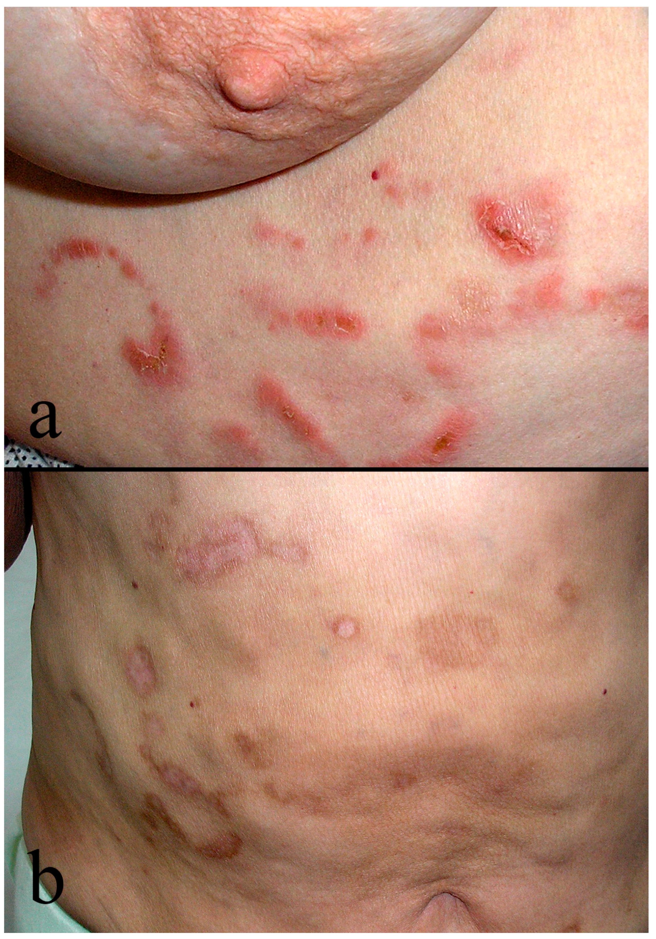
2. Bullous Mycosis Fungoides
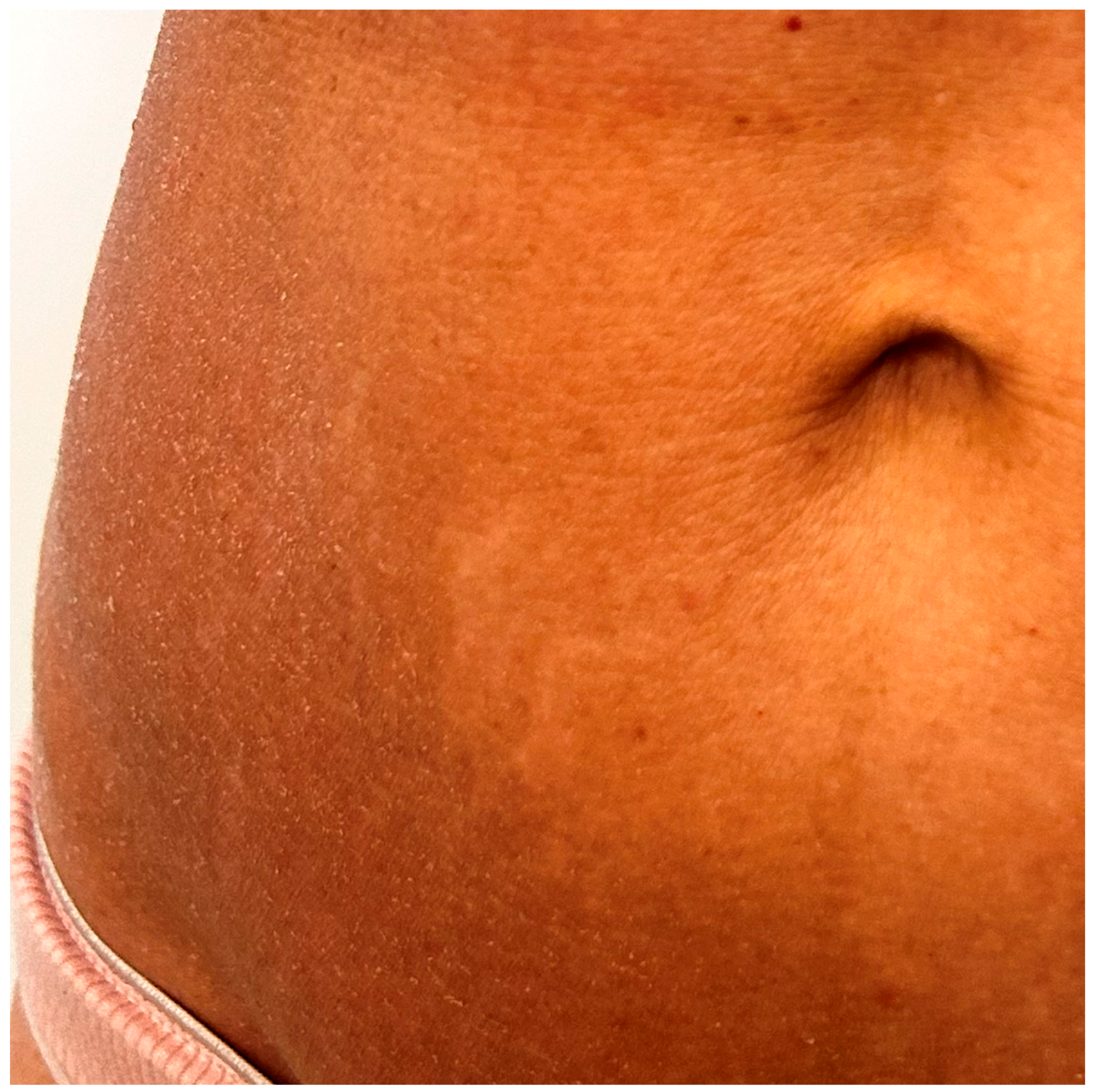
3. Ichthyosiform Mycosis Fungoides
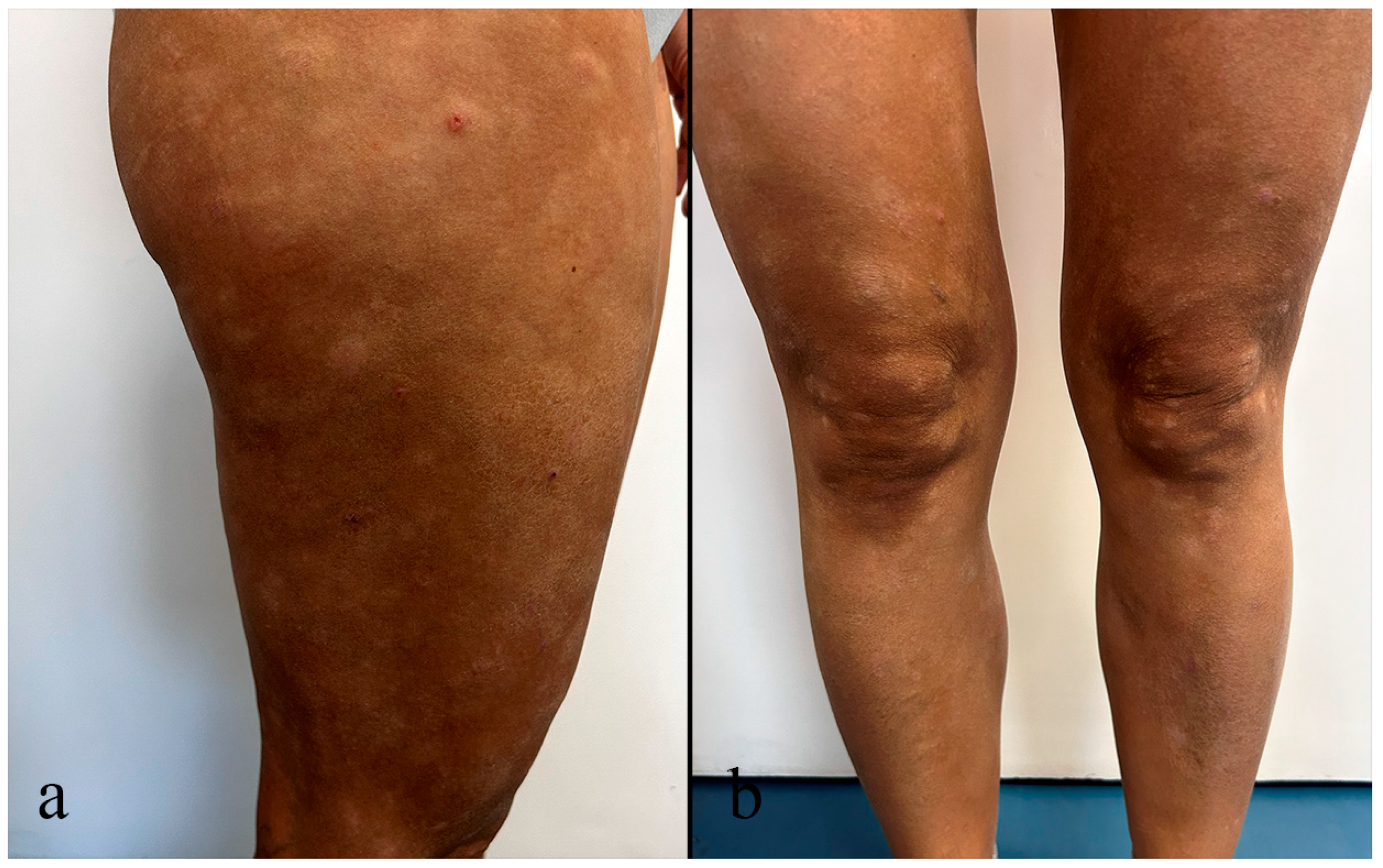
4. Hypopigmented Mycosis Fungoides
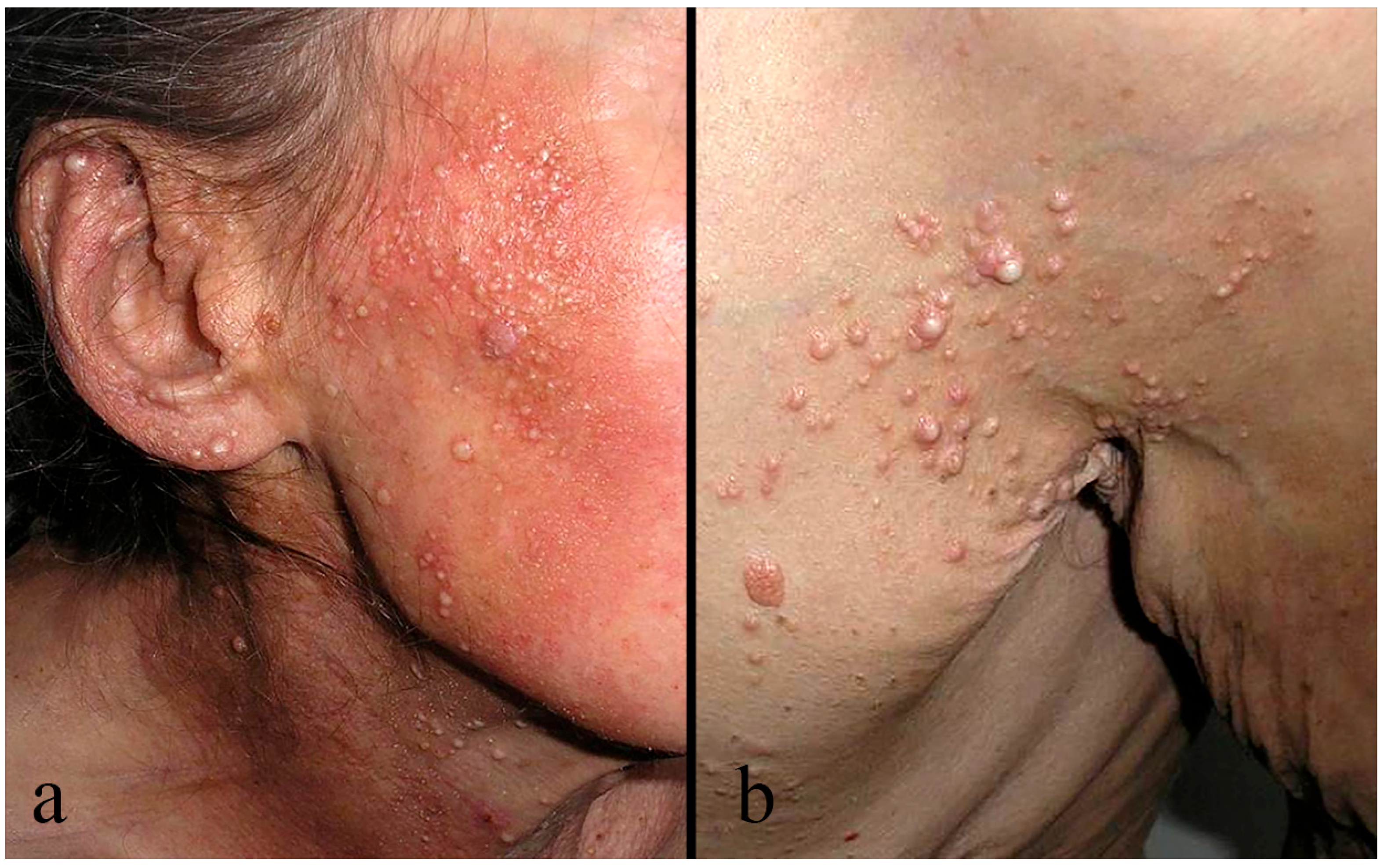
5. Folliculotropic Mycosis Fungoides
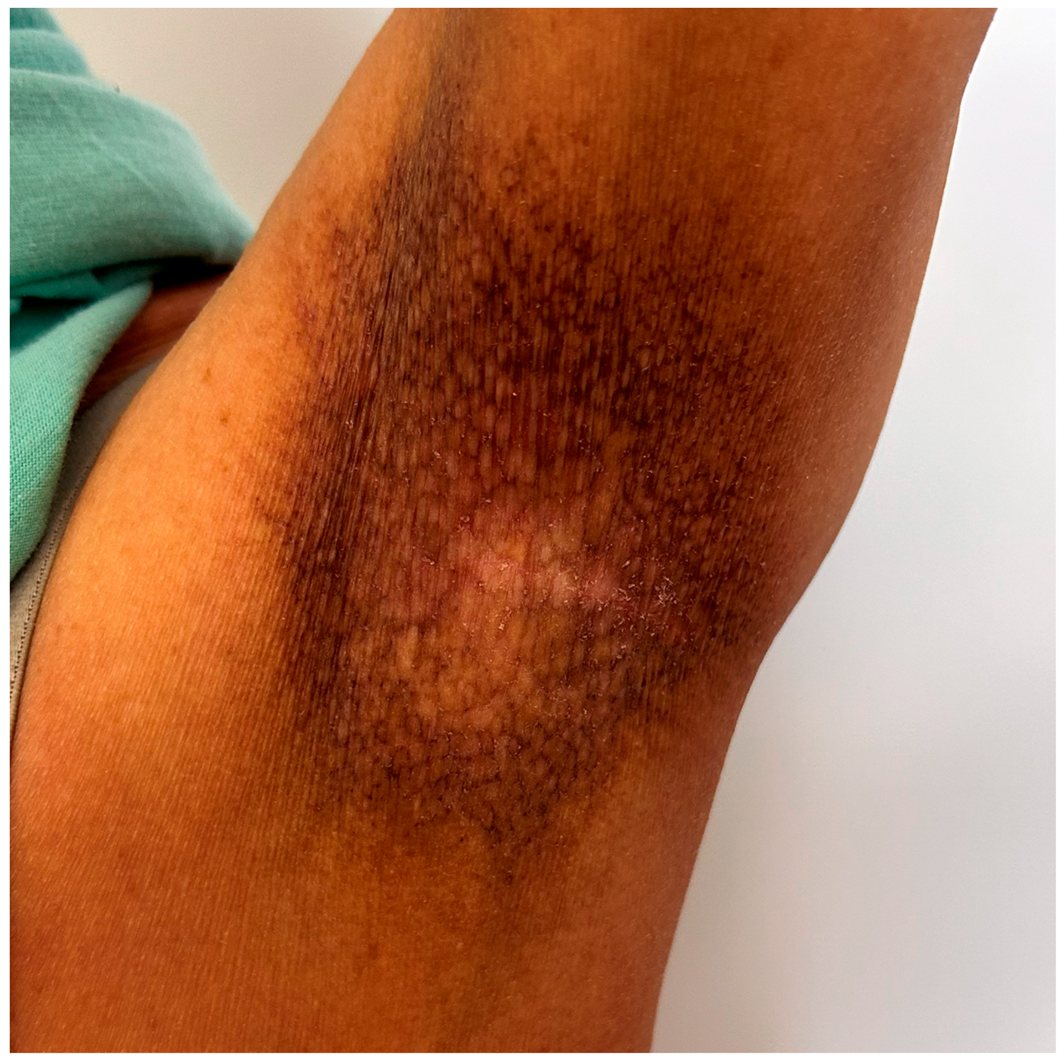
6. Poikilodermatous Mycosis Fungoides
7. Other Rare Forms of MF
8. Management and Future Therapeutic Strategies
9. Discussion
10. Conclusions
Author Contributions
Funding
Institutional Review Board Statement
Informed Consent Statement
Data Availability Statement
Acknowledgments
Conflicts of Interest
Abbreviations
| MF | mycosis fungoides |
| pCTCL | primary cutaneous T-cell lymphoma |
| HP | histopathology/histopathological |
| IHC | immunohistochemistry/immunohistochemically |
| CMF | classic mycosis fungoides |
| SS | Sézary syndrome |
| BMF | bullous mycosis fungoides |
| Th2 | T-helper type 2 |
| IL-5 | interleukin-5 |
| IL-10 | interleukin-10 |
| IFN-γ | interferon-γ |
| IMF | ichthyosiform mycosis fungoides |
| STD | skin-directed therapy |
| AI | acquired ichthyosis |
| ATLL | adult T-cell leukemia/lymphoma |
| HTLV-1 | human T-cell leukemia virus type |
| HMF | hypopigmented mycosis fungoides |
| PLC | pityriasis lichenoides chronica |
| FMF | folliculotropic mycosis fungoides |
| WHO-EORTC | World Health Organization—European Organization on Research and Treatment of Cancer |
| PMF | poikilodermatous mycosis fungoides |
| DLE | discoid lupus erythematosus |
| PPD | pigmented purpuric dermatosis |
| cGVHD | chronic graft-versus-host disease |
| GSS | granulomatous slack skin |
| GMF | granulomatous mycosis fungoides |
| PR | pagetoid reticulosis |
| PCAETCL | primary cutaneous aggressive epidermotropic CD8+ T-cell lymphoma |
| STMF | syringotropic mycosis fungoides |
| SLHA | syringolymphoid hyperplasia with alopecia |
| APEC | asymmetric periflexural exanthem of childhood |
| TME | tumor microenvironment |
| SEER-18 analysis | surveillance, epidemiology, and end results-18 analysis |
| WHO-HAEM5 | 5th edition of the World Health Organization classification of hematolymphoid tumors |
| ICC | international consensus classification |
| PUVA | psoralen + ultraviolet A(radiation) |
| AlloSCT | allogeneic stem cell transplantation |
| FDA | the Food and Drug Administration |
References
- Willemze, R.; Cerroni, L.; Kempf, W.; Berti, E.; Facchetti, F.; Swerdlow, S.H.; Jaffe, E.S. The 2018 update of the WHO-EORTC classification for primary cutaneous lymphomas. Blood 2019, 133, 1703–1714. [Google Scholar] [CrossRef] [PubMed]
- Hodak, E.; Amitay-Laish, I. Mycosis fungoides: A great imitator. Clin. Dermatol. 2019, 37, 255–267. [Google Scholar] [CrossRef]
- Zengarini, C.; Tugnoli, F.; Natale, A.; Mussi, M.; Clarizio, G.; Agostinelli, C.; Sabattini, E.; Corrà, A.; Piraccini, B.M.; Pileri, A. Dermatoscopic Patterns in Mycosis Fungoides: Observations from a Case-Series Retrospective Analysis and a Review of the Literature. Diagnostics 2025, 15, 1136. [Google Scholar] [CrossRef] [PubMed]
- Sheern, C.; Levell, N.J.; Craig, P.J.; Jeffrey, P.; Mistry, K.; Scorer, M.J.; Venables, Z.C. Mycosis fungoides: A review. Clin. Exp. Dermatol. 2025, 50, 2365–2375. [Google Scholar] [CrossRef]
- Lebas, E.; Collins, P.; Somja, J.; Nikkels, A.F. A Comprehensive Update of the Atypical, Rare and Mimicking Presentations of Mycosis Fungoides. Dermatol. Ther. 2021, 11, 1931. [Google Scholar] [CrossRef]
- Cerroni, L. Mycosis fungoides-clinical and histopathologic features, differential diagnosis, and treatment. Semin. Cutan. Med. Surg. 2018, 37, 2–10. [Google Scholar] [CrossRef]
- Amorim, G.M.; Niemeyer-Corbellini, J.P.; Quintella, D.C.; Cuzzi, T.; Ramos-E-Silva, M. Clinical and epidemiological profile of patients with early stage mycosis fungoides. An. Bras. Dermatol. 2018, 93, 546–552. [Google Scholar] [CrossRef]
- Tsang, Y.; Gu, T.; Sharma, G.; Raspa, S.; Drake, B.; Tan, H. Healthcare resource utilization, costs of care, and treatment of mycosis fungoides cutaneous T-cell lymphoma patterns in a large managed care population: A retrospective US claims-based analysis. J. Dermatol. Treat. 2018, 29, 747–753. [Google Scholar] [CrossRef]
- Hodak, E.; Amitay-Laish, I.; Bagot, M.; Battistella, M.; Ben Amitai, D.; Cozzio, A.; Duvic, M.; Ferenczi, K.; Foss, F.; Geskin, L.; et al. ISCL/EORTC-CLTG/USCLC recommendations for the diagnosis, staging and treatment of early-stage paediatric mycosis fungoides: A modified Delphi consensus. Part one: Diagnosis and staging. Br. J. Dermatol. 2025, 193, 405–414. [Google Scholar] [CrossRef] [PubMed]
- Hodak, E.; Geskin, L.; Guenova, E.; Ortiz-Romero, P.L.; Willemze, R.; Zheng, J.; Cowan, R.; Foss, F.; Mangas, C.; Querfeld, C.; et al. Real-Life Barriers to Diagnosis of Early Mycosis Fungoides: An International Expert Panel Discussion. Am. J. Clin. Dermatol. 2023, 24, 5–14. [Google Scholar] [CrossRef]
- Ottevanger, R.; de Bruin, D.; Willemze, R.; Jansen, P.; Bekkenk, M.; de Haas, E.; Horvath, B.; van Rossum, M.M.; Sanders, C.J.G.; Veraart, J.C.J.M.; et al. Incidence of mycosis fungoides and Sézary syndrome in the Netherlands in the last two decades. Eur. J. Cancer 2021, 156, S32. [Google Scholar] [CrossRef]
- Iyer, A.; Hennessey, D.; O’Keefe, S.; Patterson, J.; Wang, W.; Wong, G.K.S.; Gniadecki, R. Skin colonization by circulating neoplastic clones in cutaneous T-cell lymphoma. Blood 2019, 134, 1517–1527. [Google Scholar] [CrossRef]
- Park, J.; Daniels, J.; Wartewig, T.; Ringbloom, K.G.; Martinez-Escala, M.E.; Choi, S.; Thomas, J.J.; Doukas, P.G.; Yang, J.; Snowden, C.; et al. Integrated genomic analyses of cutaneous T-cell lymphomas reveal the molecular bases for disease heterogeneity. Blood 2021, 138, 1225–1236. [Google Scholar] [CrossRef]
- Kamran, B.; Fatemeh, M.; Ahmadreza, R.; Azita, N. Bullous mycosis fungoides: A case report. Dermatol. Online J. 2008, 14, 11. [Google Scholar] [CrossRef]
- Chen, F.; Wang, R.H.; Lullo, J.J.; Cadden, S.B.; Koster, H.; Schaeffer, J.; Speiser, J.J. Vesiculobullous Eruption in Patient With Mycosis Fungoides. Am. J. Dermatopathol. 2025, 47, 965–967. [Google Scholar] [CrossRef]
- Nofal, A.; Alakad, R.; Ehab, R.; Essam, R. Mycosis fungoides bullosa: An unusual presentation of a rare entity. JAAD Case Rep. 2021, 18, 82–88. [Google Scholar] [CrossRef] [PubMed]
- Bowman, P.H.; Hogan, D.J.; Sanusi, I.D. Mycosis fungoides bullosa: Report of a case and review of the literature. J. Am. Acad. Dermatol. 2001, 45, 934–939. [Google Scholar] [CrossRef] [PubMed]
- Gerdes, A.L.; Hyun, J.; Kreuter, A.; Schaller, J.; Hillen, U.; Michalowitz, A.L.; Müller, V. Bullous mycosis fungoides—Clinical indicator for an aggressive course of disease. Dermatologie 2024, 75, 884–888. [Google Scholar] [CrossRef]
- Momtahen, S.; Nuovo, G.J.; Magro, C.M. Vesicular Mycosis Fungoides. Am. J. Dermatopathol. 2015, 37, 724–729. [Google Scholar] [CrossRef]
- Shamim, H.; Johnson, E.F.; Gibson, L.E.; Comfere, N. Mycosis fungoides with spongiosis: A potential diagnostic pitfall. J. Cutan. Pathol. 2019, 46, 645–652. [Google Scholar] [CrossRef] [PubMed]
- Bogdanov, I.; Darlenski, R.; Hristakieva, E.; Manuelyan, K. The rash that presents as a vesiculobullous eruption. Clin. Dermatol. 2020, 38, 19–34. [Google Scholar] [CrossRef]
- Pesqué, D.; Aerts, O.; Bizjak, M.; Gonçalo, M.; Dugonik, A.; Simon, D.; Ljubojević-Hadzavdić, S.; Malinauskiene, L.; Wilkinson, M.; Czarnecka-Operacz, M.; et al. Differential diagnosis of contact dermatitis: A practical-approach review by the EADV Task Force on contact dermatitis. JEADV 2024, 38, 1704–1722. [Google Scholar] [CrossRef] [PubMed]
- Sarnoff, D.S.; Defeo, C.P. Coexistence of Pemphigus Foliaceus and Mycosis Fungoides. Arch. Dermatol. 1985, 121, 669–672. [Google Scholar] [CrossRef] [PubMed]
- Telegdy, E.; Schneider, I. Association of cutaneous T cell lymphoma and bullous pemphigoid. Dermatologica 1989, 179, 220. [Google Scholar] [CrossRef]
- Gülseren, D. Histopathological Diagnosis of Mycosis Fungoides. Turk. J. Dermatol. 2025, 19, 27–35. [Google Scholar] [CrossRef]
- Nam, K.H.; Park, J.; Hong, J.S.; Roh, S.G.; Kim, D.S.; Yun, S.K. Mycosis Fungoides as an Ichthyosiform Eruption. Ann. Dermatol. 2009, 21, 182. [Google Scholar] [CrossRef]
- Kütting, B.; Metze, D.; Luger, T.A.; Bonsmann, G. Mycosis fungoides presenting as an acquired ichthyosis. J. Am. Acad. Dermatol. 1996, 34, 887–889. [Google Scholar] [CrossRef]
- Marzano, A.V.; Borghi, A.; Facchetti, M.; Alessi, E. Ichthyosiform mycosis fungoides. Dermatology 2002, 204, 124–129. [Google Scholar] [CrossRef]
- Hodak, E.; Amitay, I.; Feinmesser, M.; Aviram, A.; David, M. Ichthyosiform mycosis fungoides: An atypical variant of cutaneous T-cell lymphoma. J. Am. Acad. Dermatol. 2004, 50, 368–374. [Google Scholar] [CrossRef]
- Perandones González, H.; Sánchez Sambucety, P.; Rodríguez Prieto, M. Ichthyosiform pattern: An exceptional presentation of mycosis fungoides. Int. J. Dermatol. 2020, 59, 730–732. [Google Scholar] [CrossRef] [PubMed]
- Kazakov, D.V.; Burg, G.; Kempf, W. Clinicopathological spectrum of mycosis fungoides. JEADV 2004, 18, 397–415. [Google Scholar] [CrossRef] [PubMed]
- Nashan, D.; Faulhaber, D.; Ständer, S.; Luger, T.A.; Stadler, R. Mycosis fungoides: A dermatological masquerader. Br. J. Dermatol. 2007, 156, 1–10. [Google Scholar] [CrossRef]
- Pereira, L.B.; Sobreira, N.d.P.; Rocha, V.B. Case for diagnosis. Ichthyosiform mycosis fungoides. An. Bras. Dermatol. 2022, 97, 795–797. [Google Scholar] [CrossRef]
- Ryan, C.; Whittaker, S.; D’Arcy, C.; O’Regan, G.M.; Rogers, S. Juvenile folliculotropic and ichthyosiform mycosis fungoides. Clin. Exp. Dermatol. 2009, 34, e160–e162. [Google Scholar] [CrossRef]
- El Gemayel, M.; Hawat, T.; Cahn, B.A.; Nadir, U.; Yi, M.D.; Tsoukas, M.; Haber, R. Adult-Onset Acquired Ichthyosis Revealing an Underlying Colon Adenocarcinoma. Cureus 2024, 16, e73045. [Google Scholar] [CrossRef]
- Ko, K.D.; Kim, K.K.; Baek, J.O.; Suh, H.S.; Hwang, I.C. A Possible Case of Statin-Induced Ichthyosis in an Elderly Woman. Korean J. Fam. Med. 2018, 39, 51. [Google Scholar] [CrossRef]
- LeWitt, T.M.; Gru, A.A. A review of cutaneous manifestations of HTLV-associated adult T-cell leukemia/lymphoma. Front. Med. 2025, 12, 1603102. [Google Scholar] [CrossRef]
- Balan, K.; Lallas, A.; Akdogan, N. Patterns and pitfalls: Dermoscopic differentiation of early-stage mycosis fungoides, atopic dermatitis, and psoriasis. J. Am. Acad. Dermatol. 2025. Online ahead of print. [Google Scholar] [CrossRef]
- Morizane, S.; Setsu, N.; Yamamoto, T.; Hamada, T.; Nakanishi, G.; Asagoe, K.; Iwatsuki, K. Ichthyosiform eruptions in association with primary cutaneous T-cell lymphomas. Br. J. Dermatol. 2009, 161, 115–120. [Google Scholar] [CrossRef]
- Hodak, E.; David, M.; Maron, L.; Aviram, A.; Kaganovsky, E.; Feinmesser, M. CD4/CD8 double-negative epidermotropic cutaneous T-cell lymphoma: An immunohistochemical variant of mycosis fungoides. J. Am. Acad. Dermatol. 2006, 55, 276–284. [Google Scholar] [CrossRef] [PubMed]
- Villarreal, A.M.; Gantchev, J.; Lagacé, F.; Barolet, A.; Sasseville, D.; Ødum, N.; Charli-Joseph, Y.V.; Salazar, A.H.; Litvinov, I.V. Hypopigmented Mycosis Fungoides: Loss of Pigmentation Reflects Antitumor Immune Response in Young Patients. Cancers 2020, 12, 2007. [Google Scholar] [CrossRef] [PubMed]
- Furlan, F.C.; Sanches, J.A. Hypopigmented mycosis fungoides: A review of its clinical features and pathophysiology. An. Bras. Dermatol. 2013, 88, 954–960. [Google Scholar] [CrossRef]
- Ryan, E.A.; Sanderson, K.V.; Barták, P.; Samman, P.D. Can mycosis fungoides begin in the epidermis? A hypothesis. Br. J. Dermatol. 1973, 88, 419–429. [Google Scholar] [CrossRef]
- Hu, Z.-H.; Lu, L.; Feng, J.D.; Song, H.-B.; Zhang, S.-Y.; Yang, L.; Wang, T.; Liu, Y.-H. Real-World Clinical Characteristics, Management, and Outcomes of 44 Paediatric Patients with Hypopigmented Mycosis Fungoides. Acta Derm. Venereol. 2023, 103, adv6226. [Google Scholar] [CrossRef]
- Jayasinghe, D.R.; Dissanayake, K.; de Silva, M.V.C. Comparison between the histopathological and immunophenotypical features of hypopigmented and nonhypopigmented mycosis fungoides: A retrospective study. J. Cutan. Pathol. 2021, 48, 486–494. [Google Scholar] [CrossRef]
- Crespi, O.; Giunipero Di Corteranzo, I.; Bongiovanni, E.; Rosset, F.; Ortoncelli, M.; Santoro, F.; Ribero, S.; Quaglino, P. Hypopigmented mycosis fungoides in pediatrics. Ital. J. Dermatol. Venerol. 2025, 160, 293–294. [Google Scholar] [CrossRef]
- Allen, P.B.; Goyal, S.; Niyogusaba, T.; O’Leary, C.; Ayers, A.; Tarabadkar, E.S.; Khan, M.K.; Lechowicz, M.J. Clinical Presentation and Outcome Differences Between Black Patients and Patients of Other Races and Ethnicities With Mycosis Fungoides and Sézary Syndrome. JAMA Dermatol. 2022, 158, 1293. [Google Scholar] [CrossRef]
- Godínez-Chaparro, J.A.; Roldán-Marín, R.; De la Cruz, H.V.; Soto-Mota, L.A.; Férez, K. Dermatoscopic Patterns in Vitiligo. Dermatol. Pract. Concept. 2023, 13, e2023197. [Google Scholar] [CrossRef] [PubMed]
- Macari, C. Hypopigmented Mycosis Fungoides in Younger Patients: A Mimicker of Common Hypopigmented Inflammatory Rashes. JDPA 2021, 15, 10–14. [Google Scholar] [CrossRef]
- Amin, S.N.M.; Muhamad, R.; Abdullah, W.N.H.W.; Zulkifli, M.M.; Bakrin, I.H.; Tangam, T. A Case Report of Pityriasis Lichenoides-Like Mycosis Fungoides in Children: A Challenging Diagnosis. Korean J. Fam. Med. 2020, 42, 334. [Google Scholar] [CrossRef] [PubMed]
- Kituashvili, T.; Urushadze, T.; Liluashvili, S.; Kankava, K.; Bagdadishvili, N. A Case Report of Folliculotropic Mycosis Fungoides: Diagnostic Criteria and Treatment Strategy. Cureus 2024, 16, e71994. [Google Scholar] [CrossRef]
- Willemze, R.; Jaffe, E.S.; Burg, G.; Cerroni, L.; Berti, E.; Swerdlow, S.H.; Ralfkiaer, E.; Chimenti, S.; Diaz-Perez, J.L.; Duncan, L.M.; et al. WHO-EORTC classification for cutaneous lymphomas. Blood 2005, 105, 3768–3785. [Google Scholar] [CrossRef]
- Sidiropoulou, P.; Nikolaou, V.; Marinos, L.; Voudouri, D.; Komini, E.; Economidi, A.; Rigopoulos, D.; Stratigos, A. The different faces of mycosis fungoides: Results of a single-center study. Int. J. Dermatol. 2020, 59, 314–320. [Google Scholar] [CrossRef]
- Roccuzzo, G.; Mastorino, L.; Gallo, G.; Fava, P.; Ribero, S.; Quaglino, P. Folliculotropic Mycosis Fungoides: Current Guidance and Experience from Clinical Practice. Clin. Cosmet. Investig. Dermatol. 2022, 15, 1899. [Google Scholar] [CrossRef]
- Cerroni, L. Mycosis fungoides. In Skin Lymphoma: Illustrated Guide; Wiley-Blackwell: Oxford, UK, 2014; pp. 11–73. [Google Scholar]
- Wieser, I.; Wang, C.; Alberti-Violetti, S.; Lyons, G.; Tran, C.; Talpur, R.; Duvic, M. Clinical characteristics, risk factors and long-term outcome of 114 patients with folliculotropic mycosis fungoides. Arch. Dermatol. Res. 2017, 309, 453–459. [Google Scholar] [CrossRef]
- Charli-Joseph, Y.; Kashani-Sabet, M.; McCalmont, T.H.; Kornak, J.; Allen, I.; Ai, W.Z.; LeBoit, P.E.; Pincus, L.B. Association of a Proposed New Staging System for Folliculotropic Mycosis Fungoides With Prognostic Variables in a US Cohort. JAMA Dermatol. 2021, 157, 157–165. [Google Scholar] [CrossRef] [PubMed]
- Hodak, E.; Amitay-Laish, I.; Atzmony, L.; Prag-Naveh, H.; Yanichkin, N.; Barzilai, A.; Kershenovich, R.; Feinmesser, M. New insights into folliculotropic mycosis fungoides (FMF): A single-center experience. J. Am. Acad. Dermatol. 2016, 75, 347–355. [Google Scholar] [CrossRef] [PubMed]
- Muniesa, C.; Estrach, T.; Pujol, R.M.; Gallardo, F.; Garcia-Muret, P.; Climent, J.; Servitje, O. Folliculotropic mycosis fungoides: Clinicopathological features and outcome in a series of 20 cases. J. Am. Acad. Dermatol. 2010, 62, 418–426. [Google Scholar] [CrossRef]
- Mitteldorf, C.; Stadler, R.; Sander, C.A.; Kempf, W. Folliculotropic mycosis fungoides. J. Dtsch. Dermatol. Ges. 2018, 16, 543–557. [Google Scholar] [CrossRef] [PubMed]
- Tambe, S.A.; Zambare, U.S.; Nayak, C.S. Folliculotropic Mycosis Fungoides. Indian. Dermatol. Online J. 2021, 12, 178. [Google Scholar] [CrossRef]
- Bi, M.Y.; Curry, J.L.; Christiano, A.M.; Hordinsky, M.K.; Norris, D.A.; Price, V.H.; Duvic, M. The spectrum of hair loss in patients with mycosis fungoides and Sézary syndrome. J. Am. Acad. Dermatol. 2011, 64, 53–63. [Google Scholar] [CrossRef]
- van Doorn, R.; Scheffer, E.; Willemze, R. Follicular mycosis fungoides, a distinct disease entity with or without associated follicular mucinosis: A clinicopathologic and follow-up study of 51 patients. Arch. Dermatol. 2002, 138, 191–198. [Google Scholar] [CrossRef] [PubMed]
- Gerami, P.; Guitart, J. The spectrum of histopathologic and immunohistochemical findings in folliculotropic mycosis fungoides. Am. J. Surg. Pathol. 2007, 31, 1430–1438. [Google Scholar] [CrossRef]
- Marschalkó, M.; Erős, N.; Kontár, O.; Hidvégi, B.; Telek, J.; Hársing, J.; Jókai, H.; Bottlik, G.; Rajnai, H.; Szepesi, Á.; et al. Folliculotropic mycosis fungoides: Clinicopathological analysis of 17 patients. J. Eur. Acad. Dermatol. Venereol. 2015, 29, 964–972. [Google Scholar] [CrossRef]
- Berg, R.V.; Valente, N.Y.S.; Fanelli, C.; Wu, I.; Pereira, J.; Zatz, R.; Sanches, J.A. Poikilodermatous Mycosis Fungoides: Comparative Study of Clinical, Histopathological and Immunohistochemical Features. Dermatology 2020, 236, 117–122. [Google Scholar]
- Wakelin, S.H.; Stewart, E.J.C.; Emmerson, R.W. Poikilodermatous and verrucous mycosis fungoides. Clin. Exp. Dermatol. 1996, 21, 205–208. [Google Scholar] [CrossRef]
- Chen, Z.; Lin, Y.; Qin, Y.; Qu, H.; Zhang, Q.; Li, Y.; Wen, Y.; Sun, J.; Tu, P.; Gao, P.; et al. Prognostic Factors and Survival Outcomes Among Patients With Mycosis Fungoides in China: A 12-Year Review. JAMA Dermatol. 2023, 159, 1059–1067. [Google Scholar] [CrossRef] [PubMed]
- Pelayo, C.A.; Gabriel, M.T. Mycosis fungoides: A case report. JPDS 2006, 15, 12–14. [Google Scholar]
- Sutedja, E.K.; Sutisna, D.; Sutedja, E.; Ruchiatan, K.; Faldian, Y.; Tsaqilah, L.; Aminah, H.; Rivanzah, Y. Poikilodermatous Mycosis Fungoides: A Rare Case Treated with Low-Dose Methotrexate. Pharmacogn. J. 2024, 16, 276–279. [Google Scholar] [CrossRef]
- Xu, P.; Tan, C. Dermoscopy of poikilodermatous mycosis fungoides (MF). J. Am. Acad. Dermatol. 2016, 74, e45–7. [Google Scholar] [CrossRef]
- Böer-Auer, A.; Jones, C.; Jepson, J.; Asgari, M. Hyperpigmented Mycosis Fungoides Masquerading as Longstanding Lichen Planus Pigmentosus: A Diagnostic Pitfall. Am. J. Dermatopathol. 2023, 45, 567–571. [Google Scholar] [CrossRef] [PubMed]
- Malinić, M.; Reljić, V.; Vuković, J.; Tanasilović, S.; Živanović, D. Dermatomyositis in women: A 15-year retrospective analysis of clinical patterns, malignancy risk, and long-term outcomes at a tertiary center. Acta Dermatovenerol. Alp. Pannonica Adriat. 2025, 34, 165–169. [Google Scholar] [CrossRef] [PubMed]
- do Vale, E.C.S.; Garcia, L.C. Cutaneous lupus erythematosus: A review of etiopathogenic, clinical, diagnostic and therapeutic aspects. An. Bras. Dermatol. 2023, 98, 355–372. [Google Scholar] [CrossRef] [PubMed]
- Riyaz, N.; Sasidharanpillai, S.; Latheef, E.N.A.; Davul, H.; Ashraf, F. Pigmented purpuric dermatosis or mycosis fungoides: A diagnostic dilemma. Indian. Dermatol. Online J. 2016, 7, 183. [Google Scholar]
- Hernández Aragüés, I.; Pulido Pérez, A.; Suárez Fernández, R. Inflammatory Skin Conditions Associated with Radiotherapy. Actas. Dermosifiliogr. 2017, 108, 209–220. [Google Scholar] [CrossRef]
- Shi, C.R.; Ferreira, A.L.; Kaur, M.; Xiang, D.; Caputo, J.; Choe, H.K.; Hamad, N.; Cowen, E.W.; Kaffenberger, B.H.; Baumrin, E. Cutaneous Chronic Graft-Versus-Host Disease: Clinical Manifestations, Diagnosis, Management, and Supportive Care. Transplant. Cell. Ther. 2024, 30, S513–S533. [Google Scholar] [CrossRef]
- Martins, D.J.; Di Lazzaro Filho, R.; Bertola, D.R.; Hoch, N.C. Rothmund-Thomson syndrome, a disorder far from solved. Front. Aging 2023, 4, 1296409. [Google Scholar] [CrossRef]
- Abbott, R.A.; Sahni, D.; Robson, A.; Agar, N.; Whittaker, S.; Scarisbrick, J.J. Poikilodermatous mycosis fungoides: A study of its clinicopathological, immunophenotypic, and prognostic features. J. Am. Acad. Dermatol. 2011, 65, 313–319. [Google Scholar] [CrossRef]
- Wechsler, J.; Ingen-Housz-Oro, S.; Deschamps, L.; Brunet-Possenti, F.; Deschamps, J.; Delfau, M.H.; Calderaro, J.; Ortonne, N. Prevalence of T-cell antigen losses in mycosis fungoides and CD30-positive cutaneous T-cell lymphoproliferations in a series of 153 patients. Pathology 2022, 54, 729–737. [Google Scholar] [CrossRef] [PubMed]
- Kalay Yildizhan, I.; Sanli, H.; Akay, B.N.; Sürgün, E.; Heper, A. CD8+ cytotoxic mycosis fungoides: A retrospective analysis of clinical features and follow-up results of 29 patients. Int. J. Dermatol. 2020, 59, 127–133. [Google Scholar] [CrossRef]
- Goldsztajn, K.O.; Moritz Trope, B.; Ribeiro Lenzi, M.E.; Cuzzi, T.; Ramos-E-Silva, M. Granulomatous slack skin. Histopathology diagnosis preceding clinical manifestations by 12 years. J. Dermatol. Case Rep. 2012, 6, 108–112. [Google Scholar] [CrossRef] [PubMed]
- Motta, L.M.D.; Soares, C.T.; Nakandakari, S.; Silva, G.V.D.; Nigro, M.H.M.F.; Brandão, L.S.G. Granulomatous slack skin: A rare subtype of mycosis fungoides. An. Bras. Dermatol. 2017, 92, 694–697. [Google Scholar] [CrossRef]
- Bakr, F.; Ziaj, S.; Cardoso, J.; Wain, E.M.; Theaker, J.; Whittaker, S.; Child, F.M.; Robson, A.F. Mycosis Fungoides and Granulomatous Slack Skin: A Single Entity With Distinct Clinical Phenotypes. Am. J. Dermatopathol. 2024, 46, 14–20. [Google Scholar] [CrossRef]
- Küçük, Ö.S. Mycosis Fungoides: A Review of Clinical Findings. Turk. J. Dermatol. 2025, 19, 7–18. [Google Scholar] [CrossRef]
- Maheswari, S.U.; Sampath, V.; Ramesh, A. Granulomatous slack skin syndrome: Report of a unique case. Indian. J. Dermatol. Venereol. Leprol. 2018, 84, 169–173. [Google Scholar]
- Kempf, W.; Ostheeren-Michaelis, S.; Paulli, M.; Lucioni, M.; Wechsler, J.; Audring, H.; Assaf, C.; Rüdiger, T.; Willemze, R.; Meijer, C.J.L.M.; et al. Granulomatous mycosis fungoides and granulomatous slack skin: A multicenter study of the Cutaneous Lymphoma Histopathology Task Force Group of the European Organization for Research and Treatment of Cancer (EORTC). Arch. Dermatol. 2008, 144, 1609–1617. [Google Scholar] [CrossRef] [PubMed]
- Battesti, G.; Ram-Wolff, C.; Dobos, G.; Aubin, F.; Algros, M.P.; Guenova, E.; Joly, P.; Courville, P.; Mourah, S.; Cayuela, J.; et al. Granulomatous slack skin: Clinical characteristics, prognosis, and response to therapy. A study from the Cutaneous Lymphoma French Study Group. Br. J. Dermatol. 2022, 187, 790–793. [Google Scholar] [CrossRef] [PubMed]
- Miyashiro, D.; Sanches, J.A. Mycosis fungoides and Sézary syndrome: Clinical presentation, diagnosis, staging, and therapeutic management. Front. Oncol. 2023, 13, 1141108. [Google Scholar] [CrossRef] [PubMed]
- Osto, M.; Afify, O.; Musa, A.; Ahmed, U.; Rehman, R.; Mehregan, D. Woringer-Kolopp disease (localized pagetoid reticulosis): A systematic review. Int. J. Dermatol. 2023, 62, 312–321. [Google Scholar] [CrossRef]
- Haghighi, B.; Smoller, B.R.; Leboit, P.E.; Warnke, R.A.; Sander, C.A.; Kohler, S. Pagetoid Reticulosis (Woringer-Kolopp Disease): An Immunophenotypic, Molecular, and Clinicopathologic Study. Mod. Pathol. 2000, 13, 502–510. [Google Scholar] [CrossRef]
- Stahly, S.; Manway, M.; Lin, C.C.; Sukpraprut-Braaten, S. Pagetoid Reticulosis: A Rare Dermatologic Malignancy Presenting in a Middle-Aged Female. Cureus 2021, 13, e18524. [Google Scholar] [CrossRef]
- Mourtzinos, N.; Puri, P.K.; Wang, G.; Liu, M.L. CD4/CD8 double negative pagetoid reticulosis: A case report and literature review. J. Cutan. Pathol. 2010, 37, 491–496. [Google Scholar] [CrossRef]
- Guitart, J.; Martinez-Escala, E.; Subtil, A.; Duvic, M.; Pulitzer, M.P.; Olsen, E.A.; Kim, E.; Rook, A.H.; Samimi, S.S.; Wood, G.S.; et al. Primary cutaneous aggressive epidermotropic cytotoxic T-cell lymphomas: Reappraisal of a provisional entity in the 2016 WHO classification of cutaneous lymphomas. Mod. Pathol. 2017, 30, 761–772. [Google Scholar] [CrossRef] [PubMed]
- Lehmer, L.M.; Amber, K.T.; de Feraudy, S.M. Syringotropic Mycosis Fungoides: A Rare Form of Cutaneous T-cell Lymphoma Enabling a Histopathologic “Sigh of Relief”. Am. J. Dermatopathol. 2017, 39, 920–923. [Google Scholar] [CrossRef] [PubMed]
- de Masson, A.; Battistella, M.; Vignon-Pennamen, M.D.; Cavelier-Balloy, B.; Mouly, F.; Rybojad, M.; Bouaziz, J.-D.; Petit, A.; Saussine, A.; Ronceray, S.; et al. Syringotropic mycosis fungoides: Clinical and histologic features, response to treatment, and outcome in 19 patients. J. Am. Acad. Dermatol. 2014, 71, 926–934. [Google Scholar] [CrossRef]
- Pileri, A.; Facchetti, F.; Rütten, A.; Zumiani, G.; Boi, S.; Fink-Puches, R.; Cerroni, L. Syringotropic mycosis fungoides: A rare variant of the disease with peculiar clinicopathologic features. Am. J. Surg. Pathol. 2011, 35, 100–109. [Google Scholar] [CrossRef]
- Coustou, D.; Léauté-Labrèze, C.; Bioulac-Sage, P.; Labbé, L.; Taïeb, A. Asymmetric Periflexural Exanthem of Childhood: A Clinical, Pathologic, and Epidemiologic Prospective Study. Arch. Dermatol. 1999, 135, 799–803. [Google Scholar] [CrossRef]
- Nussbaum, D.; Eleryan, M.G.; Samaan, C.B.; Glass, F.; Prussick, R. Syringotropic Mycosis Fungoides: A Variant of Folliculotropic or a Distinct Subtype of Adnexotropic Mycosis Fungoides? J. Drugs Dermatol. 2022, 21, 1362–1364. [Google Scholar] [CrossRef]
- Schlichte, M.J.; Talpur, R.; Venkatarajan, S.; Curry, J.L.; Nagarajan, P.; Duvic, M. Verrucous presentation in patients with mycosis fungoides. Int. J. Dermatol. 2016, 55, e126–29. [Google Scholar] [CrossRef] [PubMed]
- Sun, J.; Liu, K.; Dang, J.; Xiong, S.; Pan, H.; Wang, Y. Pigmented purpura dermatosis-like mycosis fungoides: Four case reports and a review of published cases. Eur. J. Dermatol. 2023, 33, 635–641. [Google Scholar] [CrossRef]
- Latzka, J.; Assaf, C.; Bagot, M.; Cozzio, A.; Dummer, R.; Guenova, E.; Gniadecki, R.; Hodak, E.; Jonak, C.; Klemke, C.D. EORTC consensus recommendations for the treatment of mycosis fungoides/Sézary syndrome—Update 2023. Eur. J. Cancer 2023, 195, 113343. [Google Scholar] [CrossRef]
- Zengarini, C.; Baruffaldi, G.; Piraccini, B.M.; Bardazzi, F.; Mussi, M.; Hrvatin Stancic, B.; Pileri, A. Nb-UVB and PUVA therapy in treating early stages of Mycosis Fungoides: A single-center cross-sectional study. Photodermatol. Photoimmunol. Photomed. 2023, 39, 435–440. [Google Scholar] [PubMed]
- van Santen, S.; van Doorn, R.; Neelis, K.; Daniëls, L.; Horváth, B.; Bruijn, M.; Sanders, C.; van Rossum, M.; de Haas, E.; Veraart, J.; et al. Recommendations for treatment in folliculotropic mycosis fungoides: Report of the Dutch Cutaneous Lymphoma Group. Br. J. Dermatol. 2017, 177, 223–228. [Google Scholar] [CrossRef]
- Shah, A.; Safaya, A. Granulomatous slack skin disease: A review, in comparison with mycosis fungoides. J. Eur. Acad. Dermatol. Venereol. 2012, 26, 1472–1478. [Google Scholar] [CrossRef]
- Prince, H.M.; Abeyakoon, C. Allogeneic haematopoietic stem cell transplantation for advanced stage mycosis fungoides and Sézary syndrome: Never-late, never-never? Bone Marrow Transplant 2021, 56, 1232–1234. [Google Scholar] [CrossRef]
- Bagot, M. Therapeutic advances for cutaneous T-cell lymphoma. Br. J. Dermatol. 2025, 193, 11–15. [Google Scholar] [CrossRef]
- Foss, F.M.; Kim, Y.H.; Prince, H.M.; Akilov, O.E.; Querfeld, C.; Seminario-Vidal, L.; Fisher, D.C.; Kuzel, T.M.; Yannakou, C.K.; Geskin, L.J.; et al. Efficacy and Safety of Denileukin Diftitox-Cxdl, an Improved Purity Formulation of Denileukin Diftitox, in Patients With Relapsed or Refractory Cutaneous T-Cell Lymphoma. J. Clin. Oncol. 2025, 43, 1198–1209. [Google Scholar] [CrossRef]
- Şanlı, H.; Erol Mart, H.M. Treatment algorithms for mycosis fungoides and sézary syndrome. Turk. J. Dermatol. 2025, 19, 41–60. [Google Scholar] [CrossRef]
- Nielsen, P.R.; Eriksen, J.O.; Wehkamp, U.; Lindahl, L.M.; Gniadecki, R.; Fogh, H.; Fabricius, S.; Bzorek, M.; Ødum, N.; Gjerdrum, L. Clinical and histological characteristics of mycosis fungoides and sézary syndrome: A retrospective, single-centre study of 43 patients from eastern Denmark. Acta Derm. Venereol. 2019, 99, 1231–1236. [Google Scholar] [CrossRef]
- Scarisbrick, J.J.; Quaglino, P.; Prince, H.M.; Papadavid, E.; Hodak, E.; Bagot, M.; Servitje, O.; Berti, E.; Ortiz-Romero, P.; Stadler, R.; et al. The PROCLIPI international registry of early-stage mycosis fungoides identifies substantial diagnostic delay in most patients. Br. J. Dermatol. 2019, 181, 350–357. [Google Scholar] [PubMed]
- Pileri, A.; Guglielmo, A.; Grandi, V.; Violetti, S.A.; Fanoni, D.; Fava, P.; Agostinelli, C.; Berti, E.; Quaglino, P.; Pimpinelli, N. The Microenvironment’s Role in Mycosis Fungoides and Sézary Syndrome: From Progression to Therapeutic Implications. Cells 2021, 10, 2780. [Google Scholar] [CrossRef] [PubMed]
- Williams, M.N.; Joffe, D.; Mandel, J.; Scheppes, S.; Wallace, M.; Portocarrero, C.; Abinader, O.; Mitra, R.; Mishra, A.; Nikbakht, N. Spatial transcriptomics uncovers unique tumor microenvironments in folliculotropic versus classic mycosis fungoides. J. Investig. Dermatol. 2025. Online ahead of print. [Google Scholar] [CrossRef]
- Licht, P.; Mailänder, V. Multi-Omic Data Integration Suggests Putative Microbial Drivers of Aetiopathogenesis in Mycosis Fungoides. Cancers 2024, 16, 3947. [Google Scholar] [CrossRef] [PubMed]
- Łyko, M.; Jankowska-Konsur, A. The Skin Microbiome in Cutaneous T-Cell Lymphomas (CTCL)—A Narrative Review. Pathogens 2022, 11, 935. [Google Scholar] [CrossRef]
- Goyal, A.; O’Leary, D.; Goyal, K.; Rubin, N.; Bohjanen, K.; Hordinsky, M.; Chen, S.T.; Pongas, G.; Duncan, L.M.; Lazaryan, A. Increased risk of second primary hematologic and solid malignancies in patients with mycosis fungoides: A Surveillance, Epidemiology, and End Results analysis. J. Am. Acad. Dermatol. 2019, 83, 404. [Google Scholar] [CrossRef]
- Ottevanger, R.; Vermaas, E.; Willemze, R.; Schrader, A.R.; Jansen, P.M.; Goeman, J.J.; Putter, H.; Vermeer, M.H.; Quint, K.D. Mycosis Fungoides and Associated Malignancies in a Dutch Nationwide Retrospective Cohort Study. Acta Derm. Venereol. 2024, 104, adv40065. [Google Scholar] [CrossRef]
- Melchers, S.; Albrecht, J.D.; Kempf, W.; Nicolay, J.P. The fifth edition of the WHO-Classification—What is new for cutaneous lymphomas? JDDG 2024, 22, 1254–1265. [Google Scholar] [CrossRef] [PubMed]
- Campo, E.; Jaffe, E.S.; Cook, J.R.; Quintanilla-Martinez, L.; Swerdlow, S.H.; Anderson, K.C.; Brousset, P.; Cerroni, L.; de Leval, L.; Dirnhofer, S.; et al. The International Consensus Classification of Mature Lymphoid Neoplasms: A report from the Clinical Advisory Committee. Blood 2022, 140, 1229–1253. [Google Scholar] [CrossRef] [PubMed]
- Farabi, B.; Seminario-Vidal, L.; Jamgochian, M.; Akay, B.N.; Atak, M.F.; Rao, B.K.; Karagaiah, P.; Grabbe, S.; Goldust, M. Updated review on prognostic factors in mycosis fungoides and new skin lymphoma trials. J. Cosmet. Dermatol. 2022, 21, 2742–2748. [Google Scholar] [CrossRef]
- Kempf, W.; Mitteldorf, C.; Cerroni, L.; Willemze, R.; Berti, E.; Guenova, E.; Scarisbrick, J.J.; Battistella, M. Classifications of cutaneous lymphomas and lymphoproliferative disorders: An update from the EORTC cutaneous lymphoma histopathology group. JEADV 2024, 38, 1491–1495. [Google Scholar] [CrossRef]



| MF Variant | Key Clinical Features | Dermoscopic Clues * | Most Common Clinical Differential Diagnoses | Prognosis |
|---|---|---|---|---|
| Bullous | Vesicles/bullae overlying patches/plaques; trunk and extremities | Liner vessels, whitish scale, components of blister formation/resolution, ulceration | Bullous pemphigoid, pemphigus, linear IgA dermatosis, bullous drug eruption, infectious diseases, Hailey-Hailey disease | Poor |
| Ichthyosiform | Generalized scaling resembling ichthyosis; dry, scaly plaques; alone or with classic/follicular MF; trunk and extremities | White geometric scales, linear/spermatozoa vessels, and orange structureless areas | Ichthyosis vulgaris, acquired ichthyosis, chronic eczema, xerosis in chronic renal disease, and endocrinologic disorders | Favorable |
| Hypopigmented | Hypopigmented patches/plaques; trunk and limbs; most common in children | Linear vessels, spermatozoa-like vessels, orange-yellow areas, and white scales in furrows | Vitiligo, pityriasis alba, post-inflammatory hypopigmentation, lichen sclerosus | Favorable |
| Folliculotropic (early-stage disease) | Follicular papules, keratosis pilaris-like lesions, and/or acneiform lesions; face/trunk/limbs; pruritus | Dilated follicles, follicular plugs, lack of hairs, perifollicular accentuation, spermatozoa-like vessels, yellow dots | Acne, rosacea, keratosis pilaris, alopecia areata, folliculitis | Favorable |
| Folliculotropic (advanced-stage disease) | Infiltrated plaques/tumors, alopecia; head and neck preference; dense perifollicular infiltrates | Dotted dilated vessels, broken dystrophic hairs, spermatozoa-like vessels, and absence of follicular dots | Deep fungal infection, furunculosis, hidradenitis suppurativa, sarcoidosis | Poor |
| Poikilodermatous | Atrophy, telangiectasia, hyper and hypopigmentation; reticulated pattern; flexural areas and trunk | Focal white and brown structureless areas, white patchy scales, brown reticular lines, polymorphous vessels | Dermatomyositis, radiodermatitis, discoid lupus erythematosus, cGVHD | Favorable |
| Granulomatous | Patches, plaques with irregular areas of induration or tumors; trunk and distal parts of extremities | Lack of studies | GSS, CMF, sarcoidosis, granuloma annulare, interstitial granulomatous dermatitis, infectious granulomatous dermatoses | Intermediate to unfavorable ** |
| Granulomatous slack skin | Bulky, pendulous skinfolds usually located in flexural areas | Lack of studies | GMF, acquired cutix laxa, anetoderma | Intermediate to relatively favorable *** |
| Pagetoid reticulosis | Solitary, well-demarcated psoriasiform and/or hyperkeratotic patch or plaque; acral sites | Lack of studies | CMF, psoriasis, atopic dermatitis, chronic eczema, Bowen’s disease | Favorable |
| Syringotropic | Erythematous patches or plaques sometimes exhibiting a punctate appearance, alopecia; palmoplantar involvement | Lack of studies | FMF, syringotropic lichen planus, chronic eczema, psoriasis | Favorable |
Disclaimer/Publisher’s Note: The statements, opinions and data contained in all publications are solely those of the individual author(s) and contributor(s) and not of MDPI and/or the editor(s). MDPI and/or the editor(s) disclaim responsibility for any injury to people or property resulting from any ideas, methods, instructions or products referred to in the content. |
© 2026 by the authors. Published by MDPI on behalf of the Lithuanian University of Health Sciences. Licensee MDPI, Basel, Switzerland. This article is an open access article distributed under the terms and conditions of the Creative Commons Attribution (CC BY) license.
Share and Cite
Malinić, M.; Lekić, B.; Živanović, D. Mimicry in Cutaneous Malignancy—Rare Forms of Mycosis Fungoides as Diagnostic Pitfalls: A Narrative Review. Medicina 2026, 62, 616. https://doi.org/10.3390/medicina62040616
Malinić M, Lekić B, Živanović D. Mimicry in Cutaneous Malignancy—Rare Forms of Mycosis Fungoides as Diagnostic Pitfalls: A Narrative Review. Medicina. 2026; 62(4):616. https://doi.org/10.3390/medicina62040616
Chicago/Turabian StyleMalinić, Marija, Branislav Lekić, and Dubravka Živanović. 2026. "Mimicry in Cutaneous Malignancy—Rare Forms of Mycosis Fungoides as Diagnostic Pitfalls: A Narrative Review" Medicina 62, no. 4: 616. https://doi.org/10.3390/medicina62040616
APA StyleMalinić, M., Lekić, B., & Živanović, D. (2026). Mimicry in Cutaneous Malignancy—Rare Forms of Mycosis Fungoides as Diagnostic Pitfalls: A Narrative Review. Medicina, 62(4), 616. https://doi.org/10.3390/medicina62040616