Article

Prevalence of Genetic Variants Associated with Atrial Fibrillation Risk in the Asymptomatic Young Adult Population

by
Manoranjani Murugan
1,†,
Sambandam Ravikumar
1,†,
Irisappan Ganesh
1,
Yogesh Vetriselvan
1,
Arunagiri Priyadharshini
2 and
Vishnu Bhat Ballambattu
3,*
1
Department of Medical Biotechnology, Aarupadai Veedu Medical College and Hospital, Vinayaka Mission’s Research Foundation (Deemed to be University), Kirumampakkam, Puducherry 607403, India
2
Department of Biochemistry, Aarupadai Veedu Medical College and Hospital, Vinayaka Mission’s Research Foundation (Deemed to be University), Kirumampakkam, Puducherry 607403, India
3
Advisor—Medical Research and Publications, Aarupadai Veedu Medical College and Hospital, Vinayaka Mission’s Research Foundation (Deemed to be University), Kirumampakkam, Puducherry 607403, India
*
Author to whom correspondence should be addressed.
These authors contributed equally to this work.
Medicina 2025, 61(5), 900; https://doi.org/10.3390/medicina61050900
Submission received: 2 April 2025 / Revised: 11 May 2025 / Accepted: 13 May 2025 / Published: 15 May 2025
(This article belongs to the Section Genetics and Molecular Medicine)

Abstract

Background and Objectives: Atrial fibrillation (AF) is the most common cardiac arrhythmia globally, leading to a high risk of stroke and heart failure. Genetic factors are known to play an essential role in AF risk. However, studies on genetic predisposition in asymptomatic young populations remain limited. This study aimed to investigate the prevalence of genetic variants in the PITX2 (rs2200733, rs10033464, and rs13143308), TBX5 (rs883079), PRRX1 (rs3903239), ZFHX3 (rs2106261), and HAND2 (rs7698692) polymorphisms and to assess their correlation with susceptibility to AF in a young adult population in India. Materials and Methods: This cross-sectional study included 250 subjects aged 18–29. Detailed lifestyle and family histories were collected for each participant. Genetic variation was determined using a specific TaqMan SNP genotyping assay. Hardy–Weinberg equilibrium (HWE) analysis and chi-square tests were employed to assess genotype frequencies, and statistical associations with lifestyle factors (body mass index, alcohol consumption, and smoking) were evaluated using t-tests and descriptive statistics. Results: Minor allele frequencies were varied across the study population, with notable frequencies in rs2200733 T (16%), rs10033464 T (27%), rs13143308 T (32%), rs883079 T (46%), rs3903239 G (25%), rs2106261 T (26%), and rs7698692 G (14%). HWE analysis confirmed that all SNPs were in equilibrium (p > 0.05). Approximately 15% of individuals carried six or more risk alleles, indicating a significant genetic predisposition to AF despite the absence of clinical symptoms. Conclusions: This study provides new insights into the genetic predisposition to AF among young adults in India. The high prevalence of risk alleles in asymptomatic young adults highlights the necessity of early genetic screening for AF risk and the role of genetic counseling in preventing cardiac complications.

1. Introduction

Atrial fibrillation (AF) is the most predominant cardiac arrhythmia, affecting more than 33 million people worldwide, which leads to significant complications like stroke and heart failure [1,2,3]. AF is characterized by an irregular heartbeat, changes in heart tissue, or interruptions in the heart’s electrical signal [4,5]. These irregularities lead to irregular contractions of the heart’s upper chamber, resulting in the pooling of blood and the formation of clots. These complications significantly increase the risk of stroke, which is the leading cause of disability and mortality across the world [3,4,6]. Although AF is uncommon in individuals under 40, it causes unique risks even in younger populations. Recent studies conducted at tertiary care hospitals in eastern India have indicated that younger individuals with AF also have other related problems like systemic hypertension, left ventricular systolic dysfunction, and metabolic and structural heart disease [7]. These associated conditions can complicate the clinical management of AF and highlight the need for a deeper understanding of the conditions in this population. In India, AF constitutes approximately 0.5%, but studies on its prevalence among younger adults are limited [8].
Recent studies suggest that genetic factors significantly influence the hereditary risk of developing AF. Specific single-nucleotide polymorphisms (SNPs) increase the risk of susceptibility to AF [9,10,11]. Genome-wide association studies (GWASs) have identified several SNPs located near the essential genes PITX2, TBX5, PRRX1, ZFHX3, and HAND2, which are involved in cardiac development and function [12,13]. The genetic variants, such as rs2200733, rs10033464, rs13143308, rs883079, rs3903239, rs2106261, and rs7698692, are located near essential genes and are significantly associated with an elevated risk of developing AF [11,14,15,16,17,18]. These variants influence atrial electrical activity and play an essential role in the development of the heart, contributing to AF susceptibility. The genetic variants rs2200733, rs10033464, and rs13143308 are located near the PITX2 gene, which regulates atrial electrical conduction [9,19]; the genetic variant of rs883079 located in 3′ UTR of the TBX5 gene has been associated with atrial activation patterns; and TBX5 directly activates the PITX2 gene [20]. The genetic variants in PRRX1 (rs3903239), ZFHX3 (rs2106261), and HAND2 (rs7698692) modulate the electrical activity and structural development of the heart, influencing AF susceptibility [21]. Although the genetic factors’ association with AF has been identified, most AF-based genetic research has focused only on older and symptomatic populations. This opens up a knowledge gap in understanding the genetic predisposition to AF among younger and asymptomatic individuals. Studying genetic variants in the young adult population diagnoses early AF risk, enabling targeted prevention, personalized treatments, and a better understanding of gene–lifestyle interactions to mitigate AF-related problems.
To develop preventive and customized medical strategies for AF, genetic testing is highly effective in identifying monogenic defects. A genetic predisposition is likely to play an important role in cases of early-onset AF where specific cardiovascular risk factors are absent. Studying genetic variation among younger adults is necessary for the early identification of genetic risks, allowing for more timely actions to modify lifestyle or increase monitoring to mitigate disease progression. Additionally, focusing on younger populations helps to assess the influence of genetics on age-related comorbidities, thereby enhancing the precision of investigating the genetic causative factors. However, genetic research remains a powerful tool for diagnosing, stratifying risks, and managing AF, despite challenges posed by variants of unknown significance and the low diversity of participants. This research fills a significant gap in the genetics of AF among young adults by enhancing the biomarker resources customized for specific age demographics. Focusing on this young group uncovers non-standard risk factors, enables early AF detection, predicts progression, and informs management.
Herein, we aim to investigate the distribution of AF-related SNPs in the young adult population in India, who are also asymptomatic for AF. Through this analysis, we hope to identify individuals with high-risk genetic profiles who may benefit from early intervention to prevent AF complications. Additionally, this study aims to determine the frequency and distribution of SNPs associated with AF, focusing on their impact on heart health in young adults. Although most AF research on genetic variants focuses on older populations where AF is more prevalent, our study offers novel insights into variants that may influence AF risk at a younger age, particularly in individuals without risk factors for AF, such as hypertension or structural heart disease. Thus, in this study, we assessed the prevalence of both high-risk and protective alleles, particularly in the Indian community, which contributes to enhancing our understanding of the genetic predisposition to AF among younger individuals.

2. Materials and Methods

2.1. Study Subjects

This observational study was conducted among 250 young adults recruited from the students and staff of Aarupadai Veedu Medical College & Hospital, Puducherry. The inclusion criterion for this study’s participants was young adults aged between 18 and 29 years. We obtained written informed consent from all participants and confirmed their willingness to complete a comprehensive questionnaire that addressed their medical history, family history, and lifestyle habits. This study was approved by our institution’s Human Ethics Committee (IEC No: AV/IEC/2020/022). Height, weight, and hip circumference measurements were taken to calculate the body mass index (BMI). For clarification, a BMI ranging from 18.5 to 24.9 was categorized as within the normal weight range, a BMI from 25.0 to 29.9 was classified as overweight, and a BMI of 30.0 or greater as obesity. Participants were excluded from the study if they had a documented history of cardiovascular disease, metabolic disorders, or chronic illnesses. Individuals receiving medications that impact metabolic parameters, including steroids, antidiabetic agents, or antihypertensive medications, were not included. Furthermore, pregnant or lactating women, individuals who had undergone an acute illness or hospitalization within the prior three months, as well as those unwilling to provide a blood sample or complete consent forms, were excluded. Two ml of blood sample was obtained from each study participant, and the samples were either refrigerated for storage or processed immediately for DNA extraction.

2.2. DNA Isolation

The human genomic DNA was isolated from 200 μL of blood sample using the QIAamp Blood Mini Kit (Qiagen, Hilden, Germany) according to the manufacturer’s instructions. The isolated DNA samples were stored at −20 °C till further analysis was conducted. The quality of the isolated DNA was checked using agarose gel electrophoresis. The DNA concentration was quantified using a Nanodrop (BioPhotometer D30, Eppendorf AG, Hamburg, Germany).

2.3. Genotyping via Real-Time PCR

The PITX2 (rs2200733, rs10033464, and rs13143308), TBX5 (rs883079), PRRX1 (rs3903239), ZFHX3 (rs2106261), and HAND2 (rs7698692) allele frequencies were determined using a TaqMan SNP genotyping assay via quantitative real-time PCR. The PCR reaction mixture consisted of 5 μL of TaqMan genotyping PCR master mix, 0.5 μL of TaqMan genotyping assay mix, and 20 ng of purified genomic DNA in a total volume of 10 μL of DNase-free water. The real-time PCR conditions for amplification were 10 min at 95 °C, followed by 40 cycles of denaturation for 15 s at 95 °C and annealing/extension for 1 min at 60 °C. The fluorescent signal intensities were recorded, and genotyping was analyzed using Quant Studio 5 detector systems (Applied Biosystems, Thermo Fisher Scientific Inc., Waltham, MA, USA) software. The SNPs in various genes, their SNP IDs, allele changes, SNP types, and the corresponding context sequences, are presented in Table 1.

2.4. Hardy–Weinberg Equilibrium Analysis

Hardy–Weinberg equilibrium (HWE) was used to calculate the SNP genotype frequencies, which were then compared with the expected frequencies using the chi-square (χ2) test. A p-value of less than 0.05 was considered statistically significant, indicating a deviation from the Hardy–Weinberg equilibrium (HWE).

2.5. Variant Allele Frequency Analysis

The allele frequencies of the rs2200733, rs10033464, rs13143308, rs883079, rs3903239, rs2106261, and rs7698692 genetic variants were calculated and compared with those of publicly available databases, including the 1000 Genomes Project, gnomAD, and IndiGenome. Frequencies in the study population were derived via allele counting and expressed as a proportion of the total number of alleles at each locus.

2.6. Risk Allele Categorization

We evaluated each participant for the number of risk alleles across the selected seven SNPs—rs2200733, rs10033464, rs13143308, rs883079, rs3903239, rs2106261, and rs7698692—which are associated with a greater susceptibility to AF. Participants were categorized into three groups based on their total count of risk alleles: low risk (0–3 risk alleles), medium risk (4–5 risk alleles), and high risk (6–8 risk alleles). With this classification, steps can be taken to personalize preventive strategies that require different intervention levels depending on the individual’s risk.

2.7. Association Between Risk Allele Categories and Lifestyle Factors

The study participants’ height and weight were measured, and their BMI was calculated using standard methodologies and defined as normal (BMI < 25), overweight (BMI 25–29.9), or obese (BMI ≥ 30). Data on the habits of alcohol consumption and smoking were also collected. The risk allele category group was compared based on obesity, alcohol consumption, and smoking status to determine the contribution of lifestyle factors to disease progression.

2.8. Statistical Analysis

The obtained data were presented as mean ± standard deviation (SD) for continuous variables, and categorical variables were represented as numbers and percentages. The Student’s t-test was used to compare the obtained quantitative variables. The allelic frequency, genotypic frequency, and descriptive statistics tables were generated from the received data using Microsoft Office 2019. The Hardy–Weinberg equilibrium was analyzed using a chi-square test (χ2) with SPSS 28.0 (IBM, Chicago, IL, USA). A p-value of less than 0.05 was considered statistically significant.

3. Results

3.1. Demographic Characteristics of the Young Adult Population

A total of 250 young adults, with a mean age of 25.81 years (SD ± 6.19), participated in this study. Among the participants, 31% were female and 69% were male. The average body mass index (BMI) was 24.9 kg/m2 (SD ± 4.2). Among them, 38% of the population was classified as overweight, 12% as obese, and 50% as normal. Concerning lifestyle habits, 36% did not consume alcohol. Among them, 21% were non-smokers. Considering family medical history, 17% of the study participants had a heritable predisposition to obesity, 15% to cardiovascular disease, and 23% to diabetes mellitus. The demographic profile of the study population is shown in Table 2.

3.2. Distribution of Genotypic Frequency in the Young Adult Population

The PITX2 (rs2200733, rs10033464, and rs13143308) homozygous mutant genotype’s (TT, TT, and TT) frequencies were 3%, 13%, and 10%, and its minor allele (T, T, and T) frequencies were 16%, 27%, and 32%. The TBX5 (rs883079) homozygous mutant genotype’s (TT) frequency was 21%, and its minor allele (T) frequency was 46%. The PRRX1 (rs3903239) homozygous mutant genotype’s (GG) frequency was 7%, and the frequency of its minor allele (T) was 25%. The frequency of the ZFHX3 (rs2106261) homozygous mutant genotype (TT) was 7%, and the frequency of its minor allele (T) was 26%. The HAND2 (rs2106261) homozygous mutant genotype’s (GG) frequency was 1%, and its minor allele’s (G) frequency was 14%. The genotype and allele distributions of SNPs associated with AF are presented in Table 3. Detailed information on the genotyping results with the HWE values of the study group is shown in the Supplementary Table S1. The comparison of the variant allele frequency in the South Asian population with that in the 1000 Genomes, Genome AD, and IndiGenome databases is shown in Table 4.

3.3. Distribution of Risk Alleles in the Young Adult Population

To study the interaction among SNPs—including rs2200733, rs10033464, rs13143308, rs3903239, rs2106261, rs883079, and rs7698692—the prevalence of risk alleles for AF was examined in the young adult population of India. The distribution of risk alleles in the study population is shown in Table 5. The results show that the proportion of risk alleles in young adults (n = 250) varied among individuals. Only 3% of participants possessed no risk alleles, while 9% carried one risk allele and 15% carried two. Furthermore, 19% of the population had three risk alleles, and the largest group, encompassing 20% of participants, had four. Although 19% of the study group carried five risk alleles, higher risk alleles were observed in 7% with six risk alleles, 4% with seven risk alleles, and 3% with eight risk alleles.

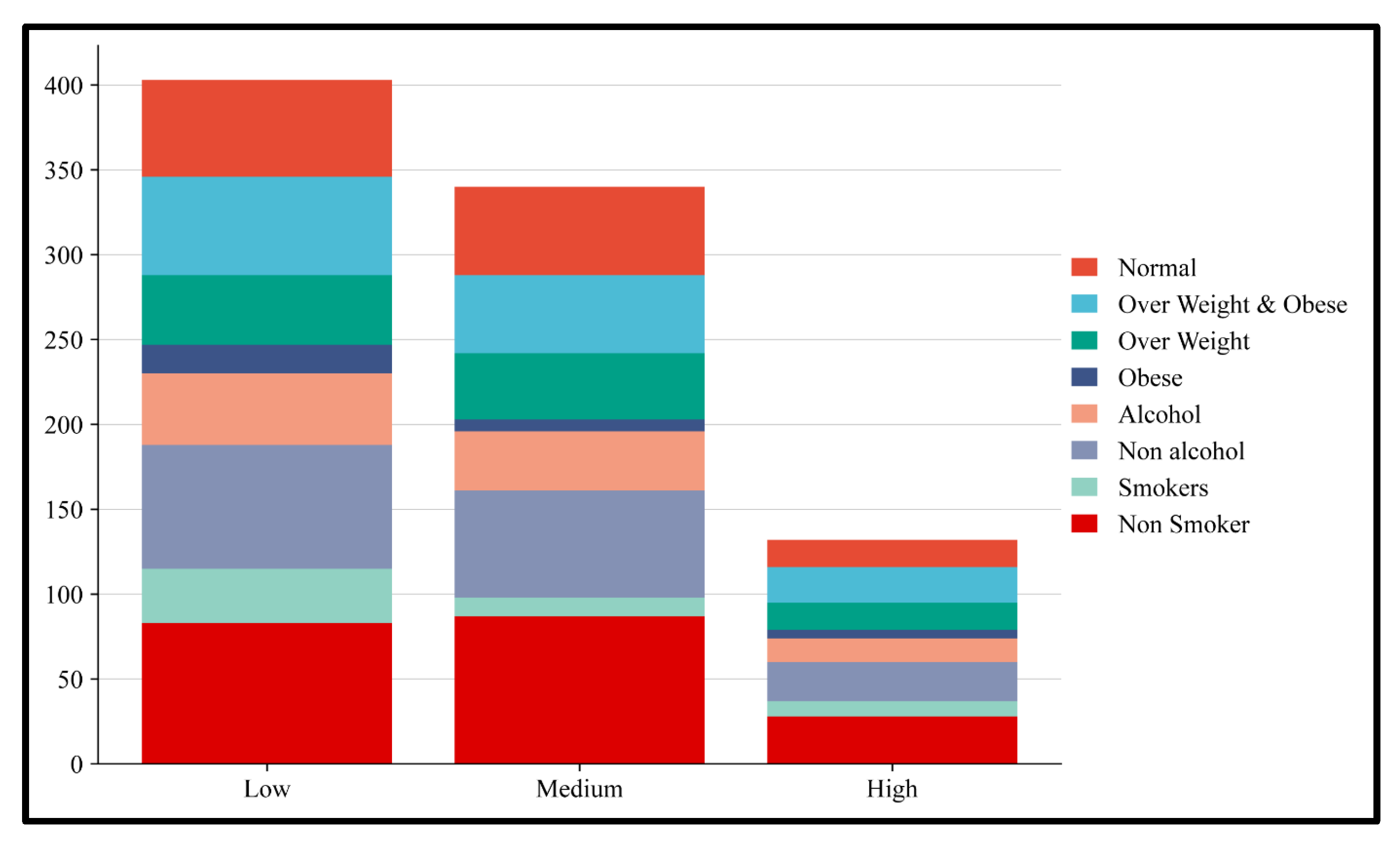
3.4. Risk Allele Association with Obesity and Alcohol

The risk alleles were further categorized into high, medium, and low-risk alleles. Among them, alleles 6, 7, and 8 were classified as high risk, alleles 4 and 5 were considered medium risk, and alleles 0, 1, 2, and 3 were considered low risk. These three groups of risk alleles were then associated with BMI, alcohol consumption, and smoking habits to assess genetic and lifestyle factors. From the results obtained, we found that 8% of individuals combining both the overweight and obese groups had a high-risk allele, whereas only 5% of the obese group had a high-risk allele. We also found that 6% of the alcohol-consuming group and 9% of the smoking group carried the high-risk allele (Figure 1 and Figure 2).

4. Discussion

Atrial fibrillation (AF) is predominantly considered an abnormal heart condition that affects mainly older people, with the risks considerably increasing with age [22]. Common conditions that increase the risk of AF include chronic kidney disease [23], diabetes [24], high blood pressure [25], overweight and obesity [26], hyperthyroidism, COPD, and other lung problems [27]. Furthermore, mutations in the genes related to the ion channels or structural proteins of the heart are associated with early-onset AF [9,28,29]. In addition to the genetic causes mentioned, conditions such as congenital heart disease [30,31], cardiomyopathies [32], and inherited syndromes (such as Brugada syndrome or Long QT syndrome) are also associated with an increased risk of AF in the young population [33,34,35]. In addition, lifestyle habits like high-intensity exercise, excessive alcohol or use of stimulants, and underlying disorders like obesity or sleep apnea cause increased susceptibility to AF in younger individuals [36]. A further literature review also suggests that younger individuals are more susceptible to AF, which is caused by genetic factors and other associated risk determinants [37,38]. This study investigated the prevalence of genetic variants associated with AF in the young adult population in India. Here, we focused on common single-nucleotide polymorphisms (SNPs) in transcription factors, including PITX2 (rs2200733, rs10033464, and rs13143308), TBX5 (rs883079), PRRX1 (rs3903239), ZFHX3 (rs2106261), and HAND2 (rs7698692). GWASs have shown that these genetic variants are linked to susceptibility to AF [39]. The findings of this study provide the prevalence and distribution of risk alleles within the young population, offering valuable insights into AF risk, particularly in the Indian community.
Genetic variants, such as rs2200733, rs10033464, and rs13143308, located on chromosome 4q25 near the PITX2 gene, are highly associated with AF. Previous studies have consistently highlighted the PITX2 gene as a key locus for AF development, particularly in European populations [11,40]. Our analysis identified the prevalence of PITX2 variants in the young adult population in India. The PITX2 gene is essential for cardiac development and function, especially in regulating left–right asymmetry and atrial electrical activity. The proximity of the variants to the PITX2 gene suggests that they influence atrial electrical remodeling and conduction, predisposing individuals to AF [9,41]. In this study, the high prevalence of the rs883079 variant (Table 2), located on chromosome 12 at the 3′ UTR of the TBX5 gene, influences the AF risk, consistent with previous findings in other ethnic groups [17]. Additionally, this variant is associated with a prolonged PR interval, which serves as an index of electrical activation from the atrial chambers [17]. The prevalence of the rs3903239 variant (Table 2), located on chromosome 1q24, 46 kb upstream of the PRRX1 gene, has been associated with a lower incidence of AF. The presence of rs3903239 likely affects the gene expression of PRRX1, resulting in atrial conduction, which prevents the development of AF, establishing it as a protective factor [21]. The prevalence of the rs2106261 variant (Table 2), located on chromosome 16q22 in the ZFHX3 gene, is associated with an increased risk of AF. Among Asians, the rs2106261 SNP affects normal conductivity, making people more susceptible to atrial fibrillation (AF). Furthermore, rs2106261 is considered a key genetic marker for AF susceptibility, supported by research studies and meta-analysis [42,43]. The prevalence of rs7698692, a variant (Table 2) located near the HAND2 gene on chromosome 4q34, is a susceptibility locus for AF, particularly among the Japanese or East Asian population [44,45]. Variants affecting the HAND2 gene may alter the heart’s conduction system, thereby increasing the risk of AF. Though the prevalence of this variant is relatively low in the Indian population, its role in heart development and conduction system differentiation makes it a relevant risk factor for AF [45].
The prevalence of minor allele frequency was compared with the IndiGenome database. Comparative analysis reveals that the frequency of the minor allele T of the SNP rs2200733 increased from 13% (in the IndiGenome database) to 16% (in our study). Similarly, the frequency of the minor allele T at rs10033464 increased from 23% to 27%. Regarding rs13143308, the minor allele T decreased in frequency from 36% to 32%. However, in this study, for rs883079, the frequency of the minor allele T was reduced from 53% to 46%. For rs3903239, the frequency of the minor allele G increased slightly from 23% to 25%. As for rs2106261, the minor allele T maintains a constant frequency of 68% in both groups. For rs7698692, the frequency of the minor allele G moderately decreased from 16% to 14%. Thus, our findings suggest that specific SNPs showed increased frequencies, while others remained consistent (Table 5).
Furthermore, this study identified the varying prevalence of homozygous mutant genotypes for SNPs linked with AF in the young adult population in India (Table 3). The rs2200733 variant (PITX2) had a low prevalence, with 3% of individuals carrying the TT genotype. In contrast, the other PITX2 gene variants, rs10033464 and rs13143308, had a high prevalence, with 10% carrying the TT genotype. Thus, the highest prevalence of these variants may suggest a strong correlation with AF susceptibility in young adults in the Indian population. The rs883079 (TBX5) variant is present in 21% of those carrying the TT genotype, suggesting genetic protection against AF. The rs3903239 (PRRX1) and rs2106261 (ZFHX3) variants, each with a frequency of 7% in the homozygous mutant GG and TT genotypes, respectively, are consistent with their roles in AF risk. The rs7698692 (HAND2) variant is rare, with only 1% of individuals having the GG genotype.
This study’s distribution of risk alleles reveals that a significant proportion of individuals (20%) possess four risk alleles. In comparison, high-risk allele groups comprise six (7%), seven (4%), or eight (3%) risk alleles. This implies that even young and healthy individuals carrying AF-related risk alleles emphasize the necessity of early genetic screening, particularly for those with a family history of AF or related cardiac conditions. Genetic counseling and preventive measures would be essential for these individuals to mitigate the onset of AF and its associated complications. As genetic predisposition plays a more prominent role in determining AF in younger adults [46], risk identification, increasing awareness, and conducting genetic testing are essential for early diagnosis and treatment. This study will contribute to implementing genetic screening for AF susceptibility in the future. This can be practically achieved by targeting the high-risk population with a family history of AF and early-onset AF using authenticated tools like polygenic risk scores or specific genetic variant panels.
Our study was conducted on a small group of young adults. Therefore, further studies are required on a larger cohort. Furthermore, to confirm the obtained observations, a larger population comprising diverse groups is essential to better understand the underlying factors contributing to allele frequency differences across various demographic and environmental contexts. The study has certain limitations, including a small sample size limited to a specific age range and locality within the Indian population, which does not represent the country adequately. A more representative cohort is needed, along with a study to enhance the generalizability of the results. A further limitation is that this study used a cross-sectional framework, which limits our ability to understand the causal associations between genetic variants and AF. In the future, case–control studies or longitudinal studies should be used to study the temporal relationships between and predictive properties of these SNPs. Since we studied individual SNPs rather than linkage disequilibrium (LD) haplotypes, we could not understand the genetic framework of AF. Functional studies are essential to evaluate the biological significance of these variants. As a result of incorporating diverse ethnic groups, we might be able to better understand these variants across a range of genetic backgrounds and enhance risk stratification models.

5. Conclusions

This study provided insight into the associations between genetic variants and atrial fibrillation (AF) in the young adult population in India. The significant distribution of a major proportion of risk alleles among young adults emphasizes the need for further research into the genetic predisposition of AF in this population. Understanding the associations between genetic factors and atrial fibrillation (AF) will enable early diagnosis and the development of personalized therapies, thereby mitigating the long-term burden of AF-related complications.

Supplementary Materials

The following supporting information can be downloaded at: https://www.mdpi.com/article/10.3390/medicina61050900/s1. Detailed information on the genotyping results with the HWE values of the study group is shown in the Supplementary Table S1.

Author Contributions

M.M.: conceptualization, investigation, methodology, writing—original draft preparation, data curation, software, and validation. S.R.: conceptualization, writing—review and editing, validation, supervision, and project administration. I.G.: data curation, software, and writing—review and editing. Y.V.: data curation, software, and formal analysis. A.P.: data curation, methodology, and formal analysis. V.B.B.: conceptualization, writing—review and editing, and resources. All authors have read and agreed to the published version of the manuscript.

Funding

The first author sincerely acknowledges the ICMR Senior Research Fellowship Program (3/1//1(25)/2022-NCD-1) for financial support. This research was supported by the ICMR Adhoc Project, “Association between PITX2, GATA-4 SNPs and atrial fibrillation in a young adult population—An Observational study” (ICMR Adhoc Project No. 5/4-35 5/1-6/2020—NCD-II).

Institutional Review Board Statement

This study was approved by our institute’s Institutional Human Ethics Committee (IEC No: AV/IEC/2020/022, approval date 19 March 2020), and we obtained informed consent from all subjects included in the study. All procedures performed in the study involving human participants were conducted based on the ethical standards of the institutional and/or national research committee in compliance with the 1964 Helsinki Declaration and its subsequent amendments or with comparable ethical standards.

Informed Consent Statement

Informed written consent was obtained from all participants in the study.

Data Availability Statement

All data will be available from the corresponding author upon reasonable request.

Acknowledgments

We thank all the participants who took part in this study.

Conflicts of Interest

The authors declare no conflicts of interest regarding this study.

Abbreviations

AFAtrial fibrillation
SNPSingle-nucleotide polymorphism
BMIBody mass index
PCRPolymerase Chain Reaction
PITX2Paired-Like Homeodomain 2
TBX5T-Box Transcription Factor 5
PRRX1Paired Related Homeobox 1
ZFHX3Zinc Finger Homeobox 3
HAND2Heart and Neural Crest Derivatives Expressed 2
OROdds ratio
CIConfidence interval
SPSSStatistical Package for the Social Sciences

References

  1. Chugh, S.S.; Havmoeller, R.; Narayanan, K.; Singh, D.; Rienstra, M.; Benjamin, E.J.; Gillum, R.F.; Kim, Y.H.; McAnulty, J.H., Jr.; Zheng, Z.J.; et al. Worldwide epidemiology of atrial fibrillation: A Global Burden of Disease 2010 Study. Circulation 2014, 129, 837–847. [Google Scholar] [CrossRef] [PubMed]
  2. Benjamin, E.J.; Muntner, P.; Alonso, A.; Bittencourt, M.S.; Callaway, C.W.; Carson, A.P.; Chamberlain, A.M.; Chang, A.R.; Cheng, S.; Das, S.R.; et al. Heart Disease and Stroke Statistics-2019 Update: A Report From the American Heart Association. Circulation 2019, 139, e56–e528. [Google Scholar] [CrossRef] [PubMed]
  3. Guo, X.J.; Qiu, X.B.; Wang, J.; Guo, Y.H.; Yang, C.X.; Li, L.; Gao, R.F.; Ke, Z.P.; Di, R.M.; Sun, Y.M.; et al. PRRX1 Loss-of-Function Mutations Underlying Familial Atrial Fibrillation. J. Am. Heart Assoc. 2021, 10, e023517. [Google Scholar] [CrossRef]
  4. Go, A.S.; Hylek, E.M.; Phillips, K.A.; Chang, Y.; Henault, L.E.; Selby, J.V.; Singer, D.E. Prevalence of diagnosed atrial fibrillation in adults: National implications for rhythm management and stroke prevention: The AnTicoagulation and Risk Factors in Atrial Fibrillation (ATRIA) Study. JAMA 2001, 285, 2370–2375. [Google Scholar] [CrossRef]
  5. Miyasaka, Y.; Barnes, M.E.; Gersh, B.J.; Cha, S.S.; Bailey, K.R.; Abhayaratna, W.P.; Seward, J.B.; Tsang, T.S. Secular trends in incidence of atrial fibrillation in Olmsted County, Minnesota, 1980 to 2000, and implications on the projections for future prevalence. Circulation 2006, 114, 119–125. [Google Scholar] [CrossRef]
  6. Saggu, D.K.; Rangaswamy, V.V.; Yalagudri, S.; Sundar, G.; Reddy, N.K.; Shah, V.; Kotti, K.; Shankar, M.; Chennapragada, S.; Narasimhan, C. Prevalence, clinical profile, and stroke risk of atrial fibrillation in rural Andhra Pradesh, India (the AP-AF study). Indian. Heart J. 2022, 74, 86–90. [Google Scholar] [CrossRef]
  7. Mohanty, S.; Banerjee, A.; Kumar, A.; Deb, P.; Samantray, H.; Das, D. Non-Valvular Atrial Fibrillation in Young Adults in Eastern India: A Clinico-Aetiological Retrospective Analysis in a Tertiary Care Hospital. Cureus 2023, 15, e36918. [Google Scholar] [CrossRef]
  8. Abdul-Aziz, A.A.; Desikan, P.; Prabhakaran, D.; Schroeder, L.F. Tackling the Burden of Cardiovascular Diseases in India. Circ. Cardiovasc. Qual. Outcomes 2019, 12, e005195. [Google Scholar] [CrossRef]
  9. Ellinor, P.T.; Lunetta, K.L.; Albert, C.M.; Glazer, N.L.; Ritchie, M.D.; Smith, A.V.; Arking, D.E.; Muller-Nurasyid, M.; Krijthe, B.P.; Lubitz, S.A.; et al. Meta-analysis identifies six new susceptibility loci for atrial fibrillation. Nat. Genet. 2012, 44, 670–675. [Google Scholar] [CrossRef]
  10. Lubitz, S.A.; Yin, X.; Lin, H.J.; Kolek, M.; Smith, J.G.; Trompet, S.; Rienstra, M.; Rost, N.S.; Teixeira, P.L.; Almgren, P.; et al. Genetic Risk Prediction of Atrial Fibrillation. Circulation 2017, 135, 1311–1320. [Google Scholar] [CrossRef]
  11. Gudbjartsson, D.F.; Arnar, D.O.; Helgadottir, A.; Gretarsdottir, S.; Holm, H.; Sigurdsson, A.; Jonasdottir, A.; Baker, A.; Thorleifsson, G.; Kristjansson, K.; et al. Variants conferring risk of atrial fibrillation on chromosome 4q25. Nature 2007, 448, 353–357. [Google Scholar] [CrossRef] [PubMed]
  12. Christophersen, I.E.; Rienstra, M.; Roselli, C.; Yin, X.; Geelhoed, B.; Barnard, J.; Lin, H.; Arking, D.E.; Smith, A.V.; Albert, C.M.; et al. Large-scale analyses of common and rare variants identify 12 new loci associated with atrial fibrillation. Nat. Genet. 2017, 49, 946–952. [Google Scholar] [CrossRef] [PubMed]
  13. Lee, J.Y.; Kim, T.H.; Yang, P.S.; Lim, H.E.; Choi, E.K.; Shim, J.; Shin, E.; Uhm, J.S.; Kim, J.S.; Joung, B.; et al. Korean atrial fibrillation network genome-wide association study for early-onset atrial fibrillation identifies novel susceptibility loci. Eur. Heart J. 2017, 38, 2586–2594. [Google Scholar] [CrossRef] [PubMed]
  14. Shen Han, Y.; Ahmed, A.M.; Chao, Z.; Jun, W.; Xiaochen, Y. Association Between rs2200733 Polymorphism of PITX2 Gene and the Risk of Atrial Fibrillation. Anatol. J. Cardiol. 2023, 27, 160–166. [Google Scholar] [CrossRef]
  15. Sun, L.; Zhang, Z.; Xu, J.; Xu, G.; Liu, X. Chromosome 4q25 Variants rs2200733, rs10033464, and rs1906591 Contribute to Ischemic Stroke Risk. Mol. Neurobiol. 2016, 53, 3882–3890. [Google Scholar] [CrossRef]
  16. Herraiz-Martinez, A.; Llach, A.; Tarifa, C.; Gandia, J.; Jimenez-Sabado, V.; Lozano-Velasco, E.; Serra, S.A.; Vallmitjana, A.; Vazquez Ruiz de Castroviejo, E.; Benitez, R.; et al. The 4q25 variant rs13143308T links risk of atrial fibrillation to defective calcium homoeostasis. Cardiovasc. Res. 2019, 115, 578–589. [Google Scholar] [CrossRef]
  17. Zhang, R.; Tian, X.; Gao, L.; Li, H.; Yin, X.; Dong, Y.; Yang, Y.; Xia, Y. Common Variants in the TBX5 Gene Associated with Atrial Fibrillation in a Chinese Han Population. PLoS ONE 2016, 11, e0160467. [Google Scholar] [CrossRef]
  18. Okubo, Y.; Nakano, Y.; Ochi, H.; Onohara, Y.; Tokuyama, T.; Motoda, C.; Amioka, M.; Hironobe, N.; Okamura, S.; Ikeuchi, Y.; et al. Predicting atrial fibrillation using a combination of genetic risk score and clinical risk factors. Heart Rhythm 2020, 17, 699–705. [Google Scholar] [CrossRef]
  19. Syeda, F.; Holmes, A.P.; Yu, T.Y.; Tull, S.; Kuhlmann, S.M.; Pavlovic, D.; Betney, D.; Riley, G.; Kucera, J.P.; Jousset, F.; et al. PITX2 Modulates Atrial Membrane Potential and the Antiarrhythmic Effects of Sodium-Channel Blockers. J. Am. Coll. Cardiol. 2016, 68, 1881–1894. [Google Scholar] [CrossRef]
  20. Nadadur, R.D.; Broman, M.T.; Boukens, B.; Mazurek, S.R.; Yang, X.; van den Boogaard, M.; Bekeny, J.; Gadek, M.; Ward, T.; Zhang, M.; et al. Pitx2 modulates a Tbx5-dependent gene regulatory network to maintain atrial rhythm. Sci. Transl. Med. 2016, 8, 354ra115. [Google Scholar] [CrossRef]
  21. Tucker, N.R.; Clauss, S.; Ellinor, P.T. Common variation in atrial fibrillation: Navigating the path from genetic association to mechanism. Cardiovasc. Res. 2016, 109, 493–501. [Google Scholar] [CrossRef] [PubMed]
  22. Curtis, A.B.; Karki, R.; Hattoum, A.; Sharma, U.C. Arrhythmias in Patients >/=80 Years of Age: Pathophysiology, Management, and Outcomes. J. Am. Coll. Cardiol. 2018, 71, 2041–2057. [Google Scholar] [CrossRef] [PubMed]
  23. Kulkarni, N.; Gukathasan, N.; Sartori, S.; Baber, U. Chronic Kidney Disease and Atrial Fibrillation: A Contemporary Overview. J. Atr. Fibrillation 2012, 5, 448. [Google Scholar] [PubMed]
  24. Bano, A.; Rodondi, N.; Beer, J.H.; Moschovitis, G.; Kobza, R.; Aeschbacher, S.; Baretella, O.; Muka, T.; Stettler, C.; Franco, O.H.; et al. Association of Diabetes With Atrial Fibrillation Phenotype and Cardiac and Neurological Comorbidities: Insights From the Swiss-AF Study. J. Am. Heart Assoc. 2021, 10, e021800. [Google Scholar] [CrossRef]
  25. Aune, D.; Mahamat-Saleh, Y.; Kobeissi, E.; Feng, T.; Heath, A.K.; Janszky, I. Blood pressure, hypertension and the risk of atrial fibrillation: A systematic review and meta-analysis of cohort studies. Eur. J. Epidemiol. 2023, 38, 145–178. [Google Scholar] [CrossRef]
  26. Goudis, C.A.; Korantzopoulos, P.; Ntalas, I.V.; Kallergis, E.M.; Ketikoglou, D.G. Obesity and atrial fibrillation: A comprehensive review of the pathophysiological mechanisms and links. J. Cardiol. 2015, 66, 361–369. [Google Scholar] [CrossRef]
  27. Tsagkaris, C.; Zacharopoulou, L. Thyroid Disease (TD), Chronic Obstructive Pulmonary Disease (COPD) and Valvular Heart Disease (VHD) as modifiable risk factors of Atrial Fibrillation. Rom. J. Intern. Med. 2020, 58, 3–4. [Google Scholar] [CrossRef]
  28. Kany, S.; Reissmann, B.; Metzner, A.; Kirchhof, P.; Darbar, D.; Schnabel, R.B. Genetics of atrial fibrillation-practical applications for clinical management: If not now, when and how? Cardiovasc. Res. 2021, 117, 1718–1731. [Google Scholar] [CrossRef]
  29. Andersen, J.H.; Andreasen, L.; Olesen, M.S. Atrial fibrillation-a complex polygenetic disease. Eur. J. Hum. Genet. 2021, 29, 1051–1060. [Google Scholar] [CrossRef]
  30. Holmgren, A.; Giang, K.W.; Fedchenko, M.; Eriksson, P.; Dellborg, M.; Mandalenakis, Z. Ischemic Stroke in Patients With Congenital Heart Disease and Atrial Fibrillation. J. Am. Heart Assoc. 2024, 13, e032813. [Google Scholar] [CrossRef]
  31. Mandalenakis, Z.; Rosengren, A.; Lappas, G.; Eriksson, P.; Gilljam, T.; Hansson, P.O.; Skoglund, K.; Fedchenko, M.; Dellborg, M. Atrial Fibrillation Burden in Young Patients With Congenital Heart Disease. Circulation 2018, 137, 928–937. [Google Scholar] [CrossRef] [PubMed]
  32. Qin, D.; Mansour, M.C.; Ruskin, J.N.; Heist, E.K. Atrial Fibrillation-Mediated Cardiomyopathy. Circ. Arrhythm. Electrophysiol. 2019, 12, e007809. [Google Scholar] [CrossRef] [PubMed]
  33. Kusano, K.F.; Taniyama, M.; Nakamura, K.; Miura, D.; Banba, K.; Nagase, S.; Morita, H.; Nishii, N.; Watanabe, A.; Tada, T.; et al. Atrial fibrillation in patients with Brugada syndrome relationships of gene mutation, electrophysiology, and clinical backgrounds. J. Am. Coll. Cardiol. 2008, 51, 1169–1175. [Google Scholar] [CrossRef] [PubMed]
  34. Platonov, P.G.; McNitt, S.; Polonsky, B.; Rosero, S.Z.; Zareba, W. Atrial Fibrillation in Long QT Syndrome by Genotype. Circ. Arrhythm. Electrophysiol. 2019, 12, e007213. [Google Scholar] [CrossRef]
  35. Knoche, J.W.; Orland, K.M.; January, C.T.; Maginot, K.R. Atrial Fibrillation and Long QT Syndrome Presenting in a 12-Year-Old Girl. Case Rep. Pediatr. 2012, 2012, 124838. [Google Scholar] [CrossRef]
  36. Yang, L.; Chung, M.K. Lifestyle changes in atrial fibrillation management and intervention. J. Cardiovasc. Electrophysiol. 2023, 34, 2163–2178. [Google Scholar] [CrossRef]
  37. Sankaranarayanan, R.; Kirkwood, G.; Dibb, K.; Garratt, C.J. Comparison of Atrial Fibrillation in the Young versus That in the Elderly: A Review. Cardiol. Res. Pract. 2013, 2013, 976976. [Google Scholar] [CrossRef]
  38. Wang, N.; Yu, Y.; Sun, Y.; Zhang, H.; Wang, Y.; Chen, C.; Tan, X.; Wang, B.; Lu, Y. Acquired risk factors and incident atrial fibrillation according to age and genetic predisposition. Eur. Heart J. 2023, 44, 4982–4993. [Google Scholar] [CrossRef]
  39. Ebana, Y.; Sun, Y.; Yang, X.; Watanabe, T.; Makita, S.; Ozaki, K.; Tanaka, T.; Arai, H.; Furukawa, T. Pathway analysis with genome-wide association study (GWAS) data detected the association of atrial fibrillation with the mTOR signaling pathway. Int. J. Cardiol. Heart Vasc. 2019, 24, 100383. [Google Scholar] [CrossRef]
  40. Lubitz, S.A.; Lunetta, K.L.; Lin, H.; Arking, D.E.; Trompet, S.; Li, G.; Krijthe, B.P.; Chasman, D.I.; Barnard, J.; Kleber, M.E.; et al. Novel genetic markers associate with atrial fibrillation risk in Europeans and Japanese. J. Am. Coll. Cardiol. 2014, 63, 1200–1210. [Google Scholar] [CrossRef]
  41. Wang, J.; Klysik, E.; Sood, S.; Johnson, R.L.; Wehrens, X.H.; Martin, J.F. Pitx2 prevents susceptibility to atrial arrhythmias by inhibiting left-sided pacemaker specification. Proc. Natl. Acad. Sci. USA 2010, 107, 9753–9758. [Google Scholar] [CrossRef] [PubMed]
  42. Wei, Y.; Wang, L.; Lin, C.; Xie, Y.; Bao, Y.; Luo, Q.; Zhang, N. Association between the rs2106261 polymorphism in the zinc finger homeobox 3 gene and risk of atrial fibrillation: Evidence from a PRISMA-compliant meta-analysis. Medicine 2021, 100, e27749. [Google Scholar] [CrossRef] [PubMed]
  43. Wu, L.; Chu, M.; Zhuang, W. Association between ZFHX3 and PRRX1 Polymorphisms and Atrial Fibrillation Susceptibility from Meta-Analysis. Int. J. Hypertens. 2021, 2021, 9423576. [Google Scholar] [CrossRef] [PubMed]
  44. Low, S.K.; Takahashi, A.; Ebana, Y.; Ozaki, K.; Christophersen, I.E.; Ellinor, P.T.; Consortium, A.F.; Ogishima, S.; Yamamoto, M.; Satoh, M.; et al. Identification of six new genetic loci associated with atrial fibrillation in the Japanese population. Nat. Genet. 2017, 49, 953–958. [Google Scholar] [CrossRef]
  45. Patel, K.K.; Venkatesan, C.; Abdelhalim, H.; Zeeshan, S.; Arima, Y.; Linna-Kuosmanen, S.; Ahmed, Z. Genomic approaches to identify and investigate genes associated with atrial fibrillation and heart failure susceptibility. Hum. Genom. 2023, 17, 47. [Google Scholar] [CrossRef]
  46. Wasmer, K.; Eckardt, L.; Breithardt, G. Predisposing factors for atrial fibrillation in the elderly. J. Geriatr. Cardiol. 2017, 14, 179–184. [Google Scholar] [CrossRef]
Figure 1. Comparison of risk allele distribution between BMI, alcohol consumption, and smoking individuals: The violin plot compares the distribution of risk alleles between two groups, such as (A) normal and overweight individuals, (B) overweight and obese individuals, (C) alcohol and non-alcohol individuals, and (D) smokers and non-smokers. Here, the x-axis indicates the different categories of risk alleles, such as low, medium, and high. The y-axis denotes the number of risk alleles, ranging from 0 to 8. The density of individuals possessing the specific number of risk alleles within each group is represented by the width of the violin. A wider range denotes a greater number of individuals possessing risk alleles.
Medicina 61 00900 g001
Figure 2. Distribution of BMI categories, alcohol consumption, and smoking status in the young adult population. A stacked bar chart shows the distribution of health-related behavioral and physiological traits: BMI (Normal, Overweight, and Obese), alcohol consumption (Alcohol and Non-alcohol), and smoking status (Smokers and Non-Smokers) among Low, Medium, and High-risk groups. Each color block represents a category. The chart reveals lifestyle and health indicator differences between the three groups, with the Low group showing higher counts in many risk-related categories.
Medicina 61 00900 g002
Table 1. Single-nucleotide polymorphisms in AF-associated transcription factor genes with allelic variants, genomic location, and sequence context.
GeneSNP IDAllele ChangeSNP TypeContext SequenceTaqMan
Assay Id
PITX2rs2200733C/TIntergenicGTGGTACTTGGGTTTTGATTTTGAT[C/T]AGAGAAAATTAGAACAGGTAATATTC_16158671_10
PITX2rs10033464T/GIntergenicTTTTTACATTGTTAGAGTCAAGAAA[T/G]AAGTGCTTTCATCAAGCTCTGAGTTC_30351088_30
PITX2rs13143308T/GRegulatory regionGATGGATAAACCAAATGCATAAATA[G/T]CTTTGCCTTATTTCTAACCTCTTTGC_31214706_10
TBX5rs883079C/T3′UTR variantATTGGGTGAAATGAAAAATCTTGTC[C/T]GTGGGGTTCTCTTGGCTACTGTCTCC_7526955_10
PRRX1rs3903239A/GIntronTTTCCTCTGCAATGTTTAATCTGCC[A/G]TTAATCCTGTCCATTGTATTTTTCAC_8919547_10
ZFHX3rs2106261C/TIntronAGATAGAGCTCGTCCAGAGAATTGT[C/T]CAACCATCCATTAAAATATCCAAGTC_16097559_10
HAND2rs7698692G/AIntergenicGGGACTTTTAATAAAACCACTTGGG[A/G]ATATTAATGTGATACGTAAGTGGCTC_310074_10
Table 2. Demographic profile of the study population.
Demographic CharacteristicsYoung Adult Population, n = 250 (%)
Age (years) (mean ± SD)25.81 ± 6.19
Male, n (%)173 (69)
Female, n (%)77 (31)
BMI (kg/m2) (mean ± SD)24.9 ± 4.2
Normal, n (%)125 (50)
Overweight, n (%)96 (38)
Obese, n (%)29 (12)
SBP (mmHg)127.4 ± 15.3
DBP (mmHg)84.8 ± 9.1
Alcohol consumption, n (%)91(36)
Smoking, n (%)52 (21)
Family History of Obesity, n (%)42 (17)
Family History of Cardiovascular Disease, n (%)38 (15)
Family History of Diabetics, n (%)58 (23)
BMI: body mass index; SBP: systolic blood pressure; and DBP: diastolic blood pressure.
Table 3. Genotype and allele distribution of SNPs responsible for atrial fibrillation.
GeneSNP IDGenotype and AllelesStudy Population95% CI
N = 250Percentage (%)LowerUpper
PITX2rs2200733CC177716082
CT65262033
TT8316
C419847592
T81161220
rs10033464GG138554665
GT88352843
TT2410614
G364736580
T136272232
rs13143308GG117473856
TG107433551
TT2610615
G341686175
T159322737
TBX5rs883079CC71282235
CT127514260
TT52211527
C269544760
T231464052
PRRX1rs3903239AA144584867
AG88352843
GG187411
A376756783
G124252029
ZFHX3rs2106261CC138554665
CT94383046
TT187411
C370746681
T130262130
HAND2rs7698692AA184746385
AG63251932
GG3103
A431867894
G69141017
CI: confidence interval; data were represented as numbers (%).
Table 4. Comparison of the variant allele frequency of AF-associated variants in South Asian population with databases.
Chr:Position:rsIDGeneVariant Allele1000
Genome
gnomADIndiGenomeIn Our Study
chr4-110789013rs2200733PITX2T0.13090.13380.13200.16
chr4-4110799605rs10033464PITX2G0.80060.77670.77060.74
chr4-110793263rs13143308PITX2G0.67590.65270.64660.68
chr12-114355435rs883079TBX5T0.51330.51980.50340.46
chr1-170600176rs3903239PRRX1G0.21680.22320.23730.25
chr16-73017721rs2106261ZFHX3T0.23930.22980.26270.26
chr4-173682953rs7698692HAND2A0.87930.87750.84830.86
Table 5. Distribution of risk alleles in the study population with a 95% confidence interval.
Number of Risk AllelesStudy Population95% CI
N = 250Percentage (%)LowerUpper
07315
1239513
237151020
348191425
450201426
548191425
6187411
711427
88316
CI: confidence interval; data were represented as numbers (%).
Disclaimer/Publisher’s Note: The statements, opinions and data contained in all publications are solely those of the individual author(s) and contributor(s) and not of MDPI and/or the editor(s). MDPI and/or the editor(s) disclaim responsibility for any injury to people or property resulting from any ideas, methods, instructions or products referred to in the content.

