Article

Comparison of Total Mesopancreatic Excision and Conventional Pancreaticoduodenectomy in the Surgical Treatment of Pancreatic Head Adenocarcinoma: Early Postoperative Outcomes

1
Department of General Surgery, Dokuz Eylul University, Izmir 35330, Türkiye
2
Department of Pathology, Dokuz Eylul University, Izmir 35330, Türkiye
3
Division of Medical Oncology, Department of Internal Medicine, Dokuz Eylul University, Izmir 35330, Türkiye
4
Department of General Surgery, Nusaybin State Hospital, Mardin 47300, Türkiye
5
Department of General Surgery, Bagcilar Training and Research Hospital, Istanbul 34200, Türkiye
*
Author to whom correspondence should be addressed.
Medicina 2025, 61(10), 1725; https://doi.org/10.3390/medicina61101725
Submission received: 20 August 2025 / Revised: 16 September 2025 / Accepted: 17 September 2025 / Published: 23 September 2025
(This article belongs to the Section Surgery)

Abstract

Background and Objectives: This study aimed to evaluate and compare the early postoperative outcomes of patients who underwent pancreaticoduodenectomy (PD) with total mesopancreatic excision (TMpE) versus conventional pancreaticoduodenectomy (Co-PD) for pancreatic head ductal adenocarcinoma (PDAC). Materials and Methods: Patients who underwent PD for pancreatic head cancer between January 2021 and December 2024 in our clinic and had a pathological diagnosis of PDAC were included. Patients were stratified into two groups according to the surgical technique performed (TMpE-PD vs. Co-PD). Demographic characteristics and early postoperative clinicopathological data were compared between the groups. Results: A total of 41 patients were included: 17 (41.5%) underwent TMpE-PD and 24 (58.5%) underwent Co-PD. Demographic and clinicopathological parameters were comparable between the groups. Although not statistically significant, the TMpE-PD group demonstrated higher R0 resection rates (58.8% vs. 45.8%; p = 0.412) and greater lymph node yield (33.9 vs. 29.1; p = 0.757) compared to the Co-PD group. Overall postoperative complications were more frequent in the TMpE-PD group (82.4% vs. 63.4%; p = 0.034). A smaller pancreatic duct diameter was associated with an increased risk of postoperative complications in both groups, approaching statistical significance (p = 0.053). Multivariable logistic regression analysis revealed that the surgical technique was not an independent risk factor for postoperative complications (OR: 0.64; 95% CI: 0.14–2.83; p = 0.56). No direct correlation was found between resection margin status (R0 vs. R1) and the development of postoperative complications. Conclusions: TMpE demonstrated non-significant trends toward higher R0 resection rates and greater lymph node yield compared with conventional PD. These findings suggest possible oncological benefits without significantly increasing perioperative morbidity.

1. Introduction

Pancreaticoduodenectomy (PD, Whipple procedure) with curative intent (margin-negative/R0 resection), combined with adjuvant or neoadjuvant chemotherapy, currently represents the standard treatment for pancreatic ductal adenocarcinoma (PDAC) located in the pancreatic head [1,2,3,4,5,6]. However, only 15–25% of patients are eligible for surgical resection at the time of diagnosis [7]. Despite advances in surgical techniques over the years, R0 resection following PD is reported in only about 50% of cases [8]. Furthermore, local recurrence develops in approximately 80% of patients after PD, even in those considered to have undergone R0 resection, suggesting that true complete resection may not have been achieved [9,10]. Esposito et al. reported that the frequency of R1 resection margins in PD specimens increased from 14% to 76% when reassessed using standardized contemporary pathological evaluation methods [11]. Even in patients undergoing combined PD and chemotherapy for pancreatic head cancer, 5-year survival rates remain around 20–25% [12,13,14]. This poor outcome is attributed to early locoregional recurrence secondary to incomplete (non-curative) resections. Ghaneh et al. demonstrated that non-curative resections significantly reduce both overall and disease-free survival in patients with pancreatic cancer [15].
In 2007, Gockel et al. first introduced the concept of the “mesopancreas” [16]. The mesopancreas is defined as a structure located posterior to the pancreatic head, consisting of neural and lymphatic tissues extending along the mesenteric vascular axis. This region represents a major pathway for tumor spread in PDAC. Gockel emphasized that incomplete resections following PD often result from residual tumor deposits in this area and highlighted the necessity of performing a “total mesopancreatic excision (TMpE)” to achieve true curative resection. Supporting this view, Gadcke et al. demonstrated that the most frequent site of R1 resection after PD was the mesopancreatic region [17]. Subsequent studies have reported that PD with TMpE increases the likelihood of achieving true R0 resection, improves lymph node yield, and thereby confers a survival advantage [18,19,20].
Nevertheless, several controversies remain regarding TMpE. The anatomical definition of the mesopancreas has not reached universal consensus, with varying descriptions proposed by different authors [21]. Consequently, the optimal extent of TMpE remains a subject of debate. Moreover, different surgical approaches to TMpE have been suggested, and an internationally standardized technique has yet to be established [22]. Furthermore, some studies have reported increased morbidity, including refractory diarrhea and lymphorrhea following TMpE, which may delay the initiation of adjuvant chemotherapy [21,23].
The present study aimed to compare the early postoperative outcomes of pancreatic head PDAC patients who underwent PD with TMpE versus conventional PD (Co-PD) in our institution.

2. Patients and Methods

2.1. Ethical Approval and Patient Selection

The study was conducted in accordance with the Declaration of Helsinki and approved by the Institutional Review Board (or Ethics Committee) of Dokuz Eylul University (Protocol code: 2025/06-26 and date of approval: 19 February 2025). This study was approved by the institutional ethics committee. Patients who underwent pancreaticoduodenectomy (PD) for pancreatic head cancer in our clinic between January 2021 and December 2024 were retrospectively reviewed. Among these, only patients with a pathological diagnosis of pancreatic ductal adenocarcinoma (PDAC) were included. Informed consent was obtained from all patients involved in the study. Patients were stratified into two groups: those who underwent PD with total mesopancreatic excision (TMpE-PD) and those who underwent conventional PD (Co-PD). All procedures were performed by experienced surgeons within our hepatopancreatobiliary (HPB) surgery unit. While some surgeons preferred the Co-PD technique, others routinely performed TMpE-PD in all eligible cases.

2.2. Surgical Technique

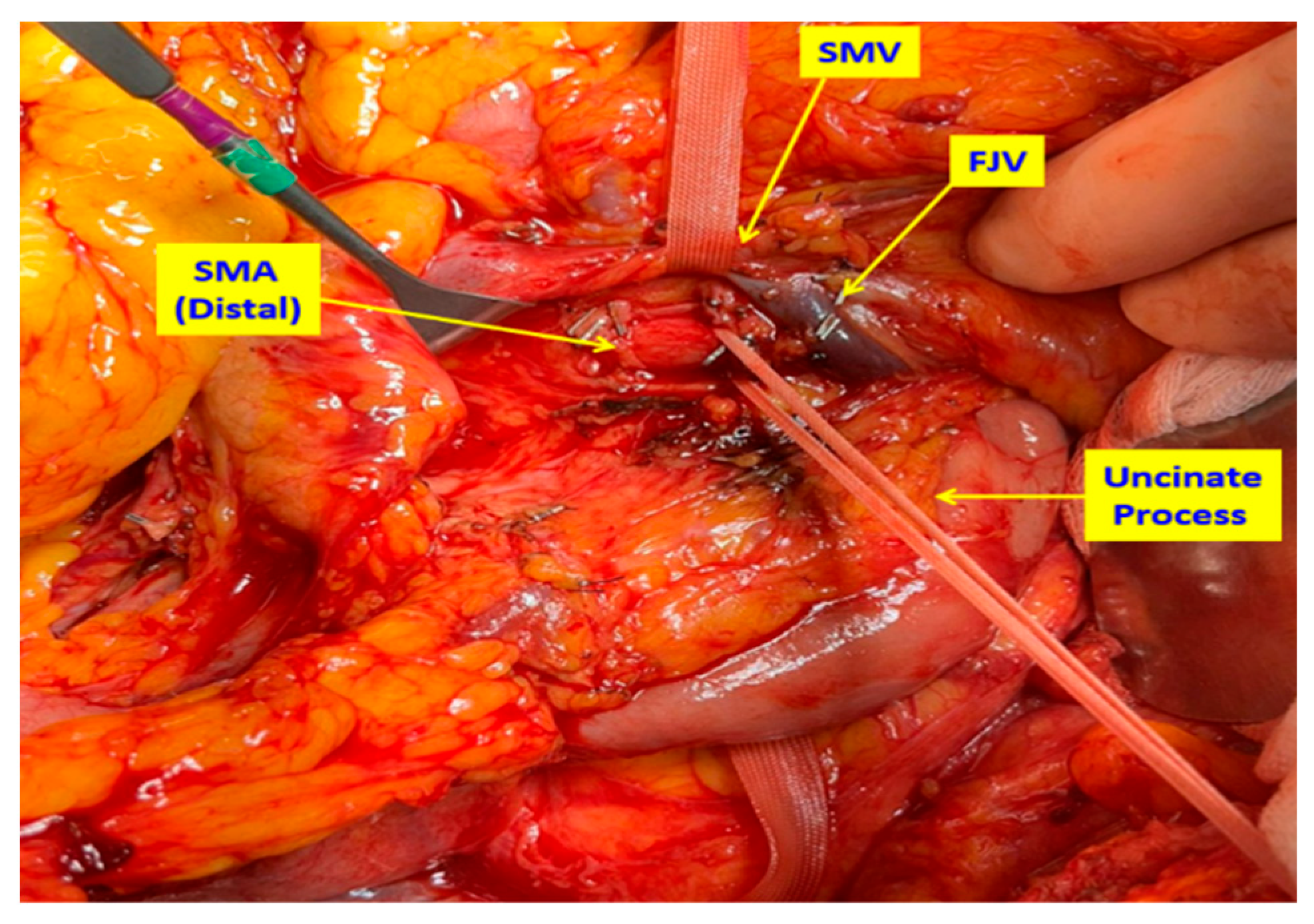
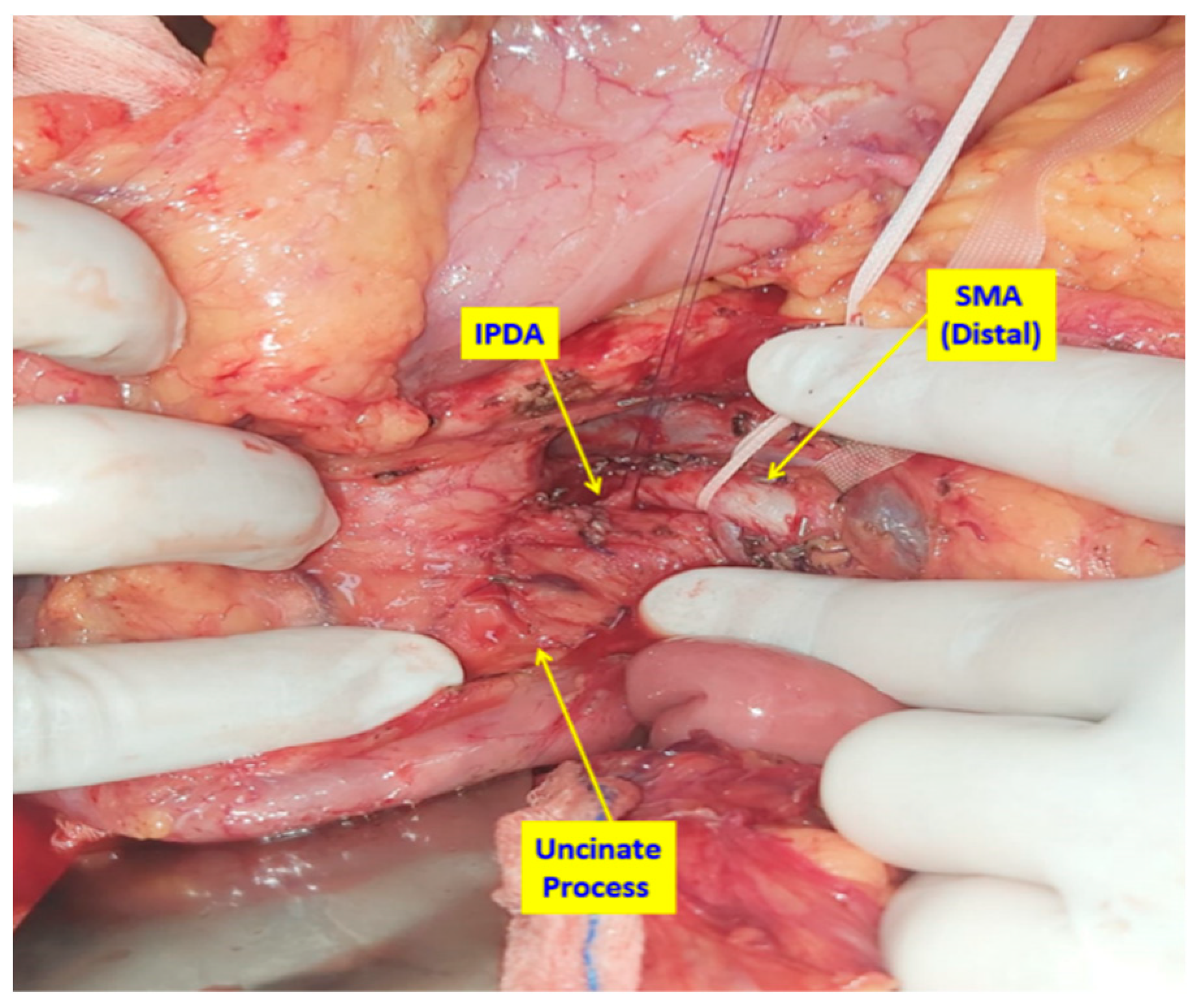
In patients undergoing conventional PD (Whipple procedure), the standard, previously described surgical technique was applied [24]. In TMpE-PD cases, surgery was initiated with an artery-first approach. Following a wide Kocher maneuver, the superior mesenteric artery (SMA) was dissected posteriorly, encircled, and suspended. The dissection was extended distally via the uncinate process, allowing both distal and proximal control of the SMA (Figure 1 and Figure 2). Dissection proceeded in a caudo-cranial manner along the pancreatic border toward the SMA origin, at which point the artery was assessed for tumor invasion.
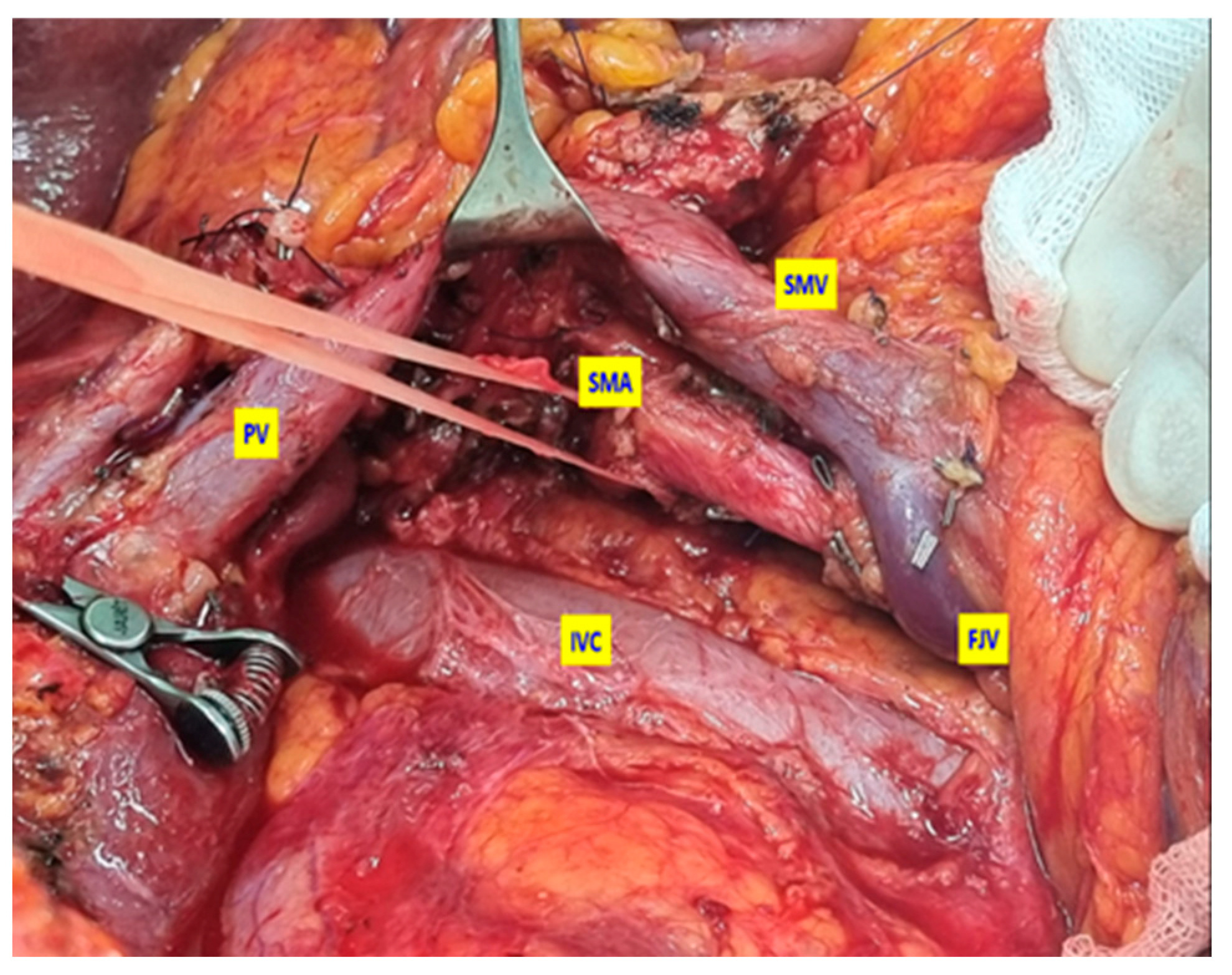
If no invasion was detected, the tissue plane between the SMA and pancreas was dissected as close as possible to the arterial wall, leaving the mesopancreatic specimen en bloc. At this stage, the inferior pancreaticoduodenal artery (IPDA) and its branches were ligated and divided (Figure 3). The first jejunal vein (FJV) was preserved unless invaded by tumor, in which case it was ligated and divided. Similarly, branches of the first jejunal artery (FJA) were preserved unless infiltration was present, requiring ligation.
During the resection phase, the uncinate process was excised from the retroperitoneum with the mesopancreas included in the specimen (Figure 4).
In cases with SMV invasion, distal and proximal vascular control was established. Following temporary clamping, en bloc resection was performed, and vascular continuity was restored with reconstruction before proceeding with the remaining anastomoses.
In both groups, lymphadenectomy was performed according to the recommendations of the International Study Group of Pancreatic Surgery (ISGPS) [25]. Pancreaticojejunostomy was performed as an end-to-side anastomosis, with the pancreatic capsule sutured to the jejunal seromuscular layer using interrupted 3/0 silk sutures, and duct-to-mucosa anastomosis performed with interrupted 5/0 polydioxanone (PDS) sutures.
Pathological specimens were evaluated macroscopically and microscopically by pathologists specialized in HPB pathology. For macroscopic evaluation, pancreatic surgical margin, retroperitoneal surgical margin, common bile duct surgical margin, and vascular bed surgical margin sampling were performed on all specimens. Microscopic evaluation included tumor type and differentiation, tumor pathologic T and N stages (according to the AJCC 8th Edition), surgical margin status, the number of dissected lymph nodes, and the number of metastatic lymph nodes. All surgical margins were inked, axial slicing of the pancreatic head was performed, and margin reporting followed international consensus definitions. Although no central review was conducted, the pathologists assessing the specimens were blinded to the surgical technique (TMpE vs. Co-PD); therefore, margin evaluation was blinded with respect to surgical approach.
Demographic features, comorbidities, preoperative factors, tumor size, TNM stage, operative time, estimated blood loss, resection margin status (R0 vs. R1), lymph node yield, length of hospital stay, and postoperative complications were recorded. Postoperative complications were classified according to the Clavien–Dindo system [26], with grade III or higher defined as major complications. Postoperative pancreatic fistula (POPF) was classified according to ISGPS criteria [27].
This study evaluated the relationship between surgical techniques and resection margin status (R0/R1) with postoperative complications and investigated risk factors affecting the development of postoperative complications.

2.3. Statistical Analysis

Logistic regression models were built using purposeful selection methods by Hosmer and Lemeshow, with clinically relevant variables retained regardless of univariable significance. Confidence intervals (95% CI) were reported for all effect estimates.
All statistical analyses were performed using jamovi (The jamovi project, Sydney, Australia) software (version 2.6), an open-source statistical package [28]. Normality of distribution for continuous variables was assessed using the Shapiro–Wilk test and visual inspection of Q–Q plots. Continuous variables were expressed as mean ± standard deviation or median (interquartile range), and categorical variables as numbers (percentages). Group comparisons were performed using Student’s t-test or the Mann–Whitney U test for continuous variables, and Chi-square or Fisher’s exact test for categorical variables.
To evaluate prognostic factors associated with complications, clinicopathological variables were first analyzed by univariable methods. Variables with p < 0.25 and those of established clinical relevance were included in the multivariable logistic regression analysis. The purposeful selection method by Hosmer and Lemeshow was applied [29]. Coefficient changes and potential interactions were examined, and clinically significant variables were retained in the final multivariable model. Results were expressed as odds ratios (ORs) with 95% confidence intervals (CIs). Two-tailed p values < 0.05 were considered statistically significant.

3. Results

A total of 41 patients were included in the study, of whom 17 (41.5%) underwent TMpE-PD and 24 (58.5%) underwent Co-PD. Demographic and clinicopathological parameters were comparable between the two groups, with no statistically significant differences (Table 1). The median operative time was slightly longer in the TMpE-PD group compared to the Co-PD group (485 min vs. 423 min; p = 0.067). Venous resections and reconstruction were performed in five patients (2 TMpE-PD, 3 Co-PD). Only one of these patients (Co-PD) had received neoadjuvant therapy; in the remaining four cases, vascular resection had not been anticipated preoperatively.
Although not statistically significant, the TMpE-PD group showed higher R0 resection rates (58.8% vs. 45.8%; p = 0.412) and greater lymph node yield (33.9 vs. 29.1; p = 0.757) compared to the Co-PD group. Pancreatic duct diameter and gland texture were similar between groups.
Postoperative outcomes are summarized in Table 2. Overall complication rates were higher in the TMpE-PD group compared with the Co-PD group (82.4% vs. 63.4%; p = 0.034). However, when only surgical complications were considered, there was no significant difference between the two groups (70.6% vs. 58.5%; p = 0.187). Although not statistically significant, the incidence of major complications (Clavien–Dindo grade ≥ III) was lower in the TMpE-PD group compared to the Co-PD group (11.8% vs. 25.0%; p = 0.414).
Readmission within the first 30 days occurred in one patient (5.9%) in the TMpE-PD group and three patients (12.5%) in the Co-PD group (p = 0.628). Clinically relevant POPF (Grade B–C) rates were low and comparable between the groups (TMpE-PD 5.9% vs. Co-PD 8.3%; p = 1.000). Chylous fistula developed in two patients (14%) in the TMpE-PD group, whereas none occurred in the Co-PD group. Neither group experienced delayed gastric emptying, refractory diarrhea, or post-pancreatectomy hemorrhage affecting clinical course.
Overall, hospital mortality due to POPF occurred in two patients (4.8%), one from each group. Additionally, two further deaths (4.8%) occurred in the Co-PD group within 90 days postoperatively, both related to comorbidities.
Analysis of parameters potentially associated with postoperative complications revealed no statistically significant predictors (Table 3).
Only a smaller pancreatic duct diameter was identified as a risk factor approaching statistical significance (p = 0.053).
After the initial evaluation, variables with p values < 0.25, including sex (OR:2.57 CI 0.69–9.55, p = 0.158), pathological stage (OR:2.73 CI 0.72–10.27, p = 0.138), and pancreatic duct diameter (OR:0.75 CI 0.56–1.00, p = 0.053) were selected for further models. The adjusted model also included pancreatic gland texture (OR:3.00 CI 0.44–20.44, p = 0.262) due to its known clinical importance.
In multivariable logistic regression analysis, the surgical technique (TMpE-PD vs. Co-PD) was not found to be an independent risk factor for the development of postoperative complications (OR: 0.64; 95% CI: 0.14–2.83; p = 0.56). Similarly, no direct association was observed between resection margin status (R0 vs. R1) and complication development (Table 4).

Discussion

Pancreatic ductal adenocarcinoma (PDAC) is one of the most aggressive malignancies, with approximately 80% of cases located in the pancreatic head [30,31]. In eligible patients, surgical resection with R0 margins remains the most effective treatment option [2,3,4,5]. Pancreaticoduodenectomy (PD) is the standard surgical procedure for pancreatic head adenocarcinomas, and technical advancements and modifications have been introduced over the years [32]. Nevertheless, even in patients undergoing curative-intent PD, early recurrence rates of 59.7–91.1% have been reported, most frequently attributed to R1 resection margins [33,34].
Total mesopancreatic excision (TMpE) has been proposed as a surgical technique with the potential to improve R0 resection rates and thereby reduce locoregional recurrence [16]. Excision of the mesopancreas during PD has been advocated to achieve more favorable oncological outcomes [17,35]. However, the definition of the mesopancreas remains controversial, with varying descriptions proposed by different authors [36,37,38]. As a result, no consensus has yet been reached regarding the optimal extent of TMpE. Furthermore, the absence of adequately powered randomized controlled trials limits the evidence on the efficacy and safety of TMpE [22].
Early reports of TMpE emphasized artery-first approaches to PD, particularly dissection beginning with the superior mesenteric artery (SMA) [18,20]. The artery-first approach offers several advantages, including early assessment of arterial invasion and avoidance of unnecessary resection in non-resectable cases [39]. For TMpE-PD, this strategy is regarded as technically advantageous. Studies comparing TMpE-PD with Co-PD have reported higher rates of R0 resection and greater lymph node yield with TMpE-PD. In a recent meta-analysis, Silva et al. also demonstrated that TMpE-PD achieved significantly higher R0 resection rates and lymph node retrieval compared with Co-PD [22].
Despite its potential oncological benefits, TMpE is associated with certain disadvantages. The procedure requires meticulous dissection along the adventitial plane of the SMA, which increases technical complexity and may prolong operative time. Furthermore, the more extensive dissection of the mesopancreatic tissue has the potential to increase perioperative morbidity. Extensive dissection around the SMA has been associated with potential morbidity, including refractory diarrhea and high-output chylous leakage due to neural and lymphatic injury [20,21,22,23]. For this reason, several authors have emphasized the need to avoid overly aggressive dissection. Refractory diarrhea not only increases morbidity but also delays the initiation of adjuvant chemotherapy [40,41,42]. Some reports have suggested that early use of antidiarrheal agents may help manage this complication [22,43]. In our series, no patients in the TMpE-PD group developed refractory diarrhea. In patients who developed diarrhea, dietary modifications, probiotics, and antidiarrheal medications were effective for symptom control. Chylous fistula occurred in two patients (14.8%) in the TMpE-PD group but in none of the Co-PD group, consistent with previous reports linking this complication to more extensive lymphatic dissection.
These findings highlight a central dilemma for surgeons: whether to accept an increased risk of postoperative morbidity—particularly refractory diarrhea—in exchange for potentially superior oncological clearance. The decision ultimately depends on defining the optimal extent of mesopancreatic dissection required for TMpE. Although multiple techniques have been described, no universally standardized and internationally accepted definition of the procedure exists to date [17,18,19,20,21,22,44,45,46].
Inoue et al. were the first to describe a systematic supracolic artery-first mesopancreatic excision for periampullary tumors, defining three levels of dissection of increasing extent [47]. They reported that TMpE-PD, compared with Co-PD, was associated with reduced operative time and blood loss. More recently, Nagakawa et al. proposed a refined TMpE technique based on four intense neurofibrous tissue (NFT) zones around the SMA (A, B, C, D areas), which provided a structured framework for dissection [48]. Compared with conventional PD, this approach achieved shorter operative times, reduced blood loss, and improved rates of R0 resection and lymphadenectomy.
In our institution, TMpE-PD is performed using a posterior and uncinate process combined artery-first approach, as described in the Methods section. The principle is to achieve early SMA control, assess resectability, and perform PD with en bloc removal of the mesopancreas. The extent of dissection is individualized intraoperatively, with the extension of resection margins determined when tumor infiltration is observed. Thus, oncological advantage is prioritized while balancing the relative risk of increased postoperative morbidity. We believe that in TMpE-PD, the extent of resection should be tailored to each patient based on intraoperative findings.
Considering that all tumors in this study were PDAC, our approach most closely resembles the level 2 dissection described by Inoue et al. Although statistical significance was not reached, TMpE-PD cases demonstrated trends consistent with the literature, including higher R0 resection rates and greater lymph node yields compared with Co-PD cases. In univariable analysis, overall complications were more frequent in the TMpE-PD group; however, when limited to surgical complications, no significant differences were observed. Moreover, major complications (Clavien–Dindo grade ≥ III) were more frequent in the Co-PD group. Multivariable logistic regression analysis demonstrated no significant differences in postoperative complication rates between the two groups. Similarly, no relationship was found between resection margin status (R0 vs. R1) and complication development. These findings suggest that more aggressive dissection to achieve R0 resection does not necessarily translate into higher postoperative morbidity. Among all cases, only a smaller pancreatic duct diameter approached significance as a risk factor for complications.
This study has several limitations. Its retrospective, single-center design and relatively small sample size limit the generalizability and statistical power of the findings. The retrospective, single-center design and relatively small sample size inherently limit causal inference and statistical power. The absence of propensity score matching may introduce selection bias. Different surgeons performed each technique, creating potential performance bias. Neoadjuvant therapy was underrepresented, reducing generalizability. Long-term outcomes (recurrence, survival) were not available due to short-term analysis. The regression model was exploratory and not validated.

4. Conclusions

Total mesopancreatic excision (TMpE) represents a promising surgical strategy for pancreatic head PDAC, offering improved locoregional clearance and the potential for superior oncological outcomes. Our findings, consistent with the current literature, suggest that TMpE-PD provides oncological advantages without increasing the risk of postoperative morbidity. However, the optimal extent of TMpE remains undefined, and further high-quality, multi-center studies with standardized protocols are warranted to establish international consensus and strengthen the evidence base. In conclusion, TMpE demonstrated non-significant trends toward higher R0 resection rates and greater lymph node yields compared with conventional PD. While these findings suggest potential oncologic benefit, definitive conclusions cannot be drawn from this dataset. Further multi-center prospective studies with standardized protocols are necessary.

Author Contributions

Conceptualization: T.E., T.U., M.O. and C.A.; Methodology: T.E. and T.U.; Software: T.E. and B.S.; Validation: T.E., G.K.K. and C.B.; Formal Analysis: T.E. and E.K.; Investigation: T.E., T.U., M.O., C.A., A.A.A., I.T.U. and O.S.; Resources: A.A.A., I.T.U. and O.S.; Data Curation: A.A.A., I.T.U., OD, C.B. and B.S.; Writing—Original Draft Preparation: T.E. and E.K.; Writing—Review and Editing: T.E. and T.U.; Visualization: T.E. and T.U.; Supervision: T.E., T.U., M.O. and C.A.; Project Administration: T.E. and G.K.K. All authors have read and agreed to the published version of the manuscript.

Funding

This research received no external funding.

Institutional Review Board Statement

The study was conducted in accordance with the Declaration of Helsinki and approved by the Institutional Review Board (or Ethics Committee) of Dokuz Eylul University (Protocol code: 2025/06-26 and date of approval: 19 February 2025). Informed consent was obtained from all subjects involved in the study.

Data Availability Statement

The data presented in this study are available on reasonable request from the corresponding author. The data is not publicly available due to privacy and ethical restrictions.

Conflicts of Interest

The authors declare no conflicts of interest.

References

  1. Are, C.; Dhir, M.; Ravipati, L. History of pancreaticoduodenectomy: Early misconceptions, initial milestones and the pioneers. HPB 2011, 13, 377–384. [Google Scholar] [CrossRef]
  2. Conroy, T.; Pfeiffer, P.; Vilgrain, V.; Lamarca, A.; Seufferlein, T.; O’Reilly, E.M.; Hackert, T.; Golan, T.; Prager, G.; Haustermans, K.; et al. Pancreatic cancer: ESMO Clinical Practice Guideline for diagnosis, treatment and follow-up. Ann. Oncol. 2023, 34, 987–1002. [Google Scholar] [CrossRef]
  3. Neoptolemos, J.P.; Stocken, D.D.; Friess, H.; Bassi, C.; Dunn, J.A.; Hickey, H.; Beger, H.; Fernandez-Cruz, L.; Dervenis, C.; Lacaine, F.; et al. A randomized trial of chemoradiotherapy and chemotherapy after resection of pancreatic cancer. N. Engl. J. Med. 2004, 350, 1200–1210. [Google Scholar] [CrossRef]
  4. Okusaka, T.; Nakamura, M.; Yoshida, M.; Kitano, M.; Ito, Y.; Mizuno, N.; Hanada, K.; Ozaka, M.; Morizane, C.; Takeyama, Y.; et al. Clinical Practice Guidelines for Pancreatic Cancer 2022 from the Japan Pancreas Society: A synopsis. Int. J. Clin. Oncol. 2023, 28, 493–511. [Google Scholar] [CrossRef]
  5. Springfeld, C.; Ferrone, C.R.; Katz, M.H.G.; Philip, P.A.; Hong, T.S.; Hackert, T.; Büchler, M.W.; Neoptolemos, J. Neoadjuvant therapy for pancreatic cancer. Nat. Rev. Clin. Oncol. 2023, 20, 318–337. [Google Scholar] [CrossRef]
  6. Strobel, O.; Neoptolemos, J.; Jäger, D.; Büchler, M.W. Optimizing the outcomes of pancreatic cancer surgery. Nat. Rev. Clin. Oncol. 2019, 16, 11–26. [Google Scholar] [CrossRef] [PubMed]
  7. Pedrazzoli, S.; DiCarlo, V.; Dionigi, R.; Mosca, F.; Pederzoli, P.; Pasquali, C.; Klöppel, G.; Dhaene, K.; Michelassi, F. Standard versus extended lymphadenectomy associated with pancreatoduodenectomy in the surgical treatment of adenocarcinoma of the head of the pancreas: A multicenter, prospective, randomized study. Ann. Surg. 1998, 228, 508–517. [Google Scholar] [CrossRef]
  8. Augustinus, S.; Schafrat, P.J.M.; Janssen, B.V.; Bonsing, B.A.; Brosens, L.A.A.; Busch, O.R.; Crobach, S.; Doukas, M.; van Eijck, C.H.; van der Geest, L.G.M.; et al. Nationwide impact of centralization, neoadjuvant therapy, minimally invasive surgery, and standardized pathology reporting on R0 resection and overall survival in pancreatoduodenectomy for pancreatic cancer. Ann. Surg. Oncol. 2023, 30, 5051–5060. [Google Scholar] [CrossRef] [PubMed]
  9. Farnell, M.B.; Pearson, R.K.; Sarr, M.G.; DiMagno, E.P.; Burgart, L.J.; Dahl, T.R.; Foster, N.; Sargent, D.J. A prospective randomized trial comparing standard pancreatoduodenectomy with pancreatoduodenectomy with extended lymphadenectomy in resectable pancreatic head adenocarcinoma. Surgery 2005, 138, 618–628. [Google Scholar] [CrossRef] [PubMed]
  10. Yeo, C.J.; Cameron, J.L.; Lillemoe, K.D.; Sohn, T.A.; Campbell, K.A.; Sauter, P.K.; Coleman, J.; Abrams, R.A.; Hruban, R.H. Pancreaticoduodenectomy with or without distal gastrectomy and extended retroperitoneal lymphadenectomy for periampullary adenocarcinoma, part 2: Randomized controlled trial evaluating survival, morbidity, and mortality. Ann. Surg. 2002, 236, 355–366. [Google Scholar] [CrossRef]
  11. Esposito, I.; Kleeff, J.; Bergmann, F.; Reiser, C.; Herpel, E.; Friess, H.; Schirmacher, P.; Büchler, M.W. Most pancreatic cancer resections are R1 resections. Ann. Surg. Oncol. 2008, 15, 1651–1660. [Google Scholar] [CrossRef]
  12. Delpero, J.R.; Bachellier, P.; Regenet, N.; Le Treut, Y.P.; Paye, F.; Carrere, N.; Sauvanet, A.; Autret, A.; Turrini, O.; Monges-Ranchin, G.; et al. Pancreaticoduodenectomy for pancreatic ductal adenocarcinoma: A French multicentre prospective evaluation of resection margins in 150 evaluable specimens. HPB 2014, 16, 20–33. [Google Scholar] [CrossRef]
  13. Kalisvaart, M.; Broadhurst, D.; Marcon, F.; Pande, R.; Schlegel, A.; Sutcliffe, R.; Marudanayagam, R.; Mirza, D.; Chatzizacharias, N.; Abradelo, M.; et al. Recurrence patterns of pancreatic cancer after pancreatoduodenectomy: Systematic review and a single-centre retrospective study. HPB 2020, 22, 1240–1249. [Google Scholar] [CrossRef] [PubMed]
  14. Versteijne, E.; Suker, M.; Groothuis, K.; Akkermans-Vogelaar, J.M.; Besselink, M.G.; Bonsing, B.A.; Buijsen, J.; Busch, O.R.; Creemers, G.-J.M.; van Dam, R.M.; et al. Preoperative chemoradiotherapy versus immediate surgery for resectable and borderline resectable pancreatic cancer: Results of the Dutch randomized phase III PREOPANC trial. J. Clin. Oncol. 2020, 38, 1763–1773. [Google Scholar] [CrossRef] [PubMed]
  15. Ghaneh, P.; Kleeff, J.; Halloran, C.M.; Raraty, M.; Jackson, R.; Melling, J.; Jones, O.; Palmer, D.H.; Cox, T.F.; Smith, C.J.; et al. The impact of positive resection margins on survival and recurrence following resection and adjuvant chemotherapy for pancreatic ductal adenocarcinoma. Ann. Surg. 2019, 269, 520–529. [Google Scholar] [CrossRef]
  16. Gockel, I.; Domeyer, M.; Wolloscheck, T.; Konerding, M.A.; Junginger, T. Resection of the mesopancreas (RMP): A new surgical classification of a known anatomical space. World J. Surg. Oncol. 2007, 5, 44. [Google Scholar] [CrossRef]
  17. Gaedcke, J.; Gunawan, B.; Grade, M.; Szöke, R.; Liersch, T.; Becker, H.; Ghadimi, B.M. The mesopancreas is the primary site for R1 resection in pancreatic head cancer: Relevance for clinical trials. Langenbeck’s Arch. Surg. 2010, 395, 451–458. [Google Scholar] [CrossRef] [PubMed]
  18. Adham, M.; Singhirunnusorn, J. Surgical technique and results of total mesopancreas excision (TMpE) in pancreatic tumors. Eur. J. Surg Oncol. 2012, 38, 340–345. [Google Scholar] [CrossRef]
  19. Bouassida, M.; Mighri, M.M.; Chtourou, M.F.; Sassi, S.; Touinsi, H.; Hajji, H.; Sassi, S. Retroportal lamina or mesopancreas? Lessons learned by anatomical and histological study of thirty-three cadaveric dissections. Int. J. Surg. 2013, 11, 834–836. [Google Scholar] [CrossRef]
  20. Kawabata, Y.; Tanaka, T.; Nishi, T.; Monma, H.; Yano, S.; Tajima, Y. Appraisal of a total meso-pancreatoduodenum excision with pancreaticoduodenectomy for pancreatic head carcinoma. Eur. J. Surg. Oncol. 2012, 38, 574–579. [Google Scholar] [CrossRef]
  21. Fernandes, E.S.M.; Strobel, O.; Girão, C.; Moraes-Junior, J.M.A.; Torres, O.J.M. What do surgeons need to know about the mesopancreas. Langenbeck’s Arch. Surg. 2021, 406, 2621–2632. [Google Scholar] [CrossRef]
  22. da Silva, L.F.L.; Belotto, M.; de Almeida, L.F.C.; Samuel, J.; Pereira, L.H.; Albagli, R.O.; de Araujo, M.S.; Ramia, J.M. Radicality and safety of total mesopancreatic excision in pancreatoduodenectomy: A systematic review and meta-analysis. World J. Surg. Oncol. 2024, 22, 217. [Google Scholar] [CrossRef]
  23. Shyr, B.U.; Shyr, B.S.; Chen, S.C.; Shyr, Y.M.; Wang, S.E. Mesopancreas level 3 dissection in robotic pancreaticoduodenectomy. Surgery 2021, 169, 362–368. [Google Scholar] [CrossRef]
  24. D’Cruz, J.R.; Misra, S.; Menon, G.; Shamsudeen, S. Pancreaticoduodenectomy (Whipple Procedure). In StatPearls; StatPearls Publishing: Treasure Island, FL, USA, 2025. [Google Scholar]
  25. Tol, J.A.; Gouma, D.J.; Bassi, C.; Dervenis, C.; Montorsi, M.; Adham, M.; Andrén-Sandberg, A.; Asbun, H.J.; Bockhorn, M.; Büchler, M.W.; et al. Definition of a standard lymphadenectomy in surgery for pancreatic ductal adenocarcinoma: A consensus statement by the International Study Group on Pancreatic Surgery (ISGPS). Surgery 2014, 156, 591–600. [Google Scholar] [CrossRef]
  26. Dindo, D.; Demartines, N.; Clavien, P.A. Classification of surgical complications: A new proposal with evaluation in a cohort of 6336 patients and results of a survey. Ann. Surg. 2004, 240, 205–213. [Google Scholar] [CrossRef] [PubMed]
  27. Bassi, C.; Marchegiani, G.; Dervenis, C.; Sarr, M.; Abu Hilal, M.; Adham, M.; Allen, P.; Andersson, R.; Absun, H.J.; Besselink, M.G.; et al. The 2016 update of the International Study Group (ISGPF) definition and grading of postoperative pancreatic fistula: Eleven years after. Surgery 2024, 176, 988–989, Erratum in Surgery 2017, 161, 584–591. [Google Scholar] [CrossRef]
  28. The jamovi Project, version 2.6; Computer Software; jamovi: Sydney, Australia, 2025; Available online: https://www.jamovi.org (accessed on 19 August 2025).
  29. Zhang, Z. Model building strategy for logistic regression: Purposeful selection. Ann. Transl. Med. 2016, 4, 111. [Google Scholar] [CrossRef] [PubMed]
  30. Salama, I.A.; Badawy, M.T.; Sweed, D.M.; Elzakaky, N.; Ibrahim, T.M. Total mesopancreas excision in pancreaticoduodenectomy and its prognostic surgical outcomes in cancer head pancreas. Egypt. J. Surg. 2025, 44, 331–341. [Google Scholar] [CrossRef]
  31. Wellner, U.F.; Krauss, T.; Csanadi, A.; Lapshyn, H.; Bolm, L.; Timme, S.; Kulemann, B.; Hoeppner, J.; Kuesters, S.; Seifert, G.; et al. Mesopancreatic stromal clearance defines curative resection of pancreatic head cancer and can be predicted preoperatively by radiologic parameters: A retrospective study. Medicine 2016, 95, e2529. [Google Scholar] [CrossRef]
  32. Fernández-del Castillo, C.; Morales-Oyarvide, V.; McGrath, D.; Wargo, J.A.; Ferrone, C.R.; Thayer, S.P.; Lillemoe, K.D.; Warshaw, A.L. Evolution of the Whipple procedure at the Massachusetts General Hospital. Surgery 2012, 152 (Suppl. S1), S56–S63. [Google Scholar] [CrossRef]
  33. Groot, V.P.; Rezaee, N.; Wu, W.; Cameron, J.L.; Fishman, E.K.; Hruban, R.H.; Weiss, M.J.; Zheng, L.; Wolfgang, C.L.; He, J. Patterns, timing, and predictors of recurrence following pancreatectomy for pancreatic ductal adenocarcinoma. Ann. Surg. 2018, 267, 936–945. [Google Scholar] [CrossRef]
  34. Tanaka, M.; Mihaljevic, A.L.; Probst, P.; Heckler, M.; Klaiber, U.; Heger, U.; Büchler, M.W.; Hackert, T. Meta-analysis of recurrence pattern after resection for pancreatic cancer. Br. J. Surg. 2019, 106, 1590–1601. [Google Scholar] [CrossRef] [PubMed]
  35. Xu, J.; Tian, X.; Chen, Y.; Ma, Y.; Liu, C.; Tian, L.; Wang, J.; Dong, J.; Cui, D.; Wang, Y.; et al. Total mesopancreas excision for the treatment of pancreatic head cancer. J. Cancer 2017, 8, 3575–3584. [Google Scholar] [CrossRef]
  36. Chowdappa, R.; Challa, V.R. Mesopancreas in pancreatic cancer: Where do we stand? Review of literature. Indian J. Surg. Oncol. 2015, 6, 69–74. [Google Scholar] [CrossRef]
  37. Peparini, N.; Chirletti, P. Mesopancreas: A boundless structure, namely R1 risk in pancreaticoduodenectomy for pancreatic head carcinoma. Eur. J. Surg. Oncol. 2013, 39, 1303–1308. [Google Scholar] [CrossRef]
  38. Yi, S.; Nagakawa, Y.; Ren, K.; Dai, Y.D.; Zhang, M.; Chen, J.F.; Wang, Z.D.; Miwa, Y.; Liu, T.; Lu, X.M. The mesopancreas and pancreatic head plexus: Morphological, developmental, and clinical perspectives. Surg. Radiol. Anat. 2020, 42, 1501–1508. [Google Scholar] [CrossRef] [PubMed]
  39. Sanjay, P.; Takaori, K.; Govil, S.; Shrikhande, S.V.; Windsor, J.A. Artery-first’ approaches to pancreatoduodenectomy. Br. J. Surg. 2012, 99, 1027–1035. [Google Scholar] [CrossRef]
  40. Kimura, W. Strategies for the treatment of invasive ductal carcinoma of the pancreas and how to achieve zero mortality for pancreaticoduodenectomy. J. Hepatobiliary Pancreat. Surg. 2008, 15, 270–277. [Google Scholar] [CrossRef]
  41. Uesaka, K.; Boku, N.; Fukutomi, A.; Okamura, Y.; Konishi, M.; Matsumoto, I.; Kaneoka, Y.; Shimizu, Y.; Nakamori, S.; Sakamoto, H.; et al. Adjuvant chemotherapy of S-1 versus gemcitabine for resected pancreatic cancer: A phase 3, open-label, randomised, non-inferiority trial (JASPAC 01). Lancet 2016, 388, 248–257. [Google Scholar] [CrossRef]
  42. Yamada, S.; Satoi, S.; Takami, H.; Yamamoto, T.; Yoshioka, I.; Sonohara, F.; Yamaki, S.; Shibuya, K.; Hayashi, M.; Hashimoto, D.; et al. Multicenter randomized phase II trial of prophylactic right-half dissection of superior mesenteric artery nerve plexus in pancreatoduodenectomy for pancreatic head cancer. Ann. Gastroenterol. Surg. 2021, 5, 111–118. [Google Scholar] [CrossRef] [PubMed]
  43. Kuroki, N.; Ono, Y.; Sato, T.; Inoue, Y.; Oba, A.; Ito, H.; Mise, Y.; Saiura, A.; Takahashi, Y. Long-term outcome of patients with postoperative refractory diarrhea after tailored nerve plexus dissection around the major visceral arteries during pancreatoduodenectomy for pancreatic cancer. World J. Surg. 2022, 46, 1172–1182. [Google Scholar] [CrossRef]
  44. Dumitrascu, T.; Popescu, I. Total mesopancreas excision in pancreatic head adenocarcinoma: The same impact as total mesorectal excision in rectal carcinoma? Eur. J. Surg. Oncol. 2012, 38, 725. [Google Scholar] [CrossRef] [PubMed]
  45. Kayahara, M.; Nagakawa, T.; Ohta, T.; Kitagawa, H.; Miyazaki, I. Surgical strategy for carcinoma of the papilla of Vater based on lymphatic spread and mode of recurrence. Surgery 1997, 121, 611–617. [Google Scholar] [CrossRef]
  46. Peparini, N.; Chirletti, P. Clearance of the retropancreatic margin in pancreatic carcinomas: Total mesopancreas excision or extended lymphadenectomy? Eur. J. Surg. Oncol. 2012, 38, 1146. [Google Scholar] [CrossRef] [PubMed]
  47. Inoue, Y.; Saiura, A.; Yoshioka, R.; Ono, Y.; Takahashi, M.; Arita, J.; Takahashi, Y.; Koga, R. Pancreatoduodenectomy with systematic mesopancreas dissection using a supracolic anterior artery-first approach. Ann. Surg. 2015, 262, 1092–1101. [Google Scholar] [CrossRef] [PubMed]
  48. Nagakawa, Y.; Yi, S.; Takishita, C.; Sahara, Y.; Osakabe, H.; Kiya, Y.; Yamaguchi, H.; Miwa, Y.; Sato, I.; Tsuchida, A. Precise anatomical resection based on structures of nerve and fibrous tissue around the superior mesenteric artery for mesopancreas dissection in pancreaticoduodenectomy for pancreatic cancer. J. Hepatobiliary Pancreat. Sci. 2020, 27, 342–351. [Google Scholar] [CrossRef]
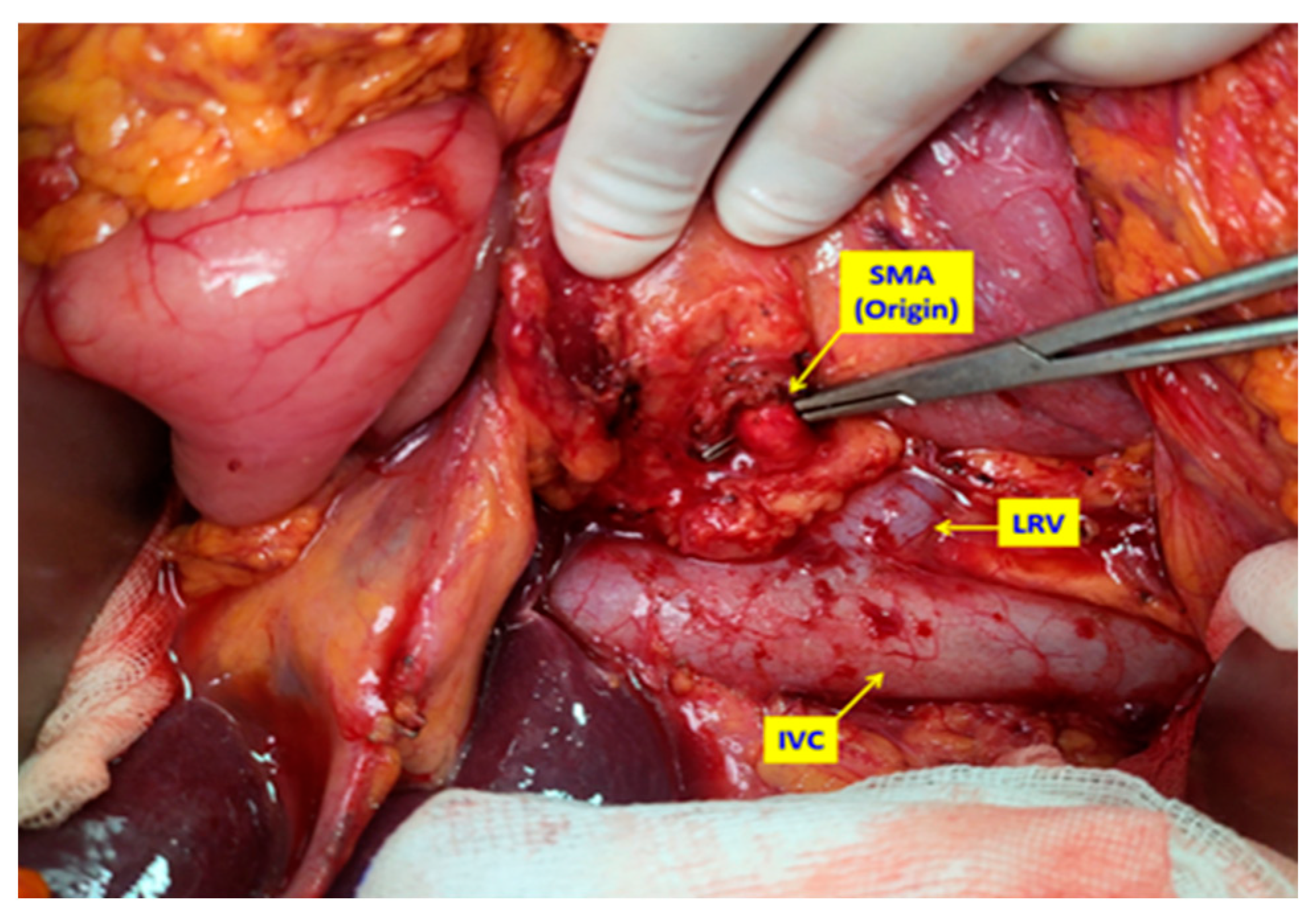
Figure 1. Intraoperative view of the first step: after the extended Kocher maneuver, the superior mesenteric artery (SMA) is dissected and encircled using the posterior artery-first approach, providing both proximal and distal vascular control and defining the dissection plane close to the arterial adventitia. LRV: Left renal vein IVC: Inferior vena cava.
Medicina 61 01725 g001
Figure 2. Intraoperative view of the second step: using the uncinate process approach, the distal portion of the superior mesenteric artery (SMA) is dissected and suspended. This maneuver ensures both proximal and distal vascular control of the SMA and facilitates safe en bloc mesopancreatic excision. FJV: First jejunal vein. SMV: Superior mesenteric vein.
Medicina 61 01725 g002
Figure 3. Intraoperative view of the dissection plane between the superior mesenteric artery (SMA) and the pancreas: the tissue is dissected as close as possible to the arterial wall, leaving the mesopancreatic tissue en bloc. The inferior pancreaticoduodenal artery (IPDA) is clearly visualized and prepared for ligation and division, highlighting the vascular control achieved during mesopancreatic excision.
Medicina 61 01725 g003
Figure 4. Intraoperative view of the surgical field after completion of the total mesopancreatic excision (TMpE), demonstrating complete clearance of the mesopancreatic tissue and the exposed adventitial surface of the superior mesenteric artery (SMA), confirming en bloc resection of the targeted anatomical unit. Intraoperative view of the surgical field after completion of the total mesopancreatic excision. SMV: Superior mesenteric vein; PV: Portal Vein; IVC: Inferior vena cava; FJV: First jejunal vein.
Medicina 61 01725 g004
Table 1. Patients’ demographic and clinicopathological characteristics.
All (n = 41)TMpE-PD
(n = 17)
Co-PD
(n = 24)
p Value
Age (years)64.6 ± 10.566.4 ± 10.863.4 ± 10.30.382
Sex
   Female16 (39.0)5 (29.4)11 (45.8)0.288
   Male25 (61.0)12 (70.6)13 (54.2)
Comorbidity31 (75.6)13 (76.5)18 (75.0)0.914
   Smoking20 (48.8)9 (52.9)11 (45.8)0.654
BMI (kg/m2)25.0 (23.0–27.0)25.4 (22.9–27.0)24.6 (23.0–27.8)0.952
Preoperative biliary drainage25 (61.0)11 (64.7)14 (58.3)0.680
Neoadjuvant chemotherapy1 (2.4)-1 (4.2)1.000
Vascular resection (Venous)5 (12.2)2 (11.8)3 (12.5)1.000
Operation duration (minutes)480 (400–495)485 (480–495)423 (398–485)0.067
R status
   R021 (51.2)10 (58.8)11 (45.8)0.412
   R120 (48.8)7 (41.2)13 (54.2)
Pathological stage
   Stage I3 (7.3)-3 (12.5)0.250
   Stage II18 (43.9)7 (41.2)11 (45.8)
   Stage III20 (48.8)10 (58.8)10 (41.7)
   Stage IV---
EBL (ml)250 (150–300)250 (150–350)250 (188–300)0.139
Number of removed lymph nodes29.0 (23.0–38.0)30.0 (25.0–39.0)26.0 (21.8–36.3)0.757
Pancreatic duct diameter (mm)4.0 (3.0–6.0)4.0 (3.0–5.0)3.5 (3.0–6.3)0.788
Pancreatic gland texture
   Firm5 (12.2)1 (5.9)4 (16.7)0.245
   Moderate32 (78.0)13 (76.5)19 (79.2)
   Soft4 (9.8)3 (17.6)1 (4.2)
Abbreviations: TMpE: Total mesopancreatic excision, PD: Pancreaticoduodenectomy, Co: Conventional, BMI: Body Mass Index, EBL: Estimated blood loss, BMI: Body mass index. Data presented as mean ± standard deviation, median (first-third quartiles) or n (%).
Table 2. Postoperative outcomes.
All (n = 41)TMpE-PD (n = 17)Co-PD (n = 24)p Value
All complications 26 (63.4)14 (82.4)12 (50.0)0.034
Surgical complications 24 (58.5)12 (70.6)12 (50.0)0.187
Surgical side infections 15 (36.6)8 (47.1)7 (29.2)0.241
Intra-abdominal abscess 3 (7.3)1 (5.9)2 (8.3)1.000
Biliary leakage x 1 (2.4)1 (5.9)-0.415
Chylous leakage 2 (4.9)2 (11.8)-0.166
POPF y 5 (12.2)2 (11.8)3 (12.5)1.000
Systemic complications 2 (4.9)2 (11.8)-0.166
Atelectasis 2 (4.9)2 (11.8)-0.166
Cardiac complications 1 (2.4)1 (2.4)-0.415
Complication grade z
I 12 (29.3)8 (47.1)4 (16.7)0.161
II 7 (17.1)4 (23.5)3 (12.5)
III-A 4 (9.8)1 (5.9)3 (12.5)
III-B 1 (2.4)-1 (4.2)
IV-A ---
IV-B ---
V 2 (4.9)1 (5.9)1 (4.2)
≥grade-III complications 7 (17.1)2 (11.8)5 (20.8)0.447
Reoperation 1 (2.4)1 (5.9)-0.415
Length of hospital stay (days) 18 (12–21)18 (12–21)19.5 (11.8–23.0)0.730
30-day readmissions 1 (2.4)-1 (4.2)1.000
90-day mortality 4 (9.8)1 (5.9)3 (12.5)0.629
Abbrevations: TMpE: Total mesopancreatic excision, PD: Pancreaticoduodenectomy, Co: Conventional, POPF: Postoperative pancreatic fistula. x Leakage of hepaticojejunal anastomosis y ISGPS (grade B/C) clinically relevant pancreatic fistulas z Patients who experienced more than one complication were classified as a higher-grade complication (Clavien–Dindo classification).
Table 3. Univariable analysis of factors associated with complications.
No Complications
(n = 15)
Complications
(n = 26)
OR (95% CI, p Value)
Age (years)64.7 ± 8.8764.6 ± 11.41.00 (0.94–1.06, p = 0.988)
Sex
   Female8 (53.3)8 (30.8)-
   Male7 (46.7)18 (69.2)2.57 (0.69–9.55, p = 0.158)
Comorbidity
   No2 (13.3)8 (30.8)-
   Yes13 (86.7)18 (69.2)0.35 (0.06–1.91, p = 0.223)
Smoking
   No9 (60.0)12 (46.2)-
   Yes6 (40.0)14 (53.8)1.75 (0.48–6.35, p = 0.395)
BMI (kg/m2)24.0 (22.9–26.6)25.2 (23.9–27.9)1.04 (0.88–1.22, p = 0.662)
Preoperative biliary drainage
   No5 (33.3)11 (42.3)-
   Yes10 (66.7)15 (57.7)0.68 (0.18–2.57, p = 0.571)
Vascular resection -
   No13 (86.7)23 (88.5)
   Yes2 (13.3)3 (11.5)0.85 (0.13–5.75, p = 0.866)
Operation duration (minutes)480 (423–488)468 (400–495)1.00 (0.99–1.01, p = 0.582)
EBL (ml)250 (175–300)250 (150–338)1.00 (0.99–1.01, p = 0.465)
Pathological stage
   Stage I–II10 (66.7)11 (42.3)-
   Stage III5 (33.3)15 (57.7)2.73 (0.72–10.27, p = 0.138)
Pancreatic duct diameter (mm)5.0 (3.5–7.0)3.5 (3.0–5.0)0.75 (0.56–1.00, p = 0.053)
Pancreatic gland texture
   Firm3 (20.0)2 (7.7)-
   Moderate/Soft12 (80.0)24 (92.3)3.00 (0.44–20.44, p = 0.262)
Number of removed lymph nodes26.0 (22.5–36.0)29.5 (23.0–37.8)1.00 (0.95–1.05, p = 0.959)
Table 4. Multivariable analysis of surgical technique and surgical border for complications.
No Complications (n = 15)Complications (n = 26)Adjusted OR * (95% CI, p Value)
Surgical technique
   Co-PD12 (80.0)12 (46.2)-
   TMpE-PD3 (20.0)14 (53.8)4.84 (0.90–25.95, p = 0.065)
R status
   R08 (53.3)13 (50.0)-
   R17 (46.7)13 (50.0)1.14 (0.27–4.86, p = 0.862)
Abbreviations: * Sex, pathological stage, pancreatic duct diameter and pancreatic gland texture were added to the model.
Disclaimer/Publisher’s Note: The statements, opinions and data contained in all publications are solely those of the individual author(s) and contributor(s) and not of MDPI and/or the editor(s). MDPI and/or the editor(s) disclaim responsibility for any injury to people or property resulting from any ideas, methods, instructions or products referred to in the content.