Case Report

Manual Therapy of Dysphagia in a Patient with Amyotrophic Lateral Sclerosis: A Case Report

1
Department of Neurology and ALS Centre, Traslational Medicine, University of Piemonte Orientale, Maggiore della Carità Hospital, 28100 Novara, Italy
2
Division of Paediatric, Manima Non-Profit Organization Social Assistance and Healthcare, 20125 Milan, Italy
3
Research Department, SOMA Istituto Osteopatia Milano, 20126 Milan, Italy
4
Principles and Practice of Clinical Research (PPCR), Harvard T.H. Chan School of Public Health–ECPE, Boston, MA 02115, USA
5
Division of Neurology and Neurorehabilitation, IRCCS Istituto Auxologico Italiano, Strada Luigi Cadorna 90, 28824 Piancavallo-Verbania, Italy
6
Department of Neurosciences “Rita Levi Montalcini”, University of Turin, 10126 Turin, Italy
*
Author to whom correspondence should be addressed.
Medicina 2024, 60(6), 845; https://doi.org/10.3390/medicina60060845
Submission received: 17 April 2024 / Revised: 12 May 2024 / Accepted: 20 May 2024 / Published: 22 May 2024
(This article belongs to the Special Issue Neuromuscular Disorders: Diagnostical Approaches and Treatments)

Abstract

:
Amyotrophic lateral sclerosis (ALS) is an incurable rare neurodegenerative condition, with 45% of cases showing the symptom of dysphagia; its clinical signs are atrophy, weakness, and fasciculations of the facial muscles, tongue, and pharynx. Furthermore, dysphagia is the main cause of aspiration pneumonia. The traditional treatment for dysphagia varies based on the patient’s difficulty of swallowing. The initial phase consists of dietary consistency adjustments, progressing to alternatives like nasogastric tubes or percutaneous endoscopic gastrostomy (PEG) in advanced stages. Osteopathic manipulative treatment (OMT) is a complementary ‘hands-on’ approach that has already shown positive results as an add-on therapy in various health conditions. This study is a case report of a man diagnosed with ALS with initial dysphagia, managed with a protocol that extraordinarily included OMT. The patient showed somatic dysfunctions in the mediastinal region, upper cervical region, and occipital area which are all anatomically related to the nervous system, especially the glossopharyngeal reflex. At the end of the rehabilitation protocol, there was a reduction in the swallowing problems measured with Strand Scale and swallowing tests, and the patient reported an improved psycho-physical well-being assessed with the Amyotrophic Lateral Sclerosis Assessment Questionnaire (ALSAQ-40). Instead, the neurological function measured with ALSFRS-S remained stable. Although the nature of this study design prevents any causal assumption, the positive results should lead to future randomized controlled trials to assess the effectiveness of OMT as an adjunctive therapeutic proposal to improve the health of ALS patients.

1. Introduction

Amyotrophic lateral sclerosis (ALS) is an incurable degenerative disease of the central nervous system, involving the upper and lower motor neurons in the cerebral cortex, brain stem, and anterior horns of the spinal cord. Over the years, a heterogeneous distribution has been identified among different areas of the world. The average incidence of ALS worldwide is approximately 1.68 cases/per year per 100,000 inhabitants. The prevalence is about 10–12 cases per 100,000 inhabitants, and individuals of all populations and ethnic groups are affected, even if a higher incidence is noted in the Caucasian ethnic group. The incidence and prevalence increase after the age of 40, with a peak between 58 and 63 years [1,2,3]. As this is an incurable disease, the approved drugs can only slow it down, and researchers continue to study other possible treatments for both the management and cure of ALS.
In addition to the already challenging condition of ALS, 45% of cases show dysphagia, particularly in the bulbar forms. It manifests as atrophy, weakness, and fasciculations of the facial muscles, tongue, and pharynx; it is the most important cause of protein–calorie malnutrition and is the leading cause of aspiration pneumonia [4,5]. The guidelines on the management of an adult dysphagic patient recommend some functional assessments of the patient: morphodynamic assessment of the structures involved in swallowing that can be explored directly (lips, tongue, hard palate, veil of the palate, mandible, larynx and muscle control of the head); evaluation of buccofacial practices; evaluation of the superficial and deep sensitivity of the peri-buccal skin, lips, tongue, and palate; evaluation of cough, vomiting, and swallowing reflexes; evaluation of the presence of pathological reflexes; and, finally, swallowing tests with substances and foods of different consistencies. The instrumental assessment is only conducted when there is a suspicion of inhalation during the functional assessment [6,7,8]. The gold standard for the treatment of late-stage dysphagia, where there is also a significant loss of body weight, is the choice of alternative feeding methods, including the use of a nasogastric tube or percutaneous endoscopic gastrostomy (PEG). Both of these methods can lead to various complications, including pressure ulcers, esophagitis, rhinitis, aspiration pneumonia, and frequent replacement by dislocation or occlusion [9].
Recent studies have started to assess the possible contribution and effect of osteopathic manipulative treatment (OMT) in various health conditions, as well as in the neurological field [10,11,12]. OMT is a ‘hands-on’ approach that treats various health conditions following the “structure–function” relationship, in accordance with the different described models: biomechanical, neurological, respiratory/circulatory, biopsychosocial, and metabolic/bioenergetic [13,14]. Its assessment and treatment are performed according to the so-called somatic dysfunction, an altered function of the body that can manifest in different areas [15,16].
In this case report, we describe the clinical case of a man diagnosed with ALS with initial dysphagia who underwent an 8-week OMT protocol in addition to the standard care, assessing neurological, logopedic, and psychological functions as well as any adverse events.

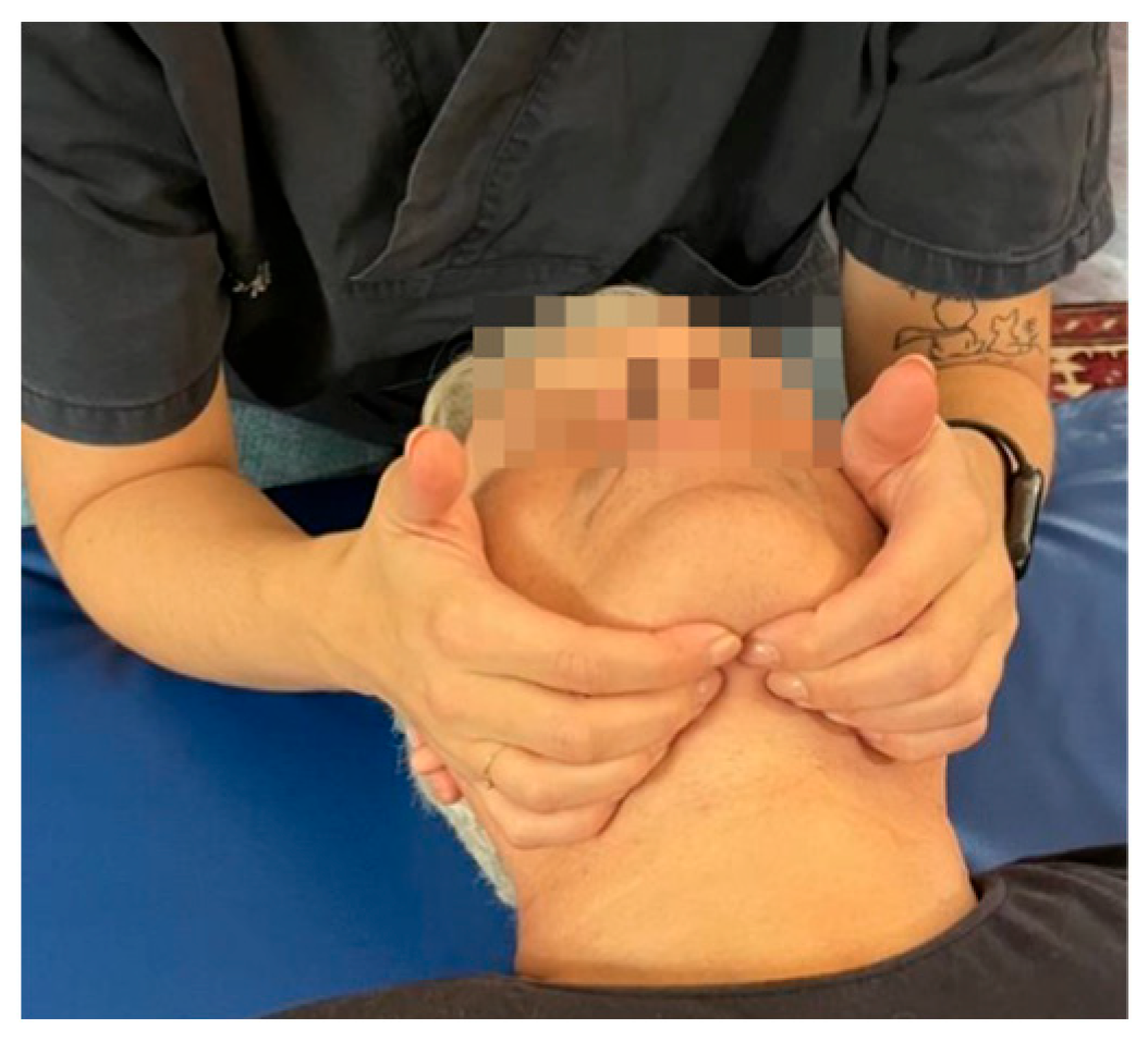
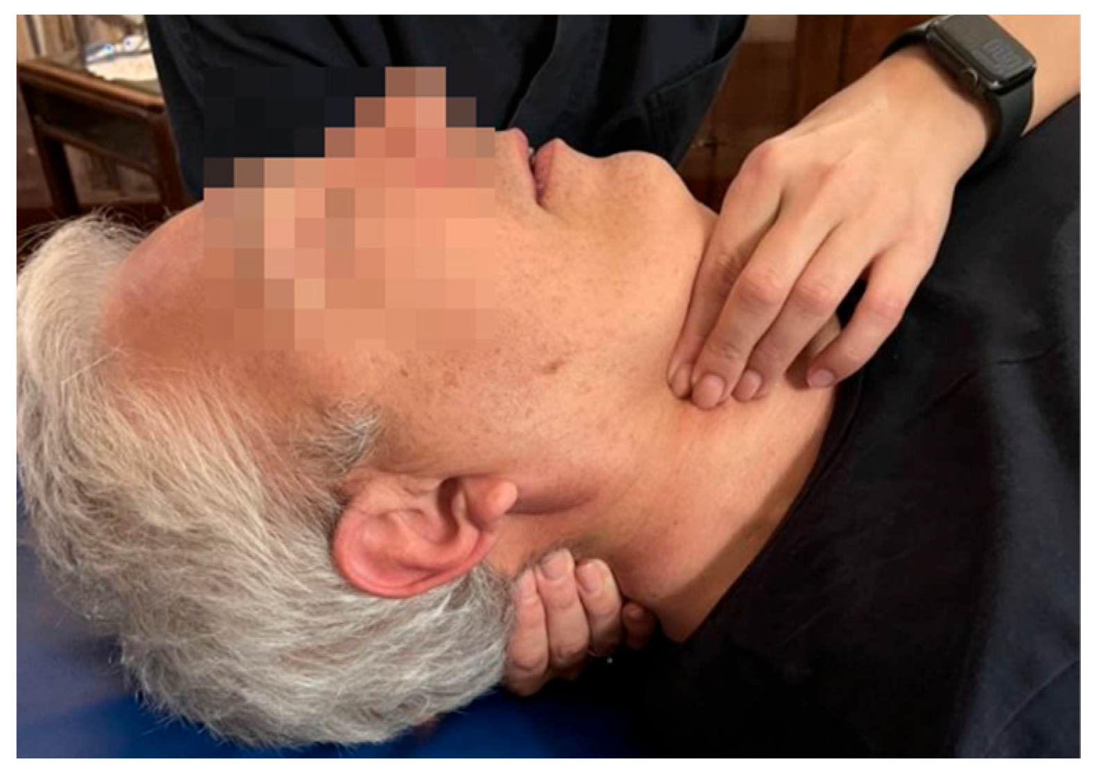
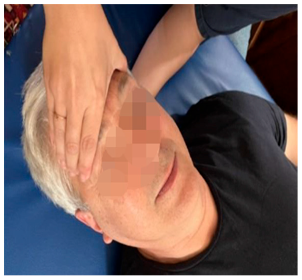
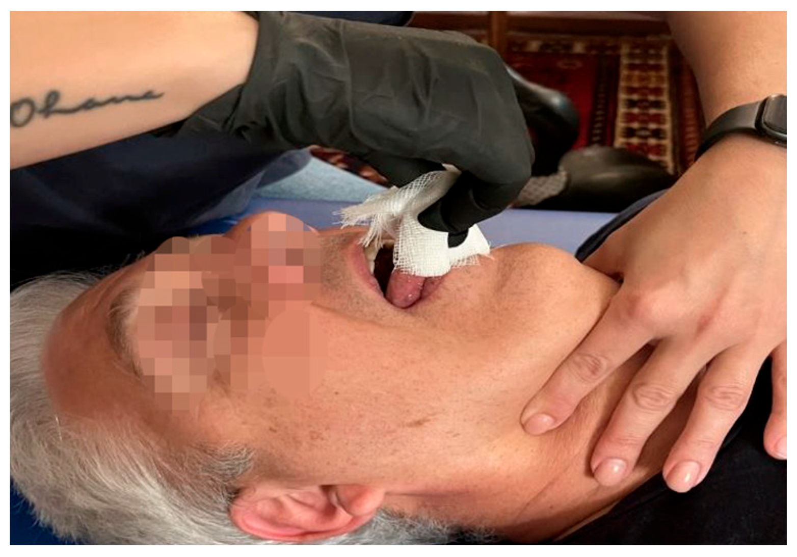
Detailed Case Presentation

The protocol for this study was approved by the local Ethical Committee (CE 179/2022) and was conducted in strict accordance with the Declaration of Helsinki. Before enrolling, the patient signed an informed consent form. This case report was reported according to the CARE Checklist.
The patient under examination was a 51-year-old Italian male, a commercial agent, not exposed to toxic agents. The first symptoms (progressive dysarthria and liquid dysphagia) began in June 2021. He came to the Amyotrophic Lateral Sclerosis Regional Expert Centre (CRESLA) of the “Maggiore della Carità” University Hospital (Novara, Italy) in February 2022. The first general neurological evaluation showed dysarthria, normal tongue with initial atrophy of the edges, and rare fasciculations. The masseter and peri-buccal reflexes were accentuated. At the spinal level, there was an initial hypotrophy with fasciculations at the right femoral quadriceps. The deep tendon reflexes were markedly accentuated in all locations. Nothing relevant was present in the previous anamnesis; the patient reported only a fall with a consequent slight head trauma during adolescence. He also denied a family history of neurodegenerative diseases.
For the diagnosis, various diagnostic tests were performed, including a brain MRI which documents bilateral frontotemporal atrophy and an electromyographic examination with diffuse active denervation in cranial, cervical, and lumbar districts. In March 2022, a genetic examination of the main involved genes in ALS (SOD1, FUS, TARDBP, C9orf72) was carried out, showing a negative result. At the first logopedic evaluation, the Strand Scale of signs and symptoms of dysphagia [17] was administered, showing slowed masticatory movements during the oral preparation and pharyngeal phases; in addition, the triggering of the pharyngeal reflex was delayed with a slight tendency to the pre-swallowing fall of the liquid bolus. According to the guidelines for dysphagia [6,7,8], for this patient, no instrumental tests were performed as they were not deemed essential at the time of enrollment. For the second neurological evaluation, the Revised Amyotrophic Lateral Sclerosis Functional Rating (ALSFRS-R) scale was used [18], showing a score of 45 out of 48. Because of the feeling of stiffness in the jaw reported by the patient and the mild sialorrhea, the neurologist prescribed amitriptyline. Finally, for the psychological evaluation, the Amyotrophic Lateral Sclerosis Assessment Questionnaire (ALSAQ-40) was administered.
In addition to the standard routine, the patient underwent an osteopathic evaluation and a weekly 45 min OMT session for a period of 8 weeks performed by a physiotherapist with 6 years of experience and attending the last year of osteopathy. The evaluation involved the functional status of the musculoskeletal, visceral, dural, and fascial systems; it was performed according to the Variability Model, assessing the somatic dysfunction. It analyzes the joint motion variability around the joint rest position, defined as the neutral zone (NZ). The different body areas were treated according to the severity of the somatic dysfunction:
  • Grade 0—No SD: symmetric motion;
  • Grade 1—Mild SD: asymmetric motion;
  • Grade 2—Moderate SD: asymmetric variability of motion in the NZ combined with altered tissue texture or tenderness;
  • Grade 3—Severe SD: asymmetric variability of motion in the NZ combined with altered tissue texture + tenderness. [15,16]
According to this evaluation, the main somatic dysfunctions were at the following levels: at the atlanto-occipital joint, hyoid bone, cricoid cartilage, occipitomastoid suture, sphenobasilar synchondrosis, resulting in compression, and at the middle and deep cervical fascia in relation to the internal tissues of the rib cage, endothoracic fascia, and pleura (see Table 1 for further details). Therefore, along with the drug regimen, the somatic dysfunctions were treated with indirect manual techniques, which are characterized by a passive movement of the dysfunctional region into a position of less tissue tension [16]. The patient mainly received balanced treatment of buccal floor ligament tension, muscle inhibition treatment with craniosacral condylar decompression, and functional visceral system release treatment (see Table 2 and Figure 1, Figure 2, Figure 3 and Figure 4).
No adverse events occurred due to OMT. Three days after the fourth OMT session, the patient reported increased dysarthria salivation in the morning; however, it was due to the missed intake of the amitriptyline for a few days, and it resolved spontaneously.
In week 8, during the standard multidisciplinary visit, the patient repeated the logopedic, psychological, and neurological evaluations: the swallowing test showed an improvement for the masticatory movements, which were now suitable for solid foods; there was only a slight tendency to fall pre-swallowing with a liquid bolus taken in the free oral cavity. Also, ALSAQ-40 showed an improvement both in the patient’s perception of fatigue during meals and in the emotional component. Instead, the ALSFRS-S remained stable (see Table 3 for further details).

2. Discussion

ALS is a rare, inevitably fatal neurodegenerative disease with no therapies to date that can modify its course. Dysphagia is one of the most critical problems in patients with neuromuscular diseases and is related to increased morbidity and mortality [18]. Among the various factors that influence this problem are the form of onset of the disease, possible genetic mutations, age of onset, and rate of progression. Typically, there are some early signs related to dysphagia, including low voice, increased salivation, and weight loss. These disorders, related to muscle weakness, lead to serious complications, such as malnutrition, dehydration, and aspiration pneumonia (one of the main causes of respiratory crises and patient hospitalization) [19].
Early detection of these symptoms should contribute to better patient management and possible prevention of comorbidities, including the impact on quality of life. Although guidelines and previous studies considered fiberoptic endoscopic evaluation and videofluoroscopic swallowing as the gold standards for dysphagia in patients with and without neurodegenerative diseases [18,20,21,22], they are diagnostic tests and not treatments.
The traditional treatment of dysphagia varies according to the patient’s difficulty in swallowing, and the different phases of swallowing are evaluated using the Scale Strand [23]; according to the obtained result, different strategies are indicated during meals.
During the initial phase, the consistency of the diet is modified from a free diet to a soft or semi-liquid diet. Then, in the advanced stage of dysphagia, there are two alternative strategies to oral feeding: the nasogastric tube or the PEG. These are two methods that exclude feeding orally, but these interventions can in turn cause patient discomfort and related recurring problems, such as rhinitis, esophagitis, dislocations, decubitus lesions, and tube occlusions [24].
To date, there is only a pilot study using OMT as an adjunctive therapy for ALS patients, even if it did not address dysphagia and did not provide positive results. However, it is interesting to note that patients showed somatic dysfunctions at the cervico-dorsal levels more frequently than the healthy matched controls [10]. Similarly, in this clinical case, the patient had altered tissue tension in the mediastinal region, which is strictly related to neck structures and the base of the skull via the fascial layers [25], suggesting an unbalanced distribution of the myofascial load. Therefore, we believe that through the application of techniques on the cervical fascias, we can influence the functioning of the peri-laryngeal muscles, normalizing the sliding of the fascial layers and then removing the fascial tensions that could hinder the laryngeal movements, the proprioceptive component, thus increasing the perception of that area and the neuromuscular coordination; consequently, with osteopathic manipulative treatment we would have obtained an effect not only on the laryngeal movement excursion, but also on the fine coordination of the intrinsic muscles [26,27]. Furthermore, the upper cervical region and the occipital area—anatomically related to the nervous system, especially to the glossopharyngeal nerve, responsible for swallowing [13]—were also dysfunctional. The basioccipital gives insertion to the superior constrictor muscle of the pharynx; hypertonia of this muscle can affect the mobility of the atlo-occipital joint, as well as the tension of the pharyngeal resonance cavity and the mandibular function. In order to normalize this muscle, it is necessary to treat its proximal and distal insertions, that is, not only the base of the skull, but also the mandible and the tongue [28]. As the swallowing difficulty is caused by the inefficiency of oral transit, reduction in the movement of the tongue base, laryngeal elevation, and slowed gag reflex during the pharyngeal phase [29], addressing the somatic dysfunctions at these levels with OMT may have contributed to improving dysphagia in the short term.
A cure for ALS has not been found; nonetheless, its variety of symptoms can be supported with different therapies [29]. OMT may join the adjunctive interventions and complement the traditional treatment of dysphagia to slow down the progression and lengthen as much as possible the use of alternative strategies to oral feeding.
The osteopathic evaluation, analyzing the functional alteration of the movement [15,16], identified the areas of greatest dysfunctionality, and OMT, affecting the fascial and neurological components, improved the functionality of the described areas, improving the patient’s condition.
We are aware that the results obtained have a limitation as this is a case report, but the introduction of OMT in therapeutic management improved the patient’s dysphagia in the short term. This case underlines the importance, at a clinical level, of considering OMT as an adjuvant therapy to the traditional treatment of dysphagia and, at a scientific research level, of being a starting point for a randomized controlled trial with a large number of patients.
Since ALS is a multisystem disease, we hypothesize that OMT could be part of the therapeutic proposals to improve the health of the patient suffering from ALS. However, to date, there are not enough studies to allow the introduction of OMT in daily practice for ALS patients. This case report outlined an improvement in the ALSAQ-40, Strand Scale, and DYALS, suggesting that OMT is a safe adjunctive therapy for ALS with dysphagia; however, the nature of the design prevents any kind of causal assumption. We suggest the development of future randomized controlled trials with OMT in ALS patients with dysphagia to evaluate its possible effects in the medium and long term.

Author Contributions

Conceptualization, I.D.M., F.B. and L.V.; methodology, F.B. and L.V.; investigation, I.D.M.; resources, I.D.M.; writing—original draft preparation, I.D.M. and F.B.; writing—review and editing, F.B., A.M., I.B. and L.V.; supervision, F.B., L.V. and A.M. All authors have read and agreed to the published version of the manuscript.

Funding

This work was supported by the Italian Ministry of Health, Ricerca Corrente.

Institutional Review Board Statement

The study was conducted in accordance with the Declaration of Helsinki, and approved by the Institutional Review Board (or Ethics Committee) of “Maggiore della Carità” University Hospital (CE 179/2022).

Informed Consent Statement

Informed consent was obtained from all subjects involved in the study.

Data Availability Statement

No new data were created or analyzed in this study.

Acknowledgments

The authors thank D.R. for his availability and active participation and thank the Regional Expert ALS Centre of the Ospedale “Maggiore della Carità” of Novara, especially L.M. for the supervision. L.V. would like to thank the PhD Programme in Experimental Medicine and Therapy of the University of Turin.

Conflicts of Interest

The authors declare no conflicts of interest. The funders had no role in the design of the study; in the collection, analyses, or interpretation of data; in the writing of the manuscript; or in the decision to publish the results.

References

  1. Boylan, K. Familial Amyotrophic Lateral Sclerosis. Neurol. Clin. 2015, 33, 807–830. [Google Scholar] [CrossRef]
  2. Oskarsson, B.; Horton, D.K.; Mitsumoto, H. Potential Environmental Factors in Amyotrophic Lateral Sclerosis. Neurol. Clin. 2015, 33, 877–888. [Google Scholar] [CrossRef]
  3. Yu, Y.; Su, F.C.; Callaghan, B.C.; Goutman, S.A.; Batterman, S.A.; Feldman, E.L. Environmental risk factors and amyotrophic lateral sclerosis (ALS): A case-control study of ALS in Michigan. PLoS ONE 2014, 9, e101186. [Google Scholar] [CrossRef]
  4. Ertekin, C.; Aydogdu, I.; Yüceyar, N.; Kiylioglu, N.; Tarlaci, S.; Uludag, B. Pathophysiological mechanisms of oropharyngeal dysphagia in amyotrophic lateral sclerosis. Brain 2000, 123 Pt 1, 125–140. [Google Scholar] [CrossRef] [PubMed]
  5. Perry, B.J.; Nelson, J.; Wong, J.B.; Kent, D.M. The cumulative incidence of dysphagia and dysphagia-free survival in persons diagnosed with amyotrophic lateral sclerosis. Muscle Nerve 2021, 64, 83–86. [Google Scholar] [CrossRef] [PubMed]
  6. EFNS Task Force on Diagnosis and Management of Amyotrophic Lateral Sclerosis; Andersen, P.M.; Abrahams, S.; Borasio, G.D.; de Carvalho, M.; Chio, A.; Van Damme, P.; Hardiman, O.; Kollewe, K.; Morrison, K.E.; et al. EFNS guidelines on the clinical management of amyotrophic lateral sclerosis (MALS)—Revised report of an EFNS task force. Eur. J. Neurol. 2012, 19, 360–375. [Google Scholar] [CrossRef] [PubMed]
  7. Stefani, L. Linee Guida sulla Disfagia (FLI e SIFEL). In Proceedings of the SIMFER, Turin, Italy, 17 May 2009; Available online: https://www.simfer.it/linee-guida-sulla-disfagia-fli-e-sifel/ (accessed on 12 May 2024).
  8. Higo, R.; Tayama, N.; Nito, T. Longitudinal analysis of progression of dysphagia in amyotrophic lateral sclerosis. Auris Nasus Larynx 2004, 31, 247–254. [Google Scholar] [CrossRef]
  9. Körner, S.; Hendricks, M.; Kollewe, K.; Zapf, A.; Dengler, R.; Silani, V.; Petri, S. Weight loss, dysphagia and supplement intake in patients with amyotrophic lateral sclerosis (ALS): Impact on quality of life and therapeutic options. BMC Neurol. 2013, 13, 84. [Google Scholar] [CrossRef]
  10. Maggiani, A.; Tremolizzo, L.; Della Valentina, A.; Mapelli, L.; Sosio, S.; Milano, V.; Bianchi, M.; Badi, F.; Lavazza, C.; Grandini, M.; et al. Osteopathic Manual Treatment for Amyotrophic Lateral Sclerosis: A Feasibility Pilot Study. Open Neurol. J. 2016, 10, 59–66. [Google Scholar] [CrossRef]
  11. Wells, M.R.; Giantinoto, S.; D’Agate, D.; Areman, R.D.; Fazzini, E.A.; Dowling, D.; Bosak, A. Standard osteopathic manipulative treatment acutely improves gait performance in patients with Parkinson’s disease. J. Am. Osteopath. Assoc. 1999, 99, 92–98. [Google Scholar] [CrossRef]
  12. Rivera-Martinez, S.; Wells, M.R.; Capobianco, J.D. A retrospective study of cranial strain patterns in patients with idiopathic Parkinson’s disease. J. Am. Osteopath. Assoc. 2002, 102, 417–422. [Google Scholar] [PubMed]
  13. Osteopathic International Alliance. Osteopathy and Osteopathic Medicine: A Global View of Practice, Patients, Education and the Contribution to Healthcare Delivery, 1st ed.; Osteopathic International Alliance (OIA): Chicago, ON, USA, 2013; Available online: https://oialliance.org/wp-content/uploads/2014/01/OIA-Stage-2-Report.pdf (accessed on 12 May 2024).
  14. World Health Organization. Benchmarks for Training in Traditional /Complementary and Alternative Medicine: Benchmarks for Training in Osteopathy, 1st ed.; World Health Organization (WHO): Geneva, Switzerland, 2010; Available online: https://www.who.int/publications-detail-redirect/9789241599665 (accessed on 12 May 2024).
  15. Bergna, A.; Vismara, L.; Parravicini, G.; Dal Farra, F. A new perspective for Somatic Dysfunction in Osteopathy: The Variability Model. J. Bodyw. Mov. Ther. 2020, 24, 181–189. [Google Scholar] [CrossRef]
  16. Vismara, L.; Bergna, A.; Tarantino, A.G.; Farra, F.D.; Buffone, F.; Vendramin, D.; Cimolin, V.; Cerfoglio, S.; Pradotto, L.G.; Mauro, A. Reliability and Validity of the Variability Model Testing Procedure for Somatic Dysfunction Assessment: A Comparison with Gait Analysis Parameters in Healthy Subjects. Healthcare 2024, 12, 175. [Google Scholar] [CrossRef] [PubMed]
  17. Ertekin, C.; Aydogdu, I. Neurophysiology of swallowing. Clin. Neurophysiol. 2003, 114, 2226–2244. [Google Scholar] [CrossRef] [PubMed]
  18. Espitalier, F.; Fanous, A.; Aviv, J.; Bassiouny, S.; Desuter, G.; Nerurkar, N.; Postma, G.; Crevier-Buchman, L. International consensus (ICON) on assessment of oropharyngeal dysphagia. Eur. Ann. Otorhinolaryngol. Head Neck Dis. 2018, 135, S17–S21. [Google Scholar] [CrossRef]
  19. Audag, N.; Goubau, C.; Toussaint, M.; Reychler, G. Screening and evaluation tools of dysphagia in adults with neuromuscular diseases: A systematic review. Ther. Adv. Chronic Dis. 2019, 10, 2040622318821622. [Google Scholar] [CrossRef]
  20. Etges, C.L.; Scheeren, B.; Gomes, E.; Barbosa Lde, R. Screening tools for dysphagia: A systematic review. Codas 2014, 26, 343–349. [Google Scholar] [CrossRef]
  21. Kertscher, B.; Speyer, R.; Palmieri, M.; Plant, C. Bedside screening to detect oropharyngeal dysphagia in patients with neurological disorders: An updated systematic review. Dysphagia 2014, 29, 204–212. [Google Scholar] [CrossRef]
  22. Bours, G.J.; Speyer, R.; Lemmens, J.; Limburg, M.; de Wit, R. Bedside screening tests vs. videofluoroscopy or fibreoptic endoscopic evaluation of swallowing to detect dysphagia in patients with neurological disorders: Systematic review. J. Adv. Nurs. 2009, 65, 477–493. [Google Scholar] [CrossRef]
  23. Burgos, R.; Bretón, I.; Cereda, E.; Desport, J.C.; Dziewas, R.; Genton, L.; Gomes, F.; Jesus, P.; Leischker, A.; Muscaritoli, M.; et al. ESPEN guideline clinical nutrition in neurology. Clin. Nutr. 2018, 37, 354–396. [Google Scholar] [CrossRef]
  24. Vilar, M.D.C.; Coutinho, K.M.D.; Vale, S.H.L.; Medeiros, G.C.B.S.; Piuvezam, G.; Leite-Lais, L.; Brandao-Neto, J. Nutritional therapy in amyotrophic lateral sclerosis: Protocol for a systematic review and meta-analysis. BMJ Open 2022, 12, e064086. [Google Scholar] [CrossRef] [PubMed]
  25. Frias Vilaça, A.; Reis, A.M.; Vidal, I.M. The anatomical compartments and their connections as demonstrated by ectopic air. Insights Imaging 2013, 4, 759–772. [Google Scholar] [CrossRef] [PubMed]
  26. Benjamin, M. The fascia of the limbs and back—A review. J. Anat. 2009, 214, 1–18. [Google Scholar] [CrossRef] [PubMed]
  27. Bordoni, B.; Zanier, E. Understanding Fibroblasts in Order to Comprehend the Osteopathic Treatment of the Fascia. Evid. -Based Complement. Altern. Med. 2015, 2015, 860934. [Google Scholar] [CrossRef]
  28. Piron, A. Techniques Ostéopathiques Appliquées à la Phoniatrie: Biomécanique Fonctionnelle et Normalisation du Larynx, 2nd ed.; Symétrie: Lyon, France, 2011. [Google Scholar]
  29. Orsini, M.; Oliveira, A.B.; Nascimento, O.J.; Reis, C.H.M.; Leite, M.A.A.; De Souza, J.A.; Pupe, C.; De Souza, O.G.; Bastos, V.H.; De Freitas, M.R.; et al. Amyotrophic Lateral Sclerosis: New Perpectives and Update. Neurol. Int. 2015, 7, 5885. [Google Scholar] [CrossRef]
Figure 1. Buccal floor inhibition technique.
Medicina 60 00845 g001
Figure 2. Anterior cervical arch technique.
Medicina 60 00845 g002
Figure 3. Decompression of the sphenobasilar synchondrosis technique.
Medicina 60 00845 g003
Figure 4. Tongue-structure fascial agreement technique.
Medicina 60 00845 g004
Table 1. Summary of the osteopathic physical examination at the initial consultation.
Fascial systemDysfunctions affecting the middle and deep cervical fascia
Visceral systemEsophageal and pharyngeal dysfunctions
Craniosacral systemCompressed sphenobasilar synchondrosis
Occiput-mastoid suture dysfunction
Musculoskeletal systemAtlo-occipital joint dysfunction
Posterior hyoid bone dysfunction
Table 2. Patient osteopathic manipulation treatment program over 8 weeks.
STAGEGOALSTECHNIQUESFREQUENCY
Weeks 1–4
  • Decompression sphenobasilar synchondrosis
  • Release of mid-cervical fascia and visceral component
  • Buccal floor release
  • Craniosacral condylar decompression
  • Release visceral component of the stomach
  • Release of the diaphragm
  • Fronto-occipital approach in decompression
  • Occiput-sternum fascial agreement technique
  • Technique of inhibition of the buccal floor and direct inhibition of the mylohyoid mm
  • Muscle inhibition technique combined with craniosacral condylar decompression
  • Seated cardiac relaxation technique
  • Functional technique of the diaphragm
Once per week for 45 min
Weeks 5–8
  • Occiput-mastoid suture release
  • Tongue fascial release in relation to the hyoid bone
  • Anterior cervical arch release
  • Visceral component release in relation to the stomach
  • Fascial technique in relation to the temporal bone and occiput
  • Tongue-structure fascial agreement technique
  • Anterior cervical arch technique
  • Relaxation of the cardia with a long lever
Once per week for 45 min
Table 3. Patient-reported outcome measure scores at the beginning and at the end of the study.
OUTCOMEPRE-TREATMENT
(Before the 1st OMT Session)
POST-TREATMENT
(After the 8th OMT Session)
Strand Scale and
Swallowing tests
Slowed chewing movements and delayed gag reflexChewing movements suitable for chewing solid foods
Slight tendency to fall pre-swallowing with liquid bolus taken with free oral cavity
DYALSDifficulty swallowing liquid foods
Cough when swallowing
solid foods and liquids
Need to drink in several sips
Cough present occasionally with fluid
intake
There is no need to drink in small sips
ALSFRS-R45/4845/48
ALSAQ-40Swallowing: 41.6, sometimes problematic
Communication: 53.5, sometimes problematic.
Emotional aspect: 24, rarely problematic
Swallowing: 25, rarely problematic
Communication: 59, problems sometimes
Emotional aspect: 15, no problem
Disclaimer/Publisher’s Note: The statements, opinions and data contained in all publications are solely those of the individual author(s) and contributor(s) and not of MDPI and/or the editor(s). MDPI and/or the editor(s) disclaim responsibility for any injury to people or property resulting from any ideas, methods, instructions or products referred to in the content.

