

Elevated Cardiac Troponin I as a Mortality Predictor in Hospitalised COVID-19 Patients

 ,
,
Abstract
1. Introduction
2. Materials and Methods
3. Results
4. Discussion
5. Conclusions
Author Contributions
Funding
Institutional Review Board Statement
Informed Consent Statement
Data Availability Statement
Conflicts of Interest
References
- Fairweather, D.; Beetler, D.J.; Di Florio, D.N.; Musigk, N.; Heidecker, B.; Cooper, L.T. COVID-19, Myocarditis and Pericarditis. Circ. Res. 2023, 132, 1302–1319. [Google Scholar] [CrossRef] [PubMed]
- Devaux, C.A.; Camoin-Jau, L. An update on angiotensin-converting enzyme 2 structure/functions, polymorphism, and duplicitous nature in the pathophysiology of coronavirus disease 2019: Implications for vascular and coagulation disease associated with severe acute respiratory syndrome coronavirus infection. Front. Microbiol. 2022, 13, 1042200. [Google Scholar] [CrossRef] [PubMed]
- Lindner, D.; Fitzek, A.; Bräuninger, H.; Aleshcheva, G.; Edler, C.; Meissner, K.; Scherschel, K.; Kirchhof, P.; Escher, F.; Schultheiss, H.-P.; et al. Association of Cardiac Infection with SARS-CoV-2 in Confirmed COVID-19 Autopsy Cases. JAMA Cardiol. 2020, 5, 1281. [Google Scholar] [CrossRef] [PubMed]
- Bearse, M.; Hung, Y.P.; Krauson, A.J.; Bonanno, L.; Boyraz, B.; Harris, C.K.; Helland, T.L.; Hilburn, C.F.; Hutchison, B.; Jobbagy, S.; et al. Factors associated with myocardial SARS-CoV-2 infection, myocarditis, and cardiac inflammation in patients with COVID-19. Mod. Pathol. 2021, 34, 1345–1357. [Google Scholar] [CrossRef] [PubMed]
- Terzic, C.M.; Medina-Inojosa, B.J. Cardiovascular Complications of Coronavirus Disease-2019. Phys. Med. Rehabil. Clin. North Am. 2023, 34, 551–561. [Google Scholar] [CrossRef] [PubMed]
- Liu, F.; Liu, F.; Wang, L. COVID-19 and cardiovascular diseases. J. Mol. Cell Biol. 2021, 13, 161–167. [Google Scholar] [CrossRef] [PubMed]
- Vosko, I.; Zirlik, A.; Bugger, H. Impact of COVID-19 on Cardiovascular Disease. Viruses 2023, 15, 508. [Google Scholar] [CrossRef] [PubMed]
- Nandy, S.; Wan, S.-H.; Brenes-Salazar, J. Cardiovascular Manifestations of COVID-19. CCR 2021, 17, e230421187503. [Google Scholar] [CrossRef] [PubMed]
- Kubiliute, I.; Vitkauskaite, M.; Urboniene, J.; Svetikas, L.; Zablockiene, B.; Jancoriene, L. Clinical characteristics and predictors for in-hospital mortality in adult COVID-19 patients: A retrospective single center cohort study in Vilnius, Lithuania. PLoS ONE 2023, 18, e0290656. [Google Scholar] [CrossRef]
- Thygesen, K.; Alpert, J.S.; Jaffe, A.S.; Chaitman, B.R.; Bax, J.J.; Morrow, D.A.; White, H.D.; Executive Group on behalf of the Joint European Society of Cardiology (ESC)/American College of Cardiology (ACC)/American Heart Association (AHA)/World Heart Federation (WHF) Task Force for the Universal Definition of Myocardial Infarction. Fourth Universal Definition of Myocardial Infarction (2018). J. Am. Coll. Cardiol. 2018, 72, 2231–2264. [Google Scholar] [CrossRef]
- Martens, C.R.; Accornero, F. Viruses in the Heart: Direct and Indirect Routes to Myocarditis and Heart Failure. Viruses 2021, 13, 1924. [Google Scholar] [CrossRef] [PubMed]
- Schultheiss, H.P.; Baumeier, C.; Pietsch, H.; Bock, C.T.; Poller, W.; Escher, F. Cardiovascular consequences of viral infections: From COVID to other viral diseases. Cardiovasc. Res. 2021, 117, 2610–2623. [Google Scholar] [CrossRef] [PubMed]
- Ilyas, S.Z.; Tabassum, R.; Hamed, H.; Rehman, S.U.; Qadri, I. Hepatitis C Virus-Associated Extrahepatic Manifestations in Lung and Heart and Antiviral Therapy-Related Cardiopulmonary Toxicity. Viral Immunol. 2017, 30, 633–641. [Google Scholar] [CrossRef] [PubMed]
- Zemaitis, L.; Alzbutas, G.; Gecyte, E.; Gecys, D.; Lesauskaite, V. SARS-CoV-2: Two Years in the Pandemic: What Have We Observed from Genome Sequencing Results in Lithuania? Microorganisms 2022, 10, 1229. [Google Scholar] [CrossRef]
- Battaglini, D.; Lopes-Pacheco, M.; Castro-Faria-Neto, H.C.; Pelosi, P.; Rocco, P.R.M. Laboratory Biomarkers for Diagnosis and Prognosis in COVID-19. Front. Immunol. 2022, 13, 857573. [Google Scholar] [CrossRef]
- Lala, A.; Johnson, K.W.; Januzzi, J.L.; Russak, A.J.; Paranjpe, I.; Richter, F.; Zhao, S.; Somani, S.; Van Vleck, T.; Vaid, A.; et al. Prevalence and Impact of Myocardial Injury in Patients Hospitalized with COVID-19 Infection. J. Am. Coll. Cardiol. 2020, 76, 533–546. [Google Scholar] [CrossRef] [PubMed]
- De Marzo, V.; Di Biagio, A.; Della Bona, R.; Vena, A.; Arboscello, E.; Emirjona, H.; Mora, S.; Giacomini, M.; Da Rin, G.; Pelosi, P.; et al. Prevalence and prognostic value of cardiac troponin in elderly patients hospitalized for COVID-19. J. Geriatr. Cardiol. 2021, 18, 338–345. [Google Scholar] [CrossRef] [PubMed]
- Shi, S.; Qin, M.; Shen, B.; Cai, Y.; Liu, T.; Yang, F.; Gong, W.; Liu, X.; Liang, J.; Zhao, Q.; et al. Association of Cardiac Injury with Mortality in Hospitalized Patients with COVID-19 in Wuhan, China. JAMA Cardiol. 2020, 5, 802. [Google Scholar] [CrossRef] [PubMed]
- Sandoval, Y.; Januzzi, J.L.; Jaffe, A.S. Cardiac Troponin for Assessment of Myocardial Injury in COVID-19. J. Am. Coll. Cardiol. 2020, 76, 1244–1258. [Google Scholar] [CrossRef]
- Li, C.; Jiang, J.; Wang, F.; Zhou, N.; Veronese, G.; Moslehi, J.J.; Ammirati, E.; Wang, D.W. Longitudinal correlation of biomarkers of cardiac injury, inflammation, and coagulation to outcome in hospitalized COVID-19 patients. J. Mol. Cell. Cardiol. 2020, 147, 74–87. [Google Scholar] [CrossRef]
- Metkus, T.S.; Sokoll, L.J.; Barth, A.S.; Czarny, M.J.; Hays, A.G.; Lowenstein, C.J.; Michos, E.D.; Nolley, E.P.; Post, W.S.; Resar, J.R.; et al. Myocardial Injury in Severe COVID-19 Compared with Non–COVID-19 Acute Respiratory Distress Syndrome. Circulation 2021, 143, 553–565. [Google Scholar] [CrossRef]
- Lombardi, C.M.; Carubelli, V.; Iorio, A.; Inciardi, R.M.; Bellasi, A.; Canale, C.; Camporotondo, R.; Catagnano, F.; Dalla Vecchia, L.A.; Giovinazzo, S.; et al. Association of Troponin Levels with Mortality in Italian Patients Hospitalized with Coronavirus Disease 2019: Results of a Multicenter Study. JAMA Cardiol. 2020, 5, 1274. [Google Scholar] [CrossRef] [PubMed]
- Papageorgiou, N.; Sohrabi, C.; Merino, D.P.; Tyrlis, A.; Atieh, A.E.; Saberwal, B.; Lim, W.-Y.; Creta, A.; Khanji, M.; Rusinova, R.; et al. High sensitivity troponin and COVID-19 outcomes. Acta Cardiol. 2022, 77, 81–88. [Google Scholar] [CrossRef] [PubMed]
- Guo, T.; Fan, Y.; Chen, M.; Wu, X.; Zhang, L.; He, T.; Wang, H.; Wan, J.; Wang, X.; Lu, Z. Cardiovascular Implications of Fatal Outcomes of Patients with Coronavirus Disease 2019 (COVID-19). JAMA Cardiol. 2020, 5, 811. [Google Scholar] [CrossRef] [PubMed]
- Davies, M.; Osborne, V.; Lane, S.; Roy, D.; Dhanda, S.; Evans, A.; Shakir, S. Remdesivir in Treatment of COVID-19: A Systematic Benefit-Risk Assessment. Drug Saf. 2020, 43, 645–656. [Google Scholar] [CrossRef] [PubMed]
- Nabati, M.; Parsaee, H. Potential Cardiotoxic Effects of Remdesivir on Cardiovascular System: A Literature Review. Cardiovasc. Toxicol. 2022, 22, 268–272. [Google Scholar] [CrossRef] [PubMed]
- Rafaniello, C.; Ferrajolo, C.; Sullo, M.G.; Gaio, M.; Zinzi, A.; Scavone, C.; Gargano, F.; Coscioni, E.; Rossi, F.; Capuano, A. Cardiac Events Potentially Associated to Remdesivir: An Analysis from the European Spontaneous Adverse Event Reporting System. Pharmaceuticals 2021, 14, 611. [Google Scholar] [CrossRef] [PubMed]
- RECOVERY Collaborative Group; Horby, P.; Lim, W.S.; Emberson, J.R.; Mafham, M.; Bell, J.L.; Linsell, L.; Staplin, N.; Brightling, C.; Ustianowski, A.; et al. Dexamethasone in Hospitalized Patients with COVID-19. N. Engl. J. Med. 2021, 384, 693–704. [Google Scholar] [CrossRef] [PubMed]
- Caiazzo, E.; Rezig, A.O.M.; Bruzzese, D.; Ialenti, A.; Cicala, C.; Cleland, J.G.F.; Guzik, T.J.; Maffia, P.; Pellicori, P. Systemic administration of glucocorticoids, cardiovascular complications and mortality in patients hospitalised with COVID-19, SARS, MERS or influenza: A systematic review and meta-analysis of randomised trials. Pharmacol. Res. 2022, 176, 106053. [Google Scholar] [CrossRef]
- Chapman, A.R.; Bularga, A.; Mills, N.L. High-Sensitivity Cardiac Troponin Can Be an Ally in the Fight against COVID-19. Circulation 2020, 141, 1733–1735. [Google Scholar] [CrossRef]
- De Michieli, L.; Jaffe, A.S.; Sandoval, Y. Use and Prognostic Implications of Cardiac Troponin in COVID-19. Cardiol. Clin. 2022, 40, 287–300. [Google Scholar] [CrossRef] [PubMed]


| Demographic and Clinical Characteristic (N = 2019) | N (%) | Laboratory Characteristics | N | Median (IQR) |
|---|---|---|---|---|
| Age, years, median (IQR) | 60 (50–69) | Haemoglobin, g/L | 2019 | 140.0 (127.0–150.0) |
| Male | 1130 (55.97) | WBC, ×109/L | 2019 | 6.34 (4.76–8.61) |
| Female | 889 (44.03) | Neutrophils, ×109/L | 2019 | 4.66 (3.28–6.69) |
| Any comorbid condition | 1000 (49.53) | Lymphocytes, ×109/L | 2019 | 1.0 (0.71–1.40) |
| Arterial hypertension | 802 (39.72) | Platelets, ×109/L | 2019 | 195.0 (153.0–253.0) |
| Coronary artery disease | 79 (3.91) | cTnI | 2019 | 9.70 (5.0–23.0) |
| Congestive heart failure | 160 (7.92) | Glucose, mmol/L | 1984 | 6.22 (5.54–7.30) |
| Atrial fibrillation | 203 (10.05) | Creatinine, µmol/L | 2019 | 81.0 (67.0–100.0) |
| Diabetes mellitus | 277 (13.72) | eGFR, mL/min/1.73 m2 | 2019 | 84.0 (64.0–96.0) |
| Obesity | 106 (5.25) | Urea, mmol/L | 1883 | 5.50 (4.07–7.91) |
| COPD | 34 (1.68) | Sodium, mmol/L | 2019 | 140.0 (137.0–143.0) |
| Chronic kidney disease | 109 (5.40) | Potassium, mmol/L | 2019 | 4.20 (3.87–4.50) |
| Previous stroke | 23 (1.14) | ALT, U/L | 1993 | 33.0 (21.0–53.0) |
| Invasive mechanical ventilation | 170 (8.42) | AST, U/L | 1986 | 37.0 (26.98–57.0) |
| Treatment with antibiotics | 1514 (74.99) | LDH, U/L | 1910 | 308.0 (243.0–410.0) |
| Treatment with antivirals (Remdesivir) | 711 (35.22) | CRP, mg/L | 2018 | 63.25 (25.45–125.53) |
| Treatment with systemic steroids | 1371 (67.90) | Ferritin, µg/L | 1939 | 471.0 (235.00–1 002.0) |
| In-hospital mortality | 247 (12.23) | IL-6, ng/L | 1892 | 30.40 (15.01–57.30) |
| Length of hospital stay, days, median (IQR) | 11 (8–16) | D-dimer, µg/L | 1972 | 490.0 (295.0–910.0) |
| Characteristic | cTnI < 19 ng/L (N = 1416), n (%) | 19 ≤ cTnI ≤ 100 ng/L (N = 431), n (%) | cTnI > 100 ng/L (N = 172), n (%) | p-Value 1 | p-Value 2 | p-Value 3 |
|---|---|---|---|---|---|---|
| Age in years, median (IQR) | 57 (47–65) | 67 (58–77) | 74 (62–81) | <0.001 | <0.001 | <0.001 |
| Male | 771 (54.45) | 253 (58.70) | 106 (61.63) | 0.120 | 0.074 | 0.508 |
| Any underlying condition | 584 (41.24) | 287 (66.59) | 129 (75.00) | <0.001 | <0.001 | 0.044 |
| Hypertension | 481 (33.97) | 220 (51.04) | 101 (58.72) | <0.001 | <0.001 | 0.088 |
| Coronary artery disease | 33 (2.33) | 27 (6.26) | 19 (11.05) | <0.001 | <0.001 | 0.046 |
| Congestive heart failure | 50 (3.53) | 72 (16.71) | 38 (22.09) | <0.001 | <0.001 | 0.122 |
| Atrial fibrillation | 74 (5.23) | 81 (18.79) | 48 (27.91) | <0.001 | <0.001 | 0.014 |
| Diabetes | 160 (11.30) | 82 (19.03) | 35 (20.35) | <0.001 | 0.001 | 0.711 |
| Obesity | 63 (4.45) | 29 (6.73) | 14 (8.14) | 0.057 | 0.033 | 0.543 |
| COPD | 17 (1.20) | 11 (2.55) | 6 (3.49) | 0.044 | 0.031 | 0.587 |
| Chronic kidney disease | 37 (2.61) | 46 (10.67) | 26 (15.12) | <0.001 | <0.001 | 0.129 |
| Previous stroke | 7 (0.49) | 10 (2.32) | 6 (3.49) | 0.002 | 0.001 | 0.410 |
| Complications | ||||||
| Pulmonary embolism | 20 (1.41) | 17 (3.94) | 16 (9.30) | 0.001 | <0.001 | 0.009 |
| Stroke | 12 (0.85) | 15 (3.48) | 3 (1.74) | <0.001 | 0.216 | 0.258 |
| Remdesivir | 513 (36.23) | 162 (37.59) | 36 (20.93) | 0.608 | <0.001 | <0.001 |
| Systemic steroids | 961 (67.87) | 315 (73.09) | 95 (55.23) | 0.040 | 0.001 | <0.001 |
| Antibiotics use | 1066 (75.28) | 336 (77.96) | 112 (65.12) | 0.255 | 0.004 | 0.001 |
| Invasive ventilation | 61 (4.31) | 66 (15.31) | 43 (25.00) | <0.001 | <0.001 | 0.005 |
| Length of hospital stay, days | 11 (7–15) | 12 (8–19) | 13 (8–21) | <0.001 | 0.001 | 0.579 |
| In-hospital mortality | 76 (5.37) | 106 (24.59) | 65 (37.79) | <0.001 | <0.001 | 0.001 |
| Variable | cTnI < 19 ng/L (N = 1416), Median (IQR) | 19 ≤ cTnI ≤ 100 ng/L (N = 431), Median (IQR) | cTnI > 100 ng/L (N = 172), Median (IQR) | p-Value 1 | p-Value 2 | p-Value 3 |
|---|---|---|---|---|---|---|
| Haemoglobin, g/L | 142.00 (130.00–151.00) | 135.00 (121.00–147.00) | 133.00 (116.00–145.75) | <0.001 | <0.001 | 0.164 |
| WBC, ×109/L | 6.15 (4.65–8.06) | 6.72 (5.02–9.41) | 8.83 (6.08–12.38) | <0.001 | <0.001 | <0.001 |
| Neutrophils, ×109/L | 4.42 (3.14–6.15) | 4.95 (3.50–7.53) | 7.10 (4.38–10.23) | <0.001 | <0.001 | <0.001 |
| Lymphocytes, ×109/L | 1.04 (0.77–1.40) | 0.90 (0.60–1.39) | 0.80 (0.53–1.30) | <0.001 | <0.001 | 0.071 |
| Platelets, ×109/L | 195.00 (154.00–252.00) | 192.00 (151.00–245.00) | 207.00 (150.25–261.25) | 0.202 | 0.495 | 0.195 |
| Glucose, mmol/L | 6.07 (5.46–6.92) | 6.67 (5.82–8.23) | 7.64 (6.02–9.76) | <0.001 | <0.001 | 0.003 |
| Creatinine, µmol/L | 78.00 (65.00–91.00) | 92.00 (72.00–125.00) | 103.50 (77.00–148.75) | <0.001 | <0.001 | 0.018 |
| eGFR, mL/min/1.73 m2 | 88.00 (73.08–98.30) | 68.40 (43.60–89.00) | 57.20 (34.00–82.00) | <0.001 | <0.001 | 0.002 |
| Urea, mmol/L | 4.91 (3.74–6.48) | 7.37 (5.24–11.14) | 10.17 (6.68–16.25) | <0.001 | <0.001 | <0.001 |
| Sodium, mmol/L | 140.75 (137.88–143.00) | 140.00 (136.10–143.00) | 139.00 (135.00–142.00) | 0.001 | 0.002 | 0.386 |
| Potassium, mmol/: | 4.15 (3.90–4.50) | 4.20 (3.80–4.60) | 4.20 (3.85–4.60) | 0.468 | 0.075 | 0.314 |
| ALT, U/L | 33.00 (21.00–53.00) | 31.00 (19.35–50.00) | 32.50 (19.00–59.75) | 0.066 | 0.676 | 0.552 |
| AST, U/L | 35.00 (25.72–53.00) | 40.00 (29.08–61.07) | 50.00 (31.00–78.75) | <0.001 | <0.001 | 0.004 |
| LDH, U/L | 295.00 (236.00–388.00) | 335.00 (264.00–487.00) | 385.50 (280.75–564.75) | <0.001 | <0.001 | 0.031 |
| CRP, mg/L | 59.00 (24.43–108.90) | 85.60 (32.60–157.60) | 92.08 (19.88–146.30) | <0.001 | 0.003 | 0.486 |
| Ferritin, µg/L | 441.00 (225.50–952.55) | 543.00 (252.50–1115.50) | 543.29 (239.70–1286.73) | 0.011 | 0.046 | 0.739 |
| IL-6, ng/L | 27.70 (13.90–51.05) | 42.50 (19.85–71.05) | 34.15 (16.85–87.73) | <0.001 | 0.001 | 0.739 |
| D-dimer, µg/L | 430.00 (270.00–720.00) | 677.50 (410.00–1330.00) | 1 255.00 (587.50–2360.00) | <0.001 | <0.001 | <0.001 |
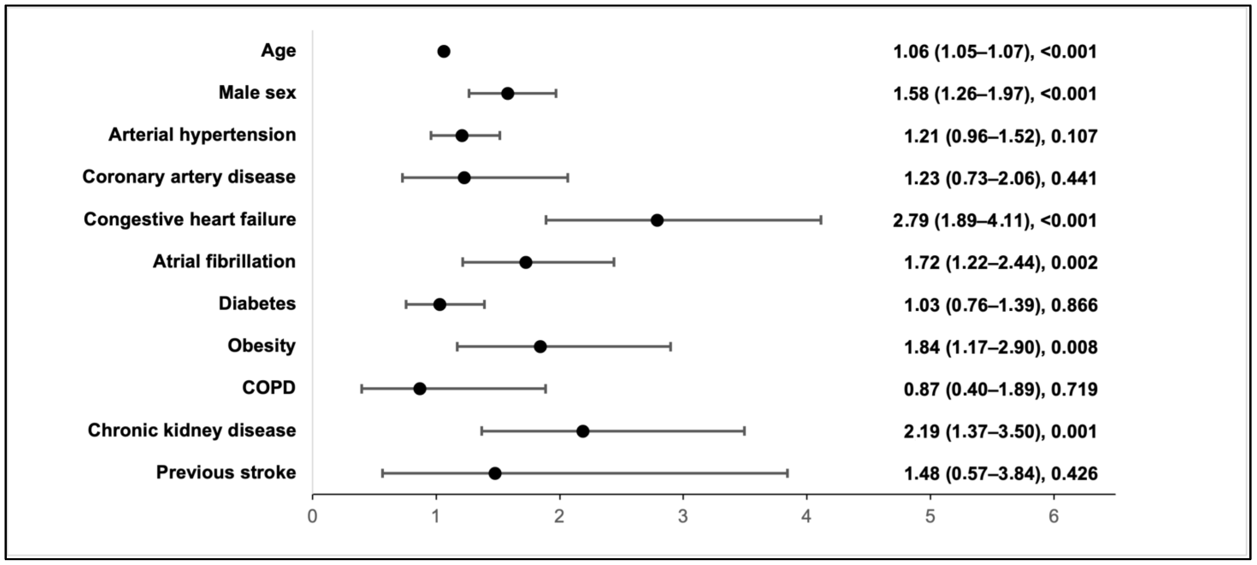
| Characteristic | In-Hospital Mortality | |
|---|---|---|
| HR (95% CI) | p-Value | |
| TnI < 19 ng/L | Reference | |
| 19 ≤ TnI ≤ 100 ng/L | 2.58 (1.83–3.62) | <0.001 |
| TnI > 100 ng/L | 2.97 (2.01–4.39) | <0.001 |
| Age in years | 1.05 (1.03–1.06) | <0.001 |
| Arterial hypertension | 0.71 (0.53–0.95) | 0.019 |
| Coronary artery disease | 1.03 (0.63–1.67) | 0.906 |
| Congestive heart failure | 1.81 (1.33–2.46) | <0.001 |
| Atrial fibrillation | 1.45 (1.06–1.98) | 0.021 |
| Diabetes | 0.92 (0.66–1.29) | 0.636 |
| Obesity | 2.40 (1.50–3.84) | <0.001 |
| COPD | 1.75 (0.96–3.18) | 0.068 |
| Chronic kidney disease | 0.70 (0.46–1.05) | 0.083 |
| Previous stroke | 1.67 (0.88–3.16) | 0.114 |
| Treatment with remdesivir | 0.61 (0.43–0.86) | 0.005 |
| Treatment with systemic steroids | 1.40 (1.00–1.96) | 0.051 |
| Antibiotics | 0.85 (0.59–1.24) | 0.413 |
Disclaimer/Publisher’s Note: The statements, opinions and data contained in all publications are solely those of the individual author(s) and contributor(s) and not of MDPI and/or the editor(s). MDPI and/or the editor(s) disclaim responsibility for any injury to people or property resulting from any ideas, methods, instructions or products referred to in the content. |
© 2024 by the authors. Licensee MDPI, Basel, Switzerland. This article is an open access article distributed under the terms and conditions of the Creative Commons Attribution (CC BY) license (https://creativecommons.org/licenses/by/4.0/).
Share and Cite
Kubiliute, I.; Urboniene, J.; Majauskaite, F.; Bobkov, E.; Svetikas, L.; Jancoriene, L. Elevated Cardiac Troponin I as a Mortality Predictor in Hospitalised COVID-19 Patients. Medicina 2024, 60, 842. https://doi.org/10.3390/medicina60060842
Kubiliute I, Urboniene J, Majauskaite F, Bobkov E, Svetikas L, Jancoriene L. Elevated Cardiac Troponin I as a Mortality Predictor in Hospitalised COVID-19 Patients. Medicina. 2024; 60(6):842. https://doi.org/10.3390/medicina60060842
Chicago/Turabian StyleKubiliute, Ieva, Jurgita Urboniene, Fausta Majauskaite, Edgar Bobkov, Linas Svetikas, and Ligita Jancoriene. 2024. "Elevated Cardiac Troponin I as a Mortality Predictor in Hospitalised COVID-19 Patients" Medicina 60, no. 6: 842. https://doi.org/10.3390/medicina60060842
APA StyleKubiliute, I., Urboniene, J., Majauskaite, F., Bobkov, E., Svetikas, L., & Jancoriene, L. (2024). Elevated Cardiac Troponin I as a Mortality Predictor in Hospitalised COVID-19 Patients. Medicina, 60(6), 842. https://doi.org/10.3390/medicina60060842