
Analyzing the Clinical Potential of Stromal Vascular Fraction: A Comprehensive Literature Review


 ,
, 
Abstract
1. Introduction
2. Materials and Methods
2.1. Search Strategy
2.2. Selection Criteria
2.3. Data Extraction
3. Results
4. Discussion
4.1. Delivery Methods
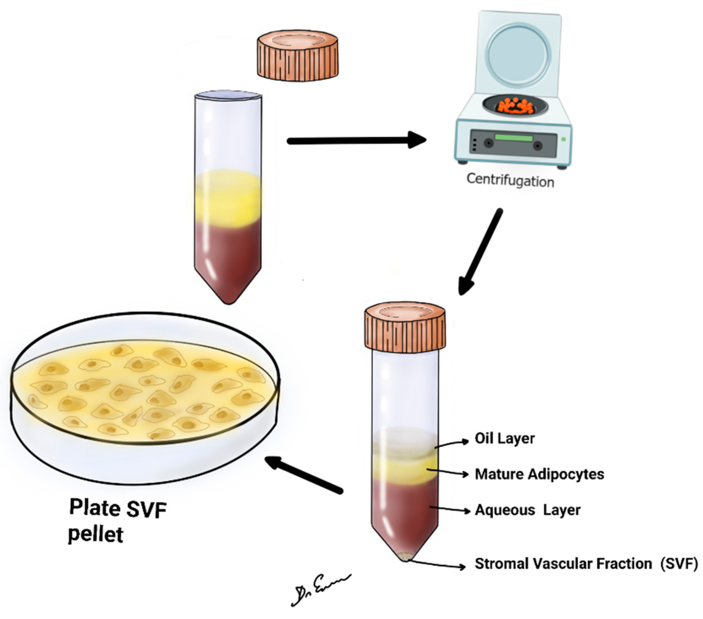
4.2. SVF Preparation Steps
4.3. Immediate Expectations
4.4. Long-Term Expectations
4.5. Future Research Directions
4.6. Limitations of this Study
5. Conclusions
Author Contributions
Funding
Institutional Review Board Statement
Informed Consent Statement
Conflicts of Interest
References
- Rodbell, M. Metabolism of isolated fat cells. I. Effects of hormones on glucose metabolism and lipolysis. J. Biol. Chem. 1964, 239, 375–380. [Google Scholar] [CrossRef]
- Zuk, P.A.; Zhu, M.; Mizuno, H.; Huang, J.; Futrell, J.W.; Katz, A.J.; Benhaim, P.; Lorenz, H.P.; Hedrick, M.H. Multilineage cells from human adipose tissue: Implications for cell-based therapies. Tissue Eng. 2001, 7, 211–228. [Google Scholar] [CrossRef]
- Bora, P.; Majumdar, A.S. Adipose tissue-derived stromal vascular fraction in regenerative medicine: A brief review on biology and translation. Stem Cell Res. Ther. 2017, 8, 145. [Google Scholar] [CrossRef]
- Baer, P.C.; Geiger, H. Adipose-derived mesenchymal stromal/stem cells: Tissue localization, characterization, and heterogeneity. Stem Cells Int. 2012, 2012, 812693. [Google Scholar] [CrossRef]
- Lee, S.H.; Lee, J.H.; Cho, K.H. Effects of Human Adipose-derived Stem Cells on Cutaneous Wound Healing in Nude Mice. Ann. Dermatol. 2011, 23, 150–155. [Google Scholar] [CrossRef] [PubMed]
- Jo, C.H.; Lee, Y.G.; Shin, W.H.; Kim, H.; Chai, J.W.; Jeong, E.C.; Kim, J.E.; Shim, H.; Shin, J.S.; Shin, I.S.; et al. Intra-articular injection of mesenchymal stem cells for the treatment of osteoarthritis of the knee: A proof-of-concept clinical trial. Stem Cells 2014, 32, 1254–1266, Erratum in Stem Cells 2017, 35, 1651–1652. [Google Scholar] [CrossRef] [PubMed]
- Gentile, P.; Garcovich, S.; Bielli, A.; Scioli, M.G.; Orlandi, A.; Cervelli, V. The Effect of Platelet-Rich Plasma in Hair Regrowth: A Randomized Placebo-Controlled Trial. Stem Cells Transl. Med. 2015, 4, 1317–1323. [Google Scholar] [CrossRef] [PubMed]
- Ude, C.C.; Shah, S.; Ogueri, K.S.; Nair, L.S.; Laurencin, C.T. Stromal Vascular Fraction for Osteoarthritis of the Knee Regenerative Engineering. Regen. Eng. Transl. Med. 2022, 8, 210–224. [Google Scholar] [CrossRef] [PubMed]
- Bai, X.; Yan, Y.; Song, Y.H.; Seidensticker, M.; Rabinovich, B.; Metzele, R.; Bankson, J.A.; Vykoukal, D.; Alt, E. Both cultured and freshly isolated adipose tissue-derived stem cells enhance cardiac function after acute myocardial infarction. Eur. Heart J. 2010, 31, 489–501. [Google Scholar] [CrossRef]
- Kølle, S.F.; Fischer-Nielsen, A.; Mathiasen, A.B.; Elberg, J.J.; Oliveri, R.S.; Glovinski, P.V.; Kastrup, J.; Kirchhoff, M.; Rasmussen, B.S.; Talman, M.L.M.; et al. Enrichment of autologous fat grafts with ex-vivo expanded adipose tissue-derived stem cells for graft survival: A randomised placebo-controlled trial. Lancet 2013, 382, 1113–1120. [Google Scholar] [CrossRef]
- Onoi, Y.; Matsumoto, T.; Sobajima, S.; Tsubosaka, M.; Hayashi, S.; Matsushita, T.; Iwaguro, H.; Kuroda, R. Clinical use of autologous adipose-derived stromal vascular fraction cell injections for hip osteoarthritis. Regen. Ther. 2023, 24, 94–102. [Google Scholar] [CrossRef]
- Kim, Y.S.; Oh, S.M.; Suh, D.S.; Tak, D.H.; Kwon, Y.B.; Koh, Y.G. Cartilage lesion size and number of stromal vascular fraction (SVF) cells strongly influenced the SVF implantation outcomes in patients with knee osteoarthritis. J. Exp. Orthop. 2023, 10, 28. [Google Scholar] [CrossRef]
- Zhang, S.; Xu, H.; He, B.; Fan, M.; Xiao, M.; Zhang, J.; Chen, D.; Tong, P.; Mao, Q. Mid-term prognosis of the stromal vascular fraction for knee osteoarthritis: A minimum 5-year follow-up study. Stem Cell Res. Ther. 2022, 13, 105. [Google Scholar] [CrossRef]
- Kwon, H.; Lee, S.; Kim, J.; Song, S.H. Efficacy and safety of stromal vascular fraction on scar revision surgery: A prospective study. J. Dermatolog. Treat. 2023, 34, 2171260. [Google Scholar] [CrossRef] [PubMed]
- Garza, J.R.; Campbell, R.E.; Tjoumakaris, F.P.; Freedman, K.B.; Miller, L.S.; Santa Maria, D.; Tucker, B.S. Clinical Efficacy of Intra-articular Mesenchymal Stromal Cells for the Treatment of Knee Osteoarthritis: A Double-Blinded Prospective Randomized Controlled Clinical Trial. Am. J. Sports Med. 2020, 48, 588–598. [Google Scholar] [CrossRef] [PubMed]
- Rodriguez-Merchan, E.C. Autologous and Allogenic Utilization of Stromal Vascular Fraction and Decellularized Extracellular Matrices in Orthopedic Surgery: A Scoping Review. Arch. Bone Jt. Surg. 2022, 10, 827–832. [Google Scholar] [PubMed]
- Perdomo-Pantoja, A.; Holmes, C.; Cottrill, E.; Rindone, A.; Ishida, W.; Taylor, M.; Tomberlin, C.; Lo, S.F.L.; Grayson, W.L.; Witham, T.F. Comparison of Freshly Isolated Adipose Tissue-derived Stromal Vascular Fraction and Bone Marrow Cells in a Posterolateral Lumbar Spinal Fusion Model. Spine 2021, 46, 631–637. [Google Scholar] [CrossRef] [PubMed]
- Choi, U.Y.; Kim, K.T.; Kim, K.G.; Lim, S.H.; Kim, Y.J.; Sonh, S.; Sheen, S.H.; Heo, C.Y.; Han, I. Safety and Tolerability of Stromal Vascular Fraction Combined with β-Tricalcium Phosphate in Posterior Lumbar Interbody Fusion: Phase I Clinical Trial. Cells 2020, 9, 2250. [Google Scholar] [PubMed]
- Rowe, G.; Heng, D.S.; Beare, J.E.; Hodges, N.A.; Tracy, E.P.; Murfee, W.L.; LeBlanc, A.J. Stromal Vascular Fraction Reverses the Age-Related Impairment in Revascularization following Injury. J. Vasc. Res. 2022, 59, 343–357. [Google Scholar] [CrossRef] [PubMed]
- Mehling, B.; Hric, M.; Salatkova, A.; Vetrak, R.; Santora, D.; Ovariova, M.; Mihalyova, R.; Manvelyan, M. A Retrospective Study of Stromal Vascular Fraction Cell Therapy for Osteoarthritis. J. Clin. Med. Res. 2020, 12, 747–751. [Google Scholar] [CrossRef] [PubMed]
- Moon, K.C.; Chung, H.Y.; Han, S.K.; Jeong, S.H.; Dhong, E.S. Tissue-engineered dermis grafts using stromal vascular fraction cells on the nose: A retrospective case-control study. J. Plast. Reconstr. Aesthetic Surg. 2020, 73, 965–974. [Google Scholar] [CrossRef] [PubMed]
- Zimmermann, S.; Fakin, R.M.; Giovanoli, P.; Calcagni, M. Outcome of Stromal Vascular Fraction-Enriched Fat Grafting Compared to Intramuscular Transposition in Painful End-Neuromas of Superficial Radial Nerve: Preliminary Results. Front. Surg. 2018, 5, 10. [Google Scholar] [CrossRef]
- Calcagni, M.; Zimmermann, S.; Scaglioni, M.F.; Giesen, T.; Giovanoli, P.; Fakin, R.M. The novel treatment of SVF-enriched fat grafting for painful end-neuromas of superficial radial nerve. Microsurgery 2018, 38, 264–269. [Google Scholar] [CrossRef]
- Jeon, H.J.; Choi, D.H.; Lee, J.H.; Lee, J.S.; Lee, J.; Park, H.Y.; Yang, J.D. Prospective Study of the Efficacy of Cell-Assisted Lipotransfer with Stromal Vascular Fraction to Correct Contour Deformities of the Autologous Reconstructed Breast. Aesthetic. Plast. Surg. 2021, 45, 853–863. [Google Scholar] [CrossRef]
- Yin, S.; Yang, X.; Bi, H.; Zhao, Z. Combined Use of Autologous Stromal Vascular Fraction Cells and Platelet-Rich Plasma for Chronic Ulceration of the Diabetic Lower Limb Improves Wound Healing. Int. J. Low. Extrem. Wounds 2021, 20, 135–142. [Google Scholar] [CrossRef]
- Aletto, C.; Giordano, L.; Quaranta, M.; Zara, A.; Notarfrancesco, D.; Maffulli, N. Short-term results of intra-articular injections of stromal vascular fraction for early knee osteoarthritis. J. Orthop. Surg. Res. 2022, 17, 310. [Google Scholar] [CrossRef]
- Francis, S.L.; Duchi, S.; Onofrillo, C.; Di Bella, C.; Choong, P.F.M. Adipose-Derived Mesenchymal Stem Cells in the Use of Cartilage Tissue Engineering: The Need for a Rapid Isolation Procedure. Stem Cells Int. 2018, 2018, 8947548. [Google Scholar] [CrossRef]
- Busato, A.; De Francesco, F.; Biswas, R.; Mannucci, S.; Conti, G.; Fracasso, G.; Conti, A.; Riccio, V.; Rioccio, M.; Sbarbati, A. Simple and Rapid Non-Enzymatic Procedure Allows the Isolation of Structurally Preserved Connective Tissue Micro-Fragments Enriched with SVF. Cells 2020, 10, 36. [Google Scholar] [CrossRef]
- Guimarães-Camboa, N.; Cattaneo, P.; Sun, Y.; Moore-Morris, T.; Gu, Y.; Dalton, N.D.; Rockenstein, E.; Masliah, E.; Peterson, K.L.; Stallcup, W.B.; et al. Pericytes of Multiple Organs Do Not Behave as Mesenchymal Stem Cells In Vivo. Cell Stem Cell 2017, 20, 345–359. [Google Scholar] [CrossRef]
- Matsuo, F.S.; de Araújo, P.H.C.; Mota, R.F.; Carvalho, A.J.R.; de Queiroz, M.S.; de Almeida, B.B.; Ferreira, K.C.d.O.S.; Metzner, R.J.M.; Ferrari, G.D.; Alberici, L.C.; et al. RANKL induces beige adipocyte differentiation in preadipocytes. Am. J. Physiol. Endocrinol. Metab. 2020, 318, E866–E877. [Google Scholar] [CrossRef]
- Contreras, G.A.; Kabara, E.; Brester, J.; Neuder, L.; Kiupel, M. Macrophage infiltration in the omental and subcutaneous adipose tissues of dairy cows with displaced abomasum. J. Dairy Sci. 2015, 98, 6176–6187. [Google Scholar] [CrossRef]
- Dey, A.; Ni, Z.; Johnson, M.S.; Sedger, L.M. A multi-colour confocal microscopy method for identifying and enumerating macrophage subtypes and adherent cells in the stromal vascular fraction of human adipose. J. Immunol. Methods 2021, 491, 112988. [Google Scholar] [CrossRef]
- Dulong, J.; Loisel, S.; Rossille, D.; Léonard, S.; Bescher, N.; Bezier, I.; Latour, M.; Monvoisin, C.; Monnier, D.; Bertheuil, N.; et al. CD40L-expressing CD4+ T cells prime adipose-derived stromal cells to produce inflammatory chemokines. Cytotherapy 2022, 24, 500–507. [Google Scholar] [CrossRef]
- Gulyaeva, O.; Dempersmier, J.; Sul, H.S. Genetic and epigenetic control of adipose development. Biochim. Biophys. Acta Mol. Cell Biol. Lipids 2019, 1864, 3–12. [Google Scholar] [CrossRef]
- Russo, A.; Condello, V.; Madonna, V.; Guerriero, M.; Zorzi, C. Autologous and micro-fragmented adipose tissue for the treatment of diffuse degenerative knee osteoarthritis. J. Exp. Orthop. 2017, 4, 33. [Google Scholar] [CrossRef]
- Stachura, A.; Paskal, W.; Pawlik, W.; Mazurek, M.J.; Jaworowski, J. The Use of Adipose-Derived Stem Cells (ADSCs) and Stromal Vascular Fraction (SVF) in Skin Scar Treatment-A Systematic Review of Clinical Studies. J. Clin. Med. 2021, 10, 3637. [Google Scholar] [CrossRef] [PubMed]
- Carstens, M.H.; Zelaya, M.; Calero, D.; Rivera, C.; Correa, D. Adipose-derived stromal vascular fraction (SVF) cells for the treatment of non-reconstructable peripheral vascular disease in patients with critical limb ischemia: A 6-year follow-up showing durable effects. Stem Cell Res. 2020, 49, 102071. [Google Scholar] [CrossRef] [PubMed]
- Choi, J.S.; Chae, D.S.; Ryu, H.A.; Kim, S.W. Transplantation of human adipose tissue derived-SVF enhance liver function through high anti-inflammatory property. Biochim. Biophys. Acta Mol. Cell Biol. Lipids 2019, 186, 158526. [Google Scholar] [CrossRef]
- Efthymiou, V.; Patti, M.E. It Is Not Just Fat: Dissecting the Heterogeneity of Adipose Tissue Function. Curr. Diab. Rep. 2022, 22, 177–187. [Google Scholar] [CrossRef] [PubMed]
- Minteer, D.; Marra, K.G.; Rubin, J.P. Adipose-derived mesenchymal stem cells: Biology and potential applications. Adv. Biochem. Eng. Biotechnol. 2013, 129, 59–71. [Google Scholar]
- Aguena, M.; Fanganiello, R.D.; Tissiani, L.A.L.; Ishiy, F.A.A.; Atique, R.; Alonso, N.; Passos-Bueno, M.R. Optimization of parameters for a more efficient use of adipose derived stem cells in regenerative medicine therapies. Stem. Cells. Int. 2012, 2012, 303610. [Google Scholar] [CrossRef]
- Food and Drug Administration, HHS. Minimal Manipulation of Human Cells, Tissues, and Cellular and Tissue-Based Products. Draft Guidance for Industry and Food and Drug Administration Staff; Food and Drug Administration: Moscow, Russia, 2014; Volume 1, p. 1. [Google Scholar]
- Tiryaki, T.; Condé-Green, A.; Cohen, S.R.; Canikyan, S.; Kocak, P. A 3-step Mechanical Digestion Method to Harvest Adipose-derived Stromal Vascular Fraction. Plast. Reconstr. Surg. Glob. Open 2020, 8, e2652. [Google Scholar] [CrossRef] [PubMed]
- Pallua, N.; Grasys, J.; Kim, B.S. Enhancement of progenitor cells by two step centrifugation of emulsified lipoaspirates. Plast. Reconstr. Surg. 2018, 142, 99–109. [Google Scholar] [CrossRef] [PubMed]
- Karina, K.; Rosliana, I.; Rosadi, I.; Schwartz, R.; Sobariah, S.; Afini, I.; Widyastuti, T.; Remelia, M.; Wahyuningsih, K.A.; Pawitan, J.A. Safety of Technique and Procedure of Stromal Vascular Fraction Therapy: From Liposuction to Cell Administration. Scientifica 2020, 2020, 2863624. [Google Scholar] [CrossRef] [PubMed]
- Chen, W.; He, Z.; Li, S.; Wu, Z.; Tan, J.; Yang, W.; Li, G.; Pan, X.; Liu, Y.; Lyu, F.-J.; et al. The Effect of Mesenchymal Stem Cells, Adipose Tissue Derived Stem Cells, and Cellular Stromal Vascular Fraction on the Repair of Acute Anal Sphincter Injury in Rats. Bioengineering 2022, 9, 318. [Google Scholar] [CrossRef] [PubMed]
- Yao, Y.; Cai, J.; Zhang, P.; Liao, Y.; Yuan, Y.; Dong, Z.; Lu, F. Adipose Stromal Vascular Fraction Gel Grafting: A New Method for Tissue Volumization and Rejuvenation. Dermatol. Surg. 2018, 44, 1278–1286. [Google Scholar] [CrossRef] [PubMed]
- Cai, Y.; Zhang, F.; Feng, J.; Wu, B.; Li, H.; Xiao, S.; Lu, F.; Wei, Z.; Deng, C. Long-term follow-up and exploration of the mechanism of stromal vascular fraction gel in chronic wounds. Stem Cell Res. Ther. 2023, 14, 163. [Google Scholar] [CrossRef] [PubMed]
- Cohen, S.R.; Hewett, S.; Ross, L.; Delaunay, F.; Goodacre, A.; Ramos, C.; Leong, T.; Saad, A. Regenerative Cells for Facial Surgery: Biofilling and Biocontouring. Aesthetic Surg. J. 2017, 37, S16–S32. [Google Scholar] [CrossRef] [PubMed]
- Mbiine, R.; Wayengera, M.; Ocan, M.; Kiwanuka, N.; Munabi, I.; Muwonge, H.; Lekuya, H.M.; Kawooya, I.; Nakanwagi, C.; Kinengyere, A.A.; et al. Adipose-derived stromal vascular fraction (SVF) in scar treatment: A systematic review proto-col. Am. J. Stem Cells. 2022, 11, 56–63. [Google Scholar]
- Pak, J.; Lee, J.H.; Pak, N.J.; Park, K.S.; Jeon, J.H.; Jeong, B.C.; Lee, S.H. Clinical Protocol of Producing Adipose Tissue-Derived Stromal Vascular Fraction for Potential Cartilage Regeneration. JoVE 2018, 139, 58363. [Google Scholar]
- Zhu, H.; Ge, J.; Chen, X.; Lu, F.; Cai, J. Mechanical Micronization of Lipoaspirates for Regenerative Therapy. J. Vis. Exp. 2019, 15, 145. [Google Scholar]
- Copcu, H.E.; Oztan, S. Not Stromal Vascular Fraction (SVF) or Nanofat, but Total Stromal-Cells (TOST): A New Definition. Systemic Review of Mechanical Stromal-Cell Extraction Techniques. Tissue Eng. Regen. Med. 2021, 18, 25–36. [Google Scholar] [CrossRef]
- Bony, C.; Cren, M.; Domergue, S.; Toupet, K.; Jorgensen, C.; Noël, D. Adipose Mesenchymal Stem Cells Isolated after Manual or Water-jet-Assisted Liposuction Display Similar Properties. Front. Immunol. 2016, 6, 655. [Google Scholar] [CrossRef]
- Aronowitz, J.A.; Ellenhorn, J.D.I. Adipose stromal vascular fraction isolation: A head-to-head comparison of four commercial cell separation systems. Plast. Reconstr. Surg. 2013, 132, 932e–939e. [Google Scholar] [CrossRef]
- Packer, J.D.; Chang, W.T.; Dragoo, J.L. The use of vibrational energy to isolate adipose-derived stem cells. Plast. Reconstr. Surg. Glob. Open 2018, 6, e1620. [Google Scholar] [CrossRef] [PubMed]
- Dragoo, J.L.; Chang, W. Arthroscopic Harvest of Adipose-Derived Mesenchymal Stem Cells from the Infrapatellar Fat Pad. Am. J. Sports Med. 2017, 45, 3119–3127. [Google Scholar] [CrossRef] [PubMed]
- Aronowitz, J.A.; Lockhart, R.A.; Hakakian, C.S.; Birnbaum, Z.E. Adipose stromal vascular fraction isolation: A head-to-head comparison of 4 cell separation systems# 2. Ann. Plast. Surg. 2016, 77, 354–362. [Google Scholar] [PubMed]
- Domenis, R.; Lazzaro, L.; Calabrese, S.; Mangoni, D.; Gallelli, A.; Bourkoula, E.; Manini, I.; Bergamin, N.; Toffoletto, B.; Beltrami, C.A. Adipose tissue derived stem cells: In vitro and in vivo analysis of a standard and three commercially available cell-assisted lipotransfer techniques. Stem Cell Res. Ther. 2015, 6, 2. [Google Scholar] [CrossRef] [PubMed]
- Fang, C.; Patel, P.; Li, H.; Huang, L.T.; Wan, H.; Collins, S.; Connell, T.L.; Xu, H. Physical, biochemical, and biologic properties of fat graft processed via different methods. Plast. Reconstr. Surg. 2020, 8, e3010. [Google Scholar] [CrossRef] [PubMed]
- Tremolada, C.; Colombo, V.; Ventura, C. Adipose tissue and mesenchymal stem cells: State of the art and Lipogems® technology development. Curr. Stem Cell Rep. 2016, 2, 304–312. [Google Scholar] [CrossRef] [PubMed]
- Magnanelli, S.; Screpis, D.; Di Benedetto, P.; Natali, S.; Causero, A.; Zorzi, C. Open-wedge high tibial osteotomy associated with lipogems® intra-articular injection for the treatment of varus knee osteoarthritis–retrospective study. Acta Biomed. Atenei Parm. 2020, 91, e2020022. [Google Scholar]
- Kavala, A.A.; Turkyilmaz, S. Autogenously derived reegenerative cell therapy for venous leg ulcers. Arch. Med. Sci. Atheroscler. Dis. 2018, 3, e156–e163. [Google Scholar] [CrossRef] [PubMed]
- Lobascio, P.; Balducci, G.; Minafra, M.; Laforgia, R.; Fedele, S.; Conticchio, M.; Palasciano, N. Adipose-derived stem cells (MYSTEM® EVO Technology) as a treatment for complex transsphincteric anal fistula. Tech. Coloproctol. 2018, 22, 373–377. [Google Scholar] [CrossRef] [PubMed]
- Stevens, H.P.; van Boxtel, J.; van Dijck, R.; van Dongen, J.A. Platelet Rich STROMA, the Combination of PRP and tSVF and Its Potential Effect on Osteoarthritis of the Knee. Appl. Sci. 2020, 10, 4691. [Google Scholar] [CrossRef]
- Copcu, H.E. Supercharged Mechanical Stromal-cell Transfer (MEST). Plast Reconstr. Surg. Glob. Open 2021, 9, e3552. [Google Scholar] [CrossRef]
- Zocchi, M.L.; Facchin, F.; Pagani, A.; Bonino, C.; Sbarbati, A.; Conti, G.; Vindigni, V.; Bassetto, F. New perspectives in regenerative medicine and surgery: The bioactive composite therapies (BACTs). Eur. J. Plast. Surg. 2022, 45, 1–25. [Google Scholar] [CrossRef]
- Rossi, M.; Roda, B.; Zia, S.; Vigliotta, I.; Zannini, C.; Alviano, F.; Bonsi, L.; Zattoni, A.; Reschiglian, P.; Gennai, A. Characterization of the Tissue and Stromal Cell Components of Micro-Superficial Enhanced Fluid Fat Injection (Micro-SEFFI) for Facial Aging Treatment. Aesthetic Surg. J. 2020, 40, 679–690. [Google Scholar] [CrossRef]
- Cohen, S.R.; Tiryaki, T.; Womack, H.A.; Canikyan, S.; Schlaudraff, K.U.; Scheflan, M. Cellular Optimization of Nanofat: Comparison of Two Nanofat Processing Devices in Terms of Cell Count and Viability. Aesthetic Surg. J. Open Forum 2019, 1, ojz028. [Google Scholar] [CrossRef]
- Tiryaki, K.T.; Cohen, S.; Kocak, P.; Canikyan Turkay, S.; Hewett, S. In-Vitro Comparative Examination of the Effect of Stromal Vascular Fraction Isolated by Mechanical and Enzymatic Methods on Wound Healing. Aesthetic Surg. J. 2020, 40, 1232–1240. [Google Scholar] [CrossRef] [PubMed]
- Simunec, D.; Salari, H.; Meyer, J. Treatment of Grade 3 and 4 Osteoarthritis with Intraoperatively Separated Adipose Tissue-Derived Stromal Vascular Fraction: A Comparative Case Series. Cells 2020, 9, 2096. [Google Scholar] [CrossRef] [PubMed]
- Sesé, B.; Sanmartín, J.M.; Ortega, B.; Matas-Palau, A.; Llull, R. Nanofat Cell Aggregates: A Nearly Constitutive Stromal Cell Inoculum for Regenerative Site-Specific Therapies. Plast Reconstr. Surg. 2019, 144, 1079–1088. [Google Scholar] [CrossRef]
- Caforio, M.; Nobile, C. Intra-Articular Administration of Autologous Purified Adipose Tissue Associated with Arthroscopy Ameliorates Knee Osteoarthritis Symptoms. J. Clin. Med. 2021, 10, 2053. [Google Scholar] [CrossRef]
- Ferguson, R.E.; Cui, X.; Fink, B.F.; Vasconez, H.C.; Pu, L.L. The viability of autologous fat grafts harvested with the LipiVage system: A comparative study. Ann. Plast Surg. 2008, 60, 594–597. [Google Scholar] [CrossRef] [PubMed]
- Agaverdiev, M.; Shamsov, B.; Mirzoev, S.; Vardikyan, A.; Ramirez, M.E.; Nurmukhametov, R.; Beilerli, A.; Zhang, B.; Gareev, I.; Pavlov, V. MiRNA regulated therapeutic potential of the stromal vascular fraction: Current clinical applications—A systematic review. Noncoding RNA Res. 2022, 8, 146–154. [Google Scholar] [CrossRef] [PubMed]
- Goncharov, E.N.; Koval, O.A.; Bezuglov, E.; Ramirez, M.d.J.E.; Engelgard, M.; Igorevich, E.I.; Saporiti, A.; Kotenko, K.V.; Montemurro, N. Stromal Vascular Fraction Therapy for Knee Osteoarthritis: A Systematic Review. Medicina 2023, 59, 2090. [Google Scholar] [CrossRef] [PubMed]
- Montemurro, N.; Pierozzi, E.; Inchingolo, A.M.; Pahwa, B.; De Carlo, A.; Palermo, A.; Scarola, R.; Dipalma, G.; Corsalini, M.; Inchingolo, A.D.; et al. New biograft solution, growth factors and bone regenerative approaches in neurosurgery, dentistry, and orthopedics: A review. Eur. Rev. Med. Pharmacol. Sci. 2023, 27, 7653–7664. [Google Scholar] [PubMed]
- Uhl, J.F.; Sufianov, A.; Ruiz, C.; Iakimov, Y.; Mogorron, H.J.; Ramirez, M.E.; Prat, G.; Lorea, B.; Baldoncini, M.; Goncharov, E.; et al. The Use of 3D Printed Models for Surgical Simulation of Cranioplasty in Craniosynostosis as Training and Education. Brain Sci. 2023, 13, 894. [Google Scholar] [CrossRef]
- Montemurro, N.; Condino, S.; Carbone, M.; Cattari, N.; D’amato, R.; Cutolo, F.; Ferrari, V. Brain Tumor and Augmented Reality: New Technologies for the Future. Int. J. Environ. Res. Public Health 2022, 19, 6347. [Google Scholar] [CrossRef] [PubMed]
- Fortunato, G.M.; Sigismondi, S.; Nicoletta, M.; Condino, S.; Montemurro, N.; Vozzi, G.; Ferrari, V.; De Maria, C. Analysis of the Robotic-Based In Situ Bioprinting Workflow for the Regeneration of Damaged Tissues through a Case Study. Bioengineering 2023, 10, 560. [Google Scholar] [CrossRef]
- Encarnacion Ramirez, M.; Ramirez Pena, I.; Barrientos Castillo, R.E.; Sufianov, A.; Goncharov, E.; Sanchez, J.A.S.; Colome-Hidalgo, M.; Nurmukhametov, R.; Céspedes, J.R.C.; Montemurro, N. Development of a 3D Printed Brain Model with Vasculature for Neurosurgical Procedure Visualisation and Training. Biomedicines 2023, 11, 330. [Google Scholar] [CrossRef]

| Author(s) (Year) | Study Type | Sample Size | Key Findings | Conclusion | Complications |
|---|---|---|---|---|---|
| Onoi et al. [11] (2023) | prospective case series | 42 | Safety of autologous SVF | SVF cell injections in the hip joint showed good short-term clinical efficacy for reducing hip OA symptoms. | no |
| Kim et al. [12] (2023) | retrospective | 43 | Cartilage repair was evaluated based on the Magnetic Resonance Observation of Cartilage Repair Tissue scoring system, using the magnetic resonance imaging from the 12-month follow-up | SVF implantation improved pain and cartilage regeneration for patients with knee osteoarthritis. The cartilage lesion size and the number of SVF cells significantly influenced the postoperative outcomes. | no |
| Zhang et al. [13] (2022) | retrospective, randomized controlled clinical trial | 126 | The VAS and WOMAC scores in the SVF group were significantly better than those in the hyaluronic acid group during the 5-year follow-up after treatment. | Up to 5 years after autologous SVF treatment, acceptable clinical state was present for approximately 60% of patients with less cartilage volume loss. In addition, the high severity of BML and high BMI increased the risk of clinical failure. Intra-articular injections of SVF do not improve subchondral BML. | no |
| Kwon et al. [14] (2023) | prospective | 20 | The 6-month follow-up following scar revision surgery revealed better results after treatment with SVF than those in the control group. | Although more research is needed, autologous SVF is a valuable source of regenerative medicine that can be swiftly and inexpensively prepared from human fat tissue. | no |
| Garza et al. [15] (2021) | prospective double-blinded randomized trial | 39 | The median percentage change in WOMAC score at 6 months after injection for the high-dose, low-dose, and placebo groups was 83.9%, 51.5%, and 25.0%, respectively. The high- and low-dose groups had statistically significant changes in WOMAC scores when compared with the placebo group (high dose, p = 0.04; low dose, p = 0.02). The improvements were dose-dependent. | Intra-articular SVF injections can significantly decrease knee OA symptoms and pain for at least 12 months. The efficacy and safety demonstrated in this placebo-controlled trial support its implementation as a treatment option for symptomatic knee OA. Magnetic resonance image review revealed no changes in cartilage thickness after treatment. | no |
| Rodriguez-Merchan et al. [16] (2022) | literature review | 28 | Intra-articular injection of SVF seems to be a safe and efficacious method for managing knee osteoarthritis (OA). Platelet-rich plasma (PRP) and SVF are safe and effective management for intractable Achilles tendinopathy in humans, although subjects treated with SVF recover earlier. | The SVF can safely be used to treat diabetic subjects suffering from chronic foot ulcers. Experimental studies indicate that SVF could be a new option to osseous regeneration. | no |
| Perdomo-Pantoja et al. [17] (2021) | prospective | 36 | The aim of this study was to compare the efficacy of freshly isolated adipose tissue-derived stromal vascular fraction (A-SVF) cells and bone marrow cells (BMCs) in achieving spinal fusion on rat models. | SVF cells yielded a comparable fusion mass volume and radiographic rate of fusion to BMCs when combined with a clinical-grade bone graft substitute. These results suggest the feasibility of using freshly isolated A-SVF cells in spinal fusion procedures. | no |
| Choi et al. [18] (2020) | prospective | 10 | Two polyetheretherketone (PEEK) cages were inserted into the intervertebral space following the complete removal of the intervertebral disc. The PEEK cage (SVF group) on the right side of the patient was filled with β-TCP in combination with SVF, and the cage on the left side (control group) was filled with β-TCP alone. Fusion rate and cage subsidence were assessed by lumbar spine X-ray and CT at 6 and 12 months postoperatively. At the 6-month follow-up, 54.5% of the SVF group (right-sided cages) and 18.2% of the control group (left-sided cages) had radiologic evidence of bone fusion (p = 0.151). | The 12-month fusion rate of the right-sided cages was 100%, while that of the left-sided cages was 91.6% (p = 0.755). Cage subsidence was not observed. Perioperative combined use of SVF with β-TCP is feasible and safe in patients who require spinal fusion surgery, and it has the potential to increase the early bone fusion rate following spinal fusion surgery. | no |
| Rowe et al. [19] (2023) | prospective | 344 | Mesenteric windows from old rats were isolated following exteriorization-induced (EI) hypoxic injury and intravenous injection of one of four cell therapies: (1) SVF from young or (2) old donors, (3) SVF from old donors depleted of or (4) enriched for T cells. Advancing age increased the SVF T-cell population but reduced revascularization following injury. | SVF represents a heterogeneous cell population shown to increase angiogenic regeneration in the researchers’ novel aged mesenteric injury model. This study provides others with a new tool for tracking vascular remodeling and can be used in conjunction with study of cell therapies or drugs in a setting of advanced age. Furthermore, the researchers show how the age of the donor should be considered not only for cellular differences but functionality as a vascular therapeutic. Age-related changes to cell dynamics and function in providing therapeutic gains—that is, the secretion of anti-inflammatory cytokines, increasing sensitivity to VEGF, increasing the migration and engraftment potential of injected cells, and endothelial cell division. | no |
| Brian et al. [20] (2020) | retrospective | 350 | Seven days after SVF cell therapy, 45.2% of subjects experienced improved pain levels and mobility. Three, six, and twelve months after therapy, improvement in pain levels reached 75.3%, 84.4%, and 84.9%, and improvement in mobility reached 75.2%, 84.4%, and 84.9%. | The treatment demonstrated a strong safety profile with no severe adverse events or complications reported. The results of the study are showing that SVF cell therapy was more effective in subjects with arthritis stage III compared to arthritis stages I, II, and IV. | no |
| Moon et al. [21] (2019) | retrospective | 77 | In the upper two-third and lower one-third zones, except for the ala, no statistically significant differences were found in any parameters. In the alar zone, statistically significant differences were detected in 10 of 21 POSAS parameters. | To cover nasal defects, the tissue-engineered dermis graft may be superior to the artificial dermis graft regarding scar quality at the ala. However, there were no significant differences in other zones. | no |
| Zimmermann et al. [22] (2018) | retrospective | 10 | In the transposition group, sustained pain reduction was not observed after an initial significant reduction 2 months post-surgery, resulting in pain relapse at 36 months and pain comparable to the preoperative assessment. In the graft group, some degree of pain reduction was observed at 2 months after the surgery and proved to be constant in the long-term outcome, although not statistically significant compared to preoperative levels. | Both SVF-enriched fat grafting and intramuscular transposition failed to prove statistically significant pain reduction in treating symptomatic neuromas of peripheral nerves. | no |
| Calcagni et al. [23] (2018) | retrospective | 5 | Pain reduction observed at 2 months after surgery was constant over time, though not statistically significant compared to preoperative levels. | SVF-enriched fat grafting represents another alternative to numerous available treatments of painful end-neuromas of the SBRN. The researchers’ preliminary results could not show any significant difference in pain reduction following SVF-enriched fat grafting. Further larger trials are required in order to evaluate the therapeutic potential of SVF-enriched fat grafting. | no |
| Jeon et al. [24] (2021) | prospective | 20 | Fat graft retention rate was higher in Group 1 than in Group 2 at both postoperative 6 months (73.8% vs. 62.2%; p = 0.03) and 12 months (65.4% vs. 48.4%; p = 0.03). Group 1 showed higher patient satisfaction. Regarding complications, fat necrosis occurred in one patient in each of the two groups. However, locoregional recurrence was not observed in any patient during follow-up. | CAL with SVF is effective in increasing survival rates of autologous fat grafts for correction of volume deficit after breast reconstruction. Moreover, it is associated with improved patient satisfaction in terms of the aesthetic aspect. | fat necrosis occurred in one patient |
| Yin et al. [25] (2021) | prospective | 5 | All patients were treated with surgical debridement, cell suspension (SVF cells suspended by platelet-rich plasma) injection into the wound, and platelet-rich plasma gel coverage. Wounds were measured every week after treatment using a two-dimensional digital camera and a three-dimensional wound measurement device. All patients were followed-up for 4 months after the treatment. | The average proportion of granulation tissue achieved 100% within 4 weeks for all cases. The wound size decreased to less than half of the original size for all cases 4 weeks after the treatment. Findings revealed that the new treatment is efficient to achieve wound healing in patients with recalcitrant chronic diabetic ulcer of lower limb. | no |
| Aletto et al. [26] (2022) | prospective clinical trial | 123 | One single injection of lipoaspirate reduces knee pain and improves function after 1 month from the injection. | The intra-articular knee injection of SVF is safe and effective to ameliorate the clinical and functional scores in patients with early knee osteoarthritis for 6 months. | no |
| Type of Cells | Functions | Authors, Year [ref.] |
|---|---|---|
| Mesenchymal progenitor/stem cells | They have the capacity to perform self-renewal and differentiation into specific cell lineages, and support the maintenance of other cells via paracrine secretion. | Francis et al., 2018 [27,32] |
| Lymphocytes | They participate in both innate and adaptive immune responses with multiple effect or functions. They produce antibodies, direct the cell-mediated killing of virus-infected and/or tumor cells, and regulate immune responses. | Busato et al., 2020 [28] |
| Smooth muscle cells | They display involuntary contractile activity to control the diameter, wall movement, and wall stiffness of specific organs. | Guimarães, 2017 [29] |
| Adipose tissue-derived stem cells | They secrete growth factors, cytokines, and antioxidant factors into a microenvironment, regulating intracellular signaling pathways in neighboring cells. Protective outcome via anti-inflammatory and immunomodulatory effects. | Bora et al., 2017 [3] |
| Preadipocytes | They promote the growth of adipose tissue by differentiating into mature and metabolically active adipocytes. Proliferating preadipocytes may also exhibit phagocytic activity towards microorganisms and behave similarly to macrophage-like cells. | Matsuo et al., 2020 [30] |
| Mφ2 macrophage | The type 2 macrophage (Mφ2) is produced by the type 2 T helper immune response and takes on an anti-inflammatory role, typically characterized by an increase in the production of interleukins (IL-4, IL-5, IL-9, and IL-13). It is also directly involved in regenerative and tissue repair processes that occur after injuries. | Contreras et al., 2015 [31]; Dey et al., 2021 [32] |
| T cells | As components of the adaptive immune system with major importance, these cells are responsible for eliminating infected host cells, activating other immune cells, and secreting cytokines that further regulate immune responses. | Dulong et al., 2022 [33] |
| Endothelial precursor cells and endothelial cells | They differentiate into functional endothelial cells and sustain vasculogenesis by incorporating themselves into the injured endothelium with the formation of functional blood vessels and through the local secretion of pro-angiogenic factors, with a paracrine effect on the cells that form the vessel. They play a critical role in vascular homeostasis as well as physiological or pathological processes such as thrombosis, inflammation, and vascular wall remodeling. Resting endothelial cells control blood flow and the passage of protein from blood into tissues, as well as inhibiting inflammation and preventing coagulation. | Gulyaeva et al., 2019 [34] |
| Regulation of pro-inflammatory molecules | Decreases IL-1b and IL-6 levels. |
| Hyaline cartilage extracellular matrix | Increases Glycosaminoglycan level. |
| Triggering of IL-1Ra | Reduces the catabolic effect of IL-1. |
| Increasing of ADAMTS-4 and -5 | Provides tissue balance (homeostasis). |
| Anti-inflammatory | Reduces tissue swelling (edema). |
| Anti-apoptotic | Reduces and stops programmed cell death. |
| Increasing of TIMPs-1, -3, and -4 metalloproteinases | Provides tissue balance (homeostasis). |
| Conventional | Modified Approach | |
|---|---|---|
| Obtaining adipose tissue |
|
|
| Mechanical separation/shredding |
|
|
| Pre-filtration |
|
|
| Washing |
|
|
| Final filtration purity |
|
|
| Collection of SVF/adipose tissue |
|
|
| Cell counting and characterization |
|
|
| Product | Company | Article |
|---|---|---|
| Cha-Station | Somnotec http://www.somnotec.net (accessed on 15th Novembre 2023) | [55] |
| Octagone D200 | Endecotts Ltd. https://www.endecotts.com (accessed on 15th Novembre 2023) | [56] |
| AdiPrep | Harvest http://www.harvest.co.kr/clinician/clinician-home/adiprep/advantages/quality.html (accessed on 15th Novembre 2023) | [57] |
| Lipokit | Medi-Khan http://www.medikanint.com (accessed on 15th Novembre 2023) | [58,59] |
| Puregraft 250 | Puregraft LLC http://www.puregraft.com (accessed on 15th Novembre 2023) | [60] |
| Lipogems | Lipogems http://understandlipogems.com (accessed on 15th Novembre 2023) | [61,62] |
| MyStem | MyStem LLC https://mystem.eu/ (accessed on 15th Novembre 2023) | [63,64] |
| Arthrex SVF | https://www.arthrex.com/orthobiologics (accessed on 15th Novembre 2023) | [65] |
| Adinizer | BSL http://biosl.com/?ckattempt=1 (accessed on 15th Novembre 2023) | [66] |
| Microlyser | Tlab https://tlab.com.tr/en/products/microlyzer-svf-kit/ (accessed on 15th Novembre 2023) | [67] |
| SEFFIE | Advanced-Maes http://www.advanced-maes.com/ (accessed on 15th Novembre 2023) | [68] |
| LIPOCUBE | STEMC https://lipocube.com/ (accessed on 15th Novembre 2023) | [69,70] |
| Q-Graft | Human Med AG https://www.humanmed.com/en/products/q-graft/ (accessed on 15th Novembre 2023) | [71] |
| Tulip Nanotransfer | Tulip Medical https://tulipmedical.com/ (accessed on 15th Novembre 2023) | [72] |
| Lipocell | Tissyou https://www.tissyou.com/portfolio_page/lipocell/ (accessed on 15th Novembre 2023) | [73] |
| LipiVage | Genesis Biosytems https://www.genesisbiosystems.com/lipivagesystem-autologous-fat-transfer/ (accessed on 15th Novembre 2023) | [74] |
Disclaimer/Publisher’s Note: The statements, opinions and data contained in all publications are solely those of the individual author(s) and contributor(s) and not of MDPI and/or the editor(s). MDPI and/or the editor(s) disclaim responsibility for any injury to people or property resulting from any ideas, methods, instructions or products referred to in the content. |
© 2024 by the authors. Licensee MDPI, Basel, Switzerland. This article is an open access article distributed under the terms and conditions of the Creative Commons Attribution (CC BY) license (https://creativecommons.org/licenses/by/4.0/).
Share and Cite
Goncharov, E.N.; Koval, O.A.; Igorevich, E.I.; Encarnacion Ramirez, M.D.J.; Nurmukhametov, R.; Valentinovich, K.K.; Montemurro, N. Analyzing the Clinical Potential of Stromal Vascular Fraction: A Comprehensive Literature Review. Medicina 2024, 60, 221. https://doi.org/10.3390/medicina60020221
Goncharov EN, Koval OA, Igorevich EI, Encarnacion Ramirez MDJ, Nurmukhametov R, Valentinovich KK, Montemurro N. Analyzing the Clinical Potential of Stromal Vascular Fraction: A Comprehensive Literature Review. Medicina. 2024; 60(2):221. https://doi.org/10.3390/medicina60020221
Chicago/Turabian StyleGoncharov, Evgeniy Nikolaevich, Oleg Aleksandrovich Koval, Eremin Ilya Igorevich, Manuel De Jesus Encarnacion Ramirez, Renat Nurmukhametov, Kotenko Konstantin Valentinovich, and Nicola Montemurro. 2024. "Analyzing the Clinical Potential of Stromal Vascular Fraction: A Comprehensive Literature Review" Medicina 60, no. 2: 221. https://doi.org/10.3390/medicina60020221
APA StyleGoncharov, E. N., Koval, O. A., Igorevich, E. I., Encarnacion Ramirez, M. D. J., Nurmukhametov, R., Valentinovich, K. K., & Montemurro, N. (2024). Analyzing the Clinical Potential of Stromal Vascular Fraction: A Comprehensive Literature Review. Medicina, 60(2), 221. https://doi.org/10.3390/medicina60020221