Evidence of Age Estimation Procedures in Forensic Dentistry: Results from an Umbrella Review

 ,
,  ,
,  ,
,  ,
, 
Abstract
:1. Introduction
2. Materials and Methods
- Eligibility criteria
- Information sources search
- Study selection
- Data extraction process and data items
- Risk of bias assessment
3. Results
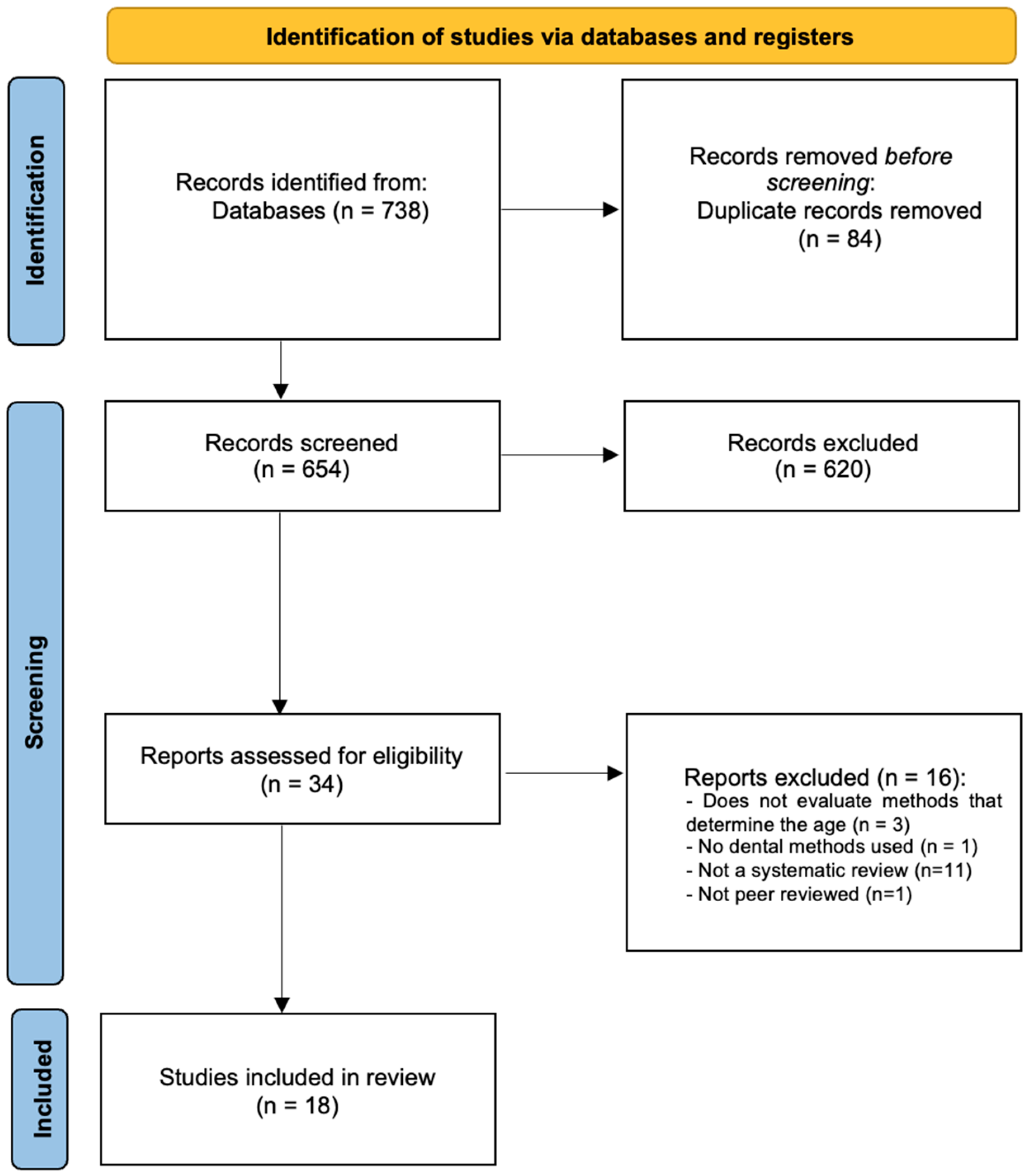
- Study selection
- SR characteristics
- Methodological Quality
- Synthesis of Results
- Panoramic radiographs-based methods
- Three-dimensional imaging methods
- AI-based methods
4. Discussion
- Strengths and limitations
5. Conclusions
Supplementary Materials
Author Contributions
Funding
Institutional Review Board Statement
Informed Consent Statement
Data Availability Statement
Conflicts of Interest
References
- Cameriere, R.; Pacifici, A.; Viva, S.; Carbone, D.; Pacifici, L.; Polimeni, A. Adult or Not? Accuracy of Cameriere’s Cut-off Value for Third Molar in Assessing 18 Years of Age for Legal Purposes. Minerva Stomatol. 2014, 63, 283–294. [Google Scholar] [PubMed]
- Cunha, E.; Baccino, E.; Martrille, L.; Ramsthaler, F.; Prieto, J.; Schuliar, Y.; Lynnerup, N.; Cattaneo, C. The Problem of Aging Human Remains and Living Individuals: A Review. Forensic Sci. Int. 2009, 193, 1–13. [Google Scholar] [CrossRef] [PubMed]
- Ritz-Timme, S.; Cattaneo, C.; Collins, M.J.; Waite, E.R.; Schütz, H.W.; Kaatsch, H.J.; Borrman, H.I. Age Estimation: The State of the Art in Relation to the Specific Demands of Forensic Practise. Int. J. Leg. Med. 2000, 113, 129–136. [Google Scholar] [CrossRef] [PubMed]
- Schmeling, A.; Geserick, G.; Reisinger, W.; Olze, A. Age Estimation. Forensic Sci. Int. 2007, 165, 178–181. [Google Scholar] [CrossRef] [PubMed]
- Angelakopoulos, N.; de Luca, S.; Velandia Palacio, L.A.; Coccia, E.; Ferrante, L.; Cameriere, R. Third Molar Maturity Index (I3M) for Assessing Age of Majority: Study of a Black South African Sample. Int. J. Leg. Med. 2018, 132, 1457–1464. [Google Scholar] [CrossRef] [PubMed]
- Yan, J.; Lou, X.; Xie, L.; Yu, D.; Shen, G.; Wang, Y. Assessment of Dental Age of Children Aged 3.5 to 16.9 Years Using Demirjian’s Method: A Meta-Analysis Based on 26 Studies. PLoS ONE 2013, 8, e84672. [Google Scholar] [CrossRef] [PubMed]
- Mani, S.A.; Naing, L.; John, J.; Samsudin, A.R. Comparison of Two Methods of Dental Age Estimation in 7–15-Year-Old Malays. Int. J. Paediatr. Dent. 2008, 18, 380–388. [Google Scholar] [CrossRef]
- Willems, G. A Review of the Most Commonly Used Dental Age Estimation Techniques. J. Forensic Odontostomatol. 2001, 19, 9–17. [Google Scholar]
- Priyadarshini, C.; Puranik, M.P.; Uma, S.R. Dental Age Estimation Methods: A Review; Lambert Academic Publisher: Saarbrücken, Germany, 2015. [Google Scholar]
- Capitaneanu, C.; Willems, G.; Thevissen, P. A Systematic Review of Odontological Sex Estimation Methods. J. Forensic Odontostomatol. 2017, 35, 1–19. [Google Scholar]
- Peckmann, T.R.; Logar, C.; Garrido-Varas, C.E.; Meek, S.; Pinto, X.T. Sex Determination Using the Mesio-Distal Dimension of Permanent Maxillary Incisors and Canines in a Modern Chilean Population. Sci. Justice 2016, 56, 84–89. [Google Scholar] [CrossRef]
- Angadi, P.V.; Hemani, S.; Prabhu, S.; Acharya, A.B. Analyses of Odontometric Sexual Dimorphism and Sex Assessment Accuracy on a Large Sample. J. Forensic Leg. Med. 2013, 20, 673–677. [Google Scholar] [CrossRef] [PubMed]
- Maber, M.; Liversidge, H.M.; Hector, M.P. Accuracy of Age Estimation of Radiographic Methods Using Developing Teeth. Forensic Sci. Int. 2006, 159, S68–S73. [Google Scholar] [CrossRef] [PubMed]
- Jeon, H.-M.; Jang, S.-M.; Kim, K.-H.; Heo, J.-Y.; Ok, S.-M.; Jeong, S.-H.; Ahn, Y.-W. Dental Age Estimation in Adults: A Review of the Commonly Used Radiological Methods. J. Oral Med. Pain 2014, 39, 119–126. [Google Scholar] [CrossRef]
- Moher, D.; Liberati, A.; Tetzlaff, J.; Altman, D.G. PRISMA Group Preferred Reporting Items for Systematic Reviews and Meta-Analyses: The PRISMA Statement. PLoS Med. 2009, 6, e1000097. [Google Scholar] [CrossRef] [PubMed]
- Burda, B.U.; Norris, S.L.; Holmer, H.K.; Ogden, L.A.; Smith, M.E.B. Quality Varies across Clinical Practice Guidelines for Mammography Screening in Women Aged 40–49 Years as Assessed by AGREE and AMSTAR Instruments. J. Clin. Epidemiol. 2011, 64, 968–976. [Google Scholar] [CrossRef] [PubMed]
- de Tobel, J.; Bauwens, J.; Parmentier, G.I.L.; Franco, A.; Pauwels, N.S.; Verstraete, K.L.; Thevissen, P.W. Magnetic Resonance Imaging for Forensic Age Estimation in Living Children and Young Adults: A Systematic Review. Pediatr. Radiol. 2020, 50, 1691–1708. [Google Scholar] [CrossRef] [PubMed]
- Diaconescu, I.; Isailă, O.-M.; Hostiuc, S. Accuracy of the Chaillet’s Method for Assessing the Age in Subadults. A Meta-Analysis. Curr. Health Sci. J. 2021, 47, 196–203. [Google Scholar] [CrossRef]
- Hostiuc, S.; Diaconescu, I.; Rusu, M.C.; Negoi, I. Age Estimation Using the Cameriere Methods of Open Apices: A Meta-Analysis. Healthcare 2021, 9, 237. [Google Scholar] [CrossRef]
- Hostiuc, S.; Edison, S.E.; Diaconescu, I.; Negoi, I.; Isaila, O.M. Accuracy of the Demirjian’s Method for Assessing the Age in Children, from 1973 to 2020. A Meta-Analysis. Leg. Med. 2021, 52, 101901. [Google Scholar] [CrossRef]
- Esan, T.A.; Yengopal, V.; Schepartz, L.A. The Demirjian versus the Willems Method for Dental Age Estimation in Different Populations: A Meta-Analysis of Published Studies. PLoS ONE 2017, 12, e0186682. [Google Scholar] [CrossRef]
- Franco, A.; de Oliveira, M.N.; Vidigal, M.T.C.; Blumenberg, C.; Pinheiro, A.A.; Paranhos, L.R. Assessment of Dental Age Estimation Methods Applied to Brazilian Children: A Systematic Review and Meta-Analysis. Dentomaxillofac. Radiol. 2021, 50, 20200128. [Google Scholar] [CrossRef] [PubMed]
- Haglund, M.; Mörnstad, H. A Systematic Review and Meta-Analysis of the Fully Formed Wisdom Tooth as a Radiological Marker of Adulthood. Int. J. Leg. Med. 2019, 133, 231–239. [Google Scholar] [CrossRef] [PubMed]
- Jayaraman, J.; Wong, H.M.; King, N.M.; Roberts, G.J. The French-Canadian Data Set of Demirjian for Dental Age Estimation: A Systematic Review and Meta-Analysis. J. Forensic Leg. Med. 2013, 20, 373–381. [Google Scholar] [CrossRef] [PubMed]
- Mohd Yusof, M.Y.P.; Wan Mokhtar, I.; Rajasekharan, S.; Overholser, R.; Martens, L. Performance of Willem’s Dental Age Estimation Method in Children: A Systematic Review and Meta-Analysis. Forensic Sci. Int. 2017, 280, 245.e1–245.e10. [Google Scholar] [CrossRef] [PubMed]
- Prasad, H.; Kala, N. Accuracy of Two Dental Age Estimation Methods in the Indian Population—A Meta-Analysis of Published Studies. J. Forensic Odontostomatol. 2019, 3, 2–11. [Google Scholar]
- Santiago, B.M.; Almeida, L.; Cavalcanti, Y.W.; Magno, M.B.; Maia, L.C. Accuracy of the Third Molar Maturity Index in Assessing the Legal Age of 18 Years: A Systematic Review and Meta-Analysis. Int. J. Legal Med. 2018, 132, 1167–1184. [Google Scholar] [CrossRef]
- Sehrawat, J.S.; Singh, M. Willems Method of Dental Age Estimation in Children: A Systematic Review and Meta-Analysis. J. Forensic Leg. Med. 2017, 52, 122–129. [Google Scholar] [CrossRef]
- Wang, J.; Ji, F.; Zhai, Y.; Park, H.; Tao, J. Is Willems Method Universal for Age Estimation: A Systematic Review and Meta-Analysis. J. Forensic Leg. Med. 2017, 52, 130–136. [Google Scholar] [CrossRef]
- Bjørk, M.B.; Kvaal, S.I. CT and MR Imaging Used in Age Estimation: A Systematic Review. J. Forensic Odontostomatol. 2018, 36, 14–25. [Google Scholar]
- Khanagar, S.B.; Vishwanathaiah, S.; Naik, S.; Al-Kheraif, A.A.; Devang Divakar, D.; Sarode, S.C.; Bhandi, S.; Patil, S. Application and Performance of Artificial Intelligence Technology in Forensic Odontology—A Systematic Review. Leg. Med. 2021, 48, 101826. [Google Scholar] [CrossRef]
- Marroquin, T.Y.; Karkhanis, S.; Kvaal, S.I.; Vasudavan, S.; Kruger, E.; Tennant, M. Age Estimation in Adults by Dental Imaging Assessment Systematic Review. Forensic Sci. Int. 2017, 275, 203–211. [Google Scholar] [CrossRef] [PubMed]
- Rolseth, V.; Mosdøl, A.; Dahlberg, P.S.; Ding, Y.; Bleka, Ø.; Skjerven-Martinsen, M.; Straumann, G.H.; Delaveris, G.J.M.; Vist, G.E. Age Assessment by Demirjian’s Development Stages of the Third Molar: A Systematic Review. Eur. Radiol. 2019, 29, 2311–2321. [Google Scholar] [CrossRef] [PubMed]
- Norris, S.A.; Frongillo, E.A.; Black, M.M.; Dong, Y.; Fall, C.; Lampl, M.; Liese, A.D.; Naguib, M.; Prentice, A.; Rochat, T.; et al. Nutrition in Adolescent Growth and Development. Lancet 2022, 399, 172–184. [Google Scholar] [CrossRef] [PubMed]
- Machado, V.; Botelho, J.; Pereira, D.; Vasques, M.; Fernandes-Retto, P.; Proença, L.; Mendes, J.J.; Delgado, A. Bolton Ratios in Portuguese Subjects among Different Malocclusion Groups. J. Clin. Exp. Dent. 2018, 10, e864–e868. [Google Scholar] [CrossRef]
- Vandevoort, F.M.; Bergmans, L.; van Cleynenbreugel, J.; Bielen, D.J.; Lambrechts, P.; Wevers, M.; Peirs, A.; Willems, G. Age Calculation Using X-ray Microfocus Computed Tomographical Scanning of Teeth: A Pilot Study. J. Forensic Sci. 2004, 49, 787–790. [Google Scholar] [CrossRef]
- Baumann, P.; Widek, T.; Merkens, H.; Boldt, J.; Petrovic, A.; Urschler, M.; Kirnbauer, B.; Jakse, N.; Scheurer, E. Dental Age Estimation of Living Persons: Comparison of MRI with OPG. Forensic Sci. Int. 2015, 253, 76–80. [Google Scholar] [CrossRef]
- Moons, K.G.M.; Altman, D.G.; Reitsma, J.B.; Ioannidis, J.P.A.; Macaskill, P.; Steyerberg, E.W.; Vickers, A.J.; Ransohoff, D.F.; Collins, G.S. Transparent Reporting of a Multivariable Prediction Model for Individual Prognosis or Diagnosis (TRIPOD): Explanation and Elaboration. Ann. Intern. Med. 2015, 162, W1–W73. [Google Scholar] [CrossRef]

| Authors (Year) | N | Search Period | Interventions | Quality Assessment Tool | Sample | Method of Analysis | Outcomes | AMSTAR2 Score * | Funding |
|---|---|---|---|---|---|---|---|---|---|
| Bjork (2018) [30] | 27 | From July 2004 to September 2017 | CT and MR imaging | NI | RCTs | SR | Although more research is needed, both CT and MR imaging may be useful tools for age estimation. | Critically Low | NI |
| De Tobel (2020) [17] | 55 | Up to September 2018 | MR imaging | EPOC overview and QUADAS-2 | 1 prospective cohort; 35 prospective CS; 19 retrospective CS | SR/MA | The performance of the age estimation was better for the multi-factorial age estimation than for the single-site age estimation. MRI allows the examination of multiple anatomical sites without the use of ionizing radiation. | Moderate | NI |
| Diaconescu (2021) [18] | 25 | From 2013 to 2019 | Chaillet’s method | STROBE | RCTs | SR/MA | Chaillet’s method showed age overestimation in both sexes, as in most ethnic groups, with delayed dental development in Asian populations, unlike European populations. | Low | NI |
| Esan (2017) [21] | 28 | Up to 28 December 2016 | Demirjian’s and Willems’ methods | STROBE | 5 comparative CS; 4 CS; 17 retrospective CS; 2 observational CS | SR/MA | While Demirjian’s method has wide application in determining maturity scores, Willems’ method provides a more accurate estimate of chronological age in different populations. | High | None |
| Franco (2020) [22] | 13 | Up to January 2019 | Demirjian, Willems, Cameriere’s, Nolla’s and Lilequist and Lundberg’s methods | JBI Critical Appraisal Tools | CS studies | SR/MA | Optimal performance was achieved by most of the international methods for dental radiographic age estimation. | Moderate | Research Grant |
| Haglund (2018) [23] | 24 | Up to 8 June 2017 | Demirjian’s method for the 3rd molar | QUADAS-2 | RCTs | SR/MA | In addition to the fact that the presence of an immature third tooth is highly indicative of adult age, there are significant numbers of young adults (over the age of 18) with immature third teeth. | Critically Low | NI |
| Hostiuc (2021) [20] | 89 | From 1973 to 2020 | Demirjian’s method | STROBE | RCTs | SR/MA | Using the Demirjian method, the age is overestimated by about half a year for both sexes. Some geographic/ethnic differences exist. Nevertheless, regardless of the ethnic profile of the subjects, this method is useful. | Low | None |
| Hostiuc (2021) [19] | 15 | From 2005 to 2019 | Cameriere’s method | STROBE | RCTs | SR/MA | At least in the 7–14 age interval, the Cameriere method is accurate enough for clinical use. However, it should not be used outside of this age range. | Low | None |
| Jayaraman (2013) [24] | 34 | From January 1973 to December 2011 | Demirjian’s method | NI | RCTs | SR/MA | Demirjian’s method overestimates the age of the subjects by more than six months, and therefore this data set should only be used with great caution when trying to estimate the age of a group of subjects in any global population. | Critically Low | None |
| Khanagar et al. (2021) [31] | 8 | From January 2000 to June 2020 | AI based models for personal age estimation | QUADAS-2 | NR | SR | The AI technology demonstrates an accuracy and precision that is equivalent to that of a trained examiner, overcoming human error and being non-invasive are additional advantages of these models. A major limitation of the present review is the lack of real-life scenarios and the experimental nature of the included studies. | Low | Research Grant |
| Marroquin (2017) [32] | 32 | From January 1995 to July 2016 | Cameriere’s, Kvaal’s method, and CBCT imaging | NI | NR | SR | More accurate results were obtained with age estimation methods based on pulp/tooth area ratio calculation. It is advised to use dental age estimation methods, firstly, pulp/tooth area ratio calculation of single first, upper canines and other single rooted teeth, and secondly, pulp/tooth length/ratio calculation. | Critically Low | NI |
| Yusof (2017) [25] | 23 | From January 2001 to September 2014 | Willems’ method | Cochrane handbook for systematic reviews-methodology review | NR | SR/MA | Given the accuracy of the Willems method across different populations, investigators, and age groups, it is appropriate to use this method to estimate age in children. | Moderate | Research Grant |
| Prasad (2019) [26] | 20 | Up to July 2018 | Demirjian’s and Willems’ methods | QUADAS-2 | NR | SR/MA | Willems’ method was more accurate in predicting chronological age than Demirjian’s method in the Indian population, regardless of sex. | High | NI |
| Rolseth (2018) [33] | 21 | Up to May 2016 | Demirjian’s method for the 3rd molar | QUADAS-2 | NR | SR | The differences in the timing of the developmental stages of the third molars according to Demirjian have often been interpreted as differences between populations and ethnic groups. | Low | None |
| Santiago (2017) [27] | 15 | Up to November 2017 | Cameriere’s method (I3M) | QUADAS-2 | CS studies | SR/MA | The Third Molar Maturity Index is a suitable and useful method for estimating adulthood because it has a high accuracy in distinguishing whether an individual has reached 18 years of age, regardless of the population studied. | High | None |
| Sehrawat (2017) [28] | 31 | From 2001 to January 2017 | Willems’ method | NI | CS and Retrospective studies | SR/MA | Compared to other methods reported in the available literature, the Willems method of dental age estimation results in comparatively less overestimation of age. | Critically Low | NI |
| Wang (2017) [29] | 11 | Up to 28 February 2017 | Willems’ method | NOS | CS and Retrospective studies | SR/MA | For both sexes, the Willems method overestimated dental age between 3.0 and 16.9 years in almost all age groups. In addition, the accuracy of the Willems method was also shown to be affected by ethnic differences. | Low | NI |
| Yan (2013) [6] | 26 | Up to 12 July 2013 | Demirjian’s method | STROBE | CS and Retrospective studies | SR/MA | The fact that Demirjian’s method overestimates actual chronological tooth age highlights the need for population-specific standards to better estimate the rate at which human teeth mature. | High | None |
| First Author | 1 | 2 | 3 | 4 | 5 | 6 | 7 | 8 | 9 | 10 | 11 | 12 | 13 | 14 | 15 | 16 | Review Quality |
|---|---|---|---|---|---|---|---|---|---|---|---|---|---|---|---|---|---|
| Bjørk et al. (2018) [30] | Y | PY | N | N | N | N | N | N | 0/0 | N | 0/0 | 0 | Y | N | 0 | Y | Critically low |
| De Tobel et al. (2020) [17] | Y | PY | Y | Y | Y | Y | Y | PY | 0/0 | N | 0/0 | 0 | Y | Y | 0 | Y | Moderate |
| Diaconescu et al. (2021) [18] | Y | PY | Y | PY | N | N | PY | N | N/0 | N | Y/0 | Y | Y | Y | Y | Y | Low |
| Esan et al. (2017) [21] | Y | Y | Y | PY | Y | Y | Y | PY | PY/PY | N | Y/Y | Y | Y | Y | Y | Y | High |
| Franco et al. (2020) [22] | Y | Y | Y | PY | N | N | Y | N | PY/PY | N | Y/Y | Y | Y | Y | Y | Y | Moderate |
| Hadlund et al. (2018) [23] | Y | PY | Y | PY | N | N | Y | PY | N/N | N | Y/Y | Y | Y | Y | Y | Y | Critically low |
| Hostiuc et al. (2021) [20] | Y | Y | Y | N | Y | Y | Y | PY | N/0 | N | Y/0 | Y | Y | Y | Y | Y | Critically low |
| Hostiuc et al. (2021) [19] | Y | PY | N | Y | Y | Y | PY | N | N/0 | N | Y/0 | Y | Y | Y | Y | Y | Low |
| Jayaraman et al. (2013) [24] | Y | N | N | N | N | N | Y | PY | N/0 | N | N/0 | N | N | N | N | Y | Critically low |
| Khanagar et al. (2020) [31] | Y | PY | N | PY | Y | Y | N | Y | PY/PY | N | 0/0 | 0 | N | N | 0 | Y | Low |
| Marroquin et al. (2017) [32] | Y | PY | N | PY | N | N | Y | PY | N/N | N | 0/0 | 0 | N | N | 0 | N | Critically low |
| Mohd Yusof et al. (2017) [25] | Y | Y | N | PY | Y | N | Y | PY | Y/Y | N | Y/Y | Y | Y | Y | Y | Y | Moderate |
| Prasad et al. (2019) [26] | Y | Y | Y | Y | Y | Y | PY | Y | Y/Y | N | Y/Y | Y | Y | Y | Y | Y | High |
| Rolseth et al. (2018) [33] | N | Y | N | PY | Y | Y | N | PY | Y/Y | N | 0/0 | 0 | Y | Y | 0 | Y | Low |
| Santiago et al. (2017) [27] | Y | Y | Y | Y | Y | Y | Y | Y | Y/Y | N | Y/Y | Y | Y | Y | Y | Y | High |
| Sehrawat et al. (2017) [28] | Y | PY | Y | PY | Y | N | PY | Y | N/N | N | Y/Y | N | N | Y | N | Y | Low |
| Wang et al. (2017) [29] | Y | Y | Y | PY | Y | Y | PY | Y | Y/Y | N | Y/Y | Y | Y | Y | N | Y | Low |
| Yan et al. (2013) [6] | Y | Y | Y | Y | Y | Y | Y | Y | Y/Y | N | Y/Y | Y | Y | Y | Y | Y | High |
Disclaimer/Publisher’s Note: The statements, opinions and data contained in all publications are solely those of the individual author(s) and contributor(s) and not of MDPI and/or the editor(s). MDPI and/or the editor(s) disclaim responsibility for any injury to people or property resulting from any ideas, methods, instructions or products referred to in the content. |
© 2023 by the authors. Licensee MDPI, Basel, Switzerland. This article is an open access article distributed under the terms and conditions of the Creative Commons Attribution (CC BY) license (https://creativecommons.org/licenses/by/4.0/).
Share and Cite
Neves, J.A.; Lopes, L.B.; Machado, V.; Botelho, J.; Delgado, A.S.; Mendes, J.J. Evidence of Age Estimation Procedures in Forensic Dentistry: Results from an Umbrella Review. Medicina 2024, 60, 42. https://doi.org/10.3390/medicina60010042
Neves JA, Lopes LB, Machado V, Botelho J, Delgado AS, Mendes JJ. Evidence of Age Estimation Procedures in Forensic Dentistry: Results from an Umbrella Review. Medicina. 2024; 60(1):42. https://doi.org/10.3390/medicina60010042
Chicago/Turabian StyleNeves, João Albernaz, Luísa Bandeira Lopes, Vanessa Machado, João Botelho, Ana Sintra Delgado, and José João Mendes. 2024. "Evidence of Age Estimation Procedures in Forensic Dentistry: Results from an Umbrella Review" Medicina 60, no. 1: 42. https://doi.org/10.3390/medicina60010042
APA StyleNeves, J. A., Lopes, L. B., Machado, V., Botelho, J., Delgado, A. S., & Mendes, J. J. (2024). Evidence of Age Estimation Procedures in Forensic Dentistry: Results from an Umbrella Review. Medicina, 60(1), 42. https://doi.org/10.3390/medicina60010042