Case Report

Management of Complete Heart Block in a Pregnant Woman with Systemic Lupus Erythematosus-Associated Complications: Treatment Considerations and Pitfalls

1
Department of Internal Medicine and Cardiology, University Hospital Brno and Faculty of Medicine, Masaryk University, Jihlavska 20, 625 00 Brno, Czech Republic
2
Department of Laboratory Methods, Faculty of Medicine, Masaryk University, Kamenice 5, 625 00 Brno, Czech Republic
3
Department of Laboratory Medicine, Masaryk Memorial Cancer Institute, Zluty Kopec 7, 656 53 Brno, Czech Republic
4
Department of Biochemistry, Faculty of Medicine, Masaryk University, Kamenice 5, 625 00 Brno, Czech Republic
5
Department of Gynecology and Obstetrics, University Hospital Brno and Faculty of Medicine, Masaryk University, Jihlavska 20, 625 00 Brno, Czech Republic
*
Author to whom correspondence should be addressed.
Medicina 2023, 59(1), 88; https://doi.org/10.3390/medicina59010088
Submission received: 23 October 2022 / Revised: 15 December 2022 / Accepted: 28 December 2022 / Published: 31 December 2022
(This article belongs to the Section Hematology and Immunology)

Abstract

:
We present a case of a pregnant woman with systemic lupus erythematosus (SLE) who was diagnosed with asymptomatic complete heart block (CHB) during pregnancy. To evaluate possible risks and benefits of pacemaker (PM) implantation, a multidisciplinary counselling board was held. Its recommendation was to perform PM implantation to prevent intra-uterine growth restriction from insufficient cardiac output using a fluoroscopic protective shield. The procedure was performed without complications and established permanent pacing on onwards ECG examinations. The patient subsequently gave birth to a healthy newborn. After a retrospective clinical case evaluation and review of relevant literature, a presumptive association between CHB and the primary diagnosis was proposed. Above that, pregnant women with SLE who develop hypertension are commonly treated with methyldopa, which may cause conduction abnormalities. Clinical recommendations for young female patients expecting pregnancy are lacking in this area. Careful diagnostic and treatment approaches should be used in the management of possible SLE-related complications in women of child-bearing age, focusing on preventable events.

1. Introduction

Abnormal conduction between the atria and the ventricles is divided into three main degrees (first, second and complete heart block). The first-degree heart block is defined on ECG by a PR interval longer than 0.20 s and is generally asymptomatic [1]. Second-degree heart block is a disease of the cardiac conduction system in which the conduction of atrial impulse through the atrioventricular (AV) node and/or His bundle is delayed or blocked. Patients with second degree heart block may present with wide range of symptoms depending on the severity of conduction defects (e.g., syncope, lightheadedness or no symptoms at all) [2].
Complete heart block (CHB) is characterized by the absence of conduction between the atria and the ventricles. CHB patients most commonly suffer from decreased perfusion, which results in symptoms related to bradycardia. Subsequent low cardiac output can lead to serious arrhythmias such as ventricular tachycardia, syncope or sudden death [3].
CHB can either be congenital or acquired (secondary) [4]. The most common cause of congenital heart block is of autoimmune origin related to the transplacental passage of maternal anti-Ro/SSA or/and anti-La/SSB antibodies with consequent immune-mediated injury to the conduction system [5]. This occurs with 2% of anti-Ro/SSA-exposed pregnancies, and the recurrence rate is nine times higher in subsequent pregnancies [6]. Amongst other causes of congenital heart block, an underlying structural disease is also common [7].
Acquired CHB manifests from coronary artery disease, autoimmune disease, cardiomyopathy, infectious heart disease (myocarditis, Lyme disease, etc.) and as a drug-induced condition, and the latter two causes may possibly be reversible [8,9]. Uncovering the CHB during pregnancy is infrequent. Since CHB is a rare condition in women of child-bearing age, there are only a few case studies published discussing the diagnosis and management of CHB during pregnancy, and thus, no clear guidelines have been established [10,11].

2. Case Report and Results

We present a case of 28 year-old woman diagnosed with SLE at the age of 20 years. The diagnosis was made via kidney biopsy due to acute renal failure (creatinine 3.8 mg/dL, blood urea nitrogen 44.88 mg/dL) with severe proteinuria (total protein in urine 7.4 g/day) and edemas preceded by urinary tract infection. Histopathological evaluation showed edematous interstitium without fibrosis, with focal lymphoplasmacellular inflammatory infiltration, enlarged glomeruli with globally enhanced mesangial and endocapillary cellularity and the focal presence of cellular sickles. The presence of huge subendothelial focal immune deposits was noted with Acid Fuchsin Orange G staining. Under electron microscopy, numerous subendothelial immune deposits were confirmed and mesangial changes were present. Initially, her condition was classified as diffuse SLE-related nephritis class IV/A (ISN/RPS,) and her laboratory results met the clinical and laboratory criteria of nephrotic syndrome (serum: albumin 24 g/L, cholesterol 348 mg/dL).
Further clinical signs encompassed SLE-related butterfly rash, pleural and pericardial serous effusions, microcytic normochromic anemia (hemoglobin 111 g/L, MCV 78 fL, MCHC 33.7 g/dL) and positive ANA (titer 1:1600), positive anti-ds DNA and anti-ss DNA antibodies and depleted complement components C3 and C4. Her echocardiography findings, brain MRI and central and peripheral joint examinations were normal. Initial treatment included six series of acute hemodialysis, five series of plasmapheresis and six pulses of cyclophosphamide 750 mg/m2 with mesna 400 mg and methylprednisolone 3 g i.v. + oral prednisone in a descending dose. Ovarian suppression was induced with goserelin-acetate 3.6 mg s.c. monthly. No biologics were used during initial treatment. Maintenance therapy included daily azathioprine 2 mg/kg with prednisone 20 mg with which the patient achieved disease remission after 3 years (no clinical signs present, normalized BUN and creatinine values, no proteinuria, negative direct Coombs test). The patient’s ANA titers dropped to 1:100 and anti-ENA decline to negative values. Antibody panel results during treatment are summarized in Table 1. The treatment continued with prednisone 20 mg daily via monotherapy (5 years, slowly reducing the dose to 2.5 mg daily). Prior to the diagnosis of SLE, the patient was otherwise healthy. Primary diagnosis-related comorbidities included secondary anemia, hyperuricemia and frequent UTIs on long-term immunosuppressive treatment, hypertension and hypothyroidism. Prior to pregnancy, the long-term medication consisted of calcitriol 0.5 μg daily, prednisone 2.5 mg once in two days, perindopril 10 mg daily, levothyroxine 25 μg daily, folic acid 10 mg weekly and omeprazole 20 mg once in two days. Her ECG findings were normal, and hypertension was well compensated. However, six months prior to a pregnancy, the patient ceased corticosteroid treatment completely despite the clinical recommendation, which resulted in a subsequent rise in ANA titers.
Early in her 28th year of life, the patient became pregnant and immediately ceased treatment with perindopril, which was swapped with methyldopa 250 mg three times per day. Hydroxychloroquine treatment was recommended to the patient to stabilize primary disease during pregnancy, but that was rejected by the patient. Entry pregnancy maternal anti-Ro/SSA or/and anti-La/SSB antibody screening to evaluate the risk of congenital CHB was negative.
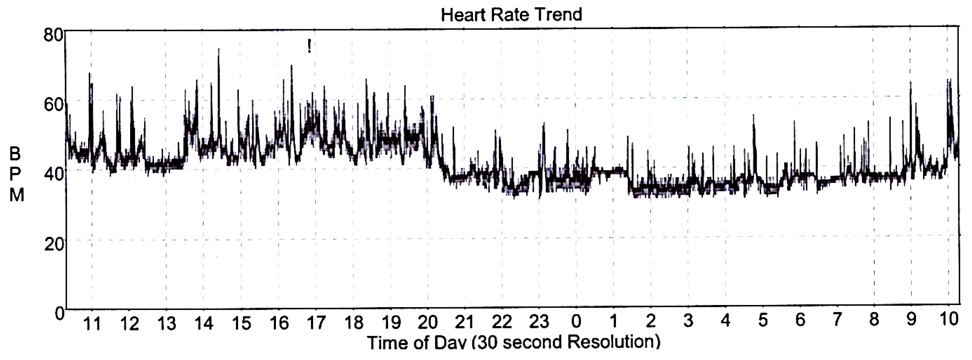
For insufficient control of blood pressure (BP), the patient had to be referred to a hypertension-treatment specialist for follow-up counselling and treatment re-evaluation during the 8th week of pregnancy. Gradual treatment escalation of methydopa to 500 mg three times per day was introduced with amlodipine 5 mg daily and acetyl-salicylic acid 150 mg daily (for preeclampsia prevention) resulting in adequate BP control. Interestingly, the incidental diagnosis of CHB was incidentally made on the regular ECG check-up (Figure 1), as the patient had not previously experienced any of the common symptoms of low cardiac output, such as fatigue, dizziness, shortness of breath or syncope. The echocardiography showed a normal ejection fraction and no left ventricle dilation and did not discover any valve pathology. Thereafter, the 24 h outpatient ECG Holter exhibited a mean heart rate of 41 beats per minute with the lowest value of 31 beats per minute (Figure 2).
The cardiology counselling board with an obstetrician and anesthesiologist evaluated and selected an appropriate treatment approach with regards to available literature. The board agreed to recommend a PM implantation to prevent the impairment of intra-uterine growth restriction from insufficient cardiac output. The Dual-chamber rate-modulated (DDDR) pacemaker (magnetic resonance imaging-compatible) was implanted during the 27th week of pregnancy as demands on the cardiovascular system in this particular pregnancy period are the highest [12]. The procedure was carried out using a protective shield covering the patient’s abdomen without complications. Post-operative ECG showed fully atrial synchronized ventricular pacing (Figure 2). During the 38th week of pregnancy, maternal hepatopathy developed, and therefore, the labor was uncomplicatedly induced. A healthy newborn 48 cm and 2930 g was delivered. On routine cardiology check-up several months afterwards and after methyldopa discontinuation, the pacemaker recording marked 100% atrial synchronized ventricular pacing (Figure 3).

3. Discussion

A major association between a congenital CHB in newborns of mothers with SLE-related autoantibodies is well described in current literature [13,14]. In adults with SLE, the manifestation of a complete heart block is a rare cardiac complication, though it is clinically important with possible life-threatening risks, particularly during pregnancy. Current SLE workup protocols include initial echocardiography for screening SLE-related pericarditis or pleural/pericardial effusions; however, no entry-ECG or regular ECG checkups with regular ECG Holter examinations as screening for SLE-related heart conduction abnormalities are neither discussed nor recommended [10,11]. A recent study by Villuendas et al. evaluated a possible link between rheumatologic disease and positive anti-Ro/SSA antibodies, previously discussed as possibly responsible for autoimmune-mediated heart conduction system damage. No association was found, and the results did not support a standard cardiac evaluation in autoimmune disease patients [15]. A study by Natsheh et al. researched case reports of CHB in lupus and found only four anti-Ro/SSA and two anti-La/SSB positive cases out of 32, therefore suggesting other possible pathophysiological mechanisms involved in SLE-induced conduction defects. Additionally, Natsheh et al. discovered that 84% of CHB cases were anti-DNA positive and 100% were ANA positive [16]. Our patient’s anti-Ro/SSA and/or anti-La/SSB antibodies during the pregnancy period were also not elevated but both anti-DNA and ANA were positive. Furthermore, a literature review of CHB cases with lupus erythematosus revealed that CHB almost exclusively involves women (30/32, 94%) and more importantly, the most common CHB manifestation in these women is syncope [16]. Since the probability of preexisting heart disease (such as ischemic heart disease, etc.) was unlikely in our patient, the association between CHB and lupus cannot be neglected. This statement is further supported by the fact that based on follow-up routine pacemaker check-ups, the CHB did not resolve after the discontinuation of methyldopa and the patient continues to have a 100% stimulated PM rhythm.
Recommendations for young women with SLE prior to pregnancy include the multidisciplinary approach of an obstetric and rheumatology team, as disease control (optimally stable disease on no or a minimal dose of oral corticosteroids) is essential for favorable pregnancy outcomes. These recommendations mention possible congenital heart block of the fetus/newborn and its prevention and management; however, no recommendations are aimed towards the possible manifestation of a CHB in the mother [17]. This issue is particularly important as pregnant women with SLE have a higher prevalence of hypertension than those without SLE [18]. Moreover, there have been reports that methyldopa, the drug of choice for pregnant women, may impair atrioventricular conduction and even cause CHB [19,20]. It is assumed that methyldopa, via its central sympatholytic effect, can impair the myocardial conduction system [19]. However, in documented cases, heart conduction defects resolved upon methyldopa discontinuation suggesting reversible impairment of the heart conduction system [19,20].
Although hemodynamic changes during pregnancy are very well known and described (the heart rate increases by 25% and plasma volume increases by more than 1 L) [21] and concomitant CHB with such increased hemodynamic demands may present a significant problem, the relevant clinical guidelines underlining this scenario are still lacking. The inability of the heart to increase the heart rate can affect prenatal development and labor. Cardiac output is based on the stroke volume and heart rate and increases by 20% at the 8th week of gestation to its maximum in the 20–28th week of gestation [12].
Pregnant women with CHB fail to exhibit compensatory heart rate increase, which results in an inadequate hemodynamic status during labor and needs to be corrected by temporary pacing [22]. Hidaka et al. described clinical cases of women with a complete heart block, who received temporary pacing during labor; however, none of them required pacing. According to their experience, it was suggested that women with a complete heart block do not routinely require temporary pacing during labor [23]. This phenomenon could possibly be explained by chronotropic competence in women with CHB during labor, even though this explanation needs to be proven by further studies.
Similarly, Adekanye et al. described a case of a pregnant woman with CHB diagnosed during pregnancy that was left unpaced until labor with temporary pacing equipment on standby. At that time, the pregnant woman was already in her 38th week of pregnancy at which time the growth of the fetus had already finished and the pacemaker was implanted during puerperium [24].
In some cases, decisions about temporary pacemaker insertion during pregnancy have been made based on a clinical history of syncope or presyncope [25]. According to a study from Suri et al., CHB is associated with premature delivery (in three out of four cases) and intrauterine growth restriction (two out of four cases) [26]. With respect to that knowledge, the decision about the implantation of a permanent pacemaker vastly outweighs the risk of intrauterine growth restriction and premature delivery [27]. However, a multidisciplinary approach with a team of cardiologists, obstetricians and anesthetists, as in our case, is essential. Certainly, the decision should consider the gestational week to evaluate the possibility of fetal growth impairment. There are case studies of progressive AV block during pregnancy; therefore, all patients should be carefully monitored [25]. According to a small study by Mandal et al. regarding concomitant CHB and pregnancy outcomes, early-phase pregnancy permanent pacemaker implantation was proposed as a safe and better option to avoid further life-threatening complications [28].
On the contrary, the teratogenic properties of fluoroscopy, routinely used during PM implantation, should not be underestimated. Either a protective shield covering the gravid uterus or a fluoroscopy-free approach using electrophysiologic signals and ECHO guidance for pacemaker insertion should be used [29,30]. Moreover, another modern fluoroscopy-free possibility is to use a cardiac mapping system during the PM implantation procedure [31].

4. Conclusions

The management of patients with CHB diagnosed in pregnancy should be performed in tertiary centers. Multidisciplinary evaluation is necessary to select optimal procedures for pregnant women with CHB, as each patient may need an individualized approach.
Due to the risk of intrauterine growth restriction and preterm birth, close fetal and maternal monitoring is essential to provide appropriate care. Women who are diagnosed with SLE are often of childbearing age and may develop CHB both prior to and during pregnancy. According to the study of Tselios et al., the risk of developing a CHB in patients with SLE reaches 1%, while the prevalence of CHB in the general population is 0,04% (including elderly people with pre-existing heart disease) [32,33]. Furthermore, impairments of the heart conduction system may result from concomitant SLE-induced damage, while the use of methyldopa may unmask clinically silent aberrations.
The etiology of lupus-associated complete heart block is unclear, and hence, recommendations are needed. Women with autoimmune disease (especially those with SLE) planning pregnancy are possible candidates for initial 24 h ECG monitoring or for modern wearables for 24 h heart rate monitoring, which may increase the probability of discovering bradycardia, as they have been shown to provide valuable clinical data [34,35]. In patients taking methyldopa or in patients with positive ANA and anti-DNA antibodies, this should be a mandatory clinical measure.
Primary benefits of such diagnostic approaches would be the prevention of severe clinical manifestations, such as syncope, heart failure of the mother and intrauterine growth restriction, fetal impairment and risks associated with PM implantation during pregnancy. Secondary outcomes include the possible elucidation of underlying pathophysiology of CHB in patients with SLE.

Author Contributions

Conceptualization, E.R. and P.V.; methodology, M.R. and E.R.; formal analysis, P.K. and P.V.; investigation, E.R., P.V., T.B. and D.K.; resources, E.R., M.R., P.V. and T.B.; writing—original draft preparation, M.R. and E.R.; writing—review and editing, E.R., P.V., T.B., M.R., D.K. and P.K.; supervision, P.V. and P.K.; funding acquisition, P.K. All authors have read and agreed to the published version of the manuscript.

Funding

This work was supported by the Ministry of Health, Czech Republic—Conceptual Development of Research Organization (FNBr. 65269705).

Institutional Review Board Statement

The study was conducted according to the guidelines of the Declaration of Helsinki and approved by the Institutional Ethics Committee of the University Hospital Brno, protocol code 08-121022/EK, date of approval 12 October 2022.

Informed Consent Statement

Informed consent was obtained from the patient involved in this study and is available in patient’s clinical documentation.

Data Availability Statement

The data presented in this study are available on request from the corresponding author.

Acknowledgments

We thank Dalibor Valik for language correction insights.

Conflicts of Interest

The authors declare no conflict of interest.

References

  1. Holmqvist, F.; Daubert, J.P. First-degree AV block-an entirely benign finding or a potentially curable cause of cardiac disease? Ann. Noninvasive Electrocardiol. 2013, 18, 215–224. [Google Scholar] [CrossRef] [PubMed]
  2. Wogan, J.M.; Lowenstein, S.R.; Gordon, G.S. Second-degree atrioventricular block: Mobitz type II. J. Emerg. Med. 1993, 11, 47–54. [Google Scholar] [CrossRef] [PubMed]
  3. Al Taweel, M.; Al Mukhaylid, S.; Alsultan, N. Congenital Complete Heart Block in Adults. J. Adv. Med. Med. Res. 2022, 34, 245–248. [Google Scholar] [CrossRef]
  4. Fischbach, P.S.; Frias, P.A.; Strieper, M.J.; Campbell, R.M. Natural history and current therapy for complete heart block in children and patients with congenital heart disease. Congenit. Heart Dis. 2007, 2, 224–234. [Google Scholar] [CrossRef]
  5. Pruetz, J.D.; Miller, J.C.; Loeb, G.E.; Silka, M.J.; Bar-Cohen, Y.; Chmait, R.H. Prenatal diagnosis and management of congenital complete heart block. Birth Defects Res. 2019, 111, 380–388. [Google Scholar] [CrossRef]
  6. De Carolis, S.; Garufi, C.; Garufi, E.; De Carolis, M.P.; Botta, A.; Tabacco, S.; Salvi, S. Autoimmune Congenital Heart Block: A Review of Biomarkers and Management of Pregnancy. Front. Pediatr. 2020, 8, 607515. [Google Scholar] [CrossRef]
  7. Brucato, A.; Jonzon, A.; Friedman, D.; Allan, L.D.; Vignati, G.; Gasparini, M.; Stein, J.I.; Montella, S.; Michaelsson, M.; Buyon, J. Proposal for a new definition of congenital complete atrioventricular block. Lupus 2003, 12, 427–435. [Google Scholar] [CrossRef]
  8. Barra, S.N.C.; Providencia, R.; Paiva, L.; Nascimento, J.; Marques, A.L. A review on advanced atrioventricular block in young or middle-aged adults. Pacing Clin. Electrophysiol. 2012, 35, 1395–1405. [Google Scholar] [CrossRef]
  9. Bamgboje, A.; Akintan, F.O.; Gupta, N.M.; Kaur, G.; Pekler, G.; Mushiyev, S. Lyme Carditis: A Reversible Cause of Acquired Third-Degree AV Block. Am. J. Case Rep. 2021, 22, e927885. [Google Scholar] [CrossRef]
  10. Fanouriakis, A.; Kostopoulou, M.; Alunno, A.; Aringer, M.; Bajema, I.; Boletis, J.N.; Cervera, R.; Doria, A.; Gordon, C.; Govoni, M.; et al. 2019 update of the EULAR recommendations for the management of systemic lupus erythematosus. Ann. Rheum. Dis. 2019, 78, 736–745. [Google Scholar] [CrossRef]
  11. Aringer, M.; Costenbader, K.; Daikh, D.; Brinks, R.; Mosca, M.; Ramsey-Goldman, R.; Smolen, J.S.; Wofsy, D.; Boumpas, D.T.; Kamen, D.L.; et al. 2019 European League Against Rheumatism/American College of Rheumatology Classification Criteria for Systemic Lupus Erythematosus. Arthritis Rheumatol. 2019, 71, 1400–1412. [Google Scholar] [CrossRef] [PubMed] [Green Version]
  12. Soma-Pillay, P.; Nelson-Piercy, C.; Tolppanen, H.; Mebazaa, A. Physiological changes in pregnancy. Cardiovasc. J. Afr. 2016, 27, 89–94. [Google Scholar] [CrossRef] [PubMed] [Green Version]
  13. Costedoat-Chalumeau, N.; Amoura, Z.; Villain, E.; Cohen, L.; Piette, J.C. Anti-SSA/Ro antibodies and the heart: More than complete congenital heart block? A review of electrocardiographic and myocardial abnormalities and of treatment options. Arthritis Res. Ther. 2005, 7, 69–73. [Google Scholar] [CrossRef] [PubMed] [Green Version]
  14. Claus, R.; Hickstein, H.; Külz, T.; Lenschow, U.; Meiske, D.; Kotitschke, A.; Thiesen, H.J.; Lorenz, P. Identification and management of fetuses at risk for, or affected by, congenital heart block associated with autoantibodies to SSA (Ro), SSB (La), or an HsEg5-like autoantigen. Rheumatol. Int. 2006, 26, 886–895. [Google Scholar] [CrossRef] [PubMed]
  15. Villuendas, R.; Martínez-Morillo, M.; Juncà, G.; Teniente-Serra, A.; Diez, C.; Heredia, S.; Riveros-Frutos, A.; Bayés-Genís, A.; Olivé, A. Usefulness of cardiac screening in patients with systemic lupus erythematosus and anti-Ro/SSA antibodies. Lupus 2021, 30, 1596–1602. [Google Scholar] [CrossRef]
  16. Natsheh, A.; Shimony, D.; Bogot, N.; Nesher, G.; Breuer, G.S. Complete heart block in lupus. Lupus 2019, 28, 1589–1593. [Google Scholar] [CrossRef]
  17. Petri, M. Pregnancy and Systemic Lupus Erythematosus. Best Pract. Res. Clin. Obstet. Gynaecol. 2020, 64, 24–30. [Google Scholar] [CrossRef]
  18. Munguia-Realpozo, P.; Mendoza-Pinto, C.; Benito, C.S.; Escarcega, R.O.; Garcia-Carrasco, M.; Martinez, S.M.; Morales, I.E.; Romero, J.L.G.; Ruiz-Arguelles, A.; Cervera, R. Systemic lupus erythematosus and hypertension. Autoimmun. Rev. 2019, 18, 102371. [Google Scholar] [CrossRef]
  19. Rosen, B.; Ovsyshcher, I.A.; Zimlichman, R. Complete atrioventricular block induced by methyldopa. Pacing Clin. Electrophysiol. 1988, 11 Pt 1, 1555–1558. [Google Scholar] [CrossRef]
  20. Cokkinos, V.; Vorides, M. Impairment of Atrioventricular Conduction by Methyldopa. CHEST J. 1978, 74, 697. [Google Scholar] [CrossRef]
  21. Hunter, S.; Robson, S.C. Adaptation of the maternal heart in pregnancy. Br. Heart J. 1992, 68, 540–543. [Google Scholar] [CrossRef] [PubMed] [Green Version]
  22. Dalvi, B.V.; Chaudhuri, A.; Kulkami, H.L.; Kale, P.A. Therapeutic guidelines for congenital complete heart block presenting in pregnancy. Obstet. Gynecol. 1992, 79 Pt 2, 802–804. [Google Scholar] [PubMed]
  23. Hidaka, N.; Chiba, Y.; Kurita, T.; Satoh, S.; Nakano, H. Is intrapartum temporary pacing required for women with complete atrioventricular block? An analysis of seven cases. BJOG 2006, 113, 605–607. [Google Scholar] [CrossRef] [PubMed]
  24. Adekanye, O.; Srinivas, K.; Collis, R.E. Bradyarrhythmias in pregnancy: A case report and review of management. Int. J. Obstet. Anesth. 2007, 16, 165–170. [Google Scholar] [CrossRef] [PubMed]
  25. Thaman, R.; Curtis, S.; Faganello, G.; Szantho, G.V.; Turner, M.S.; Trinder, J.; Sellers, S.; Stuart, G.A. Cardiac outcome of pregnancy in women with a pacemaker and women with untreated atrioventricular conduction block. Europace 2011, 13, 859–863. [Google Scholar] [CrossRef]
  26. Suri, V.; Keepanasseril, A.; Aggarwal, N.; Vijayvergiya, R.; Chopra, S.; Rohilla, M. Maternal complete heart block in pregnancy: Analysis of four cases and review of management. J. Obstet. Gynaecol. Res. 2009, 35, 434–437. [Google Scholar] [CrossRef]
  27. Carrión-Camacho, M.R.; Marín-León, I.; Molina-Doñoro, J.M.; González-López, J.R. Safety of Permanent Pacemaker Implantation: A Prospective Study. J. Clin. Med. 2019, 8, 35. [Google Scholar] [CrossRef] [Green Version]
  28. Mandal, S.; Mandal, D.; Sarkar, A. Complete Heart Block and Pregnancy Outcome: An Analysis from Eastern India. SOJ Gynaecol. Obstet. Womens Health 2015, 1, 5. [Google Scholar] [CrossRef] [Green Version]
  29. Pedrinazzi, C.; Gazzaniga, P.; Durin, O.; Tovena, D.; Inama, G. Implantation of a permanent pacemaker in a pregnant woman under the guidance of electrophysiologic signals and transthoracic echocardiography. J. Cardiovasc. Med. 2008, 9, 1169–1172. [Google Scholar] [CrossRef]
  30. Jordaens, L.J.; Vandenbogaerde, J.F.; Van De Bruaene, P.; De Buyzere, M. Transesophageal echocardiography for insertion of a physiological pacemaker in early pregnancy. Pacing Clin. Electrophysiol. 1990, 13, 955–957. [Google Scholar] [CrossRef]
  31. Guo, P.; Qiu, J.; Wang, Y.; Chen, G.; Proietti, R.; Fadhle, A.S.; Zhao, C.; Wen Wang, D. Zero-fluoroscopy permanent pacemaker implantation using Ensite NavX system: Clinical viability or fanciful technique? Pacing Clin. Electrophysiol. 2018, 41, 122–127. [Google Scholar] [CrossRef] [PubMed] [Green Version]
  32. Kojic, E.M.; Hardarson, T.; Sigfusson, N.; Sigvaldason, H. The prevalence and prognosis of third-degree atrioventricular conduction block: The Reykjavik study. J. Intern. Med. 1999, 246, 81–86. [Google Scholar] [CrossRef] [PubMed] [Green Version]
  33. Tselios, K.; Gladman, D.D.; Harvey, P.; Su, J.; Urowitz, M.B. Severe brady-arrhythmias in systemic lupus erythematosus: Prevalence, etiology and associated factors. Lupus 2018, 27, 1415–1423. [Google Scholar] [CrossRef] [PubMed]
  34. Ploux, S.; Strik, M.; Caillol, T.; Ramirez, F.D.; Abu-Alrub, S.; Marchand, H.; Buliard, S.; Haïssaguerre, M.; Bordachar, P. Beyond the wrist: Using a smartwatch electrocardiogram to detect electrocardiographic abnormalities. Arch. Cardiovasc. Dis. 2022, 115, 29–36. [Google Scholar] [CrossRef]
  35. Yerasi, C.; O’Donoghue, S.; Satler, L.F.; Waksman, R. Apple Watch detecting high-grade block after transcatheter aortic valve implantation. Eur. Heart J. 2020, 41, 1096. [Google Scholar] [CrossRef]
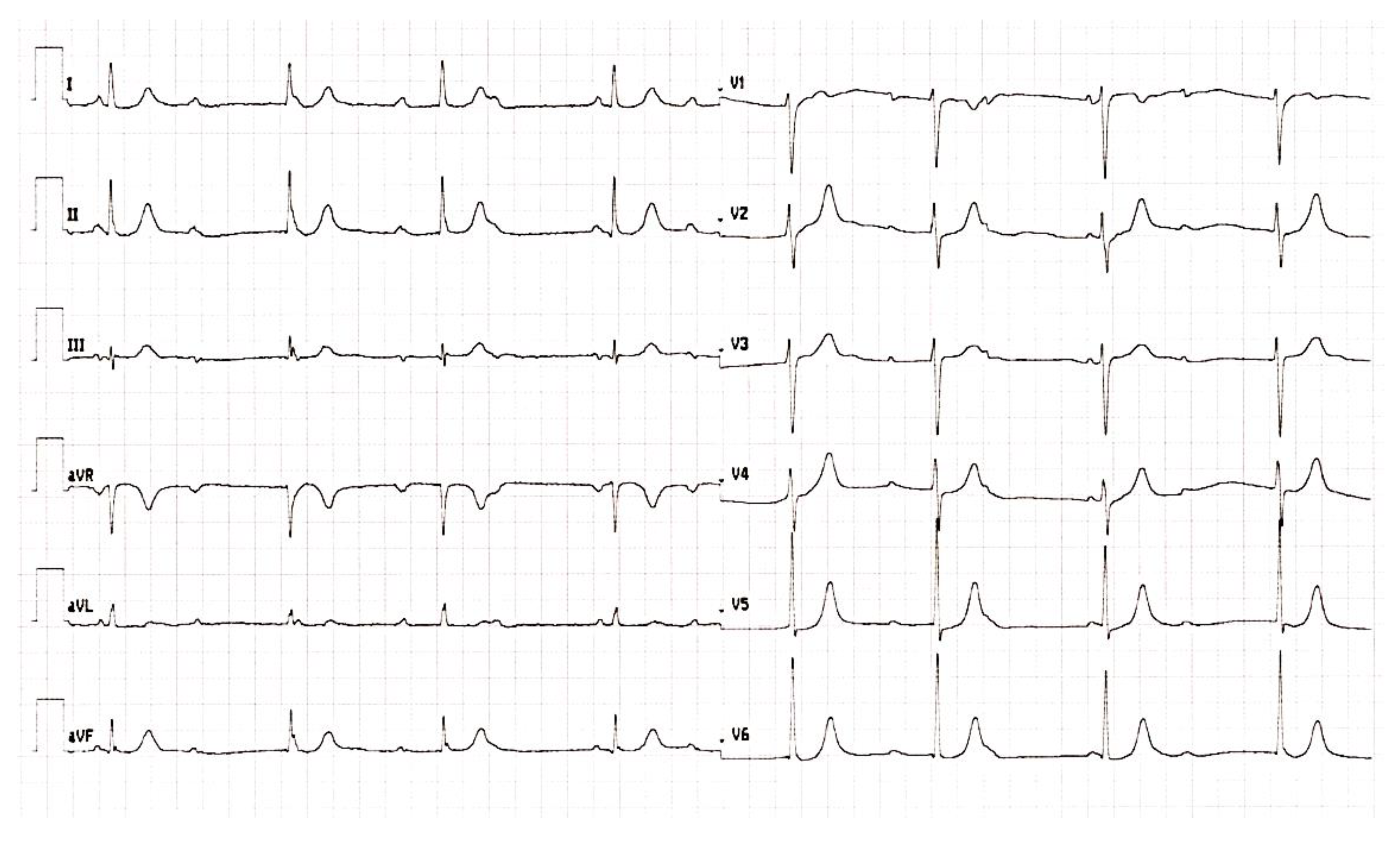
Figure 1. ECG taken at the cardiology department showing the complete heart block.
Medicina 59 00088 g001
Figure 2. Patient’s ECG 24 h Holter with a mean heart rate of 41 beats per minute and the lowest value of 31 beats per minute.
Medicina 59 00088 g002
Figure 3. ECG taken based on the routine check-up several months after pacemaker implantation with 100% atrial synchronized ventricular pacing.
Medicina 59 00088 g003
Table 1. Patient’s antibody panel results and physical parameters during treatment periods.
AutoantibodiesPhysical Parameters
ANAAnti-Ds
DNA
Anti-Ss
DNA
Anti-ENABP
(mmHg)
HR
(bpm)
At the time of the diagnosis1:1600+++/-180/10065
Clinically stable disease (3 years on maintenance treatment)1:100++-130/7570
1 month prior to the PM implantation1:1600++-140/7540
5 months after the PM implantation1:3200+n/a-135/7085
Aabbreviations: n/a, not available; BP, blood pressure; HR = heart rate; bpm = beats per minute.
Disclaimer/Publisher’s Note: The statements, opinions and data contained in all publications are solely those of the individual author(s) and contributor(s) and not of MDPI and/or the editor(s). MDPI and/or the editor(s) disclaim responsibility for any injury to people or property resulting from any ideas, methods, instructions or products referred to in the content.