
Surgical Treatment of Dysphagia Secondary to Anterior Cervical Osteophytes Due to Diffuse Idiopathic Skeletal Hyperostosis

Abstract
:1. Introduction
2. Case Reports
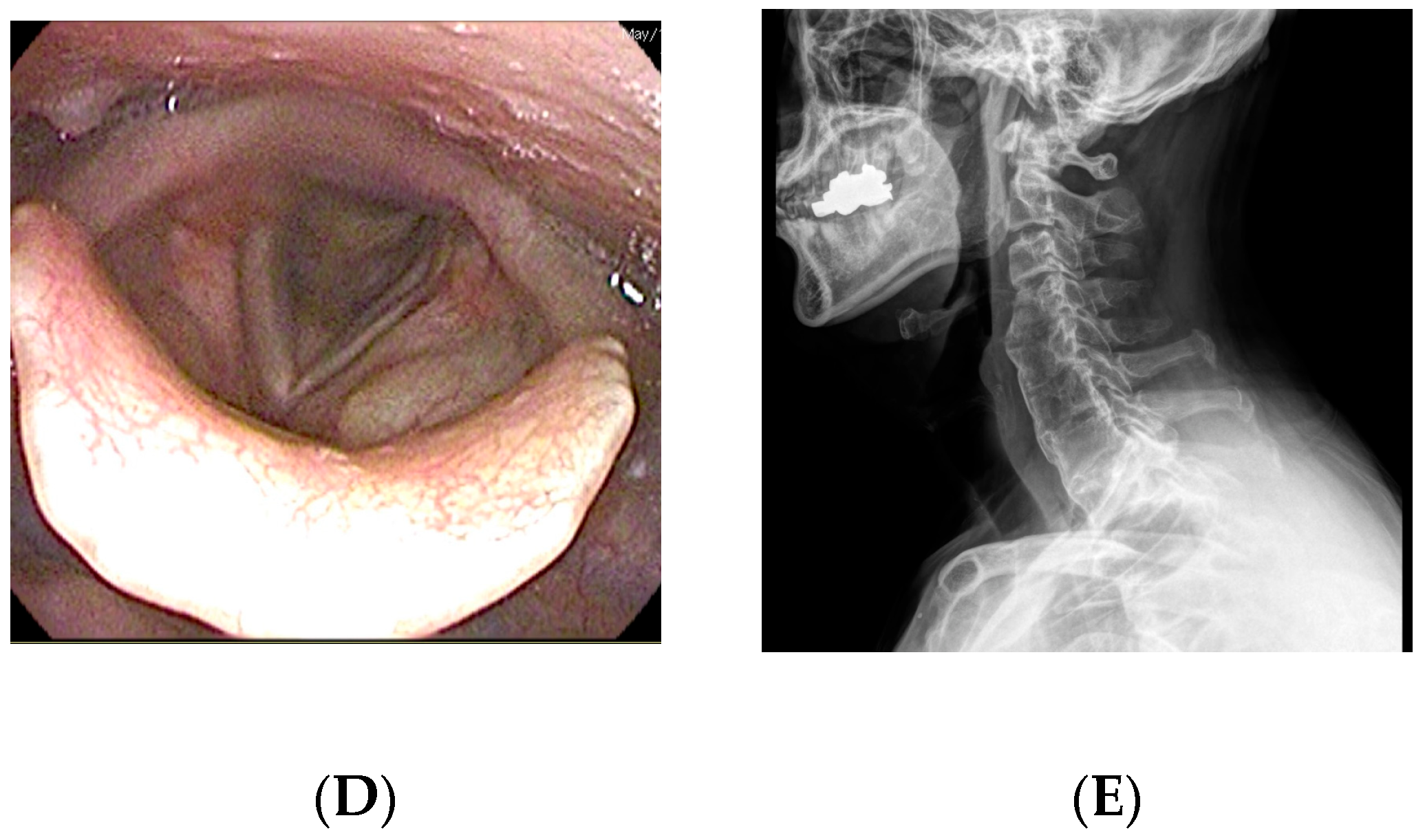
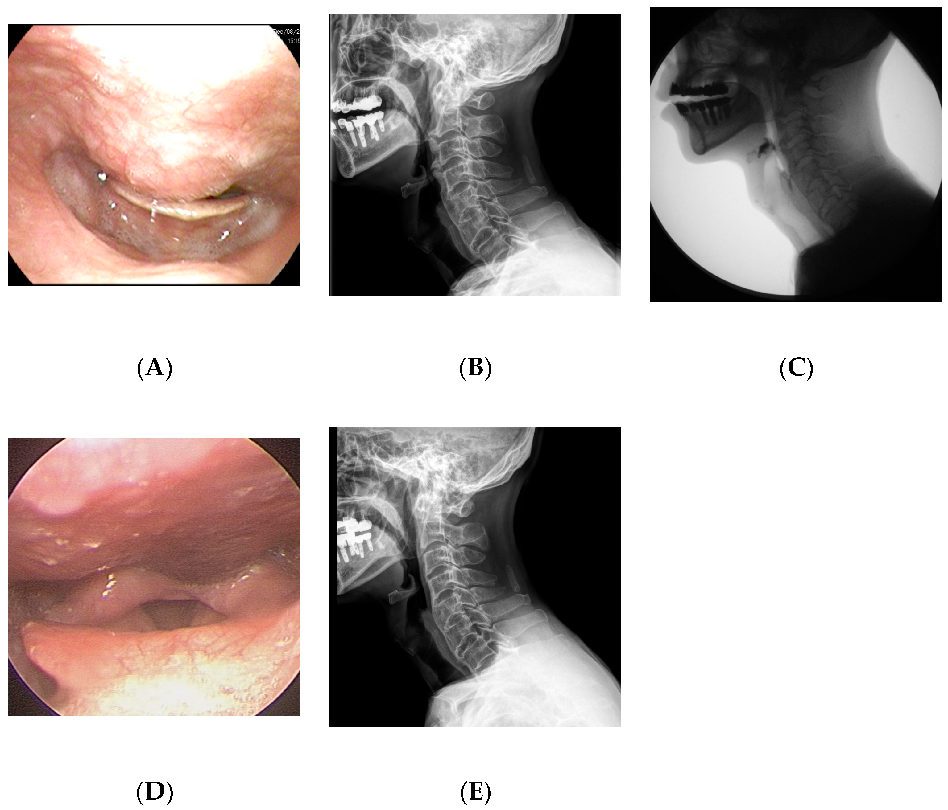
2.1. Case 1
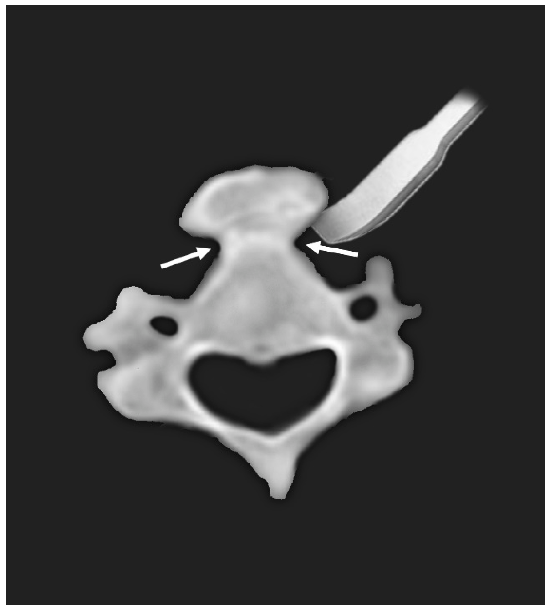
2.2. Case 2
3. Discussion
4. Conclusions
Author Contributions
Funding
Institutional Review Board Statement
Informed Consent Statement
Data Availability Statement
Conflicts of Interest
References
- Mattioli, F.; Ghirelli, M.; Trebbi, M.; Silvestri, M.; Presutti, L.; Fermi, M. Improvement of Swallowing Function After Surgical Treatment of Diffuse Idiopathic Skeletal Hyperostosis: Our Experience. World Neurosurg. 2020, 134, e29–e36. [Google Scholar] [CrossRef] [PubMed]
- Hirasawa, A.; Wakao, N.; Kamiya, M.; Takeuchi, M.; Kawanami, K.; Murotani, K.; Matsuo, T.; Deie, M. The prevalence of diffuse idiopathic skeletal hyperostosis in Japan—The first report of measurement by CT and review of the literature. J. Orthop. Sci. 2016, 21, 287–290. [Google Scholar] [CrossRef] [Green Version]
- Resnick, D.; Niwayama, G. Radiographic and pathologic features of spinal involvement in diffuse idiopathic skeletal hyperostosis (DISH). Radiology 1976, 119, 559–568. [Google Scholar] [CrossRef] [PubMed]
- Verlaan, J.J.; Boswijk, P.F.; de Ru, J.A.; Dhert, W.J.; Oner, F.C. Diffuse idiopathic skeletal hyperostosis of the cervical spine: An underestimated cause of dysphagia and airway obstruction. Spine J. 2011, 11, 1058–1067. [Google Scholar] [CrossRef] [PubMed]
- Kiss, C.; Szilagyi, M.; Paksy, A.; Poor, G. Risk factors for diffuse idiopathic skeletal hyperostosis: A case-control study. Rheumatology 2002, 41, 27–30. [Google Scholar] [CrossRef] [Green Version]
- Fahrer, H.; Markwalder, T. Dysphagia caused by diffuse idiopathic skeletal hyperostosis. Clin. Rheumatol. 1988, 7, 117–121. [Google Scholar] [CrossRef]
- Kissel, P.; Youmans, J.R. Posttraumatic anterior cervical osteophyte and dysphagia: Surgical report and literature review. J. Spinal Disord. 1992, 5, 104–107. [Google Scholar] [CrossRef]
- Lambert, J.; Tepperman, P.; Jimenez, J. Cervical Spine Disease and Dysphagia. Am. J. Gastroenterol. 1981, 76, 35–40. [Google Scholar]
- Sura, L.; Madhavan, A.; Carnaby, G.; Crary, M.A. Dysphagia in the elderly: Management and nutritional considerations. Clin. Interv. Aging 2012, 7, 287–298. [Google Scholar]
- Aydin, E.; Akdogan, V.; Akkuzu, B.; Kirbas, I.; Ozgirgin, O.N. Six cases of Forestier syndrome, a rare cause of dysphagia. Acta Otolaryngol. 2006, 126, 775–778. [Google Scholar] [CrossRef]
- Chung, Y.S.; Zhang, H.Y.; Ha, Y.; Park, J.Y. Surgical Outcomes of Dysphagia Provoked by Diffuse Idiopathic Skeletal Hyperostosis in the Cervical Spine. Yonsei Med. J. 2020, 61, 341–348. [Google Scholar] [CrossRef] [PubMed]
- Flynn, J.M. Anterior cervical osteophytes causing dysphagia. Bol. Asoc. Med. Puerto Rico 1991, 83, 47–53. [Google Scholar]
- Crary, M.A.; Groher, M.E. Introduction to Adult Swallowing Disorders; Butterworth-Heinemann Medical: Oxford, UK, 2003. [Google Scholar]
- Barczi, S.R.; Sullivan, P.A.; Robbins, J.A. How Should Dysphagia Care of Older Adults Differ? Establishing Optimal Practice Patterns. Seminars in Speech and Language; Thieme Medical Publishers: New York, NY, USA, 2000; pp. 347–364. [Google Scholar]
- Brandenberg, G.; Leibrock, L.G. Dysphagia and dysphonia secondary to anterior cervical osteophytes. Neurosurgery 1986, 18, 90–93. [Google Scholar] [CrossRef] [PubMed]
- Kibel, S.; Johnson, P. Surgery for osteophyte-induced dysphagia. J. Laryngol. Otol. 1987, 101, 1291–1296. [Google Scholar] [CrossRef]
- Ozgocmen, S.; Kiris, A.; Kocakoc, E.; Ardicoglu, O. Osteophyte-induced dysphagia: Report of three cases. Jt. Bone Spine 2002, 69, 226–229. [Google Scholar] [CrossRef]
- Egerter, A.C.; Kim, E.S.; Lee, D.J.; Liu, J.J.; Cadena, G.; Panchal, R.R.; Kim, K.D. Dysphagia Secondary to Anterior Osteophytes of the Cervical Spine. Glob. Spine J. 2015, 5, e78–e83. [Google Scholar] [CrossRef]
- Oppenlander, M.E.; Orringer, D.A.; La Marca, F.; McGillicuddy, J.E.; Sullivan, S.E.; Chandler, W.F.; Park, P. Dysphagia due to anterior cervical hyperosteophytosis. Surg. Neurol. 2009, 72, 266–270; discussion 70–71. [Google Scholar] [CrossRef]
- Lui Jonathan, Y.C.; Sayal, P.; Prezerakos, G.; Russo, V.; Choi, D.; Casey, A.T.H. The surgical management of dysphagia secondary to diffuse idiopathic skeletal hyperostosis. Clin. Neurol. Neurosurg. 2018, 167, 36–42. [Google Scholar] [CrossRef]
- Urrutia, J.; Bono, C.M. Long-term results of surgical treatment of dysphagia secondary to cervical diffuse idiopathic skeletal hyperostosis. Spine J. 2009, 9, e13–e17. [Google Scholar] [CrossRef]
- Hamouda, W.O. Timing for surgical intervention in DISHphagia. J. Craniovertebr. Junction Spine 2018, 9, 227–231. [Google Scholar] [CrossRef]
- Hirano, H.; Suzuki, H.; Sakakibara, T.; Higuchi, Y.; Inoue, K.; Suzuki, Y. Dysphagia due to hypertrophic cervical osteophytes. Clin. Orthop. Relat. Res. 1982, 167, 168–172. [Google Scholar] [CrossRef]
- Lecerf, P.; Malard, O. How to diagnose and treat symptomatic anterior cervical osteophytes? Eur. Ann. Otorhinolaryngol. Head Neck Dis. 2010, 127, 111–116. [Google Scholar] [CrossRef] [PubMed] [Green Version]
- McCafferty, R.R.; Harrison, M.J.; Tamas, L.B.; Larkins, M.V. Ossification of the anterior longitudinal ligament and Forestier’s disease: An analysis of seven cases. J. Neurosurg. 1995, 83, 13–17. [Google Scholar] [CrossRef]
- Sebaaly, A.; Boubez, G.; Sunna, T.; Wang, Z.; Alam, E.; Christopoulos, A.; Shedid, D. Diffuse Idiopathic Hyperostosis Manifesting as Dysphagia and Bilateral Cord Paralysis: A Case Report and Literature Review. World Neurosurg. 2018, 111, 79–85. [Google Scholar] [CrossRef]
- Miyamoto, K.; Sugiyama, S.; Hosoe, H.; Iinuma, N.; Suzuki, Y.; Shimizu, K. Postsurgical recurrence of osteophytes causing dysphagia in patients with diffuse idiopathic skeletal hyperostosis. Eur. Spine J. 2009, 18, 1652–1658. [Google Scholar] [CrossRef] [PubMed] [Green Version]
- Haws, B.E.; Khechen, B.; Patel, D.V.; Yoo, J.S.; Guntin, J.A.; Cardinal, K.L.; Singh, K. Swallowing Function Following Anterior Cervical Discectomy and Fusion With and Without Anterior Plating: A SWAL-QOL (Swallowing-Quality of Life) and Radiographic Assessment. Neurospine 2019, 16, 601–607. [Google Scholar] [CrossRef]
- Shriver, M.F.; Lewis, D.J.; Kshettry, V.R.; Rosenbaum, B.P.; Benzel, E.C.; Mroz, T.E. Dysphagia Rates after Anterior Cervical Diskectomy and Fusion: A Systematic Review and Meta-Analysis. Glob. Spine J. 2017, 7, 95–103. [Google Scholar] [CrossRef] [Green Version]

Publisher’s Note: MDPI stays neutral with regard to jurisdictional claims in published maps and institutional affiliations. |
© 2022 by the authors. Licensee MDPI, Basel, Switzerland. This article is an open access article distributed under the terms and conditions of the Creative Commons Attribution (CC BY) license (https://creativecommons.org/licenses/by/4.0/).
Share and Cite
Choi, H.Y.; Jo, D.J. Surgical Treatment of Dysphagia Secondary to Anterior Cervical Osteophytes Due to Diffuse Idiopathic Skeletal Hyperostosis. Medicina 2022, 58, 928. https://doi.org/10.3390/medicina58070928
Choi HY, Jo DJ. Surgical Treatment of Dysphagia Secondary to Anterior Cervical Osteophytes Due to Diffuse Idiopathic Skeletal Hyperostosis. Medicina. 2022; 58(7):928. https://doi.org/10.3390/medicina58070928
Chicago/Turabian StyleChoi, Ho Yong, and Dae Jean Jo. 2022. "Surgical Treatment of Dysphagia Secondary to Anterior Cervical Osteophytes Due to Diffuse Idiopathic Skeletal Hyperostosis" Medicina 58, no. 7: 928. https://doi.org/10.3390/medicina58070928
APA StyleChoi, H. Y., & Jo, D. J. (2022). Surgical Treatment of Dysphagia Secondary to Anterior Cervical Osteophytes Due to Diffuse Idiopathic Skeletal Hyperostosis. Medicina, 58(7), 928. https://doi.org/10.3390/medicina58070928