Article

Co-Infection of Oral Candida albicans and Porphyromonas gingivalis Is Associated with Active Periodontitis in Middle-Aged and Older Japanese People

Department of Public Oral Health, Program of Oral Health Sciences, Graduate School of Biomedical and Health Sciences, Hiroshima University, Hiroshima 734-8553, Japan
*
Author to whom correspondence should be addressed.
Medicina 2022, 58(6), 723; https://doi.org/10.3390/medicina58060723
Submission received: 11 May 2022 / Revised: 24 May 2022 / Accepted: 27 May 2022 / Published: 28 May 2022
(This article belongs to the Special Issue Recent Advances in Periodontics and Dental Implantology)

Abstract

:
Background and Objectives: Candida albicans can be detected in subgingival sites of patients with periodontitis. However, the association between oral Candida albicans and periodontitis has not been fully elucidated in Japanese adults. The aim of this study is to clarify the relationship between oral Candida albicans infection/co-infection of oral C. albicans and Porphyromonas gingivalis and periodontitis among middle-aged and older Japanese people. Materials and Methods: Eighty-six patients (mean age 70.4 years) who visited the Hiroshima University Hospital from April to September 2021 were investigated in this study. Oral swab samples were collected from the tongue surface. C. albicans and P. gingivalis DNA was detected by real-time PCR using specific DNA primer sets. C. albicans-positive participants were classified into two groups according to the presence or absence of intron insertion of C. albicans DNA by PCR analysis. Results: C. albicans was detected in 22 (25.6%) of the 86 patients. Patients in their 80s recorded a higher C. albicans-positive rate (35.3%) compared with other participants. However, there was no significant association between the C. albicans positivity rate and clinical parameters such as sex, age, systemic disease, denture use, or oral health status. Of the 22 C. albicans-positive participants, 10 participants (45.5%) had C. albicans with intron insertion; 70% of participants who had C. albicans with intron insertion exhibited ≥6 mm probing depth. C. albicans/P. gingivalis co-infection was found in 12 patients (14%). Importantly, binomial logistic regression analysis revealed that C. albicans/P. gingivalis co-infection was significantly associated with ≥6 mm periodontal pockets with bleeding on probing (p = 0.02). Conclusions: Co-infection of C. albicans and P. gingivalis is involved in active periodontitis in middle-aged and older people.

1. Introduction

It is thought that the dysbiosis of the subgingival microbiome is importantly associated with the development of periodontitis [1]. Treponema denticola, Tannerella forsythia, and Porphyromonas gingivalis, the so-called red complex bacteria, are thought to be major periodontal pathogens [2]. Recent studies revealed that not only periodontopathic bacteria but also herpes viruses (e.g., herpes simplex virus 1, Epstein-Barr virus, and human cytomegalovirus) and oral Candida albicans are involved in periodontitis [3,4,5,6]. C. albicans can be detected in subgingival sites of patients with periodontitis, indicating that C. albicans has the ability to colonize inflammatory periodontal pockets and is importantly involved in periodontitis [7]. However, the relationship between oral Candida albicans and periodontitis has not been fully elucidated in Japanese adults. Additionally, the relationship between co-infection of oral C. albicans and periodontopathic bacteria such as P. gingivalis and periodontitis remains unknown.
Co-infection of periodontopathic bacteria and herpes viruses is importantly associated with active periodontitis [8]. In the interaction between periodontopathic bacteria and herpes virus, herpes virus-related local proinflammatory cytokines enhance the growth of periodontopathic bacteria [8]. In contrast, the Epstein–Barr virus can be reactivated by P. gingivalis-produced butyric acid [9]. Additionally, P. gingivalis facilitates the growth of C. albicans under aerobic and anaerobic conditions [10]. Therefore, it is vital to examine the co-existence of periodontopathic bacteria and other microorganisms for a better understanding of the pathogenesis of periodontitis. It is hypothesized that co-infection of periodontopathic bacteria and C. albicans is importantly involved in periodontal inflammation.
C. albicans can be classified into three genotypes in accordance with the presence or absence of transposable group I introns in the 25S rDNA gene [11,12]. The association between the genotype of oral C. albicans and the virulence activity (i.e., extracellular enzyme activity, adherence to epithelial cells, and biofilm formation) has been investigated [13,14]. However, no significant association between the genotype of oral C. albicans and fungal activity has been found [13,14]. The relationship between the genotype of C. albicans and periodontal health status in Japanese people remains unknown. The specific genotype of C. albicans may be associated with the severity of periodontal inflammation. The aim of this study was to clarify the relationship between the prevalence of oral C. albicans and periodontal health among middle-aged and older Japanese adults. In addition, the association between the C. albicans genotype and periodontal health status was investigated. Furthermore, the association of co-infection of oral Candida albicans and P. gingivalis and periodontal health status was examined. Recently, the periodontal inflamed surface area (PISA) has been used to quantify the total amount of inflamed periodontal tissue in the oral cavity [15]. The PISA value is a useful indicator to assess the severity of periodontitis. Therefore, PISA was employed to assess periodontitis in this study. The null hypothesis in this study was that there is no relationship between C. albicans and periodontitis.

2. Materials and Methods

2.1. Subjects

Eighty-six patients (mean age 70.4 years, range 43–86 years) who visited the Department of Oral Health of the Hiroshima University Hospital from April 2021 to September 2021 were included. No participants exhibited pseudomembrane formation or erythematous lesions in the oral cavity, which are features of oral candidiasis. This study has been approved by the Ethical Committee of Hiroshima University (No. E-1115, date of approval: 1 March 2018) and all participants signed a consent form.

2.2. Oral Sample Collection and DNA Extraction

Oral swab samples were collected from the tongue dorsum using a Orcellex® Brush (Rovers Medical Devices, Oss, The Netherlands). The tongue dorsum was swabbed 10 times using a brush. A PureLink™ Microbiome DNA purification kit (Invitrogen; Thermo Fisher Scientific Inc., Waltham, MA, USA) was used for DNA extraction and purification.

2.3. Oral Investigation

Bleeding on probing (BOP) and probing depth were assessed at six sites (mesiobuccal, midbuccal, distobuccal, mesiolingual, midlingual, and distolingual) for each individual tooth. According to the community periodontal index for treatment needs (CPITN) [16], at least one ≥ 4 mm periodontal pocket indicates moderate periodontitis and at least one ≥ 6 mm periodontal pocket indicates severe periodontitis. Therefore, patients with ≥4 mm pockets and those with 6 mm pockets were investigated in this study. Then, the PISA and periodontal epithelial surface area (PESA) scores were calculated in accordance with the methods used in a previous study [15]. Next, plaque control record scores were recorded using a six-point method (mesiobuccal, midbuccal, distobuccal, mesiolingual, midlingual, and distolingual) using a plaque-disclosing solution to detect dental plaque accumulation. Systemic disease, denture use, and the number of remaining teeth were also recorded.

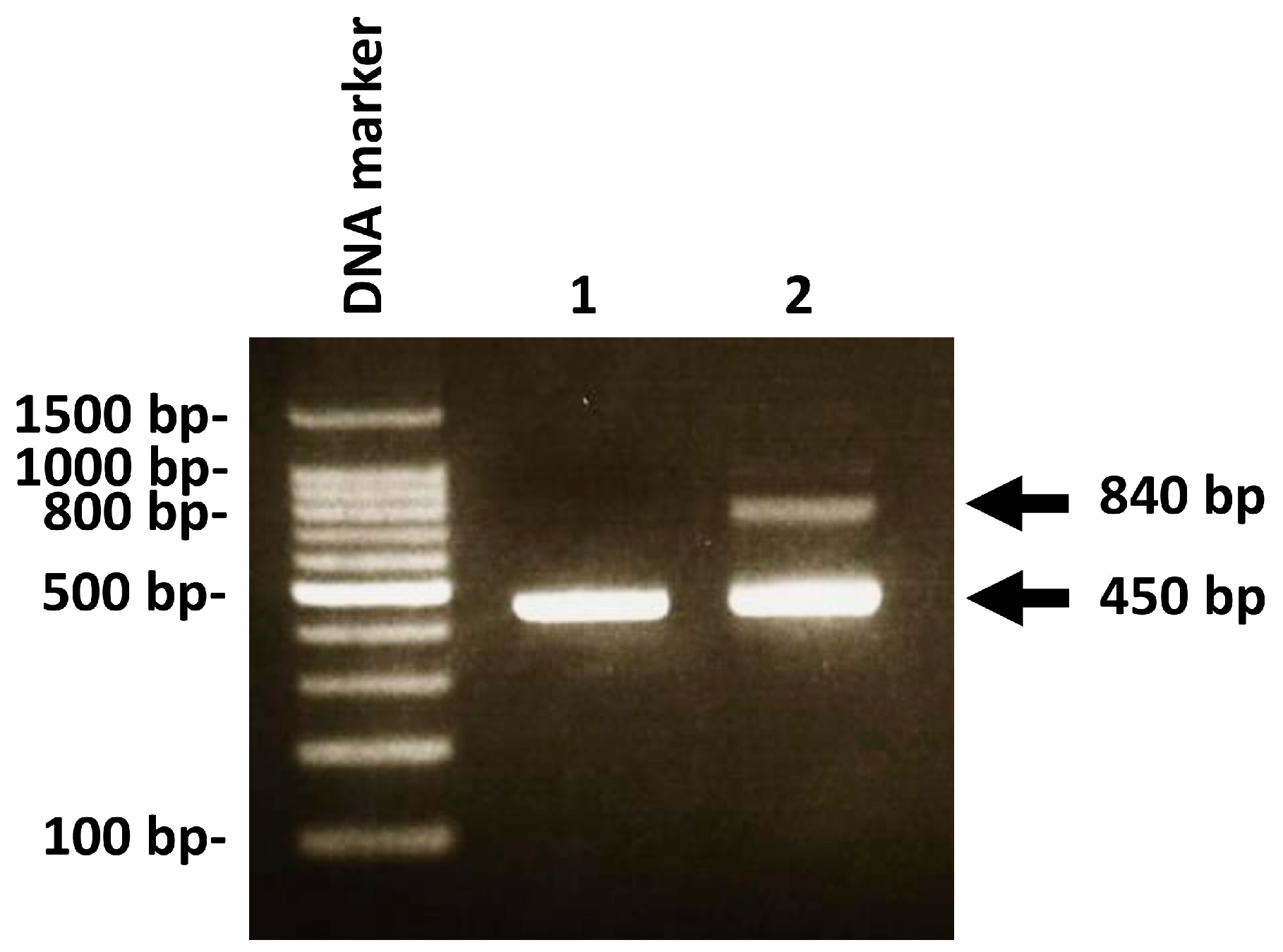
2.4. Detecting C. albicans DNA and P. gingivalis DNA by Real-Time PCR

Real-time polymerase chain reaction (PCR) analysis was carried out with a Thermal Cycler Dice® Real Time System III (Takara, Osaka, Japan). Real-time PCR was performed using a THUNDERBIRD SYBR qPCR Mix (Toyobo Life Science, Osaka, Japan) in accordance with the manufacturer’s instructions. The amplification procedure was as follows: initial denaturation at 95 °C for 2 min, then 40 cycles of 95 °C for 1 min, 60 °C for 1 min, and 72 °C for 1 min. The following PCR primer sets were employed in this study: C. albicans, 5′-TTTATCAACTTGTCACACCAGA-3′ (sense), and 5′-ATCCCGCCTTACCACTACCG-3′ (antisense) [17]; and P. gingivalis, 5′-AGGCAGCTTGCCATACTGCG-3′ (sense) and 5′-ACTGTTAGCAACTACCGATGT-3′ (antisense) [18]. As described in our recent study [19], standard curves for 10-fold serial dilutions of C. albicans DNA were used to determine C. albicans-positive samples. Ten-fold serial dilutions of a P. gingivalis DNA positive sample (94.0 ng/μL–0.0094 ng/μL) were used to generate a standard curve (Supplementary Figure S1). Real-time PCR experiments were performed in duplicate.

2.5. PCR Analysis for Genotype Identification of C. albicans

PCR analysis was carried out using an Eppendorf Mastercycler EP Gradient S Thermal Cycler (Eppendorf, Hamburg, Germany). A GoTaq® Green Master Mix (Promega, Madison, WI, USA) was used to amplify the PCR products in accordance with the manufacturer’s instructions. The amplification procedure was as follows: 95 °C for 2 min, followed by 40 cycles of 95 °C for 1 min, 60 °C for 1 min, and 72 °C for 1 min. PCR products were analyzed using 2% agarose gel electrophoresis with ethidium bromide and an ultraviolet transilluminator (FAS4; Nippon Genetics Co., Ltd., Tokyo, Japan). The Loading Quick 100 bp DNA Ladder (Toyobo, Osaka, Japan) was employed as a molecular size marker for gel electrophoresis. The following PCR primer sets were used: C. albicans-intron, 5′-ATAAGGGAAGTCGGCAAAATAGATCCGTAA-3′ (sense), and 5′-CCTTGGCTGTGGTTTCGCTAGATAGTAGAT-3′ (antisense) [12].

2.6. Statistical Analysis

SPSS software, version 24.0 (SPSS Inc., Chicago, IL, USA) was employed for statistical analysis. Fisher’s exact test or the χ2 test were employed to assess significant associations between clinical factors and C. albicans or C. albicans/P. gingivalis. The Mann–Whitney U test was employed to evaluate significant differences in age, remaining teeth, BOP (%), PISA value, and PESA value between C. albicans- or C. albicans/P. gingivalis-positive and negative cases. Logistic regression analysis was performed to adjust for the potential confounding effects in this study. Binomial logistic regression analysis with forced entry was performed to examine the relationship between independent variables and C. albicans/P. gingivalis as the dependent variable. Variables with a p value of less than 0.20 through univariate analysis were considered to be independent variables. Multicollinearity was evaluated using a variance inflation factor (VIF), and independent variables with a VIF of less than 2 were included in the logistic regression analysis. An adjusted odds ratio was obtained from logistic regression analysis. p Values of less than 0.05 were considered to be statistically significant.

3. Results

3.1. Relationship between Oral C. albicans and Clinical Parameters

Oral C. albicans positivity was determined in a total of 86 oral samples using real-time PCR. The C. albicans-positive rate was 26.1%. Table 1 summarizes the association between C. albicans and clinical factors. Participants in their 80s had a higher C. albicans-positive rate (35.3%) compared with other participants. There was no significant difference between the C. albicans positivity rate and clinical factors such as sex, age, systemic disease, or denture use. The association between oral C. albicans and oral health status was also investigated (Table 1). Participants with ≥6 mm periodontal pockets with BOP exhibited a higher C. albicans-positive rate (38.5%) compared with those without ≥6 mm periodontal pockets with BOP (23.3%). However, no significant association between C. albicans positivity and ≥6 mm periodontal pockets with BOP was found. C. albicans-positive participants recorded lower plaque control record scores compared with C. albicans-negative participants. No significant association was found between the C. albicans positivity rate and plaque control record scores.

3.2. Relationship between Intron Insertion of C. albicans and Clinical Parameters

The C. albicans genotype can be classified into the following three types: genotype A (single band of 450 bp), genotype B (single band of 840 bp), and genotype C (two bands of 450 and 840 bp) in accordance with a previously reported study [11]. However, it was impossible to classify C. albicans into these three genotypes because DNA extracted from oral rinse samples was used for PCR analysis in this study. Therefore, C. albicans-positive participants were classified into two groups according to the presence or absence of a PCR band of 840 bp (i.e., group I intron insertion) (Figure 1).
Of the 22 C. albicans-positive participants, 12 participants (54.5%) had no C. albicans with intron insertion (Table 2). In contrast, 10 participants (45.5%) had C. albicans with intron insertion. No C. albicans-positive participants showed a single band of 840 bp alone. The mean age of participants who had C. albicans with intron insertion was greater than that of those who had C. albicans without an intron insertion. Participants who had C. albicans with an intron insertion exhibited higher plaque control record scores compared with those who had C. albicans without an intron insertion. However, no significant association was found between C. albicans with an intron insertion and plaque control record scores. Importantly, 70% of participants who had C. albicans with intron insertion exhibited ≥6 mm probing depth. In addition, participants who had C. albicans with intron insertion recorded higher PISA values than those without C. albicans with intron insertion. However, there was no significant association between intron insertion of C. albicans and PISA value in C. albicans-positive participants.

3.3. Relationship between Oral C. albicans/P. gingivalis and Clinical Parameters

The positive rate of co-infection of C. albicans and P. gingivalis was 14%. Table 3 summarizes the association between co-infection of C. albicans and P. gingivalis and clinical factors. Participants in their 80s showed a higher rate of co-infection of C. albicans and P. gingivalis (23.5%) compared with other participants. However, there was no significant difference between co-infection of C. albicans and P. gingivalis and clinical factors such as sex, age, or medical history. Denture use was significantly associated with co-infection of C. albicans and P. gingivalis (p = 0.02). Participants with ≥6 mm periodontal pockets with BOP had a higher C. albicansP. gingivalis positive rate (38.5%) compared with those without ≥6 mm periodontal pockets with BOP (9.6%). There was a significant association between co-infection of C. albicans and P. gingivalis and ≥6 mm periodontal pockets with BOP (p = 0.02). C. albicans and P. gingivalis double-positive participants exhibited higher PISA values than non-C. albicans and P. gingivalis double-positive participants, but no significant difference was found. C. albicans and P. gingivalis double-positive participants recorded lower plaque control record scores compared with non-C. albicans and P. gingivalis double-positive participants. No significant association was found between the C. albicans and P. gingivalis double-positive rate and plaque control record scores.
Next, binomial logistic regression analysis was conducted using co-infection of C. albicans and P. gingivalis as the dependent variable, and variables showing a p value of <0.20 such as remaining teeth, denture use, probing depth, and ≥6 mm periodontal pockets with BOP as independent variables. The Hosmer–Lemeshow test revealed a good fit of the model (p = 0.29). A significant association was found between co-infection of C. albicans and P. gingivalis and ≥6 mm periodontal pockets with BOP (p = 0.02) (Table 4).

4. Discussion

Although no significant association between C. albicans DNA and periodontal health status was found in this study, double infection of C. albicans and P. gingivalis was significantly associated with deep periodontal pockets with bleeding. Our results suggest that co-infection of C. albicans and P. gingivalis rather than C. albicans infection alone contributes to active periodontitis. C. albicans may be involved in periodontitis in cooperation with periodontopathic bacteria. The viability and hemagglutination activity of P. gingivalis was enhanced in the P. gingivalis/C. albicans mixed biofilm under low heme conditions, suggesting that C. albicans can enhance the virulence of P. gingivalis under the conditions of insufficient heme [20]. The capacity of P. gingivalis to invade gingival fibroblasts and epithelial cells was enhanced by mannans derived from C. albicans [21]. Therefore, C. albicans is thought to play a supportive role in the progression of periodontitis in the presence of P. gingivalis. Additionally, C. albicans may be associated with periodontopathic bacteria other than P. gingivalis. However, the relationship between C. albicans and periodontopathic bacteria other than P. gingivalis has not been investigated in this study. Possible associations between periodontopathic bacteria and the herpes virus have also not been elucidated in this study. Further additional study is necessary to clarify the relationship between periodontopathic bacteria and C. albicans/herpes virus in the pathogenesis of periodontitis.
Among the three different genotypes of C. albicans, genotype B (i.e., the genotype with a group I intron) was found to be most common in the oral cavity in both healthy participants and those with candidiasis [13]. Tantivitayakul et al. reported that C. albicans with intron insertion exhibited greater phospholipase activity and adherence capacity to epithelial cells compared with C. albicans without an intron insertion; however, there was no significant association between genotype and virulence characteristics such as phospholipase activity or adherence capacity in C. albicans [13]. In this study, participants who had C. albicans with intron insertion exhibited a greater surface area of periodontal inflammation than those without C. albicans with intron insertion. These results suggest that C. albicans with intron insertion may exhibit higher pathogenic potential. The specific genotype of C. albicans has high virulence activity and may be involved in severe periodontitis. However, it remains unknown which genotype of C. albicans is importantly associated with the severity of periodontitis. Additionally, the mean age was higher in C. albicans-positive participants who had C. albicans with intron insertion than in those who had C. albicans without an intron insertion. Increased infection of C. albicans with intron insertion may be associated with a decline in the immune response because of aging. However, it was impossible to determine the genotype of each C. albicans in this study. Further additional study will be required to identify the genotype of C. albicans using an isolated single colony of C. albicans.
Segata et al. divided the oral microbiome into three categories according to the location in the oral cavity: category 1 (microbiome of buccal mucosa, gingiva, and hard palate), category 2 (microbiome of saliva and tongue), and category 3 (microbiome of gingival plaque) [22]. The bacterial composition differed in each of the three categories, and bacterial genera associated with periodontitis were detected in the microbiome of the saliva and tongue [22]. It is thought that the microbiome of the saliva and tongue is importantly associated with the microbiome in the other groups and reflects the entire spectrum of the microbiome in the oral cavity. In addition, periodontopathic bacterial DNA can be detected by PCR using saliva and swabs from the tongue surface [23,24]. Therefore, it is thought that the sampling method (i.e., sample collection from the tongue surface) had little impact on the detection of periodontopathic bacteria such as P. gingivalis in this study.
The relationship between periodontopathic bacteria and PISA values was previously investigated in participants after adjustment for their potential confounding factors using propensity scores [25]. Participants with co-infection of Tannerella forsythia and Treponema denticola exhibited significantly higher PISA values than those without such co-infection [25]. The results highlight the importance of co-infection with red complex bacteria in periodontal inflammation severity. In contrast, no significant association was found between P. gingivalis and PISA values in participants after adjustment for their potential confounding factors [25]. Thus, P. gingivalis may determine the severity of periodontitis in cooperation with other periodontopathic pathogens such as C. albicans.

5. Conclusions

Co-infection with C. albicans and P. gingivalis rather than C. albicans infection alone is involved in active periodontitis among middle-aged and older Japanese people.

Supplementary Materials

The following supporting information can be downloaded at https://www.mdpi.com/article/10.3390/medicina58060723/s1: Figure S1: Standard curve indicating a Porphyromonas gingivalis DNA positive sample vs. CT value.

Author Contributions

I.O.: investigation, data curation, formal analysis, and writing—review and editing; H.S.: conceptualization, methodology, investigation, formal analysis, writing—original draft, and writing—review and editing; K.O.: funding acquisition, supervision, and writing—review and editing. All authors have read and agreed to the published version of the manuscript.

Funding

This research was financially supported by Hiroshima University’s grant funding (funder: Hiroshima University, no. 0G220).

Institutional Review Board Statement

The study protocol was approved by the Ethical Committee of Hiroshima University (approval no. E-1115, date of approval: 1 March 2018).

Informed Consent Statement

All participants signed a consent agreement.

Data Availability Statement

All data generated or analyzed in this study are included in this article.

Conflicts of Interest

The authors have no conflict of interest to declare. The funders had no role in the design of the study, in the collection, analyses, or interpretation of data, in the writing of the manuscript, or in the decision to publish the results.

References

  1. Curtis, M.A.; Diaz, P.I.; Van Dyke, T.E. The role of the microbiota in periodontal disease. Periodontol. 2000 2020, 83, 14–25. [Google Scholar] [CrossRef] [PubMed]
  2. Socransky, S.; Haffajee, A.; Cugini, M.; Smith, C.; Kent, R.L. Microbial complexes in subgingival plaque. J. Clin. Periodontol. 1998, 25, 134–144. [Google Scholar] [CrossRef]
  3. Slots, J.; Slots, H. Periodontal herpesvirus morbidity and treatment. Periodontol. 2000 2019, 79, 210–220. [Google Scholar] [CrossRef] [PubMed]
  4. Su, C.Y.; Shigeishi, H.; Murodumi, H.; Sugiyama, M.; Ohta, K.; Takemoto, T. Association of oral Epstein-Barr virus with periodontal health in Japanese adults. Exp. Ther. Med. 2021, 22, 767. [Google Scholar] [CrossRef]
  5. Nakamura, M.; Shigeishi, H.; Cheng-Yih, S.U.; Sugiyama, M.; Ohta, K. Oral human cytomegalovirus prevalence and its relationships with periodontitis and Porphyromonas gingivalis in Japanese adults: A cross-sectional study. J. Appl. Oral Sci. 2020, 28, e20200501. [Google Scholar] [CrossRef] [PubMed]
  6. Satala, D.; Gonzalez-Gonzalez, M.; Smolarz, M.; Surowiec, M.; Kulig, K.; Wronowska, E.; Zawrotniak, M.; Kozik, A.; Rapala-Kozik, M.; Karkowska-Kuleta, J. The Role of Candida albicans Virulence Factors in the Formation of Multispecies Biofilms with Bacterial Periodontal Pathogens. Front. Cell. Infect. Microbiol. 2022, 11, 765942. [Google Scholar] [CrossRef]
  7. Urzúa, B.; Hermosilla, G.; Gamonal, J.; Morales-Bozo, I.; Canals, M.; Barahona, S.; Cóccola, C.; Cifuentes, V. Yeast diversity in the oral microbiota of subjects with periodontitis: Candida albicans and Candida dubliniensis colonize the periodontal pockets. Med. Mycol. 2008, 46, 783–793. [Google Scholar] [CrossRef] [PubMed]
  8. Slots, J. Periodontal herpesviruses: Prevalence, pathogenicity, systemic risk. Periodontol. 2000 2015, 69, 28–45. [Google Scholar] [CrossRef]
  9. Imai, K.; Inoue, H.; Tamura, M.; Cueno, M.E.; Inoue, H.; Takeichi, O. The periodontal pathogen Porphyromonas gingivalis induces the Epstein-Barr virus lytic switch transactivator ZEBRA by histone modification. Biochimie 2012, 94, 839–846. [Google Scholar] [CrossRef]
  10. Karkowska-Kuleta, J.; Bartnicka, D.; Zawrotniak, M.; Zielinska, G.; Kieronska, A.; Bochenska, O.; Ciaston, I.; Koziel, J.; Potempa, J.; Baster, Z.; et al. The activity of bacterial peptidylarginine deiminase is important during formation of dual-species biofilm by periodontal pathogen Porphyromonas gingivalis and opportunistic fungus Candida albicans. Pathog. Dis. 2018, 76, fty033. [Google Scholar] [CrossRef]
  11. McCullough, M.J.; Clemons, K.V.; Stevens, D.A. Molecular and phenotypic characterization of genotypic Candida albicans subgroups and comparison with Candida dubliniensis and Candida stellatoidea. J. Clin. Microbiol. 1999, 37, 417–421. [Google Scholar] [CrossRef] [PubMed] [Green Version]
  12. Tamura, M.; Watanabe, K.; Mikami, Y.; Yazawa, K.; Nishimura, K. Molecular characterization of new clinical isolates of Candida albicans and C. dubliniensis in Japan: Analysis reveals a new genotype of C. albicans with group I intron. J. Clin. Microbiol. 2001, 39, 4309–4315. [Google Scholar] [CrossRef] [PubMed] [Green Version]
  13. Tantivitayakul, P.; Panpradit, N.; Maudcheingka, T.; Klaophimai, A.; Lapirattanakul, J. Genotyping of Candida albicans and Candida dubliniensis by 25S rDNA analysis shows association with virulence attributes in oral candidiasis. Arch. Oral Biol. 2019, 97, 18–24. [Google Scholar] [CrossRef] [PubMed]
  14. Abraham, S.B.; Al Marzooq, F.; Himratul-Aznita, W.H.; Ahmed, H.M.A.; Samaranayake, L.P. Prevalence, virulence and antifungal activity of C. albicans isolated from infected root canals. BMC Oral Health 2020, 20, 347. [Google Scholar] [CrossRef] [PubMed]
  15. Nesse, W.; Abbas, F.; van der Ploeg, I.; Spijkervet, F.K.; Dijkstra, P.U.; Vissink, A. Periodontal inflamed surface area: Quantifying inflammatory burden. J. Clin. Periodontol. 2008, 35, 668–673. [Google Scholar] [CrossRef] [Green Version]
  16. Cutress, T.W.; Ainamo, J.; Sardo-Infirri, J. The community periodontal index of treatment needs (CPITN) procedure for population groups and individuals. Int. Dent. J. 1987, 37, 222–233. [Google Scholar]
  17. Hsu, M.C.; Chen, K.W.; Lo, H.J.; Chen, Y.C.; Liao, M.H.; Lin, Y.H.; Li, S.Y. Species identification of medically important fungi by use of real-time LightCycler PCR. J. Med. Microbiol. 2003, 52, 1071–1076. [Google Scholar] [CrossRef] [Green Version]
  18. Yasukawa, T.; Ohmori, M.; Sato, S. The relationship between physiologic halitosis and periodontopathic bacteria of the tongue and gingival sulcus. Odontology 2010, 98, 44–51. [Google Scholar] [CrossRef]
  19. Shigeishi, H.; Ohta, K.; Sugiyama, M. Prevalence and risk factors for oral HPV16/18 and candida in Japanese people without oral cancer or premalignant lesions. Oral Sci. Int. 2022; in press. [Google Scholar] [CrossRef]
  20. Guo, Y.; Wang, Y.; Wang, Y.; Jin, Y.; Wang, C. Heme Competition Triggers an Increase in the Pathogenic Potential of Porphyromonas gingivalis in Porphyromonas gingivalis-Candida albicans Mixed Biofilm. Front. Microbiol. 2020, 11, 596459. [Google Scholar] [CrossRef]
  21. Tamai, R.; Sugamata, M.; Kiyoura, Y. Candida albicans enhances invasion of human gingival epithelial cells and gingival fibroblasts by Porphyromonas gingivalis. Microb. Pathog. 2011, 51, 250–254. [Google Scholar] [CrossRef]
  22. Segata, N.; Haake, S.K.; Mannon, P.; Lemon, K.P.; Waldron, L.; Gevers, D.; Huttenhower, C.; Izard, J. Composition of the adult digestive tract bacterial microbiome based on seven mouth surfaces, tonsils, throat and stool samples. Genome Biol. 2012, 13, R42. [Google Scholar] [CrossRef] [PubMed] [Green Version]
  23. Könönen, E.; Paju, S.; Pussinen, P.J.; Hyvönen, M.; Di Tella, P.; Suominen-Taipale, L.; Knuuttila, M. Population-based study of salivary carriage of periodontal pathogens in adults. J. Clin. Microbiol. 2007, 45, 2446–2451. [Google Scholar] [CrossRef] [PubMed] [Green Version]
  24. Su, C.Y.; Shigeishi, H.; Nishimura, R.; Ohta, K.; Sugiyama, M. Detection of oral bacteria on the tongue dorsum using PCR amplification of 16S ribosomal RNA and its association with systemic disease in middle-aged and elderly patients. Biomed. Rep. 2019, 10, 70–76. [Google Scholar] [CrossRef] [PubMed] [Green Version]
  25. Shigeishi, H.; Nakamura, M.; Oka, I.; Su, C.Y.; Yano, K.; Ishikawa, M.; Kaneyasu, Y.; Sugiyama, M.; Ohta, K. The Associations of Periodontopathic Bacteria and Oral Candida with Periodontal Inflamed Surface Area in Older Adults Receiving Supportive Periodontal Therapy. Diagnostics 2021, 11, 1397. [Google Scholar] [CrossRef] [PubMed]
Figure 1. Polymerase chain reaction (PCR) detection of group I intron insertion in C. albicans positive cases. Lane 1: PCR products of 450 bp alone were found. Lane 2: PCR products of 450 bp and 840 bp were found. PCR products of 840 bp indicate the insertion of an intron into C. albicans DNA.
Medicina 58 00723 g001
Table 1. Relationship between oral C. albicans and clinical factors.
Clinical Factor (n)C. albicansp-Value
(−) (n = 64)(+) (n = 22)
Age69.7 ± 10.372.3 ± 9.40.43
Age in years
 40–49 (4) 4 (100%)0 (0%)0.64
 50–59 (8) 6 (75%)2 (25%)
 60–69 (25) 18 (72%)7 (28%)
 70–79 (32) 25 (78.1%)7 (21.9%)
 80–89 (17)11 (64.7%)6 (35.3%)
Gender
 Male (25) 15 (60%)10 (40%)0.06
 Female (61) 49 (80.3%)12 (19.7%)
Hypertension
 No (69)53 (76.8%)16 (23.2%)0.36
 Yes (17)11 (64.7%)6 (35.3%)
Diabetes
 No (76)56 (73.7%)20 (26.3%)1.0
 Yes (10)8 (80%)2 (20%)
Dyslipidemia
 No (72)54 (75%)18 (25%)0.75
 Yes (14)10 (71.4%)4 (28.6%)
Remaining teeth23.3 ± 5.821.9 ± 5.20.10
Denture use
 Non-user (55)44 (80%)11 (20%)0.13
 User (31)20 (64.5%)11 (35.5%)
Plaque Control Record scores (%)20.3 ± 15.916.4 ± 9.10.49
Probing depth
 <4 mm (18)13 (72.2%)5 (27.8%)0.13
 ≥4 mm and <6 mm (42)35 (83.3%)7 (16.7%)
 ≥6 mm (26)16 (61.5%)10 (38.5%)
Bleeding on probing (%)8.0 ± 8.98.4 ± 9.40.92
≥4 mm periodontal pocket with BOP
 No (48)34 (70.8%)14 (29.2%)0.46
 Yes (38)30 (78.9%)8 (21.1%)
≥6 mm periodontal pocket with BOP
 No (73)56 (76.7%)17 (23.3%)0.30
 Yes (13)8 (61.5%)5 (38.5%)
 PISA (mm2)93.6 ± 107.6105.1 ± 143.90.66
 PESA (mm2)1023.0 ± 333.41007.5 ± 305.90.96
Table 2. Relationship between intron insertion of C. albicans and clinical factors in C. albicans-positive participants.
Clinical Factor (n)C. albicans (+) (n = 22)p-Value
Intron Insertion (−) (n = 12)Intron Insertion (+) (n = 10)
Age69.4 ± 9.875.8 ± 7.80.16
Age in years
 50–59 (2) 2 (100%)0 (0%)0.27
 60–69 (7) 5 (71.4%)2 (28.6%)
 70–79 (7) 3 (42.9%)4 (57.1%)
 80–89 (6)2 (33.4%)4 (66.7%)
Gender
 Male (10) 5 (50%)5 (50%)1.0
 Female (12) 7 (58.3%)5 (41.7%)
Hypertension
 No (16)9 (56.3%)7 (43.8%)1.0
 Yes (6)3 (50%)3 (50%)
Diabetes
 No (20)11 (55%)9 (45%)1.0
 Yes (2)1 (50%)1 (50%)
Dyslipidemia
 No (18)10 (55.6%)8 (44.4%)1.0
 Yes (4)2 (50%)2 (50%)
Remaining teeth20.7 ± 6.523.4 ± 2.80.46
Denture use
 Non-user (11)5 (45.6%)6 (54.5%)0.67
 User (11)7 (63.6%)4 (36.4%)
Plaque Control Record scores (%)20.7 ± 6.523.4 ± 2.80.67
Probing depth
 <4 mm (5)3 (60%)2 (40%)0.07
 ≥4 mm and <6 mm (7)6 (85.7%)1 (14.3%)
 ≥6 mm (10)3 (30%)7 (70%)
Bleeding on probing (%)7.7 ± 10.19.2 ± 8.90.97
≥4 mm periodontal pocket with BOP
 No (14)8 (57.2%)6 (42.9%)1.0
 Yes (8)4 (50%)4 (50%)
≥6 mm periodontal pocket with BOP
 No (17)10 (58.8%)7 (41.2%)0.62
 Yes (5)2 (40%)3 (60%)
 PISA (mm2)80.1 ± 146.2135.0 ± 142.70.58
 PESA (mm2)940.1 ± 342.11088.3 ± 1019.00.50
Table 3. Relationship between oral C. albicans/P. gingivalis and clinical factors.
Clinical Factor (n)C. albicans/P. gingivalisp-Value
(−) (n = 74)(+) (n = 12)
Age69.9 ± 10.073.1 ± 10.00.35
Age in years
 40–49 (4) 4 (100%)0 (0%)0.63
 50–59 (8) 7 (87.5%)1 (12.5%)
 60–69 (25) 21 (84%)4 (16%)
 70–79 (32) 29 (90.6%)3 (9.4%)
 80–89 (17)13 (76.5%)4 (23.5%)
Gender
 Male (25) 20 (80%)5 (20%)0.32
 Female (61) 54 (88.5%)7 (11.5%)
Hypertension
 No (69)59 (85.5%)10 (14.5%)1.0
 Yes (17)15 (88.2%)2 (11.8%)
Diabetes
 No (76)65 (85.5%)11 (14.5%)1.0
 Yes (10)9 (90%)1 (10%)
Dyslipidemia
 No (72)60 (83.3%)12 (16.7%)0.20
 Yes (14)14 (100%)0 (0%)
Remaining teeth23.3 ± 5.621.15 ± 6.30.16
Denture use
 Non-user (55)51 (92.7%)4 (7.3%)0.02
 User (31)23 (74.2%)8 (25.8%)
Plaque Control Record scores (%)19.6 ± 15.017.0 ± 10.70.65
Probing depth
 <4 mm (18)14 (77.8%)4 (22.2%)0.06
 ≥4 mm and <6 mm (42)40 (95.2%)2 (4.8%)
 ≥6 mm (26)20 (76.9%)6 (23.1%)
Bleeding on probing (%)7.7 ± 8.610.3 ± 11.20.52
≥4 mm periodontal pocket with BOP
 No (48)42 (87.5%)6 (12.5%)0.76
 Yes (38)32 (84.2%)6 (15.8%)
≥6 mm periodontal pocket with BOP
 No (73)66 (90.4%)7 (9.6%)0.02
 Yes (13)8 (61.5%)5 (38.5%)
 PISA (mm2)89.3 ± 106.9141.5 ± 166.50.50
 PESA (mm2)1026.4 ± 318.2973.2 ± 375.70.89
p-Values < 0.05 were considered statistically significant.
Table 4. Logistic regression analysis with C. albicans/P. gingivalis as dependent variable.
Clinical VariablesAdjusted Odds Ratio95% Confidence Intervalp-Value
≥6 mm periodontal pocket with BOP17.131.60–183.840.02
p-Values < 0.05 were considered statistically significant.
Publisher’s Note: MDPI stays neutral with regard to jurisdictional claims in published maps and institutional affiliations.

