
Analysis of the Increase of Vascular Cell Adhesion Molecule-1 (VCAM-1) Expression and the Effect of Exposure in a Hyperbaric Chamber on VCAM-1 in Human Blood Serum: A Cross-Sectional Study

 , , , and
Abstract
:1. Introduction
- Does exposure in the hyperbaric chamber affect the VCAM-1 level in the divers group and non-divers group (a group who stayed in the same chamber for the same time period and breathed in the same pattern with the same breathing mix to best reflect the effect of the same pressure during exposure and decompression)?
- Is there a correlation between the blood cell counts (BCC) and VCAM-1, before and after exposure in the chamber, in the divers group?
- Exposure in a hyperbaric chamber has an effect on the serum VCAM-1 level in the divers group and the non-divers group.
- There is a correlation between tested BCC and VCAM-1 before and after exposure in the chamber in the divers group.
2. Materials and Methods
2.1. Design of the Study
2.2. Characteristics of Subject Population
2.3. Methods
2.4. Procedural and Ethical Considerations
2.5. Statistical Analysis
3. Results
- Relationships between the neutrophil count and VCAM-1—the higher the level of neutrophils was, the lower the value of VCAM-1 was (p = 0.0456, R = −0.51);
- A similar relationship existed between the leukocyte count and VCAM-1 (it was slightly weaker and only close to the level of statistical significance);
- VCAM-1 was higher in subjects with a higher lymphocyte ratio (R = 0.44)—this correlation was close to the level of statistical significance (p = 0.0848).
- Higher increases in VCAM-1 were associated with a greater increase (in some cases, a smaller decrease) in the mean corpuscular hemoglobin (MCH) and the mean corpuscular hemoglobin concentration (MCHC)—these were relationships of average strength and statistical significance;
- Quite a strong correlation occurred between the changes in the neutrophil counts and the changes in VCAM-1—the greater the increase in the number of neutrophils was, the smaller the increase in VCAM-1 was;
- In turn, the increase in the lymphocyte count after exposure to the conditions corresponding to a 60 m dive was correlated with the increase in VCAM-1 (R = 0.54; p = 0.0304);
- Similar correlations (i.e., positive) were found for VCAM-1 and the lymphocyte and monocyte ratio (although the latter relationship was no longer statistically significant—p = 0.1156).
4. Discussion
Strengths and Limitations of the Study
5. Conclusions
- The results confirm our hypothesis that exposure to a hyperbaric chamber has an effect on VCAM-1 in blood serum in the divers group.
- There is a correlation between the tested BCC and VCAM-1 before and after exposure in the chamber in the divers group.
- We believe that exposure in a hyperbaric chamber may result in the activation of the endothelium.
Author Contributions
Funding
Institutional Review Board Statement
Informed Consent Statement
Data Availability Statement
Conflicts of Interest
References
- Kong, D.H.; Kim, Y.K.; Kim, M.R.; Jang, J.H.; Lee, S. Emerging roles of Vascular Cell Adhesion Molecule-1 (VCAM-1) in immunological disorders and cancer. Int. J. Mol. Sci. 2018, 19, 1057. [Google Scholar] [CrossRef] [Green Version]
- Gunes, A.E.; Celik, H.; Yilmaz, O.; Erbas, C.; Gumus, E. Acute and chronic effects of pressure on dynamic Thiol-Disulphide homoeostasis in divers. Fresenius Environ. Bull. 2019, 28, 2949–2956. [Google Scholar]
- Łuczyński, D.; Lautridou, J.; Hjelde, A.; Monnoyer, R.; Eftedal, I. Hemoglobin during and following a 4-week commercial saturation dive to 200 m. Front. Physiol. 2019, 10, 1494. [Google Scholar] [CrossRef]
- Ridker, P.M. Intercellular adhesion molecule (ICAM-1) and the risk of developing atherosclerotic disease. Eur. Heart J. 1998, 19, 1119–1121. [Google Scholar]
- Morisaki, N.; Saito, I.; Tamura, K.; Tashiro, J.; Masuda, M.; Kanzaki, T.; Watanabe, S.; Masuda, Y.; Saito, Y. New indicates of ischemic heart disease and aging: Studies on the serum levels of soluble intercellular adhesion molecule-1 (ICAM-1) and soluble vascular cell adhesion molecul-1 (VCAM-1) in patients with hypercholesterolemia and ischemic heart disease. Atherosclerosis 1997, 131, 43–48. [Google Scholar] [CrossRef]
- Brubakk, A.O.; Duplancic, D.; Valic, Z.; Palada, I.; Obad, A.; Bakovic, D.; Wisloff, U.; Dujic, Z. A single air dive reduces arterial endothelial function in man. J. Physiol. 2005, 566 Pt 3, 901–906. [Google Scholar] [CrossRef] [Green Version]
- Lehoux, S.; Castier, Y.; Tedgui, A. Molecular mechanisms of the vascular responses to haemodynamic forces. J. Intern. Med. 2006, 259, 381–392. [Google Scholar] [CrossRef]
- Ungvari, Z.; Wolin, M.S.; Csiszar, A. Mechanosensitive production of reactive oxygen species in endothelial and smooth muscle cells: Role in microvascular remodeling? Antioxid. Redox Signal 2006, 8, 1121–1129. [Google Scholar] [CrossRef] [PubMed]
- Freyssinet, J.M. Cellular microparticles: What are they bad or good for? J. Thromb. Haemost. 2003, 1, 1655–1662. [Google Scholar] [CrossRef]
- Horstman, L.L.; Jy, W.; Jimenez, J.J.; Ahn, Y.S. Endothelial microparticles as markers of endothelial dysfunction. Front. Biosci. 2004, 9, 1118–1135. [Google Scholar] [CrossRef] [PubMed] [Green Version]
- Madden, L.A.; Laden, G. Endothelial microparticles in vascular disease and as a potential marker of decompression illness. EJUHM 2007, 8, 6–10. [Google Scholar]
- Vince, R.V.; McNaughton, L.R.; Taylor, L.; Midgley, A.W.; Laden, G.; Madden, L.A. Release of VCAM-1 associated endothelial microparticles following simulated SCUBA dives. Eur. J. Appl. Physiol. 2009, 105, 507–513. [Google Scholar] [CrossRef]
- Bao, X.C.; Shen, Q.; Fang, Y.Q.; Wu, J.G. Human Physiological Responses to a Single Deep Helium-Oxygen Diving. Front. Physiol. 2021, 12, 735986. [Google Scholar] [CrossRef] [PubMed]
- Zhang, K.; Wang, D.; Jiang, Z.; Ning, X.; Buzzacott, P.; Xu, W. Endothelial dysfunction correlates with decompression bubbles in rats. Sci. Rep. 2016, 6, 33390. [Google Scholar] [CrossRef] [Green Version]
- Glavas, D.; Markotic, A.; Valic, Z.; Kovacic, N.; Palada, I.; Martinic, R.; Breskovic, T.; Bakovic, D.; Brubakk, A.O.; Dujic, Z. Expression of endothelial selectin ligands on human leukocytes following dive. Exp. Biol. Med. 2008, 233, 1181–1188. [Google Scholar] [CrossRef] [PubMed]
- Thorsen, E.; Haave, H.; Hofso, D.; Ulvik, R.J. Exposure to hyperoxia in diving and hyperbaric medicine—Effects on blood cell counts and serum ferritin. Undersea Hyperb. Med. 2001, 28, 57–62. [Google Scholar]
- Smith, S.M.; Davis-Street, J.E.; Fesperman, J.V.; Smith, M.D.; Rice, B.L.; Zwart, S.R. Nutritional status changes in humans during a 14-day saturation dive: The NASA extreme environment mission operations V project. J. Nutr. 2004, 134, 1765–1771. [Google Scholar] [CrossRef] [PubMed] [Green Version]
- Deb, S.; Burgess, K.; Swinton, P.; Dolan, E. Physiological responses to prolonged saturation diving: A field-based pilot study. Undersea Hyperb. Med. 2017, 44, 581–587. [Google Scholar] [CrossRef]
- Kiboub, F.Z.; Balestra, C.; Loennechen, O.; Eftedal, I. Hemoglobin and erythropoietin after commercial saturation diving. Front. Physiol. 2018, 9, 1176. [Google Scholar] [CrossRef] [Green Version]
- Olszański, R.; Konarski, M.; Kierznikowicz, B. Changes of selected morphotic parameters and blood plasma proteins in blood of divers after a single short-time operational heliox exposure. Int. Marit. Health 2002, 53, 111–121. [Google Scholar]
- Perovic, A.; Nikolac, N.; Braticevic, M.N.; Milcic, A.; Sobocanec, S.; Balog, T.; Dabelic, S.; Dumic, J. Does recreational scuba diving have clinically significant effect on routine haematological parameters? Biochem. Med. 2017, 27, 325–331. [Google Scholar] [CrossRef] [PubMed]
- Obad, A.; Palada, I.; Valic, Z.; Ivancev, V.; Baković, D.; Wisløff, U.; Brubakk, A.O.; Dujić, Z. The effects of acute oral antioxidants on diving-induced alterations in human cardiovascular function. J. Physiol. 2007, 578 Pt 3, 859–870. [Google Scholar] [CrossRef]
- Sureda, A.; Batle, J.M.; Capo, X.; Martorell, M.; Córdova, A.; Tur, J.A.; Pons, A. Scuba diving induces nitric oxide synthesis and the expression of inflammatory and regulatory genes of the immune response in neutrophils. Physiol. Genom. 2014, 46, 647–654. [Google Scholar] [CrossRef] [PubMed]
- Yuan, M.L.; Tong, Z.H.; Jin, X.G.; Zhang, J.C.; Wang, X.J.; Ma, W.L.; Yin, W.; Zhou, Q.; Ye, H.; Shi, H.Z. Regulation of CD4+ T Cells by Pleural Mesothelial Cells via Adhesion Molecule-Dependent Mechanisms in Tuberculous Pleurisy. PLoS ONE 2013, 8, e74624. [Google Scholar]
- Miyamoto, Y.; Torii, T.; Tanoue, A.; Yamauchi, J. VCAM1 acts in parallel with CD69 and is required for the initiation of oligodendrocyte myelination. Nat. Commun. 2016, 7, 13478. [Google Scholar] [CrossRef] [Green Version]
- Brett, K.D.; Nugent, N.Z.; Fraser, N.K.; Bhopale, V.M.; Yang, M.; Thom, S.R. Microparticle and interleukin-1β production with human simulated compressed air diving. Sci. Rep. 2019, 9, 13320. [Google Scholar] [CrossRef] [Green Version]
- Leitner, J.; Grabmeier-Pfistershammer, K.; Steinberger, P. Receptors and ligands implicated in human T cell costimulatory processes. Immunol. Lett. 2010, 128, 89–97. [Google Scholar] [CrossRef]
- Nasreen, N.; Mohammed, K.A.; Ward, M.J.; Antony, V.B. Mycobacterium-induced transmesothelial migration of monocytes into pleural space: Role of intercellular adhesion molecule-1 in tuberculous pleurisy. J. Infect. Dis. 1999, 180, 1616–1623. [Google Scholar] [CrossRef] [PubMed]
- Choe, N.; Zhang, J.; Iwagaki, A.; Tanaka, S.; Hemenway, D.R.; Kagan, E. Asbestos exposure up regulates the adhesion of pleural leukocytes to pleural mesothelial cells via VCAM-1. Am. J. Physiol. 1999, 277, L292–L300. [Google Scholar]


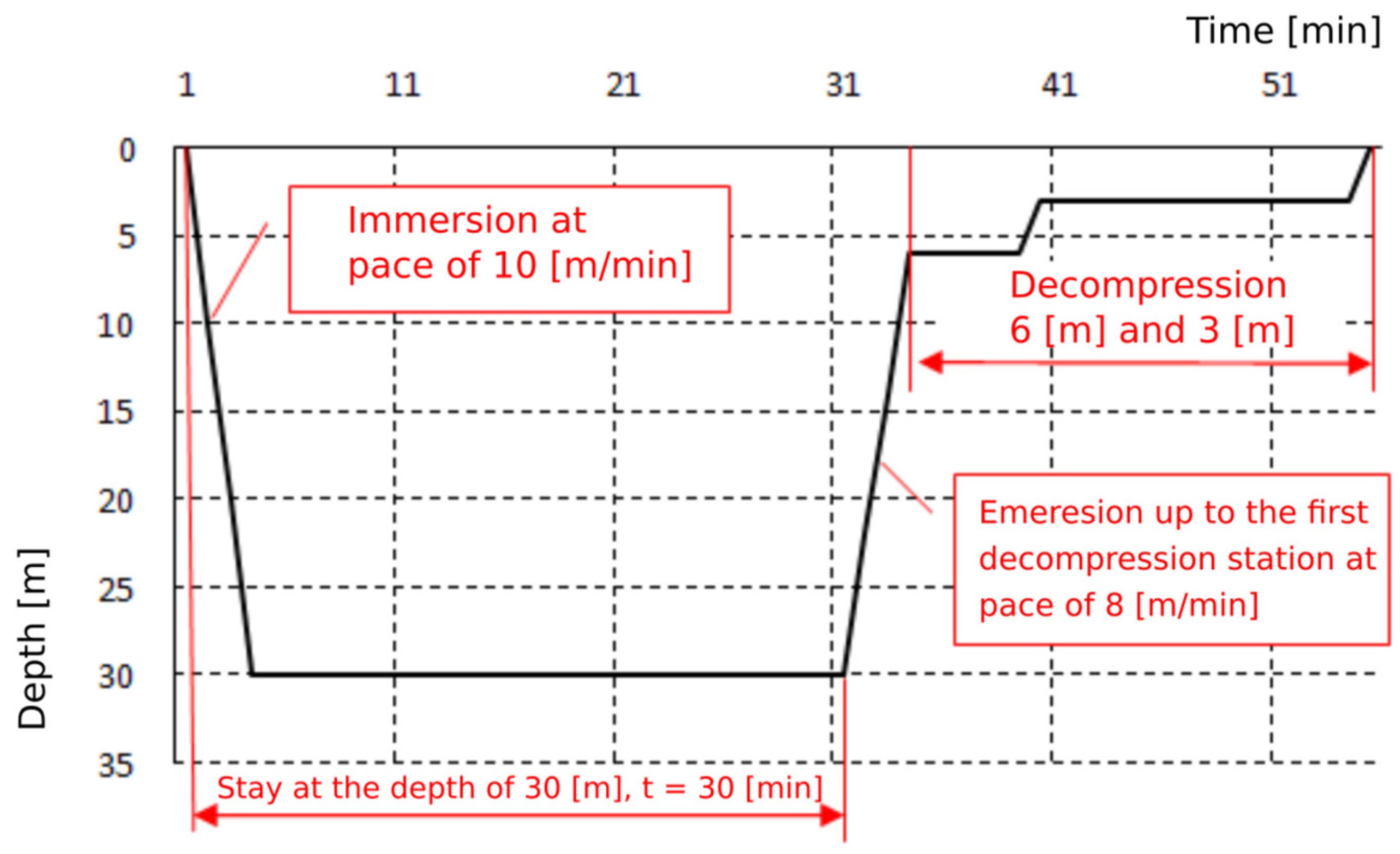
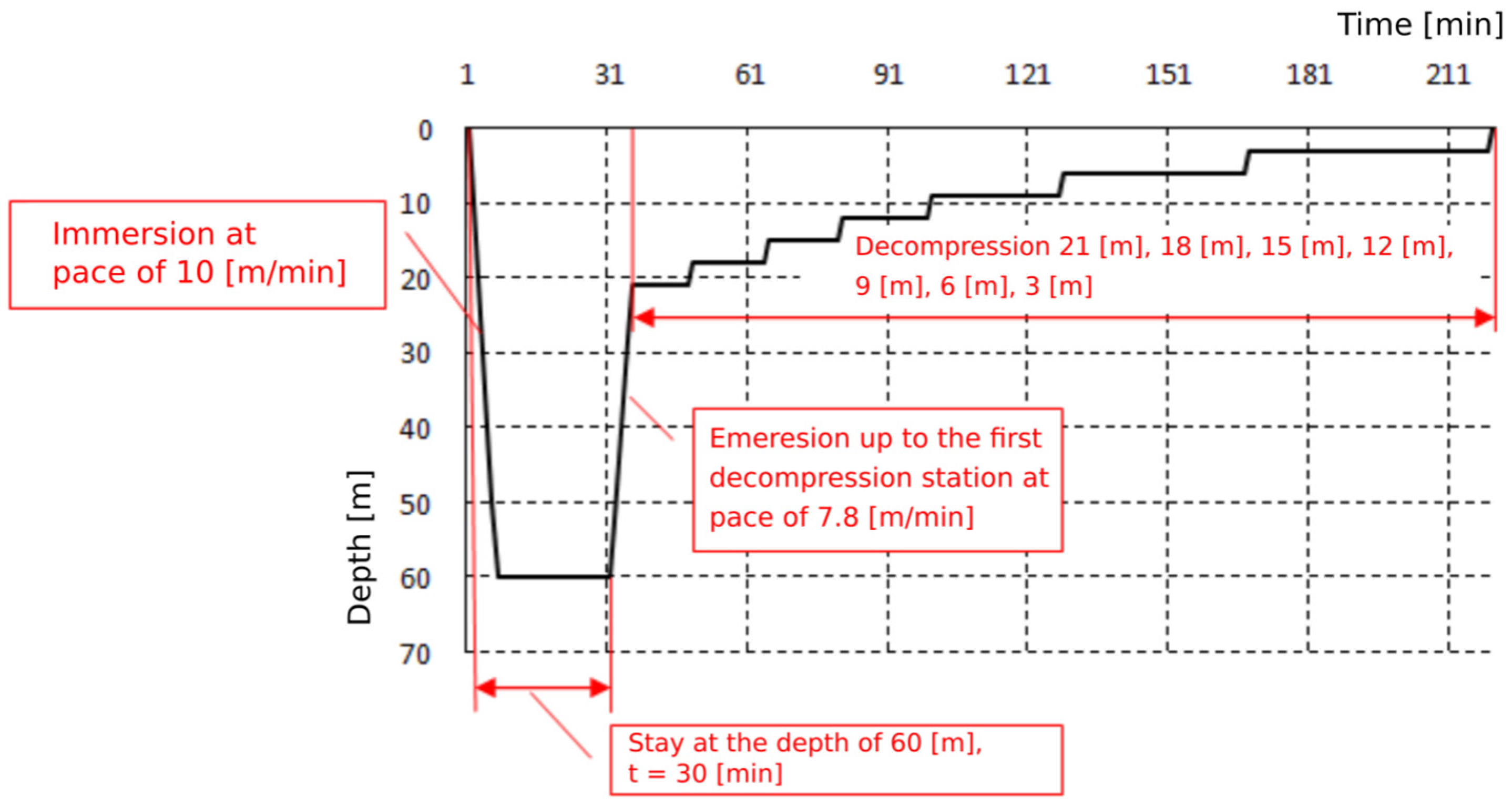
| Exposition | Depth | Bottom Time | Time to First Stop | Decompression Stops (mH2O) | Total Ascent Time | ||||||||||||||||
|---|---|---|---|---|---|---|---|---|---|---|---|---|---|---|---|---|---|---|---|---|---|
| 42 | 39 | 36 | 33 | 30 | 27 | 24 | 21 | 18 | 15 | 12 | 9 | 6 | 3 | ||||||||
| Time on the Stops (min) | Air | Oxygen | |||||||||||||||||||
| (mH2O) | (min) | (min) | Air | Air or (Oxygen) | (h) | (min) | (h) | (min) | |||||||||||||
| 30 m | 33 | 35 | 3 | 6(3) | 10(5) | 16(8) | 35 | 19 | |||||||||||||
| 60 m | 63 | 35 | 6 | 9 | 13 | 16 | 18(9) | 22(11) | 32(16) | 47(24) | 58(29) | 3 | 41 | 2 | 13 | ||||||
| Demographic and Occupational Status | Group | p | |||
|---|---|---|---|---|---|
| Divers (N = 18) | Non-Divers (N = 14) | ||||
| N | % | N | % | ||
| Sex | 0.0391 * | ||||
| female | 0 | 0.0% | 3 | 21.4% | |
| male | 18 | 100.0% | 11 | 78.6% | |
| Age (years) | 33.9 ± 6.6 | 33.0 ± 8.4 | 0.5521 | ||
| Place of residence | 0.0515 | ||||
| Rural | 5 | 29.4% | 0 | 0.0% | |
| municipality up to 50,000 inhabitants | 8 | 47.1% | 5 | 35.7% | |
| municipality 50,000–100,000 inhabitants | 1 | 5.9% | 1 | 7.1% | |
| municipality over 100,000 inhabitants | 3 | 17.6% | 8 | 57.1% | |
| Education | 0.1739 | ||||
| Secondary | 8 | 44.4% | 3 | 21.4% | |
| Higher | 10 | 55.6% | 11 | 78.6% | |
| Occupational seniority (years) | 10.4 ± 7.3 | 10.6 ± 7.8 | 0.9254 | ||
| Type of work | 0.0436 * | ||||
| physical | 15 | 83.3% | 7 | 50.0% | |
| mental | 3 | 16.7% | 7 | 50.0% | |
| Type of physical effort | 0.0043 ** | ||||
| Intensive | 3 | 17.0% | 2 | 14.3% | |
| Moderate | 4 | 22.0% | 2 | 14.3% | |
| Variable | 3 | 17.0% | 0 | 0.0% | |
| Aerobic | 0 | 0.0% | 8 | 57.1% | |
| None | 8 | 44.0% | 2 | 14.3% | |
| Lifestyle | Group | p | |||
|---|---|---|---|---|---|
| Divers (N = 18) | Non-Divers (N = 14) | ||||
| N | % | N | % | ||
| Drinking coffee | 0.3365 | ||||
| None | 6 | 33.3% | 4 | 28.6% | |
| Once a day | 4 | 22.2% | 3 | 21.4% | |
| Twice a day | 6 | 33.3% | 2 | 14.3% | |
| More | 2 | 11.1% | 5 | 35.7% | |
| Tobacco smoking | 0.7876 | ||||
| Yes | 2 | 11.1% | 2 | 14.3% | |
| No | 16 | 88.9% | 12 | 85.7% | |
| Self-assessment of physical condition | 0.0842 | ||||
| Ideal | 1 | 5.6% | 4 | 28.6% | |
| Good | 17 | 94.4% | 8 | 57.1% | |
| Intermediate | 0 | 0.0% | 1 | 7.1% | |
| Hard to tell | 0 | 0.0% | 1 | 7.1% | |
| VCAM-1 (ng/mL) | 95% CI |
|---|---|
| Before exposure—30 m | (10.7; 20.5) |
| After exposure—30 m | (12.3; 21.9) |
| Before exposure—60 m | (9.2; 16.4) |
| After exposure—60 m | (12.8; 24.6) |
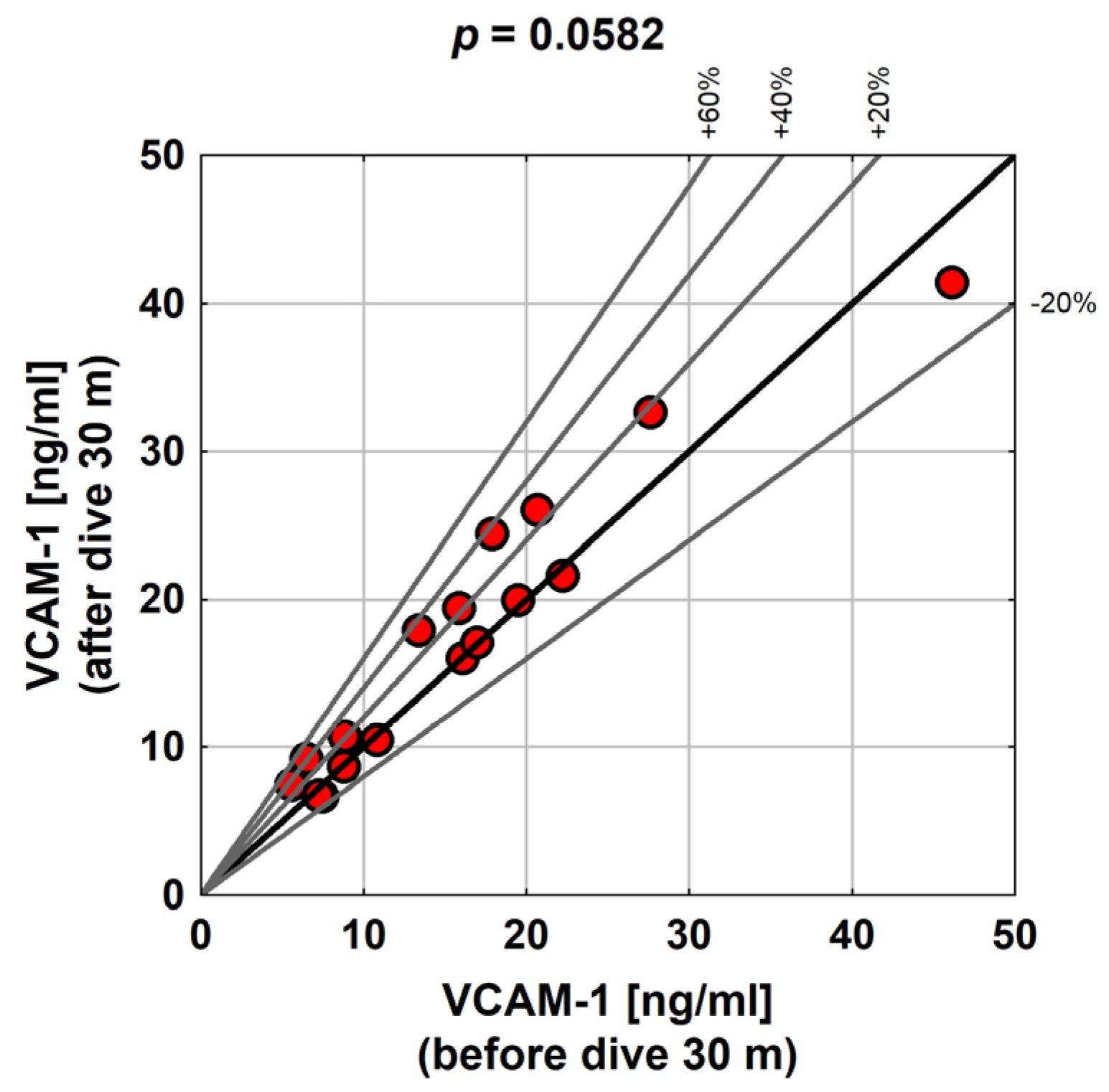
| VCAM-1 [ng/mL] 30 | M | Me | SD | Min. | Max. | |
| Before dive | 15.6 | 14.6 | 9.9 | 5.6 | 46.1 | |
| After dive | 17.1 | 16.6 | 9.6 | 6.7 | 41.5 | |
| Change (p = 0.0582) | 1.5 | 1.1 | 2.8 | −4.6 | 6.6 | |
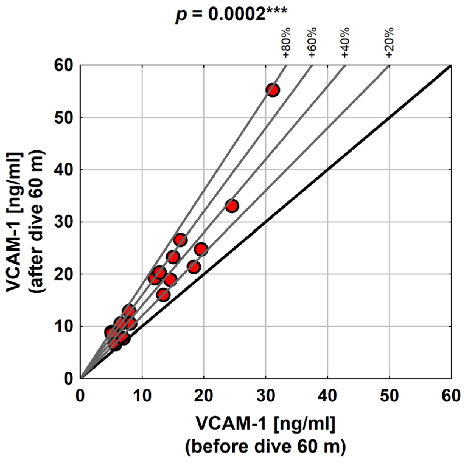
| VCAM-1 [ng/mL] 60 | Before dive | 12.8 | 12.4 | 7.2 | 5.0 | 31.1 |
| After dive | 18.7 | 17.5 | 11.8 | 6.7 | 55.3 | |
| Change (p = 0.0002 ***) | 5.9 | 4.2 | 5.3 | 0.8 | 24.2 |
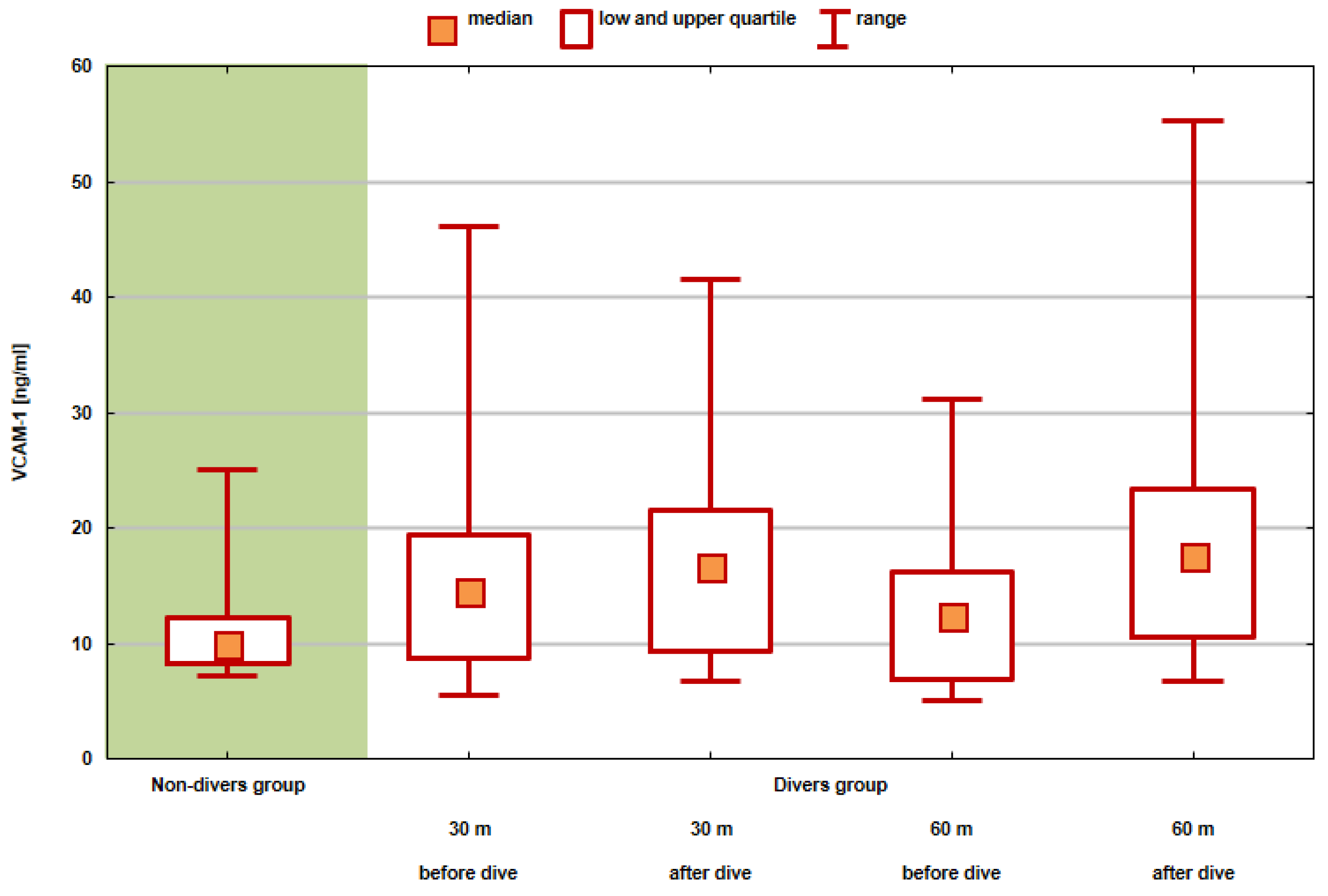
| VCAM-1 (ng/mL) | Group | p | |||||||||
|---|---|---|---|---|---|---|---|---|---|---|---|
| Divers (N = 18) | Non-Divers (N = 14) | ||||||||||
| Me | s | Min | Max | Me | s | Min | Max | ||||
| before exposure—30 m | 15.6 | 14.6 | 9.9 | 5.6 | 46.1 | 11.1 | 9.9 | 4.5 | 7.2 | 25.0 | 0.2666 |
| after exposure—30 m | 17.1 | 16.6 | 9.6 | 6.7 | 41.5 | 0.1251 | |||||
| before exposure—60 m | 12.8 | 12.4 | 7.2 | 5.0 | 31.1 | 0.8662 | |||||
| after exposure—60 m | 18.7 | 17.5 | 11.8 | 6.7 | 55.3 | 0.0494 * | |||||
| M | Me | SD | Min. | Max. | ||
|---|---|---|---|---|---|---|
| Leukocytes [G/L] | Before dive 30 m | 6.20 | 6.19 | 1.05 | 4.23 | 9.03 |
| After dive 30 m | 6.43 | 6.22 | 1.23 | 4.42 | 8.87 | |
| Change (p = 0.2485) | 0.23 | 0.18 | 0.74 | −0.76 | 1.56 | |
| Before dive 60 m | 5.95 | 6.01 | 0.96 | 3.70 | 7.56 | |
| After dive 60 m | 7.10 | 7.26 | 1.28 | 4.55 | 9.33 | |
| Change (p = 0.0004 ***) | 1.17 | 0.94 | 0.85 | 0.12 | 3.20 | |
| Hemoglobin [g/dL] | Before dive 30 m | 15.2 | 15.3 | 0.8 | 14.0 | 16.8 |
| After dive 30 m | 15.0 | 15.0 | 0.9 | 13.4 | 16.8 | |
| Change (p = 0.0098 **) | −0.2 | −0.2 | 0.3 | −1.0 | 0.4 | |
| Before dive 60 m | 15.2 | 15.2 | 0.8 | 13.8 | 16.4 | |
| After dive 60 m | 15.0 | 15.1 | 0.8 | 13.8 | 16.3 | |
| Change (p = 0.1673) | −0.1 | 0.0 | 0.3 | −0.9 | 0.3 | |
Publisher’s Note: MDPI stays neutral with regard to jurisdictional claims in published maps and institutional affiliations. |
© 2022 by the authors. Licensee MDPI, Basel, Switzerland. This article is an open access article distributed under the terms and conditions of the Creative Commons Attribution (CC BY) license (https://creativecommons.org/licenses/by/4.0/).
Share and Cite
Van Damme-Ostapowicz, K.; Cybulski, M.; Kozakiewicz, M.; Krajewska-Kułak, E.; Siermontowski, P.; Sobolewski, M.; Kaczerska, D. Analysis of the Increase of Vascular Cell Adhesion Molecule-1 (VCAM-1) Expression and the Effect of Exposure in a Hyperbaric Chamber on VCAM-1 in Human Blood Serum: A Cross-Sectional Study. Medicina 2022, 58, 95. https://doi.org/10.3390/medicina58010095
Van Damme-Ostapowicz K, Cybulski M, Kozakiewicz M, Krajewska-Kułak E, Siermontowski P, Sobolewski M, Kaczerska D. Analysis of the Increase of Vascular Cell Adhesion Molecule-1 (VCAM-1) Expression and the Effect of Exposure in a Hyperbaric Chamber on VCAM-1 in Human Blood Serum: A Cross-Sectional Study. Medicina. 2022; 58(1):95. https://doi.org/10.3390/medicina58010095
Chicago/Turabian StyleVan Damme-Ostapowicz, Katarzyna, Mateusz Cybulski, Mariusz Kozakiewicz, Elżbieta Krajewska-Kułak, Piotr Siermontowski, Marek Sobolewski, and Dorota Kaczerska. 2022. "Analysis of the Increase of Vascular Cell Adhesion Molecule-1 (VCAM-1) Expression and the Effect of Exposure in a Hyperbaric Chamber on VCAM-1 in Human Blood Serum: A Cross-Sectional Study" Medicina 58, no. 1: 95. https://doi.org/10.3390/medicina58010095
APA StyleVan Damme-Ostapowicz, K., Cybulski, M., Kozakiewicz, M., Krajewska-Kułak, E., Siermontowski, P., Sobolewski, M., & Kaczerska, D. (2022). Analysis of the Increase of Vascular Cell Adhesion Molecule-1 (VCAM-1) Expression and the Effect of Exposure in a Hyperbaric Chamber on VCAM-1 in Human Blood Serum: A Cross-Sectional Study. Medicina, 58(1), 95. https://doi.org/10.3390/medicina58010095