
Morphometry of the Entire Internal Carotid Artery on CT Angiography


Abstract
1. Introduction
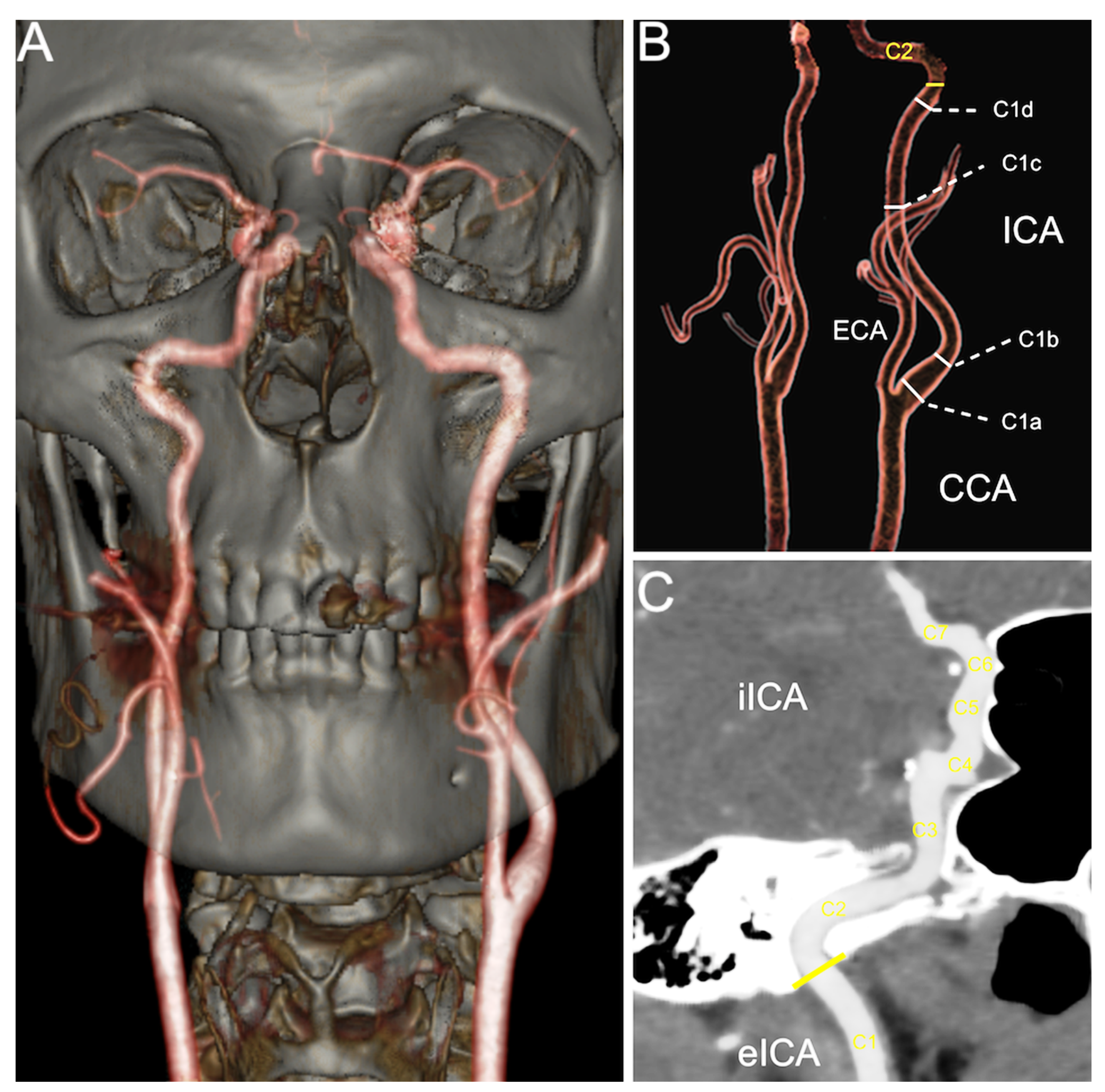
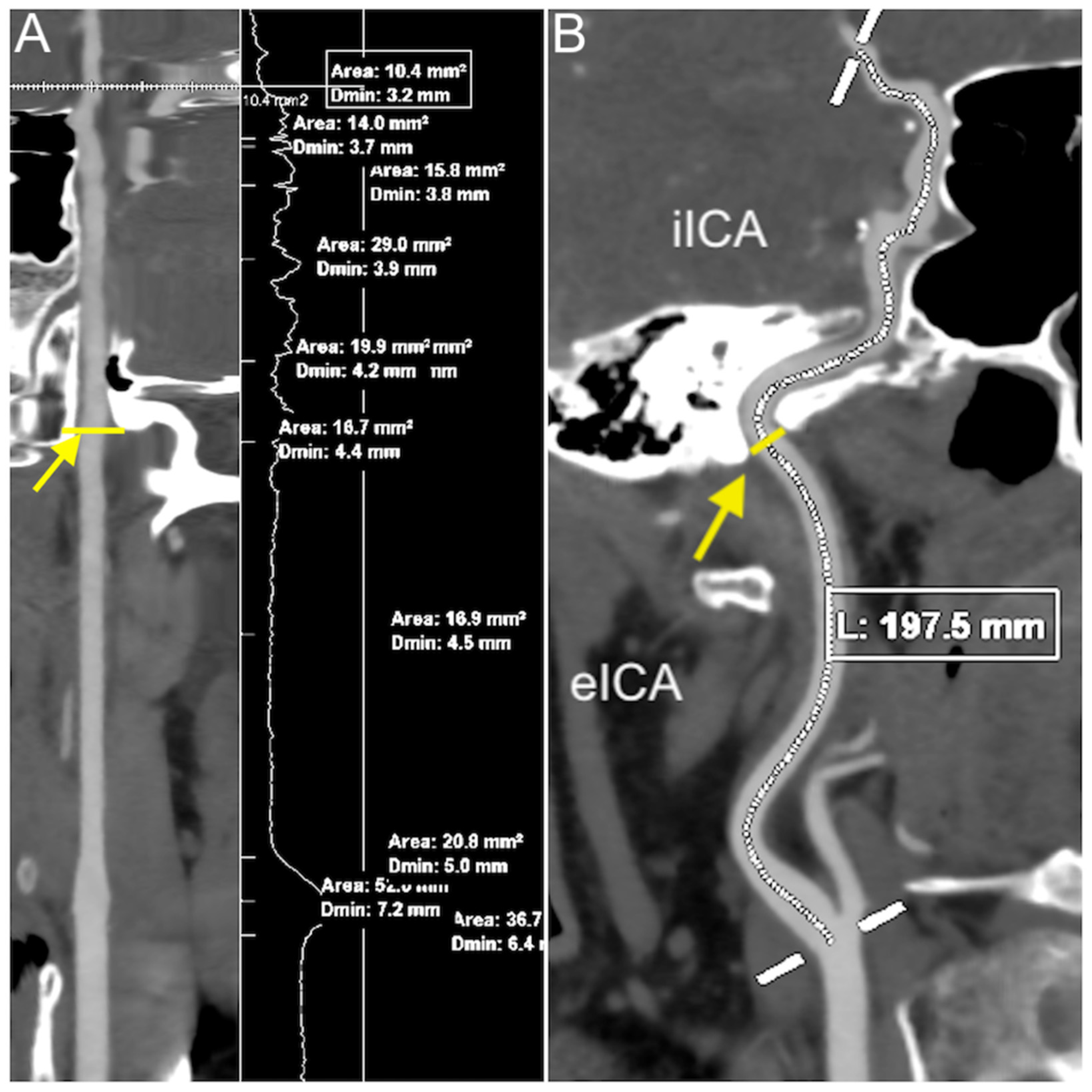
2. Materials and Methods
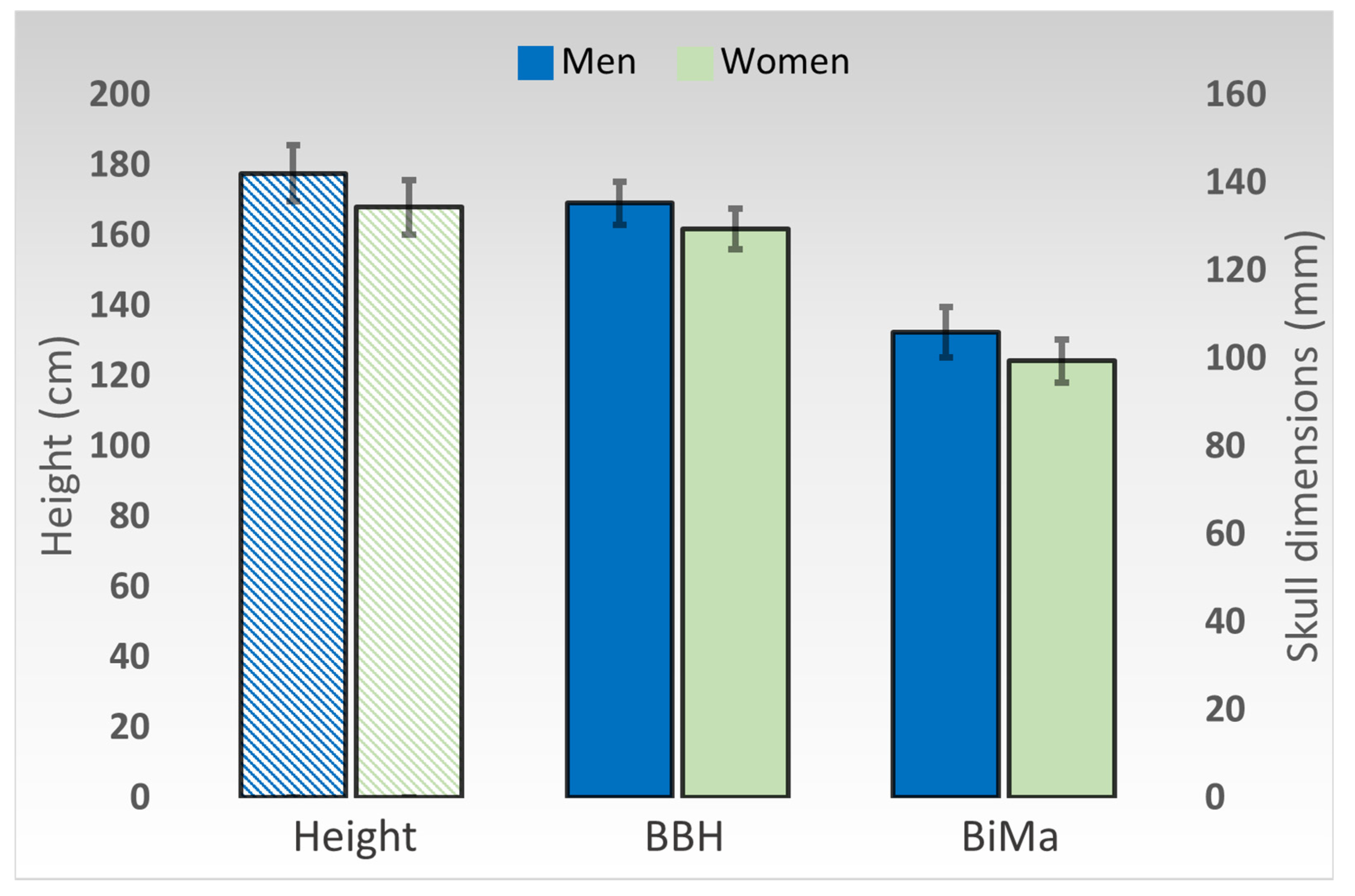
3. Results
4. Discussion
5. Conclusions
Author Contributions
Funding
Institutional Review Board Statement
Informed Consent Statement
Data Availability Statement
Conflicts of Interest
References
- Benjamin, E.J.; Blaha, M.J.; Chiuve, S.E.; Cushman, M.; Das, S.R.; Deo, R.; de Ferranti, S.D.; Floyd, J.; Fornage, M.; Gillespie, C.; et al. Heart Disease and Stroke Statistics-2017 Update: A Report From the American Heart Association. Circulation 2017, 135, e146–e603. [Google Scholar] [CrossRef] [PubMed]
- Virani, S.S.; Alonso, A.; Aparicio, H.J.; Benjamin, E.J.; Bittencourt, M.S.; Callaway, C.W.; Carson, A.P.; Chamberlain, A.M.; Cheng, S.; Delling, F.N.; et al. Heart Disease and Stroke Statistics—2021 Update. Circulation 2021, 143, e254–e743. [Google Scholar] [CrossRef] [PubMed]
- Gupta, A.; Gialdini, G.; Lerario, M.P.; Baradaran, H.; Giambrone, A.; Navi, B.B.; Marshall, R.S.; Iadecola, C.; Kamel, H. Magnetic resonance angiography detection of abnormal carotid artery plaque in patients with cryptogenic stroke. J. Am. Heart Assoc. 2015, 4, e002012. [Google Scholar] [CrossRef] [PubMed]
- Bartlett, E.S.; Walters, T.D.; Symons, S.P.; Fox, A.J. Quantification of carotid stenosis on CT angiography. Am. J. Neuroradiol. 2006, 27, 13–19. [Google Scholar]
- Pretterklieber, B.; Pretterklieber, M.L. A Phylogenetic and Ontogenetic Perspective of the Unique Accumulation of Arterial Variations in One Human Anatomic Specimen. Medicina 2020, 56, 449. [Google Scholar] [CrossRef]
- Cobiella, R.; Quinones, S.; Aragones, P.; León, X.; Abramovic, A.; Vazquez, T.; Ramón Sanudo, J.; Maranillo, E.; Olewnik, L.; Simon de Blas, C.; et al. Anatomic mapping of the collateral branches of the external carotid artery with regard to daily clinical practice. Ann. Anat. 2021, 238, 151789. [Google Scholar] [CrossRef]
- Bijari, P.B.; Wasserman, B.A.; Steinman, D.A. Carotid bifurcation geometry is an independent predictor of early wall thickening at the carotid bulb. Stroke 2014, 45, 473–478. [Google Scholar] [CrossRef]
- Wollschlaeger, P.B.; Wollschlaeger, G. Anterior cerebral-internal carotid artery and middle cerebral-internal carotid artery ratios. Acta Radiol. Diagn. 1966, 5, 615–620. [Google Scholar] [CrossRef]
- Spanos, K.; Petrocheilou, G.; Karathanos, C.; Labropoulos, N.; Mikhailidis, D.; Giannoukas, A. Carotid Bifurcation Geometry and Atherosclerosis. Angiology 2017, 68, 757–764. [Google Scholar] [CrossRef]
- Krejza, J.; Arkuszewski, M.; Kasner, S.E.; Weigele, J.; Ustymowicz, A.; Hurst, R.W.; Cucchiara, B.L.; Messe, S.R. Carotid artery diameter in men and women and the relation to body and neck size. Stroke 2006, 37, 1103–1105. [Google Scholar] [CrossRef]
- Limbu, Y.R.; Gurung, G.; Malla, R.; Rajbhandari, R.; Regmi, S.R. Assessment of carotid artery dimensions by ultrasound in non-smoker healthy adults of both sexes. Nepal Med. Coll. J. 2006, 8, 200–203. [Google Scholar]
- Koskinen, S.M.; Soinne, L.; Valanne, L.; Silvennoinen, H. The normal internal carotid artery: A computed tomography angiographic study. Neuroradiology 2014, 56, 723–729. [Google Scholar] [CrossRef]
- White, J.H.; Bartlett, E.S.; Bharatha, A.; Aviv, R.I.; Fox, A.J.; Thompson, A.L.; Bitar, R.; Symons, S.P. Reproducibility of semi-automated measurement of carotid stenosis on CTA. Can. J. Neurol. Sci. 2010, 37, 498–503. [Google Scholar] [CrossRef][Green Version]
- Lloyd, K.D.; Barinas-Mitchell, E.; Kuller, L.H.; Mackey, R.H.; Wong, E.A.; Sutton-Tyrrell, K. Common carotid artery diameter and cardiovascular risk factors in overweight or obese postmenopausal women. Int. J. Vasc. Med. 2012, 2012, 169323. [Google Scholar] [CrossRef] [PubMed]
- Kato, M.; Dote, K.; Habara, S.; Takemoto, H.; Goto, K.; Nakaoka, K. Clinical implications of carotid artery remodeling in acute coronary syndrome: Ultrasonographic assessment of positive remodeling. J. Am. Coll. Cardiol. 2003, 42, 1026–1032. [Google Scholar] [CrossRef]
- Sasaki, R.; Yamano, S.; Yamamoto, Y.; Minami, S.; Yamamoto, J.; Nakashima, T.; Takaoka, M.; Hashimoto, T. Vascular remodeling of the carotid artery in patients with untreated essential hypertension increases with age. Hypertens. Res. 2002, 25, 373–379. [Google Scholar] [CrossRef] [PubMed]
- Deng, Y.; Wang, X.M.; Wu, L.B.; Sun, C.; Duan, Y.H.; Cheng, Z.P.; Wu, D.W. Significance of the preoperative guidance of dual-source CT in carotid body tumor. Chin. Med. J. 2010, 123, 2816–2819. [Google Scholar]
- Bouthillier, A.; van Loveren, H.R.; Keller, J.T. Segments of the internal carotid artery: A new classification. Neurosurgery 1996, 38, 425–433. [Google Scholar] [CrossRef]
- Martin, R.; Saller, K. Lehrbuch der Anthropologie II; Gustav Fischer: Stuttgart, Germany, 1957. [Google Scholar]
- Ring, B.A.; Waddington, M.M. Intraluminal diameters of the intracranial arteries. Vasc. Surg. 1967, 1, 137–151. [Google Scholar] [CrossRef]
- Gabrielsen, T.O.; Greitz, T. Normal size of the internal carotid, middle cerebral and anterior cerebral arteries. Acta Radiol. Diagn. 1970, 10, 1–10. [Google Scholar] [CrossRef]
- Markert, M.S.; Della-Morte, D.; Cabral, D.; Roberts, E.L., Jr.; Gardener, H.; Dong, C.; Wright, C.B.; Elkind, M.S.; Sacco, R.L.; Rundek, T. Ethnic differences in carotid artery diameter and stiffness: The Northern Manhattan Study. Atherosclerosis 2011, 219, 827–832. [Google Scholar] [CrossRef]
- Zhang, Y.; Zhang, X.; Chang, R.; Cang, P.; Liu, X.; Xia, Q. Diameter measurements of cerebral arteries on three-dimensional time-of-flight MR angiograms. Chin. J. Radiol. 2003, 37, 394–398. [Google Scholar]
- Williams, M.A.; Nicolaides, A.N. Predicting the normal dimensions of the internal and external carotid arteries from the diameter of the common carotid. Eur. J. Vasc. Surg. 1987, 1, 91–96. [Google Scholar] [CrossRef]
- Choudhry, F.A.; Grantham, J.T.; Rai, A.T.; Hogg, J.P. Vascular geometry of the extracranial carotid arteries: An analysis of length, diameter, and tortuosity. J. Neurointerv. Surg. 2016, 8, 536–540. [Google Scholar] [CrossRef] [PubMed]
- Mujagić, S. The inner diameter of arteries of the Circle of Willis regarding gender and age on Magnetic Resonance Angiography. Acta Med. Salin. 2013, 42, 6–12. [Google Scholar]
- Takegoshi, H.; Kikuchi, S. An anatomic study of the horizontal petrous internal carotid artery: Sex and age differences. Auris Nasus Larynx 2007, 34, 297–301. [Google Scholar] [CrossRef] [PubMed]
- Vijaywargiya, M.; Deopujari, R.; Athavale, S.A. Anatomical study of petrous and cavernous parts of internal carotid artery. Anat. Cell Biol. 2017, 50, 163–170. [Google Scholar] [CrossRef] [PubMed]
- Arat, Y.O.; Arat, A.; Aydin, K. Angiographic Morphometry of Internal Carotid Artery Circulation in Turkish Children. Turk. Neurosurg. 2015, 25, 608–616. [Google Scholar] [CrossRef]
- McNamara, J.R.; Fulton, G.J.; Manning, B.J. Three-dimensional computed tomographic reconstruction of the carotid artery: Identifying high bifurcation. Eur. J. Vasc. Endovasc. Surg. 2015, 49, 147–153. [Google Scholar] [CrossRef]
- Toda, T.; Tsuda, N.; Nishimori, I.; Leszczynski, D.E.; Kummerow, F.A. Morphometrical analysis of the aging process in human arteries and aorta. Acta Anat. 1980, 106, 35–44. [Google Scholar] [CrossRef]
- Kamenskiy, A.V.; Pipinos, I.I.; Carson, J.S.; MacTaggart, J.N.; Baxter, B.T. Age and disease-related geometric and structural remodeling of the carotid artery. J. Vasc. Surg. 2015, 62, 1521–1528. [Google Scholar] [CrossRef]
- Sterpetti, A.V. Eversion endarterectomy of the internal carotid artery combined with open endarterectomy of the common carotid artery. Am. J. Surg. 2010, 200, e44–e47. [Google Scholar] [CrossRef] [PubMed]
- Darling, R.C., 3rd; Mehta, M.; Roddy, S.P.; Paty, P.S.; Kreienberg, P.B.; Ozsvath, K.J.; Chang, B.B.; Shah, D.M. Eversion carotid endarterectomy: A technical alternative that may obviate patch closure in women. Cardiovasc. Surg. 2003, 11, 347–352. [Google Scholar] [CrossRef]
- Morr, S.; Lin, N.; Siddiqui, A.H. Carotid artery stenting: Current and emerging options. Med. Devices 2014, 7, 343–355. [Google Scholar] [CrossRef]


| Segment | Right Side | Left Side | p-Value | Male | Female | p-Value |
|---|---|---|---|---|---|---|
| Carotid bulb (C1a) | 7.56 (±0.97) | 7.61 (±1.01) | 0.6092 | 7.94 (±1.05) | 7.27 (±0.82) | 0.0001 |
| Post-bulbar section (C1b) | 5.49 (±0.64) | 5.52 (±0.62) | 0.6183 | 5.74 (±0.67) | 5.31 (±0.50) | 0.0001 |
| Midpoint of C1 (C1c) | 4.84 (±0.54) | 4.85 (±0.53) | 0.7750 | 5.01 (±0.56) | 4.70 (±0.46) | 0.0006 |
| Endpoint of C1 (C1d) | 4.65 (±0.46) | 4.69 (±0.47) | 0.4373 | 4.81 (±0.50) | 4.55 (±0.40) | 0.0013 |
| C2 | 4.53 (±0.43) | 4.53 (±0.50) | 0.8731 | 4.66 (±0.50) | 4.42 (±0.41) | 0.0026 |
| C3 | 4.32 (±0.41) | 4.34 (±0.49) | 0.7105 | 4.48 (±0.48) | 4.20 (±0.38) | 0.0002 |
| C4 | 4.25 (±0.41) | 4.28 (±0.48) | 0.5618 | 4.43 (±0.46) | 4.13 (±0.38) | 0.0001 |
| C5 | 4.13 (±0.46) | 4.17 (±0.50) | 0.3886 | 4.34 (±0.46) | 3.98 (±0.43) | <0.0001 |
| C6 | 2.89 (±0.38) | 2.88 (±0.41) | 0.4700 | 2.91 (±0.41) | 2.86 (±0.38) | 0.5307 |
| C7 | 2.72 (±0.35) | 2.70 (±0.38) | 0.2770 | 2.73 (±0.37) | 2.70 (±0.37) | 0.6190 |
| Extracranial length | 86.33 (±18.76) | 85.87 (±18.53) | 0.5540 | 90.21 (±20.66) | 82.43 (±15.76) | 0.0151 |
| Intracranial length | 69.40 (±8.95) | 69.33 (±8.42) | 0.8370 | 71.29 (±7.97) | 67.64 (±8.93) | 0.0122 |
| Total ICA length | 154.89 (±26.17) | 155.20 (±23.57) | 0.7895 | 160.62 (±28.56) | 150.07 (±19.83) | 0.0141 |
| Segment | Measurement 1 | Height | BBH 2 | BiMa 3 |
|---|---|---|---|---|
| Carotid bulb (C1a) | 7.59 (±0.99) | p = 0.0005 | p = 0.0080 | p = 0.0226 |
| Post-bulbar section (C1b) | 5.51 (±0.63) | p = 0.0356 | p = 0.4014 | p = 0.5521 |
| Midpoint of C1 (C1c) | 4.85 (±0.53) | p = 0.0365 | p = 0.0730 | p = 0.1512 |
| Endpoint of C1 (C1d) | 4.67 (±0.47) | p = 0.0224 | p = 0.1031 | p = 0.1539 |
| C2 | 4.53 (±0.47) | p = 0.0598 | p = 0.3029 | p = 0.2993 |
| C3 | 4.33 (±0.45) | p = 0.0150 | p = 0.1846 | p = 0.1744 |
| C4 | 4.27 (±0.45) | p = 0.0075 | p = 0.0889 | p = 0.0984 |
| C5 | 4.15 (±0.48) | p = 0.0027 | p = 0.0166 | p = 0.0506 |
| C6 | 2.88 (±0.40) | p = 0.4606 | p = 0.1686 | p = 0.2540 |
| C7 | 2.71 (±0.37) | p = 0.3660 | p = 0.3329 | p = 0.6489 |
| Extracranial length | 86.10 (±18.65) | p < 0.0001 | p = 0.0052 | p = 0.0197 |
| Intracranial length | 69.36 (±8.69) | p < 0.0001 | p < 0.0001 | p < 0.0001 |
| Total ICA length | 155.04 (±24.90) | p < 0.0001 | p < 0.0001 | p = 0.0004 |
Publisher’s Note: MDPI stays neutral with regard to jurisdictional claims in published maps and institutional affiliations. |
© 2021 by the authors. Licensee MDPI, Basel, Switzerland. This article is an open access article distributed under the terms and conditions of the Creative Commons Attribution (CC BY) license (https://creativecommons.org/licenses/by/4.0/).
Share and Cite
Baz, R.A.; Scheau, C.; Niscoveanu, C.; Bordei, P. Morphometry of the Entire Internal Carotid Artery on CT Angiography. Medicina 2021, 57, 832. https://doi.org/10.3390/medicina57080832
Baz RA, Scheau C, Niscoveanu C, Bordei P. Morphometry of the Entire Internal Carotid Artery on CT Angiography. Medicina. 2021; 57(8):832. https://doi.org/10.3390/medicina57080832
Chicago/Turabian StyleBaz, Radu Andrei, Cristian Scheau, Cosmin Niscoveanu, and Petru Bordei. 2021. "Morphometry of the Entire Internal Carotid Artery on CT Angiography" Medicina 57, no. 8: 832. https://doi.org/10.3390/medicina57080832
APA StyleBaz, R. A., Scheau, C., Niscoveanu, C., & Bordei, P. (2021). Morphometry of the Entire Internal Carotid Artery on CT Angiography. Medicina, 57(8), 832. https://doi.org/10.3390/medicina57080832