
Cardiac Amyloidosis with Discordant QRS Voltage between Frontal and Precordial Leads


Abstract
:1. Introduction
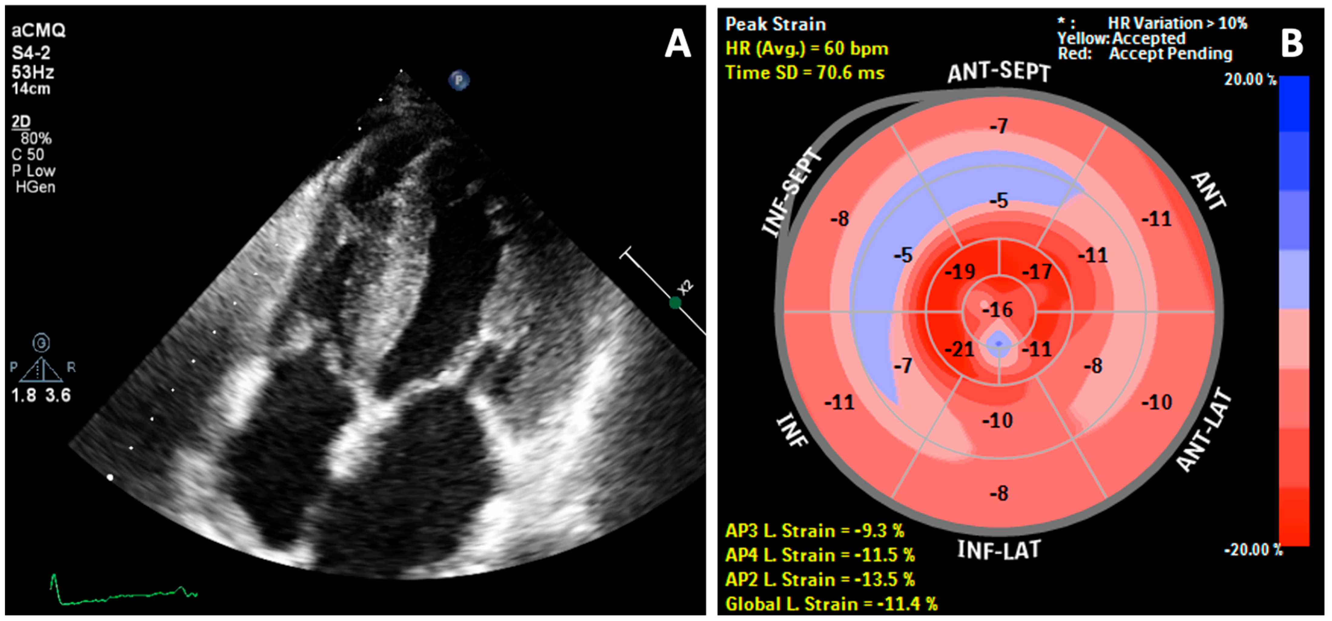
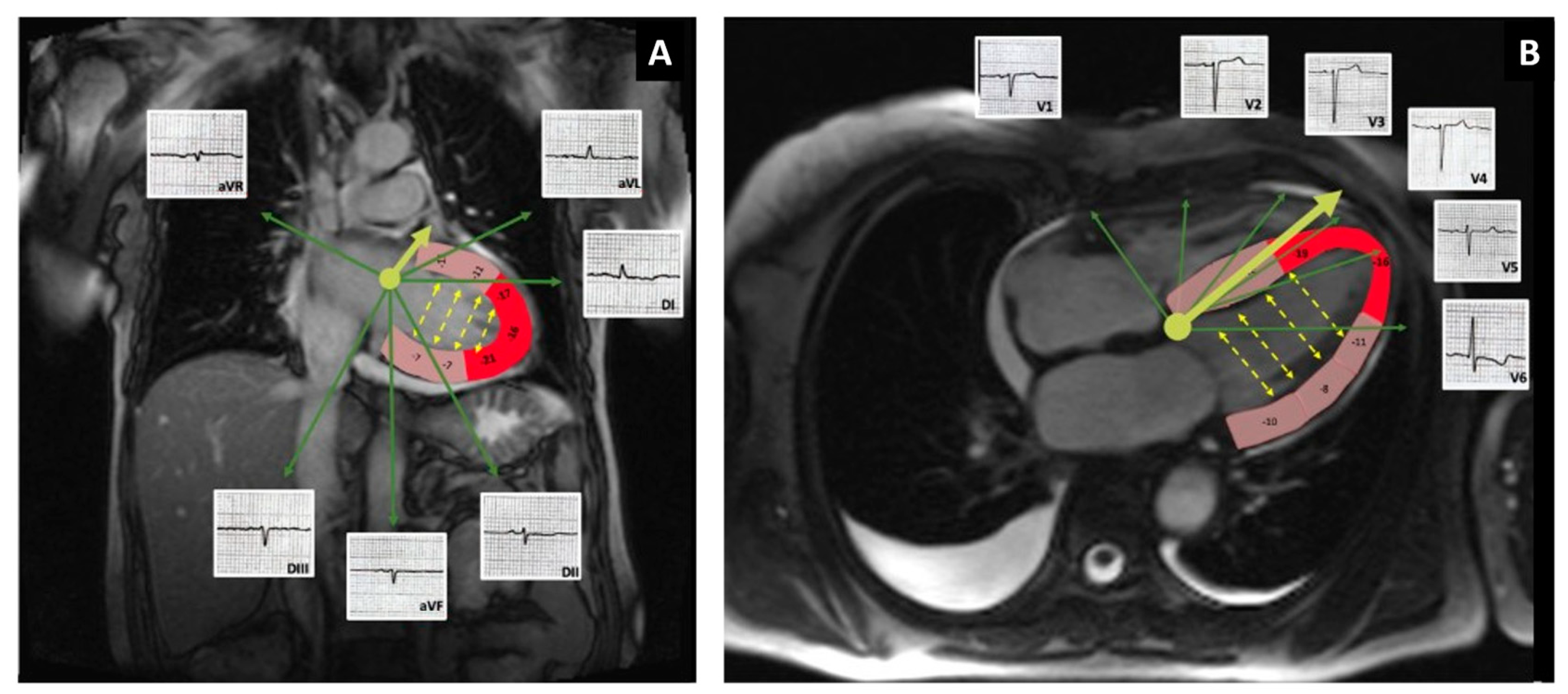
2. Case Report
3. Discussion
3.1. QRS Voltage
3.2. Atrial Fibrillation
3.3. Other Factors Related to Disease Progression in CA
4. Conclusions
Author Contributions
Funding
Institutional Review Board Statement
Informed Consent Statement
Data Availability Statement
Conflicts of Interest
References
- Ablasser, K.; Verheyen, N.; Glantschnig, T.; Agnetti, G.; Rainer, P.P. Unfolding cardiac amyloidosis—From pathophysiology to cure. Curr. Med. Chem. 2019, 26, 2865–2878. [Google Scholar] [CrossRef]
- Martinez-Naharro, A.; Hawkins, P.N.; Fontana, M. Cardiac amyloidosis. Clin. Med. (Lond.) 2018, 18, s30–s35. [Google Scholar] [CrossRef]
- Hirata, Y.; Kusunose, K.; Miki, H.; Yamada, H. Improvement of global longitudinal strain following high-dose chemotherapy and autologous peripheral blood stem cell transplantation in patients with amyloid light-chain cardiac amyloidosis: A case report. Eur. Heart J. Case Rep. 2019, 3, 1–6. [Google Scholar] [CrossRef] [PubMed]
- Kristen, A.V. Amyloid cardiomyopathy. Herz 2020, 45, 267–271. [Google Scholar] [CrossRef]
- Maurea, N.; Riva, L. Venous thromboembolism and atrial fibrillation in patients with cancer. G Ital. Cardiol. (Rome) 2018, 19, 3S–6S. [Google Scholar] [CrossRef]
- Falk, R.H.; Alexander, K.M.; Liao, R.; Dorbala, S. AL (Light-Chain) Cardiac Amyloidosis: A Review of Diagnosis and Therapy. J. Am. Coll. Cardiol. 2016, 68, 1323–1341. [Google Scholar] [CrossRef] [PubMed]
- Dorbala, S.; Cuddy, S.; Falk, R.H. How to Image Cardiac Amyloidosis: A Practical Approach. JACC Cardiovasc. Imaging 2020, 13, 1368–1383. [Google Scholar] [CrossRef] [PubMed]
- Tahir, U.A.; Doros, G.; Kim, J.S.; Connors, L.H.; Seldin, D.C.; Sam, F. Predictors of Mortality in Light Chain Cardiac Amyloidosis with Heart Failure. Sci. Rep. 2019, 9, 8552. [Google Scholar] [CrossRef] [PubMed]
- Maurer, M.S.; Elliott, P.; Comenzo, R.; Semigran, M.; Rapezzi, C. Addressing Common Questions Encountered in the Diagnosis and Management of Cardiac Amyloidosis. Circulation 2017, 135, 1357–1377. [Google Scholar] [CrossRef] [Green Version]
- Cheng, Z.; Zhu, K.; Tian, Z.; Zhao, D.; Cui, Q.; Fang, Q. The findings of electrocardiography in patients with cardiac amyloidosis. Ann. Noninvasive Electrocardiol. 2013, 18, 157–162. [Google Scholar] [CrossRef]
- Gao, M.; Liu, Q.; Chen, L. Cardiac amyloidosis as a rare cause of heart failure: A case report. Medicine (Baltimore) 2019, 98, e15036. [Google Scholar] [CrossRef] [PubMed]
- Cappelli, F.; Vignini, E.; Martone, R.; Perlini, S.; Mussinelli, R.; Sabena, A.; Morini, S.; Gabriele, M.; Taborchi, G.; Bartolini, S.; et al. Baseline ECG Features and Arrhythmic Profile in Transthyretin Versus Light Chain Cardiac Amyloidosis. Circ. Heart Fail. 2020, 13, e006619. [Google Scholar] [CrossRef] [PubMed]
- Brock, M.S.V.; Berk, J.; Cui, H.; Gopal, D.; O’Hara, C.; Ruberg, F.L.; Siddiqi, O.K. Left Ventricular Hypertrophy in a Patient with AL Amyloidosis: A Rare Overlap of Two Cardiomyopathies. Int. Arch. Cardiovasc. Dis. 2019, 3, 020. [Google Scholar] [CrossRef]
- Bacharova, L.; Szathmary, V.; Kovalcik, M.; Mateasik, A. Effect of changes in left ventricular anatomy and conduction velocity on the QRS voltage and morphology in left ventricular hypertrophy: A model study. J. Electrocardiol. 2010, 43, 200–208. [Google Scholar] [CrossRef] [PubMed]
- Garrahy, I.; Forman, D.; Swierczynski, S. Low voltage criteria EKG as a harbinger of systemic disease. J. Community Hosp. Intern. Med. Perspect 2019, 9, 226–229. [Google Scholar] [CrossRef] [PubMed]
- Fontana, M.; Pica, S.; Reant, P.; Abdel-Gadir, A.; Treibel, T.A.; Banypersad, S.M.; Maestrini, V.; Barcella, W.; Rosmini, S.; Bulluck, H.; et al. Prognostic Value of Late Gadolinium Enhancement Cardiovascular Magnetic Resonance in Cardiac Amyloidosis. Circulation 2015, 132, 1570–1579. [Google Scholar] [CrossRef] [PubMed]
- Boynton, S.J.; Geske, J.B.; Dispenzieri, A.; Syed, I.S.; Hanson, T.J.; Grogan, M.; Araoz, P.A. LGE Provides Incremental Prognostic Information Over Serum Biomarkers in AL Cardiac Amyloidosis. JACC Cardiovasc. Imaging 2016, 9, 680–686. [Google Scholar] [CrossRef]
- Martinez-Naharro, A.; Baksi, A.J.; Hawkins, P.N.; Fontana, M. Diagnostic imaging of cardiac amyloidosis. Nat. Rev. Cardiol. 2020, 17, 413–426. [Google Scholar] [CrossRef] [PubMed]
- Ash, S.; Shorer, E.; Ramgobin, D.; Vo, M.; Gibbons, J.; Golamari, R.; Jain, R.; Jain, R. Cardiac amyloidosis—A review of current literature for the practicing physician. Clin. Cardiol. 2021, 44, 322–331. [Google Scholar] [CrossRef]
- Longhi, S.; Quarta, C.C.; Milandri, A.; Lorenzini, M.; Gagliardi, C.; Manuzzi, L.; Bacchi-Reggiani, M.L.; Leone, O.; Ferlini, A.; Russo, A.; et al. Atrial fibrillation in amyloidotic cardiomyopathy: Prevalence, incidence, risk factors and prognostic role. Amyloid 2015, 22, 147–155. [Google Scholar] [CrossRef] [PubMed]
- Russo, C.; Jin, Z.; Sera, F.; Lee, E.S.; Homma, S.; Rundek, T.; Elkind, M.S.; Sacco, R.L.; Di Tullio, M.R. Left Ventricular Systolic Dysfunction by Longitudinal Strain Is an Independent Predictor of Incident Atrial Fibrillation: A Community-Based Cohort Study. Circ. Cardiovasc. Imaging 2015, 8, e003520. [Google Scholar] [CrossRef] [Green Version]
- Maurea, N.; Riva, L. Edoxaban in patients with atrial fibrillation and cancer. G Ital. Cardiol. (Rome) 2018, 19, 13S–19S. [Google Scholar] [CrossRef]
- Kumar, S.; Dispenzieri, A.; Lacy, M.Q.; Hayman, S.R.; Buadi, F.K.; Colby, C.; Laumann, K.; Zeldenrust, S.R.; Leung, N.; Dingli, D.; et al. Revised prognostic staging system for light chain amyloidosis incorporating cardiac biomarkers and serum free light chain measurements. J. Clin. Oncol. 2012, 30, 989–995. [Google Scholar] [CrossRef] [PubMed] [Green Version]
- Patel, K.S.; Hawkins, P.N. Cardiac amyloidosis: Where are we today? J. Intern. Med. 2015, 278, 126–144. [Google Scholar] [CrossRef] [PubMed]


| On Presentation | Reference Range | |
|---|---|---|
| Alanine aminotransferase (U/L) | 44 | <35 |
| Aspartate aminotransferase (U/L) | 48 | <35 |
| Alkaline phosphatase (U/L) | 136 | 30–120 |
| Glutamyl transpeptidase (U/L) | 53 | <38 |
| Hemoglobin (g/dl) | 14.7 | 12–15.5 |
| Hematocrit (%) | 44.9 | 37–47 |
| White blood cell count (/mm3) | 11.58 | 4–10 |
| Platelet count (/mm3) | 393 | 150–400 |
| Sodium (mmol/L) | 141 | 136–146 |
| Potassium (mmol/L) | 5.52 | 3.5–5.1 |
| Chloride (mmol/L) | 100 | 101–109 |
| Urea nitrogen (mg/d) | 69 | 17–43 |
| Serum creatinine (mg/dl) | 0.93 | 0.51–0.95 |
| Glucose (mg/dl) | 93 | 74–106 |
| NTproBNP (pg/mL) | 14.036 | <125 |
| Involved/uninvolved free light chains (dFLC) (mg/L) | 296 |
Publisher’s Note: MDPI stays neutral with regard to jurisdictional claims in published maps and institutional affiliations. |
© 2021 by the authors. Licensee MDPI, Basel, Switzerland. This article is an open access article distributed under the terms and conditions of the Creative Commons Attribution (CC BY) license (https://creativecommons.org/licenses/by/4.0/).
Share and Cite
Eötvös, C.-A.; Lazar, R.-D.; Zehan, I.-G.; Lévay-Hail, E.-B.; Pastiu, G.; Pop, M.; Bojan, A.S.; Pop, S.; Blendea, D. Cardiac Amyloidosis with Discordant QRS Voltage between Frontal and Precordial Leads. Medicina 2021, 57, 660. https://doi.org/10.3390/medicina57070660
Eötvös C-A, Lazar R-D, Zehan I-G, Lévay-Hail E-B, Pastiu G, Pop M, Bojan AS, Pop S, Blendea D. Cardiac Amyloidosis with Discordant QRS Voltage between Frontal and Precordial Leads. Medicina. 2021; 57(7):660. https://doi.org/10.3390/medicina57070660
Chicago/Turabian StyleEötvös, Csilla-Andrea, Roxana-Daiana Lazar, Iulia-Georgiana Zehan, Erna-Brigitta Lévay-Hail, Giorgia Pastiu, Mihaela Pop, Anca Simona Bojan, Sorin Pop, and Dan Blendea. 2021. "Cardiac Amyloidosis with Discordant QRS Voltage between Frontal and Precordial Leads" Medicina 57, no. 7: 660. https://doi.org/10.3390/medicina57070660
APA StyleEötvös, C.-A., Lazar, R.-D., Zehan, I.-G., Lévay-Hail, E.-B., Pastiu, G., Pop, M., Bojan, A. S., Pop, S., & Blendea, D. (2021). Cardiac Amyloidosis with Discordant QRS Voltage between Frontal and Precordial Leads. Medicina, 57(7), 660. https://doi.org/10.3390/medicina57070660