
The Role of miRNAs as Therapeutic Tools in Sickle Cell Disease


Abstract
:1. Introduction
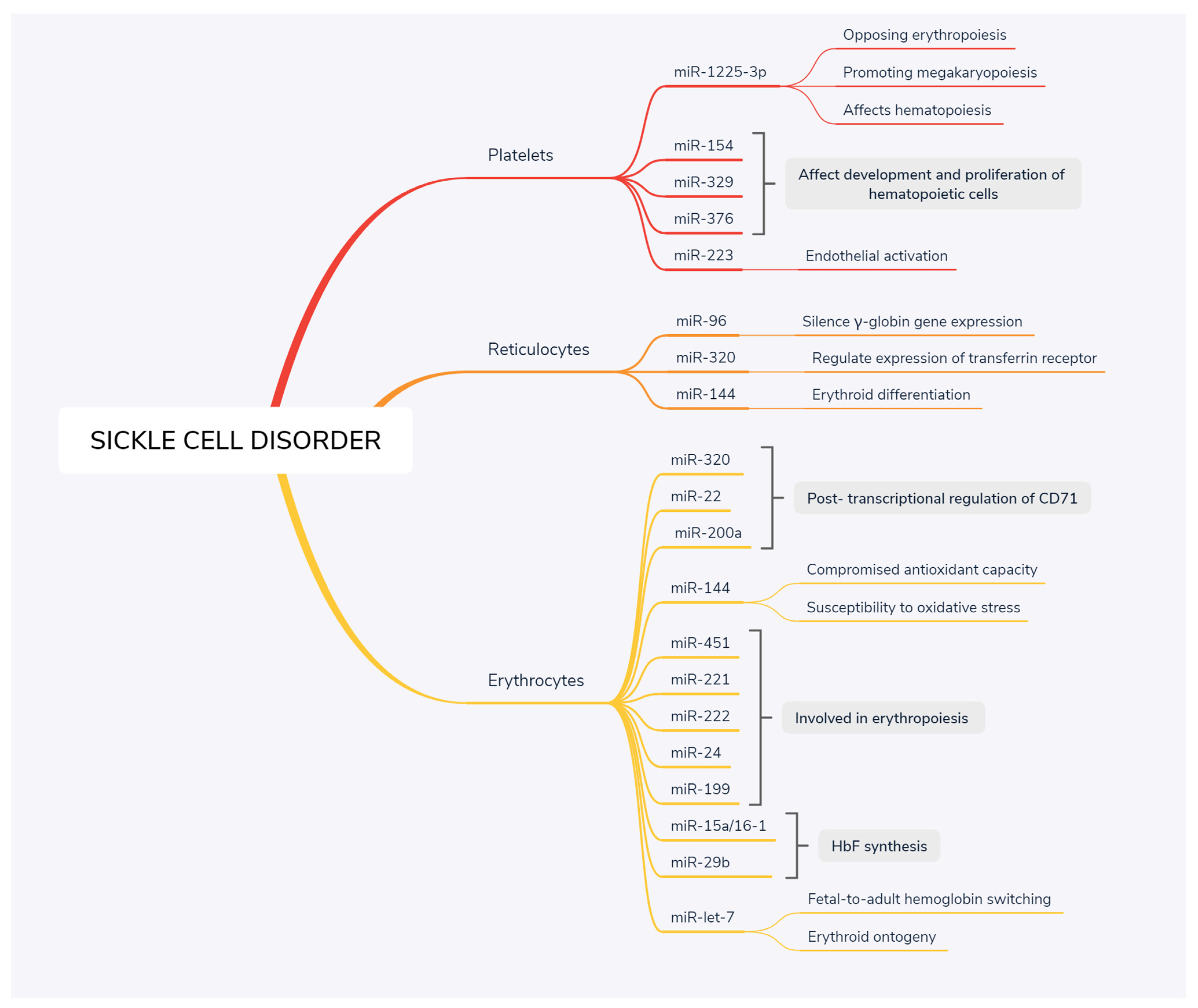
1.1. miRNA-144
1.2. miRNA-451
1.3. miRNA-29b
1.4. miRNA-320
1.5. Let-7 Family
1.6. miRNA-1225-3p
1.7. miRNA-221/-222 Cluster
1.8. miRNA-15a/16-1 Cluster
1.9. miRNA-96
1.10. miRNA-199
1.11. Other miRNAs
2. Conclusions
Funding
Institutional Review Board Statement
Informed Consent Statement
Data Availability Statement
Conflicts of Interest
References
- Schaeffer, E.K.; West, R.J.; Conine, S.J.; Lowrey, C.H. Multiple physical stresses induce γ-globin gene expression and fetal hemoglobin production in erythroid cells. Blood Cells Mol. Dis. 2014, 52, 214–224. [Google Scholar] [CrossRef]
- Jastaniah, W. Epidemiology of Sickle Cell Disease in Saudi Arabia. Ann. Saudi Med. 2011, 31, 289–293. [Google Scholar] [CrossRef]
- Gill, F.M.; Sleeper, L.A.; Weiner, S.J.; Brown, A.K.; Bellevue, R.; Grover, R.; Pegelow, C.H.; Vichinsky, E. Clinical events in the first decade in a cohort of infants with sickle cell disease. Blood 1995, 86, 776–783. [Google Scholar] [CrossRef] [Green Version]
- Vichinsky, E.P.; Styles, L.A.; Colangelo, L.H.; Wright, E.C.; Castro, O.; Nickerson, B. Acute chest syndrome in sickle cell disease: Clinical presentation and course. Blood 1997, 89, 1787–1792. [Google Scholar] [CrossRef] [PubMed] [Green Version]
- Wilson, M.; Forsyth, P.; Whiteside, J. Haemoglobinopathy and sickle cell disease. Contin. Educ. Anaesth. Crit. Care Pain. 2009, 10, 24–28. [Google Scholar] [CrossRef] [Green Version]
- Lobo, C.L.; Pinto, J.F.; Nascimento, E.M.; Moura, P.G.; Cardoso, G.P.; Hankins, J.S. The effect of hydroxycarbamide therapy on survival of children with sickle cell disease. Br. J. Haematol. 2013, 161, 852–860. [Google Scholar] [CrossRef] [PubMed]
- Niihara, Y.; Miller, S.T.; Kanter, J.; Lanzkron, S.; Smith, W.R.; Hsu, L.L.; Gordeuk, V.R.; Viswanathan, K.; Sarnaik, S.; Osunkwo, I.; et al. A Phase 3 Trial of l-Glutamine in Sickle Cell Disease. N. Engl. J. Med. 2018, 379, 226–235. [Google Scholar] [CrossRef] [PubMed]
- Torres, L.; Conran, N. Emerging pharmacotherapeutic approaches for the management of sickle cell disease. Expert Opin. Pharmacother. 2019, 20, 173–186. [Google Scholar] [CrossRef]
- Frangoul, H.; Altshuler, D.; Cappellini, M.D.; Chen, Y.S.; Domm, J.; Eustace, B.K.; Foell, J.; de la Fuente, J.; Grupp, S.; Handgretinger, R.; et al. CRISPR-Cas9 Gene Editing for Sickle Cell Disease and β-Thalassemia. N. Engl. J. Med. 2021, 384, 252–260. [Google Scholar] [CrossRef]
- Bonneau, E.; Neveu, B.; Kostantin, E.; Tsongalis, G.J.; De Guire, V. How close are miRNAs from clinical practice? A perspective on the diagnostic and therapeutic market. EJIFCC 2019, 30, 114–127. [Google Scholar]
- Treiber, T.; Treiber, N.; Meister, G. Regulation of microRNA biogenesis and its crosstalk with other cellular pathways. Nat. Rev. Mol. Cell Biol. 2019, 20, 5–20. [Google Scholar] [CrossRef]
- Azzouzi, I.; Schmugge, M.; Speer, O. MicroRNAs as components of regulatory networks controlling erythropoiesis. Eur. J. Haematol. 2012, 89, 1–9. [Google Scholar] [CrossRef]
- Gebert, L.F.R.; MacRae, I.J. Regulation of microRNA function in animals. Nat. Rev. Mol. Cell Biol. 2019, 20, 21–37. [Google Scholar] [CrossRef] [PubMed]
- Bartel, D.P. MicroRNAs: Genomics, biogenesis, mechanism, and function. Cell 2004, 116, 281–297. [Google Scholar] [CrossRef] [Green Version]
- Chen, S.-Y.; Wang, Y.; Telen, M.J.; Chi, J.-T. The Genomic Analysis of Erythrocyte microRNA Expression in Sickle Cell Diseases. PLoS ONE 2008, 3, e2360. [Google Scholar]
- Sangokoya, C.; Telen, M.J.; Chi, J.T. microRNA miR-144 modulates oxidative stress tolerance and associates with anemia severity in sickle cell disease. Blood 2010, 116, 4338–4348. [Google Scholar] [CrossRef] [PubMed] [Green Version]
- LaMonte, G.; Philip, N.; Reardon, J.; Lacsina, J.R.; Majoros, W.; Chapman, L.; Thornburg, C.D.; Telen, M.J.; Ohler, U.; Nicchitta, C.V.; et al. Translocation of sickle cell erythrocyte microRNAs into Plasmodium falciparum inhibits parasite translation and contributes to malaria Resistance. Cell Host. Microbe 2012, 12, 187–199. [Google Scholar] [CrossRef] [Green Version]
- Masaki, S.; Ohtsuka, R.; Abe, Y.; Muta, K.; Umemura, T. Expression patterns of microRNAs 155 and 451 during normal human erythropoiesis. Biochem. Biophys. Res. Commun. 2007, 364, 509–514. [Google Scholar] [CrossRef]
- Bruchova, H.; Yoon, D.; Agarwal, A.M.; Mendell, J.; Prchal, J.T. Regulated expression of microRNAs in normal and polycythemia vera erythropoiesis. Exp. Hematol. 2007, 35, 1657–1667. [Google Scholar] [CrossRef] [Green Version]
- Garzon, R.; Pichiorri, F.; Palumbo, T.; Iuliano, R.; Cimmino, A.; Aqeilan, R.; Volinia, S.; Bhatt, D.; Alder, H.; Marcucci, G.; et al. MicroRNA fingerprints during human megakaryocytopoiesis. Proc. Natl. Acad. Sci. USA 2006, 103, 5078–5083. [Google Scholar] [CrossRef] [Green Version]
- Visone, R.; Rassenti, L.Z.; Veronese, A.; Taccioli, C.; Costinean, S.; Aguda, B.D.; Volinia, S.; Ferracin, M.; Palatini, J.; Balatti, V.; et al. Karyotype-specific microRNA signature in chronic lymphocytic leukemia. Blood 2009, 114, 3872–3879. [Google Scholar] [CrossRef] [Green Version]
- Rossi, S.; Shimizu, M.; Barbarotto, E.; Nicoloso, M.S.; Dimitri, F.; Sampath, D.; Fabbri, M.; Lerner, S.; Barron, L.L.; Rassenti, L.Z.; et al. microRNA fingerprinting of CLL patients with chromosome 17p deletion identify a miR-21 score that stratifies early survival. Blood 2010, 116, 945–952. [Google Scholar] [CrossRef]
- Ha, T.Y. MicroRNAs in human diseases: From lung, liver and kidney diseases to infectious disease, sickle cell disease and endometrium disease. Immune Netw. 2011, 11, 309–323. [Google Scholar] [CrossRef] [PubMed] [Green Version]
- De Rie, D.; Abugessaisa, I.; Alam, T.; Arner, E.; Arner, P.; Ashoor, H.; Åström, G.; Babina, M.; Bertin, N.; Burroughs, A.M.; et al. An integrated expression atlas of miRNAs and their promoters in human and mouse. Nat. Biotechnol. 2017, 35, 872–878. [Google Scholar] [CrossRef] [PubMed]
- Merkerova, M.; Belickova, M.; Bruchova, H. Differential expression of microRNAs in hematopoietic cell lineages. Eur. J. Haematol. 2008, 81, 304–310. [Google Scholar] [CrossRef] [PubMed]
- Landry, P.; Plante, I.; Ouellet, D.L.; Perron, M.P.; Rousseau, G.; Provost, P. Existence of a microRNA pathway in anucleate platelets. Nat. Struct. Mol. Biol. 2009, 16, 961–966. [Google Scholar] [CrossRef] [Green Version]
- Zhan, M.; Miller, C.P.; Papayannopoulou, T.; Stamatoyannopoulos, G.; Song, C.Z. MicroRNA expression dynamics during murine and human erythroid differentiation. Exp. Hematol. 2007, 35, 1015–1025. [Google Scholar] [CrossRef] [PubMed] [Green Version]
- Noh, S.J.; Miller, S.H.; Lee, Y.T.; Goh, S.H.; Marincola, F.M.; Stroncek, D.F.; Reed, C.; Wang, E.; Miller, J.L. Let-7 microRNAs are developmentally regulated in circulating human erythroid cells. J. Transl. Med. 2009, 7, 98. [Google Scholar] [CrossRef] [PubMed] [Green Version]
- Li, B.; Zhu, X.; Ward, C.M.; Starlard-Davenport, A.; Takezaki, M.; Berry, A.; Ward, A.; Wilder, C.; Neunert, C.; Kutlar, A.; et al. MIR-144-mediated NRF2 gene silencing inhibits fetal hemoglobin expression in sickle cell disease. Exp. Hematol. 2019, 70, 85–96.e5. [Google Scholar] [CrossRef] [PubMed]
- Rasmussen, K.D.; Simmini, S.; Abreu-Goodger, C.; Bartonicek, N.; Di Giacomo, M.; Bilbao-Cortes, D.; Horos, R.; Von Lindern, M.; Enright, A.J.; O’Carroll, D. The miR-144/451 locus is required for erythroid homeostasis. J. Exp. Med. 2010, 207, 1351–1358. [Google Scholar] [CrossRef]
- Vannucchi, A.M.; Bianchi, L.; Cellai, C.; Paoletti, F.; Carrai, V.; Calzolari, A.; Centurione, L.; Lorenzini, R.; Carta, C.; Alfani, E.; et al. Accentuated response to phenylhydrazine and erythropoietin in mice genetically impaired for their GATA-1 expression (GATA-1(low) mice). Blood 2001, 97, 3040–3050. [Google Scholar] [CrossRef] [PubMed] [Green Version]
- Dore, L.C.; Amigo, J.D.; Dos Santos, C.O.; Zhang, Z.; Gai, X.; Tobias, J.W.; Yu, D.; Klein, A.M.; Dorman, C.; Wu, W.; et al. A GATA-1-regulated microRNA locus essential for erythropoiesis. Proc. Natl. Acad. Sci. USA 2008, 105, 3333–3338. [Google Scholar] [CrossRef] [PubMed] [Green Version]
- Lee, J.M.; Johnson, J.A. An important role of Nrf2-ARE pathway in the cellular defense mechanism. J. Biochem. Mol. Biol. 2004, 37, 139–143. [Google Scholar] [CrossRef] [PubMed] [Green Version]
- Byon, J.C.; Papayannopoulou, T. MicroRNAs: Allies or foes in erythropoiesis? J. Cell Physiol. 2012, 227, 7–13. [Google Scholar] [CrossRef] [Green Version]
- Wang, T.; Wu, F.; Yu, D. miR-144/451 in hematopoiesis and beyond. ExRNA 2019, 1, 16. [Google Scholar] [CrossRef] [Green Version]
- Svasti, S.; Masaki, S.; Penglong, T.; Abe, Y.; Winichagoon, P.; Fucharoen, S.; Umemura, T. Expression of microRNA-451 in normal and thalassemic erythropoiesis. Ann. Hematol. 2010, 89, 953–958. [Google Scholar] [CrossRef]
- Rathjen, T.; Nicol, C.; McConkey, G.; Dalmay, T. Analysis of short RNAs in the malaria parasite and its red blood cell host. FEBS Lett. 2006, 580, 5185–5188. [Google Scholar] [CrossRef]
- Pasvol, G.; Weatherall, D.J.; Wilson, R.J. Cellular mechanism for the protective effect of haemoglobin S against P. falciparum malaria. Nature 1978, 274, 701–703. [Google Scholar] [CrossRef]
- Alijani, S.; Alizadeh, S.; Kazemi, A.; Khatib, Z.K.; Soleimani, M.; Rezvani, M.; Minayi, N.; Karami, F.; Tayebi, B. Evaluation of the effect of miR-26b upregulation on HbF expression in erythroleukemic K-562 cell line. Avicenna J. Med. Biotechnol. 2014, 6, 53–56. [Google Scholar]
- Sankaran, V.G.; Menne, T.F.; Scepanovic, D.; Vergilio, J.A.; Ji, P.; Kim, J.; Thiru, P.; Orkin, S.H.; Lander, E.S.; Lodish, H.F. MicroRNA-15a and-16-1 act via MYB to elevate fetal hemoglobin expression in human trisomy 13. Proc. Natl. Acad. Sci. USA 2011, 108, 1519–1524. [Google Scholar] [CrossRef] [Green Version]
- Mnika, K.; Mazandu, G.K.; Jonas, M.; Pule, G.D.; Chimusa, E.R.; Hanchard, N.A.; Wonkam, A. Hydroxyurea-Induced miRNA Expression in Sickle Cell Disease Patients in Africa. Front. Genet. 2019, 10, 509. [Google Scholar] [CrossRef] [PubMed]
- Starlard-Davenport, A.; Smith, A.; Vu, L.; Li, B.; Pace, B.S. MIR29B mediates epigenetic mechanisms of HBG gene activation. Br. J. Haematol. 2019, 186, 91–100. [Google Scholar] [CrossRef] [PubMed] [Green Version]
- Farh, K.K.; Grimson, A.; Jan, C.; Lewis, B.P.; Johnston, W.K.; Lim, L.P.; Burge, C.B.; Bartel, D.P. The widespread impact of mammalian MicroRNAs on mRNA repression and evolution. Science 2005, 310, 1817–1821. [Google Scholar] [CrossRef] [PubMed] [Green Version]
- Giraldez, A.J.; Mishima, Y.; Rihel, J.; Grocock, R.J.; Van Dongen, S.; Inoue, K.; Enright, A.J.; Schier, A.F. Zebrafish MiR-430 Promotes Deadenylation and Clearance of Maternal mRNAs. Science 2006, 312, 75–79. [Google Scholar] [CrossRef] [PubMed] [Green Version]
- Wu, L.; Fan, J.; Belasco, J.G. From the Cover: MicroRNAs direct rapid deadenylation of mRNA. Proc. Natl. Acad. Sci. USA 2006, 103, 4034–4039. [Google Scholar] [CrossRef] [Green Version]
- Schaar, D.G.; Medina, D.J.; Moore, D.F.; Strair, R.K.; Ting, Y. miR-320 targets transferrin receptor 1 (CD71) and inhibits cell proliferation. Exp. Hematol. 2009, 37, 245–255. [Google Scholar] [CrossRef]
- Lee, Y.T.; de Vasconcellos, J.F.; Yuan, J.; Byrnes, C.; Noh, S.J.; Meier, E.R.; Kim, K.S.; Rabel, A.; Kaushal, M.; Muljo, S.A.; et al. LIN28B-mediated expression of fetal hemoglobin and production of fetal-like erythrocytes from adult human erythroblasts ex vivo. Blood 2013, 122, 1034–1041. [Google Scholar] [CrossRef] [PubMed] [Green Version]
- De Vasconcellos, J.F.; Byrnes, C.; Lee, Y.T.; Allwardt, J.M.; Kaushal, M.; Rabel, A.; Miller, J.L. Tough decoy targeting of predominant let-7 miRNA species in adult human hematopoietic cells. J. Transl. Med. 2017, 15, 169. [Google Scholar] [CrossRef] [PubMed]
- Goh, S.H.; Josleyn, M.; Lee, Y.T.; Danner, R.L.; Gherman, R.B.; Cam, M.C.; Miller, J.L. The human reticulocyte transcriptome. Physiol. Genom. 2007, 30, 172–178. [Google Scholar] [CrossRef] [PubMed] [Green Version]
- Azzouzi, I.; Moest, H.; Winkler, J.; Fauchère, J.C.; Gerber, A.P.; Wollscheid, B.; Stoffel, M.; Schmugge, M.; Speer, O. MicroRNA-96 directly inhibits γ-globin expression in human erythropoiesis. PLoS ONE 2011, 6, e22838. [Google Scholar]
- Ginder, G.D. Epigenetic regulation of fetal globin gene expression in adult erythroid cells. Transl. Res. 2015, 165, 115–125. [Google Scholar] [CrossRef] [Green Version]
- Jain, S.; Kapetanaki, M.G.; Raghavachari, N.; Woodhouse, K.; Yu, G.; Barge, S.; Coronnello, C.; Benos, P.V.; Kato, G.J.; Kaminski, N.; et al. Expression of Regulatory Platelet MicroRNAs in Patients with Sickle Cell Disease. PLoS ONE 2013, 8, e60932. [Google Scholar]
- Antonucci, R.; Walker, R.; Herion, J.; Orringer, E. Enhancement of sickle erythrocyte adherence to endothelium by autologous platelets. Am. J. Hematol. 1990, 34, 44–48. [Google Scholar] [CrossRef] [PubMed]
- Blann, A.D.; Marwah, S.; Serjeant, G.; Bareford, D.; Wright, J. Platelet activation and endothelial cell dysfunction in sickle cell disease is unrelated to reduced antioxidant capacity. Blood Coagul. Fibrinolysis 2003, 14, 255–259. [Google Scholar] [CrossRef] [PubMed]
- Novelli, E.M.; Kato, G.J.; Ragni, M.V.; Zhang, Y.; Hildesheim, M.E.; Nouraie, M.; Barge, S.; Meyer, M.P.; Hassett, A.C.; Gordeuk, V.R.; et al. Plasma thrombospondin-1 is increased during acute sickle cell vaso-occlusive events and associated with acute chest syndrome, hydroxyurea therapy, and lower hemolytic rates. Am. J. Hematol. 2012, 87, 326–330. [Google Scholar] [CrossRef]
- Schermuly, R.T.; Dony, E.; Ghofrani, H.A.; Pullamsetti, S.; Savai, R.; Roth, M.; Sydykov, A.; Lai, Y.J.; Weissmann, N.; Seeger, W.; et al. Reversal of experimental pulmonary hypertension by PDGF inhibition. J. Clin. Investig. 2005, 115, 2811–2821. [Google Scholar] [CrossRef] [Green Version]
- Felli, N.; Fontana, L.; Pelosi, E.; Botta, R.; Bonci, D.; Facchiano, F.; Liuzzi, F.; Lulli, V.; Morsilli, O.; Santoro, S.; et al. MicroRNAs 221 and 222 inhibit normal erythropoiesis and erythroleukemic cell growth via kit receptor down-modulation. Proc. Natl. Acad. Sci. USA 2005, 102, 18081–18086. [Google Scholar] [CrossRef] [Green Version]
- Wang, Q.; Huang, Z.; Xue, H.; Jin, C.; Ju, X.L.; Han, J.D.; Chen, Y.G. MicroRNA miR-24 inhibits erythropoiesis by targeting activin type I receptor ALK4. Blood 2007, 111, 588–595. [Google Scholar] [CrossRef] [PubMed]
- Gabbianelli, M.; Testa, U.; Morsilli, O.; Pelosi, E.; Saulle, E.; Petrucci, E.; Castelli, G.; Giovinazzi, S.; Mariani, G.; Fiori, M.E.; et al. Mechanism of human Hb switching: A possible role of the kit receptor/miR 221-222 complex. Haematologica 2010, 95, 1253–1260. [Google Scholar] [CrossRef] [Green Version]
- Cho, W.C. OncomiRs: The discovery and progress of micro-RNAs in cancers. Mol. Cancer 2007, 6, 60. [Google Scholar] [CrossRef] [Green Version]
- Tallack, M.R.; Perkins, A.C. Three fingers on the switch: Kruppel-like factor 1 regulation of γ-globin to β-globin gene switching. Curr. Opin. Hematol. 2013, 20, 193–200. [Google Scholar] [CrossRef]
- Esteghamat, F.; Gillemans, N.; Bilic, I.; van den Akker, E.; Cantu, I.; van Gent, T.; Klingmüller, U.; van Lom, K.; von Lindern, M.; Grosveld, F.; et al. Erythropoiesis and globin switching in compound Klf1, Bcl11a mutant mice. Blood 2013, 121, 2553–2562. [Google Scholar] [CrossRef]
- Saki, N.; Abroun, S.; Soleimani, M.; Kavianpour, M.; Shahjahani, M.; Mohammadi-Asl, J.; Hajizamani, S. MicroRNA expression in β-thalassemia and sickle cell disease: A role in the induction of fetal hemoglobin. Cell J. 2016, 17, 583–592. [Google Scholar]
- Li, Y.; Bai, H.; Zhang, Z.; Li, W.; Dong, L.; Wei, X.; Ma, Y.; Zhang, J.; Yu, J.; Sun, G.; et al. The up-regulation of miR-199b-5p in erythroid differentiation is associated with GATA-1 and NF-E2. Mol. Cells 2014, 37, 213–219. [Google Scholar] [CrossRef] [Green Version]
- Walker, A.L.; Steward, S.; Howard, T.A.; Mortier, N.; Smeltzer, M.; Wang, Y.D.; Ware, R.E. Epigenetic and molecular profiles of erythroid cells after hydroxyurea treatment in sickle cell anemia. Blood 2011, 118, 5664–5670. [Google Scholar] [CrossRef] [Green Version]
- Emami, S.A.; Mohammadi, S.; Kavyani, S.; Soleimani, M.; Alizadeh, S.; Dejbakhsh, E.; Kouhkan, F.; Mossahebi, M.; Dargahi, H. Investigating the relationship between miR210 upregulation and hemoglobin gamma chain expression. Payavard-Salamat 2011, 241, 20–26. [Google Scholar]
- Bianchi, N.; Zuccato, C.; Lampronti, I.; Borgatti, M.; Gambari, R. Expression of miR-210 during erythroid differentiation and induction of gamma-globin gene expression. BMB Rep. 2009, 42, 493–499. [Google Scholar] [CrossRef] [PubMed] [Green Version]
- Sarakul, O.; Vattanaviboon, P.; Tanaka, Y.; Fucharoen, S.; Abe, Y.; Svasti, S.; Umemura, T. Enhanced erythroid cell differentiation in hypoxic condition is in part contributed by miR-210. Blood Cells Mol. Dis. 2013, 51, 98–103. [Google Scholar] [CrossRef] [PubMed]
- Rogers, H.M.; Yu, X.; Wen, J.; Smith, R.; Fibach, E.; Noguchi, C.T. Hypoxia alters progression of the erythroid program. Exp. Hematol. 2008, 36, 17–27. [Google Scholar] [CrossRef] [PubMed] [Green Version]
- Lulli, V.; Romania, P.; Morsilli, O.; Cianciulli, P.; Gabbianelli, M.; Testa, U.; Giuliani, A.; Marziali, G. MicroRNA-486-3p Regulates γ-Globin Expression in Human Erythroid Cells by Directly Modulating BCL11A. PLoS ONE 2013, 8, e60436. [Google Scholar] [CrossRef] [Green Version]
- Ma, Y.; Wang, B.; Jiang, F.; Wang, D.; Liu, H.; Yan, Y.; Dong, H.; Wang, F.; Gong, B.; Zhu, Y. A feedback loop consisting of microRNA 23a/27a and the β-like globin suppressors KLF3 and SP1 regulates globin gene expression. Mol. Cell Biol. 2013, 33, 3994–4007. [Google Scholar] [CrossRef] [PubMed] [Green Version]
- Ward, C.M.; Li, B.; Pace, B.S. Original Research: Stable expression of miR-34a mediates fetal hemoglobin induction in K562 cells. Exp. Biol. Med. 2016, 241, 719–729. [Google Scholar] [CrossRef] [PubMed] [Green Version]
- Juzenas, S.; Venkatesh, G.; Hübenthal, M.; Hoeppner, M.P.; Du, Z.G.; Paulsen, M.; Rosenstiel, P.; Senger, P.; Hofmann-Apitius, M.; Keller, A.; et al. A comprehensive, cell specific microRNA catalogue of human peripheral blood. Nucleic Acids Res. 2017, 45, 9290–93017. [Google Scholar] [CrossRef] [PubMed] [Green Version]

| microRNA | Target (mRNA or Protein) | Biological Effect | Cell Type | Reference |
|---|---|---|---|---|
| Up-Regulated | ||||
| Let-7 | BCL11A | γ globin gene switching | Erythrocyte | [47] |
| miRNA-486-3p | BCL11A | Increasing expression of γ globin gene | Erythrocyte | [41,63,70] |
| miRNA-96 | γ globin (CDS region) | Suppressing γ globin gene expression | Reticulocyte | [50] |
| miRNA-29b | DNMT, MYB | Increasing expression of γ globin gene | Erythroid progenitor, Reticulocyte | [42] |
| miRNA-144 | NRF2 | Interference with antioxidant capacity; susceptibility to oxidative stress, hemolysis and severe anemia anemic phenotype | Erythrocyte, Reticulocyte | [16,29] |
| miRNA-221/-222 | KLFD | Decreasing of erythroblast proliferation and suppress HbF production | Erythrocyte | [57,59] |
| miRNA-199a | KLF3, GATA-1 | Regulating of human erythropoiesis and decrease HbF levels | Erythrocyte | [33,64] |
| miRNA-451 | GATA-1 | Inducing γ globin gene transcription and suppress α globin, Glycophorin-A | Erythrocyte | [13,30,32] |
| miRNA-320 | CD71 | Hemolysis-induced irregularity in erythropoiesis | Reticulocyte | [15,34] |
| miRNA-1225-3p | PBXIPI | Maintenance of hematopoiesis and lineage commitment by opposing erythropoiesis and promoting megakaryopoiesis | Platelet | [52] |
| miRNA-26b | GATA1 | Increasing γ globin gene expression | Erythrocyte | [65] |
| miRNA-210 | γ globin | γ globin gene switching | Reticulocyte, Erythrocyte | [66,67] |
| Down-Regulated | ||||
| miRNA-23a | SP1 | Increases γ and ε globin expression | Erythrocyte | [71] |
| miRNA-27a | KLF3 | Regulating HbF expression and erythropoiesis | Erythrocyte | [71] |
| miRNA-34a | STAT3 | γ-globin activation | Erythrocyte | [72] |
| miRNA-15a/-16-1 | MYB | Increasing γ globin gene expression | Erythroid progenitor | [49,61,62] |
Publisher’s Note: MDPI stays neutral with regard to jurisdictional claims in published maps and institutional affiliations. |
© 2021 by the author. Licensee MDPI, Basel, Switzerland. This article is an open access article distributed under the terms and conditions of the Creative Commons Attribution (CC BY) license (https://creativecommons.org/licenses/by/4.0/).
Share and Cite
Cyrus, C. The Role of miRNAs as Therapeutic Tools in Sickle Cell Disease. Medicina 2021, 57, 1106. https://doi.org/10.3390/medicina57101106
Cyrus C. The Role of miRNAs as Therapeutic Tools in Sickle Cell Disease. Medicina. 2021; 57(10):1106. https://doi.org/10.3390/medicina57101106
Chicago/Turabian StyleCyrus, Cyril. 2021. "The Role of miRNAs as Therapeutic Tools in Sickle Cell Disease" Medicina 57, no. 10: 1106. https://doi.org/10.3390/medicina57101106
APA StyleCyrus, C. (2021). The Role of miRNAs as Therapeutic Tools in Sickle Cell Disease. Medicina, 57(10), 1106. https://doi.org/10.3390/medicina57101106