
Dermoscopic Features of Lichen Amyloidosis in Caucasians—A Case Series and Literature Review


Abstract
:1. Introduction
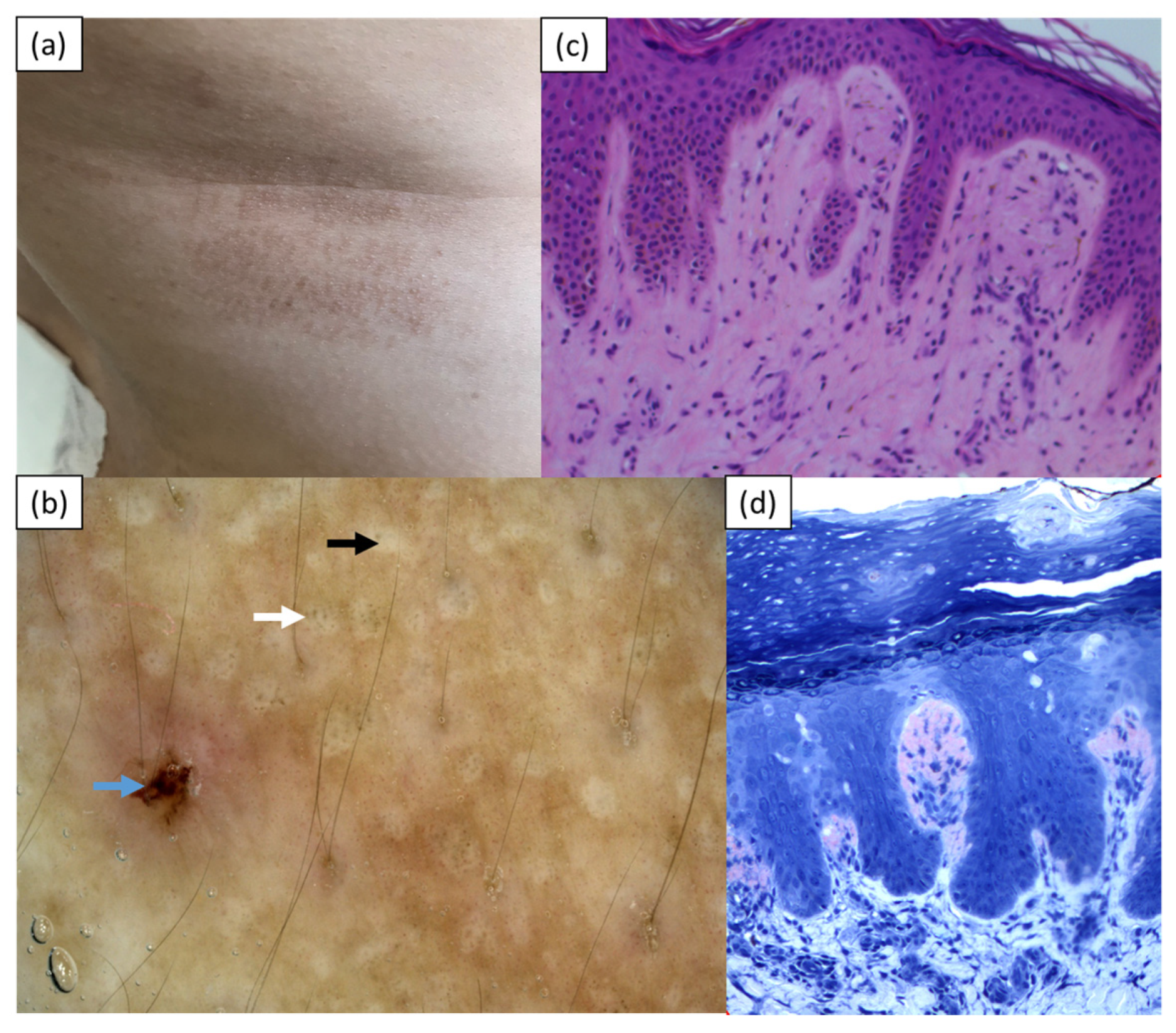
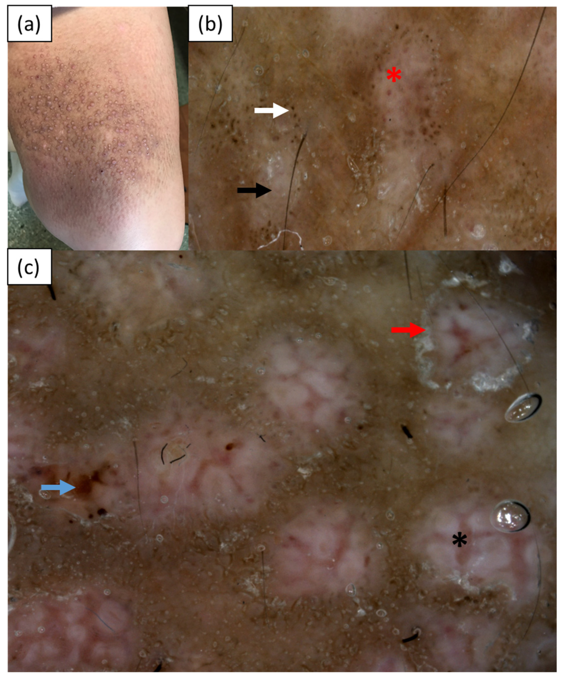
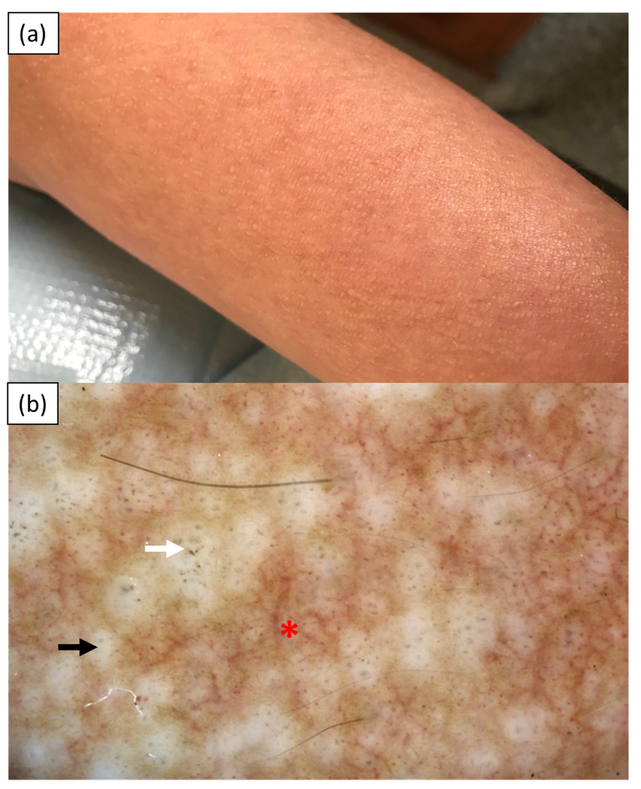
2. Case Reports
3. Discussion
4. Conclusions
Author Contributions
Funding
Institutional Review Board Statement
Informed Consent Statement
Data Availability Statement
Conflicts of Interest
References
- Arnold, S.J.; Bowling, J.C.R. ‘Shiny white streaks’ in lichen amyloidosis: A clue to diagnosis. Australas J. Dermatol. 2012, 53, 272–273. [Google Scholar] [CrossRef] [PubMed]
- Chuang, Y.Y.; Lee, D.D.; Lin, C.S.; Chang, Y.J.; Tanaka, M.; Chang, Y.T.; Liu, H.N. Characteristic dermoscopic features of primary cutaneous amyloidosis: A study of 35 cases. Br. J. Dermatol. 2012, 167, 548–554. [Google Scholar] [CrossRef] [PubMed]
- Wang, X.; Wang, H.; Zhong, Z.; Zheng, L.; Wang, Y.; Guo, Z.; Li, H.; Gao, M. Case report: Diagnosis of primary cutaneous amyloidosis using dermoscopy and reflectance confocal microscopy. Front. Med 2021, 7, 619907. [Google Scholar] [CrossRef] [PubMed]
- Rongioletti, F.; Atzori, L.; Ferreli, C.; Pinna, A.; Aste, N.; Pau, M. A unique dermoscopy pattern of primary cutaneous nodular amyloidosis mimicking a granulomatous disease. J. Am. Acad. Dermatol. 2016, 74, e9–e10. [Google Scholar] [CrossRef] [PubMed]
- Di Meo, N.; Noal, C.; Fadel, M.; Trevisan, G. Yellow teardrop-like structures in primary nodular skin amyloidosis. G. Ital. Derm. Venereol. 2018, 153, 118–119. [Google Scholar]
- Wang, L.; Jiang, X.; Zhang, N.; Liu, L.; Zhou, H.; Liu, H.J. Case of amyloidosis cutis dyschromica with dermoscopy. J. Dermatol. 2019, 46, e77–e79. [Google Scholar] [CrossRef] [PubMed]
- Moscarella, E.; Ronchi, A.; Agozzino, M.; Franco, R.; Argenziano, G. Image gallery: Dermoscopy of lichen amyloidosis. Br. J. Dermatol. 2018, 179, e231. [Google Scholar] [CrossRef] [PubMed] [Green Version]
- Kaliyadan, F.; Alkhateeb, A.; Kuruvilla, J.; Swaroop, K.; Alabdulsalam, A.A. Dermoscopy of primary cutaneous amyloidosis in skin of color. Dermatol. Pr. Concept 2019, 9, 232–234. [Google Scholar] [CrossRef] [PubMed] [Green Version]
- Ferreira, I.L.O.; Fernandes, E.L.; Lapins, J.; Benini, T.; Silva, L.C.; Lanzoni, M.A.; Steiner, D. Primary localized cutaneous nodular amyloidosis on a toe: Clinical presentation, histopathology, and dermoscopy findings. Dermatol. Pr. Concept 2019, 9, 235–236. [Google Scholar] [CrossRef] [PubMed] [Green Version]
- Sonthalia, S.; Agrawal, M.; Sehgal, V.N. Dermoscopy of macular amyloidosis. Indian Dermatol. Online J. 2020, 12, 203–205. [Google Scholar] [PubMed]
- Madarkar, M.S.; Koti, V.R. FotoFinder dermoscopy analysis and histopathological correlation in primary localized cutaneous amyloidosis. Dermatol. Pr. Concept 2021, 11, e2021057. [Google Scholar] [CrossRef] [PubMed]
- Behera, B.; Kumari, R.; Mohan Thappa, D.; Gochhait, D.; Hanuman Srinivas, B.; Ayyanar, P. Dermoscopic features of primary cutaneous amyloidosis in skin of colour: A retrospective analysis of 48 patients from South India. Australas J. Dermatol. 2021, 6. [Google Scholar] [CrossRef]


| Lichen Amyloidosis | ||||||
|---|---|---|---|---|---|---|
| Arnold et al., 2012 (India/NA) n = 2 | Chuang et al., 2012 (Taiwan) n = 17 | Moscarella et al., 2018 (Italy) n = 1 | Madarkar et al., 2021 (India) n = 18 | Behera et al., 2021 (India) n = 30 | Current cases (Poland) n = 3 | |
| Type of examination | polarized | Polarized and nonpolarized | N/A | Contact polarized | Contact nonpolarized | Contact nonpolarized |
| Two-zone pattern | - | - | - | - | 25 | - |
| Jigsaw puzzle pattern | - | - | - | - | 1 | - |
| Ridge and groove pattern | - | - | - | - | 4 | - |
| Hub and spoke pattern | - | - | - | - | 5 | - |
| Central hub | - | - | - | - | - | 3 |
| -white | - | 7 | 1 | 5 | - | 3 |
| -brown | - | - | - | 2 | - | - |
| -white and brown | - | - | - | 1 | - | - |
| White scar-like center | - | 10 | - | 10 | - | - |
| White structureless areas | - | 7 | - | 10 | - | - |
| Pigment dots/globules/ peppering | - | 3 | 1 | - | 30 | 3 |
| -clustered | - | - | - | - | 18 | 2 |
| -discrete | - | - | - | - | 23 | 1 |
| -starbust | - | - | - | - | 12 | - |
| -perieccrine | - | - | - | - | 3 | - |
| Blue-grey ovoid nest-like area | - | - | - | - | 5 | - |
| Concentric structures | - | - | - | - | 5 | - |
| White keratotic areas | - | - | - | - | 5 | - |
| Rim of white collarette | - | 3 | - | present | - | 1 |
| Diffuse white scaling | - | - | - | present | - | - |
| Comedo-like opening | - | - | - | - | 1 | - |
| Milia-like cyst | - | - | - | - | 1 | - |
| Red/purple dots and globules | - | - | - | - | 7 | 2 |
| Thick branching vessels | - | - | - | - | - | 2 |
| Shiny white streaks | 2 | - | - | - | - | - |
| Rosettes | - | - | - | 1 | - | - |
| Grayish-brown background | - | - | - | present | - | 1 |
| ‘Daily lily flower’ structures | - | - | - | present | - | - |
| Erosions | - | - | - | - | - | 2 |
| Macular Amyloidosis | ||||||
| Chuang et al., 2012 (Taiwan) n = 18 | Madarkar et al., 2021 (India) n = 12 | Wang et al., 2021 (China) n = 1 | Behera et al., 2021 (India) n = 18 | Sonthalia et al., 2020 (India) n = 2 | ||
| Type of examination | Polarized and nonpolarized | Contact polarized | NA | Contact nonpolarized | polarized | |
| Jigsaw puzzle pattern | - | - | - | 17 | - | |
| Ridge and groove pattern | - | - | - | 8 | - | |
| Hub and spoke pattern | - | - | - | 4 | 1 | |
| Central hub | 18 | - | 1 | - | 1 | |
| -white | 11 | 3 | - | - | 1 | |
| -brown | 4 | 8 | - | - | 1 | |
| -white and brown | 3 | 1 | - | - | 1 | |
| Pigment dots/globules/ peppering | - | 5 | 1 | 18 | - | |
| -clustered | - | - | - | 17 | - | |
| -discrete | - | - | - | 10 | - | |
| -starbust | - | - | - | 8 | - | |
| -perieccrine | - | - | - | 7 | - | |
| Peripheral pigmentation (fine streaks/leaf-like extensions/bulbous projections) | 18 | - | - | - | 2 | |
| Semicircular hyperpigmented structures | - | 10 | - | - | - | |
| Blue-grey ovoid nest-like area | - | - | - | 1 | - | |
| ‘daily lily flower’ structures | - | 2 | - | - | - | |
| Erythematous background | - | 3 | - | - | - | |
| Perifollicular depigmented halo | - | - | - | - | 1 | |
| Fine white scales | - | - | - | - | 1 | |
| White-to-off white colored intersecting lines | - | - | - | - | 1 | |
| white-to-light brown colored structureless areas | - | - | - | - | 1 | |
| Nodular Amyloidosis | ||||||
| Rongioletti et al., 2016 (Italy) n = 1 | Ferreira et al., 2019 (Brazil) n = 1 | DI Meo et al., 2018 (Italy) n = 1 | ||||
| Type of examination | polarized | polarized | NA | |||
| Shiny white streaks | - | 1 | - | |||
| Orange-yellowish homogenous background | 1 | - | - | |||
| Yellow teardrop-like structures | - | - | 1 | |||
| Orange-pink background | - | 1 | - | |||
| Elongated teleangiectasias | 1 | - | - | |||
| Serpentine teleangiectasias | 1 | - | - | |||
| Biphasic amyloidosis | ||||||
| Kaliyadan et al., 2019 (Saudi Arabia) n = 1 | Wang et al., 2021 (China) n = 1 | |||||
| Type of examination | polarized | NA | ||||
| Brown stripes | - | 1 | ||||
| Irregular brown center | - | 1 | ||||
| White central hub | 1 | 1 | ||||
| Dotted/globular pigmentation | 1 | 1 | ||||
| Amyloidosis Cutis Dyschromica | ||||||
| Wang et al., 2019 (China) n = 1 | ||||||
| Type of examination | NA | |||||
| Elliptical/round white spots | 1 | |||||
| Dotted/globular pigmentation | 1 | |||||
| Reticular pigment spots | 1 | |||||
| Striped hypopigmented spots | 1 | |||||
| Monotonous hypopigmented spots | 1 | |||||
Publisher’s Note: MDPI stays neutral with regard to jurisdictional claims in published maps and institutional affiliations. |
© 2021 by the authors. Licensee MDPI, Basel, Switzerland. This article is an open access article distributed under the terms and conditions of the Creative Commons Attribution (CC BY) license (https://creativecommons.org/licenses/by/4.0/).
Share and Cite
Żychowska, M.; Pięta, K.; Rudy, I.; Skubisz, A.; Reich, A. Dermoscopic Features of Lichen Amyloidosis in Caucasians—A Case Series and Literature Review. Medicina 2021, 57, 1027. https://doi.org/10.3390/medicina57101027
Żychowska M, Pięta K, Rudy I, Skubisz A, Reich A. Dermoscopic Features of Lichen Amyloidosis in Caucasians—A Case Series and Literature Review. Medicina. 2021; 57(10):1027. https://doi.org/10.3390/medicina57101027
Chicago/Turabian StyleŻychowska, Magdalena, Karolina Pięta, Izabela Rudy, Aleksandra Skubisz, and Adam Reich. 2021. "Dermoscopic Features of Lichen Amyloidosis in Caucasians—A Case Series and Literature Review" Medicina 57, no. 10: 1027. https://doi.org/10.3390/medicina57101027
APA StyleŻychowska, M., Pięta, K., Rudy, I., Skubisz, A., & Reich, A. (2021). Dermoscopic Features of Lichen Amyloidosis in Caucasians—A Case Series and Literature Review. Medicina, 57(10), 1027. https://doi.org/10.3390/medicina57101027