Abstract
Background and objectives: Natural products such as essential oils with antioxidant potential can reduce the level of oxidative stress and prevent the oxidation of biomolecules. In the present study, we investigated the antioxidant potential of Lantana montevidensis leaf essential oil (EOLM) in chemical and biological models using Drosophila melanogaster. Materials and methods: in addition, the chemical components of the oil were identified and quantified by gas chromatography coupled to mass spectrometry (GC-MS), and the percentage compositions were obtained from electronic integration measurements using flame ionization detection (FID). Results: our results demonstrated that EOLM is rich in terpenes with Germacrene-D (31.27%) and β-caryophyllene (28.15%) as the major components. EOLM (0.12–0.48 g/mL) was ineffective in scavenging DPPH radical, and chelating Fe(II), but showed reducing activity at 0.24 g/mL and 0.48 g/mL. In in vivo studies, exposure of D. melanogaster to EOLM (0.12–0.48 g/mL) for 5 h resulted in 10% mortality; no change in oxidative stress parameters such as total thiol, non-protein thiol, and malondialdehyde contents, in comparison to control (p > 0.05). Conclusions: taken together, our results indicate EOLM may not be toxic at the concentrations tested, and thus may not be suitable for the development of new botanical insecticides, such as fumigants or spray-type control agents against Drosophila melanogaster.
1. Introduction
Reactive species are known to cause damage to cellular membranes (lipid peroxidation), DNA, proteins, and mitochondria, and have been found to be involved in the pathophysiology of a wide range of diseases such as brain ischemia, carcinogenesis, diabetes, etc. [1,2]. Natural products, especially from plant origin, can prevent some of the harmful effects of reactive species [3,4,5,6]. Reports indicate that the consumption of food antioxidants can reduce the incidence of various degenerative diseases, as well as the degenerative process associated with aging [7,8,9]. One of the mechanisms by which these antioxidants exert their beneficial effects is by avoiding the oxidation of biomolecules, which would break the chain reaction of the pathogenesis in the deterioration of the physiological functions [10,11].
People’s interest in natural compounds rather than synthetic ones is growing considerably [12], especially because they are regarded as safe with no side effects and possess a variety of therapeutic actions [13]. Indeed, natural products such as vegetable oils with antioxidant potential can help the organism to modify the oxidative state in imbalance conditions [14,15]. In addition, plant essential oils exhibit low mammalian toxicity and can affect the reproduction, growth rate, and behavior of insects [16,17,18,19,20]. Lantana montevidensis Briq., popularly known as “chumbinho” in Brazil, is used to treat rheumatism, bronchitis, and gastric disorders [21]. Studies carried out with the leaf extract or leaf essential oil of L. montevidensis demonstrated their antibacterial activities and their potential to modulate antibiotics drugs used in clinical infections [22,23,24,25]. In addition, leaf extract of L. montevidensis was shown to exhibit antioxidant activity [9], while flavonoids from its leaves were reported to exert antiproliferative activity against gastric adenocarcinoma, human uterus carcinoma, and murine melanoma cells, in vitro [21].
In spite of the fact that numerous studies have reported the insecticidal activity of essential oils against mosquitos and flies, there is a lack of knowledge about the effect of the essential oil from L. montevidensis on Drosophila melanogaster. Many basic biological, physiological, and neurological properties are conserved between mammals and D. melanogaster, and about 75% of the genes causing human diseases have a functional counterpart in the fly [26,27]. Thus, D. melanogaster has been widely used as a model in genetic research to study the physiopathology of neurodegenerative diseases such as Parkinson’s disease and Alzheimer’s disease [28,29]. In addition, D. melanogaster is accessible to large-scale insecticide screening operations, and it is physiologically, biochemically, and genetically similar to mosquitoes and flies [20,30,31]. Therefore, D. melanogaster provides an excellent model system to experimentally evaluate possible fumigant insecticides.
Considering the abovementioned information, the objective of this study was to investigate the chemical characterization and antioxidant potential of L. montevidensis leaf essential oil (EOLM) in vitro, as well as its possible toxic effects using Drosophila melanogaster as a model. Particularly, we evaluated the toxic effect of the essential oil from L. montevidensis on the cell viability (MTT) and on biomarkers of oxidative stress such as lipid peroxidation, iron levels, total thiols, and non-protein thiols (NPSH).
2. Materials and Methods
2.1. Plant Material
The leaves of L. montevidensis were collected in the Medicinal Plants Garden of the Regional University of Cariri—URCA, Crato—CE Brazil (7°22′ S; 39°28′ W and 492 m above sea level), at 9:30 am. After identification, a voucher was deposited in the HCDAL (Herbarium Dárdano de Andrade Lima-URCA) (Crato, Brazil), with number #7518. The leaves were dried in the shade.
2.2. Extraction of L. montevidensis Leaf Essential Oil
The dried leaves were crushed and submitted to a hydrodistillation system in a modified Clevenger type apparatus. Three hundred (300) grams of the sample were placed in a 5.0 L glass flask along with 2.0 L of distilled water and heated to boiling for 3 h [32]. The oil was collected using a glass Pasteur pipette, after which the yield was calculated. The obtained essential oil was treated with anhydrous sodium sulphate (Na2SO4) and stored at −4 °C until the chemical analyzes were carried out. The essential oil showed a yield of 0.19%.
2.3. Composition and Identification of the Constituents of the Essential Oil
After obtaining the essential oil, it was submitted to GC/MS analysis according the procedure described in the literature [33].
2.4. Reagents
Glutathione (GSH), 1,1,3,3-Tetramethoxypropane (TMP), thiobarbituric acid (TBA), 5,5′-Dithiobis(2-nitrobenzoic acid), (3-(4,5-Dimethylthiazolyl-2)-2,5-diphenyltetrazolium bromide) (MTT), and 1,10-Phenanthroline were purchased from Sigma Aldrich (St. Louis, MO, USA). All the reagents were of analytical grade.
2.5. Antioxidant Activity in Chemical Model
2.5.1. Antioxidant Capacity in Chemical Model: 1,1-Diphenyl-2-picrylhydrazyl (DPPH) Radical Scavenging Activity
The radical scavenging ability of L. montevidensis leaf essential oil was performed using the stable free radical DPPH (1,1-Diphenyl-2-picrylhydrazyl) as described by Kamdem et al. [34].
2.5.2. Fe2+ Chelating Activity of L. montevidensis Leaf Essential Oil
The chelating capacity of essential oil from the leaves of L. montevidensis was determined according to the modified methods of Dinis et al. [35] and Kamdem et al. [36]. To summarize, the schedule for the evaluation of Fe2+ chelation or oxidation by the oil is presented in (Figure 1).
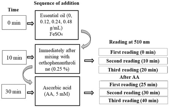
Figure 1.
Flowchart of the steps for the assays using Fe2+/Fe3++ by L. montevidensis leaf essential oil.
2.5.3. Fe3+ Reducing Power of L. montevidensis Leaf Essential Oil
To further investigate the reductive potential of the essential oil, the same reaction mixture as described above but using FeCl3(110 µM) instead of FeSO4(110 µM) in the reaction mixture and was determined using a modified method of Kamdem et al. [36].
2.6. Biological Assays with Drosophila Melanogaster
Fumigant Toxicity
Different concentrations of the essential oil of L. montevidensis (EOLM) (0, 0.12, 0.24, and 0.48 g/mL) were prepared by dissolving it in 4% DMSO. The filter paper fragment was treated with 300 μL of different concentrations of EOLM and placed at the bottom of the flask. Then, 50 adult flies (males and females) of 3–5 days were exposed to the oil. The top of the flask was sealed with adhesive foam to prevent evaporation of the essential oil. The control group was exposed to the filter paper soaked with 4% DMSO alone. The flasks were maintained in a light/dark cycle of 12 h, 25 °C ± 1 °C, and 60% of relative humidity. The experiment was performed in triplicate, and the number of death flies was counted after 30 min, 1 h, 2 h, 3 h, 4 h, and 5 h.
2.7. Determination of Total Thiol and Non-Protein Thiols (NPSH)
Twenty flies from each group were homogenized in 1 mL of 0.1 M potassium phosphate buffer, pH 7.4, at a ratio of 1:10 (1 mg of flies for 10 µL), and then centrifuged at 10,000 rpm for 10 min. For the determination of total thiols, 50 μL of the obtained supernatant was added to 190 μL of 0.1 M potassium phosphate buffer (pH 7.4), and then 10 μL of 5 mM DTNB (5,5′-Dithiobis(2-nitrobenzoic acid) was added to the mixture. The reaction mixture was incubated for 30 min at room temperature (protected from light), and the absorbance was measured at 405 nm using microplate reader. Glutathione (GSH) was used as standard, and the results were expressed as ηmol GSH/g of tissue. For the measurement of NPSH level, the obtained supernatant was missed with equal volume of 10% trichloroacetic acid (TCA) and centrifuged for 3 min at 10,000 rpm. The clear supernatant was used for NPSH determination as described for the total thiol.
2.8. Assessment of Cell Viability
Cell viability was assessed with MTT [3-(4,5-Dimethylthiazol, 2-yl)-2,5-diphenyl-2′-tetrazolium bromide], according to the method described by Mosman [37]. A volume of 20 μL of the supernatant from treated and untreated flies was added to 170 μL of potassium phosphate buffer (0.1 M, pH 7.4), followed by the addition of 10 μL of 1 mg/mL MTT prepared in ethanol. The reaction medium was incubated for 120 min, and then 150 μL of the mixture was pipetted and added to 50 μL of DMSO. After 10 min of incubation at room temperature, the readings were carried out at 492 nm and 630 nm, respectively, in an ELISA microplate reader.
2.9. Determination of Iron Levels
The iron (II) ions content was determined by measuring the intensity of the orange complexe formed with 1,10-phenanhroline and free Fe2+ in the supernatant of control and treated groups with EOLM. The free Fe2+ content was determined using a modified method of Kamdem et al. [36] and Klimaczewski et al. [38]. Briefly, ten microliters (10 μL) of 1,10-Phenanthroline (0.25%) was added to the reaction mixture containing 110 μL of saline solution (0.9% NaCl), 60 μL of 0.1 M Tris-HCl (pH 7.4), and 20 μL of the supernatant, and then incubated for 60 min at room temperature. Iron(II) sulfate was used to construct the standard curve. Absorbance was measured after incubation at 492 nm using microplate reader, and the results were expressed in ηmol of Fe (II)/g tissue.
2.10. Measurement of Malondialdehyde (MDA)
Thiobarbituric acid reactive substances (TBARS) were measured to determine lipid peroxidation products as a measure of oxidative stress. Ten flies per group, in triplicate, were homogenized and centrifuged at 10,000 rpm for 10 min. Briefly, the reaction mixture containing 100 μL of the supernatant, 100 μL of 10% trichloroacetic acid (TCA), and 100 μL of 0.75% of 2-thiobarbituric acid (TBA, prepared in 0.1 M HCl) was incubated at 95 °C for 1 h. After cooling, they were centrifuged at 10,000 rpm for 10 min, and the absorbance was measured at 405 nm using 250 μL of the reaction mixture [39]. MDA used for the standard curve was obtained by hydrolysis of 1,1,3,3-tetramethoxypropane (TMP). The results were expressed as ηmol MDA (malondialdehyde)/g of tissue.
2.11. Statistical Analysis
Results were expressed as mean ± standard error of the mean (SEM). Statistical analysis was performed using one-way (ANOVA), with multiple comparisons of Bonferroni, in order to detect significant differences between controls and treatments, and two-way, for chelating and reducing power. The probability of p < 0.05 was considered statistically significant.
All protocols were approved by the Commission of Ethics in Research in Animals (CEUA) of the Regional University of Cariri (00029/2017.1) on 23 October 2017.
3. Results
3.1. Chemical Characterization of L. montevidensis Leaf Essential Oil
The essential oil from the dried leaves of Lantana montevidensis (EOLM) yielded 0.19%, in which 32 compounds were identified. As it can be seen in Table 1, the chemical composition of EOLM revealed that germacrene D (31.27%), β-caryophyllene (28.15%), biciclogermacrene (6.04%), α-copaene (5.98%), α-humulene (5.81%), and caryophyllene oxide (5.07%) are the major phytochemicals. However, the least represented ones were spathulenol (0.96%), β-elemene (0.84%), t-sabinene hydrate (0.71%), camphor (0.43%), sabinene (0.29%), and camphene (0.11%).

Table 1.
Composition of the Lantana montevidensis essential oil.
From the results, it is possible to state that sesquiterpenes (e.g., germacrene D and β-caryophyllene) are the major groups of compounds found in EOLM (Table 1, Figure 2).
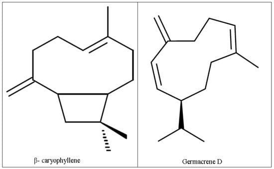
Figure 2.
Chemical structures of major compounds in L. montevidensis leaf essential oil.
3.2. Antioxidant Activity
3.2.1. Scavenging Activity of the Essential Oil from L. montevidensis on DPPH Radical
The effect of essential oil of L. montevidensis and ascorbic acid on DPPH reduction is shown in Figure 3. Ascorbic acid exhibited DPPH radical scavenging activity in a concentration-dependent manner (Figure 3), with IC50 value of 0.042 g/mL. However, L. montevidensis leaf essential oil did not exhibit DPPH radical scavenging activity at all the concentrations tested (Figure 3).
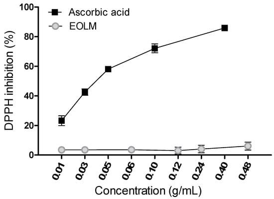
Figure 3.
Quenching of 1,1-Diphenyl-2-picrylhydrazyl (DPPH) color by the essential oil from L. montevidensis. Mean ± SEM of n = 4 independent experiments.
3.2.2. Fe2+ Chelation or Oxidation Potential of L. montevidensis Leaf Essential Oil
In Fe2+ chelating assay, the rate of the reduction in the absorbance of the orange complex formed by the interaction of Fe2+ and ortho-phenanthroline allows estimation of a possible chelator. Surprisingly, the incubation of L. montevidensis leaf essential oil (0.24 and 0.48 g/mL) with Fe2+ significantly increased the absorbance of the complex Fe2+ orthophenanthroline formed, when compared with that of Fe2+ alone (Figure 4). The absorbance remained unchanged after 20 min of incubation, but dramatically raised after addition of the reducing agent (ascorbic acid (Figure 4)). This finding may suggest that the leaf essential oil of L. montevidensis is oxidizing Fe2+ in the reaction medium.
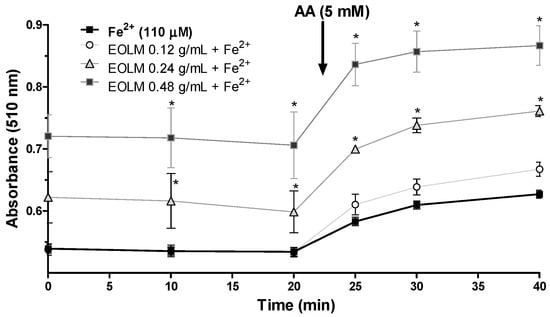
Figure 4.
Oxidation of Fe2+ by L. montevidensis leaf essential oil (0.12–0.48 g/mL). The values represent the mean ± SEM of three experiments that were performed in duplicate.
After 20 min incubation of Fe2+ with EOLM, ascorbic acid (AA) was added to the reaction medium to confirm whether or not the increase in absorbance in the presence of the oil was attributed to Fe2+. Absorbance did not change at 5, 10, and 20 min after addition of AA, suggesting that EOLM directly stimulated the oxidation of Fe2+ to Fe3+ during the incubation periods (prior to AA addition) (Figure 4).
3.2.3. Fe3+ Reducing Properties of L. montevidensis Leaf Essential Oil
The Fe3+ reducing properties of L. montevidensis is shown in Figure 5. Similar to that observed with Fe2+, the incubation of the essential oil of L. montevidensis with Fe3+ in the presence of ortho-phenanthroline resulted in a significant increase in the absorbance in a dose-dependent manner (p < 0.05) in comparison to that of Fe3+ alone. However, the absorbance of the complex formed with Fe3+ and ortho-phenanthroline was lower than that obtained with Fe3+ and ortho-phenanthroline (Figure 5). The addition of ascorbic acid to the reaction medium dramatically increased the absorbance of the sample (Figure 5), suggesting that the component(s) of the essential oil of L. montevidensis may have released Fe(II) in the medium or have reduced Fe3+ to Fe2+.
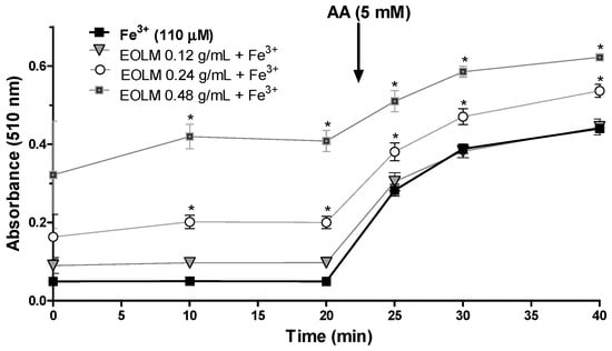
Figure 5.
Reduction of Fe3+ to Fe2+ (110 µM) by L. montevidensis leaf essential oil (0.12–0.48 g/mL). The oil was incubated with for 10 min.
3.3. Biological Assays with Drosophila Melanogaster
3.3.1. Fumigant Activity of Leaf Essential Oil L. montevidensis
The fumigant toxicity of the essential oil extracted from leaves of EOLM was investigated in Drosophila melanogaster. Exposure of flies to EOLM at all the concentrations tested (0.12, 0.24 and 0.48 g/mL) for up to 5 h, caused mortality below 10% (data not shown).
3.3.2. Quantification of Total Thiol and Non-Protein Thiols (GSH and NPSH)
Considering that antioxidant activity has been reported for L. montevidensis [9], we investigated the effects of EOLM on the indirect biomarker of oxidative stress, total thiol. Additionally, the levels of GSH, an antioxidant found in the intracellular medium, that acts to defend the cell from oxidative stress, were estimated in homogenates of D. melanogaster. The results presented in the Figure 6A,B revealed that the treatment with EOLM (0.12–0.48 g/mL) did not significantly alter the levels of total thiol (Figure 6A) and non-protein thiol (NPSH) (Figure 6B), when compared with the control (p < 0.05).
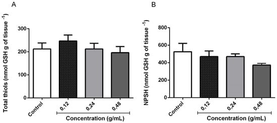
Figure 6.
Total thiol (A) and non-protein thiol (NPSH) content (B) in flies homogenates following 5 h exposure of D. melanogaster to L. montevidensis leaf essential oil.
3.3.3. Cell Viability of D. melanogaster Supernatant
MTT is reduced by the action of the enzyme succinate dehydrogenase in living cells to generate the purple chromophore, which is used to determine cell viability. The formation of the colored product is proportional to the number of viable cells in the supernatant. The EOLM at all concentrations tested did not change cellular viability of the supernatants when compared to the control (Figure 7).
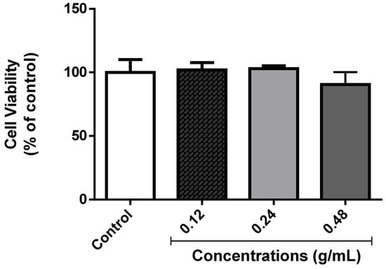
Figure 7.
Effect of EOLM on cellular viability of D. melanogaster homogenates.
3.3.4. Effect of EOLM on Fe2+ Content
To investigate possible oxidative damage, after exposure of D. melanogaster to different concentrations of EOLM, the levels of free Fe2+ ions were measured. As shown in Figure 8, exposure of flies to EOLM (0.12–0.48 g/mL) did not cause significant change in total iron content compared with the control group (p < 0.05).
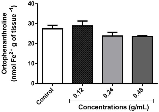
Figure 8.
Total iron levels in D. melanogaster treated with Lantana montevidensis leaf essential oil (EOLM).
3.3.5. Determination of Lipid Peroxidation
MDA, one of the well-known side products of LPO, is used as an index of lipid damage. For this reason, we measured the effect of EOLM on MDA levels following exposure. As shown in Figure 9, the EOLM at different concentrations tested did not alter MDA level in comparison with the control (p > 0.05).
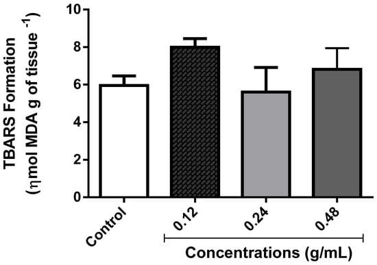
Figure 9.
Determination of the content of malondialdehyde (MDA).
4. Discussion
The use of in vivo alternative models in order to perform toxicological tests is increasing [15,40]. In this scenario, several model organisms have been used to identify the pharmacological properties of plants material or active components. Drosophila melanogaster is one of them, and has been used for more than 110 years to study the biological effects of compounds and the underlying pathophysiology of numerous diseases, including Alzheimer’s and Parkinson’s diseases [31,41,42].
Medicinal plants have been widely used for the prevention and/or treatment of various diseases, on the basis that they do not present genotoxic risks when consumed for long time [43]. In the current study, the antioxidant activity of Lantana montevidensis leaf essential oil was evaluated in vitro, while its potential toxic effect was investigated in vivo using D. melanogaster. In general, the essential oil of the leaves of L. montevidensis appeared to be rich in terpenes, (mainly, monoterpenes and sesquiterpenes), which can justify its strong odor [44]. EOLM showed only small differences in composition compared to data reported in the literature [24,25]. For instance, in the study carried out by Bezerra et al. [45], the chemical composition of the essential oil of the leaves of Lantana montevidensis was β-caryophyllene (34.96%), germacrene D (25.49%), and bicyclogermacrene (9.48%), while in the study by Sousa et al. [24], it was β-caryophyllene (31.5%), germacreno D (27.5%), and bicyclerecyroreno (13.9%). Such differences can be attributed to intrinsic and/or extrinsic factors such as environmental conditions and time and place of leaf collection [46,47,48]. β-caryophyllene (28.15%), one of the major component of L. montevidensis leaf essential oil, is a sesquiterpene present in essential oils of several plants families. Plants species that have this constituent exhibit a variety of pharmacological activities such as anti-inflammatory, antinociceptive, and antioxidant, among others [49,50,51]. d-germacrene (31.27%), the major component found in L. montevidensis leaf essential oil, is a common compound in plants, being considered a precursor for the biosynthesis of many sesquiterpenes [52]. DPPH is a stable radical, exhibiting a maximum absorbance at 517 nm. Such absorption by the action of antioxidants is taken as a measure of antioxidant activity [53]. It has widespread use in the assessment of radical elimination [54,55]. Here, the essential oil of L. montevidensis showed very low DPPH scavenging activity, and Fe(II) chelating activity, but demonstrated higher reducing power. The study carried out by Barros et al. [9] demonstrated the antioxidant activity of the aqueous and ethanolic extracts of L. montevidensis against DPPH radical, and its capacity to inhibit lipid peroxidation in rat brain. Our results are in agreement with that of Hossain and Shah [40], who found low antioxidant activity for other essential oils. Consistent with this, essential oils rich in monoterpenes (e.g., from Cupressus sempervirens, Phyllostachys nigra, Eucalyptus globulus, and Psidium guayava) have been shown to be ineffective in scavenging DPPH radical [14].
Plant essential oils (EOs) have been tested against a wide range of arthropod pests, with promising results. EOs have been reported to possess high efficacy, multiple mechanisms of action, low toxicity in non-target vertebrates, and the potential to act as reducing agents and stabilizers for the synthesis of nanopesticides [56]. Therefore, the reducing power of EOLM observed in this study could justify its potential use as a natural reducing agent.
It is widely recognized that reactive oxygen species (ROS) or reactive nitrogen species (RNS) induce oxidative stress by causing significant damage to cell structure directly or indirectly, leading to a number of diseases [57,58,59]. The major targets of ROS during oxidative stress are thought to be DNA, RNA, proteins, and lipids. Different reactive species have different degrees of reactivity with the cellular components, and the availability of free iron in the form of Fe2+ is considered to be of utmost importance in ROS toxicity due to its participation in the Fenton reaction that drives the formation of hydroxyl radicals [60]. Although free Fe2+ ions are known to exert vital functions in the organism at low concentrations [61], high level of free Fe2+ ions has been detected in several neurodegenerative diseases such as Alzheimer’s disease [62]. Therefore, compounds able to chelate Fe2+ to render it unavailable or less available for the participation in the Fenton reaction are of particular interest. Unfortunately, EOLM did not exhibit Fe2+ chelating activity.
Glutathione (GSH) is an essential compound in the maintenance of cellular homeostasis because of its reducing properties [63]. Protein thiol groups are inherently very reactive, and can be oxidized by reactive species. The balance between oxidized and free thiols is important not only for the maintenance of protein and enzymatic functions, but also for cellular redox balance [64]. The results obtained in this study showed that EOLM at all the concentrations tested did not alter the total thiol and NPSH levels and the MDA content of flies homogenates, indicating that EOLM does not cause oxidative stress since the total thiol depletion is associated with increased lipid peroxidation [65]. Thus, it is possible to presume that the beneficial effects of EOLM may be attributed at least, in part, to its reducing ability. In spite of the fact exposure of flies to EOLM (0.12–0.48 g/mL) did not affect MDA content (an index of lipid peroxidation), there is report showing the potential of L. montevidensis to decrease LPO in egg phospholipid [25], and to exert antioxidant activity in vitro [9,25].
5. Conclusions
In conclusion, the present study demonstrated that Lantana montevidensis leaf essential oil was ineffective in scavenging the DPPH radical, but exhibited reducing activity in vitro. Exposure of Drosophila melanogaster to L. montevidensis leaf essential oil (0.12–0.48 g/mL) for 5 h did not significantly alter markers of oxidative, as evidenced by there being no change in total thiol, GSH, and MDA contents.
Author Contributions
Conceptualization M.R.C.d.O.; C.D.M.O.T.; V.A.P.d.O. and B.A.F.d.S., prepared the essential oil and chemical analysis M.G.d.L.S. and A.A.B., writing – Original Draft Preparation M.R.C.d.O.; H.D.M.C.; A.O.B.P.B., designed the study I.R.A.d.M.; L.M.B.; A.E.D. and J.P.K., review and editing I.R.A.d.M.; supervision, I.R.A.d.M. and H.D.M.C.
Funding
This research received no external funding.
Acknowledgments
This research was funded by Coordenação de Aperfeiçoamento de Pessoal de Nível Superior-Brasil (CAPES), Fundação Cearense de Apoio ao Desenvolvimento Científico e Tecnológico (FUNCAP), Conselho Nacional de Desenvolvimento Científico e Tecnológico (CNPq) and Financiadora de Estudos e Projetos-Brasil (FINEP). The authors would like to thank the professors from NAPO (Center for Analysis and Organic Research at UFSM) for providing the GC/MS chromatograms and spectra and A.F. Morel (Department of Chemistry at UFSM) for the assessment of the n-alkane series.
Conflicts of Interest
The authors declare no conflict of interest.
References
- Hua, H.; Xing, F.; Selvara, J.N.; Wang, Y.; Zhao, Y.; Zhou, L.; Liu, X.; Liu, Y. Inhibitory effect of essential oils on Aspergillus ochraceus growth and ochratoxin a production. PLoS ONE 2014, 9, e108285. [Google Scholar] [CrossRef] [PubMed]
- Kamdem, J.P.; Abolaji, A.O.; Elekofehinti, O.O.; Omotuyi, I.O.; Ibrahim, M.; Hassan, W.; Barbosa, N.V.; Souza, D.O.; Rocha, J.B.T. Therapeutic Potential of Plant Extracts and Phytochemicals Against Brain Ischemia-Reperfusion Injury: A Review. Nat. Prod. J. 2016, 6, 250–284. [Google Scholar] [CrossRef]
- Fang, Y.Z.; Yang, S.; Wu, G. Freeradicals, antioxidants, andnutrition. Nutrition 2002, 18, 872–879. [Google Scholar] [PubMed]
- Rao, K.M.; Shashidhara, L.S. Human APC sequesters β-catenin even in the absence of GSK-3β in a Drosophila model. Oncogene 2008, 27, 2488–2493. [Google Scholar] [CrossRef] [PubMed][Green Version]
- Chikezie, P.C.; Uwakwe, A.A. Membrane stability of sickle erythrocytes incubated in essential oil of three medicinal plants: Anacardium occidentale, Psidium guajava, and Terminalia catappa. Pharmacogn. Mag. 2011, 7, 121. [Google Scholar] [CrossRef] [PubMed]
- De Beer, D.; Joubert, E.; Gelderblom, W.C.A.; Manley, M. Phenolic compounds: A review of their possible role as in vivo antioxidants of wine. S. Afr. J. Enol. Vitic. 2017, 23, 48–61. [Google Scholar] [CrossRef][Green Version]
- Bekhit, A.E.D.A.; Cheng, V.J.; McConnell, M.; Zhao, J.H.; Sedcole, R.; Harrison, R. Antioxidant activities, sensory and anti-influenza activity of grape skin tea infusion. Food Chem. 2011, 129, 837–845. [Google Scholar]
- Vasilescu, I.; Eremia, S.A.; Albu, C.; Radoi, A.; Litescu, S.C.; Radu, G.L. Determination of the antiradical properties of olive oils using an electrochemical method based on DPPH radical. Food Chem. 2015, 166, 324–329. [Google Scholar] [CrossRef] [PubMed]
- Barros, L.M.; Duarte, A.E.; Waczuk, E.P.; Roversi, K.; da Cunha, F.A.B.; Rolon, M.; Coronel, C.; Gomez, M.C.V.; de Menezes, I.R.A.; da Costa, J.G.M.; et al. Safety assessment and antioxidant activity of Lantana montevidensis leaves: Contribution to its phytochemical and pharmacological activity. EXCLI J. 2017, 16, 566–582. [Google Scholar] [PubMed]
- Scarfiotti, C.; Fabris, F.; Cestaro, B.; Giuliani, A. Free radicals, atherosclerosis, ageing, and related dysmetabolic pathologies: Pathological and clinical aspects. Eur. J. Cancer Prev. 1997, 6, 31–36. [Google Scholar] [CrossRef]
- Garcia, G.R.; Goodale, B.C.; Wiley, M.W.; La Du, J.K.; Hendrix, D.A.; Tanguay, R.L. In vivo characterization of an AHR-dependent long non-coding RNA required for proper Sox9b expression. Mol. Pharmacol. 2017, 91, 609–619. [Google Scholar] [CrossRef] [PubMed]
- Carmona-Jiménez, Y.; García-Moreno, M.V.; Igartuburu, J.M.; Barroso, C.G. Simplification of the DPPH assay for estimating the antioxidant activity of wine and wine by-products. Food Chem. 2014, 165, 198–204. [Google Scholar] [CrossRef] [PubMed]
- Musa, K.H.; Abdullah, A.; Kuswandi, B.; Hidayat, M.A. A novel high throughput method based on the DPPH dry reagent array for 2 determination of antioxidant activity. Food Chem. 2013, 141, 4102–4106. [Google Scholar] [CrossRef] [PubMed]
- Sacchetti, G.; Maietti, S.; Muzzoli, M.; Scaglianti, M.; Manfredini, S.; Radice, M.; Bruni, R. Comparative evaluation of 11 essential oils of different origin as functional antioxidants, antiradicals and antimicrobials in foods. Food Chem. 2005, 91, 621–632. [Google Scholar] [CrossRef]
- Yassa, N.; Masoomi, F.; Rankouhi, S.R.; Hadjiakhoondi, A. Chemical composition and antioxidant activity of the extract and essential oil of Rosa damascena from Iran, population of Guilan. DARU J. Pharm. Sci. 2015, 17, 175–180. [Google Scholar]
- Xie, Y.J.; Wang, K.; Huang, Q.Y.; Lei, C.L. Evaluation toxicity of monoterpenes to subterranean termite, Reticulitermes chinensis Snyder. Ind. Crops Prod. 2014, 53, 163–166. [Google Scholar] [CrossRef]
- Neahoua-Bougherra, H.H.; Bedini, S.; Cosci, F.; Flamini, G.; Belhamel, K.; Conti, B. Enhancing the insecticidal efficacy of inert dusts against stored food insect pest by the combined action with essential oils. IOBC-WPRS Bull. 2015, 111, 31–38. [Google Scholar]
- Mansoura, S.A.; El-Sharkawy, A.Z.; Abdel-Hamid, N.A. Toxicity of essential plant oils, in comparison with conventional insecticides, against the desert locust, Schistocerca gregaria (Forskål). Ind. Crops Prod. 2015, 63, 92–99. [Google Scholar] [CrossRef]
- Peixoto, M.G.; Bacci, L.; Blank, A.F.; Araújo, A.P.A.; Alves, P.B.; Silva, J.H.S.; Santos, A.A.; Oliveira, A.P.; da Costa, A.S.; Blank, M.F.A. Toxicity and repellency of essential oils of Lippia alba chemotypes and their major monoterpenes against stored grain insects. Ind. Crops Prod. 2015, 71, 31–36. [Google Scholar] [CrossRef]
- Zhang, Z.; Yang, T.; Zhang, Y.; Wang, L.; Xie, Y. Fumigant toxicity of monoterpenes against fruitfly, Drosophila melanogaster. Ind. Crops Prod. 2016, 81, 147–151. [Google Scholar] [CrossRef]
- Nagao, T.; Abe, F.; Kinjo, J.; Okabe, H. Antiproliferative constituents in plants. 10 flavones from the leaves of Lantana montevidensis BRIQ. and consideration of structure activity relationship. Biol. Pharm. Bull. 2002, 25, 875–879. [Google Scholar] [CrossRef]
- Barreto, F.S.; Sousa, E.O.; Rodrigues, F.F.G.; Costa, J.G.M.; Campos, A.R. Antibacterial Activity of Lantana camara Linn Lantana montevidensis Briq extracts from Cariri-Ceara, Brazil. J. Young Pharm. 2010, 2, 42–44. [Google Scholar] [CrossRef]
- Sousa, E.O.; Almeida, T.S.; Rodrigues, F.F.; Campos, A.R.; Lima, S.G.; Costa, J.G. Lantana montevidensis Briq improves the aminoglycoside activity against multiresistant Escherichia coli and Staphylococcus aureus. Indian J. Pharmacol. 2011, 43, 180–182. [Google Scholar]
- Sousa, E.O.; Barreto, F.S.; Rodrigues, F.F.; Campos, A.R.; Costa, J.G. Chemical composition of the essential oils of Lantana camara L. and Lantana montevidensis Briq. and their synergistic antibiotic effects on aminoglycosides. J. Essent. Oil Res. 2012, 24, 447–452. [Google Scholar] [CrossRef]
- Sousa, E.O.; Rodrigues, F.F.G.; Campos, A.R.; Lima, S.G.; da Costa, J.G.M. Chemical composition and synergistic interaction between aminoglycosides antibiotics and essential oil of Lantana montevidensis Briq. Nat. Prod. Res. 2013, 27, 942–945. [Google Scholar] [CrossRef]
- Nichols, C.D. Drosophila melanogaster neurobiology, neuropharmacology, and how the fly can inform central nervous system drug discovery. Pharmacol. Ther. 2006, 112, 677–700. [Google Scholar] [CrossRef]
- Pandey, U.B.; Nichols, C.D. Human disease models in Drosophila melanogaster and the role of the fly in therapeutic drug discovery. Pharmacol. Rev. 2011, 63, 411–436. [Google Scholar] [CrossRef]
- Feany, M.B.; Bender, W.W. A Drosophila model of Parkinson’s disease. Nature 2000, 404, 394–398. [Google Scholar] [CrossRef]
- Tiwari, S.; Gondhalekar, A.D.; Mann, R.S.; Scharf, M.E.; Stelinski, L.L. Characterization of five CYP4 genes from Asian citrus psyllid and their expression levels in Candidatus Liberibacter asiaticus-infected and uninfected psyllids. Insect Mol. Biol. 2011, 20, 733–744. [Google Scholar] [CrossRef]
- Zolfaghari Emameh, R.; Syrjänen, L.; Barker, H.; Supuran, C.T.; Parkkila, S. Drosophila melanogaster: A model organism for controlling Dipteran vectors and pests. J. Enzyme Inhib. Med. Chem. 2015, 30, 505–513. [Google Scholar] [CrossRef]
- Panchal, K.; Tiwari, A.K. Drosophila melanogaster “a potential model organism” for identification of pharmacological properties of plants/plant-derived components. Biomed. Pharmacother. 2017, 89, 1331–1345. [Google Scholar] [CrossRef]
- Matos, F.J.A. Introduction to Experimental Phytochemistry, 2nd ed.; UFC: Fortaleza, Brazil, 2002. [Google Scholar]
- Adams, R.P. Identification of Essential Oil Components by Gas Chromatography/Mass Spectrometry; Allured Publishing Corporation: Carol Stream, IL, USA, 2007. [Google Scholar]
- Kamdem, J.P.; Stefanello, S.T.; Boligon, A.A.; Wagner, C.; Kade, I.J.; Pereira, R.P.; Preste, A.S.; Roos, D.H.; Waczuk, E.P.; Appel, A.S.; et al. In vitro antioxidant activity of stem bark of Trichilia catigua Adr. Juss. Acta Pharm. 2012, 62, 371–382. [Google Scholar] [CrossRef] [PubMed]
- Dinis, T.C.P.; Madeira, V.M.C.; Almeida, M.L.M. Action of phenolic derivates (acetoaminophen, salycilate and 5-aminosalycilate) as inhibitors of membrane lipid peroxidation and as peroxyl radical scavengers. Arch. Biochem. Biophys. 1994, 315, 161–169. [Google Scholar] [CrossRef]
- Kamdem, J.P.; Adeniran, A.; Boligon, A.A.; Klimaczewsk, C.V.; Elekofehinti, O.O.; Hassan, W.; Ibrahim, M.; Waczuk, E.P.; Meinerz, D.F.; Athayde, M.L. Antioxidant activity, genotoxicity and cytotoxicity evaluation of lemon balm (Melissa officinalis L.) ethanolic extract: Its potential role in neuroprotection. Ind. Crops Prod. 2013, 51, 26–34. [Google Scholar] [CrossRef]
- Mosmann, T. Rapid colorimetric assay for cellular growth and survival: Application to proliferation and cytotoxicity assays. J. Immunol. Methods 1983, 65, 55–63. [Google Scholar] [CrossRef]
- Klimaczewsk, C.V.; Saraiva, R.A.; Roos, D.H.; Boligon, A.; Athayde, M.L.; Kamdem, J.P.; Barbosa, N.V.; Rocha, J.B.T. Antioxidant activity of Peumus boldus extract and alkaloid boldine against damage induced by Fe (II)–citrate in rat liver mitochondria in vitro. Ind. Crops Prod. 2014, 54, 240–247. [Google Scholar] [CrossRef]
- Barbosa Filho, V.M.; Waczuk, E.P.; Kamdem, J.P.; Abolaji, A.O.; Lacerda, S.R.; da Costa, J.G.; de Menezes, I.R.; Boligon, A.A.; Athayde, M.L.; da Rocha, J.B.; et al. Phytochemical constituents, antioxidant activity, cytotoxicity and osmotic fragility effects of Caju (Anacardium microcarpum). Ind. Crops Prod. 2014, 55, 280–288. [Google Scholar] [CrossRef]
- Hossain, M.A.; Shah, M.D. A study on the total phenols content and antioxidant activity of essential oil and different solvent extracts of endemic plant Merremia borneensis. Arab. J. Chem. 2015, 8, 66–71. [Google Scholar] [CrossRef]
- Siddique, Y.H.; Mujtaba, S.F.; Jyoti, S.; Naz, F. GC-MS analysis of Eucalyptus citriodora leaf extract and its role on the dietary supplementation in transgenic Drosophila model of Parkinson’s disease. Food Chem. Toxicol. 2013, 55, 29–35. [Google Scholar] [CrossRef]
- Liu, Q.F.; Lee, J.H.; Kim, Y.M.; Lee, S.; Hong, Y.K.; Hwang, S.; Oh, Y.; Lee, K.; Yun, H.S.; Lee, I.S.; et al. In vivo screening of traditional medicinal plants for neuroprotective activity against Aβ42 cytotoxicity by using Drosophila models of Alzheimer’s disease. Biol. Pharm. Bull. 2015, 38, 1891–1901. [Google Scholar] [CrossRef] [PubMed]
- Bakkali, F.; Averbeck, S.; Averbeck, D.; Idaomar, M. Biological effects of essential oils—A review. Food Chem. Toxicol. 2008, 46, 446–475. [Google Scholar] [CrossRef]
- Guilhon de Castro, H.; Borges de Moura Perini, V.; Rodrigues dos Santos, G.; Castro Alves Barros Leal, T. Avaliação do teor e composição do óleo essencial de Cymbopogon nardus (L.) em diferentes épocas de colheita. Revista Ciência Agronômica, 2010; 41, 308–314. [Google Scholar]
- Bezerra, J.W.A.; Rodrigues, F.C.; Costa, A.R.; Boligon, A.A.; da Rocha, J.B.T.; Barros, L.M. Estudo químico-biológico do óleo essencial de Lantana montevidensis (chumbinho) (Spreng.) Briq. (Verbenaceae) contra Drosophila melanogaster. Rev. Bras. Plantas Med. 2017, 22, 1–13. [Google Scholar]
- Charles, D.J.; Simon, J.E. Comparison of extraction methods for the rapid determination of essential oil content and composition of basil. J. Am. Soc. Hortic. Sci. 1990, 115, 458–462. [Google Scholar] [CrossRef]
- Jorge, S.S.A.; Nardes, P.R.B.; Guarim, N.G.; Macedo, M. O uso medicinal da arnica, Brickelia brasiliensis (Spreng.) Robinson (Asteraceae). Revista Saúde e Ambiente 1998, 1, 107–121. [Google Scholar]
- Facanali, R.; Campos, M.M.S.; Pocius, O.; Ming, L.C.; Soares-Scott, M.D.; Marques, M.O.M. Biologia reprodutiva de populações de Ocimum selloi Benth. Rev. Bras. Plantas Med. 2009, 11, 141–146. [Google Scholar] [CrossRef]
- Passos, G.F.; Fernandes, E.S.; da Cunha, F.M.; Ferreira, J.; Pianowski, L.F.; Campos, M.M.; Calixto, J.B. Anti-inflammatory and anti-allergic properties of the essential oil and active compounds from Cordia verbenacea. J. Ethnopharmacol. 2007, 110, 323–333. [Google Scholar] [CrossRef]
- Marinho, D.F.; Oliveira, E.C.P.D.; Araújo, J.A.D.S.; Pinto, I.F.; Lima, H.S.D.; Moraes, W.P.; Ambrósio, C.E.; Morini, A.C. Evaluation of ultrasonic transmission of Copaifera duckei Dwyer herbal gel. Pesqui. Veterinária Bras. 2017, 37, 516–520. [Google Scholar] [CrossRef][Green Version]
- Martins, A.O.; Rodrigues, L.B.; Cesário, F.R.; de Oliveira, M.R.; Tintino, C.D.; e Castro, F.F.; Alcântara, I.S.; Fernandes, M.N.; de Albuquerque, T.R.; da Silva, M.S.; et al. Anti-edematogenic and anti-inflammatory activity of the essential oil from Croton rhamnifolioides leaves and its major constituent 1, 8-cineole (eucalyptol). Biomed. Pharmacother. 2017, 96, 384–395. [Google Scholar] [CrossRef]
- Steliopoulos, P.; Wüst, M.; Adam, K.P.; Mosandl, A. Biosynthesis of the sesquiterpene germacrene D in Solidago canadensis: 13C and 2H labeling studies. Phytochemistry 2002, 60, 13–20. [Google Scholar] [CrossRef]
- Frankel, E.N.; Meyer, A.S. The problem of using one- dimensional methods to evaluate multifunctional food and biological antioxidants. J. Sci. Food Agric. 2000, 80, 1925–1941. [Google Scholar] [CrossRef]
- Siddhuraju, P.; Becker, K. Antioxidant properties of various solvent extracts of total phenolic constituents from three different agroclimatic origins of drumstick tree (Moringa oleifera Lam.) leaves. J. Agric. Food Chem. 2003, 51, 2144–2155. [Google Scholar] [CrossRef]
- Siddhuraju, P.; Becker, K. The antioxidant and free radical scavenging activities of processed cowpea (Vignaunguiculata (L.) Walp.) seed extracts. Food Chem. 2007, 101, 10–19. [Google Scholar] [CrossRef]
- Pavela, R.; Benelli, G. Essential oils as ecofriendly biopesticides? Challenges and constraints. Trends Plant Sci. 2016, 21, 1000–1007. [Google Scholar] [CrossRef]
- Pistón, M.; Machado, I.; Branco, C.S.; Cesio, V.; Heinzen, H.; Ribeiro, D.; Fernandes, E.; Chisté, R.C.; Freitas, M. Infusion, decoction and hydroalcoholic extracts of leaves from artichoke (Cynara cardunculus L. subsp. cardunculus) are effective scavengers of physiologically relevant ROS and RNS. Food Res. Int. 2014, 64, 150–156. [Google Scholar]
- Gupta, D.K.; Palma, J.M.; Corpas, F.J. Reactive Oxygen Species and Oxidative Damage in Plants under Stress; Springer: Heidelberg, Germany, 2015; pp. 1–22. [Google Scholar]
- Wang, J.; Hu, S.; Nie, S.; Yu, Q.; Xie, M. Reviews on mechanisms of in vitro antioxidant activity of polysaccharides. Oxidative Med. Cell. Longev. 2016, 2016. [Google Scholar] [CrossRef]
- Mittler, R. ROS are good. Trends Plant Sci. 2017, 22, 11–19. [Google Scholar] [CrossRef]
- Oliveira, F.; Rocha, S.; Fernandes, R. Iron metabolism: from health to disease. J. Clin. Lab. Anal. 2014, 28, 210–218. [Google Scholar] [CrossRef]
- Connor, J.R.; Malecki, E.A.; Cable, E.E.; Isom, H.C. The lipophilic iron compound TMH-ferrocene [(3, 5, 5-trimethylhexanoyl) ferrocene] increases iron concentrations, neuronal l-ferritin, and heme oxygenase in brains of BALB/c mice. Biol. Trace Elem. Res. 2002, 86, 73–84. [Google Scholar] [CrossRef]
- Rooney, J.P. The role of thiols, dithiols, nutritional factors and interacting ligands in the toxicology of mercury. Toxicology 2007, 234, 145–156. [Google Scholar] [CrossRef]
- Ferreira, I.C.; Abreu, R. Oxidative stress, antioxidants and phytochemicals. Bioanálise 2007, 4, 32–39. [Google Scholar]
- Ghorbel, I.; Khemakhem, M.; Boudawara, O.; Marrekchi, R.; Jamoussi, K.; Amar, R.B.; Boudawara, T.; Zeghal, N.; Kamoun, N.G. Effects of dietary extra virgin olive oil and its fractions on antioxidant status and DNA damage in the heart of rats co-exposed to aluminum and acrylamide. Food Funct. 2015, 6, 3098–3108. [Google Scholar] [CrossRef]
© 2019 by the authors. Licensee MDPI, Basel, Switzerland. This article is an open access article distributed under the terms and conditions of the Creative Commons Attribution (CC BY) license (http://creativecommons.org/licenses/by/4.0/).