Review

Expanding the Role of Ultrasound for the Characterization of Renal Masses
by
Eduard Roussel
1,*,
Riccardo Campi
2,
Daniele Amparore
3,
Riccardo Bertolo
4,
Umberto Carbonara
5,
Selcuk Erdem
6,
Alexandre Ingels
7,
Önder Kara
8,
Laura Marandino
9,
Michele Marchioni
10,
Stijn Muselaers
11,
Nicola Pavan
12,
Angela Pecoraro
3,
Benoit Beuselinck
13,
Ivan Pedrosa
14,15,16,
David Fetzer
14,
Maarten Albersen
1 and
on behalf of the European Association of Urology (EAU) Young Academic Urologists (YAU) Renal Cancer Working Group
1
Department of Urology, University Hospitals Leuven, 3000 Leuven, Belgium
2
Unit of Urological Robotic Surgery and Renal Transplantation, Careggi Hospital, University of Florence, 50134 Firenze, Italy
3
Department of Urology, San Luigi Gonzaga Hospital, University of Turin, 10043 Orbassano, Italy
4
Department of Urology, San Carlo Di Nancy Hospital, 00165 Rome, Italy
5
Department of Emergency and Organ Transplantation-Urology, Andrology and Kidney Transplantation Unit, University of Bari, 70121 Bari, Italy
6
Division of Urologic Oncology, Department of Urology, Istanbul University Istanbul Faculty of Medicine, 34093 Istanbul, Turkey
7
Department of Urology, University Hospital Henri Mondor, 94000 Créteil, France
8
Department of Urology, Kocaeli University School of Medicine, 41001 Kocaeli, Turkey
9
Department of Medical Oncology, Fondazione IRCCS Istituto Nazionale dei Tumori, 20133 Milan, Italy
10
Department of Medical, Oral and Biotechnological Sciences, G. d’Annunzio University of Chieti, 66100 Chieti, Italy
11
Department of Urology, Radboud University Medical Center, 6525 GA Nijmegen, The Netherlands
12
Urology Clinic, Department of Medical, Surgical and Health Science, University of Trieste, 34127 Trieste, Italy
13
Department of General Medical Oncology, University Hospitals Leuven, 3000 Leuven, Belgium
14
Department of Radiology, University of Texas Southwestern Medical Center, Dallas, TX 75390, USA
15
Advanced Imaging Research Center, University of Texas Southwestern Medical Center, Dallas, TX 75390, USA
16
Department of Urology, University of Texas Southwestern Medical Center, Dallas, TX 75390, USA
*
Author to whom correspondence should be addressed.
Membership of the European Association of Urology (EAU) Young Academic Urologists (YAU) Renal Cancer Working Group is provided in Acknowledgments.
J. Clin. Med. 2022, 11(4), 1112; https://doi.org/10.3390/jcm11041112
Submission received: 24 December 2021 / Revised: 16 February 2022 / Accepted: 17 February 2022 / Published: 19 February 2022
(This article belongs to the Special Issue Hot Topics and New Frontiers in Kidney Cancer)

Abstract

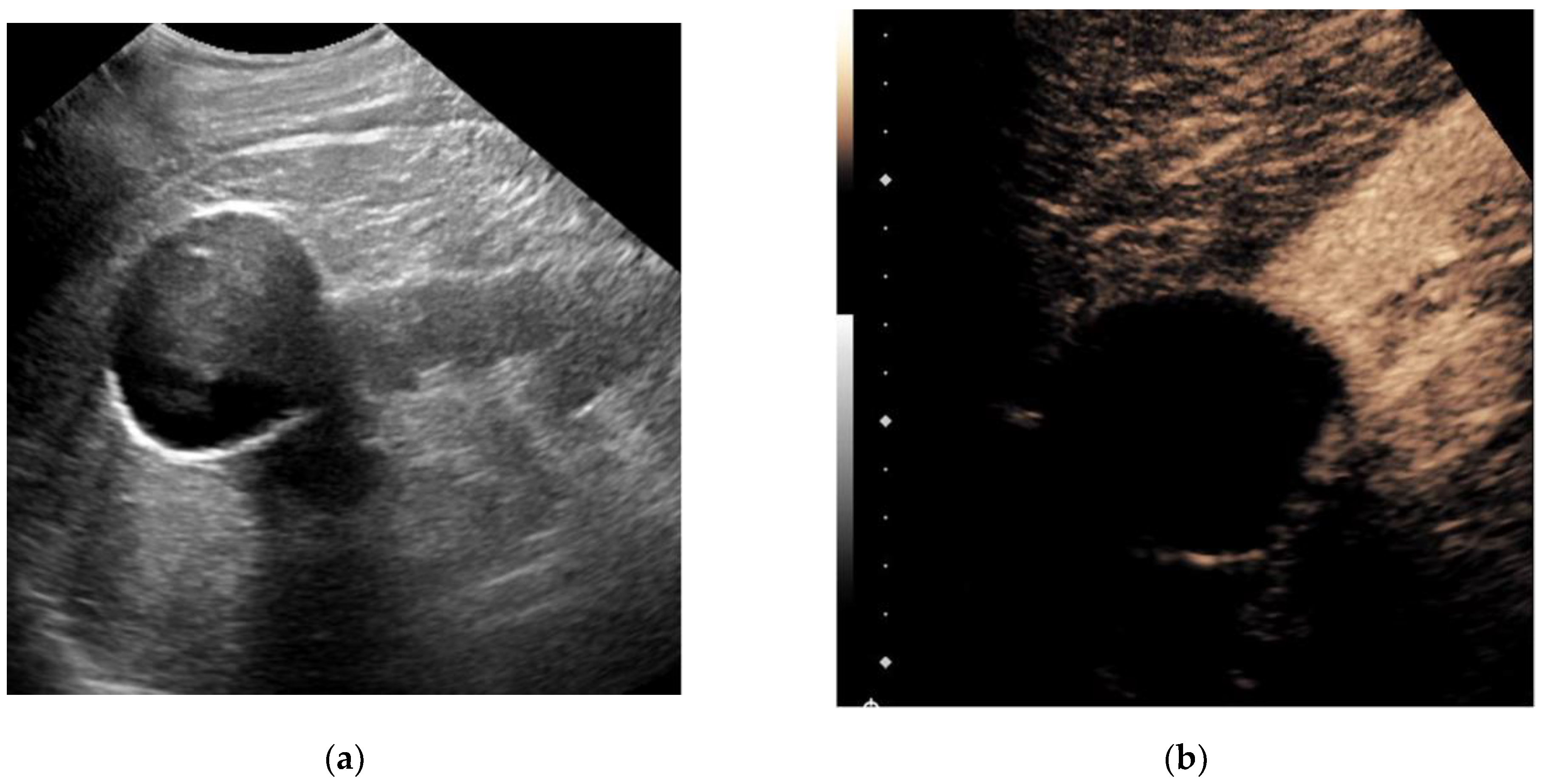
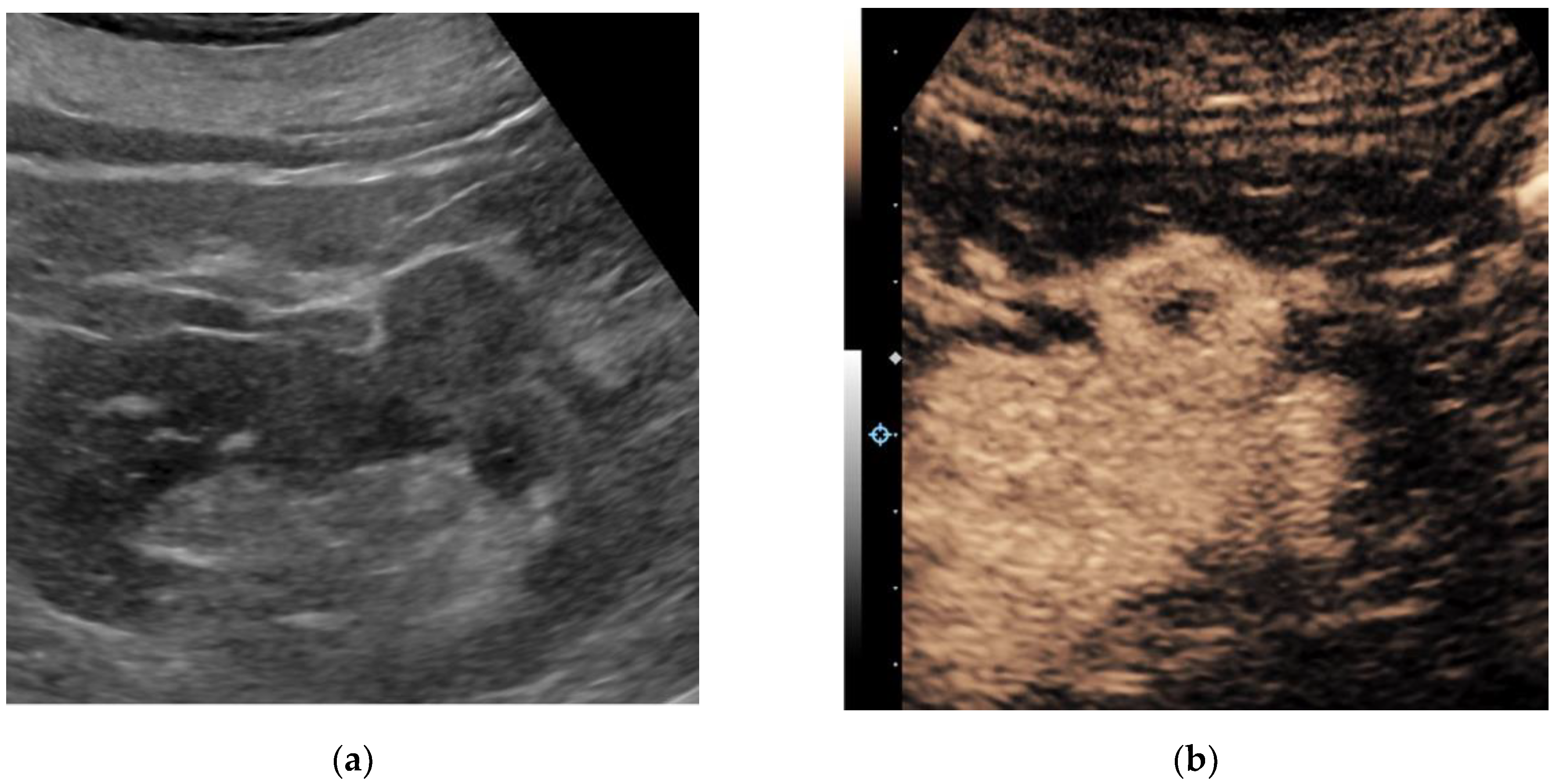
The incidental detection of renal masses has been steadily rising. As a significant proportion of renal masses that are surgically treated are benign or indolent in nature, there is a clear need for better presurgical characterization of renal masses to minimize unnecessary harm. Ultrasound is a widely available and relatively inexpensive real-time imaging technique, and novel ultrasound-based applications can potentially aid in the non-invasive characterization of renal masses. Evidence acquisition: We performed a narrative review on novel ultrasound-based techniques that can aid in the non-invasive characterization of renal masses. Evidence synthesis: Contrast-enhanced ultrasound (CEUS) adds significant diagnostic value, particularly for cystic renal masses, by improving the characterization of fine septations and small nodules, with a sensitivity and specificity comparable to magnetic resonance imaging (MRI). Additionally, the performance of CEUS for the classification of benign versus malignant renal masses is comparable to that of computed tomography (CT) and MRI, although the imaging features of different tumor subtypes overlap significantly. Ultrasound molecular imaging with targeted contrast agents is being investigated in preclinical research as an addition to CEUS. Elastography for the assessment of tissue stiffness and micro-Doppler imaging for the improved detection of intratumoral blood flow without the need for contrast are both being investigated for the characterization of renal masses, though few studies have been conducted and validation is lacking. Conclusions: Several novel ultrasound-based techniques have been investigated for the non-invasive characterization of renal masses. CEUS has several advantages over traditional grayscale ultrasound, including the improved characterization of cystic renal masses and the potential to differentiate benign from malignant renal masses to some extent. Ultrasound molecular imaging offers promise for serial disease monitoring and the longitudinal assessment of treatment response, though this remains in the preclinical stages of development. While elastography and emerging micro-Doppler techniques have shown some encouraging applications, they are currently not ready for widespread clinical use.
Keywords: ultrasound; ultrasonography; contrast-enhanced ultrasound; micro-Doppler; molecular ultrasound; renal mass; renal tumor; renal cell carcinoma ultrasound; ultrasonography; contrast-enhanced ultrasound; micro-Doppler; molecular ultrasound; renal mass; renal tumor; renal cell carcinoma