Article

Pseudostichococcus Stands Out from Its Siblings Due to High Salinity and Desiccation Tolerance

Institute for Biological Sciences, Applied Ecology and Phycology, University of Rostock, 18059 Rostock, Germany
*
Author to whom correspondence should be addressed.
Phycology 2022, 2(1), 108-119; https://doi.org/10.3390/phycology2010007
Submission received: 30 November 2021 / Revised: 26 January 2022 / Accepted: 9 February 2022 / Published: 11 February 2022

Abstract

:
Desiccation and high salinity are two abiotic stressors that are related in terms of their effect on water homeostasis within cells. The success of certain aeroterrestrial microalgae is influenced by their ability to cope with desiccation, and in some cases, high salinity. The microalgae of the Stichococcus clade are ubiquitous in terrestrial habitats and are known to withstand desiccation and salinity stress by accumulating secondary metabolites. Nevertheless, it remains unclear if those two related stressors have a synergistic effect. Hence, we studied the effect of salinity on desiccation on various representative taxa within the Stichococcus clade. The results showed that in contrast to other Stichococcus taxa, Pseudostichococcus was able to recover fully after desiccation, with and without salinity stress. This observation was connected to elevated proline production under salinity stress and higher proline:sorbitol ratio in Pseudostichococcus to the other strains tested. In the other taxa, increasing salinity reduced their ability to withstand desiccation. This might have severe effects on microalgae in (semi)arid regions, where salinization of soils is an increasing threat also for agriculture. The results encourage further research to be done on the possible applications of this genus in salinity bioremediation, as it seems to be comparable to other halotolerant green algae used for this purpose.

1. Introduction

Microalgae inhabiting terrestrial environments must cope with irregular water availability and, depending on the specific habitat, significant salt stress. The ecological success of terrestrial algae may be attributed to their ability to withstand repeated and extended drought-rewetting cycles, as well as UV and temperature stress. Salt stress is related to dehydration, but is defined as “physiological drought” [1], where osmotic stress disrupts normal cellular ion concentrations, even if water in the environment is plentiful. In addition, dissociated Na+ and Cl ions are cytotoxic in high concentrations [2]. There has been extensive research done on the adaptive mechanisms in terrestrial microalgae to desiccation [3,4,5,6] and salt stress [7,8,9]. However, the focus on these two stressors in combination has been primarily in crop plants and marine seaweeds [10,11,12,13].
Desiccation resistance in plants is well-studied. Two strategies exist, namely the growth of protective physical barriers as well as rapid cellular repair mechanisms that are activated upon rehydration [14,15]. Similarly, strategies to cope with salt stress can be broadly grouped into ion exclusion and tissue tolerance. In the former, Na+ and Cl are kept as out of vital tissues and compatible solutes are produced to maintain osmotic balance. In the latter, salt is assimilated to levels higher than the outside environment and stored in vacuoles and K+ is used to maintain osmotic pressure [16]; some plants as well as many marine seaweeds implement vacuolar ATP-driven Na+/K+ pumps to actively transport Na+ ions to the cellular exterior [13,17]. Salt sensitive plants, as many microalgae do, rely primarily on ion exclusion, particularly through the production of compatible solutes. Halophytes rely instead on tissue tolerance, as tissue tolerance requires constant exposure and extensive adaptations to high salinities; furthermore, research on plant growth in arid and salinized environments has focused heavily on species that are halophytic or halotolerant, as the precise factors, mechanisms and genetic basis behind salinity tolerance in plants are still not fully understood [12,16,18,19].
Except for studies on the salt and desiccation tolerance of Dunaliella salina [20,21,22], there has been comparatively little research done on this stressor combination in green microalgae, perhaps due to lack of economic pressure. While halotolerant algae have chiefly been used to bioremediate saline waters and salinized soils, crop yield increase is also a growing field in applied phycology [12,23,24,25]. However, recent work has shown that some algae were able to form young biocrusts on hypersaline potash tailing heaps, with ramifications for salinity bioremediation [26,27,28]. Among the organisms isolated form salt heaps were Chloroidium ellipsoideum, the cyanobacterium Nostoc, representatives from Stichococcus and Pseudostichococcus [27].
The taxonomic history of the genus Stichococcus Nägeli, 1849 deserves mentioning here, as it has been frequently revised since its first description [29]. Pseudostichococcus L. Moewus 1951, was distinguished from Stichococcus Nägeli initially based on morphological and physiological differences [30], and then later via molecular data [31]. Pröschold and Darienko created from Stichococcus the genera Protostichococcus, Deuterostichococcus, Tritostichococcus and Tetratostichococcus on the basis of molecular and morphological differences [31]. In publications, these Stichococcus-adjacent genera have been broadly grouped into the “Prasiola-clade”, i.e., Prasiolales, or even generally to the Trebouxiophycaeae [31,32,33,34]. However, the validly published family name Stichococcaceae has been recently rediscovered and the authority of the family determined to be K. Bohlin 1901 [35] (M. Guiry and S. Heesch, personal communication, July 2020). The members of the Stichococcaceae comprise the “Stichococcus-like” genera.
The discovery of Stichococcus and Pseudostichococcus in a highly saline habitat is unusual, because Stichococcus has hitherto been considered euryhaline [36,37,38]. As with the other Stichococcus-like organisms, Pseudostichococcus are present in a variety of habitats, such as such as tree bark, surfaces of buildings and outdoor structures, diverse soils and are also components of biocrusts. A previous study indicated that Pseudostichococcus may be more halotolerant than previously assumed, as those strains retained vitality despite cultivation at very high salinities [39].
Stichococcus uses the ion exclusion strategy, producing sorbitol and proline as major compatible solutes to withstand osmotic stress [8,37,40]. It was confirmed that members of the seven newly described Stichococcus-like genera have the same osmolyte pattern, with the addition of small amounts of sucrose under high salinity. Strains from this clade were highly resistant to strong dehydration and high recovery rates after rehydration, as well as certain strains being halotolerance.
In this study we extend our work done previously [39] and profile osmolyte production and strain vitality under desiccation stress following cultivation in nonsaline and saline media, with a focus on various strains within Pseudostichococcus. The combination of stressors is intended to reflect conditions in saline terrestrial habitats, which may guide further efforts in algal bioremediation of, for example, salt heaps or salinized soils. In addition, a comparison between Pseudostichococcus and other Stichococcaceae strains expressed as ecophysiological differences will be examined in more detail. When possible, authentic strains were used to maximize taxonomic value, since these are type strains confirmed to be isolated or deposited into a culture collection by the epithet authority.

2. Materials and Methods

Nine established unialgal strains of Pseudostichococcus and other Stichococcaceae were used (Table 1). All cultures were maintained at 20–22 °C on 1.5% modified Bold’s Basal agar (3N BBM+V, to have triple nitrate concentration) [41], modified by Starr and Zeikus [42] with a 16:8 light:dark photoperiod, with ca. 30 µmol photons m−2 s−1 (Lumilux Cool Daylight L18W/840, OSRAM, Munich, Germany). For the experiments following, a saline stock medium was made by adding 90 g NaCl (NaCl, ≥99%, Carl Roth GmbH + Co. KG, Karlsruhe, Germany) to one liter of prepared 3N BBM+V to closely approximate 90 SA. For simplicity, all media salinities will be expressed in absolute salinity (SA).

2.1. Cultivation and Inducing Osmolyte Production

Two cohorts per strain at 0 and 30 SA were established, for a total of 18 flasks filled with unialgal suspensions. Algal biomass from agar plates was transferred to 300 mL Erlenmeyer flasks filled with 200 mL 0 SA 3N BBM+V under culture conditions and left to grow for 4 weeks. For the 30 SA cohort, 100 mL of 90 SA 3N BBM+V medium was added to flasks to achieve a net salinity of 30 SA and left to grow for four days; the 0 SA cohort received an additional 100 mL of 0 SA 3N BBM+V. Following this, the 300 mL suspensions were used for the experiments described below.

2.2. Sorbitol Quantification

Sorbitol content was determined via HPLC quantification, following a previously published protocol [43]. In summary, ca. 7–10 mg dried algal biomass was extracted at 70 °C in 1 mL 70% ethanol for 4 h, centrifuged, from which 700 µL was removed. The supernatant was removed and dried under vacuum evaporation, then resuspended with 700 µL MiliQ water. The extract was analyzed with an Agilent 1260 system (Agilent Technologies, Inc., Santa Clara, CA, USA) equipped with a differential refractive index detector. Sorbitol standards were prepared from sorbitol (D-sorbitol, BioUltra quality, Sigma-Aldrich Chemie, Schnellorf, Germany) dissolved in MiliQ water to 5 mM and quantified by peak areas and retention times. Peak areas in the chromatograms were integrated and the resulting concentrations were expressed as µmol·g−1 algal dry weight (DW). The limit of quantitation (LoQ) was 0.24 mM for sorbitol; the limit of detection (LoD) was 0.08 mM for sorbitol.

2.3. Proline Quantification

The proline content of cultures at 0 and 30 SA was determined spectrophotometrically using ninhydrin, adapted from Bačkor et al. and Lee et al. [44,45]. 5–30 mL algal suspensions were filtered through incinerated Whatman GF/F 25 mm filters and dried at 40 °C overnight. The filter was extracted at 70 °C in 1 mL 1% w/v sulfosalicylic acid for 1 h, then left at 4 °C overnight to allow for maximal proline extraction. The mixture was then centrifuged for 10 min at 13,000× g. 1 mL of ninhydrin reagent (1.25% w/v ninhydrin in 80% glacial acetic acid) was added to 500 µL of the decanted supernatant and heated at 100 °C in a drying oven for 60 min. The reaction was stopped by putting the samples on ice for 10 min. Absorbance was read at 510 nm with a spectrophotometer (Shimadzu UV-2401 PC, Kyoto, Japan).
Absolute amounts of proline were calculated from absorbances using the calibration standard curve. The calibration standard curve (R2 = 0.9969) for proline covered the concentration range of 0.01–5 mM; dissolved proline (L-proline, ReagentPlus® quality, Sigma-Aldrich Chemie, Schnellorf, Germany) in 1% sulfosalicylic acid was reacted with ninhydrin as with the samples.
Concentrations of proline were standardized to algal dry weight (µmol·g−1 DW) instead of cell concentration to bypass difficulties in counting filamentous versus unicellular strains, as well as to have a direct comparison with the sorbitol measurements.

2.4. Desiccation and Recovery Experiments

This experimental setup for desiccation tolerance and recovery largely followed that of Karsten et al. [46], with minor changes to the setup and measurement protocol outlined below. Both 0 SA and 30 SA cohorts from (2.1) were investigated and three replicates were measured consecutively over three days.
For the dehydration experiment, polypropylene desiccation chambers were filled with 100 g freshly activated silica; a perforated metal disc was positioned upon four columns within the chambers. Eight Whatman GF/F 25 mm fiberglass filters were distributed along the edge of the metal disk and 200 µL suspensions of each strain were dropped onto the filters, so that one filter corresponded to one strain. Relative humidity (RH) was maintained at ~10% and 20–22 °C, respectively. Throughout the duration of the experiment, all polystyrene chambers and algae were kept in a relatively low-light environment (ca. 40 µmol photons m−2 s−1), as close to culturing conditions as possible, since it was not possible to perfectly replicate the growing conditions in the experimental setup.
The effective quantum yield of PSII (Y(II), ΔF/Fm’) was measured on a PAM 2500 and its accompanying software (Walz, Effeltrich, Germany), with actinic light set at 650 nm (red light) and pulse of 800 ms. The strains were measured every 30 min until no yield was recorded (360 min). The ΔF/Fm’ calculation was done within the Walz software using equations previously described [47,48].
For the rehydration experiment immediately following the dehydration period, silica gel was replaced with 100 mL tap water in a new chamber to create a moist atmosphere (RH ~95%). The filters were moved to the high-humidity chambers and rehydrated with 200 µL of algae growth medium, after which the effective quantum yield was measured every 30 min up to 2 h; a final measurement was done after 24 h of recovery.
The two control replicates consisted of algal suspensions at 0 and 30 SA dropped onto filters as described above. They were held in the desiccation chambers filled with 100 mL water for 360 min, instead of in silica. Measurements were at the beginning and end of the experiment (5 days), to account for fluctuations in culture health under the experimental setup.

2.5. Statistical Analyses

Statistical calculations were performed in R (Version 4.1.2) implemented in R Studio [49]. Mean Y(II)) values of the three replicates per strain and salinity was expressed as a percent against the mean of the respective salinity control values. Absolute Y(II) values were taken directly from the Walz software. Similarly, the mean and standard deviation of sorbitol and proline content were calculated from the three replicates in 2.2 and 2.3. To simplify the proportion calculation, only the mean was used.
A two-way ANOVA was used on the proline measurements to find the statistical variation of means of samples with respect to salinity and strain. A p-value of less than 0.05 was considered significant (α = 0.05).
Similarly, a one-way ANOVA followed by a Tukey’s multiple comparison test was performed on the samples in the dehydration experiment with respect to salinity. A Pearson correlation analysis was conducted to determine the relationship between salinity and final recovery values in the dehydration experiments.

3. Results

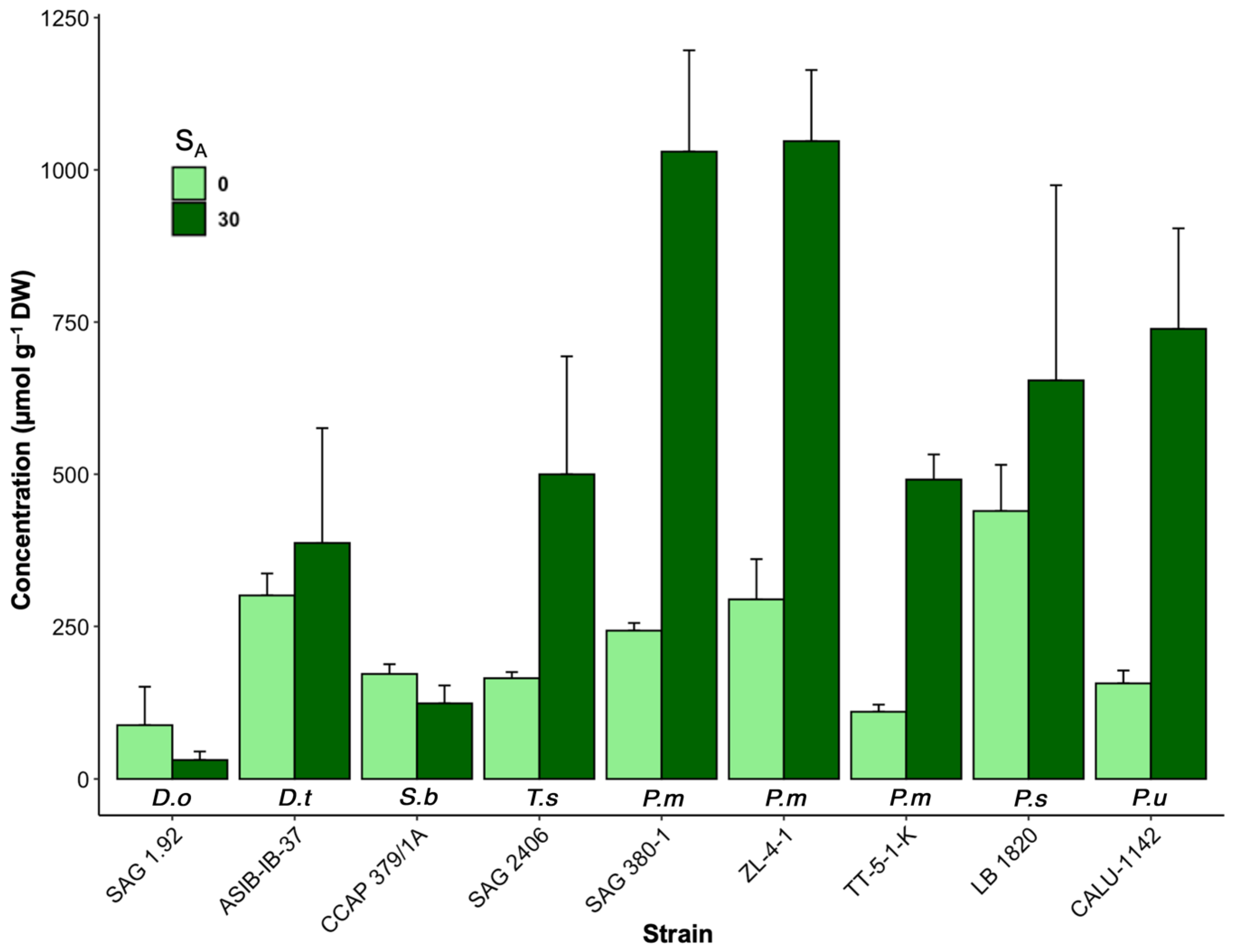
3.1. Changes in Osmolyte Concentrations under Salinity Stress

Nine strains were grown at 0 and 30 SA and their proline concentration (Figure 1) and proline:sorbitol ratio (Figure 2) was measured using HPLC. A two-way ANOVA yielded a main effect for the salinity, F(1, 14) = 154.91, p < 2 × 10⁻¹⁶, such that the average proline amount produced was significantly higher at 30 SA (M = 425.33 µmol·g−1 DW, SD = 356.40) than at 0 SA (M = 172.26 µmol·g−1 DW, SD = 108.46). The main effect of strain was also significant, F(1, 14) = 27.72, p < 2 × 10⁻¹⁶. Finally, the interaction effect was significant, F(1, 14) = 14.85, p = 1.45 × 10⁻¹⁴, indicating that the variability was due to taxon-specific differences.
Growth at 30 SA resulted in increased proline concentrations compared to 0 SA across all strains except for SAG 1.92 Desomocccus olivaceus. The most drastic increases were from Pseudostichococcus strains (SAG 380-1, ZL-4-1, TT-5-1-K and CALU-1142), where each strain had a ~3-fold increase in proline compared to 0 SA medium. A smaller increase in proline concentration (~33%) was observed in P. sequoieti LB 1820. Outside of this genus, only SAG 406 Tritostichocccus solitus had a similar proline increase. Pseudostichococcus strains maintained a relatively steady proportion of proline:sorbitol under both salinities, while Stichococcus, Desmococcus, Deuterostichococcus, and Tritostichococcus increased their proportions of sorbitol at 30 SA.

3.2. Desiccation and Rehydration

Dehydrating the strains for 6 h at ca. 10% RH resulted in complete inhibition PSII and rehydration led to recovery of all strains after 24 h with varying end values (Figure 3). For reference, the range of starting Y(II) values in the control strains was 0.365–0.660 (average 0.571 ± 0.078) across all replicates. In the non-control strains, this corresponded to 0.461–0.668 (0.602 ± 0.061) in the 0 SA strains and 0.379–0.660 (0.556 ± 0.068) in the 30 SA strains. The end phase of desiccation where Y(II) dropped was more gradual and began ca. 30 min earlier in the 30 SA group compared to the 0 SA group. The range of end values after recovery phase were narrower for strains grown in 0 SA (ca. 40–80% of control Y(II)) while the 30 SA strains ranged from ca. 25–80%, with strains outside the genus Pseudostichococcus having lower values up to ca. 60%.
Three strains in the 30 SA cohort (SAG 1.92, CCAP 379/1A, ASIB-IB-37) consistently had lower average starting Y(II) values (0.483, 0.359, and 0.517, respectively) in the desiccation experiment compared to the other strains. This was not the case at 0 SA, where the strains had comparably high average starting Y(II) values.
The one-way ANOVA analysis revealed that there was overall no significant difference in average final recovery values between the 0 and 30 SA cohorts (p = 0.678), and no correlation between final recovery values and salinity (r = −0.05). However, when removing Pseudostichococcus strains, final recovery Y(II) was somewhat negatively correlated with increased salinity in (r = −0.518, p = 0.00953). Y(II) activity in most Pseudostichococcus strains was virtually fully restored after 24 h following rehydration, except for strain CALU-1142, which had lower final recovery values at both salinities.

4. Discussion

4.1. Effect of Salinity on Proline Production

Osmolyte production is upregulated with increasing salinity and is a protective response to osmotic stress in microalgae. The data on salt-induced physiological changes are scarce for the Stichococcaceae group. This is limited to descriptions of morphological changes induced by salinity [30,31], with more extensive salt stress experiments being done mostly on Stichococcus bacillaris, the most known taxon in this group [36,37,50].
Two works from Brown and Hellebust on S. bacillaris showed that sucrose, proline, and sorbitol were its major osmolytes, with minor contributions from other amino acids [36,37]. At roughly 30 SA equivalent (1234 mOsmol/kg), S. bacillaris had intracellular concentration of 520 mM for sorbitol, 278 mM for proline, and less than 0.1 mM of sucrose. Unfortunately, as the authors did not use dry weight but rather intracellular concentration, we could not directly compare our values. Nevertheless, our measured proportion of proline:sorbitol for S. bacillaris (0.86) at 0 SA is similar to the 0.97 ratio reported by Brown and Hellebust [37], so it is likely that the results from this study are comparable.
Our recent studies expanded this idea to the Stichococcaceae. In short, sucrose, proline and sorbitol are characteristic osmolytes, independent of salinity [39,40]. Of these three, sucrose was consistently present at lower concentrations than proline or sorbitol. At 30 SA, sorbitol almost always increased to the range of ca. 48–376 µmol·g−1 DW; increased sucrose quantities were within the range of 12–135 µmol·g−1 DW [39]. Here we supplemented this finding with proline quantification, whose contribution is higher than that of both sorbitol and sucrose combined (20–1218 µmol·g−1 DW), with an average increase of 425.33 µmol·g−1 DW.
The interaction between salinity and strain accounted for the differential proline response. 30 SA medium induced higher proline production all strain except for Desmococcus and Stichococcus strains (Figure 1) but not in the other strains. The major osmolyte for these two strains under salinity seems to be sorbitol and not proline, compared to, Tritostichococcus and Pseudostichococcus. In these strains, the proportion of proline increases at higher salinity (Figure 2). While sucrose was not directly measured in this experiment, our previous study showed that sucrose generally decreased at 30 SA in the same strains here [39]. Deuterostichococcus had a high proportion of proline at both salinities, but the proportion of proline decreases~10% at 30 SA. [8,37,43].
Within Pseudostichococcus, strains SAG 380-1 and ZL-4-1 P. monallantoides produced the highest concentration of proline at 30 SA. However, as Pseudostichococcus was, until recently, monotypic, all Pseudostichococcus strains obligatorily belong to monallantoides [40]. Furthermore, the strains LB 1820 and CALU-1142 are the only known representatives of P. sequoieti and P. undulatus, respectively. Molecular verification of the P. monallantoides strains in this study will be necessary for clearer taxonomy-based ecophysiological data on osmolyte composition within this genus.
While the experiments in this study were conducted at a maximum of 30 SA, results from our previous study demonstrated that Pseudostichococcus and Tritostichococcus can grow at salinities up to 75 SA [39]. Experimental results recorded the survival of Stichococcus of up to 108‰ and 1.6 osmol/kg, roughly equivalent to 10 SA and 120 SA, respectively, and that sorbitol and then proline were the main osmolytes, with a minimal contribution from sucrose [36,51]. Proline is energetically “inexpensive” to synthesize, biologically inert and its accumulation is a response to environmental stressors, which includes UV, temperature, drought and salinity [52,53]. Strains that produce more proline under salinity may be able to better adapt to harsh environmental conditions.
Our previous study with these strains grown at 30 SA show signs of salinity stress, such as enlarged vacuoles and cell conformation changes [39]. Nevertheless, because no growth experiments were performed in this study, the production of proline and other osmolytes cannot be said to directly cause differential fitness in saline conditions within the Stichococcaceae, or to what degree. It is possible that there were strain variation effects for each taxon.

4.2. Salinity and Desiccation

Physiologically, the ability to continually recover following periods of drought is one of the most important requirements for survival in terrestrial environments [54]. Green algal communities in the wild, often in the form of biocrusts or biofilms, lie dormant and can rapidly restart photosynthetic activity following precipitation to maximize growth and before the next dehydration event [46]. Salinity, in turn, exacerbates the effects of dehydration by further upsetting osmotic balance within cells [9]. Previous experiments on the Stichococcaceae and other aeroterrestrial algae [7,33,46,55] have demonstrated that these algae are resistant to and can recovery from desiccation, although the effect of salinity has not been studied in this context.
The current study extends upon our previous work [39] and shows that salinity did not have a strong effect on average recovery potential across the 0 and 30 SA groups (p = 0.678) (Figure 3). The earlier study showed that SAG 380-1 P. monallantoides and SAG 2406 T. solitus, the most halotolerant strains, had comparable recovery potential to other Stichococcaceae strains, when grown at 0 SA. From the current results, when Pseudostichococcus strains were removed from the calculations, there was a slight negative trend between increased salinity and final recovery values (r = −0.518, p = 0.00953). With both groups, there was no significant effect noted (p = 0.678). This was because Pseudostichococcus strains were almost unaffected by salinity at 30 SA compared to 0 SA, in terms of recovery.
Because Stichococcus is homiochlorophyllous [56], its chloroplasts are not significantly damaged upon desiccation, which allows for immediate recovery of photosynthesis. This contrasts with other alga, for example some Klebsormidium and Tetradesmus, which require more time to recover post-rewetting [57,58]. This is despite the strong and rapid desiccation to ~ 10% RH, comparable to a desert environment. First, the similar recovery profiles of all strains indicates that the other genera of the Stichococcaceae are homiochlorophyllous. Second, salinity seems to have little impact on the chloroplasts or repair mechanisms, since final recovery potential was almost unaffected.
The strains that started with lower initial yields (SAG 1.92, CCAP/379 1A, ASIB-IB-37) had lower final recovery yields at both salinities, indicating that they were already stressed prior to dehydration. Here salinity did not further lower recovery potential. Hence, the mechanisms for salinity and desiccation tolerance appear to be unrelated in the Stichococcaceae and differences are present only on the trends le.
The slightly stronger response of the P. monallantoides strain SAG 380-1 to salinity and dehydration compared to the other Stichococcaceae is possibly related to its marine origin [30]. It was isolated after being found in association with Enteromorpha compressa and was found to survive cultivation in salinities up to 5% seawater, equivalent to 50 SA, with a growth limit of 70 SA [30]. This could mean that the taxon inherently has genetic mechanisms for salt adaptation, when compared to P. undulatus and P. sequoieti strains, which are both natively aeroterrestrial. It is, however, not possible to conclude that proline upregulation alone is the primary mechanism that protects against salinity and dehydration. The genetic and molecular basis of halotolerance for microalgae, such as the upregulation of genes coding for protective proteins, have only recently begun to be discovered [59,60,61].
Halotolerant algae produce a wide variety of secondary metabolites beneficial to salinized agriculture [23] and may be utilized in bioremediation [21,24,62], including desalination [63]. P. monallantoides, which was successfully used to grow young biocrusts on saline substrates from potash tailing heaps [26]. Hence, the soil desalinization with this genus is not purely conjectural. The natural biodiversity of understudied taxonomic groups is far from fully explored and the field of algal biotechnology eagerly awaits more useful organisms. Though simple, the experiment in this study suggests that salinity has a negative impact on desiccation tolerance on Stichococcaceae strains, except for Pseudostichococcus and possibly Tritostichococcus, which may also have a role to play in the biotechnology sector.
Finally, an important consideration here is that all experiments were performed on algal cultures grown in stale laboratory conditions, kept as free as possible from biotic competition and additional abiotic stressors. It is possible that additional stressors, such as UV radiation and frost, could significantly change the response to desiccation and osmotic stress. The relative rarity and recent taxonomic status of the genera within the Stichococcaceae means that few empirical results exist on their ecophysiology. More extensive comparison, especially on Pseudostichococcus, should be done to determine if they are halotolerance outliers within the Stichococcus-like group and if they have biotechnology application potential.

5. Conclusions

At 30 SA, a plausible natural salinity, most Pseudostichococcus strains produce proline as the major osmolyte, in contrast to the other Stichococcaceae. Furthermore, proline contributes to a higher proportion of osmolytes in Pseudostichococcus and Deuterostichococcus in nonsaline and saline conditions. We showed here that salinity is only slightly correlated with reduced ability to withstand desiccation, hence there are likely other protective factors at work. The results from the desiccation experiment support that the Stichococcaceae are euryhaline, as all the strains could strongly recover from desiccation despite being exposed to salinity.

Author Contributions

Conceptualization, A.T.V. and K.G.; methodology, A.T.V.; software, A.T.V.; validation, A.T.V. and K.G.; formal analysis, A.T.V.; investigation, A.T.V.; data curation, A.T.V.; writing—original draft preparation, A.T.V.; writing—review and editing, A.T.V. and K.G.; visualization, A.T.V.; supervision, K.G.; funding acquisition, K.G. All authors have read and agreed to the published version of the manuscript.

Funding

This project was funded by a grant from the German Research Foundation, under Special Priority Project: 1991 Taxon-Omics (CrustAlgaeTax—GL909/1-1).

Institutional Review Board Statement

Not applicable.

Informed Consent Statement

Not applicable.

Data Availability Statement

Data is contained within the article.

Acknowledgments

Warm thanks to Veronika Sommer for her technical expertise in the planning phase of the salinity stress experiments.

Conflicts of Interest

The authors declare no conflict of interest.

References

  1. Kirst, G. Salinity Tolerance of Eukaryotic Marine Algae. Annu. Rev. Plant Biol. 1990, 41, 21–53. [Google Scholar] [CrossRef]
  2. Tavakkoli, E.; Fatehi, F.; Coventry, S.; Rengasamy, P.; McDonald, G.K. Additive Effects of Na+ and Cl Ions on Barley Growth under Salinity Stress. J. Exp. Bot. 2011, 62, 2189–2203. [Google Scholar] [CrossRef] [PubMed] [Green Version]
  3. Gasulla, F.; de Nova, P.G.; Esteban-Carrasco, A.; Zapata, J.M.; Barreno, E.; Guéra, A. Dehydration Rate and Time of Desiccation Affect Recovery of the Lichenic Algae Trebouxia Erici: Alternative and Classical Protective Mechanisms. Planta 2009, 231, 195–208. [Google Scholar] [CrossRef] [PubMed]
  4. González-Hourcade, M.; del Campo, E.M.; Casano, L.M. The Under-Explored Extracellular Proteome of Aero-Terrestrial Microalgae Provides Clues on Different Mechanisms of Desiccation Tolerance in Non-Model Organisms. Microb. Ecol. 2021, 81, 437–453. [Google Scholar] [CrossRef]
  5. Holzinger, A.; Karsten, U. Desiccation Stress and Tolerance in Green Algae: Consequences for Ultrastructure, Physiological and Molecular Mechanisms. Front. Plant Sci. 2013, 4, 327. [Google Scholar] [CrossRef] [Green Version]
  6. Tamaru, Y.; Takani, Y.; Yoshida, T.; Sakamoto, T. Crucial Role of Extracellular Polysaccharides in Desiccation and Freezing Tolerance in the Terrestrial Cyanobacterium Nostoc Commune. Appl. Environ. Microbiol. 2005, 71, 7327–7333. [Google Scholar] [CrossRef] [PubMed] [Green Version]
  7. Häubner, N.; Schumann, R.; Karsten, U. Aeroterrestrial Microalgae Growing in Biofilms on Facades—Response to Temperature and Water Stress. Microb. Ecol. 2006, 51, 285–293. [Google Scholar] [CrossRef]
  8. Hellebust, J.A. Mechanisms of Response to Salinity in Halotolerant Microalgae. In Biosalinity in Action: Bioproduction with Saline Water; Springer: Dordrecht, The Netherlands, 1985; pp. 69–81. [Google Scholar]
  9. Shetty, P.; Gitau, M.M.; Maróti, G. Salinity Stress Responses and Adaptation Mechanisms in Eukaryotic Green Microalgae. Cells 2019, 8, 1657. [Google Scholar] [CrossRef] [Green Version]
  10. Diehl, N.; Karsten, U.; Bischof, K. Impacts of Combined Temperature and Salinity Stress on the Endemic Arctic Brown Seaweed Laminaria Solidungula, J. Agardh. Polar Biol. 2020, 43, 647–656. [Google Scholar] [CrossRef]
  11. Kumar, M.; Kumari, P.; Reddy, C.; Jha, B. Salinity and Desiccation Induced Oxidative Stress Acclimation in Seaweeds. Adv. Bot. Res. 2014, 71, 91–123. [Google Scholar]
  12. Shabala, S. Learning from Halophytes: Physiological Basis and Strategies to Improve Abiotic Stress Tolerance in Crops. Ann. Bot. 2013, 112, 1209–1221. [Google Scholar] [CrossRef] [PubMed]
  13. van Ginneken, V. Some Mechanism Seaweeds Employ to Cope with Salinity Stress in the Harsh Euhaline Oceanic Environment. Am. J. Plant Sci. 2018, 9, 1191–1211. [Google Scholar] [CrossRef] [Green Version]
  14. Moore, J.P.; Le, N.T.; Brandt, W.F.; Driouich, A.; Farrant, J.M. Towards a Systems-Based Understanding of Plant Desiccation Tolerance. Trends Plant Sci. 2009, 14, 110–117. [Google Scholar] [CrossRef] [PubMed]
  15. Muscolo, A.; Sidari, M.; Panuccio, M.R.; Santonoceto, C.; Orsini, F.; Pascale, S.D. Plant Responses in Saline and Arid Environments: An Overview. Eur. J. Plant Sci. Biotechnol. 2011, 5, 1–11. [Google Scholar]
  16. Munns, R.; James, R.A.; Gilliham, M.; Flowers, T.J.; Colmer, T.D. Tissue Tolerance: An Essential but Elusive Trait for Salt-Tolerant Crops. Funct. Plant Biol. 2016, 43, 1103. [Google Scholar] [CrossRef] [Green Version]
  17. Schilling, R.K.; Marschner, P.; Shavrukov, Y.; Berger, B.; Tester, M.; Roy, S.J.; Plett, D.C. Expression of the A Rabidopsis Vacuolar H+-pyrophosphatase Gene (AVP 1) Improves the Shoot Biomass of Transgenic Barley and Increases Grain Yield in a Saline Field. Plant Biotechnol. J. 2014, 12, 378–386. [Google Scholar] [CrossRef]
  18. Hanley, M.E.; Sanders, S.K.D.; Stanton, H.-M.; Billington, R.A.; Boden, R. A Pinch of Salt: Response of Coastal Grassland Plants to Simulated Seawater Inundation Treatments. Ann. Bot. 2019, 125, 267–275. [Google Scholar] [CrossRef]
  19. Alpert, P. Constraints of Tolerance: Why Are Desiccation-Tolerant Organisms so Small or Rare? J. Exp. Biol. 2006, 209, 1575–1584. [Google Scholar] [CrossRef] [Green Version]
  20. Chen, H.; Jiang, J.-G.; Wu, G.-H. Effects of Salinity Changes on the Growth of Dunaliella Salina and Its Isozyme Activities of Glycerol-3-Phosphate Dehydrogenase. J. Agric. Food Chem. 2009, 57, 6178–6182. [Google Scholar] [CrossRef]
  21. Kaushik, G.; Raza, K. Potential of Novel Dunaliella Salina from Sambhar Salt Lake, India, for Bioremediation of Hexavalent Chromium from Aqueous Effluents: An Optimized Green Approach. Ecotoxicol. Environ. Saf. 2019, 180, 430–438. [Google Scholar]
  22. Mishra, A.; Mandoli, A.; Jha, B. Physiological Characterization and Stress-Induced Metabolic Responses of Dunaliella Salina Isolated from Salt Pan. J. Ind. Microbiol. Biotechnol. 2008, 35, 1093. [Google Scholar] [CrossRef] [PubMed]
  23. Carillo, P.; Ciarmiello, L.F.; Woodrow, P.; Corrado, G.; Chiaiese, P.; Rouphael, Y. Enhancing Sustainability by Improving Plant Salt Tolerance through Macro- and Micro-Algal Biostimulants. Biology 2020, 9, 253. [Google Scholar] [CrossRef] [PubMed]
  24. Das, C.; Naseera, K.; Ram, A.; Meena, R.M.; Ramaiah, N. Bioremediation of Tannery Wastewater by a Salt-Tolerant Strain of Chlorella Vulgaris. J. Appl. Phycol. 2017, 29, 235–243. [Google Scholar] [CrossRef]
  25. Meng, F.; Liu, D.; Huang, W.; Lei, Z.; Zhang, Z. Effect of Salinity on Granulation, Performance and Lipid Accumulation of Algal-Bacterial Granular Sludge. Bioresour. Technol. Rep. 2019, 7, 100228. [Google Scholar] [CrossRef]
  26. Sommer, V.; Palm, A.; Schink, A.; Leinweber, P.; Gose, N.; Karsten, U.; Glaser, K. Artificial Biocrust Establishment on Materials of Potash Tailings Piles along a Salinity Gradient. J. Appl. Phycol. 2021, 13, 1–7. [Google Scholar] [CrossRef]
  27. Sommer, V.; Mikhailyuk, T.; Glaser, K.; Karsten, U. Uncovering Unique Green Algae and Cyanobacteria Isolated from Biocrusts in Highly Saline Potash Tailing Pile Habitats, Using an Integrative Approach. Microorganisms 2020, 8, 1667. [Google Scholar] [CrossRef]
  28. Sommer, V.; Karsten, U.; Glaser, K. Halophilic Algal Communities in Biological Soil Crusts Isolated from Potash Tailings Pile Areas. Front. Ecol. Evol. 2020, 8, 46. [Google Scholar] [CrossRef] [Green Version]
  29. Moewus, V.L. Systematische Bestimmung Einzelliger Grüner Algen Auf Grund von Kulturversuchen. Bot. Not. 1951, 4, 287–318. [Google Scholar]
  30. Proeschold, T.; Darienko, T. The Green Puzzle Stichococcus (Trebouxiophyceae, Chlorophyta): New Generic and Species Concept among This Widely Distributed Genus. Phytotaxa 2020, 441, 113–142. [Google Scholar] [CrossRef]
  31. Hotter, V.; Glaser, K.; Hartmann, A.; Ganzera, M.; Karsten, U. Polyols and UV-Sunscreens in the Prasiola-Clade (Trebouxiophyceae, Chlorophyta) as Metabolites for Stress Response and Chemotaxonomy. J. Phycol. 2018, 54, 264–274. [Google Scholar] [CrossRef] [Green Version]
  32. Medwed, C.; Holzinger, A.; Hofer, S.; Hartmann, A.; Michalik, D.; Glaser, K.; Karsten, U. Ecophysiological, Morphological, and Biochemical Traits of Free-Living Diplosphaera Chodatii (Trebouxiophyceae) Reveal Adaptation to Harsh Environmental Conditions. Protoplasma 2021, 258, 1–13. [Google Scholar] [CrossRef] [PubMed]
  33. Neustupa, J.; Elias, M.; Sejnohova, L. A Taxonomic Study of Two Stichococcus Species (Trebouxiophyceae, Chlorophyta) with a Starch-Enveloped Pyrenoid. Nova Hedwig. 2007, 84, 51–64. [Google Scholar] [CrossRef]
  34. Bohlin, K. Utkast Till de Gröna Algernas Och Arkegoniaternas Fylogeni; Folded chart; Akademisk Afhandling: Uppsala, Sweden, 1901; p. 43 + iv. [Google Scholar]
  35. Brown, L.M.; Hellebust, J.A. Some New Taxonomic Characteristics Applied to Stichococcus bacillaris (Chlorophyceae). Can. J. Bot. 1980, 58, 1405–1411. [Google Scholar] [CrossRef]
  36. Brown, L.M.; Hellebust, J.A. Sorbitol and Proline as Intracellular Osmotic Solutes in the Green Alga Stichococcus bacillaris. Can. J. Bot. 1978, 56, 676–679. [Google Scholar] [CrossRef]
  37. Ahmad, I.; Hellebust, J.A. The Relationship between Inorganic Nitrogen Metabolism and Proline Accumulation in Osmoregulatory Responses of Two Euryhaline Microalgae. Plant Physiol. 1988, 88, 348–354. [Google Scholar] [CrossRef] [Green Version]
  38. Van, A.T.; Sommer, V.; Glaser, K. The Ecophysiological Performance and Traits of Genera within the Stichococcus-like Clade (Trebouxiophyceae) under Matric and Osmotic Stress. Microorganisms 2021, 9, 1816. [Google Scholar] [CrossRef]
  39. Van, A.T.; Karsten, U.; Glaser, K. A Chemosystematic Investigation of Selected Stichococcus-like Organisms (Trebouxiophyta). Algae 2021, 36, 123–135. [Google Scholar] [CrossRef]
  40. Bischoff, H. Some Soil Algae from Enchanted Rock and Related Algal Species. In Phycological Studies IV; University of Texas Publisher No. 6318: Austin, TX, USA, 1963; Volume 6318, pp. 1–95. [Google Scholar]
  41. Starr, R.C.; Zeikus, J.A. UTEX—The Culture Collection of Algae at the University of Texas at Austin 1993 List of Cultures 1. J. Phycol. 1993, 29, 1–106. [Google Scholar] [CrossRef]
  42. Gustavs, L.; Eggert, A.; Michalik, D.; Karsten, U. Physiological and Biochemical Responses of Green Microalgae from Different Habitats to Osmotic and Matric Stress. Protoplasma 2010, 243, 3–14. [Google Scholar] [CrossRef]
  43. Bačkor, M.; Fahselt, D.; Wu, C.T. Free Proline Content Is Positively Correlated with Copper Tolerance of the Lichen Photobiont Trebouxia Erici (Chlorophyta). Plant Science 2004, 167, 151–157. [Google Scholar] [CrossRef]
  44. Lee, M.R.; Kim, C.S.; Park, T.; Choi, Y.-S.; Lee, K.-H. Optimization of the Ninhydrin Reaction and Development of a Multiwell Plate-Based High-Throughput Proline Detection Assay. Anal. Biochem. 2018, 556, 57–62. [Google Scholar] [CrossRef] [PubMed]
  45. Karsten, U.; Herburger, K.; Holzinger, A. Living in Biological Soil Crust Communities of African Deserts—Physiological Traits of Green Algal Klebsormidium Species (Streptophyta) to Cope with Desiccation, Light and Temperature Gradients. J. Plant Physiol. 2016, 194, 2–12. [Google Scholar] [CrossRef] [PubMed] [Green Version]
  46. Schreiber, U.; Bilger, W. Progress in Chlorophyll Fluorescence Research: Major Developments during the Past Years in Retrospect. In Progress in Botany/Fortschritte der Botanik; Springer: Berlin/Heidelberg, Germany, 1993; pp. 151–173. [Google Scholar]
  47. Genty, B.; Briantais, J.-M.; Baker, N.R. The Relationship between the Quantum Yield of Photosynthetic Electron Transport and Quenching of Chlorophyll Fluorescence. Biochim. Biophys. Acta (BBA)-Gen. Subj. 1989, 990, 87–92. [Google Scholar] [CrossRef]
  48. RStudio Team. RStudio: Integrated Development for R; RStudio, PBC: Boston, MA, USA, 2020. [Google Scholar]
  49. Gustavs, L.; Görs, M.; Karsten, U. Polyol Patterns in Biofilm-Forming Aeroterrestrial Green Algae (Trebouxiophyceae, Chlorophyta): Polyols in Aeroterrestrial Trebouxiophyceae. J. Phycol. 2011, 47, 533–537. [Google Scholar] [CrossRef]
  50. Hayward, J. Studies on the Growth of Stichococcus Bacillaris Naeg in Culture. J. Mar. Biol. Ass. 1974, 54, 261–268. [Google Scholar] [CrossRef]
  51. Ben Rejeb, K.; Abdelly, C.; Savouré, A. How Reactive Oxygen Species and Proline Face Stress Together. Plant Physiol. Biochem. 2014, 80, 278–284. [Google Scholar] [CrossRef]
  52. Szabados, L.; Savouré, A. Proline: A Multifunctional Amino Acid. Trends Plant Sci. 2010, 15, 89–97. [Google Scholar] [CrossRef]
  53. Hoekstra, F.A.; Golovina, E.A.; Buitink, J. Mechanisms of Plant Desiccation Tolerance. Trends Plant Sci. 2001, 6, 431–438. [Google Scholar] [CrossRef]
  54. Gray, D.W.; Lewis, L.A.; Cardon, Z.G. Photosynthetic Recovery Following Desiccation of Desert Green Algae (Chlorophyta) and Their Aquatic Relatives. Plant Cell Environ. 2007, 30, 1240–1255. [Google Scholar] [CrossRef]
  55. Ritchie, R.J.; Heemboo, M. Trentepohlia sp., a Terrestrial Chlorophyte Growing on Galvanised Iron Lamp Posts. Phycologia 2021, 60, 48–61. [Google Scholar] [CrossRef]
  56. Pierangelini, M.; Glaser, K.; Mikhailyuk, T.; Karsten, U.; Holzinger, A. Light and Dehydration but Not Temperature Drive Photosynthetic Adaptations of Basal Streptophytes (Hormidiella, Streptosarcina and Streptofilum) Living in Terrestrial Habitats. Microb. Ecol. 2019, 77, 380–393. [Google Scholar] [CrossRef] [PubMed] [Green Version]
  57. Terlova, E.F.; Holzinger, A.; Lewis, L.A. Terrestrial Green Algae Show Higher Tolerance to Dehydration than Do Their Aquatic Sister-Species. Microb. Ecol. 2021, 82, 770–782. [Google Scholar] [CrossRef] [PubMed]
  58. Holzinger, A.; Kaplan, F.; Blaas, K.; Zechmann, B.; Komsic-Buchmann, K.; Becker, B. Transcriptomics of Desiccation Tolerance in the Streptophyte Green Alga Klebsormidium Reveal a Land Plant-like Defense Reaction. PLoS ONE 2014, 9, e110630. [Google Scholar] [CrossRef] [PubMed]
  59. Peredo, E.L.; Cardon, Z.G. Shared Up-Regulation and Contrasting down-Regulation of Gene Expression Distinguish Desiccation-Tolerant from Intolerant Green Algae. Proc. Natl. Acad. Sci. USA 2020, 117, 17438–17445. [Google Scholar] [CrossRef]
  60. Gasulla, F.; del Campo, E.M.; Casano, L.M.; Guéra, A. Advances in Understanding of Desiccation Tolerance of Lichens and Lichen-Forming Algae. Plants 2021, 10, 807. [Google Scholar] [CrossRef]
  61. Bwapwa, J.; Jaiyeola, A.; Chetty, R. Bioremediation of Acid Mine Drainage Using Algae Strains: A Review. S. Afr. J. Chem. Eng. 2017, 24, 62–70. [Google Scholar] [CrossRef]
  62. Figler, A.; B-Béres, V.; Dobronoki, D.; Márton, K.; Nagy, S.A.; Bácsi, I. Salt Tolerance and Desalination Abilities of Nine Common Green Microalgae Isolates. Water 2019, 11, 2527. [Google Scholar] [CrossRef] [Green Version]
  63. Gan, X.; Shen, G.; Xin, B.; Li, M. Simultaneous Biological Desalination and Lipid Production by Scenedesmus Obliquus Cultured with Brackish Water. Desalination 2016, 400, 1–6. [Google Scholar] [CrossRef]
Figure 1. Proline concentrations within the respective strains at growth in 0 and 30 SA media. Error bars indicate standard deviation (n = 3). Differences in the means were significantly correlated to the combined effect of taxon and salinity. Labels beneath the bars are taxon initials and refer to the full names Table 1.
Phycology 02 00007 g001
Figure 2. Mean proportions of proline:sorbitol produced in the respective strains at growth in 0 and 30 SA media. The text labels on the x-axis indicates growth at 0 SA and “30” indicates the same strain response at 30 SA. Labels above the bars are taxon initials and refer to the full names Table 1.
Phycology 02 00007 g002
Figure 3. Comparison of Y(II) after desiccation and rehydration in Stichococcaceae strains grown in 0 SA and 30 SA media. Each row represents the same strain. Magenta lines show average effective quantum yield (Y(II)) per strain during dehydration, while green lines show the average Y(II) in the recovery phase; grey lines in the background represent the other strains for comparison. Error bars represent standard deviation (n = 3).
Phycology 02 00007 g003
Table 1. The strains used in this study and their culture ID, localities and collector information. Strains marked with “*” are authentic strains.
Strain IDSpecies AssignmentLocality and HabitatCollector/Isolator
SAG 1.92Desmococcus olivaceus *Vienna, Austria; subaerialW. Vischer, before 1960
ASIB-IB-37Deuterostichococcus tetrallantoides *Dauphin Island, Alabama, USA; soilT.R. Deason, 1969
SAG 380-1Pseudostichococcus monallantoides *Germany; marineL. Moewus, 1951
ZL-4-1Pseudostichococcus monallantoidesTeutschenthal, Germany; potash tailing heap surroundingsV. Sommer, 2018
TT-5-1-KPseudostichococcus monallantoidesZielitz, Germany; potash tailing heap surroundingsV. Sommer, 2018
LB 1820Pseudostichococcus sequoieti *USA; redwood forest soilG. Arce, 1971
CALU-1142Pseudostichococcus undulates *Dolomite Mountains, ItalyG. Vinatzer, 1975
CCAP 379/1AStichococcus bacillaris *Likely SwitzerlandW. Vischer, before 1936
SAG 2406Tritostichococcus solitus *Northeim, Germany; karstwater stream rock surfaceK. Mohr, 2003
Publisher’s Note: MDPI stays neutral with regard to jurisdictional claims in published maps and institutional affiliations.

