Article

Computational Exploration of Bacterial Compounds Targeting Arginine-Specific Mono-Adp-Ribosyl-Transferase 1 (Art1): A Pathway to Novel Therapeutic Anticancer Strategies

1
Department of Natural and Life Sciences, Larbi Ben M’Hidi University, Oum El Bouaghi 04000, Algeria
2
Higher National School of Forest, Khenchela 40000, Algeria
3
Biotechnology, Water, Environment and Health Laboratory, Abbes Laghrour University, Khenchela 40000, Algeria
4
Laboratory of Biomaterials and Transport Phenomena (LBMTP), University Yahia Fares, Médéa 26000, Algeria
5
Laboratoire de Génie des Procédés Chimiques, Département de Génie des Procédés, Faculté de Technologie, Université Ferhat Abbas, Sétif-1, Sétif 19000, Algeria
6
Univ Rennes, Ecole Nationale Supérieure de Chimie de Rennes, CNRS, ISCR—UMR6226, 35000 Rennes, France
7
Department of Biochemistry, College of Science, King Saud University, Riyadh 11451, Saudi Arabia
8
School of Engineering, Merz Court, Newcastle University, Newcastle upon Tyne NE1 7RU, UK
*
Authors to whom correspondence should be addressed.
Curr. Issues Mol. Biol. 2025, 47(8), 634; https://doi.org/10.3390/cimb47080634
Submission received: 13 June 2025 / Revised: 11 July 2025 / Accepted: 4 August 2025 / Published: 8 August 2025

Abstract

Cancer is a multifaceted and life-threatening disease characterized by the unregulated proliferation of malignant cells. Developing new therapies and diagnostic methods for cancer remains a critical focus of research. Proteins involved in cancer progression are being targeted to facilitate the discovery of effective biological treatments. Among these, the ART1 protein plays a critical role in promoting cancer progression, establishing it as a key target for drug therapy. Actinomycetes, known for their anticancer activity, were explored in this study for their potential to inhibit ART1. One hundred bioactive secondary metabolites derived from actinomycetes were subjected to in silico screening to evaluate their potential anticancer activity through inhibition of ART1. The three-dimensional structure of ART1 was generated using the SWISS-MODEL tool and validated through the Save server 6.0 and ProSa web. The structural stability of the ART1 protein was evaluated through molecular dynamics analysis using the iMod server. The potential active sites within the ART1 structure were mapped using the Computed Atlas of Surface Topography of Proteins (CASTp). Molecular docking and protein–ligand interaction studies were performed using AutoDock Vina. Additionally, pharmacophore modeling was conducted using the Pharmit server to identify promising compounds. Toxicity predictions and in silico drug-likeness assessments were carried out using Swiss-ADME and ADMET Lab which evaluate Absorption, Distribution, Metabolism, Excretion, and Toxicity (ADMET) properties. Molecular dynamics simulations results for the ART1 protein demonstrated high stability over time. Additionally, resistomycin, borrelidin, tetracycline, and oxytetracycline were identified as the top-ranking ligands, exhibiting binding energies between −8.9 kcal/mol and −9.3 kcal/mol. These ligands exhibited favorable pharmacophore profiles, drug-likeness, and ADMET properties, indicating their potential safety and efficacy in humans. In conclusion, the selected actinomycete-derived ligands show promise for further research and development as potential anticancer agents targeting ART1.

1. Introduction

Cancer remains one of the most pressing global health challenges and continues to be a leading cause of mortality worldwide [1,2]. Among men, the most commonly diagnosed cancers include prostate, lung, bronchial, colorectal, and bladder cancers, whereas in women, breast, lung, bronchial, colorectal, uterine, and thyroid cancers are the most prevalent [3]. Numerous enzymes play crucial roles in tumor development and progression, making them attractive targets for the development of novel therapeutic agents.
Mono-ADP-ribosyltransferases (mono-ARTs) represent a class of enzymes that catalyze the transfer of a single ADP-ribose moiety from nicotinamide adenine dinucleotide (NAD+) to specific target proteins, thereby modulating their function and activity. Among these, Arginine-Specific Mono-ADP-Ribosyltransferase 1 (ART1) is particularly noteworthy. ART1 is primarily expressed in epithelial and immune cells, as well as in select tissues [4]. To date, seven mono-ART enzymes (ART1 to ART7) have been identified in mammals, with five of them—ART1, ART2, ART5, ART6, and ART7—exhibiting specificity for arginine residues [4]. In humans, ART1 is typically expressed at low levels under normal physiological conditions [5]. However, its overexpression has been increasingly associated with various pathological states, including numerous forms of cancer [5,6].
ART1, a glycosylphosphatidylinositol (GPI)-anchored tumor cell surface protein, has been implicated in cancer progression [7]. It modulates immune cell function by catalyzing the mono-ADP-ribosylation of the P2X7 receptor on CD8+ T cells, ultimately leading to T cell apoptosis [8]. Meta-iodo-benzyl-guanidine (MIBG) has been reported as an ART inhibitor that suppresses interleukin-6 (IL-6) release through the inhibition of ART1 activity [9].
Moreover, ART1 has been associated with enhanced tumor development and metastasis in colorectal cancer (CRC) [10]. Lin et al. [9] demonstrated that silencing ART1 expression significantly reduced IL-6-induced proliferation of colorectal cancer cells. This effect was linked to decreased levels of glycoprotein 130 (gp130), a key signal transducer in the IL-6 signaling pathway, suggesting that ART1 may facilitate colorectal cancer progression by modulating inflammation-associated signaling cascades [9].
Traditional cancer treatment modalities—including chemotherapy, radiotherapy, and surgical intervention—have led to substantial advances in patient outcomes. However, challenges such as drug resistance, tumor heterogeneity, and off-target effects continue to limit their efficacy, highlighting the urgent need for more personalized and targeted therapeutic strategies [11,12]. In recent years, growing attention has been directed toward natural therapeutics as an alternative or complementary approach to reduce reliance on synthetic compounds.
Nature serves as an abundant source of pharmacologically active molecules, with approximately 60% of currently approved drugs having originated from natural products [13]. These bioactive compounds are derived from a variety of sources, including animals, plants, and microorganisms. Among microbial sources, actinomycetes—a group of Gram-positive filamentous bacteria—are particularly noteworthy for their ability to produce structurally diverse and biologically potent secondary metabolites [14]. Of the roughly 23,000 bioactive microbial secondary metabolites identified to date, actinomycetes account for over 10,000 compounds [15]. These metabolites encompass a broad range of activities, including immunosuppressive, antitumor, herbicidal, pesticidal, and cosmetic applications, as well as vitamin biosynthesis [16].
To reduce dependence on synthetic compounds, natural therapeutic strategies are gaining increasing attention. Natural products—particularly secondary metabolites produced by microorganisms such as actinomycetes—represent a rich source of bioactive molecules with promising anticancer potential. Although inherently chemical, these compounds offer remarkable structural diversity and target specificity, making them highly valuable in modern drug discovery efforts. Notably, approximately 60% of currently approved pharmaceutical agents are derived from natural sources [17].
Recent advances in bioinformatics have further accelerated the exploration of natural compounds by enabling the identification of molecular targets and supporting in silico methodologies such as virtual screening, molecular docking, and pharmacokinetic prediction in cancer research [18,19,20]. These computational techniques allow researchers to predict molecular interactions, perform high-throughput screening of potential drug candidates, and simulate cancer-related processes under various therapeutic scenarios [21]. Compared to conventional in vitro and in vivo experiments, in silico approaches offer significant advantages in terms of cost-efficiency and time-effectiveness, particularly during the early stages of drug discovery and development [22].
In this study, we aim to investigate the potential of actinomycete-derived bioactive compounds as inhibitors of ART1, a key enzyme implicated in cancer progression. To this end, we employed an integrative in silico approach combining molecular modeling, docking, and pharmacokinetic analysis. Initially, homology modeling was performed to predict the three-dimensional structure of ART1, followed by validation of its stereochemical properties to ensure structural reliability. A library of secondary metabolites derived from actinomycetes was then subjected to molecular docking simulations to identify potential inhibitors capable of interacting with the catalytic site of ART1. The top-ranking candidates were further assessed for their pharmacokinetic properties and ADMET (Absorption, Distribution, Metabolism, Excretion, and Toxicity) profiles to evaluate their drug-likeness and safety. Finally, molecular dynamics simulations were carried out to examine the structural stability and dynamic behavior of the most promising enzyme–ligand complexes over time. This study provides a comprehensive computational framework for the identification of novel natural compounds with potential anticancer activity and offers new insights into the inhibition of ART1 as a targeted therapeutic strategy.

2. Materials and Methods

2.1. Selection of Target and Template Sequences

The amino acid sequence of ADP-ribosyltransferase 1 (ART1) was retrieved from the UniProt database. Template identification for homology modeling was carried out using the BLASTp algorithm [23], querying the Protein Data Bank (PDB) [24] to identify structurally homologous proteins. The PDB file of the selected template, along with its corresponding FASTA sequence, was downloaded for further analysis. Multiple sequence alignment between the target protein and the selected template was performed using Clustal Omega, available through the European Bioinformatics Institute (EBI) sequence analysis platform (https://www.ebi.ac.uk/Tools/msa/clustalo/, accessed on 25 December 2024).

2.2. Protein Profile Analysis

The amino acid sequence of the target protein, ART1, was submitted to the PROSITE database (http://prosite.expasy.org/, accessed on 25 December 2024) to analyze its domains, families, and functional motifs. PROSITE provides computational tools for the identification and prediction of protein profiles, facilitating the characterization of structural and functional features of the protein [25].

2.3. Homogy Modeling of Target Protein

Homology modeling of the ADP-ribosyltransferase 1 (ART1) protein was carried out using the SWISS-MODEL web server (https://swissmodel.expasy.org/) [26]. The quality of the generated models was evaluated using the Qualitative Model Energy ANalysis (QMEAN) score, which guided the selection of the most reliable structural model. Visualization and structural analysis of the 3D model were performed using UCSF Chimera and PyMOL [27].

2.4. Validation of 3D Target Protein

The structural quality of the predicted ART1 model was evaluated using a combination of computational validation tools. PROCHECK was employed to generate a Ramachandran plot, providing insights into the stereochemical quality of the model by analyzing backbone dihedral angles (https://saves.mbi.ucla.edu/, accessed on 26 December 2024) [28]. ERRAT was used to assess the model based on non-bonded atomic interaction statistics (https://saves.mbi.ucla.edu/, accessed on 26 December 2024) [29], while Verify3D evaluated the compatibility between the 3D structure and its amino acid sequence (https://saves.mbi.ucla.edu/, accessed on 26 December 2024) [30]. Additionally, ProSA-web was utilized to calculate the Z-score, offering a quantitative measure of the overall quality and reliability of the 3D structural model (https://prosa.services.came.sbg.ac.at/prosa.php/, accessed on 26 December 2024) [31].

2.5. Analysis of Physicochemical Characteristics

The physicochemical properties of the target protein, including molecular weight (MW), isoelectric point (pI), extinction coefficient, number of positively and negatively charged residues (+R and −R, respectively), aliphatic index (AI), grand average of hydropathicity (GRAVY), and instability index (II), were analyzed using the ProtParam tool available on the ExPASy server (https://web.expasy.org/protparam/, accessed on 26 December 2024) [32].

2.6. Molecular Dynamics Simulation

Molecular dynamics (MD) simulations were performed to evaluate the stability and dynamic behavior of the ART1–ligand complex. The iMODS server (https://imods.iqf.csic.es/, accessed on 26 December 2024) was used to assess the deformability and structural rigidity of the ART1 protein through normal mode analysis (NMA) in internal coordinates [33,34]. This approach provided insights into the protein’s intrinsic flexibility and overall stability. The analysis generated a range of outputs, including deformability plots, eigenvalues, B-factors, variance profiles, covariance matrices, and atomic-scale elastic network models. Additionally, residue-level indices were calculated to characterize both the magnitude and directionality of structural fluctuations within the protein complex [34].

2.7. Prediction of Active Site

The active site of the target protein was identified using the Computed Atlas of Surface Topography of Proteins (CASTp) server [35], which provides detailed information on pocket geometry and surface accessibility. Visualization and analysis of the predicted binding sites were performed using PyMOL version 1.7.4.5 (Schrödinger, LLC) and the Discovery Studio Modeling Environment [36], allowing for accurate inspection of the spatial orientation and topological features of the active site.

2.8. Preparation of Target Protein

The target protein was prepared for molecular docking by adding hydrogen atoms and assigning partial atomic charges using the AMBER ff14SB force field [37]. This step ensured appropriate protonation states and optimized the structure for docking simulations. The finalized protein model was then saved in MOL2 format, which preserves essential information such as atomic charges and bond orders, making it suitable for subsequent docking analyses.

2.9. Preparation of Ligands

The chemical structures of actinomycete-derived compounds were retrieved from the PubChem database (https://pubchem.ncbi.nlm.nih.gov/) accessed on 27 December 2024 using their canonical SMILES representations. Ligand preparation was carried out in UCSF Chimera by adding hydrogen atoms and assigning Gasteiger partial charges. The finalized ligand structures were then saved in MOL2 format to preserve atomic coordinates and charge information for downstream molecular docking analysis.

2.10. Molecular Docking

Molecular docking analyses were performed using AutoDock Vina, following the protocol described by [38]. Both the receptor and ligands were prepared in MOL2 format prior to docking. A grid box was defined around the predicted active site using AutoDock Tools, with dimensions set to (10.2 × 37.4 × 12.9) Å and centered at coordinates (20, 20, 20) Å. Meta-iodo-benzylguanidine, a well-characterized chemical inhibitor, was used as a reference compound to benchmark binding affinities. During docking simulations, ligand flexibility was allowed, whereas the protein structure was kept rigid. The resulting protein–ligand complexes were saved in PDB format for subsequent visualization and interaction analysis.

2.11. Visualization of Protein–Ligand Interaction

The interactions between the receptor and ligands within the binding pocket—including hydrogen bonds, hydrophobic contacts, electrostatic interactions, and van der Waals forces—were analyzed using BIOVIA Discovery Studio Visualizer [36]. This analysis provided detailed insights into the binding modes and the nature of molecular interactions stabilizing the protein–ligand complexes.

2.12. Biological Activity Prediction

The potential biological activities of the studied compounds were predicted using the PASS (Prediction of Activity Spectra for Substances) online server (http://way2drug.com/passonline) accessed on 28 December 2024. This tool evaluates a wide range of pharmacological effects and estimates possible toxicities based on the structural features of each compound. The results are expressed as Pa (probability of activity) and Pi (probability of inactivity). A compound is considered to have potential pharmacological activity when its Pa value exceeds its Pi value [39].

2.13. Pharmacophore Study

The Pharmit web platform (http://pharmit.csb.pitt.edu/) accessed on 28 December 2024 was utilized to screen compound libraries based on pharmacophore models and molecular shape filters. Pharmacophore modeling in Pharmit integrates both ligand-based and structure-based features to account for the flexibility of residues within the catalytic site. The pharmacophore features were defined according to the chemical properties of functional groups and classified as aromatic (ARO), hydrogen bond donor (HD), hydrogen bond acceptor (HA), hydrophobic (HP), negatively charged (NI), or positively charged (PI), as identified by the Pharmit server [40].

2.14. Drug-Likeness and ADMET Study

The drug-likeness and physicochemical properties of the selected ligands were evaluated based on Lipinski’s Rule of Five. According to these guidelines, compounds are predicted to exhibit favorable oral bioavailability if they possess no more than five hydrogen bond donors, no more than ten hydrogen bond acceptors, a molecular weight below 500 g/mol, and a calculated partition coefficient (CLogP) of less than 5 [41].
To advance promising drug candidates toward clinical development, it is essential to assess their Absorption, Distribution, Metabolism, Excretion, and Toxicity (ADMET) profiles. These pharmacokinetic parameters are critical for predicting a compound’s behavior and safety in the human body, and help eliminate molecules with undesirable physiological or toxicological properties early in the drug development process. In this study, the in silico ADMET profiles of the most promising ART1 inhibitory compounds were predicted using two widely used online platforms: SwissADME (http://www.swissadme.ch/index.php) [42] and ADMETlab (https://admetmesh.scbdd.com/) [43] accessed on 29 December 2024.

3. Results and Discussion

3.1. Selection of Target and Template Sequences

The amino acid sequence of the target protein, Arginine-Specific Mono-ADP-Ribosyltransferase 1 (ART1), was retrieved from the UniProt database (accession number: P52961). This human protein consists of 327 amino acid residues and was obtained in FASTA format for subsequent structural and functional analyses. The complete FASTA sequence is provided in the Supporting Information (Supplementary File S1).
Template selection for homology modeling was based on maximizing sequence similarity between the target protein and available structural templates. The ART1 amino acid sequence retrieved from the UniProt database was used as input for BLASTp searches against the Protein Data Bank (PDB) to identify suitable templates. Three high-confidence templates with sequence identity above 30% were identified: PDB ID 1GXY (39.92% identity, E-value = 4 × 10−40), 1OG4 (39.52% identity, E-value = 4 × 10−39), and 1OG3 (39.52% identity, E-value = 8 × 10−39). According to established criteria [44], sequence identities above 30%, combined with E-values approaching zero, are considered statistically significant and suitable for reliable homology-based modeling.
The selected templates were retrieved in the FASTA format and aligned with the target sequence using Clustal Omega. Conserved amino acid regions between the target and templates were highlighted in red (Figure 1), facilitating the identification of structurally relevant regions for model construction.

3.2. Protein Profile Analysis

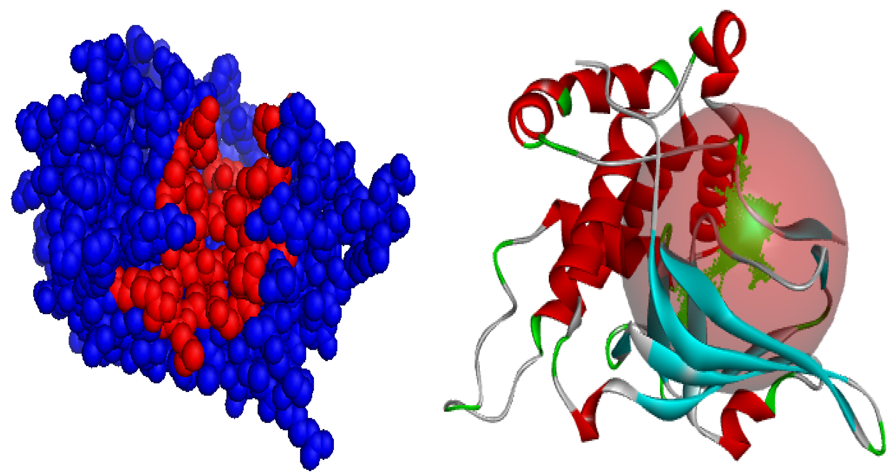
The PROSITE database was employed to analyze the domain architecture, family classification, and functional sites of the ART1 protein. The analysis identified a conserved domain spanning residues 73 to 273. Within this region, three key residues—arginine (R144), serine (S202), and glutamic acid (E203)—were predicted to constitute the protein’s active site. These residues are likely involved in catalytic activity or substrate binding. A visual representation of the predicted active site is shown in Figure 2 and Supplementary File S2.

3.3. Homology Modeling and Validation

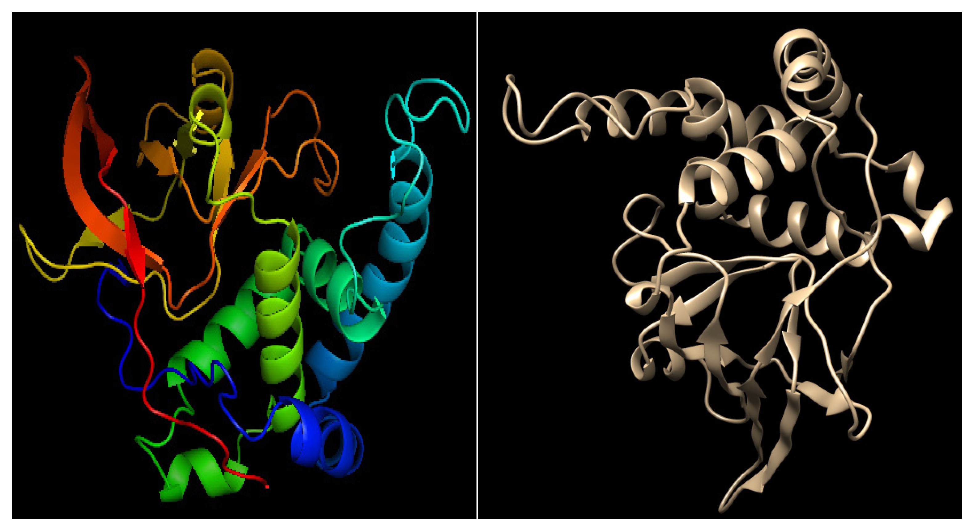
Homology modeling of the target protein was performed using the SWISS-MODEL server, a widely used and reliable platform for comparative protein structure prediction (Figure 3 and Supplementary File S3). The structural quality of the generated model was assessed using the QMEAN Z-score, which estimates the degree of similarity between the predicted model and experimentally resolved protein structures. A QMEAN Z-score within an acceptable range supports the validity of the model, while more negative values typically reflect greater agreement with high-resolution experimental data [45].
The ART1 model exhibited a QMEAN Z-score of −3.37, indicating good structural quality and reliability. The three-dimensional structure of the modeled protein was further visualized and analyzed using UCSF Chimera and PyMOL (Figure 4), allowing for detailed inspection of its structural features.
To assess the structural quality of the predicted ART1 model, multiple bioinformatics tools were employed, as illustrated in Figure 5 and detailed in Supplementary File S4. Stereochemical validation using a Ramachandran plot revealed that 86.1% of the amino acid residues were located in the most favored regions, 12.5% in additionally allowed regions, 1.4% in generously allowed regions, and none in disallowed regions. This analysis serves as a standard criterion for evaluating the reliability of protein structural models. The Ramachandran plot provides a visual representation of the backbone dihedral angles (ϕ–ψ) of all residues in the protein structure, excluding terminal residues. Glycine residues, due to their high conformational flexibility, are represented as triangles to indicate their ability to occupy otherwise restricted regions of the conformational space [46]. The most favorable regions of the plot correspond to well-defined secondary structural elements, typically distributed as follows: (1) β-sheet conformations in the upper-left quadrant, (2) right-handed α-helices in the lower-left quadrant, and (3) left-handed α-helices in the upper-right quadrant [47]. A model is generally considered reliable when more than 80% of residues fall within the favored regions [48]. Based on these criteria, the predicted ART1 model demonstrates high structural quality and reliability (Figure 5).
To further validate the structural integrity of the predicted model, the ERRAT tool was employed. ERRAT assesses the quality of protein structures by analyzing non-bonded atomic interactions and identifying potential error regions using a squared error function [49,50]. A score above the commonly accepted threshold of 80.00 is indicative of a high-quality and reliable model [51]. In this study, the ERRAT analysis produced a quality score of 91.7391, confirming the overall accuracy and robustness of the predicted ART1 structure (Figure 5).
To further assess the structural integrity of the predicted ART1 model, the ERRAT validation tool was employed. ERRAT evaluates the quality of protein structures by analyzing non-bonded atomic interactions and identifying regions with potential errors using a squared error function [49,50]. A commonly accepted quality threshold is 80.00, with higher scores reflecting greater structural reliability [51,52]. In the present study, the predicted model achieved an ERRAT score of 91.7391, indicating a high-quality structure and reinforcing the reliability of the homology model (Figure 5).
To further evaluate the overall structural accuracy of the predicted model, ProSA-web was employed. This tool assesses model quality by comparing the predicted structure’s potential energy to that of experimentally determined protein structures in the Protein Data Bank (PDB) [53,54]. ProSA is widely recognized for its ability to detect structural inconsistencies and evaluate the overall reliability of protein models [31]. The ART1 model yielded a Z-score of −6.4, which falls within the range typically observed for high-quality experimentally resolved structures. More negative Z-scores reflect better agreement with native protein conformations, as low-energy residue environments contribute significantly to the stability of the tertiary structure [55].

3.4. Analysis of Physicochemical Characteristics

The physicochemical properties of the modeled ART1 protein were analyzed using the ExPASy ProtParam server. The protein consists of 327 amino acid residues, with a calculated molecular weight of 36,334.67 Daltons. The theoretical isoelectric point (pI) was determined to be 8.53, suggesting that the protein is slightly basic under physiological conditions. The pI corresponds to the pH at which the protein carries no net electric charge, typically resulting in minimal solubility and mobility during isoelectric focusing. At this point, proteins are typically compact and structurally stable [56]. The analysis also revealed the presence of 26 negatively charged residues (Asp and Glu) and 29 positively charged residues (Arg and Lys), contributing to the protein’s overall charge distribution. The instability index was calculated at 46.08, exceeding the threshold of 40 and thus categorizing the protein as unstable in vitro [57]. The aliphatic index (AI), which reflects thermal stability based on the relative volume of aliphatic side chains (Ala, Val, Ile, and Leu), was found to be 82.17. This relatively high AI suggests that the protein may possess significant thermal stability [58]. Finally, the grand average of hydropathicity (GRAVY) was calculated to be −0.113, indicating a predominantly hydrophilic nature. Lower GRAVY values are generally associated with enhanced solubility and favorable behavior in aqueous environments.

3.5. Molecular Dynamic Simulation

Molecular docking provides a static snapshot of ligand positioning within a protein’s active site, offering an initial perspective on potential binding interactions. However, it does not account for the dynamic nature of proteins and ligands under physiological conditions. To gain a more comprehensive understanding of protein–ligand interactions and their temporal stability, molecular dynamics (MD) simulations are widely employed. These simulations enable the exploration of conformational flexibility and provide insights into the stability and behavior of complexes in biologically relevant environments. Studies have shown that a high docking score does not necessarily correlate with ligand efficacy, as some compounds may dissociate from the binding site during simulation. This underscores the importance of MD simulations as a critical complement of molecular docking, providing a more realistic assessment of binding stability and molecular behavior over time [59]. In this study, the dynamic behavior of the ART1 protein was analyzed using the iMODS server (Figure 6). iMODS applies normal mode analysis (NMA) to evaluate internal molecular motions and predict conformational changes with minimal computational cost [33]. The simulation output provided detailed data on key dynamic parameters, including deformability, B-factor distribution, eigenvalues, variance, covariance matrix, and elastic network properties. These results offered valuable insights into the intrinsic flexibility and structural dynamics of ART1, a protein implicated in cancer progression.
The deformability plot (Figure 6A) presents the flexibility of individual residues based on normal mode calculations. Peaks in the green curve indicate regions with high flexibility, while valleys represent more rigid regions. The ART1 protein exhibits several highly flexible regions, particularly at residue indices around 50, 150, and 225, which likely correspond to loop regions or terminal domains that facilitate structural rearrangements. These flexible regions are essential for protein–ligand interactions and functional adaptability in dynamic cellular environments. According to [2], the highest peaks could correspond to unstructured loops, which often contribute to dynamic interactions in biological systems (e.g., active sites of enzymes or ligand binding pockets). The relatively flat regions between the peaks indicate a well-ordered structural organization, which may play a crucial role in preserving the protein’s folding and overall stability. The rigid backbone suggests a framework that provides structural integrity, resisting deformation while allowing localized movement for biological activity [60]. The B-factor, also known as the temperature factor, quantifies atomic displacement or flexibility within a molecular structure. In molecular dynamics simulations and normal mode analysis (NMA) using iMODS, the B-factor reflects atomic fluctuations within the protein, providing insights into its flexible and rigid regions [61]. The B-factor plot results (Figure 6B) compare the theoretical B-factor from NMA (red) with experimental crystallographic B-factors (gray). The overlapping trend suggests that the normal mode predictions align well with experimentally observed flexibility patterns. Notably, residues near indices 50 and 200 exhibit prominent peaks, reinforcing the idea that these regions contribute significantly to ART1’s dynamic behavior. High B-factor values indicate areas prone to conformational changes, which may play a crucial role in the protein’s biological function. This computational B-factor is often compared to experimental B-factors obtained from X-ray crystallography, providing validation for structural predictions [62]. B-factor analysis in iMOD is derived from normal mode calculations, where low-frequency modes correspond to large-scale collective motions of the protein, and high-frequency modes capture localized vibrations [2]. If a protein contains functionally important flexible regions, such as binding pockets or catalytic sites, they often exhibit higher B-factor values [63]. The eigenvalue plot (Figure 6C) provides insight into the energy required for conformational transitions. The first eigenvalue is 2.11279 × 10⁻⁴, indicating that ART1 has a relatively low energy barrier for large-scale motion. This suggests that the protein can undergo significant conformational shifts with minimal energy expenditure, an important feature for proteins that interact dynamically with other biomolecules. The low eigenvalue implies structural flexibility and functional adaptability, which could be critical for ligand binding or signal transduction. The eigenvalue plot shown above presents a normalized trend where eigenvalues are divided by the first eigenvalue (Eigenvalue (1)). This scaling allows for an intuitive comparison of how higher modes relate to the fundamental mode. The gradual increase in the eigenvalue ratio suggests that higher modes exhibit increased stiffness or energy levels, which is a common characteristic in modal analysis [64]. The variance distribution plot (Figure 6D) illustrates the relative contribution of each normal mode to the overall motion. The first few modes account for a substantial percentage of total variance, highlighting that global motions dominate the protein’s flexibility. The cumulative variance suggests that the first 10 modes contribute over 75% of the total motion, which means that the primary movements of ART1 can be well-characterized by these dominant modes. The purple bars represent the individual contribution to the variance of the low-contributing modes. They show that the first modes explain a significant part of the variance. In biological systems, these modes generally correspond to major conformational changes or dominant collective motions [2]. The steadily increasing green bars indicate how variance accumulates across successive modes. The curve indicates that a limited number of dominant modes account for the majority of structural variation, whereas higher-order modes contribute progressively less. In electron tomography or cryo-EM, this often means that the dataset can be efficiently analyzed using a reduced set of dominant modes, thereby improving computational efficiency [65]. The variance distribution of ART1 suggests that a few dominant modes govern its functionally relevant conformational dynamics, while higher modes contribute minimally. This idea is consistent with previous findings that biomolecular motions essential for function are captured in a small set of normal modes, highlighting the importance of variance analysis in protein structural biology. The covariance matrix (Figure 6E) represents correlated (red) and anti-correlated (blue) motions between residue pairs. The red diagonal line signifies strong correlated movements within the same structural domains, whereas off-diagonal blue and red regions indicate interactions between different parts of the protein. This suggests that specific residues move in a concerted fashion, which could be relevant for ART1’s functional dynamics. Regions with strong negative correlations (blue) suggest possible hinge-like motions, where one domain moves in the opposite direction relative to another, facilitating large-scale conformational changes. Regions of positive correlation (red) suggest cooperative motions between residues, implying structural rigidity or concerted domain movements. Conversely, negative correlations (blue) highlight anti-correlated motions, which often signify hinge points or flexible regions that facilitate large-scale conformational changes [2]. These insights are particularly useful in studying allosteric regulation, where distant residues communicate dynamically to modulate protein function [66]. The observed covariance patterns can help predict biologically relevant conformational changes, ligand-binding sites, and potential allosteric pathways. Such analyses have been widely used to study enzyme mechanisms, receptor activation, and drug design [67]. Furthermore, the integration of MD simulations with experimental data, such as X-ray crystallography or cryo-EM, enhances the reliability of structural models and provides a comprehensive understanding of protein dynamics [68]. The presence of strong off-diagonal correlations may also indicate a potential evolutionary adaptation to maintain function while allowing for necessary conformational flexibility. The Elastic Network Model (ENM) is a computationally efficient approach employed in molecular dynamics simulations to study the large-scale motions of biomolecules [2]. The iMod server utilizes ENM to generate a normal mode analysis (NMA), helping to predict collective motions within a protein structure. This method is advantageous because it reduces computational complexity while maintaining accuracy in representing functional motions [69]. The elastic network results (Figure 6F) provide insights into the connectivity between different residues. The black and gray intensity map represents the strength of interactions between atom pairs, where darker shades indicate stronger connections. The diagonal dominance signifies that the protein core is structurally stable, while the lighter regions suggest areas with higher flexibility, likely corresponding to functionally important sites. The localized weaker interactions near flexible residues (e.g., around residue indices 50, 100, and 200) further support the hypothesis that these regions contribute to ART1’s conformational adaptability. Flexible loop regions and terminal ends often exhibit significant displacement, while secondary structure elements, such as α-helices and β-sheets, generally exhibit greater rigidity [70]. These results align with known protein dynamics, where functional sites and allosteric regions are frequently associated with higher flexibility [2]. The identified pathways often correspond to biologically relevant conformational changes, such as domain opening and closing, hinge bending, or twisting motions. These results support the hypothesis that protein function is intimately linked to its intrinsic dynamics, a concept well supported by normal mode analysis studies [70]. These findings support the hypothesis that ART1 exhibits structural adaptability and is capable of undergoing significant conformational rearrangements. The analysis revealed that no atom displayed substantial distortion within the ART1 protein structure, suggesting a balanced combination of rigidity and flexibility, which may facilitate its catalytic activity. These insights contribute to a deeper understanding of ART1’s potential biological functions, particularly in protein–ligand interactions, signal transduction, and molecular recognition.

3.6. Prediction of Active Site

The active site of an enzyme comprises specific amino acid residues responsible for substrate recognition, binding, and catalysis. Binding site residues typically establish transient interactions with the substrate, whereas catalytic residues facilitate the chemical transformation through mechanisms such as proton transfer, nucleophilic attack, or transition state stabilization [71]. In the present study, the active site of the modeled ART1 protein was identified using the CASTp server, which detects surface-accessible pockets and predicts potential ligand-binding sites based on the three-dimensional structure. The analysis revealed that 49 residues contribute to the formation of the active site (Figure 7 and Supplementary File S5). To gain further insight into the spatial organization of the active pocket, the protein structure was visualized using PyMOL and Discovery Studio Visualizer, enabling precise identification of interaction sites and cavity geometry (Figure 8 and Supplementary File S6).
Analysis of Chain A revealed a well-defined cluster of residues forming the protein’s active site, localized in a region enriched in catalytically relevant amino acids. Notably, residues such as histidine (H), serine (S), aspartate (D), lysine (K), and tyrosine (Y) were identified—amino acids commonly implicated in catalytic processes due to their ability to participate in acid–base chemistry and stabilization of transition states. Additionally, the presence of numerous hydrophobic residues, including valine (V), leucine (L), isoleucine (I), and phenylalanine (F), suggests the formation of a hydrophobic core within the binding pocket, favoring the accommodation of nonpolar or amphipathic ligands. Surrounding this core, polar and charged residues such as glutamate (E), glutamine (Q), and lysine (K) create an electrostatic environment conducive to substrate positioning and catalytic function. Collectively, these features indicate that the ART1 active site consists of a moderately sized hydrophobic cavity encased by a polar shell, which together provide both specificity and catalytic efficiency.

3.7. Visualization of Molecular Docking and Interaction

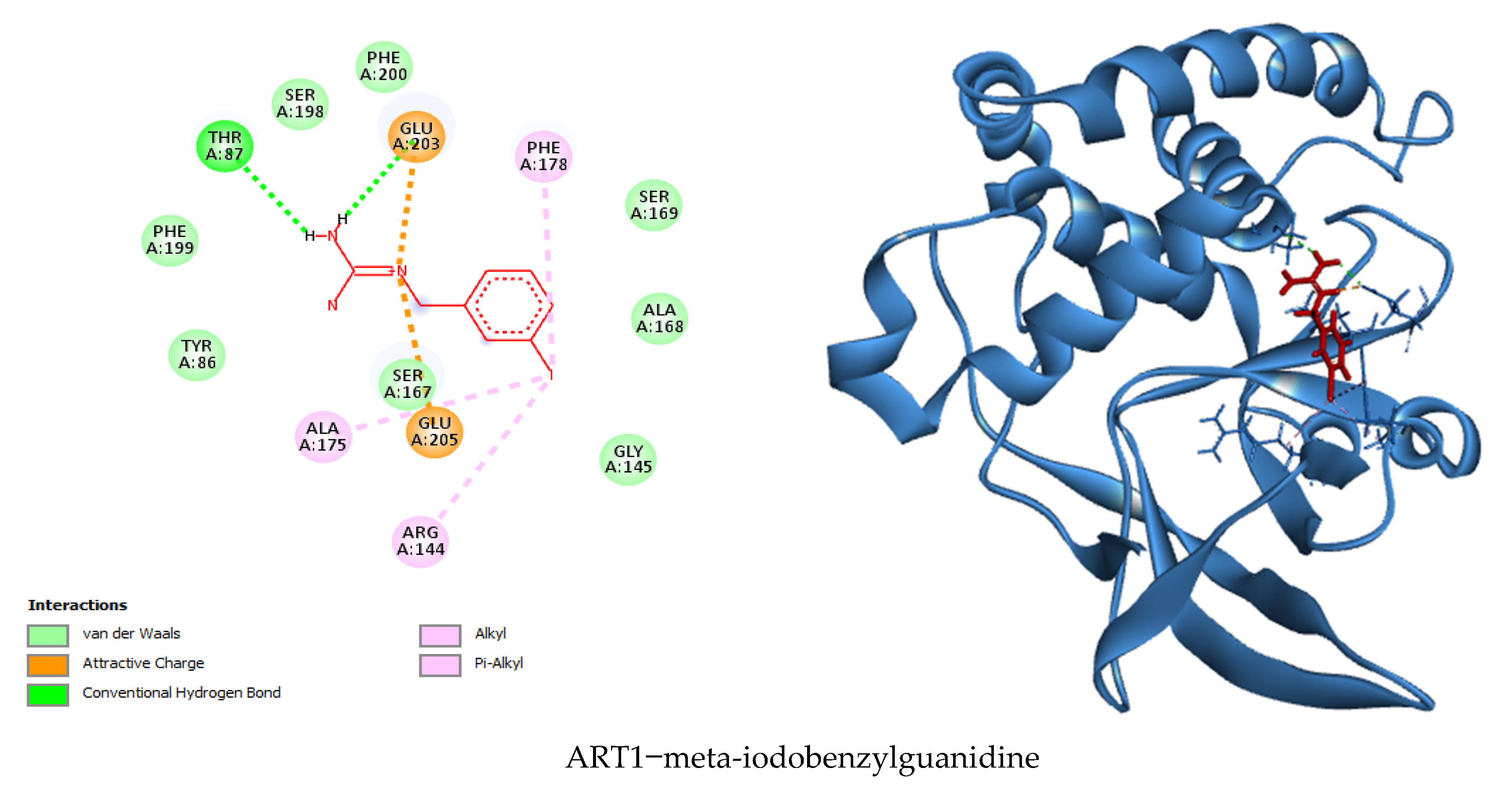
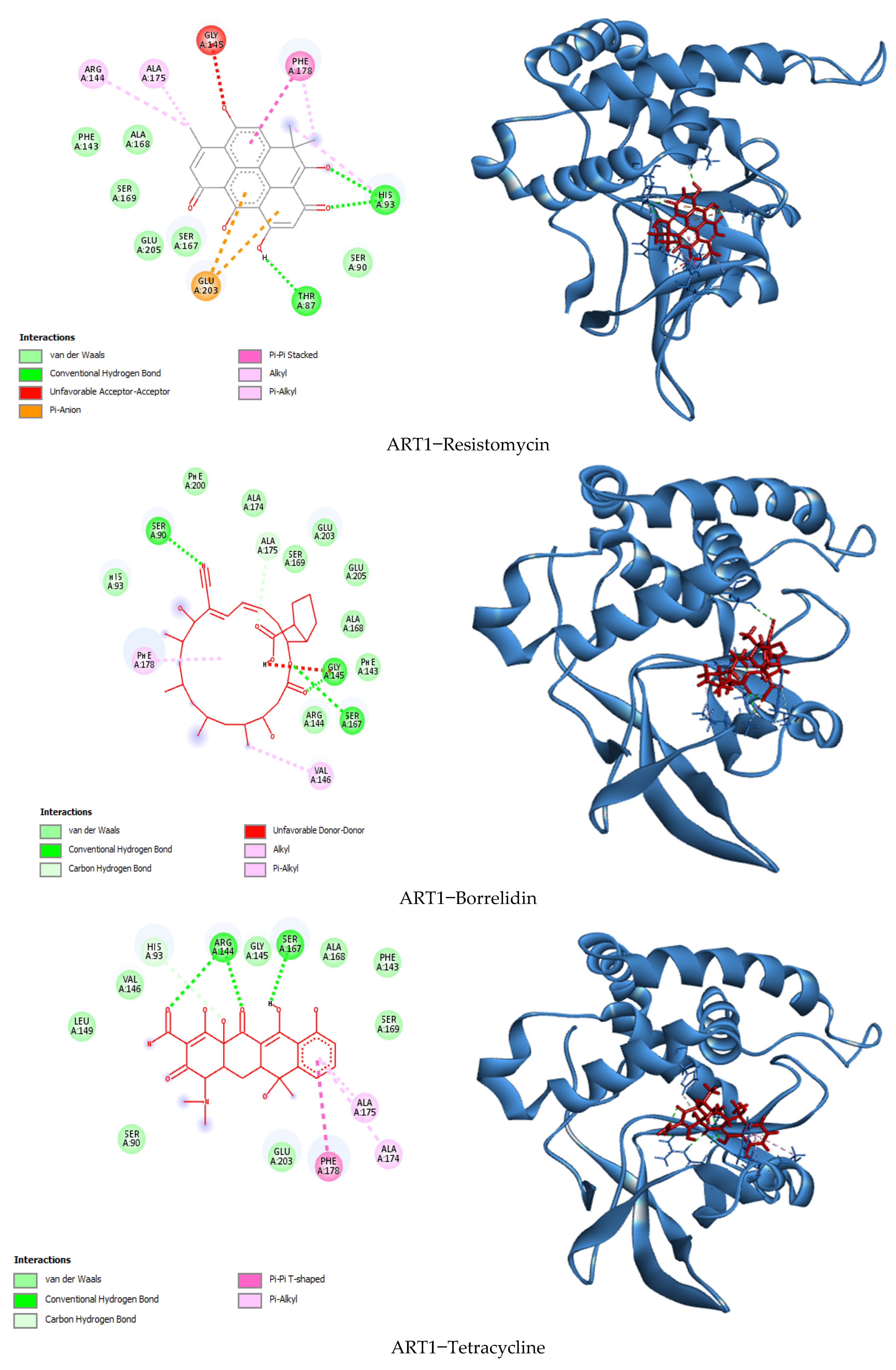
In this study, a molecular docking analysis was performed on 100 secondary metabolites derived from various actinomycete species to evaluate their potential inhibitory activity against the ART1 enzyme. The docking simulations were conducted using UCSF Chimera as the interface, with AutoDock Vina serving as the docking engine. To validate the docking protocol, meta-iodo-benzyl-guanidine—a well-characterized ART1 inhibitor—was used as a reference compound, yielding a binding energy of −6.1 kcal/mol. Following successful validation, the actinomycetal compounds were docked against the ART1 model. Among the tested molecules, four compounds demonstrated the most favorable binding affinities, with binding energies ranging from −8.9 kcal/mol to −9.3 kcal/mol. Resistomycin exhibited the strongest interaction with ART1, displaying a binding energy of −9.3 kcal/mol. This was followed by borrelidin and tetracycline, both with binding energies of −9.0 kcal/mol, and oxytetracycline with −8.9 kcal/mol (Table 1). These values suggest stronger and potentially more stable interactions with the ART1 active site compared to the reference molecule, highlighting these compounds as promising candidates for further investigation.
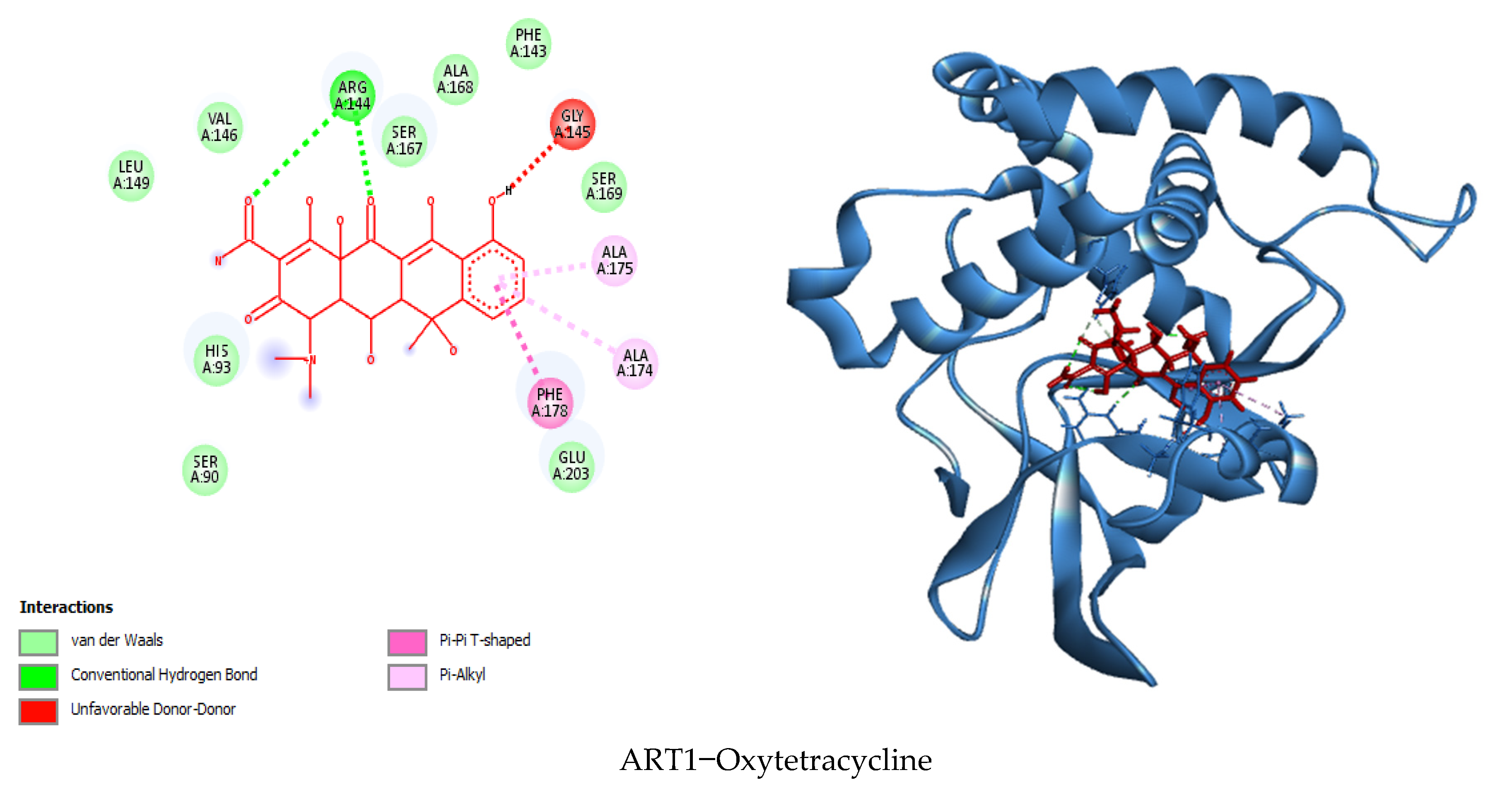
The molecular docking analysis determined the binding affinities of four secondary metabolites, resistomycin, borrelidin, tetracycline, and oxytetracycline, with the enzyme ART1, with binding energies between −8.9 and −9.3 kcal/mol, indicating a stronger binding affinity than the reference molecule meta-iodo-benzyl-guanidine (−6.1 kcal/mol), suggesting that these metabolites interact more effectively with the enzyme. The docking results were further examined by analyzing the types of interactions involved, including hydrogen bonding, hydrophobic interactions, electrostatic interactions, and van der Waals forces (Figure 9, Supplementary File S7), resistomycin demonstrated the strongest binding with the enzyme, forming hydrogen bonds with Thr87, His93, and His93, hydrophobic bonds with His93, Phe178, Ala175, and Arg144, and electrostatic bonds with Glu203 and Glu205. Additionally, it formed 16 van der Waals interactions, including residues like Ser90, Ser167, Glu205, Phe143, Ser169, and Ala168, leading to its highest binding energy of −9.3 kcal/mol. Borrelidin and tetracycline both showed similar binding energies of −9.0 kcal/mol, with borrelidin forming hydrogen bonds with Gly145, Ser167, Ser90, and Ala175, and hydrophobic bonds with Val146 and Phe178, while tetracycline interacted with Ser167, Arg144, His93, Ala175, and Phe178. Although borrelidin and tetracycline did not establish electrostatic bonds, they exhibited significant van der Waals interactions with residues such as Arg144, Phe143, Ala168, Glu205, Glu203, Ser169, and Ala174. Oxytetracycline, with a binding energy of −8.9 kcal/mol, exhibited fewer hydrogen bonds and van der Waals interactions, leading to a slightly lower binding affinity compared to the other three metabolites. Meta-iodo-benzyl-guanidine, serving as a reference, had only two hydrogen bonds and a relatively weaker set of interactions, which could explain its lower binding energy. Overall, the analysis suggests that the key residues interacting with the enzyme ART1 are Thr87, His93, Phe178, Ala175, Arg144, Glu203, and Ser167, which play a critical role in the binding and stability of the enzyme–ligand complexes. These findings suggest that resistomycin is the most promising candidate for further experimental validation, followed closely by borrelidin, tetracycline, and oxytetracycline, all of which could potentially serve as enzyme modulators.

3.8. Prediction of Biological Activity

The PASS (Prediction of Activity Spectra for Substances) online tool was employed to evaluate the anticancer potential of the selected actinomycete-derived compounds. This tool utilizes Molecular Neighborhood Analysis (MNA) descriptors to estimate two key probabilities: Pa (probability of activity) and Pi (probability of inactivity), both ranging from 0.000 to 1.000. A higher Pa relative to Pi suggests a stronger likelihood that the compound will exhibit the predicted biological activity [72]. Widely applied in preclinical drug discovery, PASS enables early-stage screening of compounds based on their chemical structures, facilitating the identification of molecules with potential therapeutic relevance.
In this study, PASS analysis was conducted on the four top-scoring ligands identified through molecular docking (Table 2). The results indicated that resistomycin and borrelidin displayed Pa values exceeding 0.7, a threshold commonly considered indicative of strong biological activity. Such values typically suggest structural similarity to known pharmacologically active compounds, reinforcing their potential as effective anticancer agents. These findings are consistent with previously reported experimental data, lending further support to their relevance in cancer drug development.
Tetracycline exhibited a Pa value in the range of 0.5–0.7, which indicates moderate confidence in its anticancer potential. While such values suggest that the compound may display biological activity, its efficacy may be limited or context-dependent, warranting further investigation.
In contrast, oxytetracycline presented a Pa value below 0.5, suggesting a low likelihood of known anticancer activity. Although this may reflect limited therapeutic potential within the currently established frameworks, it does not rule out novel mechanisms of action. Indeed, oxytetracycline could represent a candidate for exploratory studies aimed at identifying unconventional or previously uncharacterized anticancer pathways. Overall, the PASS predictions aligned well with known pharmacological profiles, supporting the prioritization of resistomycin and borrelidin for further experimental validation.

3.9. Pharmacophore Study

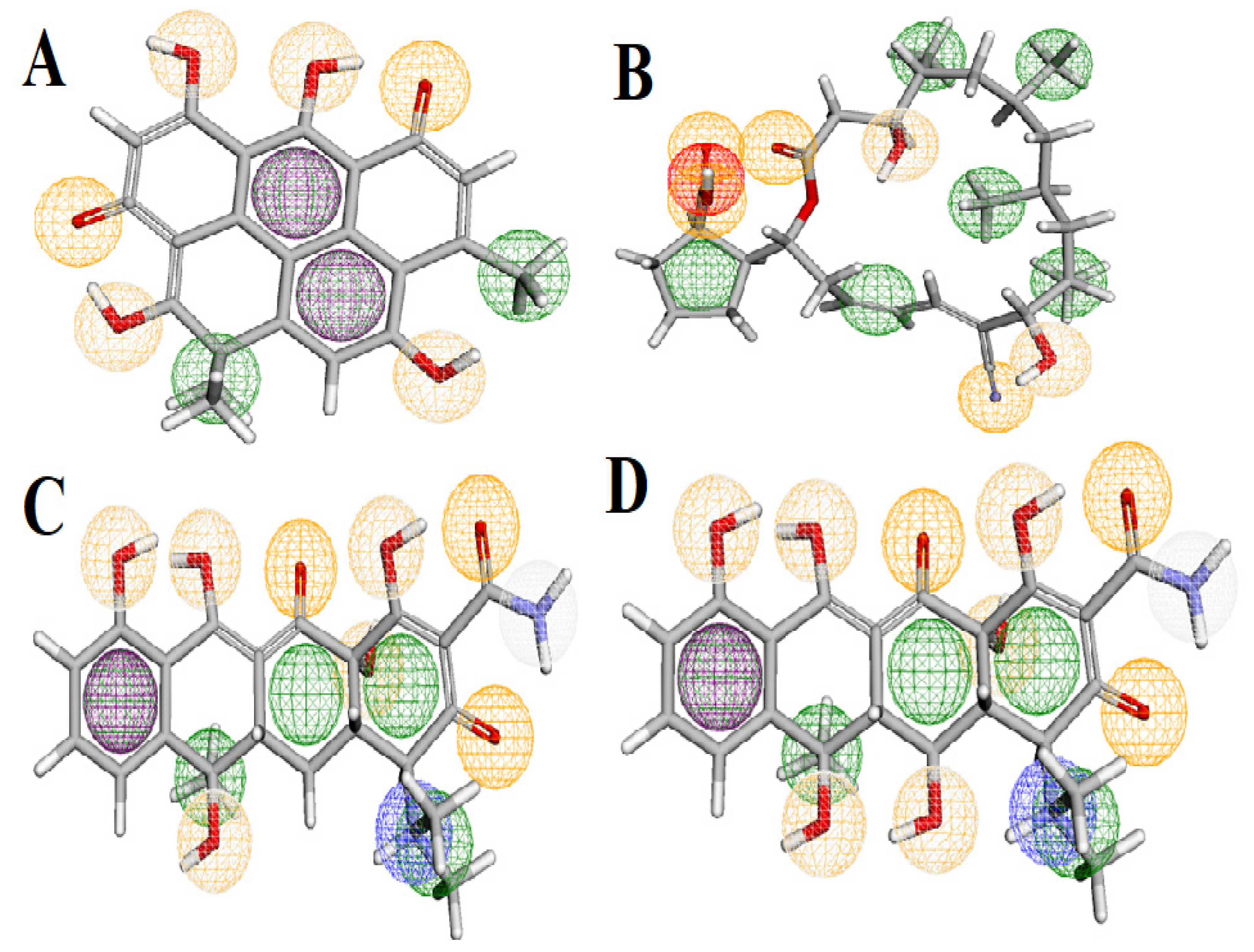
A pharmacophore is a collection of spatial and electronic features crucial for interacting with a macromolecular target, leading to a biological response [73]. It defines the key characteristics required for molecular interactions, which include the precise spatial arrangement of features such as hydrogen bond acceptor and donor, aromatic groups, charges, and hydrophobic regions. The Pharmit server is a widely used tool for visualizing these interactions, and it assigns distinct colors to various types of molecular features. Aromatic bonds are colored purple, indicating regions involved in π-π stacking interactions, while hydrogen bond donors are represented in white, and hydrogen bond acceptors are depicted in orange, typically around hydroxyl (-OH) or amine (-NH) groups. Hydrophobic interactions are shown in green, negative ions in red, and positive ions in blue [74]. In our study, the pharmacophore analysis provided detailed insights into the molecular features of the compounds tested for their potential as ART1 inhibitors. Resistomycin exhibited two aromatic groups, four hydrogen bond donors, six hydrogen bond acceptors, and four hydrophobic interactions. This suggests that aromaticity and hydrogen bonding are crucial for its biological activity, highlighting the compound’s capability for specific molecular recognition through π-π stacking interactions and hydrogen bonding (Figure 10). Borrelidin exhibited two hydrogen bond donors, six hydrogen bond acceptors, one negative ion, and six hydrophobic interactions. These characteristics indicate that the molecule’s flexibility, potentially enhanced by its ability to engage in both hydrogen bonding and hydrophobic interactions, may improve its adaptability to various biological targets, reinforcing its potential as a versatile inhibitor (Figure 10). Tetracycline exhibited one aromatic bond, seven hydrogen bond donors, eight hydrogen bond acceptors, one positive ion, and five hydrophobic interactions. The presence of both aromatic bonds and extensive hydrogen bonding indicates that these interactions are central to its biological function. The compound’s ability to form multiple hydrogen bonds enhances its potential for effective binding to target proteins (Figure 10). Oxytetracycline, characterized by one aromatic bond, eight hydrogen bond donors, nine hydrogen bond acceptors, one positive ion, and five hydrophobic interactions, exhibited the most optimal pharmacophore model, totaling 23 interactions. This model underscores the importance of conjugated systems for achieving higher binding affinity and suggests that this compound has the strongest potential for inhibition, with a robust set of interactions that may facilitate more stable binding to the ART1 target (Figure 10).
To validate the pharmacophore model, the reference compound meta-iodobenzylguanidine (MIBG)—a known ART1 substrate—was used as a control. The model successfully recognized and matched the key interaction features present in MIBG, including its hydrogen bond donors, positive ionic regions, and aromatic moieties. The structural alignment and pharmacophoric overlap between MIBG and the predicted active compounds confirmed that the model accurately captures the essential features required for ART1 binding. This validation step supports the reliability and predictive power of the constructed pharmacophore model for virtual screening applications.
Pharmacophore models are an essential tool for large-scale compound screening, as they apply various filters to identify promising candidates based on their spatial and physicochemical characteristics [75].
A compound that satisfies all the necessary criteria for biological activity is considered a “good” candidate. By matching compounds to a well-defined pharmacophore, it becomes possible to narrow down those most likely to exhibit the desired biological effect. These candidates can then undergo more focused pharmacokinetic studies to assess their stability and suitability for further development, including parameters such as the most favorable binding energy. This approach allows for the identification of compounds that may achieve similar biological effects at lower concentrations, improving the efficiency of drug development [76].

3.10. Drug-Likeness and ADMET Study

The most selective ligands inhibiting ART1 were evaluated based on Lipinski’s, Veber’s, Egan’s, and Ghose’s rules, along with their ADMET properties. The results are presented in Table 3.
Lipinski’s Rule of Five is a widely adopted framework in drug discovery for evaluating the drug-likeness and oral bioavailability potential of small molecules [77]. According to this rule, compounds are considered likely to exhibit favorable oral bioavailability if they possess a log p ≤ 5, a molecular weight ≤ 500 Da, no more than 10 hydrogen bond acceptors, and no more than 5 hydrogen bond donors [78]. In the present study, resistomycin, borrelidin, and tetracycline complied fully with Lipinski’s criteria, while oxytetracycline exceeded the hydrogen bond donor threshold (7 donors), indicating potential bioavailability limitations.
Topological Polar Surface Area (TPSA), a key determinant of molecular polarity and absorption, should ideally fall within the range of 20–130 Å2 [79]. Resistomycin (115.06 Å2) and borrelidin (127.85 Å2) met this requirement, whereas tetracycline and oxytetracycline exceeded the upper limit, suggesting potential issues with membrane permeability. All compounds exhibited a number of rotatable bonds (nRB) below 10, reflecting favorable conformational flexibility.
According to Ghose’s filter, an optimal drug candidate should have a log p between −0.4 and 5.6, molecular weight between 160 and 480 Da, molar refractivity between 40 and 130, and an atomic count between 20 and 70 [80]. Only resistomycin satisfied all these criteria, while the other three compounds showed deviations, indicating potential challenges in terms of drug-likeness.
Veber’s rule, which considers oral bioavailability based on ≤10 rotatable bonds and TPSA ≤ 140 Å2, was fulfilled by resistomycin and borrelidin, again suggesting more favorable pharmacokinetics for these molecules [81]. Egan’s rule, which evaluates bioavailability potential based on a log p between −1.0 and 5.8 and TPSA ≤ 130 Å2, also confirmed compliance for resistomycin and borrelidin [82].
Finally, Muegge’s filter, which assesses drug-likeness based on structural features and classifies compounds with a drug score between 2 and 7 as drug-like [83], identified resistomycin as a drug-like molecule, while borrelidin, tetracycline, and oxytetracycline were categorized as non-drugs.
Taken together, these analyses highlight resistomycin as the most promising candidate with favorable physicochemical and pharmacokinetic properties, fully aligning with multiple drug-likeness filters, while borrelidin shows good potential but with minor deviations. In contrast, tetracycline and oxytetracycline displayed less favorable profiles, suggesting that their pharmacological development may require structural optimization or alternative delivery strategies.
The observed discrepancies in drug-likeness parameters underscore the need for a more comprehensive evaluation of the pharmacokinetic and safety profiles of the selected compounds. Key physicochemical properties such as molecular weight and topological polar surface area (TPSA) play critical roles in determining a molecule’s ability to permeate biological membranes. Higher molecular weight and elevated TPSA are generally associated with reduced membrane permeability, whereas lower values favor improved absorption and bioavailability [84]. Similarly, the partition coefficient (LogP) reflects molecular lipophilicity, with moderate values typically indicating better passive diffusion. The balance between hydrogen bond donors and acceptors is also essential for membrane permeability, as excessive polarity can hinder transcellular transport. Furthermore, the number of rotatable bonds (nRB) is indicative of molecular flexibility; an optimal nRB supports favorable bioavailability by maintaining a balance between conformational adaptability and rigidity [84].
To address these aspects systematically, in silico prediction of ADMET (Absorption, Distribution, Metabolism, Excretion, and Toxicity) properties has become a cornerstone of early-phase drug discovery. Conducting experimental ADMET evaluations for large compound libraries is both time-consuming and resource-intensive, making computational screening tools an indispensable alternative [85]. Platforms such as SwissADME (http://www.swissadme.ch/index.php) and ADMETlab (https://admetmesh.scbdd.com/) provide rapid, reliable predictions of a compound’s pharmacokinetic behavior and potential toxicity, offering valuable guidance during the lead optimization process.
In this study, the ADMET properties of the top four candidate ligands were evaluated using these platforms. The predictive results, summarized in Table 4, offer insight into key parameters including solubility, gastrointestinal absorption, blood–brain barrier permeability, metabolic stability, hepatotoxicity, and interaction with cytochrome P450 enzymes and drug transporters. These in silico analyses facilitate the early identification of promising compounds while deprioritizing those with suboptimal pharmacokinetic or safety profiles, thus streamlining the drug development process and increasing the likelihood of downstream success.
Caco-2 permeability, assessed using human colon-derived adenocarcinoma cells, serves as a well-established in vitro model for predicting intestinal absorption due to the morphological and functional similarity of Caco-2 cells to human enterocytes [86]. Compounds with predicted permeability values above −5.15 log cm/s are typically considered to have good absorption profiles [87,88]. In this study, all tested compounds, except oxytetracycline, demonstrated Caco-2 permeability consistent with favorable intestinal absorption. Human Intestinal Absorption (HIA), an integrated parameter derived from excretion ratios in urine, bile, and feces [89], confirmed that all compounds except borrelidin achieved HIA values > 30%, supporting their classification as highly absorbable [90].
Regarding distribution, all four compounds were predicted as BBB−, indicating their inability to cross the blood–brain barrier, a characteristic commonly observed for non-lipophilic or high-molecular-weight compounds [91]. In terms of plasma protein binding (PPB), all compounds except borrelidin exhibited positive binding, suggesting efficient systemic distribution through reversible interactions with plasma proteins such as albumin and glycoproteins, which influence drug half-life and bioavailability [92,93].
Metabolic analysis revealed that tetracycline and oxytetracycline were not predicted to inhibit any cytochrome P450 (CYP450) isoforms, whereas resistomycin and borrelidin showed potential inhibition of one or more isoforms, which may influence drug–drug interactions and hepatic clearance [94,95,96]. Excretion parameters were assessed via predicted total clearance (Cl), where resistomycin, tetracycline, and oxytetracycline displayed low clearance (<5 mL/min/kg), suggesting prolonged systemic retention, while borrelidin exhibited high clearance (>15 mL/min/kg), indicating faster elimination [97].
Toxicological profiling was conducted to evaluate the compounds’ safety risks. The Ames mutagenicity test predicted non-mutagenic outcomes for all compounds except resistomycin, suggesting a favorable genetic safety profile for most candidates [98]. In carcinogenicity assessments, all four molecules tested negative, indicating no predicted risk of cancer-related toxicity [99]. However, borrelidin was flagged as a potential hERG channel blocker, which may pose cardiotoxicity risks due to the inhibition of potassium ion channels critical for cardiac repolarization [100]. The remaining compounds tested negative for hERG inhibition, indicating minimal cardiotoxic potential. In terms of hepatotoxicity, oxytetracycline was the only compound predicted to be non-hepatotoxic, while resistomycin, tetracycline, and borrelidin exhibited potential hepatotoxicity risks, underscoring the need for further in vitro and in vivo validation [101,102].
Collectively, these in silico ADMET predictions provide a comprehensive overview of the pharmacokinetic behavior and toxicity risks associated with each compound. Resistomycin and tetracycline emerged as promising candidates with favorable absorption and distribution profiles, but their potential hepatic liabilities merit additional safety evaluations. Borrelidin, while demonstrating excellent intestinal permeability and high clearance, presents concerns related to hERG inhibition and hepatotoxicity. Oxytetracycline, despite a lower permeability profile, showed the most favorable toxicity profile among the tested compounds.

4. Conclusions

Based on the results obtained in this study, we demonstrate the potential of actinomycete-derived bioactive compounds as inhibitors of ART1, a critical enzyme involved in cancer progression. Through an in silico approach combining molecular modeling, docking simulations, pharmacokinetic analysis, and molecular dynamics simulations, we identified several promising compounds with the potential to target ART1 effectively. The selected ligands resistomycin, borrelidin, tetracycline, and oxytetracycline showed favorable binding affinities, drug-likeness, and ADMET profiles, suggesting their potential as safe and effective therapeutic agents. Molecular dynamics simulations by the iMod server confirmed the stability of the enzyme. The analysis of key parameters, including deformability, B-factor distribution, eigenvalues, variance, covariance matrix, and elastic network properties, further supported the stability and compactness of the ART1 protein. This study highlights the potential of these actinomycete-derived compounds as anticancer agents targeting ART1, offering a promising avenue for future research and development of novel cancer therapies. Additional experimental validation and in vivo studies are necessary to confirm the efficacy and safety of these compounds, but the in silico results present a strong foundation for their potential as new anticancer drugs.

Supplementary Materials

The following supporting information can be downloaded at: https://www.mdpi.com/article/10.3390/cimb47080634/s1.

Author Contributions

Conceptualization, N.M.; Data curation, N.M. and J.Z.; Formal analysis, N.M. and O.B.; Funding acquisition, M.S.O.; Investigation, N.M., O.B., H.T. and J.Z.; Methodology, N.M., O.B. and S.L.; Resources, O.B., S.L. and H.T.; Software, N.M., O.B. and S.L.; Supervision, O.B.; Validation, N.M., O.B., H.T., M.S.O., J.Z. and A.A.; Visualization, S.L. and A.A.; Writing—original draft, N.M., O.B., S.L. and A.A.; Writing—review and editing, O.B., S.L., M.S.O. and J.Z. All authors have read and agreed to the published version of the manuscript.

Funding

This research was supported by the Research Funding Program (ORF-2025-710), King Saud University, Riyadh, Saudi Arabia.

Institutional Review Board Statement

Not applicable.

Informed Consent Statement

Not applicable.

Data Availability Statement

The original contributions of this study are available in the article and its Supplementary Materials. For additional information, please contact the corresponding authors.

Acknowledgments

The authors wish to express their gratitude to the Algerian Ministry of Higher Education and Scientific Research (MESRS, DGRSDT) for its support. The authors are grateful to Ongoing Research Funding Program (ORF-2025-710), King Saud University, Riyadh, Saudi Arabia.

Conflicts of Interest

The authors declare no conflicts of interest.

References

  1. Lei, W.; Yang, C.; Wu, Y.; Ru, G.; He, X.; Tong, X.; Wang, S. Nanocarriers Surface Engineered with Cell Membranes for Cancer Targeted Chemotherapy. J. Nanobiotechnol. 2022, 20, 45. [Google Scholar] [CrossRef]
  2. Lekmine, S.; Benslama, O.; Kadi, K.; Martín García, A.I.; Shamsul Ola, M.; Abdullah Yilmaz, M.; Ali, A. Therapeutic Potential of Hyoscyamus niger-Derived Compounds: Targeting Ovarian Cancer through Antioxidant Activity and EGFR Tyrosine Kinase Inhibition. J. King Saud Univ. Sci. 2024, 36, 103103. [Google Scholar] [CrossRef]
  3. Li, Z.; Fan, J.; Xiao, Y.; Wang, W.; Zhen, C.; Pan, J.; Wu, W.; Liu, Y.; Chen, Z.; Yan, Q.; et al. Essential Role of Dhx16-Mediated Ribosome Assembly in Maintenance of Hematopoietic Stem Cells. Leukemia 2024, 38, 2699–2708. [Google Scholar] [CrossRef] [PubMed]
  4. Lin, X.; Liao, Y.; Chen, X.; Long, D.; Yu, T.; Shen, F. Regulation of Oncoprotein 18/Stathmin Signaling by ERK Concerns the Resistance to Taxol in Nonsmall Cell Lung Cancer Cells. Cancer Biother. Radiopharm. 2016, 31, 37–43. [Google Scholar] [CrossRef]
  5. Wang, K.; Ning, S.; Zhang, S.; Jiang, M.; Huang, Y.; Pei, H.; Li, M.; Tan, F. Extracellular Matrix Stiffness Regulates Colorectal Cancer Progression via HSF4. J. Exp. Clin. Cancer Res. 2025, 44, 30. [Google Scholar] [CrossRef] [PubMed]
  6. Ling, F.; Tang, Y.; Li, M.; Li, Q.-S.; Li, X.; Yang, L.; Zhao, W.; Jin, C.-C.; Zeng, Z.; Liu, C.; et al. Mono-ADP-Ribosylation of Histone 3 at Arginine-117 Promotes Proliferation through Its Interaction with P300. Oncotarget 2017, 8, 72773–72785. [Google Scholar] [CrossRef]
  7. Wennerberg, E.; Mukherjee, S.; Spada, S.; Hung, C.; Agrusa, C.J.; Chen, C.; Valeta-Magara, A.; Rudqvist, N.-P.; Van Nest, S.J.; Kamel, M.K.; et al. Expression of the Mono-ADP-Ribosyltransferase ART1 by Tumor Cells Mediates Immune Resistance in Non–Small Cell Lung Cancer. Sci. Transl. Med. 2022, 14, eabe8195. [Google Scholar] [CrossRef]
  8. Sun, D.Y.; Hu, Y.J.; Li, X.; Peng, J.; Dai, Z.J.; Wang, S. Unlocking the Full Potential of Memory T Cells in Adoptive T Cell Therapy for Hematologic Malignancies. Int. Immunopharmacol. 2025, 144, 113392. [Google Scholar] [CrossRef]
  9. Lin, T.; Zhang, S.; Tang, Y.; Xiao, M.; Li, M.; Gong, H.; Xie, H.; Wang, Y. ART1 Knockdown Decreases the IL-6-Induced Proliferation of Colorectal Cancer Cells. BMC Cancer 2024, 24, 354. [Google Scholar] [CrossRef]
  10. Yang, H.; Li, Q.; Chen, X.; Weng, M.; Huang, Y.; Chen, Q.; Liu, X.; Huang, H.; Feng, Y.; Zhou, H.; et al. Targeting SOX13 Inhibits Assembly of Respiratory Chain Supercomplexes to Overcome Ferroptosis Resistance in Gastric Cancer. Nat. Commun. 2024, 15, 4296. [Google Scholar] [CrossRef]
  11. Hu, X.; Zhang, L.; Tian, J.; Ma, J. Long Non-Coding RNA PART1 Predicts a Poor Prognosis and Promotes the Malignant Progression of Pancreatic Cancer by Sponging miR-122. World J. Surg. Oncol. 2021, 19, 122. [Google Scholar] [CrossRef]
  12. Boussekine, S.; Lekmine, S.; Gasmi, S.; Benkhedir, A.; Saker, H.; Lidoughi, A. The Protective Effect of Selenium on Diabetic Nephropathy in Wistar Rats. J. Microbiol. Biotechnol. Food Sci. 2022, 12, e5960. [Google Scholar] [CrossRef]
  13. Mekersi, N.; Addad, D.; Kadi, K.; Casini, S.; Hackenberger, D.K.; Boumaza, A.; Lekmine, S. Effects of Olive Mill Wastewater and Olive Mill Pomace on Soil Physicochemical Properties and Soil Polyphenols. J. Mater. Cycles Waste Manag. 2023, 25, 1404–1416. [Google Scholar] [CrossRef]
  14. Aftab, U.; Zechel, D.L.; Sajid, I. Antitumor Compounds from Streptomyces sp. KML-2, Isolated from Khewra Salt Mines, Pakistan. Biol. Res. 2015, 48, 58. [Google Scholar] [CrossRef]
  15. Moussa, H.; Hamid, S.; Mameri, A.; Lekmine, S.; Tahraoui, H.; Kebir, M.; Touzout, N.; Dahmoune, F.; Ola, M.S.; Zhang, J.; et al. From Green Chemistry to Healthy Environments: Silver Nanoparticles as a Dual Antioxidant and Antibacterial Agents for Advancing Biomedicine and Sustainable Wastewater Treatment. Bioengineering 2024, 11, 1205. [Google Scholar] [CrossRef]
  16. Priyanka, S.; Jayashree, M.; Shivani, R.; Anwesha, S.; Rao, K.V.B.; Arnold, E.I. Characterisation and Identification of Antibacterial Compound from Marine Actinobacteria: In Vitro and In Silico Analysis. J. Infect. Public Health 2019, 12, 83–89. [Google Scholar] [CrossRef]
  17. Norbakhsh, R.; Moradi Gardeshi, T.; Dalvand, S.; Boroughani, Z. Soil Actinomycetes-Derived Secondary Metabolites-Induced Apoptosis in Human Lung Cancer Cells. Int. J. Med. Lab. 2022, 9, 226–236. [Google Scholar] [CrossRef]
  18. Ghosh, S. Computational Immunology: Applications; CRC Press: Boca Raton, FL, USA, 2020. [Google Scholar] [CrossRef]
  19. Xia, X. Bioinformatics and Drug Discovery. Curr. Top. Med. Chem. 2017, 17, 1709–1726. [Google Scholar] [CrossRef] [PubMed]
  20. He, B.; Hou, F.; Ren, C.; Bing, P.; Xiao, X. A Review of Current In Silico Methods for Repositioning Drugs and Chemical Compounds. Front. Oncol. 2021, 11, 711225. [Google Scholar] [CrossRef]
  21. Gagic, Z.; Ruzic, D.; Djokovic, N.; Djikic, T.; Nikolic, K. In Silico Methods for Design of Kinase Inhibitors as Anticancer Drugs. Front. Chem. 2020, 7, 873. [Google Scholar] [CrossRef]
  22. Guo, L.; Fu, Z.; Li, H.; Wei, R.; Guo, J.; Wang, H.; Qi, J. Smart Hydrogel: A New Platform for Cancer Therapy. Adv. Colloid Interface Sci. 2025, 323, 103470. [Google Scholar] [CrossRef]
  23. Altschul, S.F.; Gish, W.; Miller, W.; Myers, E.W.; Lipman, D.J. Basic Local Alignment Search Tool. J. Mol. Biol. 1990, 215, 403–410. [Google Scholar] [CrossRef] [PubMed]
  24. Berman, H.M.; Burley, S.K. Protein Data Bank (PDB): Fifty-Three Years Young and Having a Transformative Impact on Science and Society. Q. Rev. Biophys. 2024, 57, e2. [Google Scholar] [CrossRef]
  25. Sigrist, C.J.; De Castro, E.; Cerutti, L.; Cuche, B.A.; Hulo, N.; Bridge, A.; Bougueleret, L.; Xenarios, I. New and Continuing Developments at PROSITE. Nucleic Acids Res. 2013, 41, D344–D347. [Google Scholar] [CrossRef] [PubMed]
  26. Waterhouse, A.; Bertoni, M.; Bienert, S.; Studer, G.; Tauriello, G.; Gumienny, R.; Heer, F.T.; de Beer, T.A.P.; Rempfer, C.; Bordoli, L.; et al. SWISS-MODEL: Homology Modelling of Protein Structures and Complexes. Nucleic Acids Res. 2018, 46, W296–W303. [Google Scholar] [CrossRef]
  27. Schrödinger, LLC. The PyMOL Molecular Graphics System, version 1.8; Schrödinger, LLC: New York, NY, USA, 2015. [Google Scholar]
  28. Laskowski, R.A.; MacArthur, M.W.; Moss, D.S.; Thornton, J.M. PROCHECK: A Program to Check the Stereochemical Quality of Protein Structures. J. Appl. Crystallogr. 1993, 26, 283–291. [Google Scholar] [CrossRef]
  29. Colovos, C.; Yeates, T.O. Verification of Protein Structures: Patterns of Nonbonded Atomic Interactions. Protein Sci. 1993, 2, 1511–1519. [Google Scholar] [CrossRef]
  30. Lüthy, R.; Bowie, J.U.; Eisenberg, D. Assessment of Protein Models with Three-Dimensional Profiles. Nature 1992, 356, 83–85. [Google Scholar] [CrossRef]
  31. Wiederstein, M.; Sippl, M.J. ProSA-web: Interactive Web Service for the Recognition of Errors in Three-Dimensional Structures of Proteins. Nucleic Acids Res. 2007, 35, W407–W410. [Google Scholar] [CrossRef]
  32. Gasteiger, E.; Hoogland, C.; Gattiker, A.; Duvaud, S.; Wilkins, M.R.; Appel, R.D.; Bairoch, A. Protein Identification and Analysis Tools on the ExPASy Server. In The Proteomics Protocols Handbook; Walker, J.M., Ed.; Humana Press: Totowa, NJ, USA, 2005; pp. 571–607. [Google Scholar] [CrossRef]
  33. López-Blanco, J.R.; Aliaga, J.I.; Quintana-Ortí, E.S.; Chacón, P. iMODS: Internal Coordinates Normal Mode Analysis Server. Nucleic Acids Res. 2014, 42, W271–W276. [Google Scholar] [CrossRef]
  34. López-Blanco, J.R.; Garzón, J.I.; Chacón, P. iMOD: Multipurpose Normal Mode Analysis in Internal Coordinates. Bioinformatics 2011, 27, 2843–2850. [Google Scholar] [CrossRef] [PubMed]
  35. Binkowski, T.A.; Naghibzadeh, S.; Liang, J. CASTp: Computed Atlas of Surface Topography of Proteins. Nucleic Acids Res. 2003, 31, 3352–3355. [Google Scholar] [CrossRef]
  36. BIOVIA, D.S. Discovery Studio Modeling Environment; release 2016; Dassault Systèmes: San Diego, CA, USA, 2016. [Google Scholar]
  37. Kang, L.; Gao, X.H.; Liu, H.R.; Men, X.; Wu, H.N.; Cui, P.W.; Oldfield, E.; Yan, J.Y. Structure–Activity Relationship Investigation of Coumarin–Chalcone Hybrids with Diverse Side-Chains as Acetylcholinesterase and Butyrylcholinesterase Inhibitors. Mol. Divers. 2018, 22, 893–906. [Google Scholar] [CrossRef]
  38. Benslama, O.; Lekmine, S.; Mansouri, N. Phytochemical Constituents of Astragalus monspessulanus and Integrative Analysis for Its Antioxidant, Photoprotective, and Antityrosinase Activities: Experimental and Computational Investigation. Eur. J. Integr. Med. 2023, 60, 102247. [Google Scholar] [CrossRef]
  39. Djeghim, H.; Bellil, I.; Benslama, O.; Lekmine, S.; Temim, E.; Boufendi, H.; Postigo, I.; Sánchez, P.; Khelifi, D. Effects of Genetic Diversity on the Allergenicity of Peanut (Arachis hypogaea) Proteins: Identification of the Hypoallergenic Accessions Using BALB/c Mice Model and In Silico Analysis of Ara h 3 Allergen Cross-Reactivity. J. Proteom. 2024, 306, 105264. [Google Scholar] [CrossRef]
  40. Serseg, T.; Benarous, K.; Serseg, M.; Rehman, H.M.; El Bakri, Y.; Goumri-Said, S. Discovery of Inhibitors against SARS-CoV-2 Associated Fungal Coinfections via Virtual Screening, ADMET Evaluation, PASS, Molecular Docking, Dynamics and Pharmacophore Studies. Arab. J. Basic Appl. Sci. 2022, 29, 337–350. [Google Scholar] [CrossRef]
  41. Lipinski, C.A.; Lombardo, F.; Dominy, B.W.; Feeney, P.J. Experimental and Computational Approaches to Estimate Solubility and Permeability in Drug Discovery and Development Settings. Adv. Drug Deliv. Rev. 1997, 23, 3–25. [Google Scholar] [CrossRef]
  42. Daina, A.; Michielin, O.; Zoete, V. SwissADME: A Free Web Tool to Evaluate Pharmacokinetics, Drug-Likeness and Medicinal Chemistry Friendliness of Small Molecules. Sci. Rep. 2017, 7, 42717. [Google Scholar] [CrossRef]
  43. Dong, J.; Wang, N.-N.; Yao, Z.-J.; Zhang, L.; Cheng, Y.; Ouyang, D.; Lu, A.-P.; Cao, D.-S. ADMETlab: A Platform for Systematic ADMET Evaluation Based on a Comprehensively Collected ADMET Database. J. Cheminform. 2018, 10, 29. [Google Scholar] [CrossRef] [PubMed]
  44. Adebiyi, M.; Olugbara, O. Binding Site Identification of COVID-19 Main Protease 3D Structure by Homology Modeling. Indones. J. Electr. Eng. Comput. Sci. 2021, 21, 1713–1719. [Google Scholar] [CrossRef]
  45. Mansouri, N.; Benslama, O.; Arhab, R. Homology Modeling, Docking and Molecular Dynamics Studies of Some Secondary Metabolites of Actinomycetes as Biocontrol Agents against the 3HNR Enzyme of the Phytopathogenic Fungus Alternaria alternata. J. Biomol. Struct. Dyn. 2023, 41, 871–883. [Google Scholar] [CrossRef] [PubMed]
  46. Lavadié-González, C.E.; Serrat-Díaz, M.d.J.; Azcanio-Fuentes, L. Homology Modelling and In Silico Structural Characterization of Lanosterol 14α-Demethylase from Cryptococcus neoformans var. grubii. Rev. Cubana Quím. 2021, 33, 198–226. [Google Scholar]
  47. Elsliger, M.-A.; Wilson, I. 1.8 Structure Validation and Analysis. Compr. Biophys. 2012, 116–135. [Google Scholar] [CrossRef]
  48. Moussa, H.; Dahmoune, F.; Lekmine, S.; Mameri, A.; Tahraoui, H.; Hamid, S.; Benzitoune, N.; Moula, N.; Zhang, J.; Amrane, A. Optimization of Ultrasound-Assisted Extraction of Bioactive Compounds from Carthamus caeruleus L. Rhizome: Integrating Central Composite Design, Gaussian Process Regression, and Multi-Objective Grey Wolf Optimization Approaches. Process Biochem. 2024, 147, 476–488. [Google Scholar] [CrossRef]
  49. Sunil, L.; Vasu, P. In silico designing of therapeutic protein enriched with branched-chain amino acids for the dietary treatment of chronic liver disease. J. Mol. Graph. Modell. 2017, 76, 192–204. [Google Scholar] [CrossRef]
  50. Hasan, A.; Mazumder, H.H.; Khan, A.; Hossain, M.U.; Chowdhury, H.K. Molecular characterization of legionellosis drug target candidate enzyme phosphoglucosamine mutase from Legionella pneumophila (strain Paris): An in silico approach. Genom. Inform. 2014, 12, 268. [Google Scholar] [CrossRef]
  51. Sun, W.; Jang, M.S.; Zhan, S.; Liu, C.; Sheng, L.; Lee, J.H.; Fu, Y.; Yang, H.Y. Tumor-Targeting and Redox-Responsive Photo-Cross-Linked Nanogel Derived from Multifunctional Hyaluronic Acid–Lipoic Acid Conjugates for Enhanced In Vivo Protein Delivery. Int. J. Biol. Macromol. 2025, 314, 144444. [Google Scholar] [CrossRef]
  52. Elengoe, A.; Naser, M.A.; Hamdan, S. Modeling and docking studies on novel mutants (K71L and T204V) of the ATPase domain of human heat shock 70 kDa protein 1. Int. J. Mol. Sci. 2014, 15, 6797–6814. [Google Scholar] [CrossRef]
  53. Beg, A.; Shivangi, F.A.; Meena, L.S. Structural and functional annotation of Rv1514c gene of Mycobacterium tuberculosis H37Rv as glycosyl transferases. J. Adv. Res. Biotechnol. 2018, 3, 1–9. [Google Scholar] [CrossRef]
  54. De Oliveira, C.C.S.; Pereira, G.R.C.; De Alcantara, J.Y.S.; Antunes, D.; Caffarena, E.R.; De Mesquita, J.F. In silico analysis of the V66M variant of human BDNF in psychiatric disorders: An approach to precision medicine. PLoS ONE 2019, 14, e0215508. [Google Scholar] [CrossRef]
  55. Kadi, K.; Mrah, R.; Hamli, S.; Lekmine, S.; Dib, D.; Addad, D.; Boukeria, S.; Gueboudji, Z.; Hafsaoui, I. Evaluation of the Anticoagulant Activity of Margins from Olives Extraction in the Khenchela Region. J. Fundam. Appl. Sci. 2020, 12, 8. [Google Scholar] [CrossRef]
  56. Gupta, S.; Agnihotri, S.; Ali, S.M.; Yadav, R. Physicochemical analysis and homology modeling of antioxidant proteins of foxtail millet (Setaria italica). Vegetos 2016, 29, 1–6. [Google Scholar] [CrossRef]
  57. Mhade, S.; Panse, S.; Tendulkar, G.; Awate, R.; Narasimhan, Y.; Kadam, S.; Yennamalli, R.M.; Kaushik, K.S. Amping up the search: A structural and functional repository of antimicrobial peptides for biofilm studies, and a case study of its application to Corynebacterium striatum, an emerging pathogen. Front. Cell. Infect. Microbiol. 2021, 11, 803774. [Google Scholar] [CrossRef] [PubMed]
  58. Ghosh, R.; Upadhayay, A.; Roy, A. In silico analysis and characterization of fresh water fish ATPases and homology modelling. Ann. Proteom. Bioinform. 2017, 1, 018–024. [Google Scholar] [CrossRef]
  59. Khedraoui, M.; Abchir, O.; Nour, H.; Yamari, I.; Errougui, A.; Samadi, A.; Chtita, S. An in silico study based on QSAR and molecular docking and molecular dynamics simulation for the discovery of novel potent inhibitor against AChE. Pharmaceuticals 2024, 17, 830. [Google Scholar] [CrossRef]
  60. Tama, F.; Sanejouand, Y.-H. Conformational change of proteins arising from normal mode calculations. Protein Eng. 2001, 14, 1–6. [Google Scholar] [CrossRef]
  61. Collier, T.A.; Piggot, T.J.; Allison, J.R. Molecular dynamics simulation of proteins. In Protein Nanotechnology: Protocols, Instrumentation, and Applications; Academic Press: Cambridge, MA, USA, 2020; pp. 311–327. [Google Scholar] [CrossRef]
  62. Hinsen, K. Analysis of domain motions by approximate normal mode calculations. Proteins Struct. Funct. Bioinform. 1998, 33, 417–429. [Google Scholar] [CrossRef]
  63. Kondrashov, D.A.; Van Wynsberghe, A.W.; Bannen, R.M.; Cui, Q.; Phillips, G.N. Protein structural variation in computational models and crystallographic data. Structure 2007, 15, 169–177, Erratum in Structure 2007, 15, 637. [Google Scholar] [CrossRef]
  64. Kitao, A.; Go, N. Investigating protein dynamics in collective coordinate space. Curr. Opin. Struct. Biol. 1999, 9, 164–169. [Google Scholar] [CrossRef]
  65. Strang, G. Computational Science and Engineering; Wellesley-Cambridge Press: Wellesley, MA, USA, 2007. [Google Scholar]
  66. Dashti, A.; Mashayekhi, G.; Shekhar, M.; Ben Hail, D.; Salah, S.; Schwander, P.; Georges, A.; Singharoy, A.; Frank, J.; Ourmazd, A. Retrieving functional pathways of biomolecules from single-particle snapshots. Nat. Commun. 2020, 11, 4734. [Google Scholar] [CrossRef]
  67. Atilgan, A.R.; Durell, S.; Jernigan, R.L.; Demirel, M.C.; Keskin, O.; Bahar, I. Anisotropy of fluctuation dynamics of proteins with an elastic network model. Biophys. J. 2001, 80, 505–515. [Google Scholar] [CrossRef]
  68. Skjaerven, L.; Martinez, A.; Reuter, N. Principal component and normal mode analysis of proteins: A quantitative comparison using the GroEL subunit. Proteins Struct. Funct. Bioinform. 2011, 79, 232–243. [Google Scholar] [CrossRef]
  69. Lindorff-Larsen, K.; Piana, S.; Palmo, K.; Maragakis, P.; Klepeis, J.L.; Dror, R.O.; Shaw, D.E. Improved side-chain torsion potentials for the AMBER ff99SB protein force field. Proteins Struct. Funct. Bioinform. 2010, 78, 1950–1958. [Google Scholar] [CrossRef]
  70. Lindahl, E.; Delarue, M. Refinement of docked protein–ligand and protein–DNA structures using low frequency normal mode amplitude optimization. Nucleic Acids Res. 2005, 33, 4496–4506. [Google Scholar] [CrossRef] [PubMed]
  71. Toumi, S.; Lekmine, S.; Touzout, N.; Moussa, H.; Elboughdiri, N.; Boudraa, R.; Benslama, O.; Kebir, M.; Danish, S.; Zhang, J.; et al. Harnessing Deep Learning for Real-Time Water Quality Assessment: A Sustainable Solution. Water 2024, 16, 3380. [Google Scholar] [CrossRef]
  72. Prakasia, P.P. Modeling and structural analysis of acetylcholinesterase enzyme of fishes. Int. J. Pharm. Pharm. Sci. 2020, 12, 36–44. [Google Scholar] [CrossRef]
  73. Kumarachari, R.K.; Peta, S.; Surur, A.S.; Mekonnen, Y.T. Synthesis, characterization and in silico biological activity of some 2-(N,N-dimethyl guanidinyl)-4,6-diaryl pyrimidines. J. Pharm. Bioallied Sci. 2016, 8, 181–187. [Google Scholar] [CrossRef] [PubMed]
  74. Lekmine, S.; Benslama, O.; Kadi, K.; Brik, A.; Djeffali, O.; Ounissi, M.; Slimani, M.; Ola, M.S.; Eldahshan, O.A.; Martín-García, A.I.; et al. Preliminary Investigation of Astragalus arpilobus subsp. hauarensis: LC-MS/MS Chemical Profiling, In Vitro Evaluation of Antioxidant, Anti-Inflammatory Properties, Cytotoxicity, and In Silico Analysis against COX-2. Antioxidants 2024, 13, 654. [Google Scholar] [CrossRef]
  75. Sunseri, J.; Koes, D.R. Pharmit: Interactive exploration of chemical space. Nucleic Acids Res. 2016, 44, 442–448. [Google Scholar] [CrossRef]
  76. Dammene Debbih, O.; Mazouz, W.; Benslama, O.; Zouchoune, B.; Selatnia, I.; Bouchene, R.; Sid, A.; Bouacida, S.; Mosset, P. Hydrazone analogs as DNA gyrase inhibitors and antioxidant agents: Structure–activity relationship and pharmacophore modeling. J. Chem. Sci. 2024, 136, 32. [Google Scholar] [CrossRef]
  77. Dotolo, S.; Cervellera, C.; Russo, M.; Russo, G.L.; Facchiano, A. Virtual screening of natural compounds as potential PI3K-AKT1 signaling pathway inhibitors and experimental validation. Molecules 2021, 26, 492. [Google Scholar] [CrossRef] [PubMed]
  78. Ibrahim, Z.Y.; Uzairu, A.; Shallangwa, G.; Abechi, S. Molecular docking studies, drug-likeness and in silico ADMET prediction of some novel β-amino alcohol grafted 1,4,5-trisubstituted 1,2,3-triazoles derivatives as elevators of p53 protein levels. Sci. Afr. 2020, 10, e00570. [Google Scholar] [CrossRef]
  79. Lekmine, S.; Benslama, O.; Kadi, K.; Martín-García, A.I.; Yilmaz, M.A.; Akkal, S.; Boumegoura, A.; Alhomida, A.S.; Ola, M.S.; Ali, A. LC/MS-MS analysis of phenolic compounds in Hyoscyamus albus L. extract: In vitro antidiabetic activity, in silico molecular docking, and In Vivo investigation against STZ-induced diabetic mice. Pharmaceuticals 2023, 16, 1015. [Google Scholar] [CrossRef]
  80. Akşöz, B.E.; Onurdağ, F.K.K.; Akşöz, E.; Özgen, S. In silico ADME screening and evaluation of antimicrobial and antimycobacterial activities of 3,5-diphenyl pyrazoline derivatives. Süleyman Demirel Univ. Sağlık Bilim. Derg. 2021, 12, 184–191. [Google Scholar] [CrossRef]
  81. Ghose, A.K.; Viswanadhan, V.N.; Wendoloski, J.J. A knowledge-based approach in designing combinatorial or medicinal chemistry libraries for drug discovery. 1. A qualitative and quantitative characterization of known drug databases. J. Comb. Chem. 1999, 1, 55–68. [Google Scholar] [CrossRef]
  82. Veber, D.F.; Johnson, S.R.; Cheng, H.-Y.; Smith, B.R.; Ward, K.W.; Kopple, K.D. Molecular properties that influence the oral bioavailability of drug candidates. J. Med. Chem. 2002, 45, 2615–2623. [Google Scholar] [CrossRef]
  83. Egan, W.J.; Merz, K.M.; Baldwin, J.J. Prediction of drug absorption using multivariate statistics. J. Med. Chem. 2000, 43, 3867–3877. [Google Scholar] [CrossRef] [PubMed]
  84. Muegge, I.; Heald, S.L.; Brittelli, D. Simple selection criteria for drug-like chemical matter. J. Med. Chem. 2001, 44, 1841–1846. [Google Scholar] [CrossRef] [PubMed]
  85. Hossain, S.; Sarkar, B.; Prottoy, M.N.I.; Araf, Y.; Taniya, M.A.; Ullah, M.A. Thrombolytic activity, drug likeness property and ADME/T analysis of isolated phytochemicals from ginger (Zingiber officinale) using in silico approaches. Mod. Res. Inflamm. 2019, 8, 29–43. [Google Scholar] [CrossRef]
  86. Wang, Y.; Xing, J.; Xu, Y.; Zhou, N.; Peng, J.; Xiong, Z.; Liu, X.; Luo, X.; Luo, C.; Chen, K.; et al. In silico ADME/T modelling for rational drug design. Q. Rev. Biophys. 2015, 48, 488–515. [Google Scholar] [CrossRef]
  87. Wang, N.-N.; Dong, J.; Deng, Y.-H.; Zhu, M.-F.; Wen, M.; Yao, Z.-J.; Lu, A.-P.; Wang, J.-B.; Cao, D.-S. ADME properties evaluation in drug discovery: Prediction of Caco-2 cell permeability using a combination of NSGA-II and boosting. J. Chem. Inf. Model. 2016, 56, 763–773. [Google Scholar] [CrossRef]
  88. Han, Y.; Zhang, J.; Hu, C.Q.; Zhang, X.; Ma, B.; Zhang, P. In silico ADME and toxicity prediction of ceftazidime and its impurities. Front. Pharmacol. 2019, 10, 434. [Google Scholar] [CrossRef]
  89. Xiong, G.; Wu, Z.; Yi, J.; Fu, L.; Yang, Z.; Hsieh, C.; Yin, M.; Zeng, X.; Wu, C.; Lu, A.; et al. ADMETlab 2.0: An integrated online platform for accurate and comprehensive predictions of ADMET properties. Nucleic Acids Res. 2021, 49, W5–W14. [Google Scholar] [CrossRef]
  90. Yuan, H.; Chen, Y.; Hu, Y.; Li, Y.; Zhang, H.; Zhang, S.; Chen, Q.; Zhou, W.; Sun, J.; He, Z.; et al. Disulfide Bond-Driven Nanoassembly of Lipophilic Epirubicin Prodrugs for Breast Cancer Therapy. J. Pharm. Investig. 2025, 55. online ahead of print. [Google Scholar] [CrossRef]
  91. Piechota, P. Development of In Silico Models for the Prediction of Toxicity Incorporating ADME Information. Ph.D. Thesis, Liverpool John Moores University, Liverpool, UK, 2015. [Google Scholar] [CrossRef]
  92. Pardridge, W.M. Drug transport across the blood–brain barrier. J. Cereb. Blood Flow Metab. 2012, 32, 1959–1972. [Google Scholar] [CrossRef] [PubMed]
  93. Ghafourian, T.; Amin, Z. QSAR models for the prediction of plasma protein binding. BioImpacts 2013, 3, 21. [Google Scholar] [CrossRef]
  94. Li, J.; Yanagisawa, K.; Yoshikawa, Y.; Ohue, M.; Akiyama, Y. Plasma protein binding prediction focusing on residue-level features and circularity of cyclic peptides by deep learning. Bioinformatics 2022, 38, 1110–1117. [Google Scholar] [CrossRef]
  95. Zadorozhnii, P.V.; Kiselev, V.V.; Kharchenko, A.V. In silico ADME profiling of salubrinal and its analogues. Future Pharmacol. 2022, 2, 160–197. [Google Scholar] [CrossRef]
  96. Flores-Holguín, N.; Frau, J.; Glossman-Mitnik, D. Computational pharmacokinetics report, ADMET study and conceptual DFT-based estimation of the chemical reactivity properties of marine cyclopeptides. ChemistryOpen 2021, 10, 1142–1149. [Google Scholar] [CrossRef]
  97. Malik, A.; Manan, A.; Mirza, M.U. Molecular docking and in silico ADMET studies of silibinin and glycyrrhetic acid anti-inflammatory activity. Trop. J. Pharm. Res. 2017, 16, 67–74. [Google Scholar] [CrossRef]
  98. Duran-Iturbide, N.A.; Díaz-Eufracio, B.I.; Medina-Franco, J.L. In silico ADME/Tox profiling of natural products: A focus on BioFAsqquim. ACS Omega 2020, 5, 16076–16084. [Google Scholar] [CrossRef]
  99. Halder, S.; Narayana, P.V. A comprehensive review of current and emerging analytical techniques for the identification, quantification, and assessment of genotoxic impurities in drug substances. Eur. Chem. Bull. 2023, 12, 12526–12564. [Google Scholar]
  100. Li, X.; Du, Z.; Wang, J.; Wu, Z.; Li, W.; Liu, G.; Shen, X.; Tang, Y. In silico estimation of chemical carcinogenicity with binary and ternary classification methods. Mol. Inform. 2015, 34, 228–235. [Google Scholar] [CrossRef] [PubMed]
  101. Wang, S.; Li, Y.; Wang, J.; Chen, L.; Zhang, L.; Yu, H.; Hou, T. ADMET evaluation in drug discovery. 12. Development of binary classification models for prediction of hERG potassium channel blockage. Mol. Pharm. 2012, 9, 996–1010. [Google Scholar] [CrossRef] [PubMed]
  102. Cheng, A.; Dixon, S.L. In silico models for the prediction of dose-dependent human hepatotoxicity. J. Comput. Aided Mol. Des. 2003, 17, 811–823. [Google Scholar] [CrossRef] [PubMed]
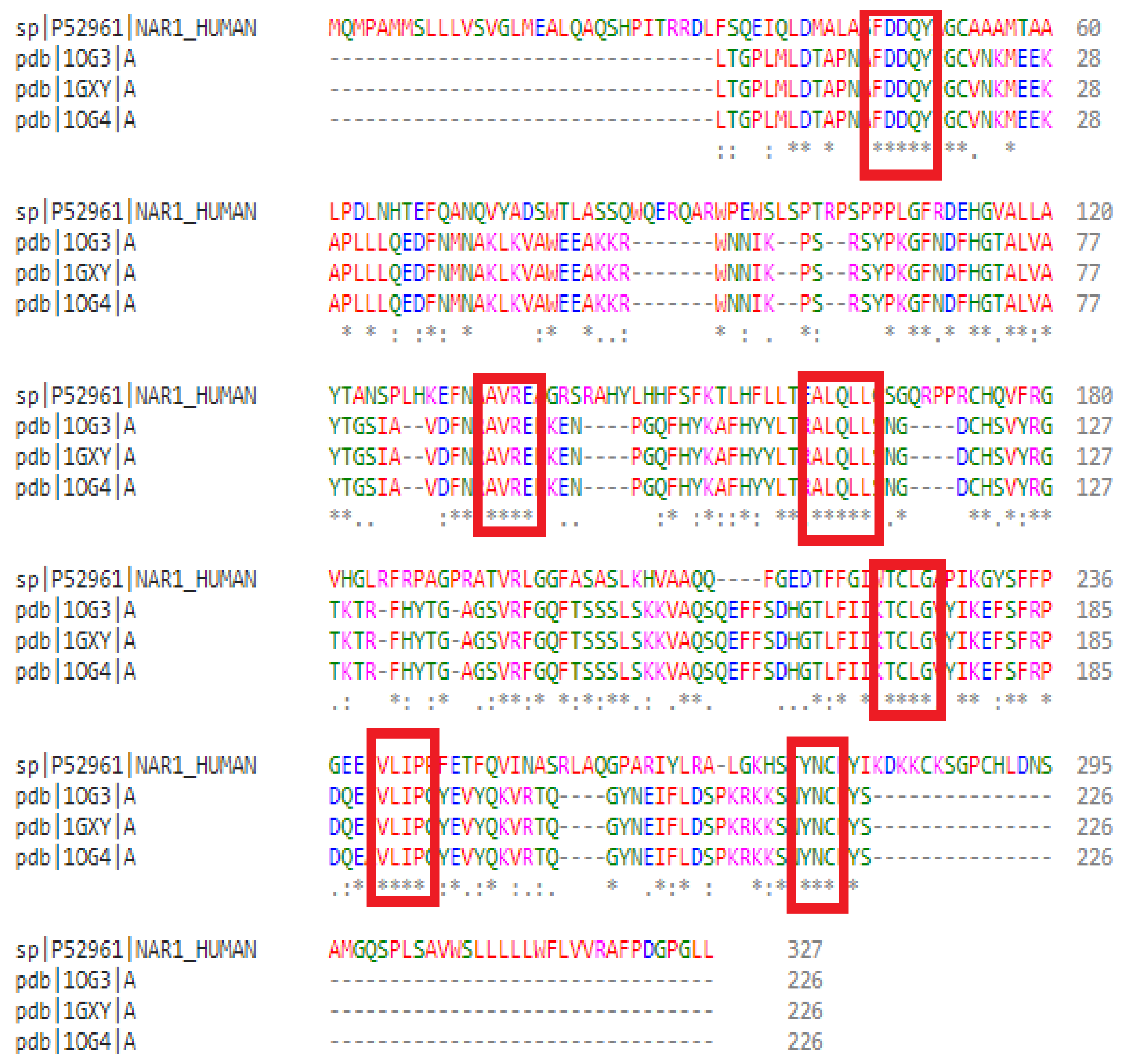
Figure 1. Sequence alignment performed with Clustal Omega between the query sequence and the three selected templates. Conservation levels are indicated as follows: (.) semi-conserved, (:) conserved, and (*) fully conserved. Red boxes indicate conserved amino acid regions between the target and template sequences.
Cimb 47 00634 g001
Figure 2. Graphical representation of Prosite result. The three red diamond-shaped markers indicate the positions of arginine (R144), serine (S202), and glutamic acid (E203), as predicted by PROSITE to constitute the protein’s active site. These residues lie within the conserved domain spanning residues 73 to 273 and are likely involved in catalytic activity or substrate binding.
Cimb 47 00634 g002
Figure 3. The result of QMEAN Z-score of the model.
Cimb 47 00634 g003
Figure 4. Predicted 3D structure of ART1 modeled through comparative modeling using SWISSMODEL and visualized using Discovery Studio and UCSF Chimera.
Cimb 47 00634 g004
Figure 5. Model validation results: (A) Ramachandran plot analysis generated by PROCHECK. Black square symbols represent the φ (phi) and ψ (psi) dihedral angles of non-glycine and non-proline residues. Red regions indicate the most favored regions, yellow the additionally allowed regions, and light yellow the generously allowed regions. Residues labeled in red represent outliers. (B) ERRAT evaluation. Different colors represent the error values across the amino acid sequence; darker regions indicate higher error scores. (C) VERIFY-3D assessment. The yellow line shows the 3D-1D compatibility score along the protein sequence; regions above the threshold line are considered structurally acceptable. (D) ProSA analysis. The black point represents the overall Z-score of the model, indicating its quality compared to experimentally solved structures.
Cimb 47 00634 g005
Figure 6. Molecular dynamics simulation results of the ART1 protein using iMODS: (A) deformability graphs, (B) B-factor plots, (C) eigenvalue plots, (D) variance maps, (E) covariance matrix plots, and (F) elastic network representation.
Cimb 47 00634 g006
Figure 7. Active site of the predicted protein. (A) The pocket of the protein’s active site. (B) The marked amino acid residues in the active site of protein.
Cimb 47 00634 g007
Figure 8. Visualization of the predicted protein’s active site using PyMOL and Discovery Studio.
Cimb 47 00634 g008
Figure 9. Two- and three-dimensional visualizations of the interactions between ART1 (in blue) and the top-ranked ligands (in red), emphasizing key binding features.
Cimb 47 00634 g009aCimb 47 00634 g009bCimb 47 00634 g009c
Figure 10. Results of pharmacophore study. Hydrophobic interactions are shown in green, negative ions in red, positive ions in blue, hydrophobic bonds in purple, and hydrogen donors and acceptors in white and orange, respectively ((A): resistomycin, (B): borrelidin, (C): tetracycline, (D): oxytetracycline).
Cimb 47 00634 g010
Table 1. Result of molecular docking of the best ligands with the predicted ART1 model.
Binding Energy (Kcal/mol)Hydrogen BondsHydrophobic BondsElectrostatic BondsVan der Waals BondsTotal Number
of Bonds
Meta-iodo-benzyl-guanidine−6.1Thr87, Glu203Phe178, Ala175, Arg144Glu203, Glu205Gly145, Ala168, Ser169, Phe200, Ser198, Phe199, Tyr86, Ser16715
Resistomycin−9.3Thr87, His93, His93His93, Phe178, Phe178, Ala175, Arg144Glu203, Glu203Ser90, Ser167, Glu205, Ser169, Ala168, Phe143.16
Borrelidin−9.0Gly145, Ser167, Ser90, Ala175Val146, Phe178/Arg144, Phe143, Ala168, Glu205, Glu203, Ser169, Ala174, Phe200, His9315
Tetracycline−9.0Ser167, Arg144, Arg144, His93Ala175, Ala175, Phe178/Ser169, Phe143, Ala163, Gly145, Val146, Leu149, Ser90, Glu20315
Oxytetracycline−8.9Arg144, Arg144Ala174, Ala175, Phe178/Ser169, Phe143, Ala168, Ser167, Val146, Leu149, His93, Ser90, Glu20314
Table 2. Results of biological activity prediction.
Biological ActivityResistomycin BorrelidinTetracyclineOxytetracycline
PaPiPaPiPaPiPaPi
Anticancer activity0.8960.0050.7990.0120.5290.0630.4650.082
PA—Probability of Activity; Pi—Probability of Inactivity.
Table 3. Drug-likeness characteristics of the selected molecules.
Molar Weight (g/mol)LogPLogSHBAHBDTPSA (Å2)AMRnRBLpinskiGhoseVeberEganMuegge
Resistomycin376.362.89−4.61 MS64115.06104.730Yes Yes YesYes Yes
Borrelidin489.643.61−2.53 S73127.85136.662YesNo Yes Yes No
Tetracycline444.43−0.34−1.82 S96181.62110.792Yes NoNo No No
Oxytetracycline460.63−1.01−1.0 S107201.85111.952No No No No No
Table 4. Results of ADMET study.
ResistomycinBorrelidinTetracyclineOxytetracycline
AbsorptionCaco2Yes Yes Yes No
HIAYes No Yes Yes
DistributionBBBNo No No No
PPBYes No Yes Yes
MetabolismCYP1A2 inhibitorYes No No No
CYP2C19 inhibitorNo No No No
CYP2C9 inhibitorYes No No No
CYP2D6 inhibitoryNo No No No
CYP3A4 inhibitorYes Yes No No
ExcretionCl0.493 (L)7.91 (M)2.238 (L)1.704 (L)
ToxicityAmes testYes No No No
CarcinogencityNo No No No
hERG BlockersNo Yes No No
H-HTYes Yes Yes No
Disclaimer/Publisher’s Note: The statements, opinions and data contained in all publications are solely those of the individual author(s) and contributor(s) and not of MDPI and/or the editor(s). MDPI and/or the editor(s) disclaim responsibility for any injury to people or property resulting from any ideas, methods, instructions or products referred to in the content.