Review

Elucidating the Role of CNOT2 in Regulating Cancer Cell Growth via the Modulation of p53 and c-Myc Expression

1
CytoGen lnc., Songpa-gu, Seoul 05610, Republic of Korea
2
Thrive Foot and Ankle Inc., Chino, CA 91710, USA
3
College of Korean Medicine, Kyung Hee University, Seoul 02447, Republic of Korea
*
Author to whom correspondence should be addressed.
These authors contributed equally to this work.
Curr. Issues Mol. Biol. 2025, 47(8), 615; https://doi.org/10.3390/cimb47080615 (registering DOI)
Submission received: 14 May 2025 / Revised: 11 July 2025 / Accepted: 24 July 2025 / Published: 4 August 2025
(This article belongs to the Section Biochemistry, Molecular and Cellular Biology)

Abstract

CNOT2, a central component of the CCR4-NOT transcription complex subunit 2, plays a pivotal role in the regulation of gene expression and metabolism. CNOT2 is involved in various cellular processes, including transcriptional regulation, mRNA deadenylation, and the modulation of mRNA stability. CNOT2 specifically contributes to the structural integrity and enzymatic activity of the CCR4-NOT complex with transcription factors and RNA-binding proteins. Recent studies have elucidated its involvement in cellular differentiation, immune response modulation, and the maintenance of genomic stability. Abnormal regulation of CNOT2 has been implicated in a spectrum of pathological conditions, including oncogenesis, neurodegenerative disorders, and metabolic dysfunctions. This review comprehensively examines the interplay between CNOT2 and p53, elucidating their collaborative and antagonistic interactions in various cellular contexts. CNOT2 is primarily involved in transcriptional regulation, mRNA deadenylation, and the modulation of mRNA stability, thereby influencing diverse biological processes such as cell proliferation, apoptosis, and differentiation. Conversely, p53 is renowned for its role in maintaining genomic integrity, inducing cell cycle arrest, apoptosis, and senescence in response to cellular stress and DNA damage. Emerging evidence suggests that CNOT2 can modulate p53 activity through multiple mechanisms, including the regulation of p53 mRNA stability and the modulation of p53 target gene expression. The dysregulation of CNOT2 and p53 interactions has been implicated in the pathogenesis and progression of various cancers, highlighting their potential as therapeutic targets. Additionally, CNOT2 regulates c-Myc, a well-known oncogene, in cancer cells. This review shows the essential roles of CNOT2 in maintaining cancer cellular homeostasis and explores its interactions within the CCR4-NOT complex that influence transcriptional and post-transcriptional regulation. Furthermore, we investigate the potential of CNOT2 as a biomarker and therapeutic target across various disease states, highlighting its significance in disease progression and treatment responsiveness.
Keywords:
CNOT2; p53; c-Myc; cancer

1. Introduction

Cancer is a multifaceted disease arising from genetic and epigenetic alterations that drive uncontrolled cell proliferation, invasion, and metastasis. Benign tumors are typically characterized by slow growth and a lack of invasive properties, preventing them from infiltrating nearby tissues or spreading to distant organs. However, depending on their size, anatomical location, and the risk of malignant transformation, medical intervention, such as surgical removal, may be warranted. Certain benign tumors, including adenomas and particular meningiomas, have the potential to cause clinical complications by exerting pressure on adjacent structures or progressing into malignant forms. In contrast, malignant tumors exhibit rapid proliferation, local tissue invasion, and the capacity for distant metastasis through the bloodstream or lymphatic system. These aggressive characteristics necessitate prompt therapeutic interventions, including surgery, chemotherapy, and radiation therapy, to control disease progression and improve patient outcomes [1,2]. Recent studies suggest that cancer progression is influenced not only by genetic mutations but also by regulatory interactions between key oncogenic and tumor suppressor pathways. Among these, CNOT2 (CCR4-NOT Transcription Complex Subunit 2) and p53 play CNOT2 (CCR4-NOT Transcription Complex Subunit 2) and p53 are pivotal regulators in cancer biology, influencing tumor progression, metastasis, and treatment response.
CNOT2 is a core component of the CCR4-NOT complex, a multi-subunit protein complex involved in mRNA degradation, transcriptional regulation, and protein ubiquitination. While the CCR4-NOT complex has been extensively studied, recent research has highlighted its emerging role in cancer pathophysiology. CNOT2 has been identified as a critical regulator of oncogenic pathways, contributing to tumor progression, immune evasion, and resistance to anticancer therapies [3,4]. Notably, studies indicate that CNOT2 modulates c-Myc stability, thereby impacting metabolic reprogramming and tumor cell proliferation [3,5].
The tumor suppressor p53 is one of the most extensively investigated genes in cancer research. Across diverse scientific disciplines—including molecular biology, pharmacology, biochemistry, and genetics—ongoing studies continue to elucidate novel regulatory mechanisms of p53 [1,2,6]. p53 functions as a key tumor suppressor by responding to cellular stressors such as DNA damage, ribosomal stress, and oncogenic activation. It orchestrates diverse cellular processes, including apoptosis, cell cycle arrest, and senescence, while also inhibiting angiogenesis, tumor cell migration, and metastasis [7,8,9,10,11,12].
However, TP53 mutations are among the most common genetic alterations in human malignancies, contributing to loss of tumor suppressive function and gain-of-function oncogenic activity [13,14]. Recent findings indicate that certain p53 mutations not only abolish tumor-suppressive activities but may also confer oncogenic properties that promote cancer cell survival and metastasis [14].
Recent research has uncovered a direct regulatory relationship between CNOT2 and p53, providing critical insights into tumorigenesis. CNOT2 depletion has been shown to induce apoptosis in p53 wild-type (WT-p53) cancer cells, whereas it exhibits an opposing effect in p53-null cells, suggesting a context-dependent role in tumor progression [14,15,16]. While CNOT2 has been implicated in transcriptional repression and RNA metabolism, its role in EMT and metastasis is only beginning to be elucidated. Recent studies suggest potential involvement in regulating cell motility and EMT-related gene networks [17,18]. Accumulating evidence also suggests that the interaction between CNOT2 and p53 may modulate chemotherapy sensitivity, with studies demonstrating that CNOT2 inhibition enhances the efficacy of chemotherapeutic agents such as Doxorubicin and 5-Fluorouracil (5-FU) [14].
Furthermore, recent investigations suggest that CNOT2 contributes to immune evasion in tumors by regulating cytokine production and T-cell infiltration within the tumor microenvironment. This growing body of evidence underscores the potential of targeting CNOT2, either independently or in combination with p53-restoring therapies, as a novel therapeutic strategy for cancer treatment.
c-Myc is a master regulator of transcription, cell cycle progression, and metabolism, and is one of the most frequently deregulated oncogenes in human cancers. It promotes the expression of genes involved in ribosomal biogenesis, glycolysis, and survival [13]. c-Myc also enhances ribosomal biogenesis and protein synthesis, whereas c-Myc is regulated by ribosomal proteins as feedback regulators [19].
Recent studies highlight the importance of transcriptional and post-transcriptional regulators in shaping the tumor microenvironment and determining cancer cell fate. CNOT2, as a core component of the CCR4–NOT complex, has emerged as a key modulator of gene expression programs that influence proliferation, apoptosis, and metabolic adaptation in tumor cells. Rather than focusing on general tumor classifications, this review emphasizes the functional role of CNOT2 in cancer progression and its interplay with major regulatory molecules such as p53 and c-Myc.

2. Oncogenic Functions of CNOT2 in Cancer Cells

Cancer can occur when a proto-oncogene is mutated and turned into an oncogene, and cells divide and multiply out of control. Here, the oncogene is a mutant gene with the potential for tumor development. Before an oncogene is mutated, it is called a proto-oncogene, and it plays a role in dividing normal cells [20]. CNOT2 has been implicated in oncogenic processes, including mRNA degradation, apoptosis regulation, and ER stress response, highlighting its role in cancer progression. When CNOT2 is depleted, the formation of P-body presumed to be mRNA degradation is suppressed. In addition, CHOP mRNA transcription is upregulated to induce endoplasmic reticulum (ER) stress and caspase-dependent apoptosis [21]. In hepatocellular carcinoma cell lines (HepG2 and Huh7), CNOT2 deficiency was found to enhance the antitumor effects of MID1IP1 depletion while reducing apoptosis markers, suggesting a functional interplay between CNOT2 and MID1IP1 in liver cancer cells [17]. Further studies were conducted based on previous papers that confirmed a close relationship between CNOT2 and MID1IP1. It was confirmed that the inhibition of CNOT2 activates p53 and induces apoptosis through MID1IP1 [12]. In non-small cell lung cancer (NSCLC) cells (H1299), atorvastatin treatment was found to be associated with apoptotic and autophagic pathways, where CNOT2 played a crucial regulatory role. The overexpression of HA-CNOT2 suppressed apoptotic and autophagic processes, whereas CNOT2 depletion enhanced these functions [16]. Moreover, in NSCLC cell lines (A549, H1299, H596, and H460), CNOT2 depletion was linked to apoptosis, SHP1 activation, and ER stress induction. In TRAIL-resistant lung cancer cells, CNOT2 knockdown sensitized cells to TRAIL-mediated apoptosis by modulating SHP1-STAT3 signaling and activating death receptor 5 (DR5) through intracellular stress responses [22]. Recent studies have also investigated the role of CNOT2 in drug response. Ophiopogon (OP) is a substance extracted from the root mass of ophiopogon japonica and has an anticancer effect. Ophiophogonin D (OP-D) of Ophiophogon (OP) induced p53 expression through ribosomal proteins L5 and L11 and inhibited c-Myc expression independently of CNOT2 capacity. It is notable here that the depletion of CNOT2 increased the Opiopogon D (Op-D) effect on c-Myc in colon cancer cells [23]. Similarly, Brasinin extracted from Chinese cabbage was dose- and time-dependently correlated with CNOT2 and p53 expression in HCT116 colorectal cancer cells, further underscoring the therapeutic potential of targeting CNOT2 in cancer treatment [18]. In summary, CNOT2 is implicated in cancer progression through its roles in mRNA degradation, apoptosis regulation, and ER stress response. The depletion of CNOT2 enhances apoptosis via interactions with MID1IP1, p53 activation, and modulation of SHP1-STAT3 signaling pathways in various cancers (Figure 1).

3. CNOT2 Promotes Cancer Cell Proliferation, Angiogenesis, and EMT

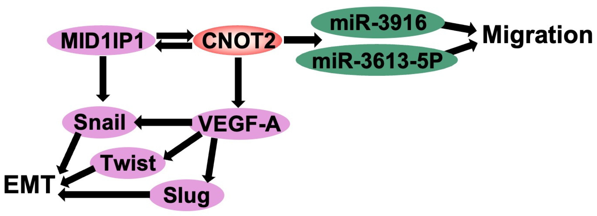
A tumor is defined as an abnormal mass of cells that evades normal cell cycle regulation and undergoes uncontrolled proliferation. Tumors are broadly classified into malignant and benign types [7]. Benign tumors are generally well-differentiated, slow-growing, and non-invasive, lacking the ability to metastasize to distant organs. However, depending on their size, location, and potential for malignant transformation, medical intervention may be required. In contrast, malignant tumors proliferate uncontrollably, invade surrounding tissues, and metastasize through the bloodstream or lymphatic system, necessitating prompt therapeutic intervention. [9]. In breast cancer, angiogenesis and epithelial–mesenchymal transition (EMT) are critical processes that drive tumor progression and metastasis. Vascular endothelial growth factor A (VEGF-A) is a key regulator of angiogenesis, stimulating new blood vessel formation to sustain tumor growth and enhance its aggressiveness [8]. Recent studies have demonstrated that CNOT2, a core subunit of the CCR4-NOT complex, is a crucial modulator of these oncogenic processes [16,17,18]. CNOT2 was found to be overexpressed in breast cancer cell lines (MCF-7 and MDA-MB-231), promoting tumor proliferation and metastasis. Conversely, CNOT2 depletion led to reduced motility in MDA-MB-231 cells, the downregulation of proliferation-related gene expression, and the suppression of VEGF signaling [15]. The reduction in VEGF expression upon CNOT2 depletion led to decreased angiogenic potential, suggesting that CNOT2 is a critical upstream regulator of VEGF-A-mediated angiogenesis. EMT is characterized by the loss of epithelial markers and the acquisition of mesenchymal traits, enabling cancer cells to invade and metastasize. EMT-related genes, including Snail, Slug, and Twist, were found to be significantly downregulated in MDA-MB-231 cells lacking CNOT2. Furthermore, miRNA profiling of these cells revealed that has-miR-3613-5p and has-miR-3916 play critical roles in relieving migration inhibition. These miRNAs may serve as downstream effectors of CNOT2, acting as regulatory nodes in breast cancer cell migration and EMT [24]. Given its regulatory involvement in tumor proliferation, angiogenesis, and metastasis, CNOT2 emerges as a potential therapeutic target for breast cancer. Inhibiting CNOT2 could effectively suppress tumor growth and metastatic progression, providing a multifaceted approach to cancer treatment. Future studies should focus on elucidating the precise molecular mechanisms by which CNOT2 modulates oncogenic pathways and developing targeted inhibitors to disrupt its function (Figure 2).

4. Knockdown of CNOT2 Induces p53 Activation

Numerous reports in relation to p53 have been available for 40 years since its first discovery, the year 1979 [25]. p53 plays a critical role as a guardian of cell genome, responding to DNA double strand breaks and regulating various target genes as a transcription factor [26]. It has been determined that p53 mediates tumor-suppressing mechanisms such as apoptosis and cell cycle [27]. The half-life of the p53 protein is tightly regulated and significantly prolonged in response to cellular stress signals such as DNA damage [28]. One of the most well-known mechanisms regulating p53 is autoregulatory feedback loops with MDM2 [29]. The N-terminal domain of MDM2 binds to N and C terminal sites of p53 and regulates its proteolysis [30]. In this section, recent studies about p53 were organized including MDM2 as well as ribosomal proteins, ubiquitylation, and neddylation and un-masked in association with interactive characteristics between p53 and CNOT2.

4.1. Regulation of p53-MDM2 Loop by Ribosomal Proteins

A total of 47S/45S pre-rRNA, RP-encoding mRNAs, 5S rRNA, non-ribosomal factors, and small nucleolar RNAs were recruited, comprising the 90S pre-ribosomes in the nucleolus, and then matured to 60S and 40S subunits for protein translation [31]. Recently, various ribosomal proteins have been studied to determine what inhibits and activates p53. It has been found that ribosomal biogenesis stimulates p53 via the suppression of MDM2 E3 ligase in combination with ribosomal proteins including RPL5, RPL11, RPL23 [32], S7 [33], S14 [34], RPS25 [35], RPS15 [36], RPL22 [37], RPS2 [38], and RPL34 [39]. On the other hand, p53 was shown to be activated upon disruption of specific ribosome proteins containing S27L [40], RPL37 [41], RPL31 [42], RPL4 [43], and RPL40 [44] (Table 1). This regulatory relationship is particularly relevant in the context of CNOT2, as recent data indicate that CNOT2 knockdown leads to the upregulation of RPL5 and RPL11. These proteins stabilize p53 by blocking its degradation via MDM2, providing a mechanistic link between CNOT2 depletion and enhanced p53-mediated tumor suppression.

4.2. Regulation of p53-MDM2 Loop: Ubiquitylation and Neddylation

Fuchs et al. found that p53 is degraded by MDM2 whose degradation was downregulated when sites between 150 and 461 aa were deleted [45]. Next, specific sites of MDM2 regulating p53 were determined to be the main residues between 202 and 302 functioning as MDM2 RING (really interesting genes) finger [46]. Also, it was identified that p53 binds this region of MDM2, which acts as an E3 ubiquitin ligase and is then subsequently degraded by the 26S proteasome [47]. The ubiquitin-conjugating enzymes (E2s) as well as the E3 ligase MDM2 also support MDM2 auto-ubiquitination of p53. Mark et al. showed that depletion of the ubiquitin-conjugating enzyme UbcH5B and -C inhibits ubiquitination of p53 [48]. The ubiquitin-activating enzyme (E1) UBE1L2 activates ubiquitin, transfers it onto UbcH5b, and interacts with MDM2 and ubiquitinate p53 [49]. In addition, many E2 that regulate the ubiquitination of p53 have been consistently discovered and demonstrated. In addition, various mechanisms were also identified. FATS promoted the stabilization of p53 as an E2-independent ubiquitin ligase [50]. Binding SUMO1 to Mdm2 was followed by the conjugation of Su-mo-1 to MDM2 by Ubc9 [51]. It was determined that the neddylation mechanism similar to ubiquitination mediates the ubiquitin–proteasome system as an important signaling pathway. Neddylation is caused by neural precursor cell-expressed, developmentally downregulated protein 8 (NEDD8) E1 activating enzyme (NAE), and NEDD8 E2/E3 enzymes with NEDD8. Interestingly, in our previous study, it was documented that NEDD8 E2 enzyme UBE2M blocks p53 while binding to MDM2 and RPL11 [52]. These classical mechanisms of p53 regulation through MDM2 and ribosomal proteins provide a crucial framework for understanding the emerging role of CNOT2. Notably, CNOT2 knockdown has been shown to enhance the expression of ribosomal proteins such as RPL5 and RPL11, both of which are known to inhibit MDM2 and stabilize p53. This suggests that CNOT2 may indirectly suppress p53 activity by modulating the availability or function of these ribosomal protein regulators.

4.3. CNOT2 Knockdown Induces p53 in Cancer Cells

Building on this regulatory framework, recent evidence indicates that CNOT2 directly influences the stability and activity of p53 in a ribosomal protein-dependent manner. In colorectal cancer cells, CNOT2 knockdown leads to the activation of p53 and apoptosis, supporting its role as an upstream modulator of the p53 axis through both transcriptional and translational mechanisms. CNOT2 has been identified as a key regulator in various cancer-related pathways. Previous studies have demonstrated that CNOT2 plays a critical role in atorvastatin-induced apoptosis in non-small cell lung cancer cells [16]. Additionally, CNOT2 has been reported to promote breast cancer cell proliferation and angiogenesis [15]. Moreover, it has been implicated in liver cancer progression through its regulation of c-Myc via ribosomal proteins [17].
Given these findings, we postulate that CNOT2 knockdown may contribute to p53 activation, thereby promoting apoptosis in cancer cells. Consistent with this hypothesis, Rosa Puca et al. reported that HIPK2, a well-established activator of p53, limits CNOT2-dependent mRNA decay, leading to increased p53 stability and activity [53]. Further supporting this, bioinformatics analysis has demonstrated a significant correlation between CNOT2 expression and the p53 signaling pathway, suggesting a regulatory association between these two factors [38]. Several studies have employed RNA interference (siRNA) to investigate the role of CNOT2 in p53 regulation. In colorectal cancer cells, siRNA-mediated knockdown of CNOT2 resulted in prolonged p53 half-life and enhanced apoptosis. Additionally, co-immunoprecipitation (co-IP) experiments have been used to detect protein–protein interactions between CNOT2 and p53 regulatory elements, including MID1IP1 and ribosomal proteins such as RPL5 and RPL11. Moreover, transcriptomic and pathway enrichment analyses have demonstrated a strong positive correlation between CNOT2 expression and the downregulation of p53 signaling pathways across multiple cancer datasets. These findings underscore the relevance of both direct and indirect regulatory interactions between CNOT2 and p53 in cancer pathophysiology.
Moreover, experimental evidence has shown that silencing CNOT2 enhances TRAIL sensitivity in cancer cells, thereby counteracting mechanisms that allow tumor cells to evade p53-mediated apoptosis [37]. Recent studies have also indicated that the depletion of CNOT2 results in p53 accumulation and heightened apoptotic activity in colorectal cancer cells, as evidenced by a prolonged p53 half-life in CNOT2-deficient cells compared to controls [39]. Additionally, MID1IP1 has been identified as a crucial modulator of p53 stability, acting through its interaction with CNOT2.
Collectively, these findings suggest that CNOT2 plays an essential role in regulating p53-mediated apoptosis in cancer cells. The suppression of CNOT2 not only stabilizes p53 but also enhances its pro-apoptotic function, highlighting its potential as a therapeutic target for cancer treatment.

4.4. Modulation of p53 Activity Through CNOT2 Inhibition as Potential Anticancer Strategy

Accumulating evidence suggests a functional interplay between CNOT2 and the tumor suppressor p53 in cancer cells. Experimental studies have demonstrated that the downregulation of CNOT2 can influence p53-associated signaling pathways. In particular, treatment with Brassinin, a phytoalexin compound, was reported to reduce the expression levels of both CNOT2 and p53, and its co-treatment with Doxorubicin led to enhanced cytotoxic effects in vitro, implying a potential for combination-based therapeutic strategies [18].
A variety of chemotherapeutic agents are known to exert their effects through the activation of the p53 pathway. Among them, Doxorubicin (Adriamycin) [54], 5-Fluorouracil [55,56], Docetaxel (Taxotere), and Cisplatin [57,58] have been approved for clinical use and are widely applied in standard cancer treatment protocols. Although specific small-molecule inhibitors of CNOT2 have not yet been developed, experimental approaches such as siRNA-mediated gene silencing have been utilized to investigate its biological function and therapeutic relevance (Figure 1).
Further studies are warranted to elucidate the molecular mechanisms through which CNOT2 modulates p53 stability and activity, and to determine whether targeting CNOT2 could enhance the responsiveness to conventional p53-based therapies. Additionally, the identification of biomarkers associated with CNOT2 expression or activity may contribute to the development of more personalized and effective therapeutic interventions.

5. CNOT2 Regulates c-Myc Expression via Ribosomal Proteins

5.1. Function of c-Myc in Cancer Cells

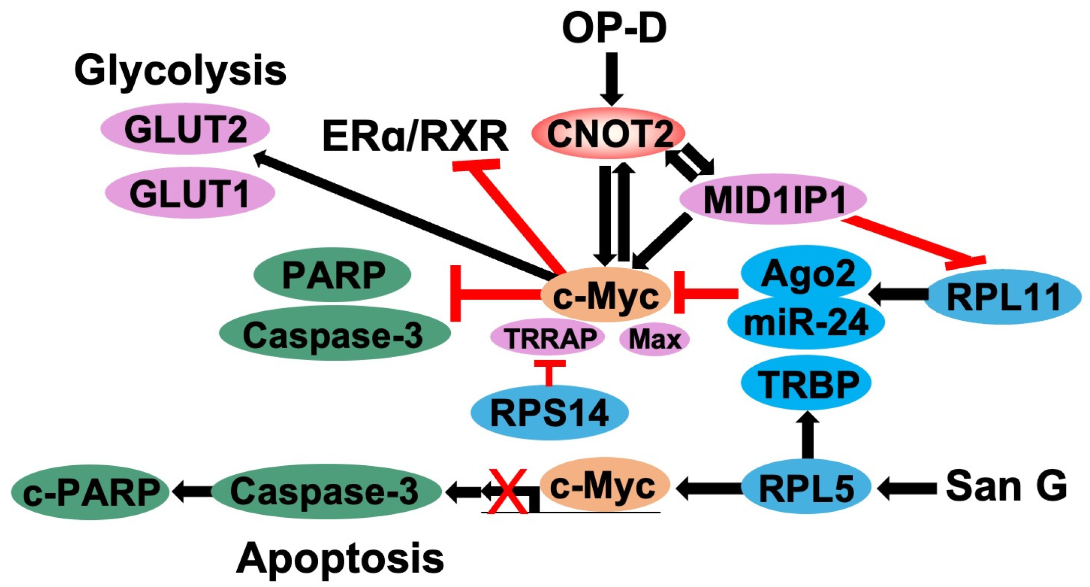
c-Myc is a transcription factor belonging to the basic helix–loop–helix (bHLH) family and is mainly present in the nucleus of the cell. In addition, c-Myc is overexpressed in cancer cells and regulates cell growth, differentiation, metabolism, and apoptosis [59]. The regulation of c-Myc expression is primarily dependent on its protein stability. c-Myc is a short-lived protein, with a half-life of approximately 30 min for protein, making it an efficient mechanism for gene regulation [60]. When genetic alterations in c-Myc occur in non-coding regulatory regions rather than protein coding sequences, the regulation of c-Myc expression causes more cell proliferation effects [61]. c-Myc plays a key role in promoting the transcription of genes encoding essential proteins. Since these genes are involved in ribosomal biogenesis, ribosomal protein translation, and overall ribosomal function, c-Myc can activate ribosomal biogenesis and protein synthesis. This is the primary mechanism through which c-Myc regulates cell growth and proliferation [62]. c-Myc is considered a promising target for cancer treatment because it is highly active in various types of cancer (Figure 3). Consequently, drugs targeting c-Myc can be developed as potential therapies for a wide range of malignancies [63].

5.2. Regulation of c-Myc by Ribosomal Proteins

Certain ribosomal proteins can be regulated by c-Myc, which is responsible for promoting ribosomal RNA and protein transcription to facilitate ribosomes biogenesis [62]. Since ribosomal proteins are affected by oncogenic factors and dysregulated translation processes, emerging evidence suggests that mutations in ribosomal proteins are critically involved in ribosomopathies and carcinogenesis [17]. Several ribosomal proteins affected in cancer are associated with widely known oncogenes, tumor suppressors [63]. Specifically, ribosomal proteins RPL5, RPL11, and RPS14 regulate c-Myc by recruiting it to RISC for c-Myc expression and c-Myc mRNA degradation [62].
Recently, it has been shown that one of the functions of ribosomal protein L11 (RPL11) is to act as a feedback regulator of c-Myc [64]. RPL11, a ribosomal protein, plays a key role not only in ribosome function but also in transmitting ribosomal stress signals to the p53-dependent cell cycle checkpoint by inhibiting MDM2, a major negative regulator of p53 [65]. RPL11 interacts with c-Myc in the promoter regions of c-Myc’s target genes and represses its transcriptional activity in response to ribosomal stress [62,66,67]. Additionally, RPL11 binds to miR-24 as well as c-Myc mRNA at 3′ untranslated region(3′UTR), a key component of the microRNA-induced silencing complex (miRISC) argonaute 2 (Ago2), which induces reduction by recruiting miR-24-loaded miRISCs and then degrading c-Myc mRNA [66]. The ablation of ago2 abrogated RPL11-mediated reduction in c-Myc mRNA. On the other hand, the knockdown of RPL11 significantly increases the mRNA level and stability of miR-24-mediated c-Myc mRNA in cells [65,66].
Ribosomal protein L5 (RPL5) also has a role in regulating c-Myc. A study on the effects of SanG found that it inhibits the proliferation of non-small cell lung cancer cells and induces cell apoptosis through a combination of caspase3 activation and RPL5-mediated inhibition of c-Myc [68]. RPL5 mediates the targeting of c-Myc mRNA via miRNAs by binding to the 3′UTR of c-Myc mRNA, the HIV-1 TAR RNA Binding Protein (TRBP), and Ago2. The knockdown of RPL5 induced c-Myc expression at both the mRNA and protein levels, while the overexpression of RPL5 suppressed c-Myc expression and activity [62].
The ribosomal proteins RPL5 and RPL11 function together to co-suppress c-Myc expression. RPL5 cooperates with RPL11 to recruit the RNA-induced silencing complex (RISC) to c-Myc mRNA, facilitating its degradation and thereby suppressing c-Myc activity [62]. Concerning the depletion of MID1IP1 in HepG2, Huh7 cells activated RPL5 and RPL11 and reduced c-Myc. The upregulation of RPL5 or RPL11 is regulated through the depletion of MID1IP1, and the depletion of RPL5 and RPL11 activates c-Myc in MID1IP1-depleted HepG2 and Huh7 cells, suggesting that RPL5 and RPL11 are responsible for the regulation of c-Myc as tumor suppressors [17].
Also, RPS14 is a ribosomal protein that regulates c-Myc. RPS14 prevents c-Myc and its cofactor TRRAP from binding to target gene promoters. A deficiency of RPS14 regulates both the mRNA and protein levels of c-Myc. The Myc homology box II (MBII) of the oncogene c-Myc and C-terminal basic helix–loop–helix leucine zipper (bHLH-LZ) domains interact with RPS14. When c-Myc transcription is inhibited by RPS14, c-Myc-driven cell proliferation is also inhibited. RPS14, which directly inhibits transcriptional activity and mediates mRNA degradation via miRNAs, abrogates the function of c-Myc [69]. Collectively, ribosomal proteins such as RPL5, RPL11, and RPS14 regulate c-Myc by influencing its transcriptional activity and promoting degradation through interactions with the miRNA-induced silencing complex (RISC). RPL5 and RPL11 collaborate to suppress c-Myc expression via miRNA-mediated mRNA degradation, thus acting as tumor suppressors. RPS14 inhibits c-Myc activity by blocking its binding to gene promoters and facilitating its degradation, thereby restraining c-Myc-driven cancer cell proliferation.

5.3. Knockdown of CNOT2 Effects c-Myc Expression

In the metabolic processes of eukaryotic cells, the CCR4-NOT complex controls several steps in mRNA production [10]. CNOT2, one of the nine subunits of the CCR4-NOT complex, acts to regulate transcription and translation, but can also work as on oncogene to promote proliferation, lipid metabolism, angiogenesis, and autophagy [11,13,19].
Recent studies have shown that CNOT2 functions as an oncogene when proliferation and tumor angiogenesis was inhibited through Vascular Endothelial Growth Factor (VEGF) signaling are suppressed in CNOT2-suppressed human cancer cells (Figure 2) [25]. In addition, previous studies have shown that CNOT2 can regulate the expression of c-Myc and thus induce apoptosis in cancer cells. In tumor tissue, CNOT2 is overexpressed, and in pancreatic cancer, CNOT2 regulates the expression of c-Myc. And there have also been studies suggesting that liver cancer growth can regulate c-Myc in the liver, mediated by CNOT2 [17]. Furthermore, researchers studied the oncogenic potential of MID1IP1 in hepatocellular carcinoma cell (HCC) growth in relation to ribosomal protein L5 and L11 and CNOT2-mediated c-Myc signaling and found that CNOT2 knockdown could further inhibit the downregulation of MID1IP1 by c-Myc. The inhibition of CNOT2 potentiated the antitumor effect produced by reduced MID1IP1 and downregulated c-Myc, pro-caspase3, and pro-PARP in MID1IP1-depleted HepG2, Huh7, and HCT116 cells. MID1IP1 supports the cooperation of proto-oncogene c-Myc, mediated by RPL5, RPL11, and CNOT2, which acts as a potent oncogene molecule, in the progression of liver cancer [17]. Moreover, CNOT2 can also inhibit the ligand-dependent transcriptional activation of ERα and retinoid X receptor (RXR) target genes such as c-Myc in MCF-7 human breast cancer cells [15] (Table 2).
These results suggest that the knockdown of CNOT2 may have a significant effect on the expression of c-Myc and that the colocalization of CNOT2 and c-Myc may regulate cell death in tumors (Figure 4).

6. Conclusions

Recent studies have revealed the critical role of CNOT2 in cancer progression, highlighting its involvement in multiple oncogenic processes. CNOT2, as a key component of the CCR4–NOT complex, influences tumorigenesis primarily through its regulation of mRNA stability and transcription. Its modulation of p53 and c-Myc activity has been shown to support cancer cell survival, proliferation, and therapy resistance.
Beyond its role in transcriptional and post-transcriptional regulation, CNOT2 has been implicated in drug resistance, immune evasion, and metabolic reprogramming, underscoring its potential as a key factor in tumor adaptation and therapy resistance. Notably, recent studies indicate that CNOT2 depletion enhances chemosensitivity in certain cancer types, suggesting that targeting CNOT2 may improve treatment efficacy when combined with conventional anticancer agents.
Interestingly, recent studies demonstrate that CNOT2 may exert opposite biological effects depending on tumor context. In breast cancer models, CNOT2 has been shown to promote proliferation and angiogenesis through VEGF signaling [15], whereas in colorectal cancer cells, CNOT2 depletion activates p53 signaling via MID1IP1, leading to apoptosis [12]. These findings suggest that CNOT2 may function either as an oncogene or a tumor suppressor, contingent on cancer type and cellular environment. This tumor-type specificity highlights the importance of context-dependent investigations when evaluating CNOT2 as a therapeutic target.
Furthermore, CNOT2 undergoes MK2-dependent phosphorylation in response to cellular stress, which suppresses deadenylase activity and induces apoptosis [70]. These findings reveal additional layers of post-translational regulation and suggest that CNOT2 contributes to stress responses in cancer cells.
Structurally, the CCR4–NOT complex functions as a multi-subunit regulatory platform composed of enzymatic and scaffolding components. CNOT1 acts as the central hub, organizing other subunits including CNOT2, CNOT3, CNOT6/6L, and CNOT7 to coordinate deadenylation, transcriptional repression, and RNA surveillance [71]. Moreover, the complex has been proposed to operate as a versatile “chaperone platform” that integrates RNA metabolism with broader cellular functions such as DNA repair, protein degradation, and chromatin remodeling [72].
Despite these advancements, several questions remain unanswered. The precise molecular mechanisms governing CNOT2-mediated oncogenic signaling, its potential interaction with other cancer-associated pathways, and its context-dependent roles across different tumor types require further investigation. Additionally, given the dual role of CNOT2 in both oncogenic and tumor-suppressive mechanisms depending on the cellular context, future research should explore its function in a broader spectrum of malignancies.
Although CNOT2 has shown promise in regulating cancer-related signaling pathways such as p53 and c-Myc, its clinical utility as a biomarker or therapeutic target remains an emerging hypothesis. Future studies incorporating transcriptomics, proteomics, and metabolomics will be essential to validate CNOT2’s diagnostic and prognostic potential and to uncover its broader network-level interactions in various tumor contexts.
Taken together, CNOT2 emerges as a key regulator that integrates oncogenic signaling with transcriptional, post-transcriptional, and stress-response pathways. Given its dual role in supporting oncogenesis or enabling tumor suppression depending on the cellular context, CNOT2 represents a compelling but nuanced target whose therapeutic exploitation will require precise, tumor-specific strategies.

Author Contributions

Conceptualization, J.H.J.; writing—original draft preparation, J.H.J., Y.J.L., J.J.O., J.L. and J.-H.K.; writing—review and editing, J.H.J., Y.J.L., Y.J.H. and J.J.O.; visualization, J.-H.K.; project administration, J.H.J.; funding acquisition, J.H.J. All authors have read and agreed to the published version of the manuscript.

Funding

This work was supported by a grant from the National Research Foundation of Korea (NRF) grant funded by the Korea government (No. RS-2023-00209169).

Conflicts of Interest

Author Jihyun Lee is employed by CytoGen lnc., and Ju-Ha Kim is employed by Thrive Foot and Ankle Inc., The authors declare no conflict of interest.

References

  1. Nguyen, D.; Liao, W.; Zeng, S.X.; Lu, H. Reviving the guardian of the genome: Small molecule activators of p53. Pharmacol. Ther. 2017, 178, 92–108. [Google Scholar] [CrossRef]
  2. Zilfou, J.T.; Lowe, S.W. Tumor suppressive functions of p53. Cold Spring Harb. Perspect. Biol. 2009, 1, a001883. [Google Scholar] [CrossRef]
  3. Yoon, K.W.; Byun, S.; Kwon, E.; Hwang, S.Y.; Chu, K.; Hiraki, M.; Jo, S.H.; Weins, A.; Hakroush, S.; Cebulla, A.; et al. Control of signaling-mediated clearance of apoptotic cells by the tumor suppressor p53. Science 2015, 349, 1261669. [Google Scholar] [CrossRef]
  4. Galluzzi, L.; Bravo-San Pedro, J.M.; Kroemer, G. Ferroptosis in p53-dependent oncosuppression and organismal homeostasis. Cell Death Differ. 2015, 22, 1237–1238. [Google Scholar] [CrossRef]
  5. Gnanapradeepan, K.; Basu, S.; Barnoud, T.; Budina-Kolomets, A.; Kung, C.P.; Murphy, M.E. The p53 Tumor Suppressor in the Control of Metabolism and Ferroptosis. Front. Endocrinol. 2018, 9, 124. [Google Scholar] [CrossRef] [PubMed]
  6. Powell, E.; Piwnica-Worms, D.; Piwnica-Worms, H. Contribution of p53 to metastasis. Cancer Discov. 2014, 4, 405–414. [Google Scholar] [CrossRef] [PubMed]
  7. Chen, J. The Cell-Cycle Arrest and Apoptotic Functions of p53 in Tumor Initiation and Progression. Cold Spring Harb. Perspect. Med. 2016, 6, a026104. [Google Scholar] [CrossRef]
  8. Farhang Ghahremani, M.; Goossens, S.; Nittner, D.; Bisteau, X.; Bartunkova, S.; Zwolinska, A.; Hulpiau, P.; Haigh, K.; Haenebalcke, L.; Drogat, B.; et al. p53 promotes VEGF expression and angiogenesis in the absence of an intact p21-Rb pathway. Cell Death Differ. 2013, 20, 888–897. [Google Scholar] [CrossRef]
  9. Muller, P.A.; Vousden, K.H.; Norman, J.C. p53 and its mutants in tumor cell migration and invasion. J. Cell Biol. 2011, 192, 209–218. [Google Scholar] [CrossRef] [PubMed]
  10. Collart, M.A. The CCR4-NOT complex is a key regulator of eukaryotic gene expression. Wiley Interdiscip. Rev. RNA 2016, 7, 438–454. [Google Scholar] [CrossRef]
  11. Jayne, S.; Zwartjes, C.G.; van Schaik, F.M.; Timmers, H.T. Involvement of the SMRT/NCoR-HDAC3 complex in transcriptional repression by the CNOT2 subunit of the human CCR4-NOT complex. Biochem. J. 2006, 398, 461–467. [Google Scholar] [CrossRef]
  12. Jung, J.H.; Lee, D.; Ko, H.M.; Jang, H.J. Inhibition of CNOT2 Induces Apoptosis via MID1IP1 in Colorectal Cancer Cells by Activating p53. Biomolecules 2021, 11, 1492. [Google Scholar] [CrossRef] [PubMed]
  13. Zwartjes, C.G.; Jayne, S.; van den Berg, D.L.; Timmers, H.T. Repression of promoter activity by CNOT2, a subunit of the transcription regulatory CCR4-NOT complex. J. Biol. Chem. 2004, 279, 10848–10854. [Google Scholar] [CrossRef]
  14. Ito, K.; Inoue, T.; Yokoyama, K.; Morita, M.; Suzuki, T.; Yamamoto, T. CNOT2 depletion disrupts and inhibits the CCR4-NOT deadenylase complex and induces apoptotic cell death. Genes Cells Devoted Mol. Cell. Mech. 2011, 16, 368–379. [Google Scholar] [CrossRef] [PubMed]
  15. Sohn, E.J.; Jung, D.B.; Lee, H.; Han, I.; Lee, J.; Lee, H.; Kim, S.H. CNOT2 promotes proliferation and angiogenesis via VEGF signaling in MDA-MB-231 breast cancer cells. Cancer Lett. 2018, 412, 88–98. [Google Scholar] [CrossRef]
  16. Lee, J.; Jung, J.H.; Hwang, J.; Park, J.E.; Kim, J.H.; Park, W.Y.; Suh, J.Y.; Kim, S.H. CNOT2 Is Critically Involved in Atorvastatin Induced Apoptotic and Autophagic Cell Death in Non-Small Cell Lung Cancers. Cancers 2019, 11, 1470. [Google Scholar] [CrossRef] [PubMed]
  17. Jung, J.H.; Lee, H.J.; Kim, J.H.; Sim, D.Y.; Im, E.; Kim, S.; Chang, S.; Kim, S.H. Colocalization of MID1IP1 and c-Myc is Critically Involved in Liver Cancer Growth via Regulation of Ribosomal Protein L5 and L11 and CNOT2. Cells 2020, 9, 985. [Google Scholar] [CrossRef]
  18. Park, W.Y.; Park, J.E.; Jung, J.H. Apoptotic Effect of Brassinin via Inhibition of CNOT2 and Activation of p53 and Its Combination Effect with Doxorubicin. Appl. Sci. 2021, 11, 10036. [Google Scholar] [CrossRef]
  19. Marsh, B.M. Characterization of CNOT1 and CNOT2, Candidate Colorectal Cancer Genes Identified in a Transposon-Based Genetic Screen in Mice. Master’s Thesis, University of Minnesota, Minneapolis, MN, USA, 2010. [Google Scholar]
  20. Faraji, F.; Hu, Y.; Wu, G.; Goldberger, N.E.; Walker, R.C.; Zhang, J.; Hunter, K.W. An integrated systems genetics screen reveals the transcriptional structure of inherited predisposition to metastatic disease. Genome Res. 2014, 24, 227–240. [Google Scholar] [CrossRef]
  21. Pastushenko, I.; Blanpain, C. EMT Transition States during Tumor Progression and Metastasis. Trends Cell Biol. 2019, 29, 212–226. [Google Scholar] [CrossRef]
  22. Kim, E.O.; Kang, S.E.; Choi, M.; Rhee, K.J.; Yun, M. CCR4-NOT transcription complex subunit 2 regulates TRAIL sensitivity in nonsmallcell lung cancer cells via the STAT3 pathway. Int. J. Mol. Med. 2020, 45, 324–332. [Google Scholar] [PubMed]
  23. Ko, H.M.; Jee, W.; Lee, D.; Jang, H.J.; Jung, J.H. Ophiopogonin D increases apoptosis by activating p53 via ribosomal protein L5 and L11 and inhibiting the expression of c-Myc via CNOT2. Front. Pharmacol. 2022, 13, 974468. [Google Scholar] [CrossRef] [PubMed]
  24. Sohn, E.J. MiRNA 3613-5P and MiRNA 3916 rescued the inhibition of cell migration in CNOT2 depleted MDA-MD-231 cells. Transl Cancer Res. 2020, 9, 4542–4549. [Google Scholar] [CrossRef] [PubMed]
  25. Levine, A.J.; Oren, M. The first 30 years of p53: Growing ever more complex. Nat. Rev. Cancer 2009, 9, 749–758. [Google Scholar] [CrossRef]
  26. Sigal, A.; Rotter, V. Oncogenic mutations of the p53 tumor suppressor: The demons of the guardian of the genome. Cancer Res. 2000, 60, 6788–6793. [Google Scholar]
  27. Kastan, M.B.; Canman, C.E.; Leonard, C.J. P53, cell cycle control and apoptosis: Implications for cancer. Cancer Metastasis Rev. 1995, 14, 3–15. [Google Scholar] [CrossRef]
  28. Porter, J.R.; Fisher, B.E.; Batchelor, E. p53 Pulses Diversify Target Gene Expression Dynamics in an mRNA Half-Life-Dependent Manner and Delineate Co-regulated Target Gene Subnetworks. Cell Syst. 2016, 2, 272–282. [Google Scholar] [CrossRef]
  29. Harris, S.L.; Levine, A.J. The p53 pathway: Positive and negative feedback loops. Oncogene 2005, 24, 2899–2908. [Google Scholar] [CrossRef]
  30. Poyurovsky, M.V.; Katz, C.; Laptenko, O.; Beckerman, R.; Lokshin, M.; Ahn, J.; Byeon, I.J.; Gabizon, R.; Mattia, M.; Zupnick, A.; et al. The C terminus of p53 binds the N-terminal domain of MDM2. Nat. Struct. Mol. Biol. 2010, 17, 982–989. [Google Scholar] [CrossRef]
  31. Zhou, X.; Liao, W.J.; Liao, J.M.; Liao, P.; Lu, H. Ribosomal proteins: Functions beyond the ribosome. J. Mol. Cell Biol. 2015, 7, 92–104. [Google Scholar] [CrossRef]
  32. Dai, M.S.; Lu, H. Inhibition of MDM2-mediated p53 ubiquitination and degradation by ribosomal protein L5. J. Biol. Chem. 2004, 279, 44475–44482. [Google Scholar] [CrossRef]
  33. Chen, D.; Zhang, Z.; Li, M.; Wang, W.; Li, Y.; Rayburn, E.R.; Hill, D.L.; Wang, H.; Zhang, R. Ribosomal protein S7 as a novel modulator of p53-MDM2 interaction: Binding to MDM2, stabilization of p53 protein, and activation of p53 function. Oncogene 2007, 26, 5029–5037. [Google Scholar] [CrossRef] [PubMed]
  34. Zhou, X.; Hao, Q.; Liao, J.; Zhang, Q.; Lu, H. Ribosomal protein S14 unties the MDM2-p53 loop upon ribosomal stress. Oncogene 2013, 32, 388–396. [Google Scholar] [CrossRef]
  35. Zhang, X.; Wang, W.; Wang, H.; Wang, M.H.; Xu, W.; Zhang, R. Identification of ribosomal protein S25 (RPS25)-MDM2-p53 regulatory feedback loop. Oncogene 2013, 32, 2782–2791. [Google Scholar] [CrossRef] [PubMed]
  36. Ljungstrom, V.; Cortese, D.; Young, E.; Pandzic, T.; Mansouri, L.; Plevova, K.; Ntoufa, S.; Baliakas, P.; Clifford, R.; Sutton, L.A.; et al. Whole-exome sequencing in relapsing chronic lymphocytic leukemia: Clinical impact of recurrent RPS15 mutations. Blood 2016, 127, 1007–1016. [Google Scholar] [CrossRef] [PubMed]
  37. Sun, Z.; Qiu, Z.; Wang, Z.; Chi, H.; Shan, P. Silencing Ribosomal Protein L22 Promotes Proliferation and Migration, and Inhibits Apoptosis of Gastric Cancer Cells by Regulating the Murine Double Minute 2-Protein 53 (MDM2-p53) Signaling Pathway. Med Sci. Monit. 2021, 27, e928375. [Google Scholar] [CrossRef]
  38. Cho, J.; Park, J.; Shin, S.C.; Kim, J.H.; Kim, E.E.; Song, E.J. Ribosomal protein S2 interplays with MDM2 to induce p53. Biochem. Biophys. Res. Commun. 2020, 523, 542–547. [Google Scholar] [CrossRef]
  39. Zhu, Y.; Ren, C.; Jiang, D.; Yang, L.; Chen, Y.; Li, F.; Wang, B.; Zhang, Y. RPL34-AS1-induced RPL34 inhibits cervical cancer cell tumorigenesis via the MDM2-P53 pathway. Cancer Sci. 2021, 112, 1811–1821. [Google Scholar] [CrossRef]
  40. Xiong, X.; Zhao, Y.; Tang, F.; Wei, D.; Thomas, D.; Wang, X.; Liu, Y.; Zheng, P.; Sun, Y. Ribosomal protein S27-like is a physiological regulator of p53 that suppresses genomic instability and tumorigenesis. Elife 2014, 3, e02236. [Google Scholar] [CrossRef]
  41. Llanos, S.; Serrano, M. Depletion of ribosomal protein L37 occurs in response to DNA damage and activates p53 through the L11/MDM2 pathway. Cell Cycle 2010, 9, 4005–4012. [Google Scholar] [CrossRef]
  42. Maruyama, Y.; Miyazaki, T.; Ikeda, K.; Okumura, T.; Sato, W.; Horie-Inoue, K.; Okamoto, K.; Takeda, S.; Inoue, S. Short hairpin RNA library-based functional screening identified ribosomal protein L31 that modulates prostate cancer cell growth via p53 pathway. PLoS ONE 2014, 9, e108743. [Google Scholar] [CrossRef]
  43. He, X.; Li, Y.; Dai, M.S.; Sun, X.X. Ribosomal protein L4 is a novel regulator of the MDM2-p53 loop. Oncotarget 2016, 7, 16217–16226. [Google Scholar] [CrossRef]
  44. Zhou, Q.; Hou, Z.; Zuo, S.; Zhou, X.; Feng, Y.; Sun, Y.; Yuan, X. LUCAT1 promotes colorectal cancer tumorigenesis by targeting the ribosomal protein L40-MDM2-p53 pathway through binding with UBA52. Cancer Sci. 2019, 110, 1194–1207. [Google Scholar] [CrossRef]
  45. Fuchs, S.Y.; Adler, V.; Buschmann, T.; Wu, X.; Ronai, Z. Mdm2 association with p53 targets its ubiquitination. Oncogene 1998, 17, 2543–2547. [Google Scholar] [CrossRef] [PubMed]
  46. Meulmeester, E.; Frenk, R.; Stad, R.; de Graaf, P.; Marine, J.C.; Vousden, K.H.; Jochemsen, A.G. Critical role for a central part of Mdm2 in the ubiquitylation of p53. Mol. Cell. Biol. 2003, 23, 4929–4938. [Google Scholar] [CrossRef]
  47. Honda, R.; Yasuda, H. Association of p19(ARF) with Mdm2 inhibits ubiquitin ligase activity of Mdm2 for tumor suppressor p53. EMBO J. 1999, 18, 22–27. [Google Scholar] [CrossRef] [PubMed]
  48. Saville, M.K.; Sparks, A.; Xirodimas, D.P.; Wardrop, J.; Stevenson, L.F.; Bourdon, J.C.; Woods, Y.L.; Lane, D.P. Regulation of p53 by the ubiquitin-conjugating enzymes UbcH5B/C in vivo. J. Biol. Chem. 2004, 279, 42169–42181. [Google Scholar] [CrossRef]
  49. Pelzer, C.; Kassner, I.; Matentzoglu, K.; Singh, R.K.; Wollscheid, H.P.; Scheffner, M.; Schmidtke, G.; Groettrup, M. UBE1L2, a novel E1 enzyme specific for ubiquitin. J. Biol. Chem. 2007, 282, 23010–23014. [Google Scholar] [CrossRef]
  50. Yan, S.; Qiu, L.; Ma, K.; Zhang, X.; Zhao, Y.; Zhang, J.; Li, X.; Hao, X.; Li, Z. FATS is an E2-independent ubiquitin ligase that stabilizes p53 and promotes its activation in response to DNA damage. Oncogene 2014, 33, 5424–5433. [Google Scholar] [CrossRef]
  51. Buschmann, T.; Lerner, D.; Lee, C.G.; Ronai, Z. The Mdm-2 amino terminus is required for Mdm2 binding and SUMO-1 conjugation by the E2 SUMO-1 conjugating enzyme Ubc9. J. Biol. Chem. 2001, 276, 40389–40395. [Google Scholar] [CrossRef] [PubMed]
  52. Kim, J.H.; Jung, J.H.; Lee, H.J.; Sim, D.Y.; Im, E.; Park, J.; Park, W.Y.; Ahn, C.H.; Shim, B.S.; Kim, B.; et al. UBE2M Drives Hepatocellular Cancer Progression as a p53 Negative Regulator by Binding to MDM2 and Ribosomal Protein L11. Cancers 2021, 13, 4901. [Google Scholar] [CrossRef]
  53. Puca, R.; Nardinocchi, L.; Sacchi, A.; Rechavi, G.; Givol, D.; D’Orazi, G. HIPK2 modulates p53 activity towards pro-apoptotic transcription. Mol. Cancer 2009, 8, 85. [Google Scholar] [CrossRef]
  54. Ravizza, R.; Gariboldi, M.B.; Passarelli, L.; Monti, E. Role of the p53/p21 system in the response of human colon carcinoma cells to Doxorubicin. BMC Cancer 2004, 4, 92. [Google Scholar] [CrossRef]
  55. Blondy, S.; David, V.; Verdier, M.; Mathonnet, M.; Perraud, A.; Christou, N. 5-Fluorouracil resistance mechanisms in colorectal cancer: From classical pathways to promising processes. Cancer Sci. 2020, 111, 3142–3154. [Google Scholar] [CrossRef]
  56. Cho, Y.H.; Ro, E.J.; Yoon, J.S.; Mizutani, T.; Kang, D.W.; Park, J.C.; Il Kim, T.; Clevers, H.; Choi, K.Y. 5-FU promotes stemness of colorectal cancer via p53-mediated WNT/beta-catenin pathway activation. Nat. Commun. 2020, 11, 5321. [Google Scholar] [CrossRef] [PubMed]
  57. Kim, I.Y.; Park, S.Y.; Kang, Y.; Thapa, D.; Choi, H.G.; Kim, J.A. Role of nonsteroidal anti-inflammatory drug-activated gene-1 in docetaxel-induced cell death of human colorectal cancer cells with different p53 status. Arch. Pharmacal Res. 2011, 34, 323–330. [Google Scholar] [CrossRef] [PubMed]
  58. Zamble, D.B.; Jacks, T.; Lippard, S.J. p53-Dependent and -independent responses to cisplatin in mouse testicular teratocarcinoma cells. Proc. Natl. Acad. Sci. USA 1998, 95, 6163–6168. [Google Scholar] [CrossRef] [PubMed]
  59. Sorolla, A.; Wang, E.; Golden, E.; Duffy, C.; Henriques, S.T.; Redfern, A.D.; Blancafort, P. Precision medicine by designer interference peptides: Applications in oncology and molecular therapeutics. Oncogene 2020, 39, 1167–1184. [Google Scholar] [CrossRef]
  60. Hann, S.R.; Eisenman, R.N. Proteins encoded by the human c-myc oncogene: Differential expression in neoplastic cells. Mol. Cell. Biol. 1984, 4, 2486–2497. [Google Scholar]
  61. Miller, D.M.; Thomas, S.D.; Islam, A.; Muench, D.; Sedoris, K. c-Myc and cancer metabolism. Clin. Cancer Res. 2012, 18, 5546–5553. [Google Scholar] [CrossRef]
  62. Dai, M.S.; Lu, H. Crosstalk between c-Myc and ribosome in ribosomal biogenesis and cancer. J. Cell. Biochem. 2008, 105, 670–677. [Google Scholar] [CrossRef]
  63. Vita, M.; Henriksson, M. The Myc oncoprotein as a therapeutic target for human cancer. Semin. Cancer Biol. 2006, 16, 318–330. [Google Scholar] [CrossRef]
  64. Liao, J.M.; Zhou, X.; Gatignol, A.; Lu, H. Ribosomal proteins L5 and L11 co-operatively inactivate c-Myc via RNA-induced silencing complex. Oncogene 2014, 33, 4916–4923. [Google Scholar] [CrossRef] [PubMed]
  65. Fancello, L.; Kampen, K.R.; Hofman, I.J.; Verbeeck, J.; De Keersmaecker, K. The ribosomal protein gene RPL5 is a haploinsufficient tumor suppressor in multiple cancer types. Oncotarget 2017, 8, 14462–14478. [Google Scholar] [CrossRef] [PubMed]
  66. Challagundla, K.B.; Sun, X.X.; Zhang, X.; DeVine, T.; Zhang, Q.; Sears, R.C.; Dai, M.S. Ribosomal protein L11 recruits miR-24/miRISC to repress c-Myc expression in response to ribosomal stress. Mol. Cell. Biol. 2011, 31, 4007–4021. [Google Scholar] [CrossRef] [PubMed]
  67. Jee, W.; Ko, H.M.; Park, D.I.; Park, Y.R.; Park, S.M.; Kim, H.; Na, Y.C.; Jung, J.H.; Jang, H.J. Momordicae Semen inhibits migration and induces apoptotic cell death by regulating c-Myc and CNOT2 in human pancreatic cancer cells. Sci. Rep. 2023, 13, 12800. [Google Scholar] [CrossRef]
  68. Park, J.E.; Jung, J.H.; Lee, H.J.; Sim, D.Y.; Im, E.; Park, W.Y.; Shim, B.S.; Ko, S.G.; Kim, S.H. Ribosomal protein L5 mediated inhibition of c-Myc is critically involved in sanggenon G induced apoptosis in non-small lung cancer cells. Phytother. Res. 2021, 35, 1080–1088. [Google Scholar] [CrossRef]
  69. Zhou, X.; Hao, Q.; Liao, J.M.; Liao, P.; Lu, H. Ribosomal protein S14 negatively regulates c-Myc activity. J. Biol. Chem. 2013, 288, 21793–21801. [Google Scholar] [CrossRef]
  70. Suzuki, T.; Hoshina, M.; Nishijima, S.; Hoshina, N.; Kikuguchi, C.; Tomohiro, T.; Fukao, A.; Fujiwara, T.; Yamamoto, T. Regulation of CCR4-NOT complex deadenylase activity and cellular responses by MK2-dependent phosphorylation of CNOT2. RNA Biol. 2022, 19, 234–246. [Google Scholar] [CrossRef]
  71. Collart, M.A.; Panasenko, O.O. The Ccr4–Not complex: Architecture and structural insights. In Macromolecular Protein Complexes: Subcellular Biochemistry; Springer: Cham, Switzerland, 2017; Volume 83, pp. 349–379. [Google Scholar]
  72. Collart, M.A.; Panasenko, O.O. The Ccr4–Not complex. Gene 2012, 492, 42–53. [Google Scholar] [CrossRef]
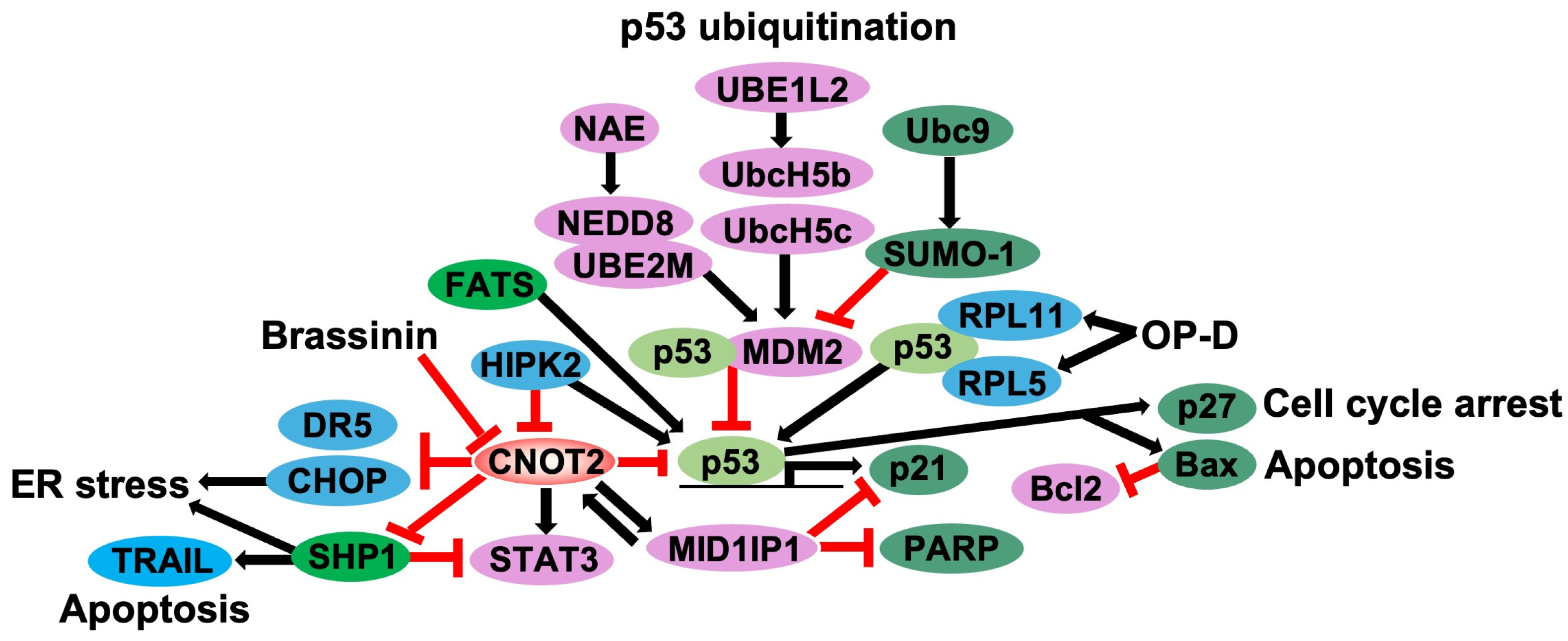
Figure 1. The scheme of CNOT2 and p53-related gene regulation. CNOT2 increases MID1IP1 and STAT3, whereas CNOT2 blocks cell cycle arrest, apoptosis, and ER stress pathways, regulating DR5, CHOP, SHP1, and p53 genes (Black arrows). MID1IP1 upregulates CNOT2 expression and suppresses p21 and PARP signaling (Black arrows). Brassinin, a single compound, inhibits CNOT2. Ophiophogonin D (OP-D) regulates RPL5 and RPL11 to increase p53 expression (Red lines). HIPK2 activates p53 and reduces CNOT2 expression. p53 ubiquitination regulates MDM2 activation with neddylation (NEDD8) and Sumoylation (SUMO-1; Red lines).
Cimb 47 00615 g001
Figure 2. The scheme of CNOT2 and EMT-related gene regulation. CNOT2 increases MID1IP1, VEGF-A, Snail, Twist, Slug, has-miR-3916, and has-miR-3613-5p to elevate the migration and EMT of cancer cells (Black arrows).
Cimb 47 00615 g002
Figure 3. The scheme of CNOT2 and c-Myc-related gene regulation. CNOT2 increases c-Myc, MID1IP1, GLUT1, and GLUT2 expression, inhibiting apoptosis and stimulating glycolysis (Black arrows), whereas CNOT2 blocks ligands of estrogen receptor alpha (ERα) and retinoid X receptor (RXR; Red lines). MID1IP1 and c-Myc upregulate CNOT2 expression. RPL11, RPL5, and RPS14 block c-Myc through Ago2/miR-24, TRBP, and RPS14 signaling. Sanggenon G (San G) induces RPL5 signaling (Red lines).
Cimb 47 00615 g003
Figure 4. Schematic of CNOT2 regulatory network. CNOT2 enhances expression of MID1IP1 and promotes EMT-related pathways (Black arrows). MID1IP1 and c-Myc, in turn, upregulate CNOT2, forming positive feedback loop (Black arrows). Although previously omitted, CNOT2 also regulates c-Myc expression by modulating its mRNA stability and protein turnover through ribosomal protein RPL5 and RPL11, indicated by reciprocal arrows (Red lines).
Cimb 47 00615 g004
Table 1. Ribosomal proteins (RPs) that modulate p53 activity. Some RPs such as RPL5, RPL11, and RPS14 stabilize p53 by inhibiting MDM2, while others downregulate p53 (p53↓). These RPs are known to be upregulated following CNOT2 knockdown, suggesting an indirect pathway through which CNOT2 regulates p53 activity (p53↑).
RPs (p53↓)RPs (p53↑)
S27LRPL5
RPL37RPL11
L31RPL23
RPL4S7
RPL40S14
RPS25
RPS15
RPL22
RPS25
RPL34
Table 2. Downregulated and upregulated genes by CNOT2 and genes downregulating and upregulating CNOT2. CNOT2 decreases the expression of p53, p21, ER/RXR and increases MID1IP1, c-Myc, VEGF, Snail, Slug, Twist, has-miR-3613-5p, has-miR-3916, S27L, RPL37, L31, RPL4, RPL40. RPL5 RPL11, and RPS14 reduce CNOT2 expression. c-Myc and MID1IP1 elevate CNOT2 expression.
Downregulated Genes by CNOT2Upregulated Genes by CNOT2Genes Downregulating CNOT2Genes Upregulating CNOT2
p53MID1IP1RPL5c-Myc
p21c-MycRPL11MID1IP1
ER/RXRVEGFRPS14
Snail
Slug
Twist
has-miR-3613-5p
has-miR-3916
S27L
RPL37
L31
RPL4
RPL40
Disclaimer/Publisher’s Note: The statements, opinions and data contained in all publications are solely those of the individual author(s) and contributor(s) and not of MDPI and/or the editor(s). MDPI and/or the editor(s) disclaim responsibility for any injury to people or property resulting from any ideas, methods, instructions or products referred to in the content.