Article

Comparative Analysis of the Transcriptomes of Persisting and Abscised Fruitlets: Insights into Plant Hormone and Carbohydrate Metabolism Regulated Self-Thinning of Pecan Fruitlets during the Early Stage

Institute of Botany, Jiangsu Province and Chinese Academy of Sciences, Nanjing 210014, China
*
Author to whom correspondence should be addressed.
Curr. Issues Mol. Biol. 2022, 44(1), 176-193; https://doi.org/10.3390/cimb44010013
Submission received: 8 November 2021 / Revised: 24 December 2021 / Accepted: 27 December 2021 / Published: 30 December 2021
(This article belongs to the Section Molecular Plant Sciences)

Abstract

:
Pecan is one of the most popular nut species in the world. The fruit drop rate of the pecan ‘Pawnee’ is more than 57%, with four fruit drop stages, which is very serious. In this study, we conducted transcriptomic profiling of persisting and abscised fruitlets in early fruit development by RNA-seq. A total of 11,976 differentially expressed genes (DEGs) were identified, 3012 upregulated and 8964 downregulated, in a comparison of abscised vs. persisting fruitlets at 35 days after anthesis (DAA). Our transcriptomic data suggest that gene subsets encoding elements involving the biosynthesis, metabolism, perception, signal transduction, and crosstalk of the plant hormones abscisic acid (ABA), auxin, cytokinin, ethylene, and gibberellin (GA) and plant growth regulators jasmonates, salicylic acid, and brassinosteroids were differentially expressed. In addition, the majority of transcriptionally activated genes involved in hormone signaling (except for ethylene and salicylic acid signaling) were downregulated in abscised fruitlets. The differential expression of transcripts coding for enzymes involved in sucrose, glucose, trehalose, starch, galactose, and galactinol metabolism shows that sucrose, galactinol, and glucose synthesis and starch content were reduced as starch biosynthesis was blocked, and retrogradation and degradation intensified. These results suggest that the abscised pecan fruitlets stopped growing and developing for some time before dropping, further indicating that their sugar supply was reduced or stopped. The transcriptome characterization described in this paper contributes to unravelling the molecular mechanisms and pathways involved in the physiological abscission of pecan fruits.

1. Introduction

Vegetative and reproductive organs that are senescent, infected, or damaged are shed from the main plant body, which is called abscission. Abscission is a specific sequence of highly complex regulated events [1]. The regulatory network activated by the abscising organ leads to the activation of abscission zones. To simplify the explanation, the abscission process includes four phases. The first step is the development of an abscission zone. The next step is the activation of abscission signaling. Then, enzymatic hydrolysis takes place in the middle lamella of the abscission zone (AZ), and AZ cells begin to enlarge. The last step is the further differentiation 1and sealing of the abscission scar [2].
Fruitlet abscission, especially for the so-called physiological drop or June drop, is very common in fruit tree development in order to control fruit load according to the nutritional status, allowing the plant to make efficient use of resources [3]. The molecular mechanisms that control and lead to early physiological fruitlet abscission are studied in model apple [3,4,5,6] and tomato [7,8] plants. The progress of physiological drop differs from senescence-driven abscission at or after ripening. Dropping of young fruits during early phases of development can be understood as a developmental response by which the plant selectively abscises fruitlets that represent weaker sinks to adapt to nutritional shortages [5]. The molecular mechanisms regulating early fruit development and the plasticity of fruit development in response to endogenous and environmental changes can be understood by studying fruit physiological drop in this scenario.
Fruit set is the process of ovary tissues undergoing transformation into fruit. It is well known that auxins and gibberellins (GAs) play a critical role in the inductive phase of fruit set. A few studies have shown that auxins trigger cell division, and their interplay with GAs sustain cell expansion [9]. However, the transformation of the ovary into fruit of tomato and Arabidopsis is prevented by a negative control exerted by auxin/indole acetic acid (AUX/IAA) and auxin response factor (ARF) proteins. This negative regulation can be removed through pollination/fertilization or treatment with auxins, leading to cell proliferation and fruit set. Ethylene and abscisic acid (ABA) biosynthesis and action appear to be significantly downregulated, and concomitantly those of auxin and GA biosynthesis and action are activated as soon as fruit set is triggered [10,11]. This suggests that plant hormones play an important role in fruit development. A few reports have shown that plant hormones auxin, ethylene, and ABA seem to play major roles, and GAs, cytokinins, and jasmonates have also been reported to be involved [3,12,13].
Sugar metabolism also plays a major role in the response to the progress of fruitlet abscission. The contents of sorbitol, glucose, fructose, and sucrose were shown to be lower in abscising fruitlets than in persisting fruitlets [6]. Transcriptomic analysis showed that genes coding trehalose-6-phosphate synthase, sorbitol transporter, UDP-glucosyltransferases, and UDP-Glc-4-epimerase were upregulated in abscising apple fruitlet [5]. The class of enzyme genes is also controlled by sugar starvation and involved in resource mobilization in other species [14,15,16].
Pecans (Carya illinoinensis (Wangenh.) K. Koch), which are native to North America and belong to the Juglandaceae family, are the most economically valuable nut trees in the world [17,18]. Pecan trees exhibit a strong tendency to produce a heavy crop one year and a light crop the following year. It is sometimes difficult to compile cultivation recommendations to minimize alternate bearing. Despite years of research, the physiology behind alternate bearing is only partially understood, and this phenomenon remains the principal challenge faced by the pecan industry year after year. It is very important to make clear the process of fruit development and the mechanism of natural fruit abscission in pecan in order to control a reasonable load. Thus, transcriptomes from persisting and abscised fruitlets were sequenced and differentially expressed genes between them were identified in order to fully understand the differences in gene expression and explore the cause of fruitlet abscission in pecan. The contents of plant hormones of persisting and abscised fruitlets were measured in this study. This study can serve to broaden our understanding of the mechanism of fruit development and natural fruit abscission.

2. Experimental Section

2.1. Plant Materials, Fruit Development, and Drop Dynamics Analysis

Experiments were carried out over 2 years on 10-year-old pecan trees (Pawnee) with spacing of 6 m × 8 m, grown with standard horticultural practices at the experimental farm of the Institute of Botany, Jiangsu Province, and Chinese Academy of Sciences (32°18′ N; 118°52′ E). Fruit drop was monitored from 7 DAA until fruit maturity at weekly intervals using 50 pre-marked fruit clusters from each of 5 homogeneous trees. Further, 10 fruits were sampled from each of 5 additional homogeneous trees from 7 DAA until fruit maturity at weekly intervals to study fruit development. Finally, 10 naturally abscised and persisting fruitlets were harvested at 36 and 48 DAA from each of 3 homogeneous trees for the study reported in this paper.

2.2. RNA Isolation and cDNA Library Preparation and Sequencing

Naturally abscised and persisting fruitlets harvested at 36 DAA were used for transcriptome analysis. Total RNA was extracted from the fruitlets using a Trizol reagent kit (Invitrogen, Carlsbad, CA, USA) according to the manufacturer’s protocol. The individual RNA samples were analyzed spectrophotometrically for protein contamination, and the samples with A260/A280 ratio values ranging from 1.9 to 2.0 were chosen. After total RNA was extracted, mRNA was enriched by oligo (dT) beads. The enriched mRNA was then fragmented into short fragments and reverse transcribed into cDNA by using the NEBNext Ultra RNA Library Prep Kit for Illumina (NEB#E7530, New England Biolabs, Ipswich, MA, USA). The purified double-stranded cDNA fragments were end repaired, poly(A) was added, and they were ligated to Illumina sequencing adapters. The ligation reaction was purified with AMPure XP Bead (1.0×). Ligated fragments were subjected to size selection by agarose gel electrophoresis and amplified PCR. The resulting cDNA library was sequenced using an Illumina HiSeq 2500 by Genedenovo Biotechnology Co., Ltd. (Guangzhou, China). The raw sequencing data generated from this study have been deposited in NCBI SRA (http://www.ncbi.nlm.nih.gov/sra, last accessed on 1 December 2021) under accession number PRJNA784729.

2.3. Analysis of Differential Gene Expression

Clean reads were obtained by filtering the raw reads, including adapters or low-quality bases, by fastp (v.18) [19]. The clean reads were mapped to a ribosome RNA database using the Bowtie2 (v.2.28) short read alignment tool [20] to identify and remove residual rRNA reads. The remaining clean reads were mapped to the pecan genome (Cil.genome.fa, ftp://parrot.genomics.cn/gigadb/pub/10.5524/100001_101000/100571/) using HISAT2.2.4 [21] with the parameter “–rna-strandness RF” and default settings for the other parameters. The unmapped reads were assembled by using StringTie v.1.3.1 [22], and the assembled sequences were regarded as novel genes. HTSeq (v.0.6.0) [23] was used to calculate the raw read counts for each gene. The gene expression was calculated and normalized to reads per kilobase per million mapped reads (RPKM) [24]. Analysis of gene expression differences between two groups was performed by DESeq2 software (European Molecular Biology Laboratory, Heidelberg, Germany) [25]. Transcripts with a false discovery rate (FDR) < 0.05 and absolute fold change > 2 were defined as differentially expressed genes (DEGs). Gene Ontology (GO) enrichment (p-value < 0.05) was studied by running all DEGs through the GO database (http://www.geneontology.org/) to further classify the genes or their products into terms (molecular function, biological process, and cellular component) to understand their biological functions. Pathway projects were performed according to the KEGG pathway database for pathway enrichment analysis of DEGs.

2.4. Plant Hormone Content Analysis

Approximately 200 mg of normal persisting fruitlets and abscised fruitlets (just shed from the main plant body) were used to measure the levels of plant hormones (ABA, IBA, GA, and TZR). Plant hormones were detected by high-performance liquid chromatography coupled with electrospray ionization tandem mass spectrometry (HPLC-ESI-MS/MS) on an Agilent 1290 HPLC (Agilent Technologies Inc., Santa Clara, CA, USA) and a Sciex QTRAP 6500 mass spectrometer (AB Sciex LLC, Framingham, MA, USA). The experiment was performed by Nanjing Innovation Biotechnology Co., Ltd. (Nanjing, China). Three biological replicates were analyzed for this experiment, each one containing 30 fruitlets. Statistically significant differences of plant hormones were calculated with GraphPad Prism 7.0 software (San Diego, CA, USA).

3. Results

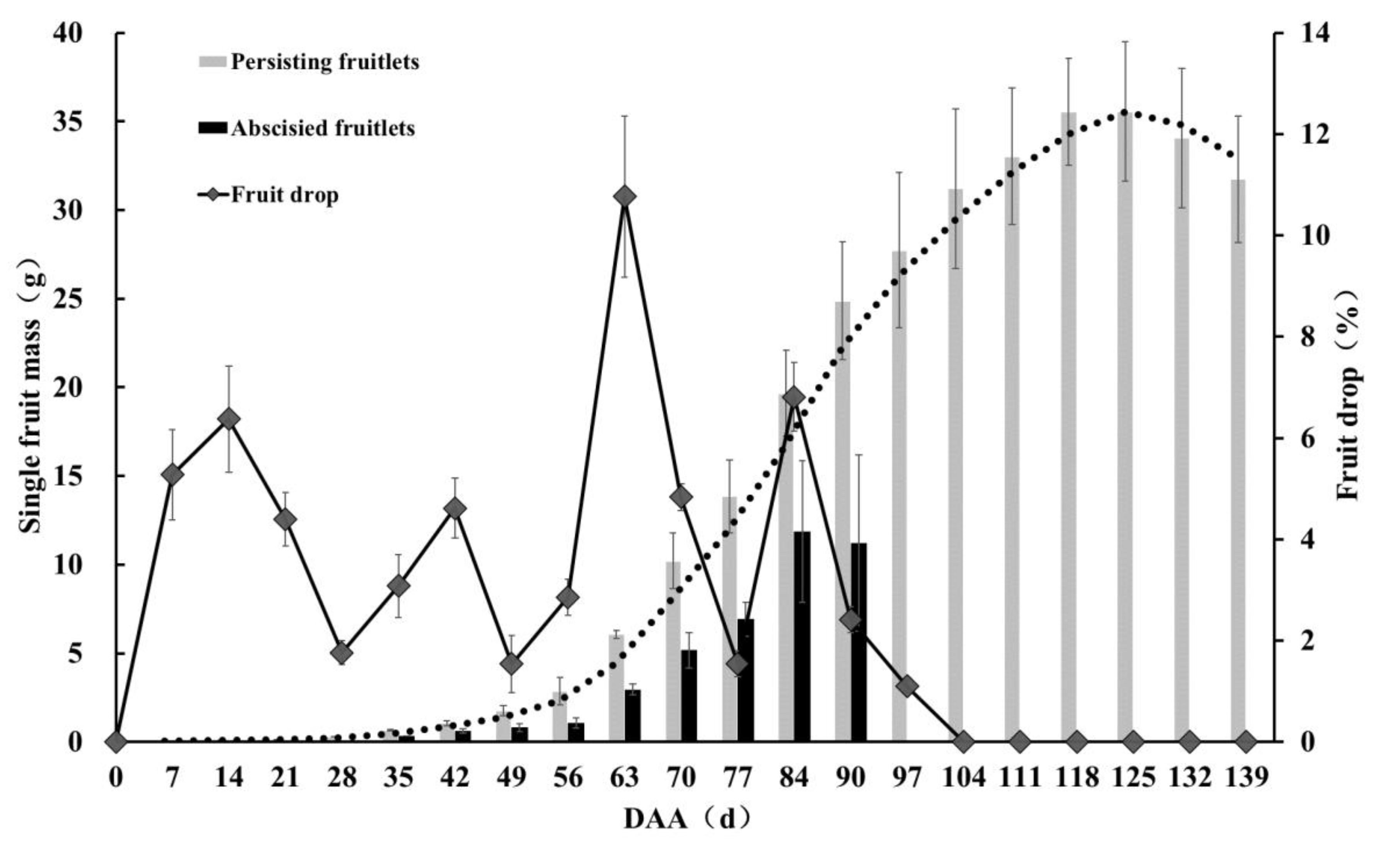
3.1. Analysis of Pecan Fruit Drop Dynamics

Pecan ‘Pawnee’ fruit drop and fruit development dynamics were investigated in this study (Figure 1). The results show that fruit growth exhibited the characteristic of a single sigmoid growth curve, presenting only one rapid growth period. The fruit drop rate was more than 57%, with four fruit drop stages, occurring at 0–28 days after anthesis (DAA), 28–49 DAA, 49–77 DAA, and 77–104 DAA. The fruit drop ratio was the largest at the third fruiting stage, accompanied by a rapid fruit growth period. The single fruit mass of persisting fruitlets was about twice that of abscised fruitlets (just shed from the main plant body) (Figure 1 and S1), indicating that the abscised fruitlets had stopped growing and developing for some time before dropping.

3.2. RNA Sequencing of Transcriptomes of Persisting and Abscised Fruitlets and Mapping of RNA Sequences to Reference Genome

To test the differentially expressed genes of persisting fruitlets (P1) and abscised fruitlets (A1) in the early stage of fruit development (35 DAA), two groups with six samples were analyzed by RNA-seq, and a total of 35.66 GB of clean data was generated by strict quality control and processing (Supplementary Table S1). Details on data and data quality before and after filtering are shown in Table 1 and Supplementary Table S1. In all, 112, 434, and 202 reads and 135, 904, and 652 reads were generated for abscised and persisting fruitlets, respectively, after filtering out duplicate sequences and ambiguous and low-quality reads (Supplementary Table S1). The numbers of clean bases and reads of persisting fruitlets were more than those of abscised fruitlets (Table 1 and Supplementary Table S1). The average GC percentage was 45.41%, with a QC30 base percentage above 93.05%. HQ clean reads were mapped to the pecan reference genome (Cil.genome.fa). Approximately 38.94 million clean reads (94.11% of the total) were mapped, and the number of mapped reads (42.57 × 10⁶) of persisting fruitlets was more than that of abscised fruitlets (35.32 × 10⁶). This indicates that the number of transcripts of persisting fruitlets was more than that of abscised fruitlets. A total of 28,624 genes were obtained from 6 transcriptome libraries, including 26,537 reference genes and 2087 novel genes (Supplementary Table S2). The numbers of reference genes, novel genes, and total genes of persisting fruitlet transcripts were more than those of abscised fruitlet transcripts (Table 1 and Supplementary Table S1).

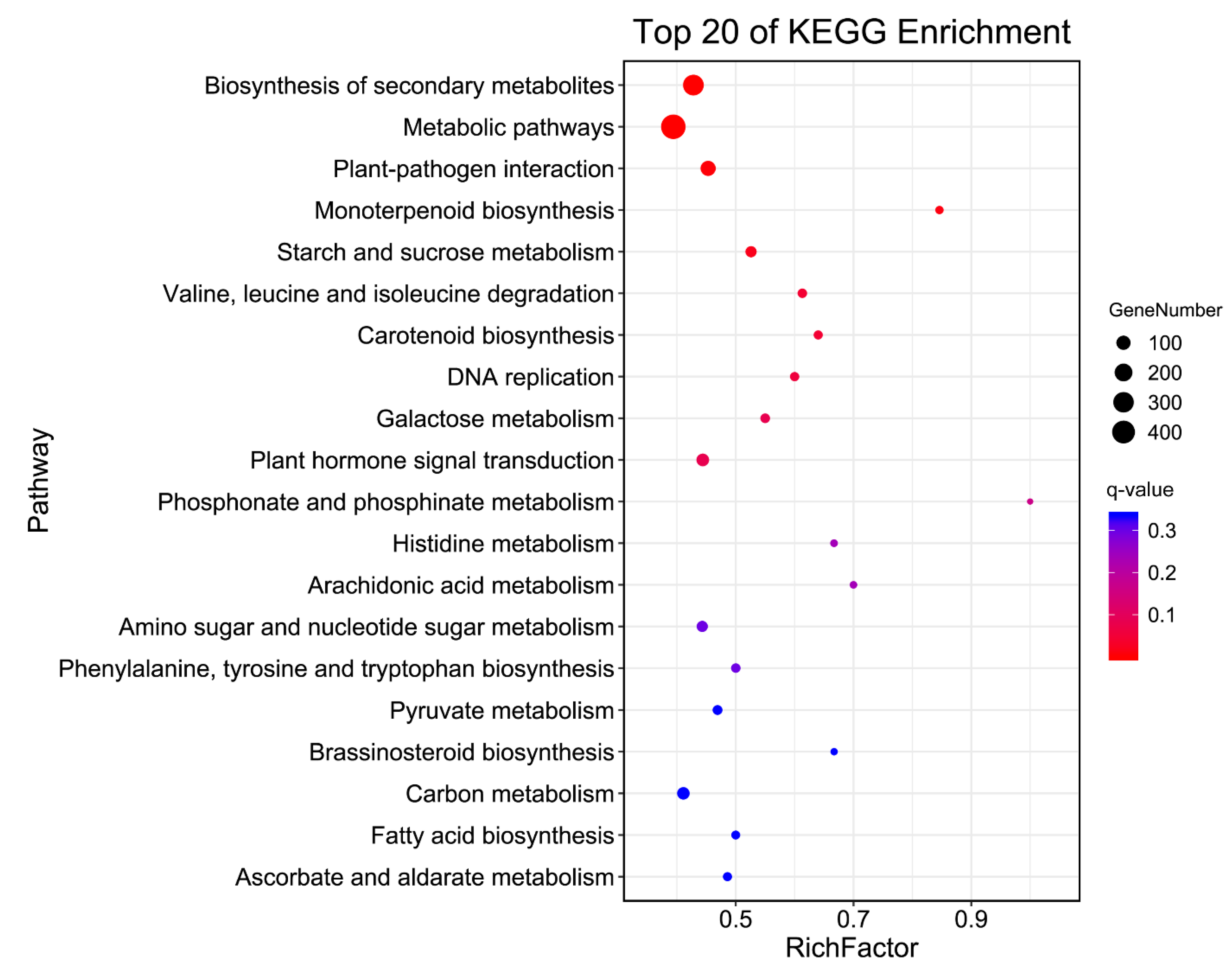
3.3. Differentially Expressed Gene Analysis

In our study, we calculated the correlation coefficient between the samples to test sample repeatability. The correlation coefficient in the repeat group was greater than 0.808 (Supplementary Figure S2), indicating consistency among the three biological replicates. Thus, the RNA-seq results were confirmed to be highly reliable for further analysis. A total of 11,976 DEGs (about 41.84% of total sequenced genes) were identified in the comparison of abscised fruitlets vs. persisting fruitlets at 35 DAA; among them, 3012 DEGs (25.15%) were upregulated and 8964 DEGs (74.85%) were downregulated (Supplementary Tables S3 and S4). There were about three times as many downregulated genes as upregulated genes, indicating that the vital signs of abscised fruitlets had diminished.
Gene Ontology (GO) functional classification included three GO trees (cellular components, molecular functions, and biological processes) and 48 functional groups (Figure 2 and Supplementary Table S5). In the category of biological processes, the largest groups were metabolic, cellular, and single-organism processes. For the cellular components, DEGs with cell, cell part, membrane, and organelle formed the major groups. Catalytic activity and binding were the dominant groups in the molecular function category. The top five enrichment categories in GO were catalytic activity, cellular process, single-organism process, primary metabolic process, and single-organism cellular process (Supplementary Table S6). In order to understand their biological function, all of the DEGs were also mapped to terms in the Kyoto Encyclopedia of Genes and Genomes (KEGG) database. Finally, 1050 DEGs were matched and assigned to 129 KEGG pathways (Supplementary Table S7). The first five biological pathways, involving biosynthesis of secondary metabolites, metabolic pathways, plant–pathogen interaction, monoterpenoid biosynthesis, and starch and sucrose metabolism, were significantly enriched in persisting fruitlets (P1) and abscised fruitlets (A1) (Figure 3 and Table 2).

3.4. Identification of Differentially Expressed Proteins Involved in Plant Hormone Signal Transduction

Gene subsets encoding elements involved in hormone biosynthesis, metabolism, perception, signal transduction, and crosstalk were found to be differentially expressed in the comparison of abscised fruitlets vs. persisting fruitlets at 35 DAA using de novo transcriptome sequencing (Table 3 and Supplementary Table S8). The statistical analyses were performed for plant hormones ABA, auxin, cytokinin, ethylene, and GA and plant growth regulators jasmonates, salicylic acid, and brassinosteroids. All 3 identified DEGs of the ABA metabolic process, 7 DEGs in response to ABA, and 8 out of 9 DEGs involved in ABA signal transduction were downregulated in abscised fruitlets vs. persisting fruitlets. All 6 identified DEGs involved in the auxin metabolic process, 7 out of 10 DEGs involved in auxin transport, and 15 out of 20 DEGs involved in auxin signal transduction were downregulated. Half of the identified DEGs involved in the gibberellin metabolic process and 10 out of 12 DEGs involved in cytokinin signal transduction were downregulated. Two out of 3 identified DEGs involved in the gibberellin metabolic process, all 4 DEGs involved in the gibberellin-mediated signaling pathway, and 2 out of 3 DEGs involved in gibberellin signal transduction were downregulated. All 3 identified DEGs involved in ethylene signal transduction were upregulated. These results show that almost all identified DEGs involved in ABA, auxin, cytokinin, and gibberellin were downregulated, and all identified DEGs involved in ethylene were upregulated. Eleven out of 20 identified DEGs involved in the SA metabolic process and 5 out of 6 DEGs involved in SA signal transduction were upregulated, but, all 9 identified DEGs involved in the JA metabolic process and 5 out of 7 DEGs involved in JA signal transduction were downregulated, indicating that SA biosynthesis and the signal transduction pathway were promoted, but were suppressed in abscised fruitlets of pecan.

3.5. Key Identified Differentially Expressed Proteins Involved in Starch and Sucrose Metabolism and Galactose Metabolism

Our analysis reveals that the transcriptional activity of genes involved in starch and sucrose metabolism and galactose metabolism were significantly regulated in the abscised fruitlets of pecan (Table 4 and Supplementary Table S4). The differential expression of transcripts coding for enzymes involved in sucrose, glucose, trehalose, starch, galactose, and galactinol metabolism was analyzed. For sucrose metabolism, the expression of two sucrose phosphate synthase (SPS) genes (EC: 2.4.1.14) were downregulated and CWINV1 (invertase, EC: 3.2.1.26) expression was upregulated in abscised fruitlets of pecan. All 4-glucan endo-1,3-beta-glucosidase genes (EC: 3.2.1.39), 6 out of 10 detected genes coding β-glucosidases (EC: 3.2.1.21), and an endoglucanase gene (EC: 3.2.1.4) for glucose metabolism were downregulated in abscised fruitlets compared with persisting fruitlets. For starch metabolism, two granule-bound starch synthase 1 (GBSS1, EC: 2.4.1.242), one GBSS2, and two 1,4-a-glucan branching enzyme (GBE, EC: 2.4.1.18) genes were downregulated in abscised fruitlets; however, two starch degradation alpha-amylase (EC: 3.2.1.1) genes were upregulated. For trehalose metabolism, the expression of trehalose 6-phosphate synthase (TPS, EC: 2.4.1.25) and trehalose-6-phosphate phosphatase (TPP, EC: 3.1.3.12) was higher in abscised fruitlets than in persisting fruitlets. The expression of alpha-galactosidase gene (EC: 3.2.1.22) was downregulated, but the expression of aldose 1-epimerase gene (EC: 5.1.3.3) was enhanced for galactose synthesis metabolism. Three out of 4 inositol 3-alpha-galactosyltransferases (GolS, EC: 4.1.123) were downregulated in abscised pecan fruitlets.

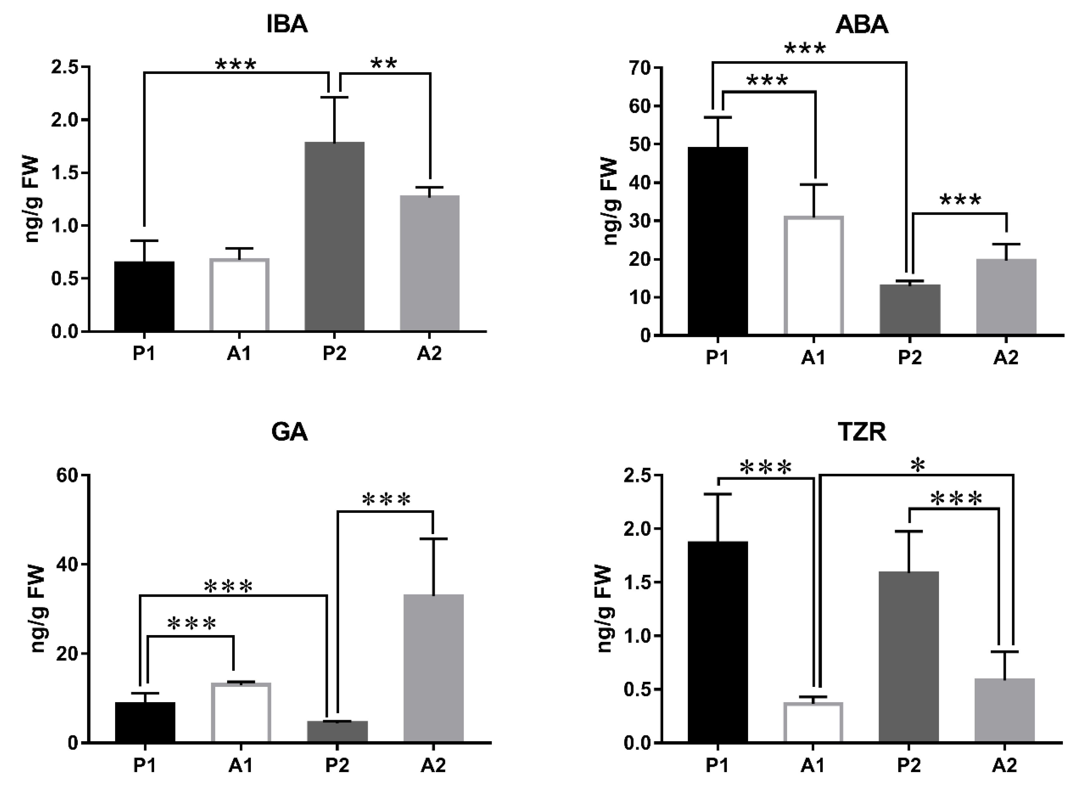
3.6. Plant Hormone Content Analysis

The contents of plant hormones 3-indolebutyric acid (IBA), ABA, GA, and Trans-zeatin riboside (TZR) were measured in abscised and persisting fruitlets at 35 and 48 DAA to further understand changes in plant hormone content after fruit shedding in pecan (Figure 4). As the fruit developed, the IBA content increased significantly, but ABA and GA content decreased significantly, indicating that increased IBA content and decreased ABA and GA might promote fruit development in pecan. Compared with persisting fruitlets, the IBA content was not changed in abscised fruitlets at 35 DAA but decreased significantly at 48 DAA. The ABA content was significantly lower in abscised fruitlets than in persisting fruitlets at 36 DAA, but the opposite was the case at 48 DAA. The GA content was significantly higher in abscised fruitlets than in persisting fruitlets, and the TZR content was significantly lower in abscised fruitlets than in persisting fruitlets at 36 and 48 DAA.

3.7. Response of Transcription Factors in the Comparison of A1 vs. P1

Differential expression of transcription factor genes was analyzed to identify transcription factors involved in abscised fruitlets in pecan (Table 4 and Supplementary Table S9). We identified 40 MYB transcription factors with significantly differential expression, 10 upregulated and 30 downregulated, suggesting that these factors might be involved in abscising in pecan. Among the NAM, ATAF1–2, and cup-shaped cotyledon 2 (NAC) transcription factor family, 13 members were upregulated and 11 members were downregulated in abscised compared with persisting fruitlets. WRKY, basic helix-loop-helix protein (bHLH), basic region/ leucine zipper motif (bZIP), C2C2, C2, ethylene response factor (ERF), zinc finger, and B3 transcription factor families were up- or downregulated in abscised fruitlets vs. persisting fruitlets, suggesting that these families may also play key roles in the transcriptional regulation of genes in abscission in the early stage of fruit development in pecan.

4. Discussion

4.1. Increased IBA Content and Decreased ABA and GA Might Promote Fruit Development in Pecan

Previous reports showed that the ethylene and ABA molecular biosynthesis and action mechanisms appear to be significantly downregulated as soon as fruit set is triggered, and concomitantly, auxin and GA molecular biosynthesis and action mechanisms are activated [10,11], indicating that fruit development appears to rely on the removal of a negative feedback regulation established by a negative control exerted mainly by ABA- and ethylene-dependent pathways of ovary growth [11]. Auxins may trigger cell division, and their interaction with GAs may be essential for sustaining cell expansion [9]. In this study, the IBA content was increased significantly, but the ABA and GA contents were decreased significantly as the fruit developed at the early stage (Figure 4), indicating that increased IBA and decreased ABA and GA might promote fruit development in pecan. Thus, further studies are needed on the molecular role of GA in the development of pecan.

4.2. Plant Hormones Seem to Play a Key Role during the Abscission Progress in Pecan

The IBA content of abscised fruitlets did not change abruptly at 35 DAA compared with persisting fruitlets, but decreased significantly at 48 DAA; however, the ABA content decreased significantly at 35 DAA but increased abruptly at 48 DAA, and the GA content was significantly higher in abscised fruitlets than in persisting fruitlets. These results show that the changes in plant hormones in abscised and persisting fruitlets have different trends at different stages of development in pecan. At 42 DAA, increased ABA and GA and decreased IBA and TZR might promote fruitlet abscission in pecan, but downregulation of GA signaling specifically in fruits induced to abscise in apple [5]. At 35 DAA, decreased ABA and no change in IAA were observed by comparing their contents in abscised and persisting fruitlets. Further studies are needed to study the initiation of abscission in pecan regarding plant hormone signaling, especially GAs, at the early stage.
Our transcriptomic data suggest that the majority of transcriptionally activated genes involved in hormone signaling were downregulated in abscised fruitlets, except for ethylene and salicylic acid signaling (Table 3 and Supplementary Table S8), indicating that hormones may play an important role during the progress of abscission in pecan. Botton et al. reported that ABA and ethylene signaling were strongly upregulated concurrently, with downregulation of GA signaling specifically in fruits induced to abscise in apple [5]. The essential role of ABA in abscission has been broadly studied in different species [4]. Exogenous ABA treatment can induce fruitlet abscission in the apple L3 class, and ABA may be involved in upstream induction of abscission [4]. Increased levels of ABA observed in BA-treated apple L3 fruitlets that were abscising, concurrently with upregulation of the ABA-responsive 9-cis-epoxycarotenoid dioxygenase 1 (MdNCED1) gene [26], suggest activation of the indirect biosynthetic pathway of ABA [4]. In our study, two NCED encoding genes were downregulated in abscised pecan fruitlets (Supplementary Table S8), consistent with the decreased ABA content (Figure 4). In addition, 8 ABA signal transduction genes were downregulated in abscised pecan fruitlets. These results show that ABA signaling is strongly downregulated in abscised pecan fruitlets, but on the contrary, it is strongly upregulated in apple fruits induced to abscise [5].
Ethylene signaling was significantly enhanced in abscised pecan fruitlets, as shown by high expression of some ethylene signaling genes (Table 3). Mitogen-activated protein kinase (MAPK) genes, which were found to be upregulated in abscised fruitlets (Supplementary Table S8), are key elements of the ethylene signal transduction pathway, probably involved in ethylene–ABA crosstalk [27]. The related proteins (14-3-3 genes), which were also found to be upregulated in abscised fruitlets (Supplementary Table S8), may trigger ABA–ethylene crosstalk and the response to sugar starvation [28]. The TPS gene, found to be overexpressed in abscised fruitlets, may also regulate ABA signaling, as found in Arabidopsis [29] and apple [5]. ABA and ethylene signaling are strongly upregulated concurrently with downregulation of GA signaling specifically in apple fruits induced to abscise [5]. Our transcriptomic data show that ethylene signaling is strongly upregulated concurrently with downregulation of GA and ABA signaling in abscised pecan fruitlets.
Auxin participates in plant organ abscission. Transcript analysis revealed that auxin may regulate the expression of early auxin-responsive gene families including AUX/IAA, gretchen Hagen3 (GH3), and small auxin up RNA (SAUR). Auxin regulates the expression of various ARFs during early abscission, whereas ethylene has the opposite effect on most of these genes in tomato [30]. SlARFs have overlapping functions in the abscission process. Meir et al. reported that tomato flower abscission was associated with the expression level of AUX/IAA genes [31]. GH3 genes are also involved in fruitlet or flower abscission. LcAUX/IAA1 and LcSAUR1 may play more important roles in abscission than LcGH3.1 in litchi, because LcAUX/IAA1 and LcSAUR1 were mostly expressed in AZ [32]. Tomato GH3 increased slightly after 8 h and maintained a low expression level during abscission, implying that it may be an effective negative regulator in IAA-induced abscission delay [33]. SAUR, an auxin-responsive gene, may serve as a marker of IAA level throughout the abscission process [33]. Two SAUR-like genes were found to be involved in shading-induced abscission by transcriptomic analysis in apple [34]. Girdling plus defoliation treatment could induce LcSAUR1 expression in AZ and fruitlet, and significantly induce litchi fruitlet abscission [32]. SAUR36 has been reported to be involved in leaf senescence in Arabidopsis [35]. Overexpression of OsSAUR39 in rice (Oryza sativa) results in phenotypes that include reductions in lateral root development, yield, and shoot and root length, suggesting that OsSAUR39 acts as a negative regulator of auxin synthesis and transport [36]. Fourteen CitSAUR genes showed obvious changes during citrus fruitlet abscission, and CitSAUR06, CitSAUR08, CitSAUR44, CitSAUR61, and CitSAUR64 were more relevant because their expression patterns under IAA treatment exhibited an opposite trend to that during fruitlet drop [37]. In our study, all 8 AUX/IAA, 3 ARF, and 2 out of 3 GH3 genes detected were downregulated, but 3 out of 4 SAUR genes were upregulated in abscised pecan fruitlets. These results show that the roles of GH3, ARF, AUX/INN, and SAUR in fruitlet abscission may vary considerably among plant species.
Beyond the GA, ABA, auxin, and ethylene signaling discussed above, cytokinin, jasmonate, salicylic acid, and brassinosteroid signaling are also involved in abscised pecan fruitlets. An association of response variables with expression data of genes regarding cytokinin, jasmonates, salicylic acid, and brassinosteroids was detected in abscising apple fruitlets [5]. Especially related to salicylic acid, many genes related to metabolism were highly expressed in abscised fruitlets. A TGA gene and 4 PR-1 genes were upregulated in abscised fruitlets, indicating that systemic acquired resistance (SAR) may be triggered in abscised pecan fruitlets.

4.3. Reduced Sugar Supply in Abscised Fruitlets Is One Reason for Fruitlet Abscission in Pecan

Sugar signaling is known to play a role in senescence regulation in a complex network. Thus, the senescence process is triggered in fruitlets once the sugar supply is decreased [38]. In apple, the sorbitol concentration of central fruitlets is higher than that of L1 pedicels, indicating that the former profit from a better supply of sugars. The increased supply of sugar to the central fruit would allow it to develop at a faster rate, thus further increasing its sink strength [6]. In our study, the single fruit mass of abscised fruitlets (just shed from the main plant body) was about half that of persisting fruitlets (Figure 1 and Supplementary Figure S1), indicating that abscised fruitlets had stopped growing and developing for some time before dropping, thus further indicating that the sugar supply is reduced or stopped in abscising pecan fruitlet.
The transcriptome analysis showed that genes coded for carbohydrate metabolism, including starch and sucrose, galactose, pyruvate, and amino sugar and nucleotide sugar metabolism, are significantly regulated in abscised pecan fruitlets (Table 5). Decreased expression of transcripts coding for enzymes involved in sucrose, glucose, and starch metabolism may be expected in slow-growing organs, considering that fruitlets act as a major carbon sink. SPS catalyzing fructose-6-phophate and UDP-glucose to synthesis sucrose-6-phosphate in plants is a key regulatory step in the control of sucrose synthesis [39]. Overexpression of SPS in Arabidopsis thaliana results in increased foliar sucrose/starch ratios and decreased foliar carbohydrate accumulation in plants after prolonged growth with CO2 enrichment [40]. Transformed tobacco plants overexpressing Arabidopsis SPS gene showed elevated transcript abundance and SPS enzyme activity, substantial pooling of soluble stem sucrose content, significantly increased stem height and greater stem diameters, longer fibers, and increased total dry biomass relative to control plants [41]. Vacuolar invertase (VINV) and cell wall invertase (CWINV, EC: 3.2.1.26) are invertases, cleaving sucrose to glucose and fructose. The transcripts and activities of CWINV, CytINV, and VINV at the fully mature stage were higher in Sweet Miriam, in agreement with the low sucrose content [42]. In our study, the expression of two SPS genes was downregulated and CWINV1 expression was upregulated in abscised fruitlets in pecan, suggesting that sucrose synthesis was reduced.
Plant class I glucan endo-1,3-β-glucosidases (β-1,3-glucanase; 1,3-β-d-glucan glucanohydrolase, EC: 3.2.1.39) have been implicated in development and the defense against pathogen attack. β-glucosidases (EC: 3.2.1.21) are glycosyl hydrolases that hydrolyze the β-O-glycosidic bond at the anomeric carbon of a glucose moiety at the nonreducing end of a carbohydrate or glycoside molecule [43]. Endoglucanases (EC: 3.2.1.4) are associated with fruit ripening, growth of cultured cells, and leaf abscission. In our study, all 4 glucan endo-1,3-β-glucosidase genes, 6 out of 10 detected β-glucosidase genes, and a endoglucanase gene were downregulated in abscised fruitlets compared with persisting fruitlets, indicating that the glucose metabolism is blocked in abscised fruitlets in pecan.
Starch is the major storage carbohydrate in most plants, with many important functions. Granule-bound starch synthase (GBSS, EC: 2.4.1.242) is the glucosyl transferase specifically responsible for elongating amylose polymers and the only protein known to be required for its biosynthesis. Protein targeting of starch is required for localizing granule-bound starch synthase to starch granules and for normal amylose synthesis in Arabidopsis [44,45]. Two GBSS1 genes and one GBSS2 gene were downregulated in abscised fruitlets in pecan, indicating that starch biosynthesis was blocked. The 1,4-α-glucan branching enzyme (GBE, EC: 2.4.1.18) is known to cleave the α-1,4 glucosidic linkage of an existing glucan chain and transfer the cut end to the 6-position of a glucose residue within the cleaved chain or within another glucan chain, creating an α-1,6 glucosidic linkage, increasing the ratio of amylopectin to amylose [46]. Amylopectin has a slower recrystallization rate than amylose. GBE treatment could retard both short- and long-term retrogradation of starch in corn [47]. It is well known that a-amylases are important enzymes for starch degradation in plants [48]. In this study, two GBE genes were downregulated and two starch degradation alpha-amylase genes were upregulated in abscised fruitlets in pecan, showing that starch retrogradation and degradation are intensified. These results show that starch biosynthesis was blocked and starch retrogradation and degradation were intensified, resulting in reduced starch content in pecan.
Trehalose is an effective signaling molecule that has been shown to function in carbohydrate storage [49]. The trehalose biosynthetic pathway is used to transfer Glc from UDP-Glc to Glc-6-P, resulting in trehalose-6-P and UDP in plants. This initial step is catalyzed by TPS (EC: 2.4.1.15), and the dephosphorylation of trehalose-6-P occurs via TPP (EC: 3.1.3.12) producing trehalose in a second step [50]. Arabidopsis class I genes (AtTPS1–4) regulate starch storage, resistance to drought, and inflorescence architecture. Class II genes (AtTPS5–11) encode multifunctional enzymes with synthase and phosphatase activity [49], induced by sugar starvation [51]. Trehalose-6-phosphate, which is a sugar signal, induces a prompt reaction to nutritional stress during the early induction of abscission. The expression levels of a class II TPS gene were found to be high in the cortex of abscising fruitlets of apple and citrus [5]. Celton et al. reported that TPP and TPS genes showed increased expression in the pedicels of apple central fruitlets, which were persisting fruits, indicating that central pedicels may be supplied from a very early stage of development in apple [6]. Overexpression of Escherichia coli TPS (OTS A) in transgenic tobacco was shown to increase photosynthetic activity, but constitutive expression of TPP (OTS B) resulted in reduced photosynthesis [52]. However, the expression of TPS and TPP in was higher in abscised fruitlets than in persisting fruitlets in pecan in our study, indicating that the functions of trehalose may vary among plants, which needs further validation in the early stage of pecan fruitlet abscission.
Previous reports showed that galactose synthesis via alpha-galactosidase (EC: 3.2.1.22) results from the hydrolysis of raffinose to yield free galactose and sucrose [53]. α-GAL and β-GAL transcripts were higher in Santa Rosa fruits and leaves than in Sweet Miriam and correlated well with the higher Santa Rosa galactose content [42]. In our study, the expression of gene coding alpha-galactosidase was downregulated, but the expression of gene coding aldose 1-epimerase (EC: 5.1.3.3) was enhanced, indicating that galactose synthesis is reduced and degradation is enhanced in abscised fruitlets. UDP-Gal together with myo-inositol are used as substrates by GolS(EC: 4.1.123) for the synthesis of galactinol [54]. Three out of 4 genes coding GolS were downregulated in abscised pecan fruitlets, showing that galactinol synthesis was also blocked.

4.4. NAC Transcription Factors Participate in Fruit Development or Abscission Process

Multiple NAC family proteins were identified in abscised fruitlets in our study. The NAC proteins, which constitute one of the largest major transcription factor families, are well-known for their roles in several developmental programs [55]. NAC TFs have been shown to play important roles in various biological processes, as well as responses to abiotic stresses, and are important regulators in a wide range of developmental processes, such as the formation of lateral roots, the development of shoot apical meristem, floral morphogenesis, embryo development, grain nutrient remobilization, and cell wall biosynthesis [56]. In kiwifruit, 74 of 142 NACs were found to be persistently expressed in fruit during the whole developmental process, and crucial candidate NAC genes were shown to be involved in fruit growth and development [57]. Several NAC transcription factors were upregulated in the pedicels of central fruits, which were persisting fruits, in Malus domestica, which might contribute to increased vascular development in the central pedicel in the early developmental stages [6]. In our study, 24 NAC transcription factors were up- or downregulated in abscised fruitlets, indicating that these genes participate in the fruit development or abscission process.

5. Conclusions

The fruit development and drop dynamics of the pecan ‘Pawnee’ were studied in this paper, and the results show that the fruit drop rate was more than 57%, suggesting that fruit drop was very serious. In order to understand the mechanism of fruitlet drop, transcriptomic profiling of persisting and abscised fruitlets in early fruit development was conducted by RNA-seq. A total of 11,976 DEGs were identified, 3012 upregulated and 8964 downregulated, in the comparison of abscised vs. persisting fruitlets at 35 DAA. Our transcriptomic data suggest that a majority of the transcriptionally activated genes involved in hormone signaling were downregulated in the abscised fruitlets, except for ethylene and salicylic acid signaling. Unlike the downregulation of GA signaling specifically in fruits induced to abscise in apple [5], the GA content was increased in abscised fruitlets in pecan. Thus, the involvement of plant hormone signaling, especially GAs, at the early stage of fruit abscission should be studied in the future. Transcripts coding for enzymes involved in sucrose, glucose, trehalose, starch, galactose, and galactinol metabolism were reduced, suggesting that abscised pecan fruitlets stopped growing and developing for some time before dropping, indicating that their sugar supply was reduced or stopped. The transcriptome characterization described in this paper contributes to unravelling the molecular mechanisms and pathways involved in the physiological abscission of pecan fruits.

Supplementary Materials

The following are available online at https://www.mdpi.com/article/10.3390/cimb44010013/s1.

Author Contributions

J.Z. and G.W. designed the experiments and wrote the manuscript; J.Z., F.Z., T.W., Y.L. and G.W. performed the experiments; J.Z. and F.Z. analyzed the data. All authors have read and agreed to the published version of the manuscript.

Funding

This research was funded by the Independent Innovation of Agricultural Science and Technology of Jiangsu Province (grant number CX(21)3046), the Science and Technology Plan Project of Nanjing (grant number 20201103), and the Science and Technology Plan Project of Luhe District of Nanjing (grant number LHZC2021N01).

Institutional Review Board Statement

Not applicable.

Informed Consent Statement

Not applicable.

Data Availability Statement

The data presented in this study are openly available in NCBI SRA (http://www.ncbi.nlm.nih.gov/sra), reference number [PRJNA784729].

Acknowledgments

Not applicable.

Conflicts of Interest

The authors declare no conflict of interest.

References

  1. Estornell, L.H.; Agusti, J.; Merelo, P.; Talon, M.; Tadeo, F.R. Elucidating mechanisms underlying organ abscission. Plant Sci. 2013, 199–200, 48–60. [Google Scholar] [CrossRef]
  2. Patharkar, O.R.; Walker, J.C. Advances in abscission signaling. J. Exp. Bot. 2018, 69, 733–740. [Google Scholar] [CrossRef]
  3. Eccher, G.; Begheldo, M.; Boschetti, A.; Ruperti, B.; Botton, A. Roles of ethylene production and ethylene receptor expression in regulating apple fruitlet abscission. Plant Physiol. 2015, 169, 125–137. [Google Scholar] [CrossRef] [Green Version]
  4. Giulia, E.; Alessandro, B.; Mariano, D.; Andrea, B.; Benedetto, R.; Angelo, R. Early induction of apple fruitlet abscission is characterized by an increase of both isoprene emission and abscisic acid content. Plant Physiol. 2013, 161, 1952–1969. [Google Scholar] [CrossRef] [Green Version]
  5. Botton, A.; Eccher, G.; Forcato, C.; Ferrarini, A.; Begheldo, M.; Zermiani, M.; Moscatello, S.; Battistelli, A.; Velasco, R.; Ramina, R.A.J.P.P. Signaling pathways mediating the induction of apple fruitlet abscission. Plant Physiol. 2011, 155, 185–208. [Google Scholar] [CrossRef] [Green Version]
  6. Celton, J.-M.; Dheilly, E.; Guillou, M.-C.; Simonneau, F.; Juchaux, M.; Costes, E.; Laurens, F.; Renou, J.-P. Additional amphivasal bundles in pedicel pith exacerbate central fruit dominance and induce self-thinning of lateral fruitlets in apple. Plant Physiol. 2014, 164, 1930–1951. [Google Scholar] [CrossRef] [Green Version]
  7. Sundaresan, S.; Philosoph-Hadas, S.; Riov, J.; Mugasimangalam, R.; Kuravadi, N.A.; Kochanek, B.; Salim, S.; Tucker, M.L.; Meir, S. De novo transcriptome sequencing and development of abscission zone-specific microarray as a new molecular tool for analysis of tomato organ abscission. Front. Plant Sci. 2015, 6, 1258. [Google Scholar] [CrossRef] [Green Version]
  8. Sundaresan, S.; Philosoph-Hadas, S.; Ma, C.; Jiang, C.Z.; Riov, J.; Mugasimangalam, R.; Kochanek, B.; Salim, S.; Reid, M.S.; Meir, S. The tomato hybrid proline-rich protein regulates the abscission zone competence to respond to ethylene signals. Hortic. Res. 2018, 5, 28. [Google Scholar] [CrossRef] [Green Version]
  9. de Jong, M.; Mariani, C.; Vriezen, W.H. The role of auxin and gibberellin in tomato fruit set. J. Exp. Bot. 2009, 60, 1523–1532. [Google Scholar] [CrossRef] [Green Version]
  10. Nitsch, L.M.; Oplaat, C.; Feron, R.; Ma, Q.; Wolters-Arts, M.; Hedden, P.; Mariani, C.; Vriezen, W.H. Abscisic acid levels in tomato ovaries are regulated by LeNCED1 and SlCYP707A1. Planta 2009, 229, 1335–1346. [Google Scholar] [CrossRef] [Green Version]
  11. Vriezen, W.H.; Feron, R.; Maretto, F.; Keijman, J.; Mariani, C. Changes in tomato ovary transcriptome demonstrate complex hormonal regulation of fruit set. New Phytol. 2008, 177, 60–76. [Google Scholar] [CrossRef]
  12. Cin, V.D.; Danesin, M.; Boschetti, A.; Dorigoni, A.; Ramina, A. Ethylene biosynthesis and perception in apple fruitlet abscission (Malus domestica L. Borck). J. Exp. Bot. 2005, 56, 2995–3005. [Google Scholar] [CrossRef] [Green Version]
  13. Kuhn, N.; Serrano, A.; Abello, C.; Arce, A.; Espinoza, C.; Gouthu, S.; Deluc, L.; Arce-Johnson, P. Regulation of polar auxin transport in grapevine fruitlets (Vitis vinifera L.) and the proposed role of auxin homeostasis during fruit abscission. BMC Plant Biol. 2016, 16, 234. [Google Scholar] [CrossRef] [Green Version]
  14. Rook, F.; Corke, F.; Baier, M.; Holman, R.; May, A.G.; Bevan, M.W. Impaired sucrose induction1 encodes a conserved plant-specific protein that couples carbohydrate availability to gene expression and plant growth. Plant J. 2006, 46, 1045–1058. [Google Scholar] [CrossRef]
  15. Rorat, T. Plant dehydrins--tissue location, structure and function. Cell. Mol. Biol. Lett. 2006, 11, 536–556. [Google Scholar] [CrossRef]
  16. Herrera-Rodriguez, M.B.; Maldonado, J.M.; Perez-Vicente, R. Light and metabolic regulation of HAS1, HAS1.1 and HAS2, three asparagine synthetase genes in Helianthus annuus. Plant Physiol. Biochem. 2004, 42, 511–518. [Google Scholar] [CrossRef]
  17. Grauke, L.J.; Wood, B.W.; Harris, M.K. Crop Vulnerability: Carya. HortScience 2016, 51, 653–663. [Google Scholar] [CrossRef] [Green Version]
  18. Poletto, T.; Muniz, M.F.B.; Poletto, I.; Baggiotto, C. Methods for overcome dormancy of pecan carya illinoinensis (wangenh.) k. koch seeds. Rev. Árvore 2015, 39, 1111–1118. [Google Scholar] [CrossRef] [Green Version]
  19. Chen, S.; Zhou, Y.; Chen, Y.; Jia, G. Fastp: An ultra-fast all-in-one FASTQ preprocessor. Bioinformatics 2018, 34, i884–i890. [Google Scholar] [CrossRef]
  20. Langmead, B.; Salzberg, S.L. Fast gapped-read alignment with Bowtie 2. Nat. Methods 2012, 9, 357–359. [Google Scholar] [CrossRef] [Green Version]
  21. Kim, D.; Langmead, B.; Salzberg, S.L. HISAT: A fast spliced aligner with low memory requirements. Nat. Methods 2015, 12, 357–360. [Google Scholar] [CrossRef] [Green Version]
  22. Pertea, M.; Pertea, G.M.; Antonescu, C.M.; Chang, T.C.; Mendell, J.T.; Salzberg, S.L. StringTie enables improved reconstruction of a transcriptome from RNA-seq reads. Nat. Biotechnol. 2015, 33, 290–295. [Google Scholar] [CrossRef] [Green Version]
  23. Anders, S.; Pyl, P.T.; Huber, W. HTSeq—A Python framework to work with high-throughput sequencing data. Bioinformatics 2015, 31, 166–169. [Google Scholar] [CrossRef]
  24. Trapnell, C.; Williams, B.A.; Pertea, G.; Mortazavi, A.; Kwan, G.; van Baren, M.J.; Salzberg, S.L.; Wold, B.J.; Pachter, L. Transcript assembly and quantification by RNA-Seq reveals unannotated transcripts and isoform switching during cell differentiation. Nat. Biotechnol. 2010, 28, 511–515. [Google Scholar] [CrossRef] [Green Version]
  25. Love, M.I.; Huber, W.; Anders, S. Moderated estimation of fold change and dispersion for RNA-seq data with DESeq2. Genome Biol. 2014, 15, 550. [Google Scholar] [CrossRef] [Green Version]
  26. Schwartz, S.H.; Qin, X.; Zeevaart, J.A. Elucidation of the indirect pathway of abscisic acid biosynthesis by mutants, genes, and enzymes. Plant Physiol 2003, 131, 1591–1601. [Google Scholar] [CrossRef] [Green Version]
  27. Xin, Z.; Zhao, Y.; Zheng, Z.L. Transcriptome analysis reveals specific modulation of abscisic acid signaling by ROP10 small GTPase in Arabidopsis. Plant Physiol. 2005, 139, 1350–1365. [Google Scholar] [CrossRef] [Green Version]
  28. Lancien, M.; Roberts, M.R. Regulation of Arabidopsis thaliana 14-3-3 gene expression by gamma-aminobutyric acid. Plant Cell Environ. 2006, 29, 1430–1436. [Google Scholar] [CrossRef] [Green Version]
  29. Avonce, N.; Leyman, B.; Mascorro-Gallardo, J.O.; Van Dijck, P.; Thevelein, J.M.; Iturriaga, G. The Arabidopsis trehalose-6-P synthase AtTPS1 gene is a regulator of glucose, abscisic acid, and stress signaling. Plant Physiol. 2004, 136, 3649–3659. [Google Scholar] [CrossRef] [Green Version]
  30. Guan, X.; Xu, T.; Gao, S.; Qi, M.; Wang, Y.; Liu, X.; Li, T. Temporal and spatial distribution of auxin response factor genes during tomato flower abscission. J. Plant Growth Regul. 2013, 33, 317–327. [Google Scholar] [CrossRef]
  31. Meir, S.; Philosoph-Hadas, S.; Sundaresan, S.; Selvaraj, K.S.; Burd, S.; Ophir, R.; Kochanek, B.; Reid, M.S.; Jiang, C.Z.; Lers, A. Microarray analysis of the abscission-related transcriptome in the tomato flower abscission zone in response to auxin depletion. Plant Physiol. 2010, 154, 1929–1956. [Google Scholar] [CrossRef]
  32. Kuang, J.F.; Wu, J.Y.; Zhong, H.Y.; Li, C.Q.; Chen, J.Y.; Lu, W.J.; Li, J.G. Carbohydrate stress affecting fruitlet abscission and expression of genes related to auxin signal transduction pathway in litchi. Int. J. Mol. Sci. 2012, 13, 16084–16103. [Google Scholar] [CrossRef]
  33. Zuo, X.; Xu, T.; Qi, M.; Lv, S.; Li, J.; Gao, S.; Li, T. Expression patterns of auxin-responsive genes during tomato flower pedicel abscission and potential effects of calcium. Aust. J. Bot. 2012, 60, 68. [Google Scholar] [CrossRef]
  34. Zhu, H.; Dardick, C.D.; Beers, E.P.; Callanhan, A.M.; Xia, R.; Yuan, R. Transcriptomics of shading-induced and NAA-induced abscission in apple (Malus domestica) reveals a shared pathway involving reduced photosynthesis, alterations in carbohydrate transport and signaling and hormone crosstalk. BMC Plant Biol. 2011, 11, 138. [Google Scholar] [CrossRef] [Green Version]
  35. Hou, K.; Wu, W.; Gan, S.S. SAUR36, a small auxin up RNA gene, is involved in the promotion of leaf senescence in Arabidopsis. Plant Physiol. 2013, 161, 1002–1009. [Google Scholar] [CrossRef] [Green Version]
  36. Kant, S.; Bi, Y.M.; Zhu, T.; Rothstein, S.J. SAUR39, a small auxin-up RNA gene, acts as a negative regulator of auxin synthesis and transport in rice. Plant Physiol. 2009, 151, 691–701. [Google Scholar] [CrossRef] [Green Version]
  37. Xie, R.; Dong, C.; Ma, Y.; Deng, L.; He, S.; Yi, S.; Lv, Q.; Zheng, Y. Comprehensive analysis of SAUR gene family in citrus and its transcriptional correlation with fruitlet drop from abscission zone A. Funct. Integr. Genom. 2015, 15, 729–740. [Google Scholar] [CrossRef]
  38. Baena-Gonzalez, E.; Rolland, F.; Thevelein, J.M.; Sheen, J. A central integrator of transcription networks in plant stress and energy signalling. Nature 2007, 448, 938–942. [Google Scholar] [CrossRef]
  39. Stitt, M.; Wilke, I.; Feil, R.; Heldt, H.W. Coarse control of sucrose-phosphate synthase in leaves: Alterations of the kinetic properties in response to the rate of photosynthesis and the accumulation of sucrose. Planta 1988, 174, 217–230. [Google Scholar] [CrossRef]
  40. Signora, L.; Galtier, N.; Skøt, L.; Lucas, H.; Foyer, C.H. Over-expression of sucrose phosphate synthase in Arabidopsis thaliana results in increased foliar sucrose/starch ratios and favours decreased foliar carbohydrate accumulation in plants after prolonged growth with CO2 enrichment. J. Exp. Bot. 1998, 49, 669–680. [Google Scholar] [CrossRef] [Green Version]
  41. Park, J.Y.; Canam, T.; Kang, K.Y.; Ellis, D.D.; Mansfield, S.D. Over-expression of an arabidopsis family A sucrose phosphate synthase (SPS) gene alters plant growth and fibre development. Transgenic Res. 2008, 17, 181–192. [Google Scholar] [CrossRef]
  42. Farcuh, M.; Li, B.; Rivero, R.M.; Shlizerman, L.; Sadka, A.; Blumwald, E. Sugar metabolism reprogramming in a non-climacteric bud mutant of a climacteric plum fruit during development on the tree. J. Exp. Bot. 2017, 68, 5813–5828. [Google Scholar] [CrossRef] [Green Version]
  43. Opassiri, R.; Pomthong, B.; Onkoksoong, T.; Akiyama, T.; Esen, A.; Ketudat Cairns, J.R. Analysis of rice glycosyl hydrolase family 1 and expression of Os4bglu12 β-glucosidase. BMC Plant Biol. 2006, 6, 33. [Google Scholar] [CrossRef] [Green Version]
  44. Seung, D.; Soyk, S.; Coiro, M.; Maier, B.A.; Eicke, S.; Zeeman, S.C. PROTEIN TARGETING TO STARCH is required for localising GRANULE-BOUND STARCH SYNTHASE to starch granules and for normal amylose synthesis in Arabidopsis. PLoS Biol. 2015, 13, e1002080. [Google Scholar] [CrossRef] [Green Version]
  45. Smith, S.M.; Fulton, D.C.; Chia, T.; Thorneycroft, D.; Chapple, A.; Dunstan, H.; Hylton, C.; Zeeman, S.C.; Smith, A.M. Diurnal changes in the transcriptome encoding enzymes of starch metabolism provide evidence for both transcriptional and posttranscriptional regulation of starch metabolism in Arabidopsis leaves. Plant Physiol. 2004, 136, 2687–2699. [Google Scholar] [CrossRef] [Green Version]
  46. Kajiura, H.; Takata, H.; Akiyama, T.; Kakutani, R.; Furuyashiki, T.; Kojima, I.; Harui, T.; Kuriki, T. In vitro synthesis of glycogen: The structure, properties, and physiological function of enzymatically-synthesized glycogen. Biologia 2011, 66, 387–394. [Google Scholar] [CrossRef] [Green Version]
  47. Li, W.; Li, C.; Gu, Z.; Qiu, Y.; Cheng, L.; Hong, Y.; Li, Z. Retrogradation behavior of corn starch treated with 1,4-α-glucan branching enzyme. Food Chem. 2016, 203, 308–313. [Google Scholar] [CrossRef] [Green Version]
  48. Chen, M.H.; Huang, L.F.; Li, H.M.; Chen, Y.R.; Yu, S.M. Signal peptide-dependent targeting of a rice α-amylase and cargo proteins to plastids and extracellular compartments of plant cells. Plant Physiol. 2004, 135, 1367–1377. [Google Scholar] [CrossRef] [Green Version]
  49. Chary, S.N.; Hicks, G.R.; Choi, Y.G.; Carter, D.; Raikhel, N.V. Trehalose-6-phosphate synthase/phosphatase regulates cell shape and plant architecture in Arabidopsis. Plant Physiol. 2008, 146, 97–107. [Google Scholar] [CrossRef] [Green Version]
  50. Mo, Z.; Feng, G.; Su, W.; Liu, Z.; Peng, F. Transcriptomic analysis provides insights into grafting union development in pecan (Carya illinoinensis). Genes 2018, 9, 71. [Google Scholar] [CrossRef] [Green Version]
  51. Osuna, D.; Usadel, B.; Morcuende, R.; Gibon, Y.; Blasing, O.E.; Hohne, M.; Gunter, M.; Kamlage, B.; Trethewey, R.; Scheible, W.R.; et al. Temporal responses of transcripts, enzyme activities and metabolites after adding sucrose to carbon-deprived Arabidopsis seedlings. Plant J. 2007, 49, 463–491. [Google Scholar] [CrossRef]
  52. Paul, M.; Pellny, T.; Goddijn, O. Enhancing photosynthesis with sugar signals. Trends Plant Sci. 2001, 6, 197–200. [Google Scholar] [CrossRef]
  53. Dai, N.; Petreikov, M.; Portnoy, V.; Katzir, N.; Pharr, D.M.; Schaffer, A.A. Cloning and expression analysis of a UDP-galactose/glucose pyrophosphorylase from melon fruit provides evidence for the major metabolic pathway of galactose metabolism in raffinose oligosaccharide metabolizing plants. Plant Physiol. 2006, 142, 294–304. [Google Scholar] [CrossRef] [Green Version]
  54. Nishizawa, A.; Yabuta, Y.; Shigeoka, S. Galactinol and raffinose constitute a novel function to protect plants from oxidative damage. Plant Physiol. 2008, 147, 1251–1263. [Google Scholar] [CrossRef] [Green Version]
  55. Puranik, S.; Sahu, P.P.; Srivastava, P.S.; Prasad, M. NAC proteins: Regulation and role in stress tolerance. Trends Plant Sci. 2012, 17, 369–381. [Google Scholar] [CrossRef]
  56. Liu, X.; Wang, T.; Bartholomew, E.; Black, K.; Dong, M.; Zhang, Y.; Yang, S.; Cai, Y.; Xue, S.; Weng, Y.; et al. Comprehensive analysis of NAC transcription factors and their expression during fruit spine development in cucumber (Cucumis sativus L.). Hortic. Res. 2018, 5, 31. [Google Scholar] [CrossRef]
  57. Jia, D.; Jiang, Z.; Fu, H.; Chen, L.; Liao, G.; He, Y.; Huang, C.; Xu, X. Genome-wide identification and comprehensive analysis of NAC family genes involved in fruit development in kiwifruit (Actinidia). BMC Plant Biol. 2021, 21, 44. [Google Scholar] [CrossRef]
Figure 1. Analysis of pecan ‘Pawnee’ fruit development and drop dynamics. Black dot line indicates the fruit development dynamic trend. Mean values (±SD) of three biological replicates are shown. DAA, days after anthesis.
Cimb 44 00013 g001
Figure 2. GO functional classification annotation of differentially expressed genes of pecan in comparison of abscised fruitlets vs. persisting fruitlets at 35 DAA.
Cimb 44 00013 g002
Figure 3. Top KEGG pathways mapping enriched differential progress.
Cimb 44 00013 g003
Figure 4. Analysis of plant hormone content in abscised and persisting fruitlets at 35 and 48 DAA. P1, persisting fruitlets at 35 DAA; P2, persisting fruitlets at 48 DAA; A1, abscised fruitlets at 35 DAA; A2, abscised fruitlets at 48 DAA. Statistically significant differences were calculated with GraphPad Prism 7.00. Data represent average ± SD of three biological repeats with three measurements. * Significant differences at p < 0.05; ** significant differences at p < 0.01; *** significant differences at p < 0.001.
Cimb 44 00013 g004
Table 1. Sequencing and statistics of transcriptome data of two groups with reference genome (Cil.genome.fa).
Group NamePersisting Fruitlets (P1)Abscised Fruitlets (A1)
No. of clean bases (×108)68.24 ± 1.0056.40 ± 0.69
No. of total reads (×106)45.30 ± 0.6837.48 ± 0.47
No. of mapped reads (×106)42.57 ± 0.6535.32 ± 0.40
Mapped percentage (%)93.98 ± 0.0.394.25 ± 0.16
No. of unique mapped reads (×106)41.45 ± 0.6234.39 ± 0.47
No. of sequenced reference genes24,307 ± 7321,723 ± 360
Percentage of sequenced reference genes (%)78.23 ± 0.2469.91 ± 1.16
No. of sequenced novel genes1874.00 ± 14.731701.33 ± 22.68
Percentage of sequenced novel genes (%)89.80 ± 0.7181.52 ± 1.09
Sequenced total genes26,181.00 ± 88.1023,424.33 ± 382.98
Percentage of sequenced total genes (%)78.96 ± 0.2770.64 ± 1.15
Table 2. KEGG pathways mapping enriched differential progress (p < 0.01).
PathwayDEGsp-Valueq-ValuePathway ID
Biosynthesis of secondary metabolites3068.64 × 10−81.11 × 10⁻⁵ko01110
Metabolic pathways4921.50 × 10−69.69 × 10⁻⁵ko01100
Plant–pathogen interaction1209.58 × 10−50.004ko04626
Monoterpenoid biosynthesis112.94 × 10−40.009ko00902
Starch and sucrose metabolism416.91 × 10−40.018ko00500
Valine, leucine, and isoleucine degradation191.99 × 10−30.043ko00280
Carotenoid biosynthesis162.40 × 10−30.044ko00906
DNA replication183.64 × 10−30.059ko03030
Galactose metabolism225.96 × 10−30.083ko00052
Plant hormone signal transduction676.40 × 10−30.083ko04075
Table 3. Identified differentially expressed proteins involved in plant hormone signal transduction (ko04075).
Gene IDDescriptionSymbolNamelog2(A/P)p-Value
Abscisic acid
CIL1308S0009Abscisic acid receptor PYL9PYR/PYLPYL9−1.938 4.18 × 10⁻¹⁵
CIL1506S0033Protein phosphatase 2C 53PP2CHAB1−2.868 6.3 × 10⁻⁹
CIL1317S0086Protein phosphatase 2C 16PP2CHAB1−1.024 0.029075
CIL1562S0028Protein phosphatase 2C 75 PP2CAHG1−2.910 2.89 × 10⁻⁸
CIL0302S0016Serine/threonine-protein kinase SAPK7 SnRK2SRK2H−2.478 6.01 × 10⁻⁸
CIL0409S0006Serine/threonine-protein kinase SAPK2SnRK2SAPK2−7.128 2.1 × 10⁻⁹
CIL0045S0005ABSCISIC ACID-INSENSITIVE 5-like protein 5 ABFDPBF32.287 1.66 × 10−59
CIL1371S0045ABSCISIC ACID-INSENSITIVE 5-like protein 6 ABFABF2−2.671 1.13 × 10⁻⁶
CIL1387S0048ABSCISIC ACID-INSENSITIVE 5-like protein 2ABFDPBF3−1.484 0.000172
Auxin
CIL1565S0004Auxin transporter-like protein 3AUX1LAX31.772 1.09 × 10⁻⁷
CIL1464S0004TRANSPORT INHIBITOR RESPONSE proteinTIRTIR1−2.584 3.62 × 10⁻⁶
CIL0202S0015Auxin-responsive protein IAA16-likeAUX/IAAIAA16−16.109 3.02 × 10⁻²³
CIL0203S0027Auxin-responsive protein IAA9-likeAUX/IAAIAA9−3.587 1.04 × 10−39
CIL0344S0014Auxin-responsive protein IAA18-likeAUX/IAAIAA26−2.671 2.16 × 10⁻¹¹
CIL0732S0001Auxin-responsive protein IAA9-likeAUX/IAAIAA9−10.617 0.000112
CIL1268S0077Auxin-responsive protein IAA27-likeAUX/IAAIAA8−3.986 6.35 × 10⁻⁵
CIL1294S0084Auxin-responsive protein IAA20-likeAUX/IAAIAA20−9.765 0.001964
CIL1320S0049Auxin-responsive protein IAA27-like AUX/IAAIAA27−4.217 2.06 × 10⁻¹¹
CIL1358S0014Auxin-responsive protein IAA29-likeAUX/IAAIAA11−10.951 8.53 × 10⁻⁷
CIL1490S0013Auxin response factor 18ARFARF9−8.323 1.83 × 10−54
CIL1564S0002Auxin response factor 9ARFARF9−3.530 1.08 × 10−38
CIL1354S0026Auxin response factor 19ARFARF7−2.515 3.06 × 10⁻¹³
CIL1313S0055Probable indole-3-acetic acid-amido synthetase GH3.6GH3GH3.62.440 5.86 × 10⁻¹⁸
CIL1405S0075Indole-3-acetic acid-amido synthetase GH3.6GH3GH3.6−4.055 6.98 × 10⁻⁷
CIL1456S0019Probable indole-3-acetic acid-amido synthetase GH3.1GH3GH3.1−2.721 1.89 × 10⁻⁶
CIL1295S0016Auxin-responsive protein SAUR36SAURSAUR363.508 3.6 × 10⁻²⁶
CIL0367S0005Auxin-induced protein X15SAURSAUR50−3.078 0.003923
MSTRG.7503Auxin-responsive protein SAUR32SAURSAUR326.343 4.96 × 10−90
CIL1294S0056Indole-3-acetic acid-induced protein ARG7SAURSAUR3610.129 0.000393
Cytokinin
CIL1595S0016Histidine kinase 2CRE1AHK2−4.205 3.31 × 10⁻¹⁹
CIL1384S0015Histidine kinase 2CRE1AHK2−1.934 0.004026
CIL0037S0021Histidine-containing phosphotransfer protein 1AHPAHP1−12.078 3.11 × 10⁻⁹
CIL1268S0039Histidine-containing phosphotransfer protein 4AHPPHP5−6.299 2.73 × 10⁻⁵
CIL1268S0040Histidine-containing phosphotransfer protein 4AHPAHP410.326 1.02 × 10⁻⁶
CIL1369S0025Histidine-containing phosphotransfer protein 1AHPAHP1−1.748 3.06 × 10⁻⁹
MSTRG.14062Histidine-containing phosphotransfer protein 1 AHPAHP11.101 0.001041
CIL1575S0022Two-component response regulator ARR12B-ARRRR23−2.035 5.52 × 10⁻¹³
CIL0004S0012Two-component response regulator ARR8A-ARRARR8−11.423 0.000187
CIL0338S0017Two-component response regulator ARR9A-ARRARR9−1.943 0.030964
CIL1308S0026Two-component response regulator ARR5A-ARRARR4−4.320 2.19 × 10⁻¹²
CIL1596S0045Two-component response regulator ARR5 A-ARRARR15−5.520 3.19 × 10⁻¹²
Ethylene
CIL1354S0046Ethylene-responsive transcription factor 1B EBF1/2ERF1B2.436 0.000283
CIL1358S0043EIN3-binding F-box protein 1EBF1/2EBF22.280 7.01 × 10−36
CIL1493S0026Ethylene receptor 2ETRETR24.072 1.15 × 10−73
Gibberellin
CIL1324S0067DELLA protein SLN1DELLA GAI1−3.061 1.6 × 10⁻¹⁶
CIL1294S0080Transcription factor PIF3TFPIL15−5.458 8.24 × 10⁻¹⁴
CIL1495S0010Transcription factor PIF1TFPIF12.298 5.46 × 10⁻⁸
Brassinosteroid
CIL1492S0038BRASSINOSTEROID INSENSITIVE 1-associated receptor kinase 1BAK1BAK11.609 1.12 × 10⁻²²
CIL1312S0044Brassinosteroid LRR receptor kinaseBRI1CURL3−8.773 1.46 × 10−58
MSTRG.23225Serine/threonine-protein kinase BSKBSK1−1.699 0.003421
CIL1383S0002BES1/BZR1 homolog protein 2BZR1/2BEH2−1.441 0.013346
CIL0182S0015HMA domain-containing proteinBZR1/25.157 0.000157
CIL1577S0019Xyloglucan endotransglucosylase/hydrolase protein 22TCH4XTH22−3.772 2.16 × 10⁻⁵
CIL1371S0059Cyclin-D3-1CYCD3CYCD3-3−11.257 4 × 10⁻⁹
JA
CIL1295S0055Protein TIFY 10AJAZTIFY10B2.236 3.57 × 10⁻¹³
CIL1338S0009Protein TIFY 6BJAZTIFY6B−4.081 4.52 × 10⁻¹⁸
CIL1390S0024Protein TIFY 6BJAZTIFY6B−5.604 1.59 × 10⁻¹⁴
CIL1565S0018Protein TIFY 6B JAZTIFY6B−3.637 1.25 × 10⁻¹²
MSTRG.9204Protein TIFY 10AJAZ−3.812 0.016461
CIL1399S0034Transcription factor MYC2MYC2BHLH14−3.273 9.07 × 10⁻¹⁰
MSTRG.9232Transcription factor MYC4MYC2MYC46.314 2 × 10⁻²²
SA
CIL1326S0021Transcription factor TGA7-likeTGATGA71.407 1.05 × 10⁻¹⁵
CIL0037S0002Pathogenesis-related protein 1PR-1PRB18.827 1.24 × 10−60
CIL0037S0003Pathogenesis-related protein 1PR-1PRB13.015 1.31 × 10⁻¹⁶
CIL0037S0006Pathogenesis-related protein 1PR-1At2g146108.831 1.4 × 10−139
CIL0037S0007Basic form of pathogenesis-related protein 1PR-1PRMS10.196 6.07 × 10⁻⁵
CIL0232S0001Pathogenesis-related protein 1PR-1PRB1−4.242 9.5 × 10⁻²⁴
Table 4. Response of transcription factors in comparison of A1 vs. P1.
Category TotalUpregulatedDownregulated
MYB401030
NAC241311
bHLH24420
WRKY24177
bZIP413
C2202
C2H2101
MADS303
ERF251114
AUX/IAA25421
zinc finger691554
B3414
Table 5. Starch and sucrose metabolism and galactose metabolism.
Gene IDDescriptionEC No.Symbollog2(fc)p-Value
CIL1395S0001Alpha-glucosidase 3.2.1.201.5940.012
MSTRG.2545Alpha-glucosidase3.2.1.20Os06g06757001.4292.71 × 10−8
CIL1595S0049Glucose-1-phosphate adenylyltransferase2.7.7.27glgC−2.7462.81 × 10−16
CIL0203S0004Glucose-1-phosphate adenylyltransferase2.7.7.27glgC−3.2811.24 × 10−10
MSTRG.12720Alpha-amylase-like isoform X13.2.1.1AMY1.13.7191.74 × 10−76
MSTRG.2528Alpha-amylase-like isoform X13.2.1.1AMY1.11.3586.01 × 10−13
CIL1383S0034Glucan endo-1,3-beta-glucosidase 13.2.1.39At1g11820−3.7351.49 × 10−32
CIL1482S0014Glucan endo-1,3-beta-glucosidase 4-like3.2.1.39At3g13560−3.4092.95 × 10−15
CIL1332S0070Glucan endo-1,3-beta-glucosidase 5-like3.2.1.39At4g31140−1.6324.40 × 10−4
CIL1347S0008Glucan endo-1,3-beta-glucosidase 63.2.1.39At5g58090−6.0065.41 × 10−32
CIL1359S0017Hexokinase-3-like [Juglans regia]2.7.1.1At1g50460−1.2113.43 × 10−3
MSTRG.14644Hexokinase-3-like isoform X32.7.1.1At1g50460−2.1252.03 × 10−7
CIL1459S0007Hexokinase-3-like isoform X22.7.1.1At1g50460−2.4115.93 × 10−7
CIL0282S0003Hexokinase-1-like isoform X22.7.1.1HXK11.9581.53 × 10−12
CIL1518S0008Hexokinase-2 2.7.1.1HXK2−2.5980.005
CIL1568S0010Probable fructokinase-7 2.7.1.4At5g518302.6232.65 × 10−36
CIL0508S0004Beta-glucosidase 12-like isoform X3 3.2.1.21BGLU12−3.7450.003
CIL1537S0001Beta-glucosidase 12-like 3.2.1.21BGLU12−8.4715.16 × 10−51
CIL0493S0002Beta-glucosidase 12-like3.2.1.21BGLU1311.9192.12 × 10−11
MSTRG.6474Beta-glucosidase 12-like 3.2.1.21BGLU1310.7371.03 × 10−7
CIL0508S0002Beta-glucosidase 13-like isoform X2 3.2.1.21BGLU13−2.9221.26 × 10−4
MSTRG.7161Beta-glucosidase 12-like 3.2.1.21BGLU24−9.0101.78 × 10−54
CIL1405S0071Beta glucosidase 41 isoform 2 3.2.1.21BGLU25−3.4441.12 × 10−11
CIL0391S0004Beta-glucosidase 42 isoform X13.2.1.21BGLU42−4.5712.02 × 10−52
CIL1320S0039Beta-glucosidase 47-like isoform X13.2.1.21BGLU474.0209.97 × 10−18
CIL1407S0038Endoglucanase 8-like 3.2.1.4CEL1−6.8228.06 × 10−27
MSTRG.23283Beta-glucosidase 3.2.1.21RE12.0921.75 × 10−3
CIL1317S0076Beta-fructofuranosidase3.2.1.26CWINV14.0178.53 × 10−14
CIL1506S0011Beta-fructofuranosidase 3.2.1.26CWINV3−5.8781.01 × 10−4
CIL1264S0043Acid beta-fructofuranosidase-like3.2.1.26INV*DC4−3.4261.78 × 10−25
CIL0525S0001Nudix hydrolase 14, chloroplastic3.6.1.21NUDT14−1.6628.32 × 10−4
CIL1568S0006Phosphoglucomutase, chloroplastic5.4.2.2PGMP−1.2050.011
CIL0360S0002Sucrose-phosphate synthase 12.4.1.14SPS1−1.7658.57 × 10−3
CIL1271S0008Sucrose-phosphate synthase 1 2.4.1.14SPS1−7.1971.16 × 10−29
CIL1417S00451,4-alpha-glucan-branching enzyme 3 2.4.1.18GBE3, glgB, SBE3−4.4665.87 × 10−16
CIL1531S00041,4-alpha-glucan-branching enzyme 12.4.1.18GBE3, glgB, SBEI−2.3214.31 × 10−18
CIL0218S0017Granule-bound starch synthase 12.4.1.242WAXY, GBSS1−3.7488.21 × 10−14
CIL0389S0009Granule-bound starch synthase 1 2.4.1.242WAXY, GBSS1−12.1356.74 × 10−12
CIL0176S0049Granule-bound starch synthase 22.4.1.21SS2−5.0831.69 × 10−20
CIL1531S0021Trehalose-phosphate phosphatase 3.1.3.12TPP1.8963.70 × 10−5
CIL1310S0034Trehalose 6-phosphate synthase2.4.1.15TPS1.5351.66 × 10−12
CIL0021S0018Alpha-galactosidase 1-like3.2.1.22AGAL1−2.0143.88 × 10−12
CIL1429S0021Aldose 1-epimerase5.1.3.3Galm1.0774.95 × 10−48
CIL0309S0003Inositol 3-alpha-galactosyltransferase 12.4.1.123GOLS11.0631.44 × 10−11
MSTRG.21476Inositol 3-alpha-galactosyltransferase 12.4.1.123GOLS1−13.9363.70 × 10−15
MSTRG.20754Inositol 3-alpha-galactosyltransferase 22.4.1.123GOLS2−2.7220.013
MSTRG.20755Inositol 3-alpha-galactosyltransferase 22.4.1.123GOLS2−2.8370.021
CIL0344S00216-phosphofructokinase 12.7.1.1PFK3−4.0902.05 × 10−8
CIL1568S0006Phosphoglucomutase5.4.2.2PGMP−1.2050.011
CIL1358S0005Raffinose synthase2.4.1.82RFS63.2112.01 × 10−33
CIL0272S0009UDP-glucose 4-epimerase GEPI48-like 5.1.3.2UGE51.3491.70 × 10−11
CIL1564S0017UDP-glucose 4-epimerase GEPI48 5.1.3.2UGE53.1598.72 × 10−43
CIL1297S0013UDP-sugar pyrophosphorylase2.7.7.64USP−3.7524.18 × 10−8
Publisher’s Note: MDPI stays neutral with regard to jurisdictional claims in published maps and institutional affiliations.

