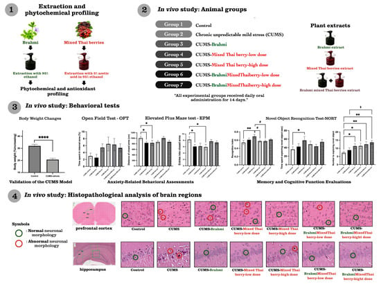
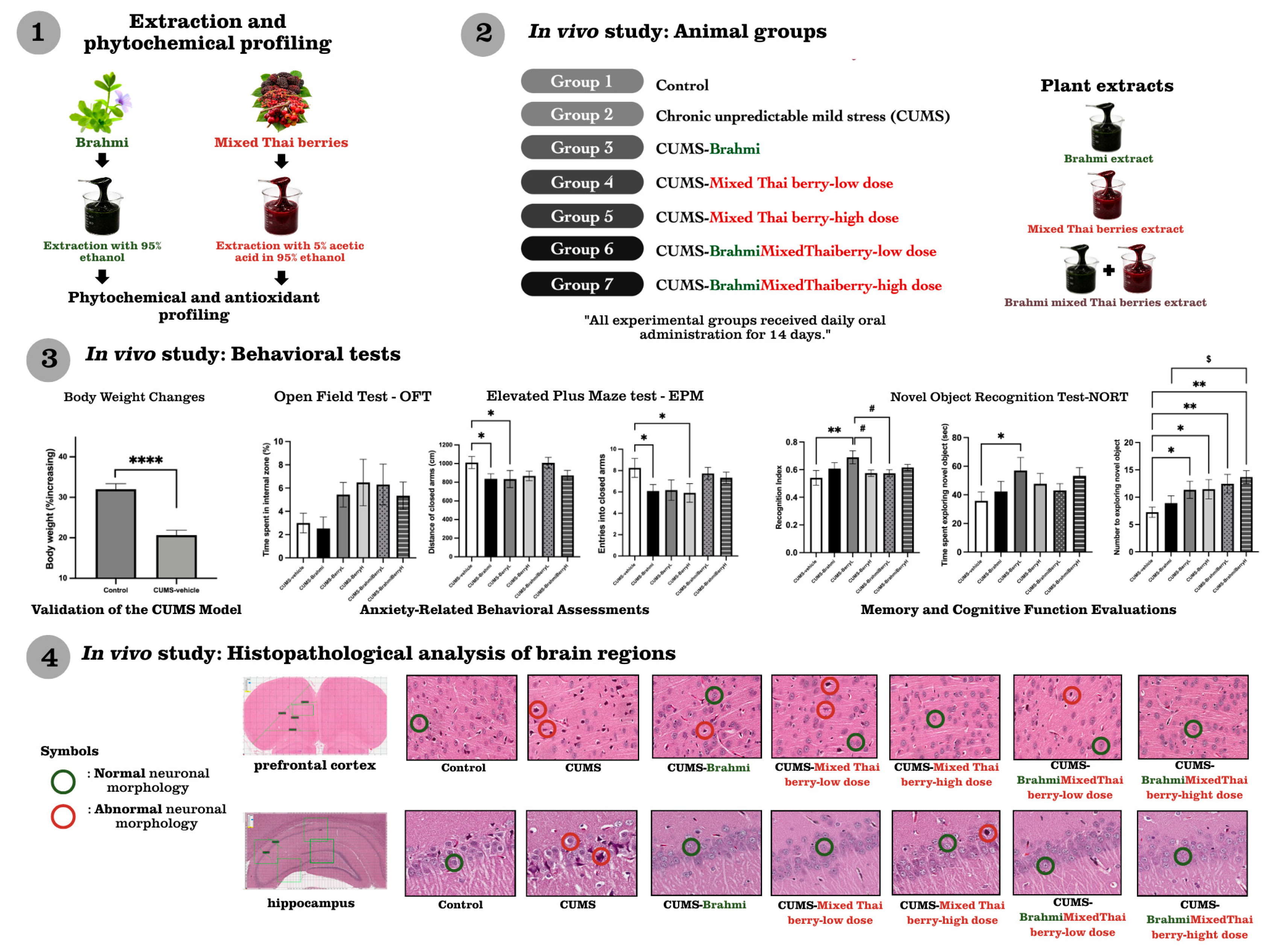
1. Introduction
Chronic stress has become a major global health concern, driven by modern lifestyles and increasing psychosocial demands, and is strongly associated with the development of stress-related mental health disorders, including anxiety and cognitive dysfunction [
1,
2]. Beyond its epidemiological significance, accumulating clinical and experimental evidence indicates that prolonged exposure to chronic stress directly contributes to the development of anxiety disorders and stress-related memory impairment [
3,
4].
At the neurobiological level, prolonged exposure to stress disrupts neuronal homeostasis in brain regions involved in emotional regulation and cognitive processing, particularly the hippocampus and prefrontal cortex [
4,
5]. Chronic stress induces oxidative stress and neuroinflammatory responses, leading to neuronal damage and impaired neuroplasticity [
5,
6,
7]. These pathological changes manifest behaviorally as anxiety-like phenotypes and deficits in learning and memory, including impaired recognition memory. Consistent with these findings, animal models of chronic stress exhibit anxiety-like behavior accompanied by cognitive impairment [
8]. Accordingly, the chronic unpredictable mild stress (CUMS) model is widely employed as a robust and translational experimental paradigm to investigate stress-induced anxiety and cognitive dysfunction [
9,
10].
Given the central role of oxidative stress and neuroinflammation in stress-related neuronal dysfunction, there is growing interest in natural products with antioxidant and neuroprotective properties as potential complementary therapeutic strategies. Plant-derived bioactive compounds have demonstrated strong antioxidant and anti-inflammatory activities, supporting neuronal survival and synaptic integrity under pathological conditions [
10,
11,
12].
Bacopa monnieri (L.) Wettst. (Plantaginaceae; Brahmi) is a well-established medicinal plant in Ayurvedic medicine, traditionally used for enhancing memory and alleviating anxiety. Previous studies have demonstrated that Brahmi exhibits antioxidant, anti-inflammatory, and neuroprotective activities, which are believed to underlie its beneficial effects on learning and memory [
13,
14]. Saponin glycosides, particularly jujubogenin and pseudojujubogenin derivatives, have been identified as the principal active constituents contributing to these neuropharmacological effects [
14,
15]. However, experimental evidence supporting the neuroprotective efficacy of standardized Brahmi extract under chronic stress conditions remains limited, highlighting the need for further investigation.
In parallel, increasing attention has been directed toward anthocyanin-rich berries as natural neuroprotective agents. Anthocyanins, including cyanidin-3-O-glucoside (C3G), possess potent antioxidant and anti-inflammatory properties and have been shown to attenuate oxidative neuronal damage and support cognitive function [
11,
15,
16,
17,
18]. Several Thai berries, including
Morus alba L. (Moraceae; mulberry),
Antidesma ghaesembilla Gaertn. (Phyllanthaceae; mamao), and
Syzygium nervosum DC. (Myrtaceae; ma-kiang), are rich sources of anthocyanins and phenolic compounds, suggesting their potential to mitigate stress-related neuronal dysfunction through antioxidant mechanisms. Previous studies consistently report strong antioxidant and neuroprotective effects of both saponin-rich Brahmi and anthocyanin-rich berry extracts [
14,
19,
20,
21].
Importantly, the use of a mixed-berry formulation offers several potential advantages over single-berry extracts, including broader phytochemical diversity and enhanced antioxidant coverage, which may support multifaceted neuroprotective activity. Combination-based plant formulations may provide a wider range of bioactive constituents than single-plant preparations, as different phenolic compounds, flavonoids, anthocyanins, and other bioactive constituents can act through complementary antioxidant, anti-inflammatory, and neuroprotective pathways. This concept is consistent with previous reports on polyherbal formulations and berry-derived phytochemicals, in which combined plant products have been proposed to exert multi-target biological effects [
22,
23]. Moreover, combining Brahmi with anthocyanin-rich berry extracts is hypothesized to broaden the spectrum of neuroprotective actions, integrating saponin-mediated neuromodulatory effects with anthocyanin-driven antioxidant and anti-inflammatory properties. To date, however, no study has systematically evaluated the combined effects of standardized Brahmi extract and a standardized anthocyanin-rich mixed Thai berry extract under chronic stress conditions, representing a significant gap in current knowledge and an opportunity to identify novel and unique natural active ingredient combinations with complementary neuroprotective potential.
Based on these considerations, this study aimed to characterize individual Thai berry extracts and the mixed extract through phytochemical profiling, antioxidant assays, and HPLC-based quantification of C3G in the mixed Thai berry extract. The in vivo effects of the standardized anthocyanin-rich mixed Thai berry extract, administered at low and high doses, alone and in combination with Brahmi extract, were then evaluated on anxiety-like behaviors and recognition memory in a CUMS rat model. Histopathological assessment of the prefrontal cortex and hippocampal subregions was also performed to support behavioral outcomes. Together, these findings are expected to advance understanding of herbal neuroprotection and support the development of evidence-based natural therapeutic strategies for stress-related anxiety and cognitive impairments.
3. Discussion
The present study integrated phytochemical characterization with behavioral and histopathological analyses to evaluate the neuroprotective effects of Brahmi and anthocyanin-rich Thai berry extracts, administered both individually and in combination, in a CUMS rat model. The antioxidant and bioactive properties of Brahmi have been well documented in previous studies, in which phenolic compounds and saponin-rich fractions were shown to exhibit significant radical scavenging and reducing activities across multiple in vitro assays [
19,
24]. Accordingly, this analysis focused on comparative profiling of Thai berry extracts, with a standardized Brahmi extract employed as a reference intervention. The present findings do not allow confirmation of pharmacological synergy. Rather, they suggest that the combined formulation may differentially affect structural and behavioral endpoints.
Phytochemical analyses demonstrated distinct differences among berry extracts, with ma-kiang exhibiting the highest antioxidant activity, corresponding to its elevated phenolic and anthocyanin contents. Mulberry and the mixed Thai berry extract exhibited moderate antioxidant capacities, whereas mamao showed lower activity. These findings are consistent with previous reports identifying phenolic compounds and anthocyanins as major contributors to antioxidant potential [
10,
11,
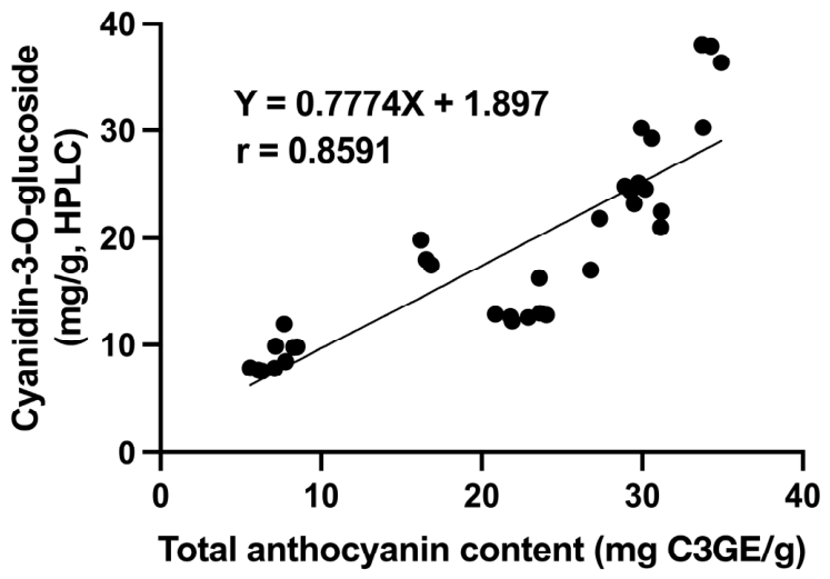
12]. Accordingly, the inclusion of ma-kiang and mulberry in the mixed Thai berry extract is supported by their strong phytochemical profiles, while mamao may contribute complementary phytochemical diversity and favorable organoleptic properties. HPLC analysis confirmed C3G as a predominant anthocyanin in ma-kiang and mulberry extracts, with intermediate levels detected in the mixed-berry extract. The strong correlation between spectrophotometric TAC and HPLC-derived C3G (r = 0.8591,
p < 0.0001) supports the reliability of the pH differential method as a rapid screening tool, while HPLC remains essential for precise quantification of individual anthocyanins. Minor deviations between methods may reflect the contribution of additional anthocyanin species beyond C3G. The stronger antioxidant capacity of ma-kiang and the mixed Thai berry extract supports their potential relevance in modulating stress-associated neurobehavioral alterations. However, the behavioral outcomes did not strictly follow the in vitro antioxidant ranking, suggesting that in vivo efficacy may also depend on bioavailability, metabolism, dose–response characteristics, and interactions among phytochemicals. This observation highlights the complexity of in vivo neuroprotective responses, in which behavioral outcomes may be influenced not only by antioxidant capacity but also by pharmacokinetic properties and interactions among phytochemical constituents.
Importantly, in vivo efficacy was evaluated using a standardized anthocyanin-rich mixed Thai berry extract, rather than individual berry extracts, to reflect the use of multi-component herbal preparations and potential phytochemical complementarity. Although individual berries such as ma-kiang and mulberry exhibited strong antioxidant activity in vitro, the mixed Thai berry extract integrates anthocyanins and phenolic compounds from the three berries, which may provide broader antioxidant coverage under in vivo conditions.
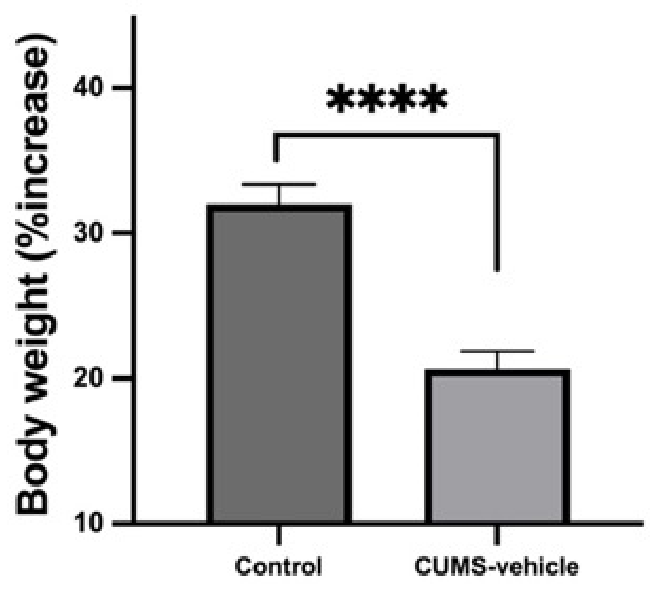
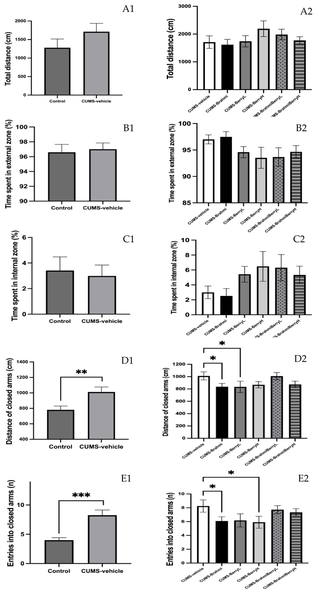
In vivo, the CUMS model was successfully validated by reduced body weight gain and increased anxiety-like behaviors, including elevated closed-arm activity in the EPM and altered exploratory patterns in the OFT, consistent with previous reports linking chronic stress to metabolic and behavioral dysregulation [
28,
29]. Previous studies have demonstrated that the behavioral and cognitive consequences of CUMS are highly dependent on stress duration, stress severity, and experimental conditions. Although recognition memory impairment has frequently been reported following prolonged CUMS exposure, cognitive outcomes remain variable across different CUMS protocols and experimental settings [
30,
31].
Accordingly, comparisons between the control and CUMS-vehicle groups were primarily used for model validation, whereas subsequent analyses among CUMS-exposed groups were intended to evaluate treatment-related cognitive modulation under chronic stress conditions.
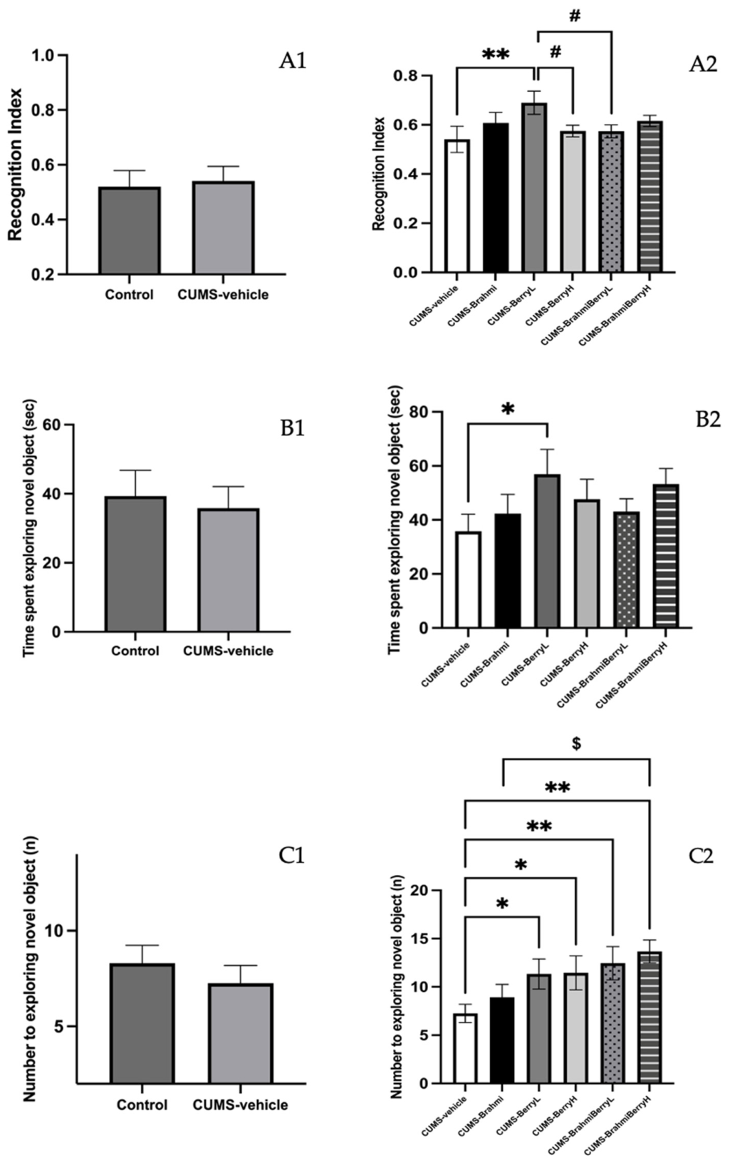
Therefore, under the present experimental conditions, the NORT paradigm may primarily reflect sensitivity to treatment-related modulation rather than reversal of established cognitive impairment. Consequently, the observed changes in recognition memory-associated parameters should be interpreted as cognitive-enhancing or modulatory effects, rather than restoration of impaired cognitive function. Regarding anxiety-related behaviors, treatment with Brahmi or Thai berry extracts alone produced measurable anxiolytic-like effects in CUMS-exposed rats. Both interventions reduced indices of anxiety in the OFT and EPM, such as closed-arm activity and exploratory avoidance, consistent with the reported neuroprotective and cognitive-enhancing effects of Brahmi, which have been linked to its antioxidant and anti-inflammatory actions [
14], and the antioxidant and anti-inflammatory actions of anthocyanins [
11,
15,
18,
32]. In contrast, combined Brahmi–berry treatments did not consistently enhance anxiolytic outcomes beyond those observed with individual extracts. This absence of additive benefit in anxiety-related measures may reflect suboptimal dose ratios under the current experimental conditions. Additionally, the relatively short CUMS duration may have constrained the detection of more robust synergistic effects on anxiety-like behavior.
In contrast to anxiety outcomes, cognitive performance assessed by the NORT revealed a distinct response pattern. Berry extracts administered alone modulated recognition memory-associated parameters, including recognition index, exploration time, and novel-object entries. The low-dose berry extract consistently demonstrated the strongest effects across several cognitive parameters, suggesting a potential non-linear or hormetic dose–response relationship, particularly with respect to recognition memory-related parameters in the low-dose berry-treated group compared with the CUMS-vehicle group. These behavioral outcomes were not directly proportional to the in vitro antioxidant ranking of the extracts, suggesting that additional factors such as phytochemical interactions, bioavailability, or neurobiological pathway modulation may contribute to the observed in vivo effects. Furthermore, the differential responses observed between single and combination treatments may indicate that combined phytochemical formulations do not necessarily produce additive behavioral effects under the present experimental conditions. In contrast, combination treatments did not uniformly outperform single treatments in behavioral outcomes, although they demonstrated more pronounced histological preservation. This discrepancy between structural and behavioral findings may reflect differential sensitivity of endpoints or suboptimal dose ratios in the combined formulation. These effects may reflect the combined influence of Brahmi-associated neuromodulatory and synaptic plasticity-supporting properties [
14,
33], together with the antioxidant and anti-inflammatory actions of berry anthocyanins [
11,
15,
32]. Brahmi has been extensively investigated for its neuroprotective and cognition-enhancing properties, which are largely attributed to saponin glycosides. Previous studies have shown that those saponin glycosides may exert antioxidant and anti-inflammatory effects, support synaptic plasticity, and protect neurons against stress-associated damage. These reported pharmacological properties may have contributed, at least in part, to the behavioral and histopathological effects observed in the present study.
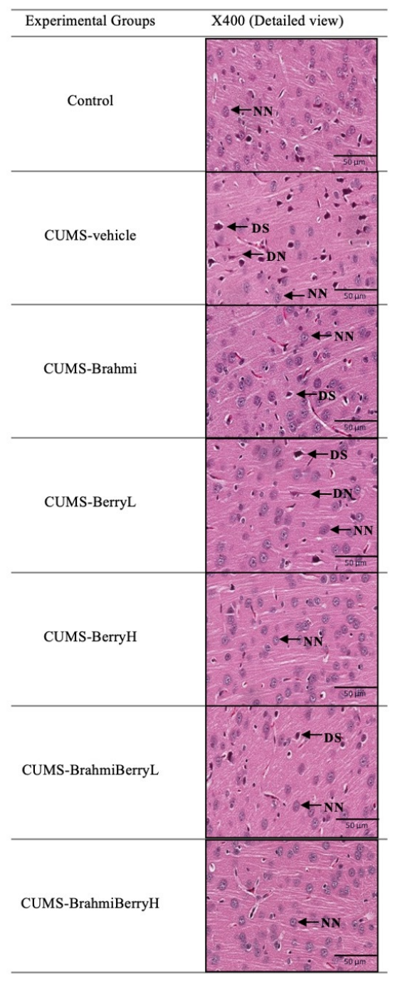
The histopathological observations were consistent with the behavioral outcomes and provided qualitative structural support for the observed functional changes. In the prefrontal cortex, CUMS exposure was associated with clear neuronal degeneration, characterized by the presence of dark shrunken and dark neurons, whereas treatment with Brahmi or Thai berry extracts alone attenuated these morphological alterations to varying degrees. Combination treatments, particularly at the higher dose, showed the greatest preservation of cortical neuronal morphology. However, this structural protection did not translate into proportionally greater anxiolytic effects, suggesting that cortical morphological integrity alone may not fully account for anxiety-related behavioral outcomes under the present experimental conditions.
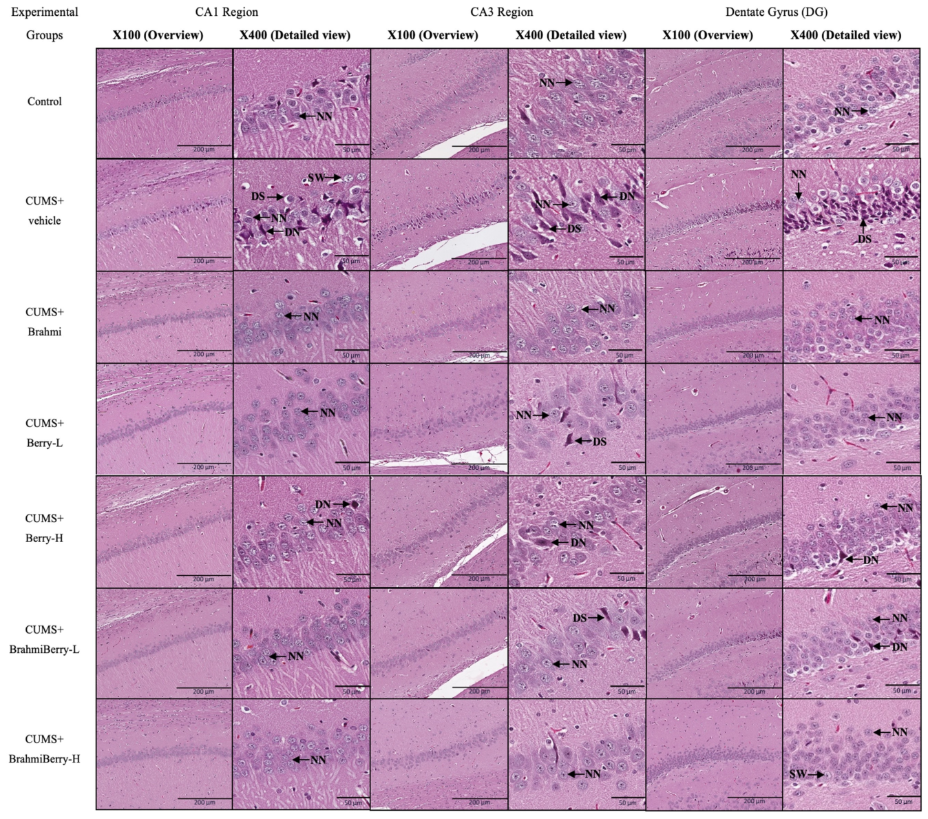
In contrast, histopathological changes in the hippocampus exhibited a clearer treatment-dependent pattern. Progressive restoration of neuronal morphology across hippocampal subregions (CA1, CA3, and dentate gyrus) was observed among treatment groups, with marked preservation in the Brahmi and combination-treated groups. This regional hippocampal preservation, particularly evident in the combination-treated groups, closely paralleled improvements in recognition memory observed in the novel object recognition test, supporting the well-established association between hippocampal neuronal integrity and cognitive performance under chronic stress conditions [
34].
The neuroprotective effects observed in this study are likely mediated, at least in part, by mechanisms previously reported for both Brahmi constituents and anthocyanins. Saponin glycosides, including bacoside A3, bacopaside X, bacopaside I, bacopaside II, and bacopasaponin C, are among the principal bioactive constituents of Brahmi and have been reported to possess antioxidant, anti-inflammatory, neuroprotective, and neurotransmitter-modulating activities that may contribute to stress resilience and cognitive function [
35,
36]. At the molecular level, previous studies have suggested that standardized Brahmi extracts may influence serotonergic and cholinergic neurotransmission, including modulation of 5-HT-related signaling, acetylcholine-associated pathways, and downstream molecular events linked to synaptic plasticity and memory formation, such as ERK/CREB signaling and synaptic protein regulation [
36]. Similarly, anthocyanins have been reported to exert neuroprotective effects through modulation of oxidative stress, neuroinflammatory pathways, and neurobiological processes associated with stress-related behaviors and cognitive performance [
37,
38]. These reported biological activities may provide a plausible explanation for the behavioral and histopathological effects observed in the present study. However, because direct biochemical, molecular, and neurotransmitter-related measurements were not performed, the proposed mechanisms remain speculative and require confirmation in future studies incorporating appropriate biochemical endpoints.
Collectively, these findings indicate that while Brahmi and Thai berry extracts individually exert anxiolytic effects under chronic stress, their combination does not further enhance anxiety-related behaviors within the present experimental framework. In contrast, cognitive-related outcomes were improved mainly by single-extract treatments, particularly the low-dose berry extract, whereas combination treatments showed stronger histological preservation than behavioral superiority. These findings reveal a differential response between behavioral and structural outcomes, underscoring the multifaceted nature of the observed neuroprotective effects. Importantly, this study introduces a novel framework integrating phytochemical standardization with functional neuroprotection in a clinically relevant stress model.
4. Materials and Methods
4.1. Chemical and Reagents
Reagents for antioxidant assays, including the Folin–Ciocalteu reagent, 2,2-diphenyl-1-picrylhydrazyl (DPPH), gallic acid, Trolox (6-hydroxy-2,5,7,8-tetramethylchroman-2-carboxylic acid), ABTS (2,2′-azino-bis(3-ethylbenzothiazoline-6-sulfonic acid)), and TPTZ (2,4,6-tri(2-pyridyl)-s-triazine), were purchased from Sigma-Aldrich (St. Louis, MO, USA). Ethanol (95%, food grade) and acetic acid (99%, food grade) were used for extraction. C3G (HPLC standard, >95% purity) was obtained from Glentham Life Sciences Ltd. (Corsham, UK). Acetonitrile (HPLC grade) was supplied by Fisher Scientific (Seoul, South Korea), and formic acid (99%, analytical grade) was purchased from CARLO ERBA Reagents (Cornaredo, Italy). Ultrapure water (18 MΩ·cm) was produced using a Milli-Q® Reference Water Purification System (Merck Millipore, Burlington, MA, USA). Reference standards for bacoside A3, bacopaside X, bacopaside I, bacopaside II, and bacopasaponin C were obtained from MedChemExpress (Monmouth Junction, NJ, USA) with purity ≥98.0%.
4.2. Plant Materials
Aerial parts of Brahmi were collected from Phitsanulok Province, Thailand. The plant material was authenticated, and a voucher specimen (Phrompittayarat001) was deposited at the Herbarium of Mahidol University (PBM), Thailand. The collected samples were thoroughly cleaned and dried in a hot-air oven at 50 °C for 24 h, then ground and sieved through a 60-mesh sieve. The resulting powder was stored at −20 °C until further phytochemical analysis and extraction, following previously established protocols [
39,
40].
Ripe fruits of Morus alba L. or mulberry (Chiang Mai variety, voucher specimen PNU 6078) were obtained from the Queen Sirikit Sericulture Center, Nan Province, Thailand. Antidesma ghaesembilla Gaertn. (Fa Prathan variety, voucher specimen PNU 6079) or mamao fruits were collected from the Sakon Nakhon Markmao Association, Sakon Nakhon Province, Thailand, and Syzygium nervosum DC. or ma-kiang fruits (voucher specimen PNU 6080) were sourced from Maejo University, Phrae Campus, Thailand. All voucher specimens were deposited at the Herbarium, Biology Department, Faculty of Science, Naresuan University, Thailand. All fruit samples were harvested at the fully ripe stage and stored at −20 °C prior to extraction.
4.3. Extract Preparation
The extraction methods were selected based on a review of previous studies, considering active compound concentration, safety, and cost-effectiveness. The Brahmi extract was prepared following established protocols [
40], with specific steps modified to improve the extraction efficiency and overall extract quality. The Brahmi extract was standardized to contain 5.0% (
w/
w) total saponin glycosides, as determined by an HPLC method (see
Section 4.4.1). The extraction yield of Brahmi extract was 10–13% based on dried plant material.
Thai berries were extracted with 5% acetic acid in 95% ethanol as previously described [
41], with minor modifications based on established methods for anthocyanin-rich extracts [
42]. The extraction yields were 9.19% for mulberry, 15.89% for mamao, and 4.40% for ma-kiang, calculated on a fresh weight basis.
The standardized anthocyanin-rich mixed Thai berry extract was prepared by combining extracts of mulberry, mamao, and ma-kiang using an optimized formulation approach designed to enhance anthocyanin content and organoleptic properties, as previously described [
41]. The formulation was standardized based on total anthocyanin content (TAC), which served as the reference parameter for dose normalization in subsequent in vivo studies. The standardized anthocyanin-rich mixed Thai berry extract showed a TAC of 41.70 ± 1.66 mg C3G equivalents (C3GE)/g extract, determined using the pH differential method.
For clarity, the standardized anthocyanin-rich mixed Thai berry extract is hereafter referred to as the mixed Thai berry extract.
4.4. Quantification of Bioactive Compounds
4.4.1. HPLC Determination of Total Saponin Glycosides in Brahmi
Total saponin glycosides in the Brahmi extract were quantified by HPLC according to a previously validated method [
39,
40]. The analysis was based on the combined determination of jujubogenin-type glycosides (bacoside A3 and bacopaside X) and pseudojujubogenin-type glycosides (bacopaside I, bacopaside II, and bacopasaponin C). The total saponin content was calculated as the sum of these marker compounds and expressed as % (
w/
w) of dried extract.
4.4.2. Determination of Total Anthocyanin Content (TAC)
TAC was quantified using the pH differential method. Extracts (250 µL) were mixed with 750 µL of either 0.025 M potassium chloride buffer (pH 1.0) or 0.04 M sodium acetate buffer (pH 4.5) and incubated in the dark for 15 min, followed by centrifugation at 2500×
g for 10 min. Absorbance was measured at 510 and 700 nm. Results were expressed as mg C3G equivalents per gram of extract (mg C3GE/g), calculated as previously described [
42,
43].
4.4.3. Determination of Total Phenolic Content (TPC)
Total phenolic content was determined using the Folin–Ciocalteu method in 96-well microplates [
44]. Briefly, 25 µL of the sample was mixed with 25 µL of diluted Folin–Ciocalteu reagent and 200 µL of water. After 5 min, 25 µL of 10.6% sodium carbonate was added, and the mixture was incubated in the dark at room temperature for 60 min. Absorbance was measured at 725 nm. Results were expressed as mg gallic acid equivalents per gram of sample (mg GAE/g).
4.4.4. HPLC Quantification of C3G
C3G was quantified by HPLC using on an Agilent 1260 Infinity system with detection set at 520 nm. Chromatographic separation was achieved on a Phenomenex C12 column (250 × 4.6 mm, 4 µm) equipped with a guard column, using a gradient of solvent A (water: acetonitrile: formic acid, 87:3:10
v/
v/
v) and solvent B (water: acetonitrile: formic acid, 40:50:10
v/
v/
v). The flow rate was 1.0 mL/min, the injection volume was 20 µL, and the column temperature was 30 °C. Sample preparation and chromatographic conditions were adapted from previous reports [
45], with minor modifications to improve analytical efficiency. The calibration curve showed good linearity over the range of 1.25–40 µg/mL, where R
2 = 0.993 LOD and LOQ were 0.039 and 1.25 µg/mL, respectively. Representative HPLC chromatograms of standardized C3G and the anthocyanin-rich mixed Thai berry extract are provided in
Supplementary Materials: Figures S1 and S2.
4.5. Correlation Analysis Between Spectrophotometric and HPLC Anthocyanin Data
To evaluate the consistency between analytical methods for anthocyanin determination, Pearson’s correlation was performed between TAC measured by the pH differential spectrophotometric method and C3G quantified by HPLC. TAC values were plotted against corresponding C3G concentrations, and correlation coefficients (r) with p-values were calculated using GraphPad Prism (version 10).
4.6. Evaluation of Antioxidant Potential
The antioxidant activities of individual berry extracts and the mixed Thai berry extract were evaluated using DPPH, ABTS, and FRAP assays according to previously established protocols [
32,
44,
46], with minor modifications as described below.
4.6.1. DPPH Radical Scavenging Method
The antioxidant activity of the extracts was evaluated using a modified DPPH radical scavenging assay [
44]. Briefly, 150 µL of 0.2 mM DPPH solution was mixed with 75 µL of diluted extract, with a reagent-only control prepared simultaneously. After incubation in the dark at room temperature for 30 min, absorbance was measured at 515 nm using a microplate reader. IC
50 values were calculated via nonlinear regression using GraphPad Prism (version 10), with Trolox serving as the positive control. Percentage inhibition was determined according to established protocols.
4.6.2. ABTS Radical Scavenging Method
ABTS radical scavenging activity was measured using a modified method adapted from previous studies [
32,
46]. The ABTS•
+ reagent was generated by reacting 7 mM ABTS with 2.45 mM potassium persulfate and incubating in the dark at room temperature for 16 h; then, it was diluted 1:24 (
v/
v) with water. Extract samples (20 µL) were mixed with 180 µL of diluted ABTS solution and incubated for 6 min at room temperature. Absorbance was recorded at 732 nm using a microplate reader. Trolox served as the positive control and IC
50 values were calculated via nonlinear regression using GraphPad Prism (version 10). Percentage inhibition was determined according to established protocols.
4.6.3. Ferric Reducing Antioxidant Power (FRAP)Method
The ferric reducing ability of berry extracts was assessed using a modified FRAP assay based on previous studies [
32,
46], Briefly, 20 µL of sample was mixed with 180 µL of FRAP reagent, prepared by combining acetate buffer (300 mM, pH 3.6), TPTZ solution (10 mM), and FeCl
3 solution (20 mM) in a 10:1:1 ratio. The mixture was incubated in the dark at room temperature for 30 min, then absorbance was measured at 595 nm using a microplate reader. A standard curve was constructed using FeSO
4·7H
2O, and results were expressed as grams of Fe
2+ equivalents per 100 g of sample. Trolox was used as a positive C.
4.7. In Vivo Study
4.7.1. Ethical Approval and Animal Care
The study protocol was approved by the Naresuan University Animal Care and Use Committee (NUACUC; approval no. NU-AE660305). A total of 84 male Sprague Dawley rats (7 weeks old) were obtained from Nomura Siam International Co., Ltd. All animals were housed at Naresuan University under controlled conditions (22 ± 1 °C, 12/12 h light–dark cycle, 55 ± 10% humidity) with ad libitum access to food and water. Animals were allowed a 7-day acclimatization period before the experiment, after which they were randomly assigned to either the chronic unpredictable mild stress (CUMS) group or the control group. All experimental procedures were conducted in compliance with relevant laws and institutional guidelines.
4.7.2. The Chronic Unpredictable Mild Stress Procedure
Chronic unpredictable mild stress (CUMS) is a validated rodent model for studying stress-related disorders, characterized by the induction of depressive- and anxiety-like behaviors through repeated exposure to mild, unpredictable stressors. It mimics the chronic and variable nature of human psychological stress and is widely used to evaluate therapeutic interventions [
47,
48]. Animals were exposed to various mild stressors in an unpredictable sequence, as detailed in
Table 3.
4.7.3. Administration of Treatment
A total of 84 male Sprague Dawley rats were randomly allocated into seven groups (
n = 12 per group). The normal control group (control) received reverse osmosis (RO) water (10 mL/kg bw, p.o.). The remaining six groups were subjected to the CUMS protocol. Among them, the CUMS control group (CUMS-vehicle) also received RO water (10 mL/kg bw, p.o.). The treatment groups consisted of the CUMS-Brahmi group receiving Brahmi extract at 20 mg/kg bw; the CUMS-BerryL and CUMS-BerryH groups, which received low and high doses of mixed Thai berry extract, corresponding to 0.043 and 0.215 mg C3GE/kg bw, respectively; and the CUMS-BrahmiBerryL and CUMS-BrahmiBerryH groups, which received Brahmi extract in combination with low and high doses of mixed Thai berry extract, respectively. All treatments were administered orally once daily in the morning for 14 consecutive days. The dose of Brahmi extract (20 mg/kg bw) was selected based on previous studies reporting cognitive-enhancing and neuroprotective effects in rodent models [
33,
49]. Dose selection for the mixed Thai berry extract was guided by reports on anthocyanin-rich berries demonstrating cognitive benefits [
50]. For the combination groups, Brahmi extract was administered at 20 mg/kg bw together with the mixed Thai berry extract at either 0.043 or 0.215 mg C3GE/kg bw. The combination was freshly prepared before administration by mixing the corresponding doses of each standardized extract in RO water to achieve the same final administration volume.
4.7.4. Body Weight Monitoring
All animals were weighed each morning before dosing for 14 consecutive days using a calibrated digital balance. Body weight (BW) data were used to calculate the percent increase from Day 0 to Day 14.
where BW0 denotes the baseline (Day 0) body weight and BW14 the Day 14 body weight; positive values indicate gain relative to the baseline, negative values indicate loss, and 0% indicates no change.
4.7.5. Behavioral Assessments
Open-Field Test (OFT)
The open-field test (OFT) is a well-established behavioral paradigm used to assess spontaneous locomotor activity and general arousal in rodents. It also provides an indirect measure of anxiety-like behavior based on the animal’s tendency to avoid open and unprotected areas of the arena [
28]. In the present study, each rat was placed individually at the center of the open-field arena and was allowed to explore freely for 10 min. The arena was divided into predefined zones, and locomotor activity and movement patterns during the last 5 min of the test were recorded for analysis.
Elevated Plus Maze Test (EPM)
The elevated plus maze (EPM) is a standard behavioral test for assessing anxiety-related responses in rodents. It consists of two open arms and two closed arms elevated above the floor. Rodents naturally avoid open arms, and the time spent and entries into the open arms serve as indices of anxiety-like behavior [
29]. In the present study, each rat was placed at the center of the maze and allowed to explore freely for 10 min. The time spent and number of entries into the open and closed arms during the last 5 min of the test were recorded for the evaluation of anxiety-like behavior.
Novel Object Recognition Test (NOR)
The novel object recognition (NOR) test is a widely used behavioral paradigm for assessing recognition memory in rodents. The test is based on the natural tendency of rodents to preferentially explore a novel object over a familiar one. Increased exploration time of the novel object reflects intact recognition memory [
51,
52]. In the present study, each rat was placed individually in the arena during the training phase and was allowed to explore two identical objects. After a retention interval of 90 min, the rat was returned to the arena for the test phase, during which one familiar object was replaced with a novel object. The time spent exploring the novel object and the number of entries into the novel object zone were recorded for the evaluation of recognition memory.
Behavioral recordings from the OFT, EPM, and NORT were analyzed using SMART video tracking software V3.0 (Panlab, Spain). To improve data reliability and minimize observer bias, behavioral parameters were independently verified by three investigators through repeated assessments. The final datasets were confirmed based on concordant evaluations obtained from both automated software analysis and investigator-based observations.
4.8. Histological Analyses
After the behavioral tests, rats were anesthetized with thiopental sodium (100 mg/kg, intraperitoneally). The prefrontal cortex and hippocampus tissues from six selected rats in each group were dissected and fixed in 10% neutral buffered formalin. The samples were then processed using a standard paraffin-embedding protocol, sectioned at approximately 5 μm thickness using a rotary microtome, and stained with hematoxylin and eosin (H&E). Finally, the sections were mounted on glass slides and examined under a bright-field light microscope (Olympus BX51, Olympus, Tokyo, Japan) to evaluate histopathological changes [
53].
4.9. Statistical Analysis
Data were presented as mean ± standard deviation and the difference between variables was analyzed using the independent sample t-test or Mann–Whitney test (non-parametric). A p < 0.05 was considered statistically significant.
All data were analyzed using GraphPad Prism version 10. Data are presented as mean ± SEM unless otherwise indicated. Comparisons between two groups were analyzed using an unpaired t-test. Comparisons among three or more groups were analyzed using one-way ANOVA followed by Fisher’s LSD post hoc test only when the overall ANOVA was significant. Statistical significance was set at p < 0.05.
5. Conclusions
This study integrated phytochemical characterization with in vivo behavioral and histopathological analyses to evaluate the neuroprotective effects of standardized Brahmi, anthocyanin-rich Thai berry extracts, and their combination in a chronic unpredictable mild stress (CUMS) rat model. Thai berry extracts exhibited high anthocyanin with strong antioxidant capacity, and HPLC analysis confirmed C3G as a major anthocyanin, supporting rapid screening approaches for anthocyanin-rich extracts.
Behaviorally, CUMS reliably induced anxiety-like phenotypes in rats. Treatment with Brahmi or berry extracts administered individually attenuated selected anxiety-related behaviors, whereas combined treatment did not consistently enhance anxiolytic outcomes beyond single treatments. In contrast, cognitive performance assessed by the novel object recognition test was most consistently improved by the low-dose berry extract, while combination treatments were more clearly associated with histological preservation. Recognition memory-associated parameters were modulated following individual treatments, with the low-dose berry group showing the most consistent effects, while exploratory analyses indicated enhanced novelty-directed exploration in combination-treated groups.
Histopathological findings in the prefrontal cortex and hippocampus supported these behavioral patterns, demonstrating reduced neuronal degeneration and improved tissue organization, particularly in the Brahmi and combination-treated groups.
Overall, these findings indicate domain-specific neuroprotective effects of Brahmi and anthocyanin-rich Thai berry extracts under chronic stress conditions, with individual treatments primarily alleviating anxiety-like behaviors and combined treatment preferentially supporting cognitive and exploratory aspects of behavior. This integrated evidence supports their potential as natural interventions for stress-related anxiety and cognitive modulation.
As for the limitations of this study, this study was conducted exclusively in male rats, precluding assessment of sex-specific responses. Histopathological evaluation was qualitative and based primarily on H&E staining, without quantitative stereological or immunohistochemical analyses. Further studies incorporating quantitative histological and immunohistochemical approaches are warranted to elucidate the underlying cellular and molecular mechanisms. Although the relatively short CUMS duration may have affected the extent of detectable neurodegenerative changes, the behavioral results still showed beneficial effects on anxiety-like behavior and recognition memory-associated parameters. Although combination treatments showed enhanced histological preservation in some brain regions, formal analyses to confirm pharmacological synergy were not performed. Although TAC and HPLC-derived C3G showed a strong overall correlation, deviations may occur at higher concentrations because the spectrophotometric method estimates total monomeric anthocyanins and may be influenced by other absorbing compounds. Therefore, TAC was used primarily for comparative screening, whereas HPLC was used for marker-based quantification. The absence of a standard pharmacological positive control (e.g., fluoxetine or diazepam) may represent a limitation in directly comparing the evaluated extracts with established reference treatments for anxiety-like behaviors. However, the current study design was intended primarily to demonstrate the protective effects of the extracts rather than their therapeutic efficacy. The inclusion of a positive control in future studies would facilitate benchmarking against established pharmacological agents and further support mechanistic interpretation. In addition, biochemical or neuroendocrine stress-related markers, such as corticosterone levels, oxidative stress markers, inflammatory cytokines, synaptic proteins, or neurotransmitter-associated parameters, were not evaluated in the present study. Therefore, confirmation of stress induction was based primarily on established physiological and behavioral alterations observed in the CUMS model.





 ,
,