Article

Unraveling the Mechanisms of Wuling Powder Against MASLD by Integrated Metabolomics–Gut Microbiota–Serum Pharmacochemistry

1
College of Pharmaceutical Science, Yunnan University of Chinese Medicine, 1076 Yuhua Road, Kunming 650500, China
2
Yunnan Key Laboratory of Southern Medicine Utilization, 1076 Yuhua Road, Kunming 650500, China
3
The First Clinical College, Yunnan University of Chinese Medicine, 1076 Yuhua Road, Kunming 650500, China
*
Authors to whom correspondence should be addressed.
These authors contributed equally to this work.
Pharmaceuticals 2026, 19(4), 557; https://doi.org/10.3390/ph19040557
Submission received: 7 March 2026 / Revised: 22 March 2026 / Accepted: 26 March 2026 / Published: 31 March 2026
(This article belongs to the Section Pharmacology)

Abstract

Background/Objective: Metabolic dysfunction-associated steatotic liver disease (MASLD) is a highly prevalent chronic liver disease with no specific therapeutics. Wuling Powder (WLP) is a classic traditional Chinese medicine prescription with therapeutic potential against MASLD, yet its molecular mechanism remains unclear. This study aims to elucidate the mechanism and possible effective substances of WLP in the treatment of MASLD. Methods: A rat MASLD model was established via high-fat diet feeding to evaluate WLP’s efficacy. Untargeted metabolomics and 16S rRNA sequencing were used to explore the effects of WLP on metabolism and gut microbiota in vivo. Serum pharmacochemistry combined with metabolomics was used to analyze the key active components and core targets of WLP against MASLD, and molecular docking and cell experiments were used to verify the relationship between them. Results: WLP reduced hepatic lipid accumulation and pathological damage, improved lipid levels in blood liver, enhanced antioxidant capacity, and alleviated inflammation in MASLD rats. Mechanistically, WLP regulated 19 metabolic pathways. It also decreased the Firmicutes/Bacteroidota ratio and reduced the abundance of potential pathogenic bacteria (Romboutsia and Turicibacter). Thirty-one WLP-derived components were identified in serum, 13 of which were key active components for treating MASLD. These components, especially 11-deoxyalisol A and 8β-methoxyatractylenolide I, alleviated hepatic steatosis by downregulating NOS2 and PLA2G2A expression. Conclusions: The alleviation of MASLD by WLP was mediated by the regulation of 8 metabolic pathways, alterations in the abundance of Romboutsia and Turicibacter, and the restoration of 20 metabolite levels, an effect primarily ascribed to 13 distinct pharmacodynamic components derived from WLP.

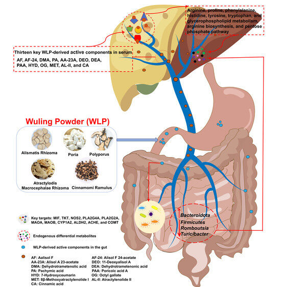
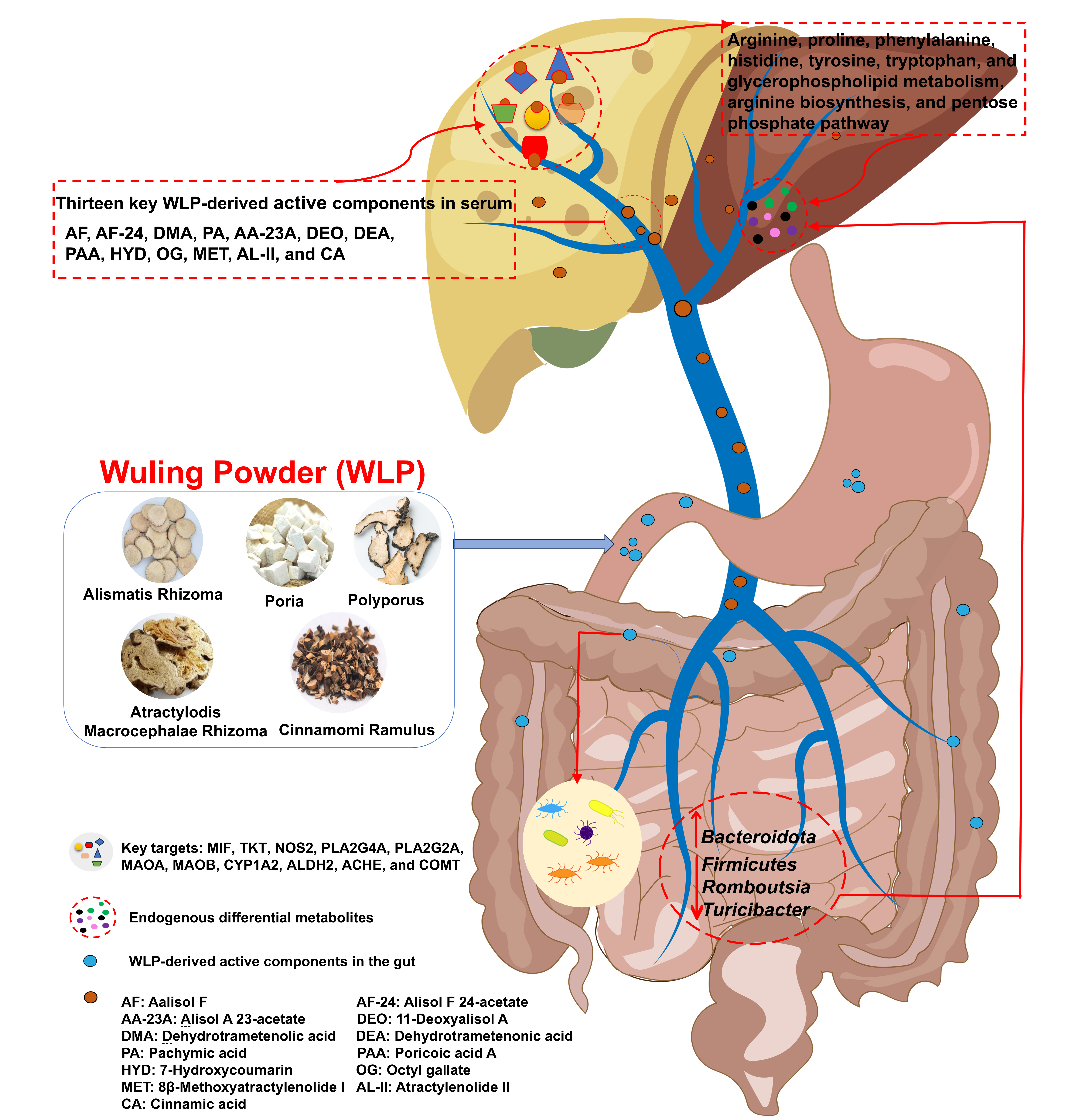
Graphical Abstract

1. Introduction

Metabolic dysfunction-associated steatotic liver disease (MASLD) was formerly termed non-alcoholic fatty liver disease (NAFLD). NAFLD is characterized by hepatic steatosis affecting more than 5% of hepatocytes in the absence of excessive alcohol consumption and other well-defined causes of liver injury. By contrast, MASLD is diagnosed based on hepatic steatosis accompanied by at least one cardiometabolic abnormality, including obesity/abdominal obesity, glycemic disorders/type 2 diabetes mellitus, hypertension, and dyslipidemia [1]. This revised definition more accurately captures the metabolic basis of the disease and its close linkage with multisystem comorbidities. As the most common chronic liver disease globally, MASLD affects up to 38% of the adult population worldwide and represents a leading cause of liver cirrhosis, end-stage liver disease, and primary liver cancer [2]. Currently, lifestyle interventions including low-carbohydrate, low-fat diets and exercise remain the mainstay for weight loss in the management of MASLD. In clinical practice, statins and sodium–glucose cotransporter 2 inhibitors are frequently prescribed for patients with MASLD accompanied by obesity and type 2 diabetes [3]. Nevertheless, statin treatment is associated with an increased risk of rhabdomyolysis, whereas sodium–glucose cotransporter 2 inhibitors may predispose patients to ketoacidosis [4,5]. For metabolic dysfunction-associated steatohepatitis (MASH, the progressive form of MASLD), thyroid hormone receptor β-selective agonists (resmetirom) and glucagon-like peptide-1 receptor agonists (semaglutide) are commonly used, yet both are associated with significant side effects and low efficacy [6,7]. Therefore, the development of safe and effective therapeutic agents for MASLD is of great clinical importance.
The pathogenesis of MASLD is complex, involving multiple factors such as insulin resistance, oxidative stress, abnormal lipid metabolism, genetic variation, and intestinal microbiota dysbiosis [8]. In recent years, traditional Chinese medicine (TCM), characterized by its multi-component, multi-target, and multi-pathway properties, has demonstrated unique advantages in addressing the complex pathogenesis of MASLD [9,10]. Wuling Powder (WLP) was first recorded in Treatise on Febrile Diseases and included in the Chinese Pharmacopoeia (2025 Edition), which is composed of Alismatis Rhizoma (AR), Poria (PA), Polyporus (PS), Atractylodis Macrocephalae Rhizoma (AMR) and Cinnamomi Ramulus (CR) in a 5:3:3:3:2 ratio. WLP has the effects of warming yang to activate qi and strengthening the spleen to dispel dampness and is a very famous dampness-resolving formula in TCM. Modern pharmacological studies have shown that WLP reduces blood lipid levels in hyperlipidemic mice, promotes reverse cholesterol transport and hepatic bile acid metabolism, reshapes the gut microbiota, and restores bile acid homeostasis to alleviate MASLD [11,12]. It also inhibits the release of inflammatory factors to alleviate inflammatory damage and regulate blood glucose and lipid levels in mice with type 2 diabetes [13]. In addition, all single herbs in the prescription can also alleviate MASLD-related symptoms [14,15,16,17]. Clinical studies have found that WLP reduces serum leptin levels in MASLD patients, improves insulin resistance, and normalizes liver function and lipid indices. Moreover, its combination with polyene phosphatidylcholine can enhance therapeutic efficacy [18,19]. Additionally, WLP is indicated for patients with MASLD of the phlegm dampness stasis obstruction type, and it is not suitable for those with heat symptoms. However, the mechanism of action and pharmacodynamic substances of WLP against MASLD remain unclear.
In this study, a MASLD rat model induced by high-fat diet feeding was established to evaluate the efficacy of WLP. Then, untargeted metabolomics and intestinal microbiota analysis were used to investigate the anti-MASLD mechanism of WLP. Furthermore, combined analysis of serum pharmacochemistry and metabolomics was conducted to identify the active components and therapeutic targets of WLP for anti-MASLD. Finally, molecular docking and cell experiments were performed to evaluate the anti-MASLD activity of these active components. This study reveals the anti-MASLD mechanism and in vivo active substances of WLP, thereby providing a scientific basis for its clinical application and market development.

2. Results

2.1. WLP Reverses the HFD-Induced Pathological Changes in MASLD Rats

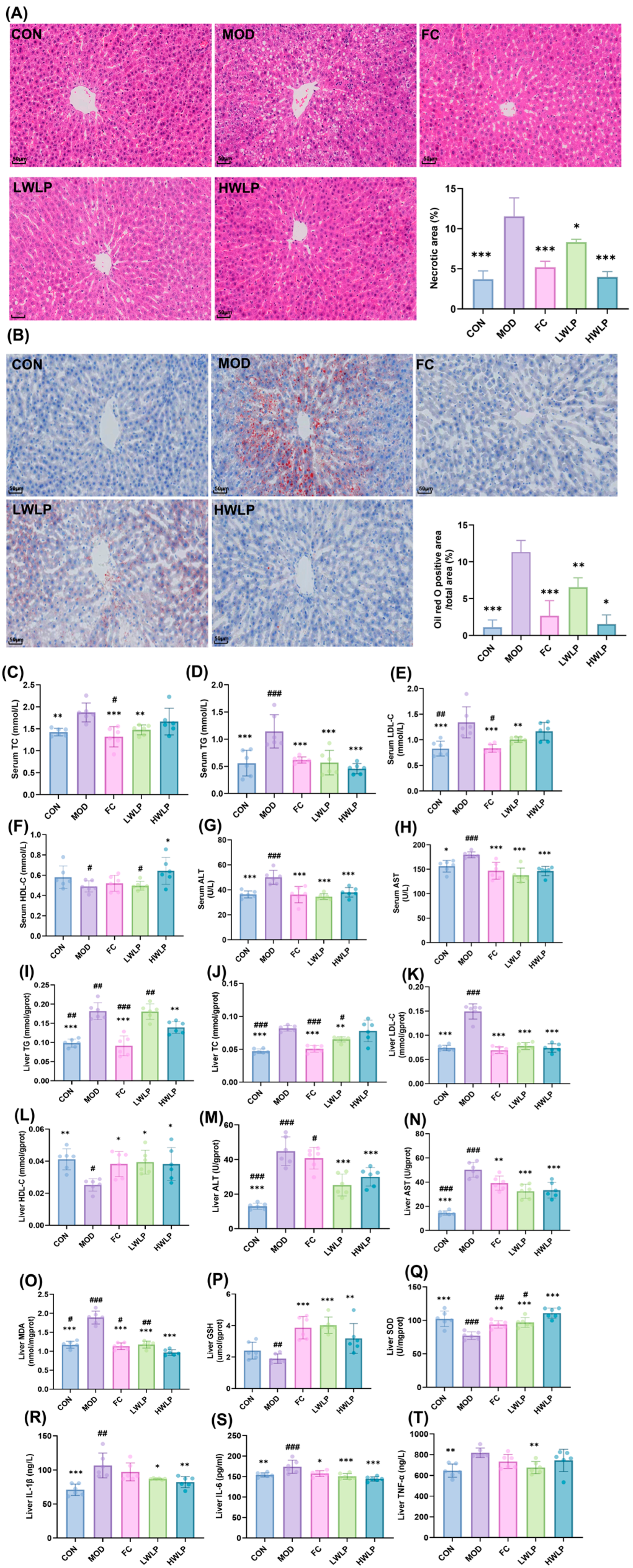
HE staining results demonstrated that following 12 weeks of HFD feeding, hepatocytes in the rat liver exhibited disorganized arrangement, accompanied by an increased number of lipid droplet vacuoles. Meanwhile, Oil Red O staining revealed that severe lipid accumulation was observed in the rat liver after 12 weeks of HFD feeding. Treatment with WLP significantly ameliorated the hepatic histomorphology of MASLD rats and alleviated hepatic lipid accumulation and damage (Figure 1A,B). In addition, as shown in Figure 1C–N, following 12 weeks of HFD feeding, the levels of TC, TG, LDL-C, ALT, and AST in the serum and liver of rats were significantly elevated (p < 0.05, p < 0.01, p < 0.001), whereas the level of HDL-C in the liver was significantly reduced (p < 0.01). Following intervention with WLP, the serum and hepatic levels of TC, TG, LDL-C, ALT, and AST were significantly decreased (p < 0.05, p < 0.01, p < 0.001), while hepatic HDL-C levels were significantly increased (p < 0.05). The HWLP group was significantly superior to the LWLP group in regulating serum HDL-C and liver TC and TG levels (p < 0.05, p < 0.01), suggesting a dose-dependent effect of WLP treatment. These findings suggest that WLP regulated serum and hepatic lipid levels and ameliorated HFD-induced lipid metabolism disorders.
As shown in Figure 1O–T, following 12 weeks of HFD feeding, the levels of MDA, TNF-α, IL-1β, and IL-6 in the livers of rats were significantly increased (p < 0.01, p < 0.001), accompanied by a marked decrease in SOD activity (p < 0.001). After treatment with WLP, the levels of MDA, TNF-α, IL-1β, and IL-6 were significantly reduced (p < 0.05, p < 0.01, p < 0.001), whereas the activities of GSH and SOD were significantly elevated (p < 0.01, p < 0.001). The HWLP group was significantly superior to the LWLP group in regulating liver SOD and MDA levels (p < 0.05, p < 0.01), indicating a dose-dependent effect of WLP treatment. These results indicate that WLP shows promise in alleviating HFD-induced hepatic oxidative stress and inflammatory response in MASLD rats.

2.2. WLP Normalizes the Abnormal Metabolic Profiles in Serum, Liver, and Urine of MASLD Rats

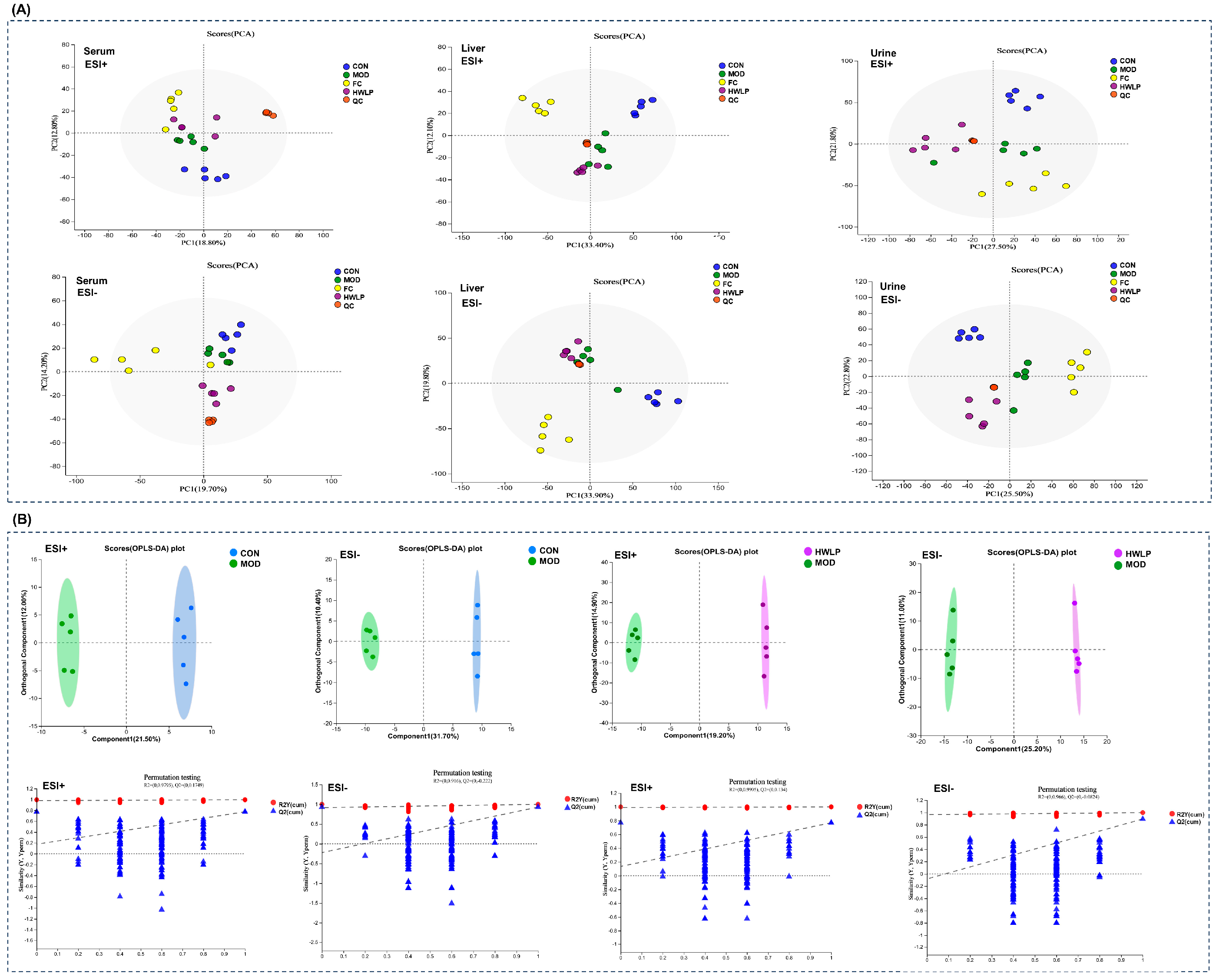
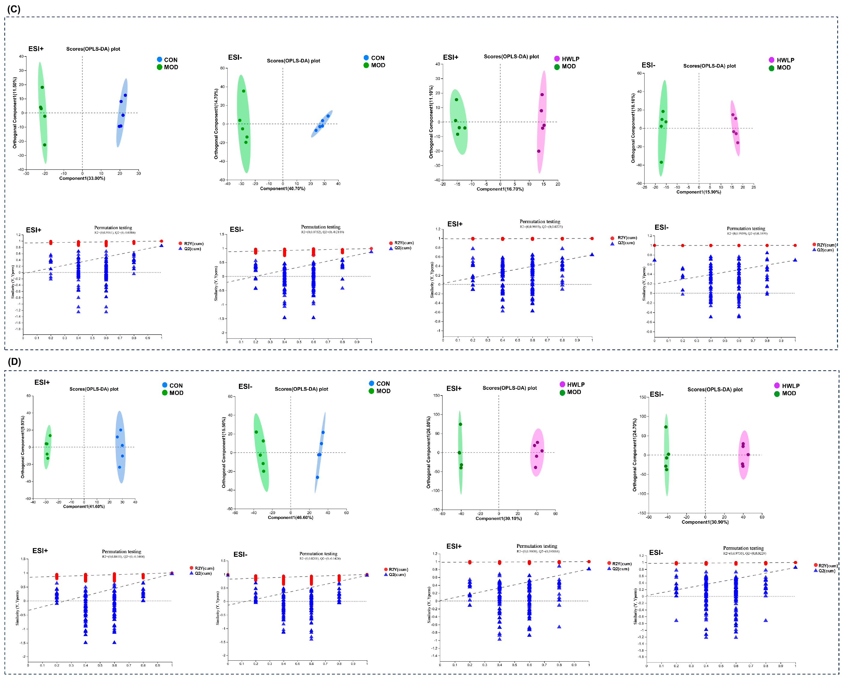
Untargeted metabolomics was employed to investigate the effects of WLP on the metabolic profiles of serum, liver, and urine in MASLD rats. PCA results reveal significant changes in metabolite composition among the CON, MOD, and WLP group (Figure 2A). To further evaluate pairwise differences in metabolic profiles between groups, OPLS-DA was conducted. This analysis showed distinct separation between the CON and MOD groups, as well as between the MOD and WLP groups, confirming that the three groups exhibited distinct metabolic profiles (Figure 2B–D). Additionally, a random permutation test (200 times) was performed for the OPLS-DA model. Model parameters demonstrated that R2Y was close to 1 and Q2 > 0.5 (Table 1), indicating that the OPLS-DA model had good explanatory and predictive abilities. In addition, the OPLS-DA models for serum ESI+ and liver tissue ESI− exhibited positive Q2 intercepts, which suggests a potential risk of overfitting. However, the overall distribution of Q2 values after random permutation was lower than that of the original model, suggesting high reliability of the models. Moreover, R2 values were greater than 0.9, indicating excellent explanatory power of the models for the data.
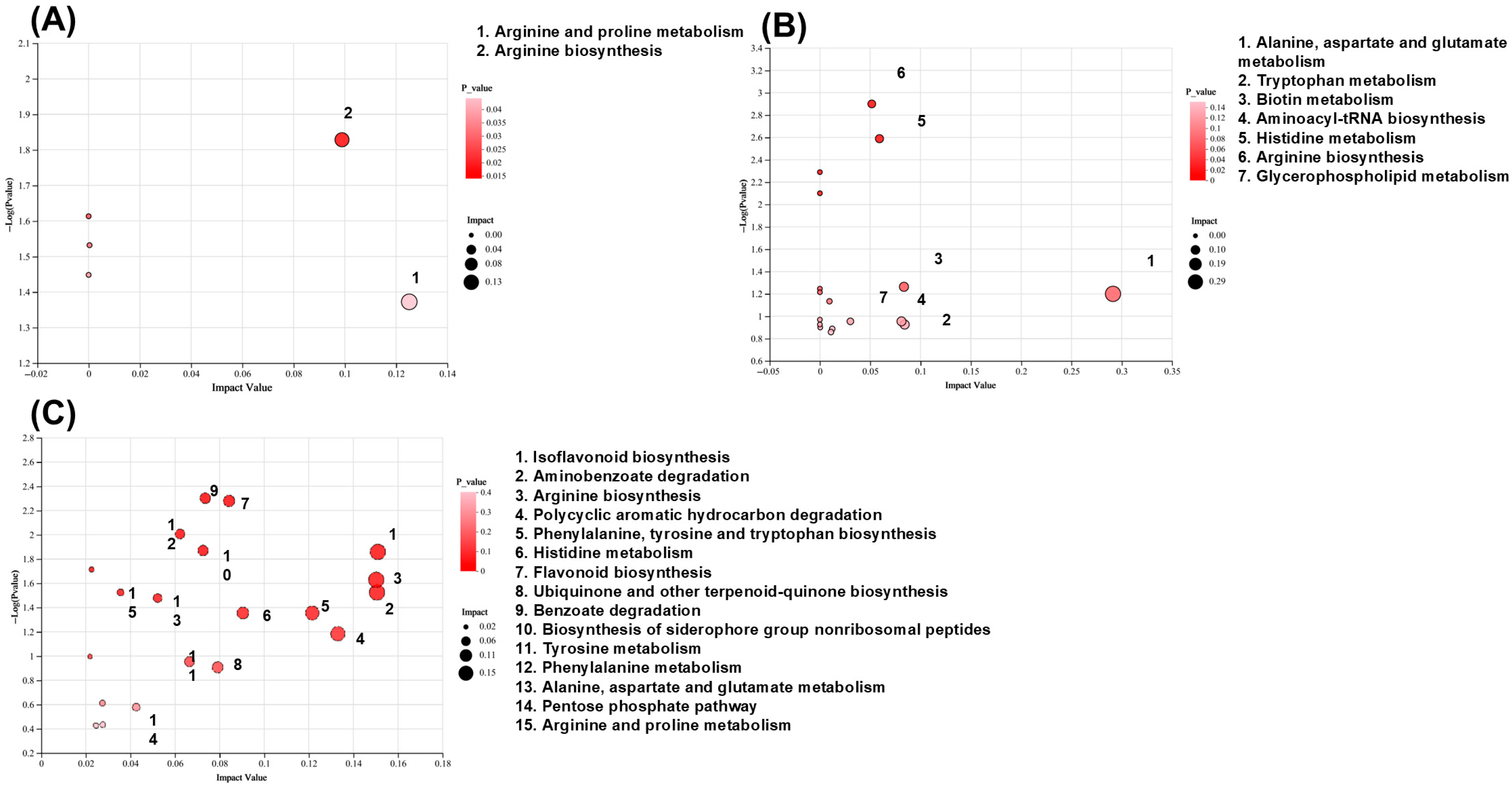
Differential metabolites were screened with the criteria of VIP > 1.0 and p < 0.05. After WLP treatment, the levels of 222 differential metabolites in MASLD rats were reversed, including 28 in serum (Table S1), 5 in liver tissues (Table S2), and 192 in urine (Table S3). Subsequently, pathway enrichment analysis was performed for these differential metabolites using the KEGG database. The results indicate that WLP regulated 19 metabolic pathways in MASLD rats, primarily involving amino acid, lipid, cofactor and vitamin metabolism, biosynthesis of other secondary metabolites, biodegradation and metabolism of xenobiotics, and translation-related pathways (Figure 3A–C).

2.3. WLP Improves the Gut Microbiota Dysbiosis of MASLD Rats

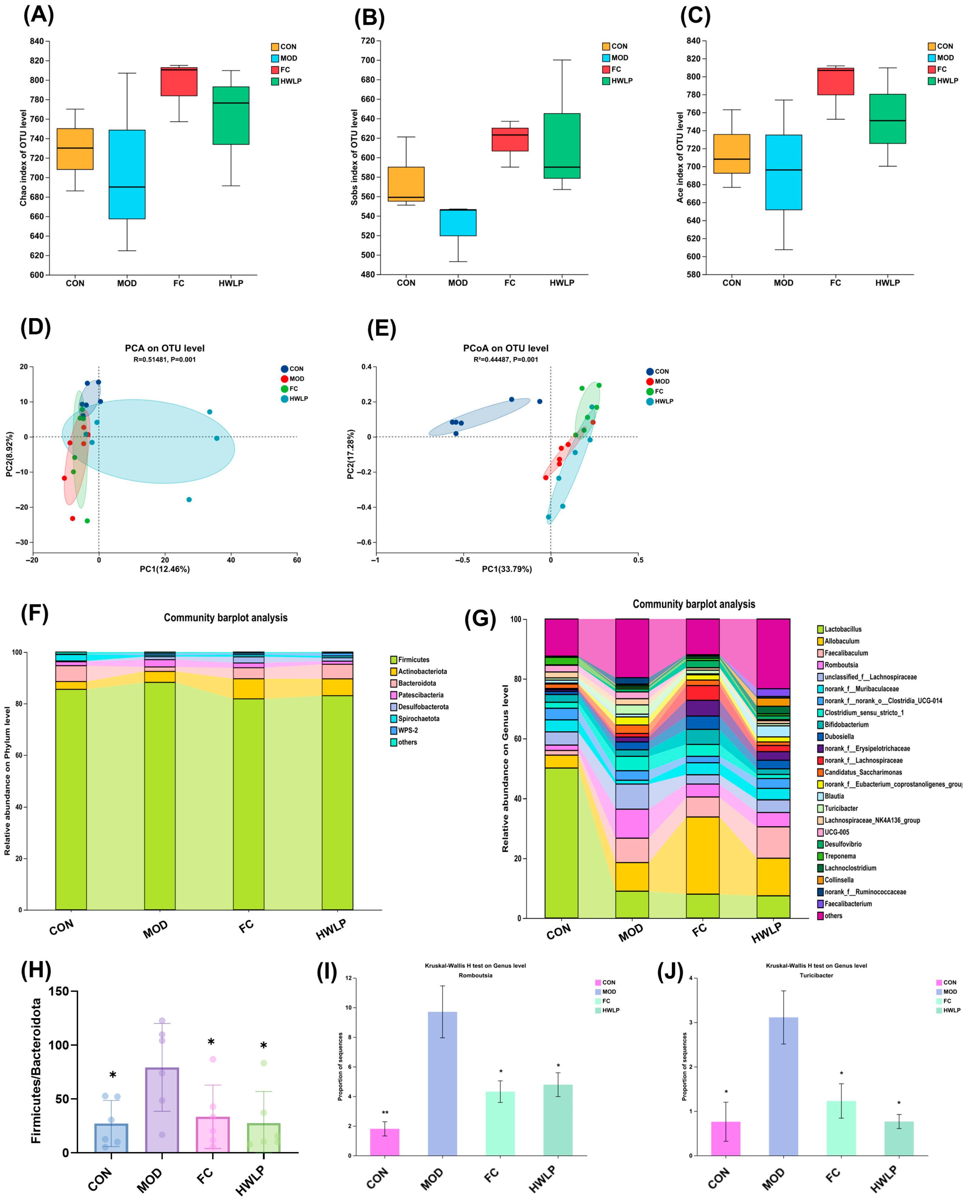
Alpha diversity analysis reveals that the Chao, Ace, and Sobs indices exhibited an increasing trend after WLP group treatment, though the difference did not reach statistical significance (Figure 4A–C). Further evaluation of the similarity or dissimilarity in the intestinal flora community structure was conducted using PCA and PCoA. Results showed a clear separation between the WLP and MOD group (Figure 4D,E), indicating that the gut microbiota structure dysbiosis in HFD-induced MASLD rats was ameliorated by WLP. Community bar charts were used to further analyze the species composition of each group across different taxonomic levels. At the phylum level, the intestinal flora of rats was predominantly composed of Firmicutes, Actinobacteriota, Bacteroidota, Patescibacteria, and Desulfobacterota (Figure 4F). Compared with the MOD group, WLP treatment resulted in a decrease in the relative abundance of Firmicutes, an increase in the relative abundance of Bacteroidota, and a significant reduction in the Firmicutes/Bacteroidota ratio (p < 0.05) (Figure 4H). At the genus level, WLP significantly decreased the relative abundances of Romboutsia and Turicibacter in the intestines of MASLD rats (p < 0.05) (Figure 4G,I–J). These findings indicate that WLP modified the species composition of intestinal flora in MASLD rats and modulated the homeostasis of the intestinal flora.

2.4. Correlation Between the Gut Microbiota, Pathological Indicators, and Differential Metabolites Regulated by WLP

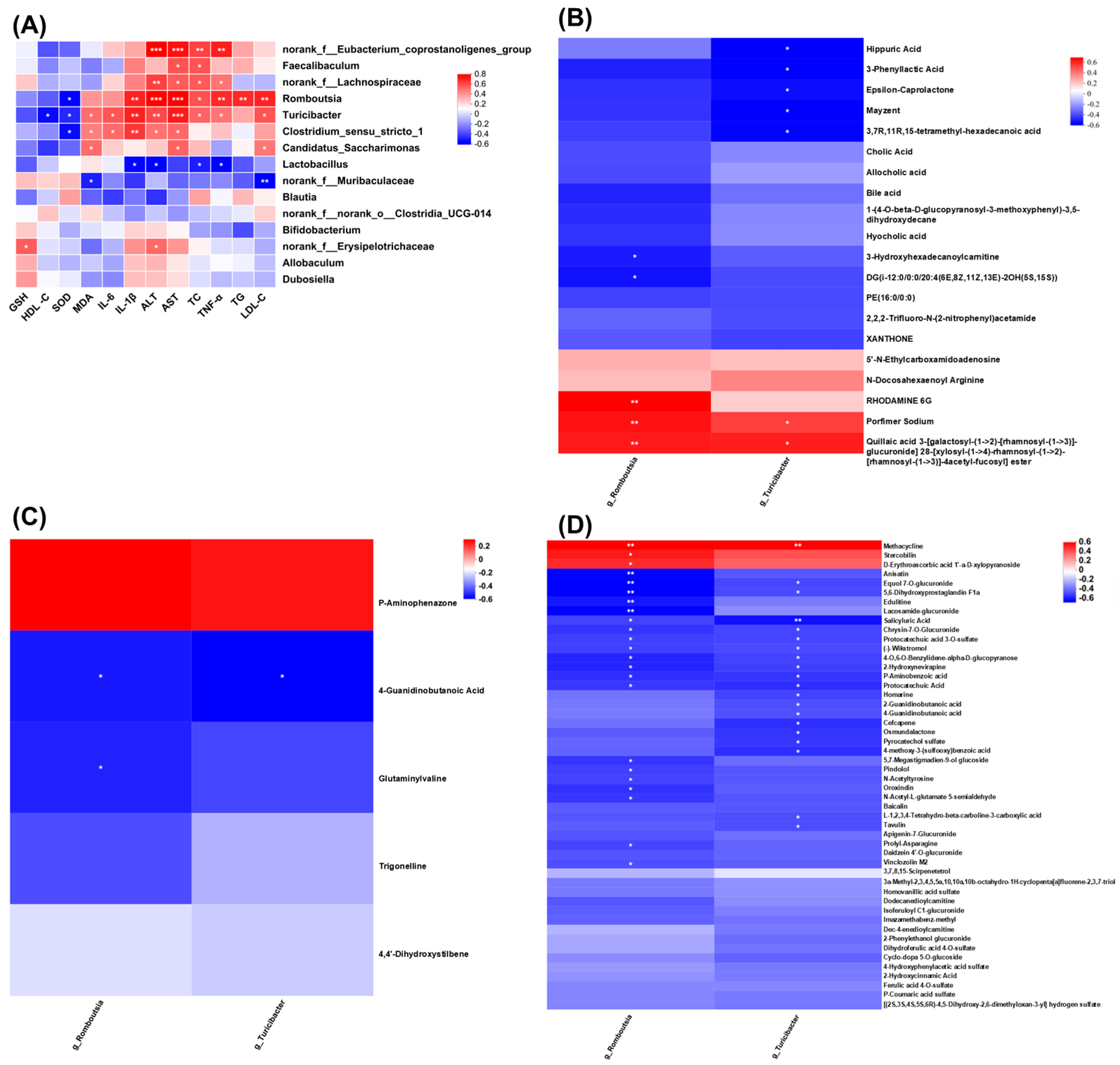
To further explore the potential association between WLP-regulated gut microbiota and MASLD, Spearman correlation analysis was performed between gut microbiota at the genus level and liver pathological indicators. As shown in Figure 5A, Romboutsia exhibited a significantly positive correlation with the levels of TC, TG, ALT, AST, LDL-C, IL-1β, and TNF-α (p < 0.05, p < 0.01, p < 0.001), while showing a significantly negative correlation with SOD levels (p < 0.05). Turicibacter had a significantly positive correlation with the levels of TC, ALT, AST, LDL-C, IL-1β, TNF-α, IL-6, and MDA (p < 0.05, p < 0.01, p < 0.001), and a significantly negative correlation with HDL-C and SOD levels (p < 0.05). These results suggest that Romboutsia and Turicibacter may be the key gut microbiota through which WLP restores gut microbiota homeostasis and alleviates MASLD.
To investigate the association between WLP-regulated endogenous metabolites and gut microbiota, Spearman correlation analysis was conducted between the differential metabolites and the gut microbiota at the genus level reversed by WLP. As shown in Figure 5B, Romboutsia showed a significant positive correlation with three serum differential metabolites, quillaic acid 3-[galactosyl-(1->2)-[rhamnosyl-(1->3)]-glucuronide]28-[xylosyl-(1->4)-rhamnosyl-(1->2)-[rhamnosyl-(1->3)]-4acetyl-fucosyl] ester, porfimer sodium, and rhodamine 6g (p< 0.01), and a significant negative correlation with DG(i-12:0/0:0/20:4(6E,8Z,11Z,13E)-2OH(5S,15S)) and 3-hydroxyhexadecanoylcarnitine (p < 0.05). For Turicibacter, it had a significant positive correlation with two serum differential metabolites, quillaic acid 3-[galactosyl-(1->2)-[rhamnosyl-(1->3)]-glucuronide]28-[xylosyl-(1->4)-rhamnosyl-(1->2)-[rhamnosyl-(1->3)]-4acetyl-fucosyl] ester and porfimer sodium (p < 0.05), as well as a significant negative correlation with 3,7R,11R,15-tetramethyl-hexadecanoic acid, mayzent, epsilon-caprolactone, hippuric acid, and 3-phenyllactic acid (p < 0.05).
As shown in Figure 5C, Romboutsia exhibited a significantly negative correlation with the liver differential metabolites 4-guanidinobutanoic acid and glutaminylvaline (p < 0.05). Turicibacter showed a significantly negative correlation with the liver differential metabolite 4-guanidinobutanoic acid (p < 0.05).
As shown in Figure 5D, Romboutsia exhibited a significantly positive correlation with the urine differential metabolites methacycline, stercobilin, and D-erythroascorbic acid 1′-α-D-xylopyranoside (p < 0.05, p < 0.01), while showing a significantly negative correlation with anisatin, equol 7-O-glucuronide, 5,6-dihydroxyprostaglandin F1α, edulitine, lacosamide-glucuronide, salicyluric acid, chrysin-7-O-Glucuronide, protocatechuic acid 3-O-sulfate, (-)-wikstromol, 4-O,6-O-benzylidene-α-D-glucopyranose, 2-hydroxynevirapine, P-aminobenzoic acid, protocatechuic acid, 5,7-megastigmadien-9-ol glucoside, pindolol, N-acetyltyrosine, oroxindin, N-acetyl-L-glutamate 5-semialdehyde, prolyl-Asparagine, and vinclozolin M2 (p < 0.05, p < 0.01). Turicibacter displayed a significantly positive correlation with the urine differential metabolite methacycline (p < 0.01), and a significantly negative correlation with equol 7-O-glucuronide, 5,6-dihydroxyprostaglandin F1α, salicyluric acid, chrysin-7-O-glucuronide, protocatechuic acid 3-O-sulfate, (-)-wikstromol, 4-O,6-O-benzylidene-α-D-glucopyranose, 2-hydroxynevirapine, P-aminobenzoic acid, protocatechuic acid, homarine, 2-guanidinobutanoic acid, 4-guanidinobutanoic acid, cefcapene, osmundalactone, pyrocatechol sulfate, 4-methoxy-3-(sulfooxy)benzoic acid, L-1,2,3,4-tetrahydro-β-carboline-3-carboxylic acid, and tavulin (p < 0.05, p < 0.01).
The above results indicate that WLP ameliorated HFD-induced MASLD by regulating the relative abundance of key gut microbiota and restoring the levels of related endogenous metabolites.

2.5. Potential Active Substances for WLP Against MASLD

A total of 31 WLP-derived components were identified in rat serum, with 8 components derived from AR, 2 from PS, 6 from PA, 12 from AMR, and 5 from CR. Among these 31 pharmacodynamic components, there were 12 triterpenoids, 6 sesquiterpenoids, 2 phenolic acids, 5 carboxylic and organic acids, 2 steroids, 1 fatty alcohol, 1 coumarin, 1 phenylpropanoic acid, and 1 glycoside (Table 2).
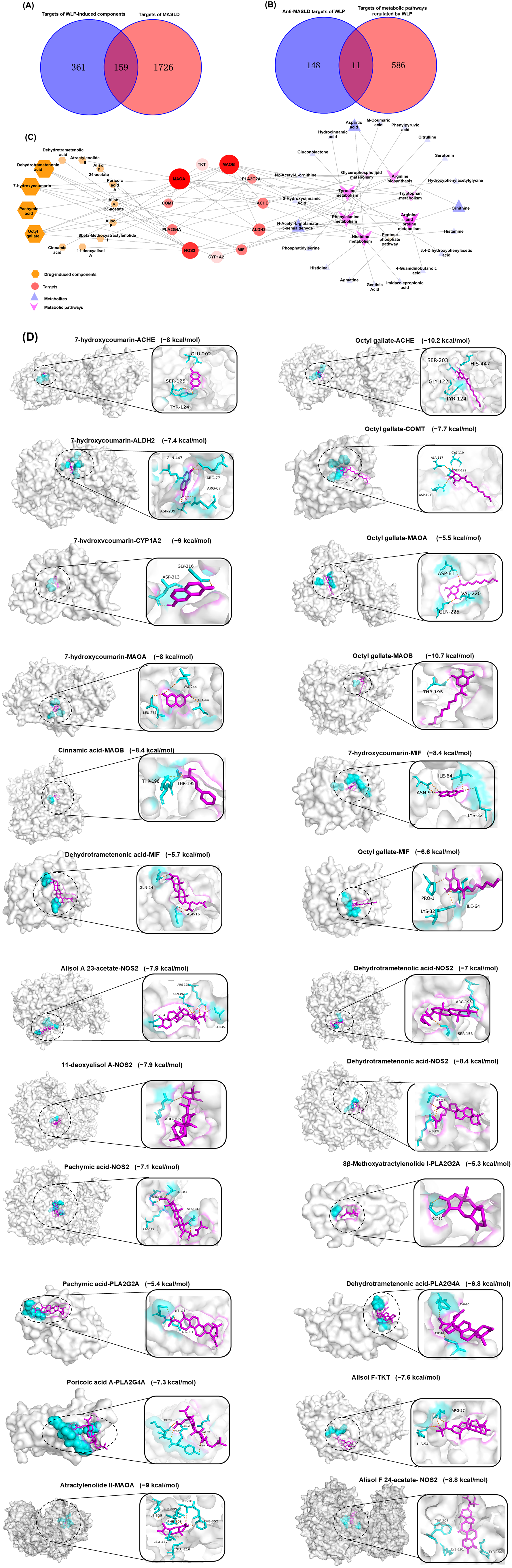
Subsequently, a network pharmacology analysis was performed on the MASLD targets and WLP-derived component targets. A total of 520 targets of WLP-derived components and 1885 MASLD-related targets were screened out. The number of overlapping targets between the two groups was 159, which are the potential targets of the WLP-derived components for anti-MASLD effect (Figure 6A). A total of 597 relevant targets were obtained from 19 metabolic pathways. These targets were subsequently overlapped with the anti-MASLD targets of the WLP-derived components, resulting in 11 targets, including TKT, NOS2, PLA2G4A, MIF, PLA2G2A, MAOA, MAOB, CYP1A2, ALDH2, ACHE, and COMT (Figure 6B).
By constructing the “component–target–metabolic pathway–metabolite” network via Cytoscape, version 3.10.1 (Figure 6C), it was found that 11 core targets were associated with 13 WLP-derived components. These components may be the key active substances for WLP to alleviate MASLD, including alisol F, alisol F 24-acetate, alisol A 23-acetate, 11-deoxyalisol A, dehydrotrametenolic acid, dehydrotrametenonic acid, pachymic acid, poricoic acid A, 7-hydroxycoumarin, octyl gallate, 8β-methoxyatractylenolide I, atractylenolide II, and cinnamic acid, with their relative contents being 1.31%, 11.04%, 2.29%, 32.14%, 10.97%, 0.62%, 2.04%, 1.34%, 4.60%, 7.71%, 2.44%, 21.99%, and 1.50%, respectively. Additionally, the core targets are involved in 8 metabolic pathways (arginine and proline, phenylalanine, histidine, tyrosine, tryptophan, and glycerophospholipid metabolism, arginine biosynthesis, and pentose phosphate pathway) and affect the levels of 20 metabolites (gluconolactone, N-acetyl-L-glutamate 5-semialdehyde, agmatine, 4-guanidinobutanoic acid, ornithine, citrulline, aspartic acid, N2-acetyl-L-ornithine, phosphatidylserine, gentisic acid, 3,4-dihydroxyphenylacetic acid, hydroxyphenylacetylglycine, phenylpyruvic acid, M-coumaric acid, hydrocinnamic acid, 2-hydroxycinnamic acid, histidinal, imidazolepropionic acid, histamine, and serotonin). Finally, molecular docking between the key active substances and the core targets shows that all binding energies were less than −5.0 kcal/mol (Table 3), suggesting that the 13 key active components derived from WLP exhibit favorable binding ability for the relevant targets, with the binding sites shown in Figure 6D.

2.6. Key Active Components Derived from WLP Alleviate FFA-Induced Steatosis in HepG2 Cells

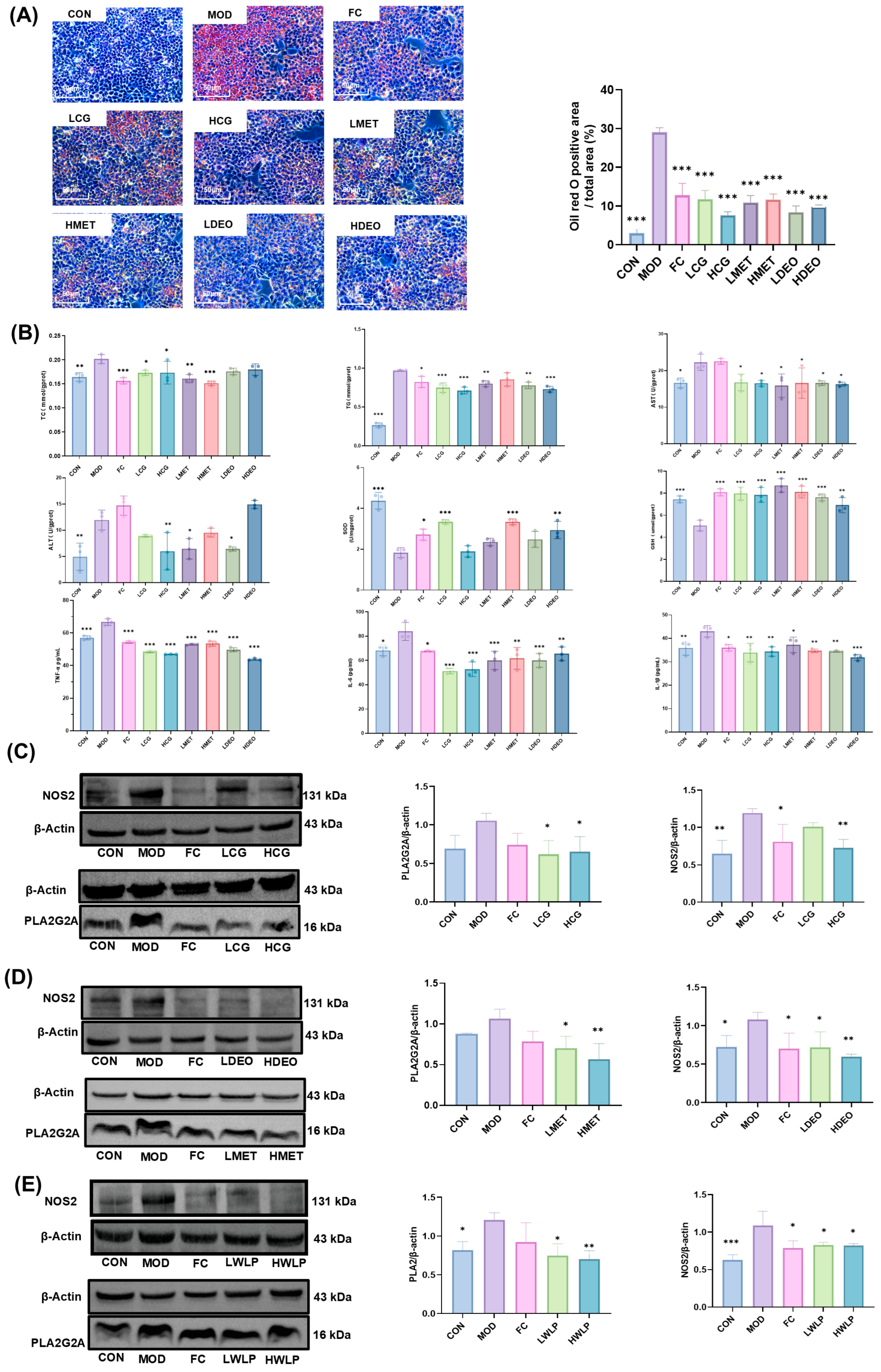
Among the 13 key active components, only DEO and MET have not been reported to exhibit anti-MASLD activity. Therefore, the anti-MASLD activity of DEO and MET was evaluated using FFA-induced HepG2 cells. Additionally, the in vitro anti-hepatic steatosis capacity of component group composed of 12 key active components (the reference standard of dehydrotrametenonic acid was unavailable and was not mixed into the component group) was assayed. Oil Red O staining results demonstrate that FFA treatment induced lipid accumulation in HepG2 cells, whereas treatment with CG, MET, and DEO significantly reduced intracellular lipid droplet accumulation (Figure 7A). In addition, CG intervention significantly decreased the intracellular levels of TC, TG, ALT, AST, TNF-α, IL-1β, and IL-6 (p < 0.05, p < 0.01, p < 0.001), while the activities of SOD and GSH (p < 0.001) were significantly increased; MET significantly decreased the intracellular levels of TC, TG, ALT, AST, TNF-α, IL-1β, and IL-6 (p < 0.05, p < 0.01, p < 0.001), while the levels of SOD and GSH were significantly increased (p < 0.001); and DEO intervention significantly lowered the levels of TG, AST, ALT, TNF-α, IL-1β, and IL-6 (p< 0.05, p < 0.01, p < 0.001), while the levels of SOD and GSH were significantly increased (p < 0.01, p < 0.001) (Figure 7B). These results indicate that CG, MET, and DEO ameliorated FFA-induced lipid accumulation, oxidative stress, and inflammation in HepG2 cells, thereby alleviating steatosis.
The regulatory effects of DEO and MET on the core targets NOS2 and PLA2G2A, respectively, were further verified by Western blot assay. CG treatment significantly down-regulated the expression levels of NOS2 and PLA2G2A in HepG2 cells (p < 0.05, p < 0.01) (Figure 7C), while DEO and MET intervention significantly down-regulated the expression of NOS2 and PLA2G2A in cells, respectively (p < 0.05, p < 0.01) (Figure 7D). In addition, after treating MASLD rats with WLP extract, the levels of NOS2 and PLA2G2A in liver tissues were significantly down-regulated (p < 0.05, p < 0.01) (Figure 7E). These results suggest that DEO and MET improved hepatocellular steatosis by regulating the protein expression of NOS2 and PLA2G2A, respectively.

3. Discussion

MASLD is a chronic liver disease characterized by excessive lipid accumulation in hepatocytes induced by metabolic dysfunction as its core pathological feature. Long-term high-calorie diet and lack of exercise have led to a continuous rise in the prevalence of MASLD, which is now the most common chronic liver disease worldwide [20]. Dysregulated lipid metabolism can lead to excessive accumulation of fatty acids in the liver, thereby inducing MASLD, often manifested as abnormal changes in lipid metabolism indicators such as TC, TG, LDL-C, HDL-C, ALT, and AST. Significantly, this study found that WLP alleviated hepatic lipid accumulation and hepatocyte pathological damage in HFD-fed MASLD rats; reduced the levels of TC, TG, LDL-C, ALT, and AST; increased the level of HDL-C; and regulated lipid metabolism disorders. In addition, oxidative stress is a key factor driving the initiation and progression of MASLD. Excessive lipid accumulation in hepatocytes can induce mitochondrial dysfunction, generate large amounts of reactive oxygen species (ROS), trigger lipid peroxidation, and impair hepatic cell membranes and organelles [21]. Furthermore, the progression of MASLD is often accompanied by the occurrence of inflammatory responses. Excessive lipid accumulation in hepatocytes can activate hepatic stellate cells and Kupffer cells to release pro-inflammatory factors, further damaging hepatocytes [22]. In this study, WLP was found to not only increase the hepatic levels of antioxidant molecules (GSH, SOD) and reduce MDA levels but also markedly suppress the expression of pro-inflammatory cytokines (TNF-α, IL-1β, and IL-6). These findings indicate that WLP ameliorated abnormal hepatic oxidative stress and inflammatory responses to mitigate HFD-induced MASLD.
Metabolomics technology enables the reflection of an organism’s physiological and pathological states through detecting dynamic changes in small-molecule metabolites within the organism. In recent years, this technology has played a crucial role in research on the modernization of TCM [23]. In this study, it was found that after intervention with WLP, 28, 5, and 192 potential differential metabolites in serum, liver, and urine could be reversed respectively, and 19 metabolic pathways were mainly regulated. Aberrant amino acid metabolism is a key driver of the pathogenesis of MASLD. Emerging evidence indicates that aberrant amino acid metabolism can trigger oxidative stress, thereby further exacerbating hepatocellular damage [24]. Citrulline, an amino acid with potential hepatoprotective effects, has been confirmed to significantly ameliorate hepatic steatosis [25]. Its precursor, ornithine, collaborates with arginine and proline in the body’s urea cycle, dynamically regulating arginine biosynthesis and proline metabolic homeostasis [26]. Additionally, the endogenous metabolites of arginine, 4-guanidinobutanoic acid and agmatine can exert hepatoprotective effects by modulating mitochondrial function and fatty acid oxidation [27,28]. In the present study, intervention with WLP was found to increase the levels of argininic acid, citrulline, ornithine, 4-guanidinobutanoic acid, and agmatine. Collectively, these findings indicate that WLP alleviates MASLD by restoring the imbalance in amino acid metabolism.
Dysregulation of lipid metabolic homeostasis is the core pathological mechanism underlying the development of MASLD. As the most abundant structural phospholipids in the body, phosphatidylcholine (PC) and phosphatidylethanolamine (PE) play a crucial role in maintaining the dynamic balance of hepatocellular lipids by regulating the glycerophospholipid metabolic pathway [29]. Specifically, studies have shown that PC participates in the assembly of TG and apolipoprotein B, promotes the synthesis of very-low-density lipoprotein (VLDL), and facilitates its conversion to HDL-C, thereby reducing hepatic TG accumulation [30]. In contrast, PE (16:0/0:0), a key precursor for PC synthesis, can regulate the homeostasis of energy metabolism by stabilizing mitochondrial membrane protein complexes [31]. Additionally, metabolites including bile acids, allocholic acid, and hyocholic acid are well-documented to regulate hepatic lipid metabolism [32,33]. Significantly, this study found that WLP intervention elevated the levels of PE(16:0/0:0), bile acids, allocholic acid, and hyocholic acid. These findings suggest that WLP alleviates MASLD by regulating hepatic lipid metabolism disorders.
Excessive carbohydrate intake is a key pathogenic factor in MASLD. Studies have shown that high carbohydrate load can promote de novo fatty acid synthesis, leading to abnormal accumulation of TG. It also produces excessive ROS and aggravates hepatocyte injury [34]. Gluconolactone, as a biosynthetic precursor of D-gluconate, plays a key role in glycolysis and the pentose phosphate pathway. Its downregulation would impair hepatic glucose metabolism function. In this study, it was found that the level of gluconolactone significantly increased after intervention with WLP.
Furthermore, cofactor and vitamin metabolism are important regulators in maintaining hepatic metabolic homeostasis. As a precursor substance for the biosynthesis of ubiquinones and terpenoquinones, 4-hydroxybenzoic acid can alleviate oxidative stress-induced hepatocellular injury by enhancing mitochondrial function [35,36]. Protocatechuic acid, a key intermediate in the synthesis of non-ribosomal peptide siderophores, can ameliorate the hepatic inflammatory microenvironment by inhibiting the release of pro-inflammatory factors [37]. WLP intervention significantly elevated the levels of 4-hydroxybenzoic acid and protocatechuic acid. Additionally, aspartic acid can reduce liver injury by modulating inflammatory cytokine levels. Its precursor, prolyl-asparagine, was significantly increased after WLP intervention. Finally, the biosynthesis of other secondary metabolites, as well as the biodegradation and metabolism of xenobiotics, play crucial roles in the pathological regulation of MASLD. Isoflavonoids such as daidzein and biochanin A can counteract MASLD by regulating hepatic lipogenesis [38,39]. Moreover, 4-hydroxybenzoic acid and protocatechuic acid, as degradation products of benzoic acid, reduce hepatic accumulation of lipotoxic metabolic intermediates by regulating the exogenous substance detoxification process mediated by cytochrome P450 enzymes [40]. WLP intervention increased the levels of daidzein, biochanin A, pinocembrin, 4-hydroxybenzoic acid, and protocatechuic acid. These findings suggest that WLP alleviated MASLD by modulating the levels of various endogenous metabolites and influencing multiple metabolic pathways.
Both the liver and intestine are important components of the digestive system, connected by the portal vein and interacting with each other. The intestine harbors a large number of intestinal flora; dysbiosis of intestinal flora can lead to disorders of bile acid metabolism, endotoxin translocation, and disturbances in metabolites, thereby promoting the progression of MASLD [41]. In this study, it was found that WLP significantly reduced the Firmicutes/Bacteroidota ratio and reduced the relative abundance of Romboutsia and Turicibacter in the intestines of MASLD rats. Furthermore, increased abundance of Romboutsia and Turicibacter has been reported in patients with MASLD [42,43]. Significantly, Romboutsia and Turicibacter showed a significant positive correlation with pathological indicators in the liver, such as TC, TG, ALT, AST, LDL-C, IL-1β, TNF-α, IL-6, and MDA.
Changes in the structure of gut microbiota can affect the levels of bacterial metabolites, thereby influencing MASLD. In this study, the results of correlation analysis between differential metabolites and key flora showed that 4-guanidinobutanoic acid, equol 7-O-glucuronide, (-)-wikstromol, and protocatechuic acid were significantly negatively correlated with the potential pathogenic bacteria Romboutsia and Turicibacter. Tercobilin was significantly positively correlated with Romboutsia, while hippuric acid and 3-phenyllactic acid were significantly negatively correlated with Turicibacter. Existing studies have found that 4-guanidinobutanoic acid, as an arginine metabolite, can prevent and treat MASLD by regulating mitochondrial dysfunction. Equol 7-O-glucuronide is a substrate for human hepatic β-glucuronidase and regulates carbohydrate metabolism in the body [44]. (-)-wikstromol, protocatechuic acid, and 3-phenyllactic acid, as intestinal metabolites, have been shown to alleviate oxidative stress, regulate intestinal microbial composition, and possess the potential to treat MASLD [45,46,47]. Additionally, increased levels of hippuric acid are negatively correlated with the prevalence of metabolic syndrome [48]. Stercobilin, on the other hand, is the terminal metabolite produced by the reduction in bilirubin by intestinal flora, and its increase can induce low-grade chronic inflammation in ob/ob mice [49]. The above results suggest that WLP may improve MASLD by regulating metabolite levels through key gut microbiota. Additionally, the differential metabolites significantly associated with gut microbiota also include porfimer sodium, rhodamine 6g, methacycline, D-erythroascorbic acid 1′-a-D-xylopyranoside, anisatin, 5,6-dihydroxyprostaglandin F1a, edulitine, lacosamide-glucuronide, salicyluric acid, chrysin-7-O-glucuronide, protocatechuic acid 3-O-sulfate, 4-O,6-O-benzylidene-alpha-D-glucopyranose, 2-hydroxynevirapine, P-aminobenzoic acid, 5,7-megastigmadien-9-ol glucoside, pindolol, oroxindin, N-acetyl-L-glutamate 5-semialdehyde, prolyl-asparagine, vinclozolin M2, glutaminylvaline, DG(i-12:0/0:0/20:4(6E,8Z,11Z,13E)-2OH(5S,15S)), 3-hydroxyhexadecanoylcarnitine, 3,7R,11R,15-tetramethyl-hexadecanoic acid, mayzent, epsilon-caprolactone, homarine, 2-guanidinobutanoic acid, cefcapene, osmundalactone, pyrocatechol sulfate, 4-methoxy-3-(sulfooxy)benzoic acid, L-1,2,3,4-Tetrahydro-beta-carboline-3-carboxylic acid, and tavulin. None of these differential metabolites have been reported to be associated with the pathogenesis of MASLD, which warrants further investigation.
Serum pharmacochemistry technology of TCM is a method that uses modern scientific technologies and approaches to identify the migrated components in serum after oral administration of TCM. In this study, a total of 31 WLP-derived components were identified in the serum of MASLD rats. Integrated analysis of serum pharmacochemistry combined with metabolomics identified 13 key WLP-derived components, which acted on 11 targets, regulating 8 metabolic pathways and 20 metabolites to exert anti-MASLD effects. Among them, alisol F, alisol F 24-acetate, pachymic acid, poricoic acid A, and octyl gallate possess anti-inflammatory activity and can alleviate MASLD [50,51,52,53,54,55,56,57,58]. Alisol A 23-acetate can improve abnormal lipid metabolism and oxidative stress in hepatocytes [59]. Atractylenolide II and dehydrotrametenolic acid can improve insulin resistance [60]. 7-hydroxycoumarin alleviates liver injury by activating Nrf2, and cinnamic acid improves MASLD by inhibiting hepatic lipogenesis and promoting fatty acid oxidation [61,62]. Moreover, we found that DEO and MET could alleviate hepatic steatosis. The component group composed of 12 key active components also prevented MASLD. This suggests that these 13 components are the key pharmacodynamic substances of WLP against MASLD by regulating 11 core targets, 8 metabolic pathways and 20 metabolites.
Additionally, DEO and MET have not been previously reported for treating MASLD. Further combined analysis revealed that DEO targets NOS2, while MET act on PLA2G2A. NOS2 (iNOS) is a key enzyme that catalyzes the production of NO from L-arginine. Excessive activation of NOS2 leads to overproduction of NO, which impairs hepatic mitochondrial function via nitrative stress, inhibits fatty acid oxidation, and results in massive accumulation of lipids such as triglycerides in the liver. Meanwhile, NO modifies insulin receptor substrates, suppresses insulin signaling, and exacerbates insulin resistance [63]. Furthermore, excess NO derived from NOS2 stimulates the secretion of pro inflammatory cytokines including TNF α, IL 1β, and IL 6, recruits macrophages and T cells into the liver, aggravates hepatic inflammation and injury, and drives the transition from simple steatosis to MASH [64]. Clinical studies demonstrate that hepatic NOS2 expression is markedly elevated in MAFLD patients compared with healthy controls, and is further upregulated in MASH and liver fibrosis relative to simple steatosis. PLA2G2A is a member of the phospholipase A2 family and catalyzes the hydrolysis of phosphatidylcholine, releasing free fatty acids and lysophospholipids, which exacerbate hepatic steatosis. Furthermore, upregulation of PLA2G2A promotes arachidonic acid production, which is further metabolized into inflammatory mediators such as leukotrienes. Previous studies have shown that PLA2G2A expression is increased in rats with liver fibrosis [65]. Thus, NOS2 and PLA2G2A may serve as promising biomarkers for evaluating disease severity and predicting prognosis in MASLD. This study innovatively found that DEO and MET downregulated the expression levels of NOS2 and PLA2G2A in HepG2 cells, respectively, thereby reducing lipid accumulation, oxidative stress damage, and inflammation. These results further describe that the 13 key components may act on their 11 core targets to relieve MASLD.
WLP is a very famous dampness-resolving formula that functions to invigorate the spleen and eliminate dampness. Notably, invigorating the spleen and resolving dampness represents an effective strategy for the prevention and treatment of MASLD; WLP can effectively treat the pathological mechanism of MASLD. In the present study, WLP significantly reduced the levels of TC, TG, and LDL-C in both serum and hepatic tissue and inhibited the release of inflammatory factors, demonstrating its dual functions of lipid lowering and anti-inflammatory effects. Further mechanistic investigations revealed that WLP exerted its therapeutic effects by modulating gut microbiota and 19 metabolic pathways, which were mainly attributed to its 13 medicinal components. Given the current clinical lack of drugs that can simultaneously regulate multiple lipid parameters including TC, TG, and LDL-C and inhibit inflammation, as well as agents capable of targeting multiple targets, gut microbiota, and diverse metabolic pathways for fatty liver disease, WLP shows great promise as a potential therapeutic agent for MASLD.
Currently, the efficacy of WLP in the treatment of MASLD has been scientifically evaluated in clinical settings. However, studies on the application of WLP in the management of MASLD complications remain limited, and relevant research regarding its adverse reactions in clinical practice is also scarce. In this study, although WLP exerted a dose-dependent effect against MASLD, only the high-dose group was included in mechanistic experiments. Further studies are therefore warranted to investigate the regulatory effects of low-dose WLP on the gut microbiota and differential metabolites, and to explore the dose-dependent manner of WLP. The precise causal relationships among molecules, gut microbiota, and metabolic pathways also deserve further validation. In addition, we established a high-fat diet-induced MASLD animal model. Whether WLP also exerts therapeutic effects on other types of fatty liver disease, such as malnutrition-induced fatty liver and toxin-induced fatty liver, remains unclear. Given the increasing prevalence of obesity among populations with long-term high-fat diets, future studies may perform clinical trials in such cohorts. Clinical trials can further validate the mechanisms by which WLP modulates gut microbiota and endogenous differential metabolites.

4. Materials and Methods

4.1. Reagents and Chemicals

Laboratory animal diet was purchased from SPF (Beijing) Biotechnology Co., Ltd. (Beijing, China). Fenofibrate capsules (FC) were purchased from French Libofoni Pharmaceutical Biological Technology Co., Ltd. (Paris, France). Commercial kits for the determination of total cholesterol (TC), triglyceride (TG), low-density lipoprotein cholesterol (LDL-C), high-density lipoprotein cholesterol (HDL-C), alanine transaminase (ALT), aspartate transaminase (AST), malondialdehyde (MDA), superoxide dismutase (SOD), and glutathione (GSH) were purchased from Nanjing Jiancheng Bioengineering Institute (Nanjing, China). Interleukin-6 (IL-6), interleukin-1β (IL-1β), and tumor necrosis factor-alpha (TNF-α) ELISA Kit were obtained from Jiangsu Meimian Industrial Co., Ltd. (Yancheng, China), and 4% Paraformaldehyde fix solution, hematoxylin and eosin (HE) stain kit, and Oil red O dye were purchased from Wuhan Servicebio Technology Co., Ltd. (Wuhan, China). The HepG2 human hepatocellular cell was purchased from the Cell Resource Center, Institute of Basic Medical Sciences, Chinese Academy of Medical Sciences. (Beijing, China). Dulbecco’s modified eagle medium (DMEN; No.6124477) was purchased from Grand Island Biological Company Co., Ltd. (Grand Island, NY, USA). Sodium oleate (No. S817542) was purchased from Shanghai Macklin Biochemical Co., Ltd. (Shanghai, China). Sodium palmitate (No. P9767) was purchased from Sigma-Aldrich (St. Louis, MO, USA). RIPA Lysis Buffer (No. 240007014) was purchased from Solarbio Co., Ltd. (Beijing, China). Tris Buffered Saline Tween (TBST; No. 240011012) was purchased from Solarbio Co., Ltd. (Beijing, China). Ultrasensitive ECL Detection Kit (No. 230718E01-02) was obtained from UElandy Co., Ltd. (Suzhou, China). PageRuler™ Plus Prestained Protein Ladder, 10 to 180 kDa (26616) was purchased from Thermo Fisher Scientific Co., Ltd. (Waltham, MA, USA). β-actin (No. 20536-1-AP) was purchased from Proteintech Group, Inc. (Wuhan, China). Goat Anti-Rabbit IgG(H + L) (No. Bs-0295G-HRP) and Anti-NOS2 (No. Bs-0162R) were purchased from Bioss Biotechnology Co., Ltd. (Beijing, China). Anti-PLA2G2A (No. TU721565S) was purchased from Abmart Inc. (Shanghai, China). Alisol F 24-acetate (No. TC0328-231129), octyl gallate (No. TC4627-240429), atractylenolide II (No. TC0526-240325), and cinnamic acid (No. TC0822-230522) were purchased from Sichuan Jingcui Tiancheng Pharmaceutical Technology Co., Ltd. (Chendu, China). Dehydrotrametenolic acid (No. QC90577), pachymic acid (No. QC99590), alisol A 23-acetate (No. QC92544), 11-deoxyalisol A (No. QC95661), poricoic acid A (No. QC92838), 7-hydroxycoumarin (No. QC97503), 8β-Methoxyatracty lenolide I (No. QC89314), and alisol F (No. QC92402) were purchased from Shanghai Qincheng Biotechnology Co., Ltd. (Shanghai, China). All the other reagents were of analytical grade or higher. The AR, PA, PS, AMR and CR samples were purchased from Kunming Luosiwan Chinese Medicinal Materials Market (Kunming, China) and authenticated by Professor Xingxin Yang at the Yunnan University of Chinese Medicine.

4.2. Preparation of WLP Extract

The powders of AR, PA, PS, AMR and CR twig were weighed and mixed at a 5:3:3:3:2 ratio, and the mixture was then decocted twice with a 10-fold volume of water for 1 h each time. Impurities were removed by filtration, and the filtrate was retained, concentrated and freeze-dried to produce a freeze-dried powder of WLP aqueous extract with a yield of 14%. The quantitative determination of cinnamaldehyde in WLP was conducted, and its content was found to be 0.36 mg/g (Figure S1, details are provided in the Supplementary Material).

4.3. Evaluation of WLP Against MASLD

4.3.1. Animals and Experimental Design

Thirty specific pathogen-free male Sprague-Dawley rats (aged 8 weeks, 180–200 g) were purchased from Beijing Speifu Biotechnology Co., Ltd. (Beijing, China; license No. SCXK, 2019-0010) and kept at 22 ± 2 °C with 60% relative humidity and 12 h light/dark cycle. After one week of adaptive feeding, 30 rats were randomly divided into the following five groups (n = 6): control group (CON, fed a normal basal diet), model group (MOD, fed a HFD for 12 weeks to induce MASLD), fenofibrate capsules group (FC, fed with HFD and intragastric administered 21 mg/kg/d of FC for 12 weeks), low-dose WLP group (LWLP, fed with HFD and intragastric administered 5.04 g/kg/d of WLP for 12 weeks), and high-dose WLP group (HWLP, fed with HFD and intragastric administered 20.16 g/kg/d of WLP for 12 weeks). The dosage of WLP was selected based on the maximum effective and safe doses from previous animal experiments and clinical studies [66,67]. The body weight of rats was monitored and recorded weekly. At the 12th week, urine was collected within 24 h in metabolic cages and stored at −80 °C. At the end of the experiment, the rats were anesthetized with pentobarbital sodium, and blood samples were collected from the abdominal aorta and centrifuged (3500 rpm, 15 min, 4 °C) to obtain serum. In addition, the liver and colon contents of the rats were collected for further analysis. All experimental animal manipulations in this study were performed in accordance with the ethical guidelines of the Experimental Animal Ethics Committee of Yunnan University of Chinese Medicine (Approval No. R-062022135).

4.3.2. Histological Analysis

Liver tissues were fixed in 4% paraformaldehyde for 24 h, washed with PBS, dehydrated in gradient ethanol, embedded in paraffin, and sliced into sections, and after hematoxylin-eosin (HE) staining, the cellular morphological changes in rat liver tissue were observed under a light microscope (Olympus Corporation, Tokyo, Japan). Furthermore, liver tissues were embedded in OTC embedding medium, frozen at −20 °C for 30 min, and sectioned at a thickness of 8 μm. The sections were rinsed with 60% isopropanol, stained with Oil Red O staining solution, and subsequently observed under an optical microscope (Olympus Corporation, Tokyo, Japan) to assess lipid accumulation in rat liver tissues. Oil Red O-positive areas and necrotic areas in HE-stained sections were quantified using ImageJ software.

4.3.3. Detection of Biochemical Indicators and Cytokines

The levels of TC, TG, HDL-C, LDL-C, AST, ALT, SOD, MDA, GSH, TNF-α, IL-6, and IL-1β were detected by biochemical kits according to the manufacturer’s instructions and protocols. Data were acquired using a SpectraMax Plus 384 microplate reader (Molecular Devices, Sunnyvale, CA, USA).

4.4. Metabolomic Analysis

4.4.1. Sample Preparation

Serum samples of 100 μL were collected from each of the CON, MOD, FC, and HWLP groups, and each sample was mixed with 400 μL extraction solution (acetonitrile: methanol = 1:1). After vortex for 30 s and ultrasonic extraction for 30 min (4 °C, 40 KHz), the samples were centrifuged (12,000 rpm, 15 min, 4 °C). The supernatants were collected, dried under nitrogen, and reconstituted with 100 µL of reconstitution solution (acetonitrile: water = 1:1) prior to analysis. Urine samples were processed in the same manner.
An amount of 50 mg of liver samples from the CON, MOD, FC, and HWLP groups was weighed respectively, and each sample was mixed with 400 μL of extraction solution (methanol: water = 4:1). The samples were ground in a tissue grinder for 6 min (−10 °C, 50 Hz), ultrasonicated for 30 min (5 °C, 40 kHz), and then centrifuged (12,000 rpm, 15 min, 4 °C). The supernatant was collected, dried with nitrogen gas, redissolved in 100 µL of a solvent mixture (acetonitrile:water = 1:1), and then analyzed.
All samples were mixed in equal volume to prepare quality control (QC) samples. During the detection process, one QC sample was run every 5 test samples to evaluate the repeatability of the analytical procedure.

4.4.2. LC-MS/MS Analyses

LC-MS/MS analyses were executed on an Thermo UHPLC-Q Exactive system (Thermo Scientific, Waltham, MA, USA) and a ACQUITYHSS T3 column (1.8 μm, 100 mm × 2.1 mm, Waters, Waltham, MA, USA). The mobile phase comprised (A) 0.1% formic acid in water:acetonitrile (95:5, v/v) and (B) 0.1% formic acid inacetonitrile: isopropanol:water (47.5:47.5:5, v/v). The following elution order was adopted: 0–3 min, 5% B→20% B; 3–4.5 min, 20% B→35% B; 4.5–5 min, 35% B→100% B; 5–6.3 min, 100% B; 6.3–6.4 min, 100% B→0% B; 6.4–8 min, 0% B. The sample injection volume was 3 μL, with a flow rate of 0.4 mL/min. Mass spectral signals of the samples were acquired in both positive and negative ion scanning modes using a Thermo UHPLC-Q Exactive Mass Spectrometer equipped with an electrospray ionization (ESI) source, and the scan mass range was 70–1050 m/z. The spray voltage was 3500 V for the positive ion mode and 2800 V for the negative ion mode. The sheath gas flow rate was 40 psi and the auxiliary gas flow rate was 10 psi. The collision energy was set to a cyclic collision energy of 20–40–60 V.

4.4.3. Data Processing

The raw LC-MS/MS data were preprocessed using Progenesis QI software (Waters Corporation, Milford, MA, USA, version v3.0). In addition, the metabolites were identified using public databases HMDB (accessed on 25 April 2023, https://hmdb.ca/), Metlin (accessed on 25 April 2023, https://metlin.scripps.edu/auth-login.html) and Majorbio. Principal component analysis (PCA) and orthogonal least partial squares discriminant analysis (OPLS-DA) were performed using the R package “ropls” (Version 1.6.2). Significant differential metabolites were identified based on variable importance in projection (VIP) values from the OPLS-DA model and raw p-values from Student’s t-test; to control false positives caused by multiple testing, the raw p-values were corrected using the Benjamini–Hochberg (BH) method, with VIP > 1 and FDR-adjusted p < 0.05 as the screening criteria. Significant differential metabolites were identified based on the variable weight value (VIP) obtained from the OPLS-DA model and Student’s t-test p value (VIP > 1, p < 0.05). KEGG (accessed on 25 April 2023, https://www.kegg.jp/kegg/pathway.html) and MetaboAnalyst6.0 (accessed on 25 April 2023, https://www.metaboanalyst.ca/) databases were used to further enrich and analyze the pathways involved in differential metabolites.

4.5. 16S rRNA Gene Sequencing Analysis

Total genomic DNA was extracted from rat colonic content samples of the CON, MOD, and HWLP groups using the PF Mag-Bind Stool DNA Kit (Omega, Norcross, GA, USA). The concentration and purity of the DNA were determined by 1% agarose gel electrophoresis. The V3–V4 region of the 16S rRNA gene was amplified by polymerase chain reaction (PCR) using primers [338F (5′-ACTCCTACGGGAGGCAGCAG-3′) and 806R (5′-GGACTACHVGGGTWTCTAAT-3′)]. The PCR product was extracted from 2% agarose gel and purified. Purified PCR products were subjected to library construction using the NEXTFLEX Rapid DNA-Seq Kit (Bioo Scientific, Austin, TX, USA) and finally sequenced on the Illumina PE300/PE250 platform (Illumina, San Diego, CA, USA). Data were clustered into different Operational Taxonomic Units (OTUs) with a 97% similarity threshold using UPARSE software (accessed on 24 April 2023, https://www.drive5.com/uparse/index.html, version 11) for cluster analysis. For each OTU, the sequence with the highest abundance was chosen as the representative sequence. After manual filtration of the OTU table, the 16S rRNA gene sequences of each sample were rarefied to 20,000 reads to reduce the impact of sequencing depth on alpha and beta diversity analyses. Taxonomic annotation of OTUs was performed using the RDP Classifier version 2.2 with a confidence threshold of 70%. The richness and diversity of microorganisms were evaluated using Alpha diversity analysis, such as Chao 1, Ace, and Sobs indices. Differences in microbial community structures among sample groups were examined through Beta diversity analysis, including PCA and Principal coordinate analysis (PCoA).

4.6. Correlation Analysis Between Gut Microbiota, Pathological Indicators, and Differential Metabolites

Based on Spearman correlation analysis (|r| > 0.6, p < 0.05), the correlations between gut microbiota, pathological indicators, and differential metabolites were analyzed.

4.7. Identification of WLP-Derived Components in Serum from MASLD Rats

4.7.1. Sample Processing

A 1.2 mL amount of MOD, HWLP serum was mixed with 3.6 mL of acetonitrile, vortexed for 30 s, allowed to stand for 20 min at 4 °C, and centrifuged (12,000 rpm, 15 min, 4 °C). The supernatant was collected, gently blow-dried under nitrogen gas, then redissolved in 200 μL of an acetonitrile–water mixture (50:50, v/v) and filtered through a 0.22 μm microporous filter membrane for LC-MS analysis.

4.7.2. LC-MS/MS Analysis

Agilent 1290 infinity II UPLC system (Agilent Technologies, Santa Clara, CA, USA) was used for the analysis. Elution was performed using the Waters Acquity UPLC HSS T3 column (100 mm × 2.1 mm, 1.8 μm, column temperature 30 ◦C, Waters, Milford, MA, USA) at a constant flow rate of 0.2 mL/min. The injection volume was 5 μL. The mobile phase was acetonitrile (A) with 0.1% formic acid water (B). Gradient elution conditions were as follows: 0–5.0 min, 5% A; 5.0–10.0 min, 20% A; 10.0–15.0 min, 40% A; 15.0–20.0 min, 50% A; 20.0–25.0 min, 60% A; 25.0–30.0 min, 70% A; 30. 0–35.0 min, 80% A; 35.0–40.0 min, 90% A; 40.0–41.0 min, 100% A; and 41.0 min, 5% A.
Mass spectrometry was performed by the Agilent G6545 Q/TOF mass spectrometer equipped with ESI (Agilent Technologies, Santa Clara, CA, USA). The positive and negative ion scanning was used to collect the sample quality spectrum information, and the scan mass range was 50–1700 m/z. The spray voltage was 3.5 kV for the positive ion mode and 2.8 kV for the negative ion mode. The drying temperature was set at 320 °C and the flow rate was 8 L/min. The sheath temperature was 350 °C and the flow rate was 11 L/min. The collision energies were set to 35 and 40 eV.

4.7.3. Data Analysis

Qualitative Analysis B.06.00 software was used to process the UPLC-Q/TOF MS data. First, the mass spectral information of the WLP treatment group was compared with that of the MOD group to eliminate interference from endogenous substances in the serum. Subsequently, the molecular weight, MS/MS data, and relative retention time of AR, PA, PS, AMR, and CR components in the reported studies were used to identify the structures of drug-derived compounds in the serum samples of rats.

4.8. Integrated Analysis of Drug-Derived Components and Metabolomics

The targets of WLP-derived components in serum were predicted by PubChem (accessed on 17 April 2024, https://pubchem.ncbi.nlm.nih.gov/) and SwissTargetPrediction (accessed on 17 April 2024, https://swisstargetprediction.ch/). MASLD-related genes were found by GeneCards (accessed on 17 April 2024, https://www.genecards.org/) and OMIM Gene Map (accessed on 17 April 2024, https://www.omim.org/) databases. In addition, related targets of WLP in regulating metabolic pathways were obtained via KEGG (accessed on 22 April 2024, https://www.genome.jp/kegg/pathway.html) website. Venny 2.1.0 (accessed on 22 April 2024, https://bioinfogp.cnb.csic.es/tools/venny/) software was used to obtain the overlapping targets of WLP-derived components, MASLD, and metabolic pathways. Finally, cytoscape was used to construct the active ingredient–target–metabolic pathway–metabolite network to determine the key active components, targets and pathways of WLP alleviating MASLD.

4.9. Molecular Docking of WLP-Derived Key Active Components with Its Core Targets

The structures of key active components from WLP were downloaded from the Pubchem (accessed on 22 April 2024, http://pubchem.ncbi.nlm.nih.gov/) website and converted to PDB format using Open Babel 3.0.1 software. Overlapping proteins were searched in the Uniprot (accessed on 22 April 2024, https://www.uniprot.org/uniprotkb) database and their crystal structures were downloaded in the Protein Data Bank (accessed on 22 April 2024, https://www.rcsb.org/). The receptor proteins were treated with Pymol software (version 2.5) to remove water molecules, demetal ions and hydrogenation using AutoDockTools (version 1.5.7). Finally, AutoDock Vina was used to dock the receptor proteins with the active components, the binding energy was calculated, and the docking results were visualized by Pymol.

4.10. Evaluation of WLP-Derived Key Active Components Against MASLD via Cell Experiments

HepG2 cells were cultured in DMEM containing 10% fetal bovine serum and 1% penicillin-streptomycin and placed in a 37 °C, 5% CO2 cell incubator. HepG2 cells were treated with 1 mM free fatty acid (FFA; oleic acid: palmitic acid = 2:1) for 24 h to establish a steatosis model when the cells were in the logarithmic phase of growth. The HepG2 cells were treated with 5 and 20 μg/mL component group (CG; consisting of AF, AF-24, DMA, PA, AA-23A, DEO, PAA, HYD, OG, MET, AL-II and CA in the ratio 1:8.42:8.37:1.56:1.75:24.53:1.02:3.51:5.88:1.86:16.78:1.14; their concentration ratios were based on the ratio of chromatographic peak areas of the WLP-derived components in the serum), 10 and 40 μM of monomers (MET, DEO), and FC (150 μM) for 24 h. Finally, lipid accumulation in cells was observed through Oil Red O staining, and the intracellular levels of TC, TG, ALT, AST, GSH, SOD, IL-1β, IL-6 and TNF-α were detected.

4.11. Detection of NOS2 and PLA2G2A Expression by Western Blotting

Livers from rats and HepG2 cell samples were lysed using RIPA lysis buffer containing protease inhibitors. After centrifuging the suspension, the supernatant was collected. The protein concentration was determined using the BCA method. Equal amounts of protein extracts were mixed with loading buffer and denatured (100 °C, 10 min). Then, the protein samples were electrophoresed on a 10% SDS-PAGE gel and transferred to a polyvinylidene fluoride membrane, which was then placed in blocking solution (5% skimmed milk) for blocking for 1 h. The membrane was incubated with primary antibodies (anti-iNOS and anti-PLA2G2A) at 4 °C overnight, followed by incubation with Goat Anti-Rabbit secondary antibody at room temperature for 1 h. Finally, images were captured using a fully automatic chemiluminescence image analysis system (Tanon, Shanghai, China), and the results were analyzed using Image J software (Image J Software, version 1.54f, Bethesda, MD, USA).

4.12. Statistical Analysis

Statistical analyses were performed using GraphPad Prism 9.0 (GraphPad Software, version 9.5.1, San Diego, CA, USA), and the results were expressed as mean ± standard deviation. For comparisons between groups, one-way ANOVA or Wilcoxon rank-sum test was used, and p < 0.05 was considered statistically significant.

5. Conclusions

WLP improved liver tissue morphology and lipid accumulation in MASLD rats, as well as regulated hepatic lipid metabolism, oxidative stress, and inflammation. Its mechanism may be associated with WLP reversing the levels of 222 endogenous metabolites in MASLD rats, regulating 19 metabolic pathways, reducing the abundance of potential pathogenic bacteria Romboutsia and Turicibacter, and restoring the structure of gut microbiota. Additionally, 31 WLP-derived components were identified in rat serum. Thirteen of these were the key active components of WLP against MASLD, which can act on 11 targets and regulate 8 metabolic pathways and 20 differential metabolites. Additionally, DEO and MET downregulated the expression of NOS2 and PLA2G2A in cells, respectively, and alleviated hepatic steatosis.

Supplementary Materials

The following supporting information can be downloaded at https://www.mdpi.com/article/10.3390/ph19040557/s1, Figure S1: Quantity control of WLP extract; Table S1: Differential metabolites in rat serum; Table S2: Differential metabolites in rat liver; Table S3: Differential metabolites in rat urine.

Author Contributions

Methodology, H.Y.; software, P.-Q.W.; formal analysis, S.-C.H.; investigation, H.Y.; data curation, P.-Q.W. and Y.-X.T.; writing—original draft preparation, H.Y. and Y.-M.T.; writing—review and editing, H.Y., H.-Q.Y., M.Z., M.L., J.Y. and X.-X.Y.; visualization, Y.-M.T. and S.-C.H.; project administration, X.-X.Y.; funding acquisition, X.-X.Y. All authors have read and agreed to the published version of the manuscript.

Funding

This research was funded by grants from the National Natural Science Foundation of China (82560801 and 82104381), Yunnan Fundamental Research Projects (202505AF350032, 202407AB110018, 202201AW070016, and 202301AS070070), and Yunnan Xingdian Talent Plan Innovation Team Project (202505AS350014).

Institutional Review Board Statement

Approval was obtained from the Ethical Committee on Animal Care and Experimentation of the Yunnan University of Chinese Medicine (R-062022135, 23 September 2022).

Informed Consent Statement

Not applicable.

Data Availability Statement

The original contributions presented in the study are included in the article/Supplementary Material; further inquiries can be directed to the corresponding authors.

Acknowledgments

We would like to thank all participants.

Conflicts of Interest

The authors declare no conflicts of interest associated with this manuscript.

Abbreviations

AA-23Aalisol A 23-acetate
AFalisol F
AF-24alisol F 24-acetate
AL-IIatractylenolide II
ALTalanine transaminase
AMRatractylodis macrocephalae rhizoma
ARAlismatis Rhizoma
ASTaspartate transaminase
BCAbicinchoninic acid
CAcinnamic acid
CGcomponent group
CONcontrol group
CRCinnamomi Ramulus
DEO11-deoxyalisol A
DMAdehydrotrametenolic acid
DMEMDulbecco’s modified eagle medium
ESIelectrospray ionization
FCfenofibrate capsules group
FFAfatty acid
GSHglutathione
HEhematoxylin-eosin
HDL-Chigh density lipoprotein-C
HWLPhigh-dose WLP group
HYD7-hydroxycoumarin
IL-1βinterleukin 1β
IL-6interleukin 6
LDL-Clow-density lipoprotein-C
LWLPlow-dose WLP group
MASLDmetabolic dysfunction-associated steatotic liver disease
MDAmalondialdehyde
MET8β-Methoxyatractylenolide I
MODmodel group
NOS2nitric oxide synthase 2
OGoctyl gallate
PLS-DAorthogonal least partial squares discriminant analysis
OTUsoperational taxonomic units
PApachymic acid
PAAporicoic acid A
PBSphosphate-buffered saline
PCphosphatidylcholine
PCAprincipal component analysis
PCoAprincipal coordinate analysis
PCRpolymerase chain reaction
PEphosphatidylethanolamine
PMporiae mushroom
PLA2G2Aphospholipase A2 group IIA
PSPolyporus
QCquality control
SODsuperoxide dismutase
TCtotal cholesterol
TCMtraditional Chinese medicine
TGtriglyceride
TNF-αtumor necrosis factor-α
VLDLvery low-density lipoprotein
WLPWuling powder

References

  1. Hsu, C.L.; Loomba, R. From NAFLD to MASLD: Implications of the new nomenclature for preclinical and clinical research. Nat. Metab. 2024, 6, 600–602. [Google Scholar] [CrossRef] [Scilit]
  2. Younossi, Z.M.; Kalligeros, M.; Henry, L. Epidemiology of metabolic dysfunction-associated steatotic liver disease. Clin. Mol. Hepatol. 2025, 31, S32–S50. [Google Scholar] [CrossRef] [Scilit]
  3. Tacke, F.; Horn, P.; Wong, V.W.-S.; Ratziu, V.; Bugianesi, E.; Francque, S.; Zelber-Sagi, S.; Valenti, L.; Roden, M.; Schick, F.; et al. EASL-EASD-EASO Clinical Practice Guidelines on the management of metabolic dysfunction-associated steatotic liver disease (MASLD). J. Hepatol. 2024, 81, 492–542. [Google Scholar] [CrossRef] [Scilit]
  4. Kobuchi, S.; Sugiyama, D.; Iima, A.; Obuchi, A.; Osaka, A.; Doi, A.; Ueta, H.; Yokoyama, S.; Hosomi, K.; Takada, M.; et al. Data Mining of the FDA Adverse Event Reporting System and Animal Experiments for Assessment of Rhabdomyolysis Risk Associated with Lipid-lowering Drugs. Int. J. Med. Sci. 2025, 22, 1485–1492. [Google Scholar] [CrossRef] [Scilit]
  5. Gao, F.M.; Ali, A.S.; Bellomo, R.; Gaca, M.; Lecamwasam, A.; Churilov, L.; Ekinci, E.I. A Systematic Review and Meta-analysis on the Safety and Efficacy of Sodium-Glucose Cotransporter 2 Inhibitor Use in Hospitalized Patients. Diabetes Care 2024, 47, 2275–2290. [Google Scholar] [CrossRef] [Scilit]
  6. Harrison, S.A.; Bedossa, P.; Guy, C.D.; Schattenberg, J.M.; Loomba, R.; Taub, R.; Labriola, D.; Moussa, S.E.; Neff, G.W.; Rinella, M.E.; et al. A Phase 3, Randomized, Controlled Trial of Resmetirom in NASH with Liver Fibrosis. N. Engl. J. Med. 2024, 390, 497–509. [Google Scholar] [CrossRef] [Scilit]
  7. Alkhouri, N. Phase 3 ESSENCE Trial: Semaglutide in Metabolic Dysfunction-Associated Steatohepatitis. Gastroenterol. Hepatol. 2024, 20, 6–7. [Google Scholar]
  8. Li, Y.; Yang, P.; Ye, J.; Xu, Q.; Wu, J.; Wang, Y. Updated mechanisms of MASLD pathogenesis. Lipids Health Dis. 2024, 23, 117. [Google Scholar] [CrossRef] [Scilit] [PubMed]
  9. Pan, M.; Deng, Y.; Qiu, Y.; Pi, D.; Zheng, C.; Liang, Z.; Zhen, J.; Fan, W.; Song, Q.; Pan, J.; et al. Shenling Baizhu powder alleviates non-alcoholic fatty liver disease by modulating autophagy and energy metabolism in high-fat diet-induced rats. Phytomedicine 2024, 130, 155712. [Google Scholar] [CrossRef] [Scilit] [PubMed]
  10. Zhou, Y.; Dai, Z.; Deng, K.; Wang, Y.; Ying, J.; Chu, D.; Zhou, J.; Tang, C. Eight Zhes Decoction ameliorates the lipid dysfunction of nonalcoholic fatty liver disease using integrated lipidomics, network pharmacology and pharmacokinetics. J. Pharm. Anal. 2023, 13, 1058–1069. [Google Scholar] [CrossRef] [Scilit]
  11. Zang, Y.; Li, K.; Lai, W.; Wang, Y.; Zheng, X.; Xu, X.; Lou, Z. Effects of Wuling Powder on Mice with Hyperlipidemia. J. Zhejiang Chin. Med. Univ. 2024, 48, 1482–1491. [Google Scholar]
  12. Luo, J.; Jin, X.; Cui, M.; He, H.; Liao, J.; Wen, W.; Li, W.; Cao, Y. Wulingsan alleviates metabolic dysfunction-associated steatotic liver disease through regulating gut microbiota-bile acid axis. Prostaglandins Other Lipid Mediat. 2026, 183, 107067. [Google Scholar] [CrossRef] [Scilit]
  13. Cai, Z.; Ma, Y.; Xiao, S. Study on the protecting effect of Wulingsan Decoction on glucose and lipid metabolism and myocardial injury in type 2 diabetic mice. Mod. J. Integr. Tradit. Chin. West. Med. 2022, 31, 474–479. [Google Scholar]
  14. Pi, D.; Liang, Z.; Pan, M.; Zhen, J.; Zheng, C.; Pan, J.; Fan, W.; Song, Q.; Yang, Q.; Zhang, Y. Atractylodes lancea Rhizome Polysaccharide Alleviates MCD Diet-Induced NASH by Inhibiting the p53/mTOR Pathway. Int. J. Mol. Sci. 2024, 25, 11112. [Google Scholar] [CrossRef] [Scilit]
  15. Li, Y.; Zhang, K.; Feng, Y.; Wu, L.; Jia, Y.; Zhao, R. Alisma orientalis Extract Ameliorates Hepatic Iron Deregulation in MAFLD Mice via FXR-Mediated Gene Repression. Nutrients 2024, 16, 2272. [Google Scholar] [CrossRef] [Scilit]
  16. Wang, T.; Zhao, J.; Li, Q.Y.; Yang, H.Q.; Li, M.; Duan, R.; Zhang, M.; Qi, Y.; Yu, J.; Yang, X.X. Poria cocos-Derived Exosome-like Nanovesicles Alleviate Metabolic Dysfunction-Associated Fatty Liver Disease by Promoting Mitophagy and Inhibiting NLRP3 Inflammasome Activation. Int. J. Mol. Sci. 2025, 26, 2253. [Google Scholar] [CrossRef] [Scilit] [PubMed]
  17. Gao, W.; Zhang, L.; Wu, J.; Xu, Y.; Qi, S.; Liu, W.; Liu, P.; Shi, S.; Wang, H.; Zhang, Q.; et al. Extraction, characterization, and anti-nonalcoholic steatohepatitis activity of a (1,3) (1,6)-β-D-glucan from the Polyporus umbellatus (Pers.) Fries. Int. J. Biol. Macromol. 2023, 230, 123252. [Google Scholar] [CrossRef] [Scilit] [PubMed]
  18. Ma, J.; Song, Y. Effect of Wuling Powder on Serum Leptin Level and Insulin Resistance Index in Patients with Nonalcoholic Fatty Liver Disease. Guangming J. Chin. Med. 2018, 33, 2389–2392. [Google Scholar]
  19. Chen, L.; Huang, Q.; Liu, S.; Lu, Z. Clinical Observation of Wuling Powder Combined with Polyene Phosphatidylcholine on Patients with Nonalcoholic Fatty Liver Disease. Chin. Arch. Tradit. Chin. Med. 2020, 38, 190–193. [Google Scholar]
  20. Zeng, J.; Qin, L.; Jin, Q.; Yang, R.X.; Ning, G.; Su, Q.; Yang, Z.; Fan, J.G. Prevalence and characteristics of MAFLD in Chinese adults aged 40 years or older: A community-based study. Hepatobiliary Pancreat. Dis. Int. 2022, 21, 154–161. [Google Scholar] [CrossRef] [Scilit]
  21. Chen, Z.; Tian, R.; She, Z.; Cai, J.; Li, H. Role of oxidative stress in the pathogenesis of nonalcoholic fatty liver disease. Free Radic. Biol. Med. 2020, 152, 116–141. [Google Scholar] [CrossRef] [Scilit]
  22. Kumar, S.; Duan, Q.; Wu, R.; Harris, E.N.; Su, Q. Pathophysiological communication between hepatocytes and non-parenchymal cells in liver injury from NAFLD to liver fibrosis. Adv. Drug Deliv. Rev. 2021, 176, 113869. [Google Scholar] [CrossRef] [Scilit]
  23. Du, H.; Shao, M.; Xu, S.; Yang, Q.; Xu, J.; Ke, H.; Zou, L.; Huang, L.; Cui, Y.; Qu, F. Integrating metabolomics and network pharmacology analysis to explore mechanism of Pueraria lobata against pulmonary fibrosis: Involvement of arginine metabolism pathway. J. Ethnopharmacol. 2024, 332, 118346. [Google Scholar] [CrossRef] [Scilit] [PubMed]
  24. Masoodi, M.; Gastaldelli, A.; Hyötyläinen, T.; Arretxe, E.; Alonso, C.; Gaggini, M.; Brosnan, J.; Anstee, Q.M.; Millet, O.; Ortiz, P.; et al. Metabolomics and lipidomics in NAFLD: Biomarkers and non-invasive diagnostic tests. Nat. Rev. Gastroenterol. Hepatol. 2021, 18, 835–856. [Google Scholar] [CrossRef] [Scilit] [PubMed]
  25. Jegatheesan, P.; Beutheu, S.; Ventura, G.; Nubret, E.; Sarfati, G.; Bergheim, I.; De Bandt, J.P. Citrulline and Nonessential Amino Acids Prevent Fructose-Induced Nonalcoholic Fatty Liver Disease in Rats. J. Nutr. 2015, 145, 2273–2279. [Google Scholar] [CrossRef] [Scilit] [PubMed]
  26. Jin, W.; Cho, S.; Laxi, N.; Bao, T.; Dai, L.; Yu, H.; Qi, R.; Zhang, J.; Ba, G.; Fu, M. Hepatoprotective Effects of Ixeris chinensis on Nonalcoholic Fatty Liver Disease Induced by High-Fat Diet in Mice: An Integrated Gut Microbiota and Metabolomic Analysis. Molecules 2022, 27, 3148. [Google Scholar] [CrossRef] [Scilit]
  27. Song, Y.; Wei, D.; Wang, Q.; Guo, J.; Zhu, Y.; Shang, E.; Duan, J.A. Luteoloside mitigates premature age-related macular degeneration by suppressing p53-p21-Rb1 axis: Insights from transcriptomic analysis, serum metabolomics and gut microbiota analysis. J. Pharm. Biomed. Anal. 2024, 248, 116296. [Google Scholar] [CrossRef] [Scilit]
  28. Wiśniewska, A.; Stachowicz, A.; Kuś, K.; Ulatowska-Białas, M.; Totoń-Żurańska, J.; Kiepura, A.; Stachyra, K.; Suski, M.; Gajda, M.; Jawień, J.; et al. Inhibition of Atherosclerosis and Liver Steatosis by Agmatine in Western Diet-Fed apoE-Knockout Mice Is Associated with Decrease in Hepatic De Novo Lipogenesis and Reduction in Plasma Triglyceride/High-Density Lipoprotein Cholesterol Ratio. Int. J. Mol. Sci. 2021, 22, 10688. [Google Scholar] [CrossRef] [Scilit]
  29. van der Veen, J.N.; Kennelly, J.P.; Wan, S.; Vance, J.E.; Vance, D.E.; Jacobs, R.L. The critical role of phosphatidylcholine and phosphatidylethanolamine metabolism in health and disease. Biochim. Biophys. Acta Biomembr. 2017, 1859, 1558–1572. [Google Scholar] [CrossRef] [Scilit]
  30. Simon, J.; Nuñez-García, M.; Fernández-Tussy, P.; Barbier-Torres, L.; Fernández-Ramos, D.; Gómez-Santos, B.; Buqué, X.; Lopitz-Otsoa, F.; Goikoetxea-Usandizaga, N.; Serrano-Macia, M.; et al. Targeting Hepatic Glutaminase 1 Ameliorates Non-alcoholic Steatohepatitis by Restoring Very-Low-Density Lipoprotein Triglyceride Assembly. Cell Metab. 2020, 31, 605–622.e10. [Google Scholar] [CrossRef] [Scilit]
  31. Qian, F.; Ouyang, B.; Cai, Z.; Zhu, D.; Yu, S.; Zhao, J.; Wei, N.; Wang, G.; Wang, L.; Zhang, J. Compound Shouwu Jiangzhi Granule regulates triacylglyceride synthesis to alleviate hepatic lipid accumulation. Phytomedicine 2024, 129, 155691. [Google Scholar] [CrossRef] [Scilit]
  32. Han, X.; Lin, C.; Liu, H.; Li, S.; Hu, B.; Zhang, L. Allocholic acid protects against α-naphthylisothiocyanate-induced cholestasis in mice by ameliorating disordered bile acid homeostasis. J. Appl. Toxicol. 2024, 44, 582–594. [Google Scholar] [CrossRef] [Scilit]
  33. Zheng, X.; Chen, T.; Zhao, A.; Ning, Z.; Kuang, J.; Wang, S.; You, Y.; Bao, Y.; Ma, X.; Yu, H.; et al. Hyocholic acid species as novel biomarkers for metabolic disorders. Nat. Commun. 2021, 12, 1487. [Google Scholar] [CrossRef] [Scilit]
  34. Gao, Y.; Hua, R.; Hu, K.; Wang, Z. Carbohydrates deteriorate fatty liver by activating the inflammatory response. Nutr. Res. Rev. 2022, 35, 252–267. [Google Scholar] [CrossRef] [Scilit]
  35. Quan, W.; Jiao, Y.; Li, Y.; Xue, C.; Liu, G.; Wang, Z.; Qin, F.; He, Z.; Zeng, M.; Chen, J. Metabolic changes from exposure to harmful Maillard reaction products and high-fat diet on Sprague-Dawley rats. Food Res. Int. 2021, 141, 110129. [Google Scholar] [CrossRef] [Scilit]
  36. Corral-Sarasa, J.; Martínez-Gálvez, J.M.; González-García, P.; Wendling, O.; Jiménez-Sánchez, L.; López-Herrador, S.; Quinzii, C.M.; Díaz-Casado, M.E.; López, L.C. 4-Hydroxybenzoic acid rescues multisystemic disease and perinatal lethality in a mouse model of mitochondrial disease. Cell Rep. 2024, 43, 114148. [Google Scholar] [CrossRef] [Scilit]
  37. Abdelrahman, R.S.; El-Tanbouly, G.S. Protocatechuic acid protects against thioacetamide-induced chronic liver injury and encephalopathy in mice via modulating mTOR, p53 and the IL-6/ IL-17/ IL-23 immunoinflammatory pathway. Toxicol. Appl. Pharmacol. 2022, 440, 115931. [Google Scholar] [CrossRef] [Scilit] [PubMed]
  38. Leng, W.; Liu, Y.; Shi, H.; Li, S.; Zhu, H.; Pi, D.; Hou, Y.; Gong, J. Aspartate alleviates liver injury and regulates mRNA expressions of TLR4 and NOD signaling-related genes in weaned pigs after lipopolysaccharide challenge. J. Nutr. Biochem. 2014, 25, 592–599. [Google Scholar] [CrossRef] [Scilit] [PubMed]
  39. Kim, M.H.; Park, J.S.; Jung, J.W.; Byun, K.W.; Kang, K.S.; Lee, Y.S. Daidzein supplementation prevents non-alcoholic fatty liver disease through alternation of hepatic gene expression profiles and adipocyte metabolism. Int. J. Obes. 2011, 35, 1019–1030. [Google Scholar] [CrossRef] [Scilit] [PubMed]
  40. Lah, L.; Podobnik, B.; Novak, M.; Korošec, B.; Berne, S.; Vogelsang, M.; Kraševec, N.; Zupanec, N.; Stojan, J.; Bohlmann, J.; et al. The versatility of the fungal cytochrome P450 monooxygenase system is instrumental in xenobiotic detoxification. Mol. Microbiol. 2011, 81, 1374–1389. [Google Scholar] [CrossRef] [Scilit]
  41. Chen, J.; Thomsen, M.; Vitetta, L. Interaction of gut microbiota with dysregulation of bile acids in the pathogenesis of nonalcoholic fatty liver disease and potential therapeutic implications of probiotics. J. Cell Biochem. 2019, 120, 2713–2720. [Google Scholar] [CrossRef] [Scilit]
  42. Gu, C.; Zhou, Z.; Yu, Z.; He, M.; He, L.; Luo, Z.; Xiao, W.; Yang, Q.; Zhao, F.; Li, W.; et al. Corrigendum: The Microbiota and It’s Correlation With Metabolites in the Gut of Mice With Nonalcoholic Fatty Liver Disease. Front. Cell Infect. Microbiol. 2022, 12, 972118. [Google Scholar] [CrossRef] [Scilit] [PubMed]
  43. Yang, S.; Cao, S.; Li, C.; Zhang, J.; Liu, C.; Qiu, F.; Kang, N. Berberrubine, a Main Metabolite of Berberine, Alleviates Non-Alcoholic Fatty Liver Disease via Modulating Glucose and Lipid Metabolism and Restoring Gut Microbiota. Front. Pharmacol. 2022, 13, 913378. [Google Scholar] [CrossRef] [Scilit] [PubMed]
  44. O’Leary, K.A.; Day, A.J.; Needs, P.W.; Sly, W.S.; O’Brien, N.M.; Williamson, G. Flavonoid glucuronides are substrates for human liver beta-glucuronidase. FEBS Lett. 2001, 503, 103–106. [Google Scholar] [CrossRef] [Scilit]
  45. Wang, Z.; Jiang, D.; Wang, X.; Jiang, Y.; Sun, Q.; Ling, W.; An, X.; Ji, C.; Li, S.; Qi, Y.; et al. Spermidine improves the antioxidant capacity and morphology of intestinal tissues and regulates intestinal microorganisms in Sichuan white geese. Front. Microbiol. 2023, 14, 1292984. [Google Scholar] [CrossRef] [Scilit]
  46. Kim, D.; Xu, H.; Li, O.; Xue, M.; Bao, Z.; Yang, F. Phenyllactic acid modulates the gut microbiota, enhances intestinal health, and alleviates physical frailty in aging mice. Eur. J. Pharmacol. 2024, 985, 177105. [Google Scholar] [CrossRef] [Scilit]
  47. Zeinalian Boroujeni, Z.; Khorsandi, L.; Zeidooni, L.; Badiee, M.S.; Khodayar, M.J. Protocatechuic Acid Protects Mice Against Non-Alcoholic Fatty Liver Disease by Attenuating Oxidative Stress and Improving Lipid Profile. Rep. Biochem. Mol. Biol. 2024, 13, 218–230. [Google Scholar] [CrossRef] [Scilit]
  48. Pallister, T.; Jackson, M.A.; Martin, T.C.; Zierer, J.; Jennings, A.; Mohney, R.P.; MacGregor, A.; Steves, C.J.; Cassidy, A.; Spector, T.D.; et al. Hippurate as a metabolomic marker of gut microbiome diversity: Modulation by diet and relationship to metabolic syndrome. Sci. Rep. 2017, 7, 13670. [Google Scholar] [CrossRef] [Scilit] [PubMed]
  49. Sanada, S.; Suzuki, T.; Nagata, A.; Hashidume, T.; Yoshikawa, Y.; Miyoshi, N. Intestinal microbial metabolite stercobilin involvement in the chronic inflammation of ob/ob mice. Sci. Rep. 2020, 10, 6479. [Google Scholar] [CrossRef] [Scilit]
  50. Bi, X.; Wang, P.; Ma, Q.; Han, L.; Wang, X.; Mu, Y.; Guan, P.; Qu, X.; Wang, Z.; Huang, X. Anti-Inflammatory Activities and Liver Protection of Alisol F and 25-Anhydroalisol F through the Inhibition of MAPK, STAT3, and NF-κB Activation In Vitro and In Vivo. Molecules 2017, 22, 951. [Google Scholar] [CrossRef] [Scilit]
  51. Jiang, Z.Y.; Zhang, X.M.; Zhang, F.X.; Liu, N.; Zhao, F.; Zhou, J.; Chen, J.J. A new triterpene and anti-hepatitis B virus active compounds from Alisma orientalis. Planta Med. 2006, 72, 951–954. [Google Scholar] [CrossRef] [Scilit] [PubMed]
  52. Wu, Y.; Ying, J.; Zhu, X.; Xu, C.; Wu, L. Pachymic acid suppresses the inflammatory response of chondrocytes and alleviates the progression of osteoarthritis via regulating the Sirtuin 6/NF-κB signal axis. Int. Immunopharmacol. 2023, 124, 110854. [Google Scholar] [CrossRef] [Scilit]
  53. Zhang, L.; Yin, M.; Feng, X.; Ibrahim, S.A.; Liu, Y.; Huang, W. Anti-Inflammatory Activity of Four Triterpenoids Isolated from Poriae Cutis. Foods 2021, 10, 3155. [Google Scholar] [CrossRef] [Scilit]
  54. Li, L.; He, Y.; Wang, N.; Li, Y.; Du, Y.; He, N.; Wang, B.; Zhang, T. Atractylone in the Atractylodes macrocephala Rhizoma Essential Oil and Its Anti-Inflammatory Activity. Molecules 2023, 28, 7340. [Google Scholar] [CrossRef] [Scilit]
  55. Park, H.; Ko, R.; Seo, J.; Ahn, G.Y.; Choi, S.W.; Kwon, M.; Lee, S.Y. Octyl gallate has potent anti-inflammasome activity by directly binding to NLRP3 LRR domain. J. Cell Physiol. 2024, 239, e31196. [Google Scholar] [CrossRef] [Scilit]
  56. Yu, W.; Yin, G.; Chen, S.; Zhang, X.; Meng, D.; Wang, L.; Liu, H.; Jiang, W.; Sun, Y.; Zhang, F. Diosgenin attenuates metabolic-associated fatty liver disease through the hepatic NLRP3 inflammasome-dependent signaling pathway. Int. Immunopharmacol. 2024, 138, 112581. [Google Scholar] [CrossRef] [Scilit]
  57. Shekari, S.; Khonsha, F.; Rahmati-Yamchi, M.; Nejabati, H.R.; Mota, A. Vanillic Acid and Non-Alcoholic Fatty Liver Disease: A Focus on AMPK in Adipose and Liver Tissues. Curr. Pharm. Des. 2021, 27, 4686–4692. [Google Scholar] [CrossRef] [Scilit] [PubMed]
  58. Sun, L.; Zong, S.B.; Li, J.C.; Lv, Y.Z.; Liu, L.N.; Wang, Z.Z.; Zhou, J.; Cao, L.; Kou, J.P.; Xiao, W. The essential oil from the twigs of Cinnamomum cassia Presl alleviates pain and inflammation in mice. J. Ethnopharmacol. 2016, 194, 904–912. [Google Scholar] [CrossRef] [Scilit]
  59. Xie, Y.; Li, G.; Duan, Y.; Li, L.; Lv, Y.; Wen, J.; Cao, L.; Li, X.; Yang, H.; Xiao, W.; et al. Alisma orientale extract alleviates lipid metabolism and oxidative stress abnormalities in free fatty acid-induced HepG2 steatosis cell model. Chin. Tradit. Herb. Drugs 2024, 55, 2002–2012. [Google Scholar]
  60. Zheng, Z.G.; Xu, Y.Y.; Liu, W.P.; Zhang, Y.; Zhang, C.; Liu, H.L.; Zhang, X.Y.; Liu, R.Z.; Zhang, Y.P.; Shi, M.Y.; et al. Discovery of a potent allosteric activator of DGKQ that ameliorates obesity-induced insulin resistance via the sn-1,2-DAG-PKCε signaling axis. Cell Metab. 2023, 35, 101–117.e11. [Google Scholar] [CrossRef] [Scilit]
  61. Li, D.; Wang, N.; Zhang, J.; Ma, S.; Zhao, Z.; Ellis, E.M. Hepatoprotective effect of 7-Hydroxycoumarin against Methyl glyoxal toxicity via activation of Nrf2. Chem. Biol. Interact. 2017, 276, 203–209. [Google Scholar] [CrossRef] [Scilit] [PubMed]
  62. Wu, Y.; Wang, M.; Yang, T.; Qin, L.; Hu, Y.; Zhao, D.; Wu, L.; Liu, T. Cinnamic Acid Ameliorates Nonalcoholic Fatty Liver Disease by Suppressing Hepatic Lipogenesis and Promoting Fatty Acid Oxidation. Evid. Based Complement. Alternat Med. 2021, 2021, 9561613. [Google Scholar] [CrossRef] [Scilit]
  63. Jin, G.; Yao, X.; Liu, D.; Zhang, J.; Zhang, X.; Yang, Y.; Bi, Y.; Zhang, H.; Dong, G.; Tang, H.; et al. Inducible nitric oxide synthase accelerates nonalcoholic fatty liver disease progression by regulating macrophage autophagy. Immun. Inflamm. Dis. 2023, 11, e1114. [Google Scholar] [CrossRef] [Scilit]
  64. Kurbatova, I.V.; Topchieva, L.V.; Dudanova, O.P.; Shipovskaya, A.A.; Kantserova, N.P.; Malysheva, I.E. Blood Level of Nitric Oxide Metabolites and Expression of Genes Regulating NO Synthesis in Early Forms of Non-Alcoholic Fatty Liver Disease. Bull. Exp. Biol. Med. 2024, 176, 555–561. [Google Scholar] [CrossRef] [Scilit]
  65. Lin, X.; Wei, Y.; Li, Y.; Xiong, Y.; Fang, B.; Li, C.; Huang, Q.; Huang, R.; Wei, J. Tormentic Acid Ameliorates Hepatic Fibrosis in vivo by Inhibiting Glycerophospholipids Metabolism and PI3K/Akt/mTOR and NF-κB Pathways: Based on Transcriptomics and Metabolomics. Front. Pharmacol. 2022, 13, 801982. [Google Scholar] [CrossRef] [Scilit]
  66. Tu, C.; Shi, H.; Chen, X.; Yu, W.; Chen, L.; Liu, Y. Effects of Wuling Powder on the level of blood lipid and activity of SOD in liver tissue of rat models of high-fat diet. J. Gansu Coll. Tradit. Chin. Med. 2011, 28, 9–11. [Google Scholar]
  67. Wang, H.; Jia, S.; Zhao, G. Sixty-Two Cases of Non-Alcoholic Fatty Liver Disease with Internal Obstruction of Phlegm and Dampness Treated with Five Substances Powder with Poria. Henan Tradit. Chin. Med. 2019, 39, 1016–1019. [Google Scholar]
Figure 1. Efficacy of WLP against MASLD. (A) HE staining sections, cell nuclei are clearly visualized by blue staining, and the cytoplasm is stained pink. Abnormal fat droplets appear as empty spaces within the cytoplasm (scale bar = 50 μm). (B) Oil Red O staining sections, Neutral lipid droplets are stained red, and nuclei are counterstained blue. (scale bar = 50 μm). Quantitative analysis of Oil Red O-positive areas and necrotic areas in HE-stained sections was performed using ImageJ software (version 1.54f). Serum levels of (C) TC, (D) TG, (E) LDL-C, (F) HDL-C, (G) ALT, and (H) AST. Hepatic levels of (I) TG, (J) TC, (K) LDL-C, (L) HDL-C, (M) ALT, (N) AST, (O) MDA, (P) GSH, (Q) SOD, (R) IL-1β, (S) IL-6, and (T) TNF-α. n = 6, and the results are expressed as mean ± SD. * p < 0.05, ** p < 0.01, *** p < 0.001 vs. the MOD group. # p < 0.05, ## p < 0.01, ### p < 0.001 vs. the HWLP group. CON, control group. MOD, model group. FC, fenofibrate capsules group. LWLP, low-dose WLP group. HWLP, high-dose WLP group.
Pharmaceuticals 19 00557 g001
Figure 2. Multivariate analysis of each group. (A) PCA score plots. OPLS-DA plot and permutation test plot of (B) serum, (C) liver, and (D) urine. CON, control group. MOD, model group. FC, fenofibrate capsules group. QC, quality control group. ESI+, positive ion mode; ESI−, negative ion mode. In the permutation test plot, the two dashed lines represent the regression lines of R2 and Q2, respectively.
Pharmaceuticals 19 00557 g002aPharmaceuticals 19 00557 g002b
Figure 3. Bubble plot of metabolic pathway enrichment analysis. (A) Serum, (B) liver, (C) urine.
Pharmaceuticals 19 00557 g003
Figure 4. Effect of WLP on gut microbiota in MASLD rats. (A) Chao1 index. (B) Sobs index. (C) Ace index. (D) PCA. (E) PCoA. (F) Species composition at the phylum level of intestinal flora in rats. (G) Species composition at the genus level of intestinal flora in rats. (H) Firmicutes/Bacteroidota ratio. (I) Relative abundance of Romboutsia. (J) Relative abundance of Turicibacter. * p < 0.05, ** p < 0.01 vs. the MOD group. CON, control group. MOD, model group. FC, fenofibrate capsules group.
Pharmaceuticals 19 00557 g004
Figure 5. Spearman’s correlation analysis among gut microbiota, pathological indicators, and differential metabolites regulated by WLP. Spearman’s correlation heatmap between gut microbiota at the genus level and (A) pathological indicators, (B) serum differential metabolites, (C) liver differential metabolites, and (D) urine differential metabolites. * p < 0.05, ** p < 0.01, *** p < 0.001.
Pharmaceuticals 19 00557 g005
Figure 6. Integrated analysis of WLP-derived components and metabolomics. (A) Venn diagram of WLP-derived component targets and MASLD targets. (B) Venn diagram of anti-MASLD targets of WLP-derived components and WLP-regulated metabolic pathway targets. (C) Component-target-pathway-metabolite network. (D) Molecular docking between key components and core targets. The gray indicate crystal structure of target, pink indicate crystal structure of active ingredient and blue indicate crystal structure of amino acid residues.
Pharmaceuticals 19 00557 g006
Figure 7. Efficacy of WLP-derived key active components and component groups against hepatic steatosis. (A) Oil Red O staining sections, Neutral lipid droplets are stained red, and nuclei are counterstained blue (scale bar = 50 μm); (B) the indicators related to lipid metabolism, oxidative stress, and inflammation, including TC, TG, AST, ALT, SOD, GSH, TNF-α, IL-6, and IL-β; (C) the expression of NOS2 and PLA2G2A in HepG2 cells after CG treatment; (D) the expression of NOS2, PLA2G2A in HepG2 cells after DEO and MET treatment; (E) the expression of NOS2 and PLA2G2A in liver tissues after WLP treatment. * p < 0.05, ** p < 0.01, *** p < 0.001 vs. the MOD group. CON, control group. MOD, model group. FC, fenofibrate capsules group. LCG, low-dose WLP-derived component group. HCG, high-dose WLP-derived component group. LMET, low-dose 8β-methoxyatractylenolide I group. HMET, high-dose 8β-Methoxyatractylenolide I group. LDEO, low-dose 11-deoxyalisol A group. HDEO, high-dose 11-deoxyalisol A group.
Pharmaceuticals 19 00557 g007
Table 1. OPLS-DA model parameters.
GroupR2YQ2
SerumLiverUrineSerumLiverUrine
ESI+CON vs. MOD0.9950.9960.9980.7740.8470.965
WLP vs. MOD0.9990.9960.9970.7680.6410.811
ESI−CON vs. MOD0.9980.9920.9930.930.870.958
WLP vs. MOD0.9990.9980.9980.8940.6880.85
CON, control group; MOD, model group; WLP, Wuling Powder group; ESI+, positive ion mode; ESI−, negative ion mode.
Table 2. WLP-derived components in serum of MASLD rats.
NORT (min)[M + H] +
([M + Na]) + m/z
ESI − MSn (+)
m/z
[M − H] −
m/z
ESI − MSn (−)
m/z
Predicted
(m/z)
Measured (m/z)Predicted FormulaDiff
(ppm)
Assigned IdentificationTypeSource
12.719263.1678203.1815, 163.0151, 145.1026, 117.0703--263.1657263.1678C16H22O37.988β-Methoxyatractylenolide ISesquiterpenoidsAMR
23.191119.0346119.0851--119.0339119.0346C4H6O45.88 Succinic acidCarboxylic acidsPS, CR
33.504149.0599149.0231, 131.9504, 103.9433, 102.9259--149.0597149.0599C9H8O21.34 Cinnamic acidPhenylpropanoic acidCR
45.128188.0707170.0583, 118.0651--188.0706188.0707C11H9NO20.53 3-Indoleacrylic acidOrganic acidsAMR
55.233217.1589217.0221, 199.0294, 175.0312, 171.8645, 143.8713, 133.0469, 121.101, 119.0344 217.1587217.1589C15H20O0.92 AtractyloneSesquiterpenoidsAMR
66.141389.2569275.1801, 229.159--389.2534389.2569C20H36O78.99 teradecylcitric acidOrganic acidsAMR
77.383475.3740457.3148--475.3782475.3740C30H50O4−8.84 11-deoxyalisol ATriterpenoidsAR
87.645455.3493437.3066 455.352455.3493C30H46O3−5.93 Dehydrotrametenolic acidTriterpenoidsPA
97.891529.3886407.7912, 295.1999 529.3888529.3886C33H52O5−0.38 Pachymic acidTriterpenoidsPA
108.956550.4046455.8768, 419.9737 550.4095550.4046C32H52O6−8.90 Alisol A 23-acetateTriterpenoidsAR
1110.545261.1123235.0274--261.1121261.1123C15H32O30.77 1,2,15-PentadecanetriolFatty alcoholAMR
1212.892163.0386135.0403, 107.0502, 103.9498--163.039163.0386C9H6O3−2.45 7-hydroxycoumarinCoumarinAMR
1313.167429.1731295.2427, 163.1099 429.1755429.1731C20H28O10−5.59 Cinnamylalcohol-6′-o-α-furanara-binose-O-β-glucopyranoside GlycosideCR
1416.906415.3217157.0079, 129.0547, 105.07--415.3207415.3217C27H42O32.41 DiosgeninSteroidsAMR
1519.879233.1535233.1535, 189.0621, 159.0804, 145.1011, 133.1012, 131.0854, 105.0698--233.1536233.1535C15H20O2−0.43 Atractylenolide IISesquiterpenoidsAMR, PS
1620.811203.1795147.0451, 119.0624 203.1794203.1795C15H220.49 Alpha-CurcumeneSesquiterpenoidsAMR
1722.182496.3416478.3297, 419.2535, 184.2901, 104.1068 496.3421496.3416C31H45NO4−1.017-[4-(11-hydroxy-undecyloxy)-phenyl]-7-pyridin-3-yl-hept-6-enoic acid ethyl esterOrganic acidsAR
1822.453507.3641507.2712, 281.2471 507.368507.3641C30H50O6−7.69 13β,17β-epoxyalisol ASteroidsAR
1926.015233.1541159.0804, 145.0641, 131.0854, 117.07, 115.0544--233.1542233.1541C15H20O2−0.43 3β-HydroxyatractylonSesquiterpenoidsAMR
2027.201453.3366435.3331, 339.2755--453.3363453.3366C30H44O30.66 Dehydrotrametenonic acidTriterpenoidsPA
2129.613455.3515437.3306--455.352455.3515C30H46O3−1.10 Alisol ITriterpenoidsAR
2231.210189.1122145.023, 127.054--189.1121189.1122C9H16O40.53 Azelaic acidCarboxylic acidsCR
2335.223531.3683435.2209--531.368531.3683C32H50O60.56 Alisol F 24-acetate TriterpenoidsAR
2435.275531.3685415.1964, 281.1909, 167.1442--531.3680531.3685C32H50O60.94 3−O−acetyl−16α,26−dihydroxytrametenolic acid TriterpenoidsPA
254.297--167.0351123.0055167.035167.0351C8H8O40.60 Vanillic acidPhenolic acidsCR
2614.047 281.1398281.2479, 191.8818281.1395281.1398C15H22O51.07 Octyl gallatePhenolic acidsAMR
2718.713--487.3430381.1656, 363.9795487.3429487.3430C30H48O50.21 Alisol FTriterpenoidsAR
2818.713--487.3430453.9094, 337.2613487.3429487.3430C30H48O50.21 15,16-dihydroalisol ATriterpenoidsAR
2918.766 487.3432451.3303487.3429487.3432C30H48O50.62 3α,16α,25-Trihydroxylanosta-8,24-dien-21-oic acidTriterpenoidsPA
3025.159 497.3272379.2705, 325.0071497.3272497.3272C31H46O50.00 Poricoic acid ATriterpenoidsPA
3126.228--265.1444265.147, 185.5014265.1445265.1444C15H22O4−0.38 6-hydroxy-3,3a-dihydro atractylenolide IIISesquiterpenoidsAMR
AR, Alismatis Rhizoma; PA, Poria; PS, Polyporus; AMR, Atractylodis Macrocephalae Rhizoma; CR, Cinnamomi Ramulus.
Table 3. Docking information between key components and core targets.
ComponentTargetBinding Energy (kcal/mol)Amino Acid Residue
Alisol FTKT−7.6HIS-54, ARG-57
Alisol F 24-acetateNOS2−8.8TYR-150, LYS-191, and TRP-206
Dehydrotrametenolic acidNOS2−7ARG-195, SER-153
Dehydrotrametenonic acidNOS2−8.4SER-453, ARG-195
Dehydrotrametenonic acidPLA2G4A−6.8TYR-96, ASP-40
Dehydrotrametenonic acidMIF−5.7GLN-24, ASP-16
Pachymic acidNOS2−7.1SER-453, GLU-450, SER-153, and ARG-195
Pachymic acidPLA2G2A−5.4LYS-115, ASN-114
Alisol A 23-acetateNOS2−7.9ARG-195, GLN-192, ASP-184, and SER-453
11-deoxyalisol ANOS2−8.4ARG-195
Poricoic acid APLA2G4A−7.3ASP-40, TYR-96, ASN-95, VAL-97, and MET-98
7-hydroxycoumarinMIF−8.4ILE-64, LYS-32, and ASN-97
7-hydroxycoumarinMAOA−8VAL-244, ALA-44, and LEU-277
7-hydroxycoumarinCYP1A2−9GLYn-6, ASP-313
7-hydroxycoumarinALDH2−7.4GLN-447, ARG -77, ARG -67, and ASP -239
7-hydroxycoumarinACHE−8GLU-202, SER-125, TYR-124
Octyl gallateMIF−6.6PRO-1, LYS-32, and ILE-64
Octyl gallateMAOA−5.5ASP-61, VAL-220, and GLM-225
Octyl gallateACHE−10.2SER-203, HIS-447, GLY-122, and TYR-124
Octyl gallateMAOB−10.7THR-195
Octyl gallateCOMT−7.7CYS-119, ALA-117, ASP-191, and SER-122
8β-Methoxyatractylenolide IPLA2G2A−5.3GLY-32
Atractylenolide IIMAOA−9ILE-180, ILEb-335, ILE-325, PHE-208, LEU-337, GLU-216, and PHE-352
Cinnamic acidMAOB−8.4THR-196, THR-195
Disclaimer/Publisher’s Note: The statements, opinions and data contained in all publications are solely those of the individual author(s) and contributor(s) and not of MDPI and/or the editor(s). MDPI and/or the editor(s) disclaim responsibility for any injury to people or property resulting from any ideas, methods, instructions or products referred to in the content.