Review

Kinetoplast-Directed Therapies: A Selective Mitochondrial Approach to Combat Leishmaniasis

by
Jenny A. Botero-Buitrago
1,
Juan Camilo Cardozo-Muñoz
1,
David Cisneros
1,
Javier Santamaría-Aguirre
2,
Koraima Torres
2,
Socorro Espuelas
3,
Javier Carrión
4,5,* and
Christophe Dardonville
1,*
1
Institute of Medicinal Chemistry, IQM-CSIC, Juan de la Cierva 3, 28006 Madrid, Spain
2
Research Group on Biodiversity, Zoonoses and Public Health (GIBCIZ), Research Institute on Zoonoses (CIZ), Faculty of Chemical Sciences (FCQ), Central University of Ecuador, Quito 170521, Ecuador
3
Department of Pharmaceutical Sciences, School of Pharmacy and Nutrition, University of Navarra, 31009 Pamplona, Spain
4
ICPVet Research Group, Department of Animal Health, Faculty of Veterinary Medicine, Complutense University of Madrid, 28040 Madrid, Spain
5
Research Institute Hospital 12 de Octubre, 28041 Madrid, Spain
*
Authors to whom correspondence should be addressed.
Pharmaceuticals 2026, 19(4), 537; https://doi.org/10.3390/ph19040537
Submission received: 22 February 2026 / Revised: 16 March 2026 / Accepted: 20 March 2026 / Published: 26 March 2026
(This article belongs to the Collection Drug Discovery and Development for Tropical Diseases (TDs))

Abstract

The leishmaniases are a group of neglected tropical diseases caused by kinetoplastid protozoa of the genus Leishmania, transmitted by phlebotomine sandflies. In the absence of a human vaccine, current chemotherapeutic options remain suboptimal due to limited target selectivity, high cost, restricted availability in endemic low-resource regions, and escalating parasite resistance. This review highlights recent advances in rational drug design directed at the kinetoplast—a distinctive mitochondrial organelle critical for parasite viability. Different targets (e.g., kDNA, G-quadruplex, topoisomerases) and innovative approaches employing mitochondrion-targeted small molecules are discussed, as well as ligand-functionalized nanoparticle delivery systems that can transport bioactive agents to the parasite’s mitochondrial microenvironment. These strategies highlight the kinetoplast’s strong translational relevance as a selective antileishmanial target. By exploiting its unique molecular machinery, these strategies may offer improved parasite selectivity, although potential mitochondrial liabilities in host cells must be carefully evaluated.

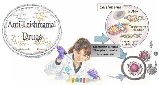
Graphical Abstract

1. Introduction

The leishmaniases are vector-borne protozoan diseases caused by multiple Leishmania species, presenting in three main clinical forms: visceral leishmaniasis (VL), cutaneous leishmaniasis (CL), and mucocutaneous leishmaniasis (MCL). VL is the most severe form, almost invariably fatal if left untreated, whereas CL and MCL cause substantial morbidity, lifelong disfigurement, and social stigma. Globally, an estimated 700,000–1,000,000 new cases occur annually, with CL accounting for the majority of infections; the disease remains endemic in more than 90 countries and disproportionately affects vulnerable populations with limited access to healthcare [1]. Despite this considerable impact, no vaccine is currently available for human leishmaniasis [2]. In contrast, several vaccines have been developed for canine leishmaniasis and are used in endemic regions as part of control strategies. However, vaccine efficacy remains variable and current research continues to focus primarily on improving chemotherapeutic options.
Kinetoplastids are a group of flagellated protozoa distinguished by the presence of the kinetoplast, a unique mitochondrial organelle that contains an intricate, catenated network of mitochondrial DNA (kDNA). Within the order Kinetoplastida, the family Trypanosomatidae is of public health relevance, as it includes Leishmania spp., Trypanosoma cruzi—the causative agent of Chagas disease—and Trypanosoma brucei, responsible for African trypanosomiasis, all of which are major pathogens associated with neglected tropical diseases [3]. Leishmania parasites alternate between two major developmental stages during their life cycle: the flagellated promastigote form that develops in the sand fly vector and the intracellular amastigote form that replicates within mammalian macrophages. These stages differ markedly in morphology, metabolism, and susceptibility to therapeutic agents. In addition, clinically relevant species such as Leishmania donovani, Leishmania infantum, Leishmania major, Leishmania tropica, and Leishmania braziliensis show important differences in geographic distribution, pathogenicity, and drug susceptibility profiles [4]. Such biological diversity has important implications for therapeutic development, particularly for strategies targeting parasite-specific organelles such as the kinetoplast and mitochondrion [5]. Current therapies against leishmaniasis and other kinetoplastid diseases remain suboptimal, challenged by increasing resistance, dose-limiting toxicity, prohibitive cost, and inconsistent availability. Therefore, deciphering parasite biology and elucidating drug–parasite interactions at both molecular and cellular levels are essential to identify novel targets and drive the development of next-generation antileishmanial agents [6].

2. Drawbacks of Current Chemotherapy of Leishmaniasis

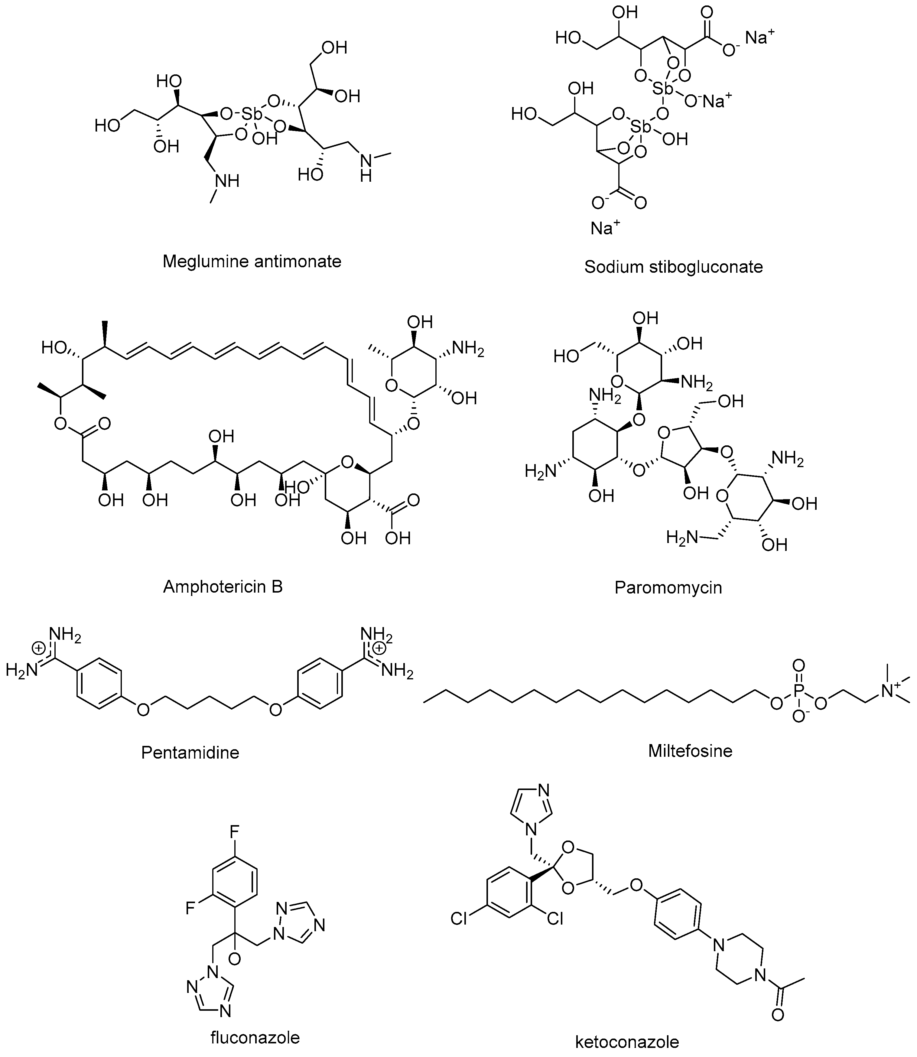
Chemotherapy remains the cornerstone of leishmaniasis management, although the position of each drug in therapeutic algorithms varies depending on the clinical form of the disease. Pentavalent antimonials (Figure 1) have historically been the standard of care and continue to represent the first-line option for CL and MCL in many endemic regions. In contrast, in VL, their use has shifted to a second-line role in areas such as the Indian subcontinent, where high levels of resistance have rendered them largely ineffective [7]. Conversely, the liposomal formulation of amphotericin B (AmBisome®, Gilead Sciences Inc., Foster City, CA, USA) is considered the first-line therapy for VL owing to its superior efficacy and reduced toxicity; however, in CL and MCL it is generally reserved as a second-line option for refractory or severe cases, since its high cost, limited availability, and requirement for cold-chain storage restrict widespread use in many endemic regions. Additional agents include oral miltefosine (MF) [8], effective against VL and CL but contraindicated in pregnancy due to teratogenicity, as well as paromomycin and pentamidine, which serve as alternatives in specific settings. The antifungal drugs fluconazole and ketoconazole (Figure 1) are other treatment options for CL [9]. Nevertheless, across all forms of the disease, current regimens remain constrained by significant obstacles [10,11]. Drug toxicity leading to systemic adverse events such as cardiotoxicity, hepatotoxicity, nephrotoxicity, pancreatic dysfunction, and gastrointestinal disorders is the most prominent one (Table 1) [12,13,14,15]. Variable efficacy across endemic regions, complex administration schedules, and the growing problem of resistance is another issue of leishmaniasis chemotherapy [16,17,18]. Other limitations include a low or variable bioavailability, poor solubility, limited and poorly characterized tissue distribution, high protein binding, or short half-lives for paromomycin and pentavalent antimonials. Such characteristics result in rapid drug clearance and subtherapeutic plasma levels, ultimately necessitating prolonged treatment regimens that compromise patient adherence and increase the risk of cumulative toxicity [19].
Additional chemical scaffolds have been explored and a few clinical leads [20], which were identified through phenotypic screens [i.e., the proteasome inhibitors GSK34942459 and LXE-408, the CDC-2-related kinase 12 (CRK12) inhibitor DNDI-6899 (GSK3186899), the oxaboroles DNDI-6174 and DNDI-6148, and the nitroimidazole DNDI-0690], have progressed to early stages of clinical development (reviewed by Ebiloma et al.) [16,21]. However, the last drugs approved for human VL, miltefosine and paromomycin, were introduced in 2002 and 2006, respectively [22].
Collectively, these shortcomings underscore the urgent need to identify vulnerable parasitic targets—molecules essential for parasite survival or virulence, ideally expressed in clinically relevant stages—to develop safer, more affordable, and more effective antileishmanial therapies [3].
Table 1. Summary of key pharmacological parameters—including route of administration, dosing regimen, therapeutic efficacy, and principal limitations—for conventional antileishmanial drugs. References provided correspond to clinical and experimental data sources.
DrugAdministration RegimenEfficacyLimitationsReferences
Pentavalent antimonials:
-
Meglumine antimonate
-
Sodium stibogluconate
intravenous (IV)
or
intramuscular (IM)
20 mg/kg/day for 30 days35–95%
(Region- and Strain-Dependent)
Painful injections
Cardiotoxicity
Nephrotoxicity
Pancreatitis
[23]
Liposomal
Amphotericin B
IV3–5 mg/kg over
6–10 infusions
60–85% in Africa
>95% in India
Rigor and chills
Nephrotoxicity
Hypokalemia
Anaphylaxis
[24,25,26,27,28]
ParomomycinIM11 mg/kg/day for 21 days63–85% in Africa
94–95% in India
Painful injections
Nephrotoxicity
Ototoxicity
[29,30,31]
Miltefosineoral2.5 mg/kg/day for 28 days≈ 85% in Africa
94% in India
Teratogenicity
Nephrotoxicity
Gastrointestinal toxicity
[32,33,34]
PentamidineIM or IV4 mg/kg/day for 7–10 daysVariable
Mostly for CL
Painful injections
Nephrotoxicity
Hypoglycemia
Cardiovascular toxicity
[35,36,37]

3. Key Structural Components of Leishmania: Kinetoplast Biology and Drug-Target Potential

Trypanosomatids of greatest concern to the WHO—including Leishmania spp., T. cruzi, and T. brucei—possess a distinctive mitochondrial organelle known as the kinetoplast, which harbors their mitochondrial DNA (kDNA). This specialized structure presents an attractive target for therapeutic intervention due to its unique composition and critical biological functions [38]. Identifying suitable therapeutic targets against Leishmania requires first an understanding of their life cycle, anatomical structures, and biological functions. These parasites alternate between two main morphological stages: the promastigote, an elongated and motile form (~5–20 µm) that develops in the sand fly vector and is characterized by a long anterior flagellum, and the amastigote, a smaller (~6–8 µm), ovoid, intracellular form with a rudimentary flagellum, specifically adapted to survive and replicate within mammalian macrophages [39]. Both forms are surrounded by a glycocalyx composed of stage-specific glycoconjugates, among which lipophosphoglycan (LPG) is the most prominent. In promastigotes, LPG facilitates attachment to the sand fly midgut, shields the parasite from complement-mediated lysis, and modulates host phagocytosis [40,41]. LPG expression is significantly downregulated in amastigotes, which instead rely on other surface molecules for immune evasion and intracellular survival [42,43]. A distinctive and evolutionarily conserved organelle in both promastigote and amastigote forms is the flagellar pocket, a plasma membrane invagination at the base of the flagellum that serves as the exclusive gateway for endocytosis and exocytosis. This specialized compartment is enriched in LPG and glycoprotein GP63, orchestrating vesicular trafficking, modulating host immune responses, and directing parasite orientation toward phagocytic cells to promote uptake [44]. As mentioned before, a defining feature of Leishmania is the kinetoplast, a unique mitochondrial organelle located adjacent to the flagellar basal body. It contains the parasite’s mitochondrial genome, arranged as a highly compact network of maxicircles (~20–40 kb) and thousands of heterogeneous minicircles (0.5–10 kb) concatenated into a disk-shaped structure [45]. Maxicircles encode mitochondrial rRNAs and essential subunits of the respiratory complexes, whereas minicircles provide the guide RNAs required for the extensive RNA editing that characterizes trypanosomatid gene expression. The kinetoplast DNA (kDNA) can constitute a substantial fraction of the total cellular DNA, underscoring its structural and functional significance in parasite survival and adaptation [46].
Overall, the unique structural features and essential functions of the kinetoplast make it a highly attractive and promising pharmacological target for the treatment of leishmaniasis. Scientific studies have highlighted the potential of targeting the kinetoplast due to its crucial role in parasite survival and its distinctive architecture. Disrupting kinetoplast integrity or its replication mechanisms can cause parasite death by impairing mitochondrial functions. The enzymes involved in kinetoplast replication, especially topoisomerases, are sufficiently different from their host counterparts, allowing for selective inhibition and a reduced risk of host toxicity. This strategy offers several advantages: the high specificity of kinetoplast enzymes minimizes off-target effects, and interfering with such a vital organelle is likely to produce a significant antiparasitic impact [38]. Therefore, drugs aimed at kinetoplast components, such as topoisomerases [47], may lead to highly effective and selective antileishmanial therapies, opening new options for the development of effective treatments. Another validated and selective drug target functionally linked to the kinetoplast, though not physically localized within it, is trypanothione reductase (TryR). By maintaining the parasite’s thiol–redox balance, TryR plays a pivotal role in safeguarding kinetoplast integrity against oxidative stress [48]. Novel chemotypes have recently been identified that inhibit Leishmania TryR with high selectivity, leading to marked reductions in intracellular amastigote viability [49,50,51].
Post-genomic analyses reveal a reduced but life-stage-adapted mitochondrial genome in Leishmania and Trypanosoma. Key processes—electron transport, membrane potential (MMP) maintenance, calcium homeostasis, and apoptosis induction—have been probed using purified parasite mitochondria. The relevance of Leishmania mitochondrion as drug target is evidenced by the number of clinically used drugs (e.g., amphotericin B, pentamidine, artemisinin, atovaquone) and investigational antileishmanial compounds (e.g., MitoTam [52]) that exert their effect by collapsing MMP and inhibiting respiration. Benzophenone bisphosphonium salts are another example of compounds that selectively target complex II of the electron transport chain in Leishmania, causing mitochondrial swelling, ATP depletion, and MMP loss [53]. Among natural products, berberine induces ROS-mediated apoptosis by inhibiting complexes I–III and depleting ATP, whereas flavonoids (luteolin, quercetin) promote kDNA loss and topoisomerase II inhibition [54,55]. Topoisomerase inhibitors further compromise kinetoplast replication and mitochondrial integrity. These multifaceted approaches underscore the translational relevance of targeting mitochondrial and kinetoplast biology and support the development of multitargeted therapies to enhance efficacy and control parasite resistance [56]. However, despite its potential, the kinetoplast poses significant challenges as a therapeutic target, mainly due to its intracellular localization within the parasite’s single mitochondrion.

4. Therapeutic Strategies Directed at the Kinetoplast

Adopting a target-based approach to identify new antileishmanial hits is a promising strategy as long as the target is validated based upon essentiality (e.g., gene, DNA, RNA,…) in addition to other parameters such as potential druggability, the availability of structural information, and, preferably, the absence of a similar target in the host to avoid possible risks of host toxicity [57]. Different targets of interest have been studied in recent years (reviewed by Marín et al. [21] and Singh et al. [58]). These include the proteasome, which in kinetoplastids can be distinguished from other eukaryotes, protein kinases, the endonuclease cleavage and polyadenylation specificity factor 3 (CPSF3), the purine salvage pathway, and the mitochondrial DNA (kDNA) [59,60,61]. In trypanosomatid parasites, kDNA replication is a critical process that occurs prior to the nuclear S phase [62]. Since the mitochondrial genome maintenance is essential for parasite survival [62], compounds that target mitochondrial DNA and interfere with kDNA replication are potentially active against kinetoplastid parasites.

4.1. DNA Minor Groove Binders

The kinetoplast is made up of thousands of DNA circles topologically linked in a disk-shaped, planar array. It contains two types of circular DNA, the maxicircles that code for rRNA and mitochondrial proteins, and thousands of small DNA minicircles that code for guide RNAs, which are used to edit the mRNA from maxicircles. The extensive AT sequences present in kDNA minicircles produce curved double-helical structures [63,64] that are potential targets for AT-specific DNA minor groove binders (MGBs) [65,66]. Given that kinetoplastid mitochondrial DNA exhibits a high A-T content compared to other organisms, it has been validated as a good target for MGBs.

4.1.1. “Classical” (Di)cationic Minor Groove Binders

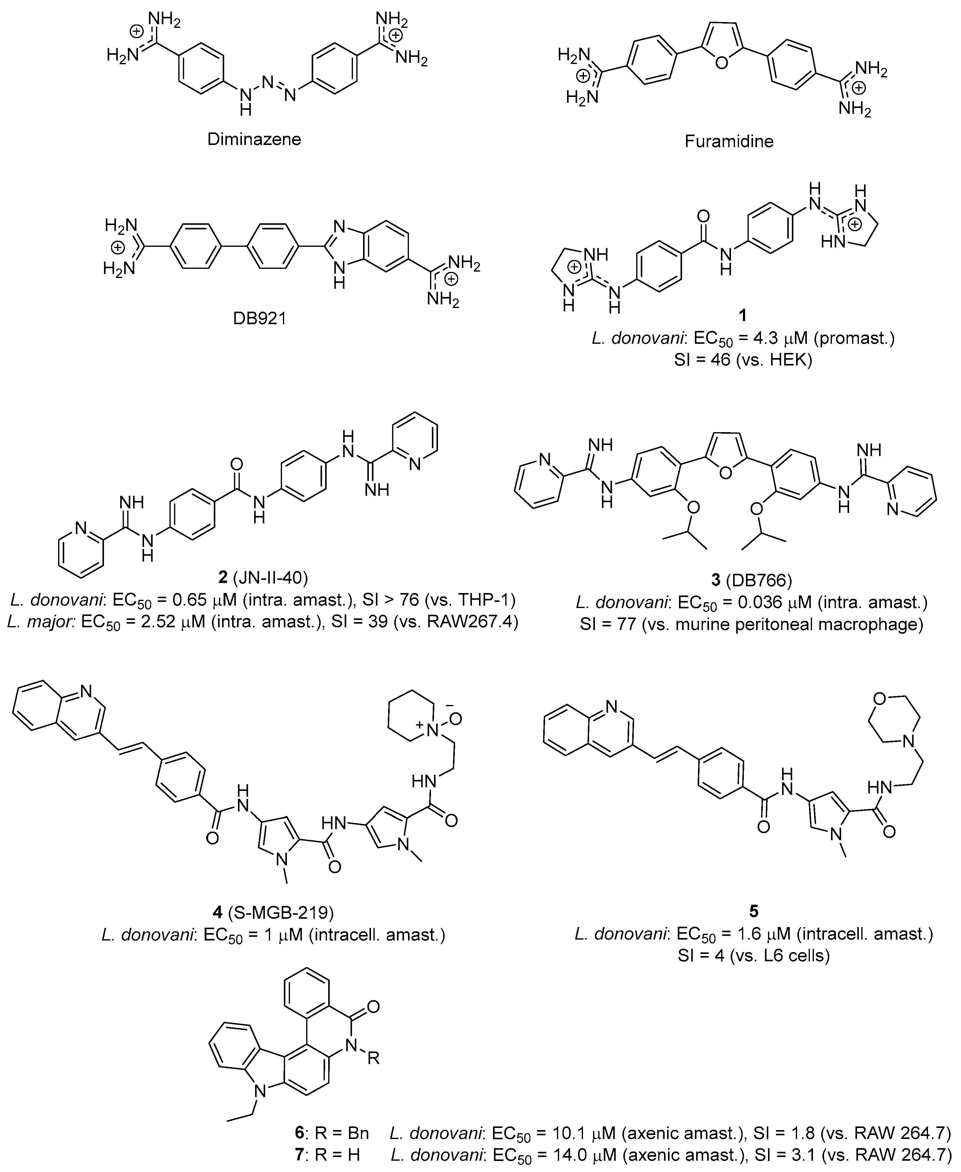
More than three decades ago, Shapiro and Englund showed in T. brucei that the diamidines pentamidine and diminazene (Figure 1 and Figure 2) generate minicircle–protein cleavable complexes specific to kDNA [67]. More recently, diminazene was also shown to impair kDNA topology and replication in T. cruzi epimastigotes, reducing parasite proliferation without affecting its viability [68].
Aromatic diamidines (e.g., diminazene, furamidine, Figure 2) are dicationic MGBs that bind preferentially to the minor groove of kDNA at AT sites of four or more base pairs and have a well-established antiparasitic activity [65,69,70,71]. In general, the concave shape of MGBs closely matches the convex minor groove shape. However, this crescent shape is not a strict requisite of MGBs because, in some cases, water can mediate the interaction between the minor groove and linear dicationic compounds via hydrogen bonds, hence forming a flexible linker resulting in an isohelical complex [71,72]. This is the case, for instance, in the dicationic near-linear biphenyl benzimidazole DB921 (Figure 2) [73,74]. Over the last two decades, new analogs of furamidine showing a strong binding to the DNA minor groove have been developed. In these analogs, the phenyl groups and the furan ring linker were replaced by heterocycles including thiophene, selenophene, indole or benzimidazole, enhancing the antiparasitic activity, both in vitro and in vivo, compared to pentamidine and furamidine [71,75].
Other classes of dicationic minor groove binding compounds such as bisguanidines and bis(2-aminoimidazolines) (1) [76,77] are also very effective against T. brucei [78,79,80,81,82,83,84], whereas bisarylimidamides (e.g., 2, 3; Figure 2) are more potent against the intracellular parasites T. cruzi [85,86,87,88,89] and Leishmania [90,91,92,93,94,95,96]. Converting amidines to arylimidamides (AIAs), which also bind to AT-DNA sequences in a structure-dependent manner, is an effective strategy to boost the activity against Leishmania. In these compounds, the DNA binding strength varies with substituent size, charge and polarity [97].
The selectivity of MGBs towards kinetoplastid parasites is driven by two main factors: the presence of cationic charges in the molecule favors their accumulation in the mitochondrion of the parasite (driven by the strong inside-negative electrostatic membrane potential) and also promotes preferential binding to AT sites of kDNA. The increased negative electrostatic potential at the floor of narrow AT-containing minor grooves is one of the factors that drives this preferential binding. As a result, these compounds accumulate in the mitochondrion of kinetoplastid parasites (although most mechanistic evidence has been obtained in Trypanosoma models), possibly disrupting replication and transcription processes by the inhibition of DNA-dependent enzymes (e.g., topoisomerases, polymerases, nucleases, and helicases) and/or compound-induced conformational changes that disrupt the kinetoplast structure [98,99].
Millan et al. have shown in T. brucei that the N-phenylbenzamide-based bis(2-aminoimidazoline) compound 1 (Figure 2), which is 100% curative in mouse models of African trypanosomiasis by oral dosage [81,100], alters the integrity of the kinetoplast and disrupts the replication of T. brucei kDNA [101]. Interference with the mitochondrial ‘high mobility group’ (HMG) box-containing protein TbKAP6 [102], which is essential for kDNA function, was suggested as possible mode of action [101]. In contrast, most bis(2-aminoimidazolines) are inactive or weakly active against Leishmania and T. cruzi intracellular amastigotes. This can be attributed to cell uptake differences because a drug targeting kDNA must cross four biological membranes to reach its target. Although the high polarity and basicity (i.e., all have pKa values > 9) of bis(2-aminoimidazolines) and related dicationic drugs (e.g., diamidines, bisguanidines) limit their diffusion across biological membranes, transporter-mediated uptake and/or binding to kDNA can influence selective cellular accumulation (see above) [103]. In fact, diamidines are known to act selectively on trypanosomes because of exclusive transport mechanisms that are absent in Leishmania species [103,104].
Bisarylimidamides (bisAIA) display distinct physicochemical properties with lower pKa (i.e., in the range 4.2−8.4) and higher logP values than diamidines, bisguanidines and bis(2-aminoimidazolines) [90]. They are usually more active against intracellular parasites, although their mode of action against T. cruzi and Leishmania is still a matter of controversy. This class of compounds binds to the DNA minor groove with more or less specificity depending on the scaffold. Daliry et al. showed that, in T. cruzi, the biological activity of very potent bisAIA compounds such as DB766 (3, Figure 2) produced deep alterations of the parasite kDNA topology. However, the study showed that the trypanocidal activity of this class of compounds did not correlate with the binding affinity to T. cruzi kDNA [105]. A lack of correlation between DNA binding affinity and antikinetoplastid activity was also observed with the structurally related compounds 1 and 2 [106]. Hence, it appears that other targets apart from kDNA may be considered to explain the antiparasitic action of bisarylimidamides. The cytochrome P450 (CYP5122A1) is an example of a target associated with ergosterol metabolism that could be implicated in the antileishmanial action of bisAIA [93].
In addition to the recognition of AT sequences, and to expand the therapeutic potential of DNA MGBs, the recent literature highlights the possibility of engineering diamidines for the sequence-specific recognition of mixed AT/GC base pairs at the DNA minor groove [72,107]. Thus, chemical approaches adding new mixed bps DNA binding motifs could be used to target the compounds to a specific kDNA sequence.
Strathclyde minor groove binders (S-MGBs) are another class of compounds showing antiparasitic activity against Leishmania donovani and against various infective agents such as bacteria, viruses, or fungi [108]. These compounds are based upon the structure of the natural product distamycin [109], in which the cationic amidine tail can be replaced by a neutral group—including an aromatic ring—and one of the amide links, can be replaced with the isosteric alkene linker. Perieteanu et al. have reported that an N-oxide derivatization of the tertiary amine tail can enhance the selective anti-leishmanial activity of S-MGBs, as exemplified by compound 4 (Figure 2) [110]. The reduction in the overall length of the molecules was also proposed to improve selectivity towards parasitic organisms, such as Trypanosoma spp. and Leishmania spp, and reduce the likelihood of the molecular aggregation of this class of compounds (e.g., 5; Figure 2) [111].

4.1.2. Non-Cationic Minor Groove Binders

Phenanthridines are nitrogenous heterocyclic compounds that can be found in many natural products and exhibit different pharmacological properties [112]. It is known that the binding of these compounds with DNA, either via classical intercalation or minor groove binding, depends on the substituents decorating the phenanthridine core [113,114]. Recently, a class of non-cationic DNA MGBs with a “hybrid” structure related to phenanthridine (i.e., indolo [3,2-a]phenanthridine derivatives 6 and 7, Figure 2) were found active against L. donovani in the micromolar range, though with limited selectivity (SI < 5). The compounds were shown to modulate the cell cycle in Leishmania parasites [115].

4.1.3. AT-Hook Proteins as Target

The “AT-hook” is a DNA-binding domain of DNA-regulating proteins that interacts in the minor groove of AT-rich sequences. It has been described in the High-Mobility Group A (HMGA) protein family and in different transcription factors and chromatin proteins [106,116,117]. Upon binding, the AT-hook causes bends in the DNA structure through the electrostatic and hydrophobic contacts of the neighboring basic residues of the central RGR core, which fits into the narrow minor groove of the DNA [118]. Since HMGA proteins are involved in a variety of cellular processes in eukaryotes, including kinetoplastid parasites, inhibitors of AT-hook binding proteins such as small-molecule MGBs could provide a therapeutic potential as antiparasitic agents [101,106].
The crystal structure of the HMGA AT-hook 1 domain bound to the minor groove of a DNA oligonucleotide has been reported recently [106]. Small-molecule DNA MGBs with antikinetoplastid activity such as 1 and 2 (Figure 2) were able to inhibit the interaction between AT-hook proteins and AT-rich DNA [101,106]. In particular, 2 is a bisarylimidamide lead compound that shows very promising activity in vitro [90,119] and in vivo ([120], Nué-Martinez et al., unpublished) against L. donovani, Leishmania infantum, and Leishmania major. Based on these results, we can envision that AT-hook proteins such as LamAT-Y, expressed in Leishmania parasites [121], are potential targets for this class of compounds [106].

4.1.4. Topoisomerase Inhibitors

Several proteins are involved in kDNA replication (reviewed by Amodeo et al. [62]), many of which are potential targets for antiparasitic chemotherapy. DNA topoisomerases of kinetoplastid parasites have been proposed as antiparasitic targets because they are structurally different from human topoisomerases, thus enabling selectivity towards the parasite [47,122,123]. These enzymes are crucial to maintain the dynamic structure of cellular DNA. They perform essential functions associated with the topological organization of kDNA, participating in the decatenation of minicircles prior to their replication for their subsequent recatenation in the kinetoplast network and contributing to the structural stability of the network during replicative processes [124]. In contrast, the function of host topoisomerases is limited to the topological modulation of non-catenated DNA and they have structural differences from those associated with the Kinetopolastida order. These functional and structural differences, combined with the specific intracellular location of the kinetoplast and the parasite’s absolute dependence on a single mitochondrion, justify the potential of these enzymes as selective therapeutic targets.
DNA topoisomerases (Topo) are divided into two main categories: they transiently cleave one strand of DNA (TopoI) or both strands (TopoII), and they transfer one DNA strand through the other from the same (TopoI) or different DNA molecules (TopoII) [47,122]. In trypanosomatids, the mitochondrial TopoII, encoded by kDNA, is essential, and a failure of this enzyme leads to the disruption in the replication of minicircles, loss of kDNA network, and death of the parasites [122,124,125]. Different classes of molecules have been reported as DNA topoisomerase inhibitors. Class I inhibitors (“topoisomerase poisons”) stabilize the DNA-topoisomerase covalent complex known as TOPcc, which causes genomic instability, leading to cell cytotoxicity. On the other hand, class II inhibitors (“catalytic inhibitors”) block the enzyme’s active site preventing enzyme–DNA interactions and interfering with the enzyme catalytic function [47].
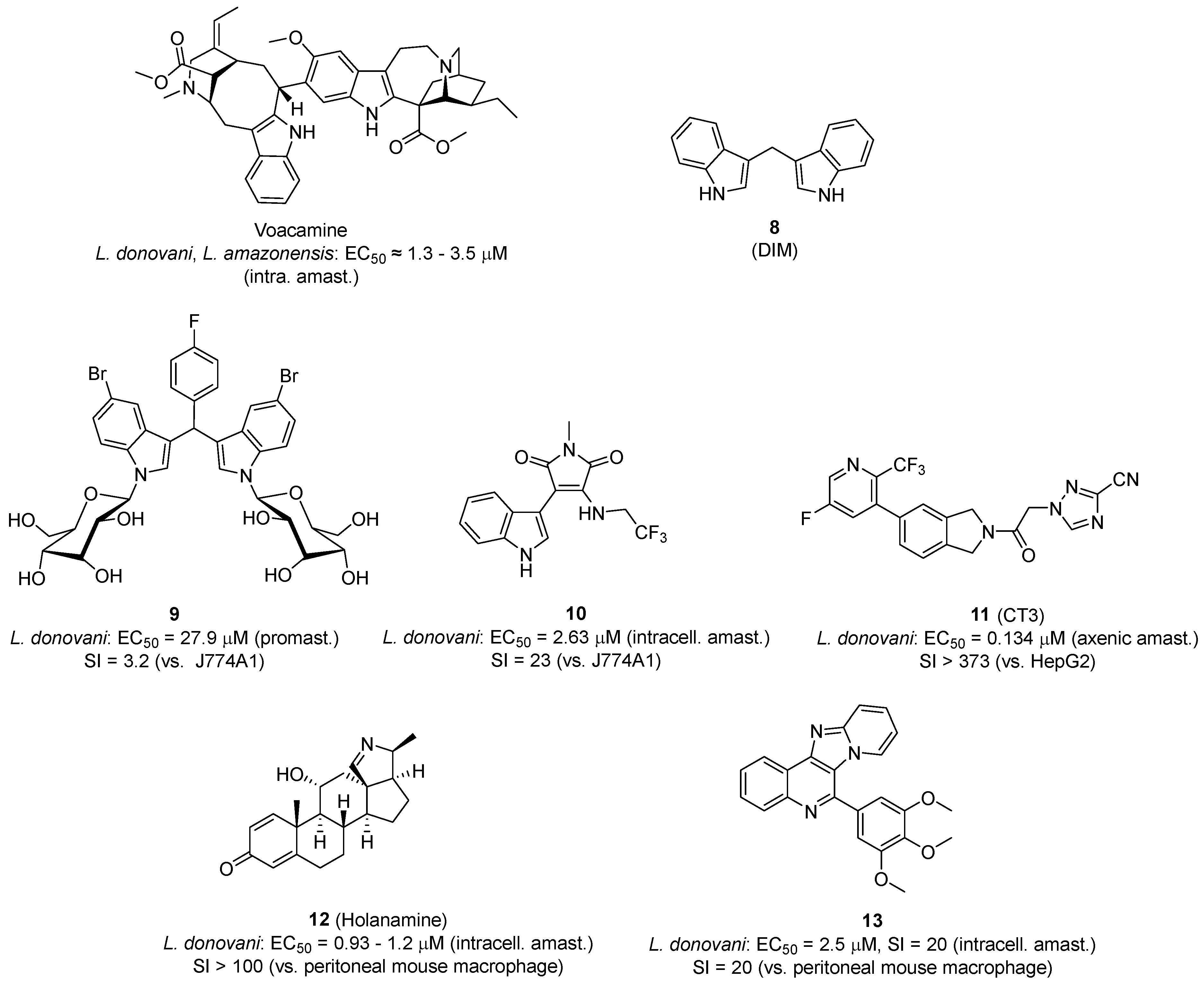
Several TopoIB inhibitors active against Leishmania have been reported in the literature (reviewed by Saha et al. [123]). For example, voacamine, an indole alkaloid isolated from the plant Tabernaemontana coronaria, is a parasite-selective uncompetitive LdTopoIB-specific poison that is effective in vitro against wild-type and drug-resistant isolates of L. donovani, Leishmania amazonensis, and T. cruzi (Figure 3). In a BALB/c mice model of VL, voacamine cleared the parasite burden in spleen and liver at an intraperitoneal dosage of 5 mg/kg [126].
Diindolylmethanes have also shown promising results against Leishmania parasites. (3,3′)-diindolylmethane (DIM) (8, Figure 3), a putative DNA MGB [127], is a natural compound that inhibits tumor growth in human cell lines and in vivo models. DIM is a non-competitive class I inhibitor of TopoI of Leishmania donovani (IC50 = 1.2 μM). It was suggested that the stabilization of the topoisomerase I–DNA cleavable complex formation hinders the DNA relaxation activity and causes the inhibition of parasite replication and transcription [128,129].
Kour et al. have shown that the glycosylation of 3,3-DIM analogues active against L. donovani promastigotes, but devoid of TopoIB inhibitory activity, resulted in reduced cytotoxicity against macrophages without affecting the anti-leishmanial activity. These glycosylated analogs (9, Figure 3) inhibited recombinant LdTopILS in the high micromolar range, suggesting that glycosylation is an interesting approach to target TopoI inhibitors to Leishmania parasites [130].
Indolylmaleimide derivatives are another class of antileishmanial compounds designed as LdTopILS inhibitors. Das et al. showed that compound 10, which is active against L. donovani intracellular amastigotes in the low micromolar range (IC50 = 2.63 µM), inhibits LdTopILS at 800 µM concentration [131]. The results of this study suggest that this compound is a class II inhibitor; however, the two orders of magnitude difference between the cellular and the enzymatic activities indicates that other targets (in addition to LdTopILS) may be involved in the antileishmanial activity of 10.
Compound CT3 (11, Figure 3) is an example of irreversible inhibitor of trypanosomal TopoII that stabilizes the covalent DNA–enzyme complex, which leads to dsDNA breaks. CT3 proved to be a very potent trypanocidal compound both in vitro and in mouse models of Chagas disease and human African trypanosomiasis. The compound was also active against axenic amastigotes of L. donovani (IC50 = 0.134 µM) [132].
Holanamine (12, Figure 3), a plant-derived steroidal alkaloid, has shown potent in vitro antileishmanial activity with IC50 = 2.66 and 3.80 µM against WT and multi-drug-resistant L. donovani promastigotes, respectively [133]. The compound inhibits the catalytic activity of L. donovani topoisomerase 1B (LdTopIB) in a noncompetitive manner (IC50 = 2.81 μM), without inhibiting the catalytic activity of human TopoI.
The pyrido [2,1′:2,3]imidazo [4,5-c]quinoline derivative 13 is a class I inhibitor that stabilizes the LdTop1−DNA covalent complex and inhibits TopoI religation activity, leading to apoptosis-like cell death in drug sensitive and antimony-resistant L. donovani clinical isolates. This compound showed in vivo antileishmanial activity in a mouse model of visceral leishmaniasis [134].
Other natural product derivatives such as terpenyl-quinones, lignans (niranthin) [135], lignan glycosides (lyoniside and saracoside) [136] and naphthyridines [137] have also shown potent anti-leishmanial activity by preventing the relaxation of supercoiled DNA, impairing the enzyme function. All these examples illustrate nicely the potential of parasite topoisomerases as a target for antikinetoplastid drugs. However, as mentioned by Kour et al. [47], many Leishmania inhibitors have been described to date, but there are not any drugs targeting leishmanial topoisomerases yet in clinical trials. Therefore, it is crucial to further investigate these promising compounds and their mechanisms of action in order support the rational development of chemotherapeutic strategies against leishmaniasis.

4.2. G-Quadruplex Stabilizers

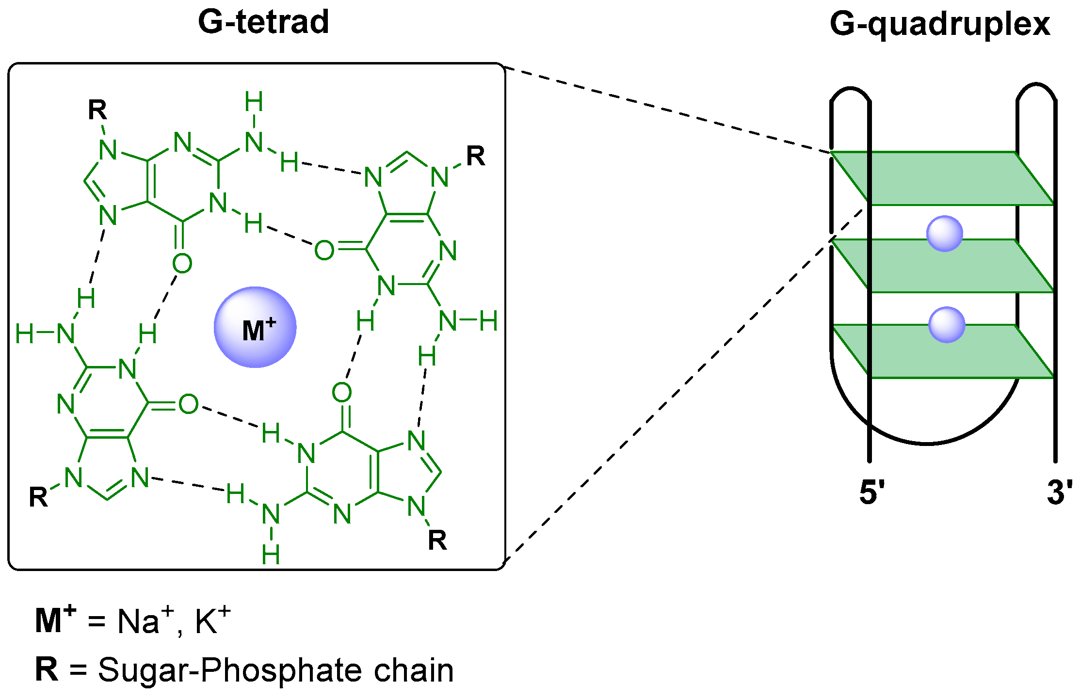
G-quadruplexes (G4) are unusual four-stranded secondary structures of guanine-rich sequences of nucleic acids. The stacking of two or more guanine tetrads, which are formed by the Hoogsteen hydrogen bonding of four guanine bases held in a planar arrangement, gives rise to a thermodynamically stable knot-like G4 structure [138]. G4 are stabilized by the presence of monovalent cations such as potassium or sodium coordinated to the O6 atom of each G in the stacked G-tetrads (Figure 4).
These structures are observed throughout the genomes of several eukaryotic species [138,139,140,141] and, in mammals, are present in the promoter regions of regulatory genes and transcription factor binding sites, as well as in 5′ untranslated regions (5′ UTR) and telomeres [142,143]. The high prevalence of putative quadruplex-forming sequences (PQSs) in the genome of protozoan parasites, especially in kinetoplastid parasites [144,145,146,147], suggests that G4s are a key transcriptional regulator of these parasites [142]. In Leishmania, whose genome is particularly GC-rich (59.6% in L. major) among trypanosomatids (46.8% in T. brucei), 16,988 observed quadruplexes (OQs) were found versus 3231 for T. brucei [138]. The studies showed that T. brucei OQs are enriched in 5′UTR regions or gene promoters. In contrast, Leishmania showed depletion at these and other (e.g., exons, 3′UTR) intragenic regions, and no enrichment at non-coding regions.
Recent studies have established that Leishmania spp. display an unconventional DNA replication program, in which the timing of the DNA replication completion is chromosome length-dependent and relies on stochastic initiation events localized in regions with high AT content and increased G4 levels [148,149]. These findings reinforce the idea that targeting G4 structures in these pathogens could have deleterious consequences for the parasite.
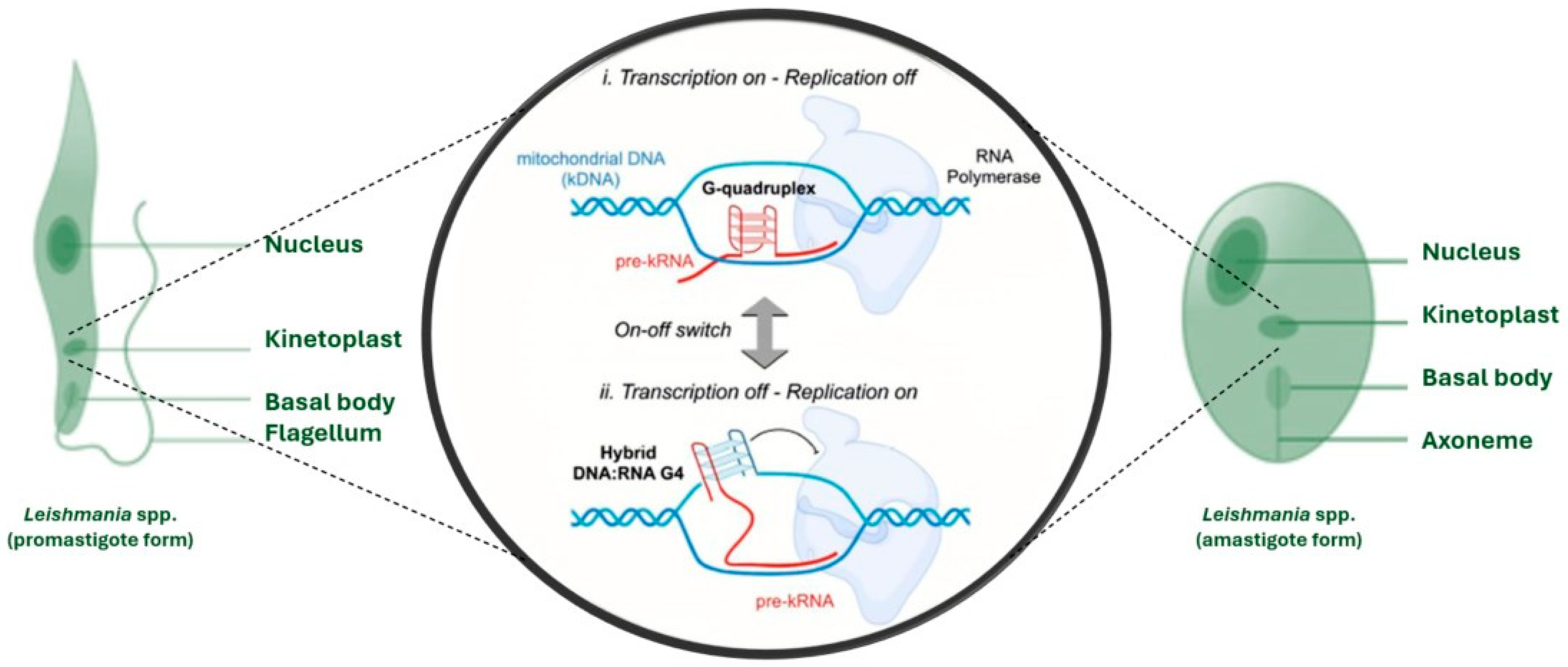
This high prevalence of G4s and the crucial roles they play in the regulation of vital processes of these organisms, including immune evasion and virulence [150], underscore G4 as a promising drug target in kinetoplastid parasites [142]. In particular, the formation of DNA/RNA hybrid G4 structures has been proposed to modulate kDNA transcription and replication in African trypanosomes [151]. Hence, G4 specific ligands, designed to selectively target hybrid DNA/RNA quadruplexes, could potentially suppress transcription, whereas enhancing the formation of RNA G-quadruplexes might instead promote transcription, as depicted in Figure 5 [142].
In general, G4 ligands are molecules with large (planar shape) hydrophobic aromatic cores that can bind efficiently to G-tetrads by π–π stacking. These molecules usually have cationic groups to stabilize the electrostatic interaction with the G4 phosphate backbone regions and some side chains or substituents that can interact with the G4 loops and improve the selectivity towards G4 structures (reducing double-stranded DNA intercalation) [152,153]. With hundreds of G4 ligands described in the literature, it has become clear that, although planarity is essential for G4 binding, the design of molecules with higher flexibility and cationic side chains of refined lengths can improve the affinity and selectivity towards specific G4 topological arrangements [152].
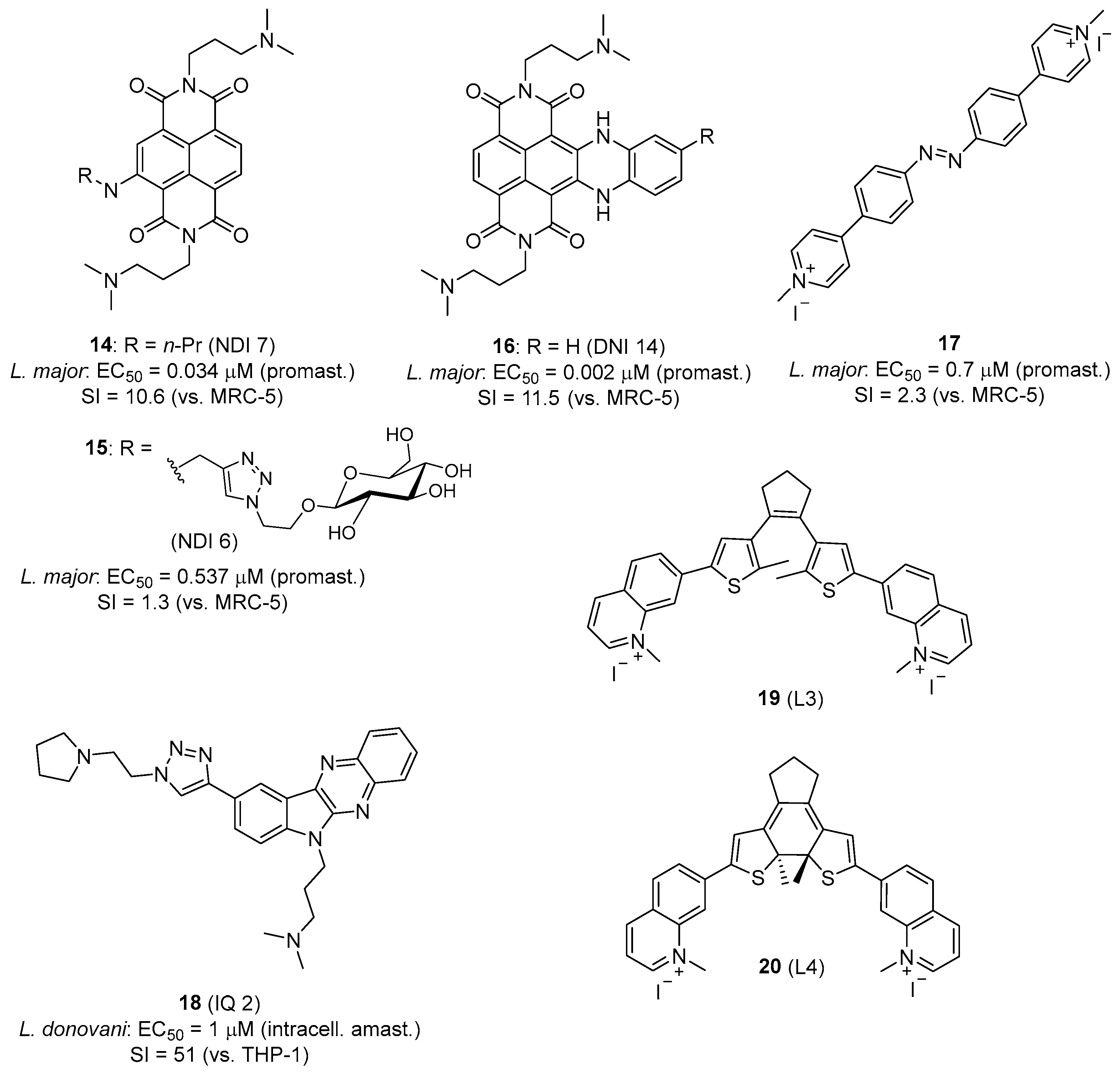
Recent studies reported the potential of G4 ligands to treat HAT and leishmaniasis by targeting PQSs in the T. brucei and Leishmania genomes [147,154,155,156,157]. Belmonte-Reche et al. conducted a search for PQSs in the genomes of T. brucei, L. major, and Plasmodium falciparum. A highly abundant repeated sequence called EBR1 (5′-GGGCAGGGGGTGATGGGGAGGAGCCAGGG-3′) was identified in T. brucei, the G4-forming capacity of which was confirmed by biophysical methods [147]. The study also evaluated carbohydrate-naphthalene diimide ligands (carb-NDI), which were shown to bind EBR1 and displayed antiparasitic activity with IC50 in the nanomolar range against T. brucei and L. major (e.g., 14 and 15, Figure 6) [147]. Confocal microscopy studies confirmed the localization of 14 and 15, mainly in the nucleus and in the kinetoplast.
In subsequent studies, more potent NDI derivatives were developed and tested against T. brucei and Leishmania. Benassi et al. showed that core-extended NDI G4 ligands such as 16 had potent low nanomolar activity against T. brucei (IC50 = 0.26 nM; SI = 88 versus MRC-5 cells), while activity against L. major promastigotes was more modest (IC50 = 0.002 μM; SI = 11.5 versus MRC-5 cells) (Figure 6). For this series, a correlation emerged between T. brucei antiparasitic activity and the affinity for T. brucei G4 [158].
Azobenzene derivatives are another class of G4 ligands with excellent activity against T. brucei (IC50 = 0.7 nM, SI = 2286 versus MRC-5 cells) and submicromolar IC50 value against L. major promastigotes (17, Figure 6). In this case, the low selectivity index towards Leishmania (SI = 2.3) is due to the relatively high cytotoxicity (CC50 = 1.6 µM) of the compound on MRC-5 cells [159]. This study highlighted the importance of the relative position of the positively charged N atoms in the structure of these compounds for G4 stabilization and selectivity.
Karmakar et al. reported a host-directed therapeutic strategy using G4 ligands to modulate the host immune response against Leishmania; for example, the indolo [2,3-b]quinoxaline derivative IQ2 (18, Figure 6) stabilizes the c-MYC G4 promoter in macrophages, which causes the suppression of c-MYC transcription and translation. As a result, this compound exerted immunomodulatory effects (“host-directed therapy”) shifting the macrophage polarization from the disease-promoting M2 phenotype to the host-protective M1 phenotype. Compound 18 showed potent antileishmanial activity against promastigotes (IC50 = 0.18 µM) and intramacrophage amastigotes of L. donovani (IC50 = 1 µM) with low toxicity to THP-1 host cells [160].
G-quadruplexes have also been proposed as a therapeutic target for Chagas disease. Approximately 174 PQSs per 100,000 nucleotides were identified in the genome of T. cruzi [144]. Among a series of fourteen G4 ligands based on the dithienyl ethene (DTE) skeleton synthesized by Pérez-Soto et al., compounds 19 and 20 (Figure 6) achieved low micromolar activity against trypomastigotes of T. cruzi (IC50 = 1.5 and 3.3 μM, respectively), with good selectivity indexes (SI = 25 and 40, respectively). Biophysical studies showed that these compounds bind to different quadruplex sequences found in the genome of T. cruzi such as RCr (5′-GGGGACGGGAATGGGGGTGCATGAGGGG-3′), MCr (5′-GTGGAGGGGGAGGGTCATGGGG-3′), and TCr (5′-GGGAGGGACGGATGGGCAGAAACGGG-3′). In addition, fluorescence microscopy revealed the localization of the compounds in the nucleus and kinetoplast of the parasites after 24 h incubation, indicating that G4s are a potential target of these molecules [144].
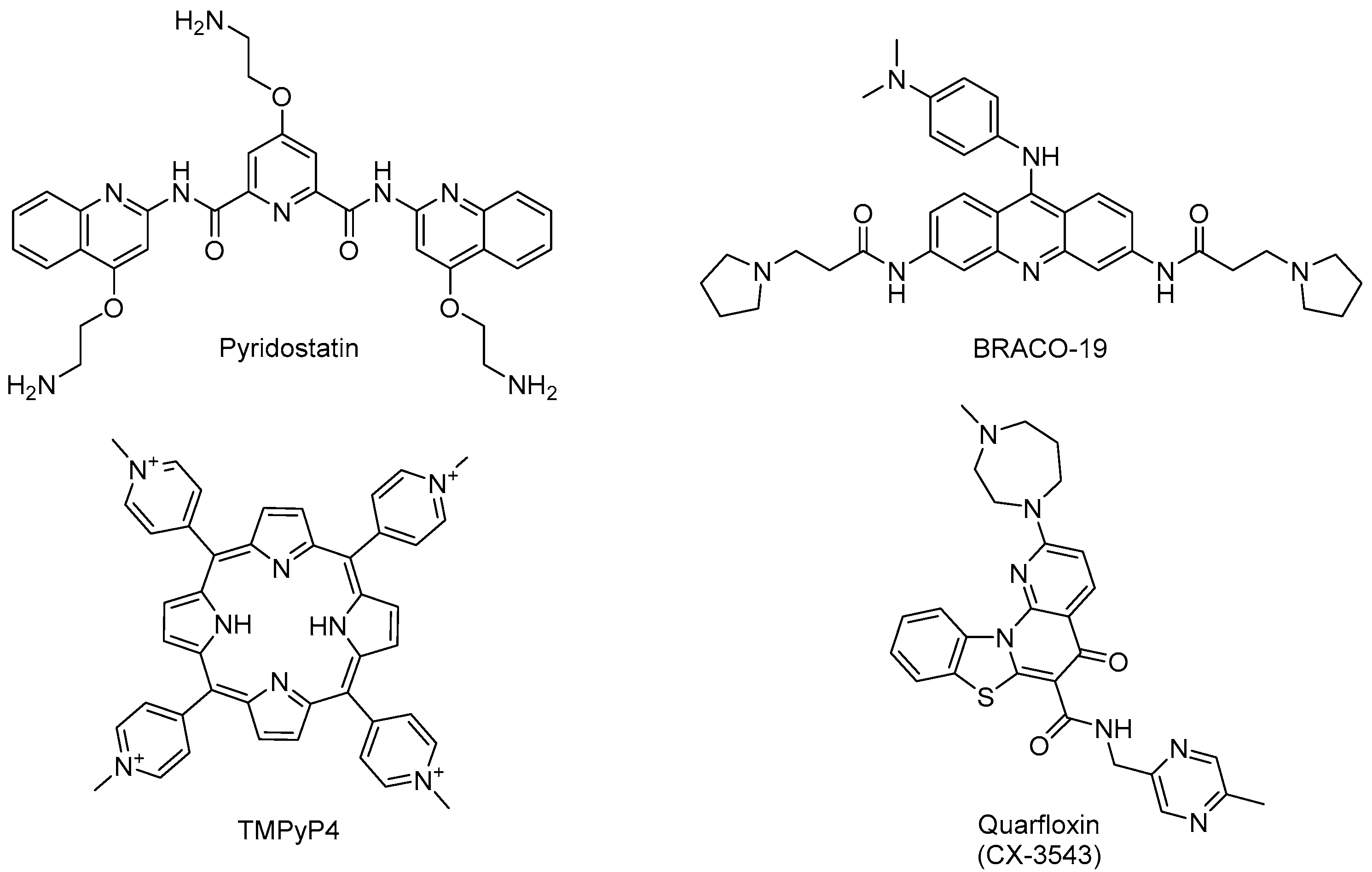
In the studies mentioned above, other “classical” G4 ligands initially used in cancer research such as pyridostatin, BRACO-19, and TMPyP4 were also tested against these parasites (Figure 7). However, these typical ligands showed much higher IC50 values against T. brucei, T. cruzi [144] or Leishmania [147], underscoring that it is possible to design selective antiparasitic G4 ligands (e.g., 14, 15) to target kinetoplastid parasites [147]. This trend was confirmed in another study on P. falciparum. Calvo and Wasserman established that TMPyP4 inhibits telomerase activity (IC50 = 5 µM) in this apicomplexan parasite, although it had a moderate effect on parasite growth (FCB-2 strain) at this concentration [161].
In the apicomplexan parasite P. falciparum, DNA G-quadruplexes can be detected in the nucleus of the parasite, which has an extremely AT-rich DNA (>80%) and therefore possesses few guanine-rich sequences with the potential to form G4s [162]. Notwithstanding, the 3D7 strain of P. falciparum is sensitive to several G-quadruplex-stabilizing drugs, including the RNA polymerase I (Pol I) inhibitor quarfloxin (Figure 7), an antitumoral G4 drug that also displays trypanocidal activity (IC50 = 155 nM) against T. brucei [141,163].
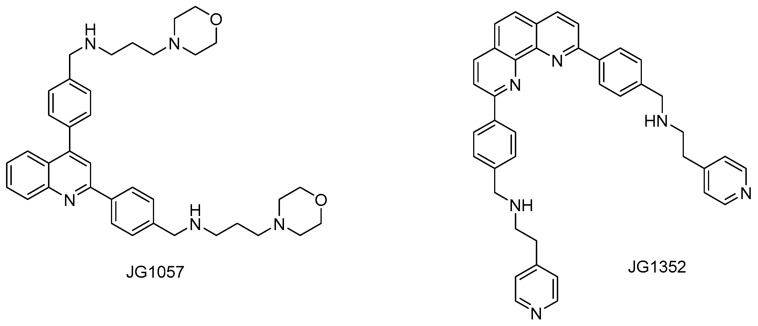
Recently, G4 formation was experimentally confirmed for sequences found in different parasitic helminths [139,140]. Small molecules able to selectively recognize G4 were found to bind to Schistosoma mansoni G4 motifs, and two of these ligands (JG1057 and JG1352, Figure 8) demonstrated potent activity against both larval and adult stages of this parasite [139].
In the last years, several studies reported the capacity of well-known MGBs to bind G4 structures. For instance, the diamidine drug diminazene (Figure 2), used for the treatment of animal trypanosomiasis, can bind G4 structures in a highly selective manner [164,165,166]. A recent study by Scott and Chalikian showed that MGBs such as netropsin and Hoechst 33258 can stabilize G-quadruplex-duplex hybrid (QDH) structures containing a hairpin duplex as a step-loop into the G4 core [167]. These studies suggest that (di)cationic minor groove binding molecules should be reassessed as G4 ligands, and may be used as templates for the design of new compounds with the sequence-specific recognition of G4s and/or QDH structures.

4.3. Mitochondrion-Targeted Small Molecules

Trypanosomatids are single-cell parasites with only one large mitochondrion containing essential enzymes involved in the energy-producing machinery. Hence, the mitochondrion is a critical organelle involved in the survival of these parasites. Accumulating evidence from our group [53,168,169,170,171,172,173,174] and others [175,176,177] demonstrates that conjugating antiparasitic agents with lipophilic cations is a highly effective chemotherapeutic strategy for selective mitochondrial targeting in protozoan parasites. However, because mitochondria-targeting strategies rely on conserved bioenergetic features shared with host cells, a careful evaluation of host mitochondrial toxicity remains essential for translational development. Among these, the triphenylphosphonium (TPP+) cation—characterized by a positive charge delocalized over a large and hydrophobic surface area—enables efficient electrophoretic transport across lipid bilayers driven by transmembrane potentials without the need for transporters. This targeting strategy has been reviewed by Zielonka et al. [178]. Another article by Cotman and co-workers nicely illustrates the recent advances in the development of mitochondrion-targeted molecules (MTMs), mainly TPP+-based and nitrogen-based heterocyclic MTMs, for different applications [179]. In particular, they highlight the effect of structural and physicochemical properties of MTMs on delivery and intrinsic bioactivity of the same.
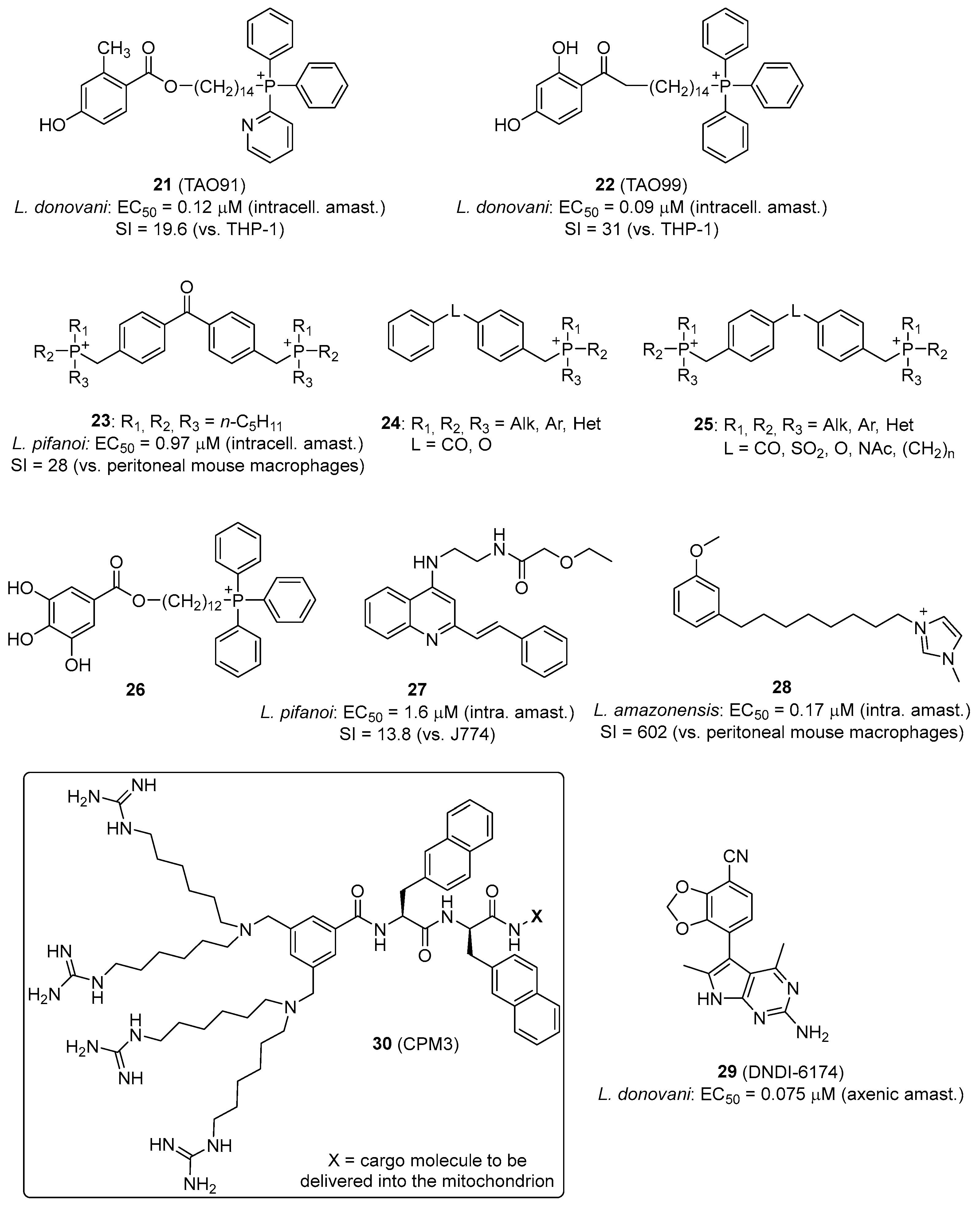
In Leishmania, energy metabolism is mostly based on oxidative phosphorylation which accounts for approximately 75% of ATP production in L. donovani promastigotes, the rest being furnished by glycolysis [180]. In contrast, the bloodstream-form trypomastigotes of T. brucei obtain their energy via glucose-dependent respiration using a unique cytochrome-independent terminal oxidase, the trypanosome alternative oxidase (TAO) [181]. In recent years, our research group has established an innovative approach to enhance the antitrypanosomal efficacy of TAO inhibitors based on the 2,4-dihydroxybenzoic acid scaffold [172]. Given that TAO is localized on the matrix-facing surface of the mitochondrial inner membrane of African trypanosomes, we exploited mitochondrion-targeting approaches to improve drug delivery [172,181]. Using TPP+, quinolinium and pyridinium cations, we were able to boost the antiparasitic activity of several structurally simple TAO inhibitors [169,170,171,182]. For instance, the methylene-linked 2-methyl-4-hydroxybenzoate 2-pyridinyldiphenylphosphonium derivative 21 (Figure 9) is the first example of an allosteric inhibitor of TAO with broad-spectrum nanomolar range activity against African trypanosomes [168].
Interestingly, this class of MTMs also displays leishmanicidal activity in vitro (21) and in vivo (22). Compound 22 is a good example of a mitochondrion-targeted phosphonium salt with in vivo efficacy (>95% reduction in parasite load in spleen and liver) in a mouse model of visceral leishmaniasis by oral administration. Mechanistic investigations demonstrated that compound 22 permeates the plasma membrane and selectively localizes within the mitochondrion of Leishmania parasites. This mitochondrial targeting induced profound alterations in bioenergetic homeostasis, evidenced by a marked reduction in intracellular ATP concentrations, mitochondrial membrane potential collapse, and the substantial generation of reactive oxygen species [183].
Other bisphosphonium compounds have been shown to display antitrypanosomal and antileishmanial activity, interfering with the mitochondrion of the parasites to produce their effect (Figure 9) [53,174]. Benzophenone-derived bisphosphonium salts 23 exhibit leishmanicidal activity through the inhibition of succinate dehydrogenase (respiratory complex II) [53]. In T. brucei, mono- (24) and bisphosphonium (25) compounds inhibit the hydrolytic activity of the mitochondrial FoF1 ATPase [173].
Cortes et al. reported a series of TPP+–gallate conjugates (26) active against T. cruzi, the activity of which was mediated by the uncoupling effect of the gallic acid pharmacophoric group and not by the inhibition of a specific enzyme of the electron transport chain [176]. The uncoupling effect of TPP+ carriers on oxidative phosphorylation has been studied by Kulkarni et al. [184]. They found that Hückel charge has a stronger impact than lipophilicity on the uncoupling activity of TPP+ conjugates. Despite higher lipophilicity, 4-CF3-TPP+ derivatives showed no membrane depolarization, highlighting the inertness of this moiety and its suitability as a mitochondrion-targeting scaffold [184].
The quinoline scaffold has also been used as a mitochondrion-targeting moiety, providing compounds with antileishmanial activity. For instance, 4-aminostyrylquinolines such as compound 27 (Figure 9) have an increased basicity at the heterocyclic nitrogen and a positive charge delocalized by resonance across the pyridine ring. Hence, these molecules behave as delocalized lipophilic cations, which allows them to cross the lipid bilayers easily without the need for transporters. Compound 27, which was active against intracellular amastigotes of Leishmania pifanoi in the low micromolar range, localized in the mitochondrion of the parasite [185]. Other quinolinium derivatives [186] and alternative cationic groups, such as imidazolium (28) and ammonium salts, were shown to display potent antiparasitic effect involving a mitochondrial target [175].
Another validated target of Leishmania is the cytochrome bc1 complex of the parasite’s electron transport chain. DNDI-6174 (29, Figure 9) is a promising pyrrolopyrimidine-derived preclinical candidate for visceral leishmaniasis that targets the cytochrome bc1. This compound is a promising drug candidate not only for Leishmania, but also for Chagas disease if combined with benznidazole [187].
Other mitochondrion delivery approaches exist apart from the “classical” mitochondrion-targeting scaffolds mentioned above. Appiah Kubi et al. reported a nonpeptidic cell-penetrating motif (30, Figure 9) consisting of four guanidinium groups and one or two hydrophobic naphthalene groups linked through a central scaffold for the specific delivery of both membrane-permeable small molecules and membrane-impermeable peptidyl cargoes into the mitochondrial matrix of mammalian cells [188,189]. Although this platform has not been used for antiparasitic compound delivery, it would deserve further study.

5. Nanomedicine-Based Strategies in Antileishmanial Therapy

Recent advances position nanomedicine as a compelling complement to conventional chemotherapy for leishmaniasis, with the explicit goals of enhancing targeted drug delivery, improving pharmacokinetic and biodistribution profiles, and circumventing mechanisms of drug resistance [190]. Liposomal formulations—exemplified clinically by liposomal amphotericin B—provide a proof of concept for efficient intracellular delivery, increasing macrophage uptake while reducing systemic toxicity [191]. Polymeric and metallic nanoparticles are another example of formulations that have demonstrated the effective targeted delivery of antimonials, amphotericin B, and miltefosine, diminishing host toxicity and mitigating efflux-mediated resistance pathways. The characteristics, advantages and limitations of such nanoparticles (NPs) have been reviewed recently [192,193].
Nanoemulsions and other nanocarriers are under active investigation for their capacity to cross biological barriers and to deliver therapeutics into parasitophorous vacuoles within infected macrophages, thereby improving intramacrophage bioavailability and antiparasitic activity [194,195]. However, despite promising preclinical and early clinical data, the translation of nanomedicine to widespread clinical use is constrained by challenges in scalable manufacturing, physicochemical and biological stability, regulatory requirements, and cost-effectiveness [193].
Nanomedicine can improve drug pharmacokinetics and the overall risk–benefit ratio by using functionalized nanocarriers for intracellular targeting, thereby increasing subcellular specificity while reducing systemic toxicity and treatment costs [196]. By tuning physicochemical properties and surface ligands, nanoparticles are selectively internalized and trafficked to specific organelles, enabling targeted accumulation and greater therapeutic precision [197]. Evidence from oncology and neurodegeneration indicates that mitochondrial targeting enhances therapeutic efficacy by promoting the selective accumulation of lipophilic cationic carriers driven by the organelle’s negative inner membrane potential, while amphiphilic mitochondriotropic motifs and targeting sequences facilitate endosomal escape and precise organelle delivery [198,199]. These studies have shown that, to deliver a bioactive molecule into the mitochondrial matrix, the nanoparticle-based delivery system must have a precise size, a lipophilic surface, a positive charge and a specific density of targeting ligands on its surface that allows for the recognition of and transport to the mitochondria [200]. Representative organelle-directed platforms—MITO porters and PLGA-bPEG-TPP+ systems, gold nanostars decorated with pro apoptotic peptides, and polymeric conjugates such as folate coated or TPP+ modified chitosan nanoparticles—illustrate how rational nanoformulations can provoke mitochondrial dysfunction and cancer cell death [201,202]. Mitochondrial-targeting principles can be applied to kinetoplastid parasites because the kinetoplast is a distinct mitochondrial subdomain essential for parasite survival [203]; mitochondrion-directed nanocarriers functionalized with targeting ligands or peptides therefore constitute a plausible strategy to deliver leishmanicidal agents with unprecedented subcellular specificity and to overcome the limited organelle selectivity of current drugs. Although most studies on mitochondrial targeting have been conducted in the context of neurodegenerative diseases and cancer, it has been demonstrated that nanoparticle accumulation within mitochondria enhances therapeutic efficacy while minimizing systemic toxicity [200,204,205,206,207]. These advances provide a conceptual framework that could be translated into the treatment of parasitic diseases such as leishmaniasis.
The active targeting of NPs offers significant potential for enhancing the specificity and efficacy of therapeutic delivery; however, its success is profoundly influenced by interactions with the biological milieu. Following systemic administration, NPs rapidly adsorb biomolecules to form a protein corona, which effectively redefines their surface characteristics. This corona can obscure engineered targeting ligands, altering cellular recognition and biodistribution, and often preventing the particles from reaching the intended cells or organs. Although positively charged NPs may facilitate mitochondrial uptake at the cellular level in vitro, in vivo they are highly susceptible to opsonization, aggregation, and nonspecific accumulation, particularly in organs such as the lungs. To mitigate these challenges, surface modifications such as PEGylation are widely employed to mask surface charges, reduce protein adsorption, and prolong circulation time. Therefore, rational nanoparticle design must integrate strategies to control corona formation, as it is a critical determinant of targeting efficiency, cellular uptake, and overall therapeutic performance. Nanoparticles that recruit specific serum proteins, such as apolipoproteins within the protein corona, have been shown to display enhanced targeting and biodistribution in vivo, with apolipoprotein-enriched coronas improving delivery efficiency in murine tumor models [208] and broader studies indicating that the modulation of corona composition can be strategically used to prolong circulation and optimize therapeutic outcomes [209].
Considering that Leishmania resides within the endolysosomal compartment of macrophages, it may be more effective to functionalize nanoparticles with macrophage-specific ligands to selectively deliver compounds targeting the kinetoplast. The effective delivery of nanocarriers to intracellular parasites such as Leishmania requires not only macrophage targeting but also trafficking toward acidic phagolysosomal compartments, where the parasitophorous vacuole is formed. This can be achieved by deliberately exploiting biological uptake pathways, including controlled opsonization to enhance FcγR/CR3-mediated phagocytosis [210], mannose receptor-mediated internalization using mannosylated surfaces [211], macrophage membrane cloaking to improve homing and uptake [212], and pH-responsive materials that trigger drug release within acidic endolysosomal environments [213,214]. Together, these strategies illustrate how rational nanodesign can exploit host cell biology to achieve predictable intracellular targeting and enhanced antiparasitic efficacy. This approach may provide a more precise and therapeutically relevant strategy than functionalizing nanoparticles solely for mitochondrial targeting, highlighting the importance of tailoring nanoparticle design to the subcellular localization and biology of the target pathogen.

5.1. Functionalized Mitochondrion-Targeted Nanoparticles

Unlike human cells, which contain multiple dynamic and functional mitochondria with free mtDNA dispersed in the cytoplasm, the protozoa of the order Kinetoplastida possess a single branched mitochondrion, indispensable for the survival of these trypanosomatids, making this subcellular structure a particular therapeutic target. This mitochondrion is distinctively characterized by the presence of the kinetoplast, which, as previously mentioned, is a specialized structure that houses the mtDNA in a highly condensed network of maxicircles and minicircles.
Despite its potential, the kinetoplast poses significant challenges as a therapeutic target, mainly due to its intracellular localization within the parasite’s single mitochondrion. Functionalized nanoparticles represent a new generation of intelligent delivery systems engineered for intracellular targeting in mitochondrial therapy [205,206,207]. These nanostructures can be selectively recognized, internalized, and accumulated within specific subcellular compartments, depending on their design features. From this perspective, the development of mitochondrion-targeted nanoparticles offers a promising strategy for delivering agents with selective activity against the kinetoplast of Leishmania spp.
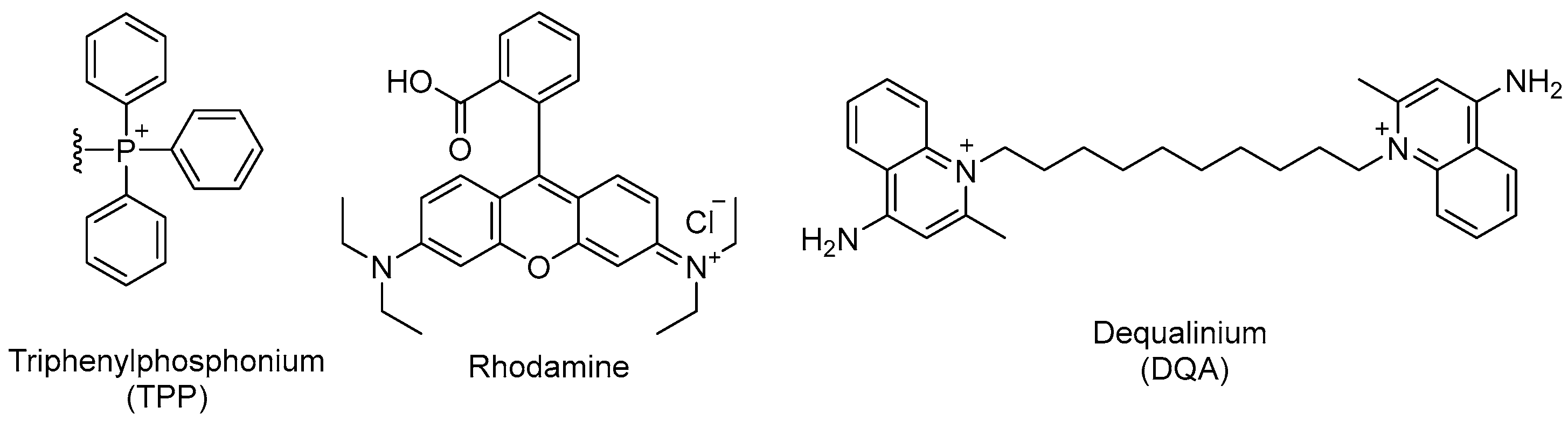
Many mitochondrial-targeting strategies exploit the highly negative potential of the inner mitochondrial membrane (IMM). This electrochemical gradient enables the accumulation of cations such as triphenylphosphonium, guanidinium, dequalinium, and rhodamine derivatives within the mitochondrial matrix (Figure 10). Most of these ligands are delocalized lipophilic cations with a positive charge, promoting electrostatic interactions with the anionic phospholipids of mitochondrial membranes and subsequent internalization (see Section 4.3) [215,216]. When conjugated with nanoparticles, these ligands allow for more precise drug delivery into mitochondria.
In eukaryotic cells, the MMP is a dynamic parameter that can change depending on the metabolic state, nutrient availability, and energy demands of the cell, and it is distributed among multiple functional mitochondria [217]. In kinetoplastids, the MMP is maintained even during life cycle stages when oxidative phosphorylation is no longer the primary source of ATP [218]. Since the MMP is an essential component of mitochondrial function and viability in eukaryotic cells (including protozoa), the selectivity of a mitochondrial-targeted nanosystem would not be based exclusively on qualitative differences in membrane potential, but rather on the parasite’s structural and functional dependence on a single organelle per cell.
Thus, nanosystems can promote high drug concentrations within intracellular mononuclear phagocyte system infected cells. Once internalized, certain nanocarriers undergo intracellular disassembly, enabling the targeted release of their cargo into mitochondrial compartments, including the kinetoplast of Leishmania spp. Collectively, these features position nanometric drug delivery systems as powerful platforms for improving antileishmanial efficacy and inform the rational design of next-generation therapeutics targeting parasite-specific mitochondrial structures [219]. Below, we summarize the principal classes of mitochondrial-targeted nanocarriers, originally developed in oncology and other mitochondrion-centered therapies, such as neurodegenerative diseases, and discuss their relevance to antiparasitic drug development.

5.1.1. Polymeric Nanoparticles

Polymeric nanoparticles are colloidal particles self-assembled from amphiphilic polymers that are highly biocompatible, minimally toxic, and capable of encapsulating therapeutic agents within their polymeric matrix or conjugating them on their surface. Their size typically falls within the submicron range (100–1000 nm). However, nanoparticles most effective in mitochondrial therapies are those <100 nm with a positive zeta potential, a condition that favors mitochondrial uptake [200]. This delivery system represents a multifunctional tool for the design of targeted therapies due to its ability to encapsulate bioactive agents, enable controlled release, and allow versatile surface functionalization to reach specific cellular compartments. Common polymeric designs in Leishmania include lipophilic blocks of polylactic acid (PLA), polyglycolic acid (PGA), poly(lactic-co-glycolic acid) (PLGA), and polycaprolactone (PCL), in combination with hydrophilic polyethylene glycol (PEG) blocks [220,221,222]. The latter provides greater stability, prolongs systemic circulation, and allows the conjugation of mitochondrion-targeting ligands (Table 2) [223]. Among these, the lipophilic cation triphenylphosphonium (TPP+, Figure 10), whose three benzene rings provide structural versatility for the incorporation of functional moieties [174], has been widely employed (see Section 4.3). In the context of Leishmania spp., accumulation within the cell can reach up to 10-fold-higher levels due to the parasite’s plasma membrane potential. Hence, this functionalization should enable nanoparticles to cross the parasite cell membrane, escape endolysosomal sequestration after internalization, and finally penetrate the highly negative double mitochondrial membrane of Leishmania spp. [207,224,225]. Such a strategy may offer significant advantages over conventional drugs by improving parasite specificity and reducing host toxicity.

5.1.2. Liposomes and DQAsomes

Liposomes are vesicular structures composed primarily of phospholipids and cholesterol [223]. They present a great functional adaptability and can be structurally modified to reach subcellular compartments such as mitochondria. Their aqueous core and one or more lipid bilayers allow them to encapsulate hydrophilic, lipophilic, and macromolecular drugs. Liposomes are widely employed in drug delivery owing to their biocompatibility (i.e., biodegradable and low toxicity), their dual-loading capacity that facilitates combination therapies, and their ability to improve the bioavailability of drugs with distinct pharmacokinetic properties [205,223]. In the context of leishmaniasis, the most successful example is the liposomal formulation of amphotericin B (AmBisome®, Foster City, CA, USA) used as first-line therapy for VL. Studies on liposomal amphotericin B formulations have shown that toxicity and efficacy against cutaneous and visceral leishmaniasis can be finely tuned by controlling lipid composition, membrane rigidity, and drug aggregation state. Rigid, cholesterol-containing liposomes with slow drug release exhibit low toxicity and high efficacy, whereas more fluid liposomes favor faster release and improved bioavailability for the topical and oral treatment of CL [237]. Among other examples of liposomal formulations of antileishmanial drugs, we can cite the following: the incorporation of meglumine antimoniate into anionic liposomes, thereby improving its antileishmanial efficacy while minimizing its cytotoxicity to macrophages [238,239]; PEGylated liposomes loaded with meglumine antimoniate, which present greater stability and time in circulation allowing prolonged distribution of the drug [240]; or topical liposomes containing miltefosine for the treatment of Leishmania major infection [241]. However, challenges regarding intracellular internalization efficiency, in vivo stability, and mitochondrial specificity persist, making their optimization an ongoing area of research [242].
The surface functionalization of liposomes is key for mitochondrial targeting. Such design must consider interactions with the mitochondrial membrane and intracellular trafficking mechanisms. Liposomes functionalized with TPP+ are able to preserve vesicular integrity while gaining the capacity to cross the cell membrane and localize to the mitochondrial matrix due to their strong affinity for the mitochondrial inner membrane [243,244]. Nonetheless, the systemic application of permanently positively charged mitochondrial-targeting moieties such as TPP+ may be constrained by unfavorable pharmacokinetic behavior. The strong lipophilic cationic character of these ligands promotes extensive binding to plasma proteins and nonspecific interactions with cellular membranes, which in turn enhances opsonization and uptake by the mononuclear phagocyte system (MPS), leading to rapid clearance from circulation and reduced half-life in vivo [245,246,247]. To overcome this, negatively charged polymers such as PEG are often used to shield the surface, thereby enhancing circulation time and improving the ability of these carriers to cross biological barriers [248]. However, PEGylated liposomes can be recognized and cleared by anti-PEG antibodies upon repeated administration [249], a phenomenon known as accelerated blood clearance [250]. As a result, PEG is increasingly being replaced by biocompatible (i.e., biodegradable, non-toxic, and non-immunogenic) natural polymers such as hyaluronic acid, providing effective protection for cationic ligands [251].
Rhodamine derivatives (Figure 10) are a class of mitochondriotropic dyes with lipophilic and cationic properties that confer high affinity for mitochondrial membranes, enabling their specific localization and accumulation within the organelle. Similar to other lipophilic cations, this accumulation is membrane-potential dependent, a feature that has been extensively exploited in bioimaging to identify metabolically active cells, to measure mitochondrial membrane potential, and as a delivery strategy in oncology [252,253]. For example, Biswas et al. showed that the surface modification of liposomes with rhodamine-123-conjugated polymers resulted in enhanced cellular uptake and facilitated penetration across the inner mitochondrial membrane, resulting in higher cytotoxicity towards cancer cells (Table 2) [229]. Moreover, rhodamine offers advantages as a biological tracer due to its commercial availability, low cost, high quantum yield, and minimal interference with underlying metabolic processes [254]. In the context of Leishmania spp., this versatility enables the experimental validation of the mitochondrial tropism of nanoparticles, while simultaneously functioning as an active ligand to transport antileishmanial agents directly into the mitochondrial microenvironment.
Dequalinium (DQA) is a dicationic amphiphilic compound composed of two quinaldinium rings linked by a decamethylene chain (Figure 10). This molecular architecture allows DQA to self-assemble into vesicular structures resembling liposomes, known as DQAsomes [228,255]. DQAsomes were the first mitochondrion-targeted vesicular nanocarriers shown to selectively accumulate in the mitochondrial matrix [255]. Their enhanced accumulation and retention are attributed to the amphiphilic nature of DQA, which enables the formation of liposome-like structures capable of transporting drugs and DNA via nonspecific endocytic pathways, driven by the highly negative electrochemical gradient of the mitochondrial membrane [200,256]. This unique combination of structural and electrochemical properties makes DQAsomes a versatile platform for localized drug encapsulation and release (Table 2).

5.1.3. Metallic Nanoparticles

Metallic nanoparticles hold great potential for the treatment and diagnosis of mitochondrial disorders due to their surface charges and capacity to conjugate with drugs, antibodies, and proteins, which not only protects them against host immune clearance but also increases their circulation time. They are typically composed of noble metals such as gold (Au) and silver (Ag), or metal oxides such as ZnO and Fe3O4. Their nanoscale dimensions confer a high surface-to-volume ratio, resulting in unique physicochemical properties, such as a large surface area per unit volume that exposes more functional groups on the surface, increasing their reactivity, adsorption, and interaction with biological molecules [257,258,259]. For instance, silver nanoparticles (AgNPs) have demonstrated antimicrobial activity, while gold nanoparticles (AuNPs) have been used to facilitate the translocation of drugs across cellular membranes.
These nanoparticles exhibit high structural stability and versatility for surface functionalization. Particle sizes between 5 and 260 nm are generally suitable for mitochondrial uptake [216]. However, their toxicity and potential to generate reactive oxygen species (ROS) in vital organs represent significant drawbacks in terms of selectivity [260]. Interestingly, this same ROS-generating capacity has been exploited in cancer therapy to selectively damage mitochondria of malignant cells and induce apoptosis. These effects are closely related to nanoparticle size and morphology, with fibrous or elongated nanoparticles larger than 400 nm being particularly difficult to eliminate from the body [260].
In the context of Leishmania spp., the physicochemical features of metallic nanoparticles, particularly their ability to generate ROS, could be strategically harnessed to induce selective damage to kDNA, provided that collateral host cell toxicity is minimized through specific mitochondrial targeting. This approach could improve both efficacy and safety profiles.
Iron oxide nanoparticles (IONPs), in particular, have demonstrated safe and effective action on mitochondria in cancer cells, overcoming challenges such as drug resistance and adverse side effects of conventional therapies. Their superparamagnetic properties enable their external guidance toward specific sites using magnetic fields, while their capacity to catalyze Haber–Weiss reactions allows the production of large amounts of ROS in cancer cells [206,216]. In the context of Leishmania spp., the physicochemical features of metallic nanoparticles, particularly their ability to generate ROS, could be strategically harnessed to induce selective damage to kDNA, provided that collateral host cell toxicity is minimized through specific mitochondrial targeting. This approach could improve both efficacy and safety profiles. The antileishmanial photodynamic therapeutic potential of ferromagnetic iron oxide nanorods was studied by Islam et al. [261]. They showed that these NPs induced oxidative stress in promastigotes and amastigotes of L. tropica, compromising parasite viability via apoptosis [261]. According to Rivas-García et al. [206], nanoparticles between 2 and 4 nm in size are optimal for entering subcellular compartments such as mitochondria.

5.2. Targeting Platforms Based on Mitochondrial Receptors and Transporters

The vast majority of mitochondrial proteins are encoded by the nuclear genome and require specialized translocation systems for their import into the organelle. Although the mitochondrial protein import systems of trypanosomatids and mammals share certain similarities, only a limited number of subunits of their machineries are conserved. These differences highlight the potential of this system, which is crucial for parasite survival and infection, as a promising drug target in trypanosomatids [262]. Chemical approaches to attack this import system could benefit from the mitochondrion-targeting strategies mentioned here.
In addition to strategies based on exogenous ligands or universal peptides, broadly acting cell-penetrating or organelle-targeting sequences have been widely employed to enhance intracellular drug delivery through conserved physicochemical mechanisms rather than cell-specific recognition. Classical cell-penetrating peptides (CPPs), such as TAT or poly-arginine, promote uptake across diverse cell types via endocytosis and membrane translocation [263]. Likewise, mitochondrial-targeting peptides (MTPs) exploit the universally conserved mitochondrial membrane potential and the TOM/TIM import machinery to drive matrix accumulation [264], while delocalized aromatic–cationic sequences such as the Szeto–Schiller peptides preferentially partition into mitochondrial membranes through electrostatic and lipophilic interactions [265]. Although these approaches efficiently improve intracellular and mitochondrial localization, their lack of inherent parasite specificity may lead to off-target effects in host cells. Consequently, there is growing evidence that endogenous components unique to trypanosomatids such as Leishmania could be exploited as selective markers or entry points for nanoparticles. These receptors and transporters, located in the kinetoplast and associated with essential survival processes of the parasite, not only could enhance mitochondrial specificity but also open new opportunities for the design of innovative therapeutic nanoplatforms. The most relevant components are summarized in Table 3, along with their function and potential applications in nanomedicine.
In summary, kinetoplastid mitochondrial transporters and receptors provide endogenous targets for directing nanoparticles to the mitochondria of Leishmania. Understanding their function, substrate specificity, and mitochondrial-targeting signals enables the rational design of more precise and effective drug delivery strategies, while minimizing off-target effects in host cells.

6. Conclusions and Future Perspectives

The kinetoplast represents a uniquely vulnerable and highly selective therapeutic target in Leishmania spp., integrating essential mitochondrial functions related to DNA maintenance, bioenergetics, redox balance, and parasite survival. Its distinctive molecular organization, together with the presence of a single mitochondrion in kinetoplastid parasites, provides a strong biological rationale for subcellularly targeted antileishmanial strategies capable of overcoming the major limitations of current chemotherapy, including systemic toxicity, limited selectivity, and the emergence of drug resistance [3,6].
Recent advances in molecular parasitology, medicinal chemistry, and nanotechnology have converged to enable innovative therapeutic approaches that exploit kinetoplast-specific vulnerabilities. As highlighted throughout this review, successful clinical translation will depend on integrating molecular pathophysiology with delivery strategies adapted to intracellular parasites and compatible with endemic, low-resource settings.

6.1. Integration of Molecular Pathophysiology and Innovative Therapies

Mitochondrial dysfunction is a central driver of cellular pathophysiology, as extensively demonstrated in cancer models, where alterations in bioenergetics, redox homeostasis, mitochondrial membrane potential, and genome integrity critically determine cell survival and therapeutic vulnerability [256]. Similar principles apply to Leishmania parasites, whose single mitochondrion integrates energy production, kinetoplast DNA (kDNA) maintenance, and oxidative stress control, rendering it particularly susceptible to targeted perturbation. Pathophysiological features such as an elevated mitochondrial membrane potential, limited antioxidant capacity, and reliance on kinetoplast-associated processes create exploitable vulnerabilities that can be selectively targeted without affecting host cells.
The therapeutic strategies discussed in this review—such as DNA minor groove binders, G-quadruplex ligands, and mitochondrion-targeted small molecules—which are summarized in Table 4, illustrate how detailed knowledge of parasite molecular biology can be translated into rational drug design [21,142]. Accumulating experimental evidence demonstrates that interference with kinetoplast-associated processes—including kDNA replication, topoisomerase activity, G-quadruplex formation, mitochondrial membrane potential maintenance, and redox homeostasis—leads to profound mitochondrial dysfunction and irreversible parasite death [38,53,62]. These tightly interconnected processes rely on molecular components that are absent or highly divergent in mammalian cells, providing a solid basis for selective pharmacological intervention. Collectively, these data position the kinetoplast not only as a validated drug target but also as a conceptual platform for mechanism-driven antiparasitic drug discovery.
In particular, mitochondrion-targeted compounds exploiting the strong negative membrane potential of the parasite enable preferential intramitochondrial accumulation, resulting in enhanced antiparasitic efficacy while limiting host toxicity [53,174]. When combined with nanomedicine-based delivery systems, these approaches further improve intracellular trafficking, subcellular specificity, and pharmacokinetic profiles, providing a realistic framework for repurposing existing chemotypes and accelerating the development of next-generation antileishmanial agents [190,196].
However, designing nanoparticles capable of selectively targeting the mitochondria of the parasite without affecting the host cell mitochondria is inherently challenging. Mitochondria are highly conserved organelles across eukaryotic cells, sharing similar membrane structures, surface proteins, and electrochemical gradients. This structural and functional similarity makes it difficult for nanoparticles to discriminate between host and parasite mitochondria once internalized. Furthermore, because Leishmania resides within the endolysosomal compartment of macrophages, nanoparticles must reach this compartment to access the parasite. Strategies that rely on general mitochondrial-targeting motifs (e.g., positive charges or lipophilic cations) may enhance uptake by all mitochondria within the host cell, increasing the risk of cytotoxicity. Consequently, indirect targeting approaches—such as directing nanoparticles to the host cell compartment containing the parasite—are often more feasible and safer than attempting to discriminate mitochondria directly [270].

6.2. Multidisciplinary Relevance and Dermatological Implications

Leishmaniasis is a paradigmatic neglected tropical disease that requires multidisciplinary solutions integrating parasitology, medicinal chemistry, pharmaceutical technology, immunology, and clinical dermatology. This need is particularly evident in cutaneous and mucocutaneous leishmaniasis, where parasite persistence within skin macrophages leads to chronic inflammation, tissue destruction, permanent scarring, and significant psychosocial burden [12].
Cutaneous involvement is not limited to visible lesions but is associated with prolonged disease duration, residual scarring, pain, pruritus, secondary infections, and a profound impairment of quality of life, as consistently captured by dermatology-specific outcome measures such as the Dermatology Life Quality Index. These dermatological manifestations underscore the need for therapeutic strategies capable of achieving efficient intralesional parasite clearance while minimizing tissue damage and long-term cosmetic and psychosocial sequelae [271]. Kinetoplast-directed therapies are especially relevant in the dermatological context, as they enable the selective elimination of intracellular amastigotes within skin lesions while minimizing systemic exposure. Nanoparticle-based delivery systems offer promising opportunities for topical, intralesional, or macrophage-targeted administration, potentially improving lesion resolution, reducing treatment duration, and enhancing patient adherence [192,195].
For the treatment of visceral leishmaniasis via parenteral administration, the localization of the parasite in the liver, spleen, and bone marrow generally aligns with the passive accumulation patterns of conventional nanoparticles, due to the fenestrated endothelium in these organs that allows nanoparticles to extravasate and reach resident macrophages [272]. However, in cutaneous leishmaniasis, infected macrophages reside in the dermis, a compartment poorly accessible to nanoparticles following either parenteral or topical administration. This limitation explains why formulations such as AmBisome are more effective for visceral leishmaniasis than cutaneous leishmaniasis [273,274], and why topical application has not been successful. Even in lesioned skin, it is difficult for nanoparticles, including very small ones, to reach the dermal layer intact and effectively interact with infected macrophages [275,276]. Therefore, for nanoparticles to be effective in the local treatment of cutaneous leishmaniasis, they must either be administered intralesionally or be designed to enhance drug diffusion and penetration, rather than relying on mitochondrial targeting. Hence, in the specific context of cutaneous leishmaniasis, drugs with intrinsic specificity for the parasite (i.e., kinetoplast) may be more effective than nanoparticles designed solely for mitochondrial targeting, emphasizing that rational therapeutic design should prioritize biological specificity over complex nanoparticle engineering.
Beyond dermatology, the principles discussed in this review are broadly applicable to other kinetoplastid diseases, reinforcing the value of shared molecular targets and delivery platforms across Leishmania, T. cruzi, and T. brucei infections [3,21]. From a global health perspective, translating kinetoplast-focused strategies into affordable and scalable therapies will require the early integration of formulation science, manufacturing feasibility, and regulatory considerations, together with sustained collaboration between academic, clinical, and public health stakeholders.
In conclusion, kinetoplast-directed therapies exemplify a mechanism-driven and subcellularly precise approach to antiparasitic drug development. By integrating molecular insight with innovative chemical and nanotechnological strategies, this framework offers a promising path toward safer, more effective, and context-appropriate treatments for leishmaniasis and other neglected tropical diseases. Moreover, such platforms may facilitate combination strategies integrating direct antiparasitic activity with host-directed immunomodulation by enabling the co-delivery of kinetoplast-targeted agents and immunomodulatory cues within infected macrophages, thereby simultaneously promoting parasite clearance and the restoration of effective local immune responses [16,160].

Author Contributions

Conceptualization, C.D. and J.C.; writing—original draft preparation, C.D., J.C., J.A.B.-B., J.C.C.-M., D.C., J.S.-A., K.T., S.E.; writing—review and editing, C.D., J.C., J.A.B.-B., J.C.C.-M., D.C., J.S.-A., K.T., S.E.; funding acquisition, C.D., J.C., S.E. All authors have read and agreed to the published version of the manuscript.

Funding

Funding by Agencia Estatal de Investigación (MCIN/AEI/10.13039/501100011033) (Co-funded by European Regional Development Fund. ERDF, “A way to build Europe”, by the “European Union”), through the grants PID2020-114207RB-I00, PID2022-136438OB-I00 and PID2023-148406OB-I00, is gratefully acknowledged. JCM is supported by a PhD scholarship (PREP2022-000704) funded by Ministerio de Ciencia e Innovación (MCIN/AEI/10.13039/501100011033). David Cisneros is supported by a PhD scholarship (PIPF-2022SAL-GL-24344) from Comunidad de Madrid, Spain.

Data Availability Statement

No new data were created or analyzed in this study. Data sharing is not applicable to this article.

Conflicts of Interest

The authors declare no conflicts of interest.

References

  1. WHO. Fact Sheet: Leishmaniasis. Available online: https://www.who.int/news-room/fact-sheets/detail/leishmaniasis (accessed on 12 December 2025).
  2. Ayala, A.; Llanes, A.; Lleonart, R.; Restrepo, C.M. Advances in Leishmania Vaccines: Current Development and Future Prospects. Pathogens 2024, 13, 812. [Google Scholar] [CrossRef]
  3. Tauheed, A.M.; Danazumi, A.U.; Adepoju, O.A.; Kobo, P.I.; Adamu, A.; Balogun, E.O. Kinetoplastid diseases: Insights into the mechanisms of drug action and resistance for novel drug discovery. Asp. Mol. Med. 2025, 5, 100071. [Google Scholar] [CrossRef]
  4. Van Assche, T.; Deschacht, M.; da Luz, R.A.; Maes, L.; Cos, P. Leishmania-macrophage interactions: Insights into the redox biology. Free Radic. Biol. Med. 2011, 51, 337–351. [Google Scholar] [CrossRef] [PubMed]
  5. Makarani, N.; Bharadava, K.; Kaushik, A.; Dave, A.; Gangawane, A.K.; Kaushal, R.S. Leishmaniasis: A multifaceted approach to diagnosis, maladies, drug repurposing and way forward. Microbe 2025, 6, 100239. [Google Scholar] [CrossRef]
  6. Singh, R.; Kashif, M.; Srivastava, P.; Manna, P.P. Recent Advances in Chemotherapeutics for Leishmaniasis: Importance of the Cellular Biochemistry of the Parasite and Its Molecular Interaction with the Host. Pathogens 2023, 12, 706. [Google Scholar] [CrossRef]
  7. Mostafavi, M.; Sharifi, I.; Farajzadeh, S.; Khazaeli, P.; Sharifi, H.; Pourseyedi, E.; Kakooei, S.; Bamorovat, M.; Keyhani, A.; Parizi, M.H.; et al. Niosomal formulation of amphotericin B alone and in combination with glucantime: In vitro and in vivo leishmanicidal effects. Biomed. Pharmacother. 2019, 116, 108942. [Google Scholar] [CrossRef]
  8. Sundar, S.; Jha, T.K.; Thakur, C.P.; Bhattacharya, S.K.; Rai, M. Oral miltefosine for the treatment of Indian visceral leishmaniasis. Trans. R. Soc. Trop. Med. Hyg. 2006, 100, S26–S33. [Google Scholar] [CrossRef]
  9. Nagle, A.S.; Khare, S.; Kumar, A.B.; Supek, F.; Buchynskyy, A.; Mathison, C.J.N.; Chennamaneni, N.K.; Pendem, N.; Buckner, F.S.; Gelb, M.H.; et al. Recent Developments in Drug Discovery for Leishmaniasis and Human African Trypanosomiasis. Chem. Rev. 2014, 114, 11305–11347. [Google Scholar] [CrossRef]
  10. Ponte-Sucre, A.; Gamarro, F.; Dujardin, J.C.; Barrett, M.P.; Lopez-Velez, R.; Garcia-Hernandez, R.; Pountain, A.W.; Mwenechanya, R.; Papadopoulou, B. Drug resistance and treatment failure in leishmaniasis: A 21st century challenge. PLoS Negl. Trop. Dis. 2017, 11, e0006052. [Google Scholar] [CrossRef]
  11. Vanaerschot, M.; Dumetz, F.; Roy, S.; Ponte-Sucre, A.; Arevalo, J.; Dujardin, J.C. Treatment failure in leishmaniasis: Drug-resistance or another (epi-) phenotype? Expert Rev. Anti-Infect. Ther. 2014, 12, 937–946. [Google Scholar] [CrossRef]
  12. Shmueli, M.; Ben-Shimol, S. Review of Leishmaniasis Treatment: Can We See the Forest through the Trees? Pharmacy 2024, 12, 30. [Google Scholar] [CrossRef]
  13. Kapil, S.; Singh, P.K.; Silakari, O. An update on small molecule strategies targeting leishmaniasis. Eur. J. Med. Chem. 2018, 157, 339–367. [Google Scholar] [CrossRef]
  14. Oliveira, L.F.; Schubach, A.O.; Martins, M.M.; Passos, S.L.; Oliveira, R.V.; Marzochi, M.C.; Andrade, C.A. Systematic review of the adverse effects of cutaneous leishmaniasis treatment in the New World. Acta Trop. 2011, 118, 87–96. [Google Scholar] [CrossRef]
  15. Laniado-Laborín, R.; Cabrales-Vargas, M.N. Amphotericin B: Side effects and toxicity. Rev. Iberoam. Micol. 2009, 26, 223–227. [Google Scholar] [CrossRef]
  16. Ebiloma, G.U.; Alhejeli, A.; de Koning, H.P. Interventions for Neglected Diseases Caused by Kinetoplastid Parasites: A One Health Approach to Drug Discovery, Development, and Deployment. Pharmaceuticals 2025, 18, 1415. [Google Scholar] [CrossRef] [PubMed]
  17. Sundar, S. Drug resistance in Indian visceral leishmaniasis. Trop. Med. Int. Health 2001, 6, 849–854. [Google Scholar] [CrossRef]
  18. Sundar, S.; More, D.K.; Singh, M.K.; Singh, V.P.; Sharma, S.; Makharia, A.; Kumar, P.C.; Murray, H.W. Failure of pentavalent antimony in visceral leishmaniasis in India: Report from the center of the Indian epidemic. Clin. Infect. Dis. 2000, 31, 1104–1107. [Google Scholar] [CrossRef]
  19. Van Bocxlaer, K.; Croft, S.L. Pharmacokinetics and pharmacodynamics in the treatment of cutaneous leishmaniasis—Challenges and opportunities. RSC Med. Chem. 2021, 12, 472–482. [Google Scholar] [CrossRef]
  20. Available online: https://dndi.org/news/2025/2024-rd-programmes-in-review-leishmaniasis/ (accessed on 20 August 2025).
  21. Marín, M.; López, M.; Gallego-Yerga, L.; Álvarez, R.; Peláez, R. Experimental structure based drug design (SBDD) applications for anti-leishmanial drugs: A paradigm shift? Med. Res. Rev. 2024, 44, 1055–1120. [Google Scholar] [CrossRef] [PubMed]
  22. Olías-Molero, A.I.; de la Fuente, C.; Cuquerella, M.; Torrado, J.J.; Alunda, J.M. Antileishmanial Drug Discovery and Development: Time to Reset the Model? Microorganisms 2021, 9, 2500. [Google Scholar] [CrossRef] [PubMed]
  23. Marques, S.A.; Merlotto, M.R.; Ramos, P.M.; Marques, M.E.A. American tegumentary leishmaniasis: Severe side effects of pentavalent antimonial in a patient with chronic renal failure. An. Bras. Dermatol 2019, 94, 355–357. [Google Scholar] [CrossRef]
  24. Mondal, D.; Alvar, J.; Hasnain, M.G.; Hossain, M.S.; Ghosh, D.; Huda, M.M.; Nabi, S.G.; Sundar, S.; Matlashewski, G.; Arana, B. Efficacy and safety of single-dose liposomal amphotericin B for visceral leishmaniasis in a rural public hospital in Bangladesh: A feasibility study. Lancet Glob. Health 2014, 2, e51–57. [Google Scholar] [CrossRef]
  25. Walsh, T.J.; Goodman, J.L.; Pappas, P.; Bekersky, I.; Buell, D.N.; Roden, M.; Barrett, J.; Anaissie, E.J. Safety, tolerance, and pharmacokinetics of high-dose liposomal amphotericin B (AmBisome) in patients infected with Aspergillus species and other filamentous fungi: Maximum tolerated dose study. Antimicrob. Agents Chemother. 2001, 45, 3487–3496. [Google Scholar] [CrossRef]
  26. Das, P.; Kandel, R.; Sikka, K.; Dey, A. Reversible ototoxicity: A rare adverse reaction of liposomal amphotericin-B used for the treatment of antimony-resistant visceral leishmaniasis in an elderly male. Clin. Med. Insights Case Rep. 2014, 7, 63–66. [Google Scholar] [CrossRef]
  27. Marking, U.; den Boer, M.; Das, A.K.; Ahmed, E.M.; Rollason, V.; Ahmed, B.N.; Davidson, R.N.; Ritmeijer, K. Hypokalaemia-induced rhabdomyolysis after treatment of post-Kala-azar dermal Leishmaniasis (PKDL) with high-dose AmBisome in Bangladesh—A case report. PLoS Negl. Trop. Dis. 2014, 8, e2864. [Google Scholar] [CrossRef][Green Version]
  28. Amphotericin-B liposomal. React. Wkly. 2020, 1794, 26. [CrossRef]
  29. Moreno, E.; Calvo, A.; Schwartz, J.; Navarro-Blasco, I.; Gonzalez-Penas, E.; Sanmartin, C.; Irache, J.M.; Espuelas, S. Evaluation of Skin Permeation and Retention of Topical Dapsone in Murine Cutaneous Leishmaniasis Lesions. Pharmaceutics 2019, 11, 607. [Google Scholar] [CrossRef]
  30. Wolf Nassif, P.; TFP, D.E.M.; Navasconi, T.R.; Mota, C.A.; Demarchi, I.G.; Aristides, S.M.A.; Lonardoni, M.V.C.; Teixeira, J.J.V.; Silveira, T.G.V. Safety and efficacy of current alternatives in the topical treatment of cutaneous leishmaniasis: A systematic review. Parasitology 2017, 144, 995–1004. [Google Scholar] [CrossRef] [PubMed]
  31. Kasabalis, D.; Chatzis, M.K.; Apostolidis, K.; Xenoulis, P.G.; Buono, A.; Petanides, T.; Leontides, L.S.; Polizopoulou, Z.S.; Steiner, J.M.; Suchodolski, J.S.; et al. Evaluation of nephrotoxicity and ototoxicity of aminosidine (paromomycin)-allopurinol combination in dogs with leishmaniosis due to Leishmania infantum: A randomized, blinded, controlled study. Exp. Parasitol. 2019, 206, 107768. [Google Scholar] [CrossRef] [PubMed]
  32. Pal, B.; Atem, T.D.; Kumari, S.; Murti, K.; Kumar, R.; Pandey, K.; Siddiqui, N.A.; Dhingra, S.; Chaudhary, V. Ophthalmic adverse effects of miltefosine in the treatment of leishmaniasis: A systematic review. Cutan. Ocul. Toxicol. 2024, 43, 190–197. [Google Scholar] [CrossRef] [PubMed]
  33. Astman, N.; Arbel, C.; Katz, O.; Barzilai, A.; Solomon, M.; Schwartz, E. Tolerability and Safety of Miltefosine for the Treatment of Cutaneous Leishmaniasis. Trop. Med. Infect. Dis. 2024, 9, 218. [Google Scholar] [CrossRef]
  34. Alonso, L.; Lemes, L.F.N.; Magoulas, G.E.; Costa, B.L.; Gomes, R.S.; Dorta, M.L.; Bolognesi, M.L.; Romeiro, L.A.S.; Calogeropoulou, T.; Alonso, A. Miltefosine analogues with comparable antileishmanial activity and significantly reduced macrophage cytotoxicity. Mem. Inst. Oswaldo Cruz 2025, 120, e240219. [Google Scholar] [CrossRef] [PubMed]
  35. Patel, T.A.; Lockwood, D.N. Pentamidine as secondary prophylaxis for visceral leishmaniasis in the immunocompromised host: Report of four cases. Trop. Med. Int. Health 2009, 14, 1064–1070. [Google Scholar] [CrossRef] [PubMed]
  36. Sundar, S.; Chatterjee, M. Visceral leishmaniasis—Current therapeutic modalities. Indian J. Med. Res. 2006, 123, 345–352. [Google Scholar] [PubMed]
  37. Gadelha, E.P.; Talhari, S.; Guerra, J.A.; Neves, L.O.; Talhari, C.; Gontijo, B.; Silva Junior, R.M.; Talhari, A.C. Efficacy and safety of a single dose pentamidine (7mg/kg) for patients with cutaneous leishmaniasis caused by L. guyanensis: A pilot study. An. Bras. Dermatol 2015, 90, 807–813. [Google Scholar] [CrossRef]
  38. Motta, M.C. Kinetoplast as a potential chemotherapeutic target of trypanosomatids. Curr. Pharm. Des. 2008, 14, 847–854. [Google Scholar] [CrossRef]
  39. Sacks, D.; Kamhawi, S. Molecular aspects of parasite-vector and vector-host interactions in leishmaniasis. Annu. Rev. Microbiol 2001, 55, 453–483. [Google Scholar] [CrossRef]
  40. Clos, J.; Grunebast, J.; Holm, M. Promastigote-to-Amastigote Conversion in Leishmania spp.—A Molecular View. Pathogens 2022, 11, 1052. [Google Scholar] [CrossRef]
  41. Paixao, A.R.; Dias, B.R.S.; Palma, L.C.; Tavares, N.M.; Brodskyn, C.I.; de Menezes, J.P.B.; Veras, P.S.T. Investigating the Phagocytosis of Leishmania using Confocal Microscopy. J. Vis. Exp. 2021, 173, e62459. [Google Scholar] [CrossRef]
  42. Silveira, M.B.; Gomes, R.S.; Shio, M.T.; Rugani, J.N.; Paranaiba, L.F.; Soares, R.P.; Ribeiro-Dias, F. Lipophosphoglycan from Dermotropic New World Leishmania Upregulates Interleukin-32 and Proinflammatory Cytokines Through TLR4 and NOD2 Receptors. Front. Cell. Infect. Microbiol. 2022, 12, 805720. [Google Scholar] [CrossRef]
  43. Bernardes, C.; Borges, V.M.; Veras, P.S.T.; Menezes, J.P.B. Membrane-associated glycoconjugates in parasitic protozoa: The central role of lipophosphoglycan in Leishmania. Curr. Top. Membr. 2025, 95, 267–287. [Google Scholar] [CrossRef]
  44. Sunter, J.D.; Yanase, R.; Wang, Z.; Catta-Preta, C.M.C.; Moreira-Leite, F.; Myskova, J.; Pruzinova, K.; Volf, P.; Mottram, J.C.; Gull, K. Leishmania flagellum attachment zone is critical for flagellar pocket shape, development in the sand fly, and pathogenicity in the host. Proc. Natl. Acad. Sci. USA 2019, 116, 6351–6360. [Google Scholar] [CrossRef]
  45. Camacho, E.; Rastrojo, A.; Sanchiz, A.; Gonzalez-de la Fuente, S.; Aguado, B.; Requena, J.M. Leishmania Mitochondrial Genomes: Maxicircle Structure and Heterogeneity of Minicircles. Genes 2019, 10, 758. [Google Scholar] [CrossRef]
  46. Yilmaz, I.C.; Dunuroglu, E.; Ayanoglu, I.C.; Ipekoglu, E.M.; Yildirim, M.; Girginkardesler, N.; Ozbel, Y.; Toz, S.; Ozbilgin, A.; Aykut, G.; et al. Leishmania kinetoplast DNA contributes to parasite burden in infected macrophages: Critical role of the cGAS-STING-TBK1 signaling pathway in macrophage parasitemia. Front. Immunol. 2022, 13, 1007070. [Google Scholar] [CrossRef]
  47. Kour, P.; Saha, P.; Sharma, D.K.; Singh, K. DNA topoisomerases as a drug target in Leishmaniasis: Structural and mechanistic insights. Int. J. Biol. Macromol. 2024, 256, 128401. [Google Scholar] [CrossRef]
  48. Santi, A.M.M.; Murta, S.M.F. Antioxidant defence system as a rational target for Chagas disease and Leishmaniasis chemotherapy. Mem. Inst. Oswaldo Cruz 2022, 117, e210401. [Google Scholar] [CrossRef]
  49. Madia, V.N.; Ialongo, D.; Patacchini, E.; Exertier, C.; Antonelli, L.; Colotti, G.; Messore, A.; Tudino, V.; Saccoliti, F.; Scipione, L.; et al. Inhibition of Leishmania infantum Trypanothione Reductase by New Aminopropanone Derivatives Interacting with the NADPH Binding Site. Molecules 2023, 28, 338. [Google Scholar] [CrossRef] [PubMed]
  50. Turcano, L.; Torrente, E.; Missineo, A.; Andreini, M.; Gramiccia, M.; Di Muccio, T.; Genovese, I.; Fiorillo, A.; Harper, S.; Bresciani, A.; et al. Identification and binding mode of a novel Leishmania Trypanothione reductase inhibitor from high throughput screening. PLoS Negl. Trop. Dis. 2018, 12, e0006969. [Google Scholar] [CrossRef]
  51. Battista, T.; Colotti, G.; Ilari, A.; Fiorillo, A. Targeting Trypanothione Reductase, a Key Enzyme in the Redox Trypanosomatid Metabolism, to Develop New Drugs against Leishmaniasis and Trypanosomiases. Molecules 2020, 25, 1924. [Google Scholar] [CrossRef]
  52. Arbon, D.; Ženíšková, K.; Šubrtová, K.; Mach, J.; Štursa, J.; Machado, M.; Zahedifard, F.; Leštinová, T.; Hierro-Yap, C.; Neuzil, J.; et al. Repurposing of MitoTam: Novel Anti-Cancer Drug Candidate Exhibits Potent Activity against Major Protozoan and Fungal Pathogens. Antimicrob. Agents Chemother. 2022, 66, e00727-22. [Google Scholar] [CrossRef] [PubMed]
  53. Luque-Ortega, J.R.; Reuther, P.; Rivas, L.; Dardonville, C. New benzophenone-derived bisphosphonium salts as leishmanicidal leads targeting mitochondria through inhibition of respiratory complex II. J. Med. Chem. 2010, 53, 1788–1798. [Google Scholar] [CrossRef]
  54. De Sarkar, S.; Sarkar, D.; Sarkar, A.; Dighal, A.; Staniek, K.; Gille, L.; Chatterjee, M. Berberine chloride mediates its antileishmanial activity by inhibiting Leishmania mitochondria. Parasitol. Res. 2019, 118, 335–345. [Google Scholar] [CrossRef]
  55. Mittra, B.; Saha, A.; Chowdhury, A.R.; Pal, C.; Mandal, S.; Mukhopadhyay, S.; Bandyopadhyay, S.; Majumder, H.K. Luteolin, an abundant dietary component is a potent anti-leishmanial agent that acts by inducing topoisomerase II-mediated kinetoplast DNA cleavage leading to apoptosis. Mol. Med. 2000, 6, 527–541. [Google Scholar] [CrossRef]
  56. Fidalgo, L.M.; Gille, L. Mitochondria and Trypanosomatids: Targets and Drugs. Pharm. Res. 2011, 28, 2758–2770. [Google Scholar] [CrossRef] [PubMed]
  57. Jones, N.G.; Catta-Preta, C.M.C.; Lima, A.P.C.A.; Mottram, J.C. Genetically Validated Drug Targets in Leishmania: Current Knowledge and Future Prospects. ACS Infect. Dis. 2018, 4, 467–477. [Google Scholar] [CrossRef] [PubMed]
  58. Singh, V.K.; Tiwari, R.; Rajneesh; Kumar, A.; Chauhan, S.B.; Sudarshan, M.; Mehrotra, S.; Gautam, V.; Sundar, S.; Kumar, R. Advancing Treatment for Leishmaniasis: From Overcoming Challenges to Embracing Therapeutic Innovations. ACS Infect. Dis. 2025, 11, 47–68. [Google Scholar] [CrossRef] [PubMed]
  59. Fairlamb, A.H.; Wyllie, S. The critical role of mode of action studies in kinetoplastid drug discovery. Front. Drug Discov. 2023, 3, 1185679. [Google Scholar] [CrossRef]
  60. De Rycker, M.; Wyllie, S.; Horn, D.; Read, K.D.; Gilbert, I.H. Anti-trypanosomatid drug discovery: Progress and challenges. Nat. Rev. Microbiol. 2023, 21, 35–50. [Google Scholar] [CrossRef]
  61. Kourbeli, V.; Chontzopoulou, E.; Moschovou, K.; Pavlos, D.; Mavromoustakos, T.; Papanastasiou, I.P. An Overview on Target-Based Drug Design against Kinetoplastid Protozoan Infections: Human African Trypanosomiasis, Chagas Disease and Leishmaniases. Molecules 2021, 26, 4629. [Google Scholar] [CrossRef]
  62. Amodeo, S.; Bregy, I.; Ochsenreiter, T. Mitochondrial genome maintenance-the kinetoplast story. FEMS Microbiol. Rev. 2023, 47, fuac047. [Google Scholar] [CrossRef]
  63. Marini, J.C.; Levene, S.D.; Crothers, D.M.; Englund, P.T. A bent helix in kinetoplast DNA. Cold Spring Harb. Symp. Quant. Biol. 1983, 47, 279–283. [Google Scholar] [CrossRef] [PubMed]
  64. Marini, J.C.; Levene, S.D.; Crothers, D.M.; Englund, P.T. Bent helical structure in kinetoplast DNA. Proc. Natl. Acad. Sci. USA 1982, 79, 7664–7668. [Google Scholar] [CrossRef]
  65. Wilson, W.D.; Tanious, F.A.; Mathis, A.; Tevis, D.; Hall, J.E.; Boykin, D.W. Antiparasitic compounds that target DNA. Biochimie 2008, 90, 999–1014. [Google Scholar] [CrossRef] [PubMed]
  66. Shapiro, T.A.; Englund, P.T. The structure and replication of kinetoplast DNA. Ann. Rev. Microbiol. 1995, 49, 117–143. [Google Scholar] [CrossRef]
  67. Shapiro, T.A.; Englund, P.T. Selective cleavage of kinetoplast DNA minicircles promoted by antitrypanosomal drugs. Proc. Natl. Acad. Sci. USA 1990, 87, 950–954. [Google Scholar] [CrossRef]
  68. Zuma, A.A.; Cavalcanti, D.P.; Zogovich, M.; Machado, A.C.L.; Mendes, I.C.; Thiry, M.; Galina, A.; de Souza, W.; Machado, C.R.; Motta, M.C.M. Unveiling the effects of berenil, a DNA-binding drug, on Trypanosoma cruzi: Implications for kDNA ultrastructure and replication. Parasitol. Res. 2015, 114, 419–430. [Google Scholar] [CrossRef]
  69. Wilson, W.D.; Nguyen, B.; Tanious, F.A.; Mathis, A.; Hall, J.E.; Stephens, C.E.; Boykin, D.W. Dications that target the DNA minor groove: Compound design and preparation, DNA interactions, cellular distribution and biological activity. Curr. Med. Chem.—Anticancer Agents 2005, 5, 389–408. [Google Scholar] [CrossRef]
  70. Soeiro, M.N.C.; De Souza, E.M.; Stephens, C.E.; Boykin, D.W. Aromatic diamidines as antiparasitic agents. Expert Opin. Invest. Drugs 2005, 14, 957–972. [Google Scholar] [CrossRef]
  71. Soeiro, M.N.C.; Werbovetz, K.; Boykin, D.W.; Wilson, W.D.; Wang, M.Z.; Hemphill, A. Novel amidines and analogues as promising agents against intracellular parasites: A systematic review. Parasitology 2013, 140, 929–951. [Google Scholar] [CrossRef]
  72. Paul, A.; Nanjunda, R.; Wilson, W.D. Binding to the DNA Minor Groove by Heterocyclic Dications: From AT Specific to GC Recognition Compounds. Curr. Protoc. 2023, 3, e729. [Google Scholar] [CrossRef] [PubMed]
  73. Miao, Y.; Lee, M.P.H.; Parkinson, G.N.; Batista-Parra, A.; Ismail, M.A.; Neidle, S.; Boykin, D.W.; Wilson, W.D. Out-of-Shape DNA Minor Groove Binders: Induced Fit Interactions of Heterocyclic Dications with the DNA Minor Groove. Biochemistry 2005, 44, 14701–14708. [Google Scholar] [CrossRef]
  74. Ismail, M.A.; Batista-Parra, A.; Miao, Y.; Wilson, W.D.; Wenzler, T.; Brun, R.; Boykin, D.W. Dicationic near-linear biphenyl benzimidazole derivatives as DNA-targeted antiprotozoal agents. Bioorg. Med. Chem. 2005, 13, 6718–6726. [Google Scholar] [CrossRef]
  75. Farahat, A.A.; Kumar, A.; Wenzler, T.; Brun, R.; Paul, A.; Guo, P.; Wilson, W.D.; Boykin, D.W. Investigation of the effect of structure modification of furamidine on the DNA minor groove binding and antiprotozoal activity. Eur. J. Med. Chem. 2023, 252, 115287. [Google Scholar] [CrossRef]
  76. Acosta-Reyes, F.J.; Dardonville, C.; de Koning, H.P.; Natto, M.; Subirana, J.A.; Campos, J.L. In and out of the minor groove: Interaction of an AT-rich DNA with the drug CD27. Acta Cryst. 2014, D70, 1614–1621. [Google Scholar] [CrossRef]
  77. Glass, L.S.; Nguyen, B.; Goodwin, K.D.; Dardonville, C.; Wilson, W.D.; Long, E.C.; Georgiadis, M.M. Crystal structure of a trypanocidal 4,4’-bis(imidazolinylamino)diphenylamine bound to DNA. Biochemistry 2009, 48, 5943–5952. [Google Scholar] [CrossRef] [PubMed][Green Version]
  78. Dardonville, C.; Nué Martinez, J.J. Bis(2-aminoimidazolines) and Bisguanidines: Synthetic Approaches, Antiparasitic Activity and DNA Binding Properties. Curr. Med. Chem. 2017, 24, 3606–3632. [Google Scholar] [CrossRef] [PubMed]
  79. Montalvo-Quirós, S.; Taladriz-Sender, A.; Kaiser, M.; Dardonville, C. Antiprotozoal Activity and DNA Binding of Dicationic Acridones. J. Med. Chem. 2015, 58, 1940–1949. [Google Scholar] [CrossRef]
  80. Nieto, L.; Mascaraque, A.; Miller, F.; Glacial, F.; Ríos Martínez, C.; Kaiser, M.; Brun, R.; Dardonville, C. Synthesis and Antiprotozoal Activity of N-Alkoxy Analogues of the Trypanocidal Lead Compound 4,4’-Bis(imidazolinylamino)diphenylamine with Improved Human Blood-Brain Barrier Permeability. J. Med. Chem. 2011, 54, 485–494. [Google Scholar] [CrossRef]
  81. Rodríguez, F.; Rozas, I.; Kaiser, M.; Brun, R.; Nguyen, B.; Wilson, W.D.; García, R.N.; Dardonville, C. New bis(2-aminoimidazoline) and bisguanidine DNA minor groove binders with potent in vivo antitrypanosomal and antiplasmodial activity. J. Med. Chem. 2008, 51, 909–923. [Google Scholar] [CrossRef] [PubMed]
  82. Dardonville, C.; Barrett, M.P.; Brun, R.; Kaiser, M.; Tanious, F.; Wilson, W.D. DNA binding affinity of bisguanidine and bis(2-aminoimidazoline) derivatives with in vivo antitrypanosomal activity. J. Med. Chem. 2006, 49, 3748–3752. [Google Scholar] [CrossRef]
  83. Dardonville, C.; Brun, R. Bisguanidine, bis(2-aminoimidazoline), and polyamine derivatives as potent and selective chemotherapeutic agents against Trypanosoma brucei rhodesiense. Synthesis and in vitro evaluation. J. Med. Chem. 2004, 47, 2296–2307. [Google Scholar] [CrossRef] [PubMed]
  84. Ríos Martínez, C.H.; Lagartera, L.; Kaiser, M.; Dardonville, C. Antiprotozoal activity and DNA binding of N-substituted N-phenylbenzamide and 1,3-diphenylurea bisguanidines. Eur. J. Med. Chem. 2014, 81, 481–491. [Google Scholar] [CrossRef] [PubMed]
  85. Guedes-da-Silva, F.H.; Batista, D.G.J.; Meuser, M.B.; Demarque, K.C.; Fulco, T.O.; Araújo, J.S.; Da Silva, P.B.; Da Silva, C.F.; Patrick, D.A.; Bakunova, S.M.; et al. In Vitro and In Vivo Trypanosomicidal Action of Novel Arylimidamides against Trypanosoma cruzi. Antimicrob. Agents Chemother. 2016, 60, 2425–2434. [Google Scholar] [CrossRef]
  86. Timm, B.L.; da Silva, P.B.; Batista, M.M.; da Silva, F.H.; da Silva, C.F.; Tidwell, R.R.; Patrick, D.A.; Jones, S.K.; Bakunov, S.A.; Bakunova, S.M.; et al. In vitro and in vivo biological effects of novel arylimidamide derivatives against Trypanosoma cruzi. Antimicrob. Agents Chemother. 2014, 58, 3720–3726. [Google Scholar] [CrossRef] [PubMed][Green Version]
  87. Liu, Z.-y.; Wenzler, T.; Brun, R.; Zhu, X.; Boykin, D.W. Synthesis and antiparasitic activity of new bis-arylimidamides: DB766 analogs modified in the terminal groups. Eur. J. Med. Chem. 2014, 83, 167–173. [Google Scholar] [CrossRef]
  88. De Araújo, J.S.; Da Silva, C.F.; Batista, D.G.J.; Da Silva, P.B.; Meuser, M.B.; Aiub, C.A.F.; Da Silva, M.F.V.; Araújo-Lima, C.F.; Banerjee, M.; Farahat, A.A.; et al. In vitro and in vivo studies of the biological activity of novel arylimidamides against Trypanosoma cruzi. Antimicrob. Agents Chemother. 2014, 58, 4191–4195. [Google Scholar] [CrossRef]
  89. Batista Dda, G.; Batista, M.M.; de Oliveira, G.M.; do Amaral, P.B.; Lannes-Vieira, J.; Britto, C.C.; Junqueira, A.; Lima, M.M.; Romanha, A.J.; Sales Junior, P.A.; et al. Arylimidamide DB766, a potential chemotherapeutic candidate for Chagas’ disease treatment. Antimicrob. Agents Chemother. 2010, 54, 2940–2952. [Google Scholar] [CrossRef]
  90. Nué-Martinez, J.J.; Cisneros, D.; Moreno-Blázquez, M.d.V.; Fonseca-Berzal, C.; Manzano, J.I.; Kraeutler, D.; Ungogo, M.A.; Aloraini, M.A.; Elati, H.A.A.; Ibáñez-Escribano, A.; et al. Synthesis and Biophysical and Biological Studies of N-Phenylbenzamide Derivatives Targeting Kinetoplastid Parasites. J. Med. Chem. 2023, 66, 13452–13480. [Google Scholar] [CrossRef]
  91. Abdelhameed, A.; Feng, M.; Joice, A.C.; Zywot, E.M.; Jin, Y.; La Rosa, C.; Liao, X.; Meeds, H.L.; Kim, Y.; Li, J.; et al. Synthesis and Antileishmanial Evaluation of Arylimidamide-Azole Hybrids Containing a Phenoxyalkyl Linker. ACS Infect. Dis. 2021, 7, 1901–1922. [Google Scholar] [CrossRef]
  92. Zhu, X.; Farahat, A.A.; Mattamana, M.; Joice, A.; Pandharkar, T.; Holt, E.; Banerjee, M.; Gragg, J.L.; Hu, L.; Kumar, A.; et al. Synthesis and pharmacological evaluation of mono-arylimidamides as antileishmanial agents. Bioorg. Med. Chem. Lett. 2016, 26, 2551–2556. [Google Scholar] [CrossRef]
  93. Pandharkar, T.; Zhu, X.; Mathur, R.; Jiang, J.; Schmittgen, T.D.; Shaha, C.; Werbovetz, K.A. Studies on the antileishmanial mechanism of action of the arylimidamide DB766: Azole interactions and role of CYP5122A1. Antimicrob. Agents Chemother. 2014, 58, 4682–4689. [Google Scholar] [CrossRef]
  94. Zhu, X.; Liu, Q.; Yang, S.; Parman, T.; Green, C.E.; Mirsalis, J.C.; Soeiro, M.D.N.C.; De Souza, E.M.; Da Silva, C.F.; Batista, D.D.G.J.; et al. Evaluation of arylimidamides DB1955 and DB1960 as candidates against visceral leishmaniasis and Chagas’ disease: In vivo efficacy, acute toxicity, pharmacokinetics, and toxicology studies. Antimicrob. Agents Chemother. 2012, 56, 3690–3699. [Google Scholar] [CrossRef]
  95. Reid, C.S.; Farahat, A.A.; Zhu, X.; Pandharkar, T.; Boykin, D.W.; Werbovetz, K.A. Antileishmanial bis-arylimidamides: DB766 analogs modified in the linker region and bis-arylimidamide structure-activity relationships. Bioorg. Med. Chem. Lett. 2012, 22, 6806–6810. [Google Scholar] [CrossRef]
  96. Banerjee, M.; Farahat, A.A.; Kumar, A.; Wenzler, T.; Brun, R.; Munde, M.M.; Wilson, W.D.; Zhu, X.; Werbovetz, K.A.; Boykin, D.W. Synthesis, DNA binding and antileishmanial activity of low molecular weight bis-arylimidamides. Eur. J. Med. Chem. 2012, 55, 449–454. [Google Scholar] [CrossRef][Green Version]
  97. Chai, Y.; Munde, M.; Kumar, A.; Mickelson, L.; Lin, S.; Campbell, N.H.; Banerjee, M.; Akay, S.; Liu, Z.; Farahat, A.A.; et al. Structure-Dependent Binding of Arylimidamides to the DNA Minor Groove. ChemBioChem 2014, 15, 68–79. [Google Scholar] [CrossRef]
  98. Rettig, M.; Germann, M.W.; Wang, S.; Wilson, W.D. Molecular Basis for Sequence-Dependent Induced DNA Bending. ChemBioChem 2013, 14, 323–331. [Google Scholar] [CrossRef]
  99. Hunt, R.A.; Munde, M.; Kumar, A.; Ismail, M.A.; Farahat, A.A.; Arafa, R.K.; Say, M.; Batista-Parra, A.; Tevis, D.; Boykin, D.W.; et al. Induced topological changes in DNA complexes: Influence of DNA sequences and small molecule structures. Nucleic Acids Res. 2011, 39, 4265–4274. [Google Scholar] [CrossRef]
  100. Ríos Martínez, C.; Miller, F.; Ganeshamoorthy, K.; Glacial, F.; Kaiser, M.; de Koning, H.; Eze, A.; Lagartera, L.; Herraiz, T.; Dardonville, C. A new non-polar N-hydroxy imidazoline lead compound with improved activity in a murine model of late stage Trypanosoma brucei brucei infection is not cross-resistant with diamidines. Antimicrob. Agents Chemother. 2015, 59, 890–904. [Google Scholar] [CrossRef] [PubMed]
  101. Millan, C.R.; Acosta-Reyes, F.J.; Lagartera, L.; Ebiloma, G.; Lemgruber, L.; Nué-Martinez, J.J.; Saperas, N.; Dardonville, C.; de Koning, H.; Campos, J.L. Functional and structural analysis of AT-specific minor groove binders that disrupt DNA-protein interactions and cause disintegration of the Trypanosoma brucei kinetoplast. Nucleic Acids Res. 2017, 45, 8378–8391. [Google Scholar] [CrossRef] [PubMed]
  102. Wang, J.; Pappas-Brown, V.; Englund, P.T.; Jensen, R.E. TbKAP6, a Mitochondrial HMG Box-Containing Protein in Trypanosoma brucei, Is the First Trypanosomatid Kinetoplast-Associated Protein Essential for Kinetoplast DNA Replication and Maintenance. Eukaryot. Cell 2014, 13, 919–932. [Google Scholar] [CrossRef] [PubMed]
  103. Delespaux, V.; de Koning, H.P. Transporters in Anti-Parasitic Drug Development and Resistance. In Trypanosomatid Diseases; Wiley-VCH Verlag GmbH & Co. KGaA: Weinheim, Germany, 2013; pp. 335–349. [Google Scholar]
  104. Munday, J.C.; Settimo, L.; de Koning, H.P. Transport proteins determine drug sensitivity and resistance in a protozoan parasite, Trypanosoma brucei. Front. Pharmacol. 2015, 6, 32. [Google Scholar] [CrossRef]
  105. Daliry, A.; Pires, M.Q.; Silva, C.F.; Pacheco, R.S.; Munde, M.; Stephens, C.E.; Kumar, A.; Ismail, M.A.; Liu, Z.; Farahat, A.A.; et al. The trypanocidal activity of amidine compounds does not correlate with their binding affinity to Trypanosoma cruzi kinetoplast DNA. Antimicrob. Agents Chemother. 2011, 55, 4765–4773. [Google Scholar] [CrossRef] [PubMed]
  106. Nué-Martinez, J.J.; Maturana, M.; Lagartera, L.; Rodríguez-Gutiérrez, J.-A.; Boer, R.; Campos, J.L.; Saperas, N.; Dardonville, C. Crystal structure of the HMGA AT-hook 1 domain bound to the minor groove of AT-rich DNA and inhibition by antikinetoplastid drugs. Sci. Rep. 2024, 14, 26173. [Google Scholar] [CrossRef]
  107. Guo, P.; Farahat, A.A.; Paul, A.; Boykin, D.W.; Wilson, W.D. Engineered modular heterocyclic-diamidines for sequence-specific recognition of mixed AT/GC base pairs at the DNA minor groove. Chem. Sci. 2021, 12, 15849–15861. [Google Scholar] [CrossRef]
  108. Suckling, C.J.; Hunter, I.S.; Scott, F.J. Multitargeted Anti-Infective Drugs: Resilience to Resistance in the Antimicrobial Resistance Era. Future Drug Discov. 2022, 4, FDD73. [Google Scholar] [CrossRef]
  109. Khalaf, A.I.; Bourdin, C.; Breen, D.; Donoghue, G.; Scott, F.J.; Suckling, C.J.; MacMillan, D.; Clements, C.; Fox, K.; Sekibo, D.A.T. Design, synthesis and antibacterial activity of minor groove binders: The role of non-cationic tail groups. Eur. J. Med. Chem. 2012, 56, 39–47. [Google Scholar] [CrossRef]
  110. Perieteanu, M.C.; McGee, L.M.C.; Shaw, C.D.; MacMillan, D.S.; Khalaf, A.I.; Gillingwater, K.; Beveridge, R.; Carter, K.C.; Suckling, C.J.; Scott, F.J. Selective Anti-Leishmanial Strathclyde Minor Groove Binders Using an N-Oxide Tail-Group Modification. Int. J. Mol. Sci. 2022, 23, 11912. [Google Scholar] [CrossRef] [PubMed]
  111. Brooke, D.P.; McGee, L.M.C.; Giordani, F.; Cross, J.M.; Khalaf, A.I.; Irving, C.; Gillingwater, K.; Shaw, C.D.; Carter, K.C.; Barrett, M.P.; et al. Truncated S-MGBs: Towards a parasite-specific and low aggregation chemotype. RSC Med. Chem. 2021, 12, 1391–1401. [Google Scholar] [CrossRef] [PubMed]
  112. Azad, I.; Ahmad, R.; Khan, T.; Saquib, M.; Hassan, F.; Akhter, Y.; Khan, A.R.; Nasibullah, M. Phenanthridine Derivatives as Promising New Anticancer Agents: Synthesis, Biological Evaluation and Binding Studies. Future Med. Chem. 2020, 12, 709–739. [Google Scholar] [CrossRef]
  113. Bailly, C.; Arafa, R.K.; Tanious, F.A.; Laine, W.; Tardy, C.; Lansiaux, A.; Colson, P.; Boykin, D.W.; Wilson, W.D. Molecular Determinants for DNA Minor Groove Recognition: Design of a Bis-Guanidinium Derivative of Ethidium That Is Highly Selective for AT-Rich DNA Sequences. Biochemistry 2005, 44, 1941–1952. [Google Scholar] [CrossRef]
  114. Prunkl, C.; Pichlmaier, M.; Winter, R.; Kharlanov, V.; Rettig, W.; Wagenknecht, H.-A. Optical, Redox, and DNA-Binding Properties of Phenanthridinium Chromophores: Elucidating the Role of the Phenyl Substituent for Fluorescence Enhancement of Ethidium in the Presence of DNA. Chem. Eur. J. 2010, 16, 3392–3402. [Google Scholar] [CrossRef]
  115. Banerjee, J.; Bhattacharjee, A.; Biswas, A.; Chattopadhyay, S.K. Synthesis, bio-physical and anti-leishmanial studies of some novel indolo[3,2-a]phenanthridine derivatives. Bioorg. Chem. 2022, 123, 105766. [Google Scholar] [CrossRef]
  116. Battista, S.; Fedele, M.; Secco, L.; Ingo, A.M.D.; Sgarra, R.; Manfioletti, G. Binding to the Other Side: The AT-Hook DNA-Binding Domain Allows Nuclear Factors to Exploit the DNA Minor Groove. Int. J. Mol. Sci. 2024, 25, 8863. [Google Scholar] [CrossRef] [PubMed]
  117. Garabedian, A.; Jeanne Dit Fouque, K.; Chapagain, P.P.; Leng, F.; Fernandez-Lima, F. AT-hook peptides bind the major and minor groove of AT-rich DNA duplexes. Nucleic Acids Res. 2022, 50, 2431–2439. [Google Scholar] [CrossRef] [PubMed]
  118. Fonfría-Subirós, E.; Acosta-Reyes, F.; Saperas, N.; Pous, J.; Subirana, J.A.; Campos, J.L. Crystal structure of a complex of DNA with one AT-hook of HMGA1. PLoS ONE 2012, 7, e37120. [Google Scholar] [CrossRef] [PubMed]
  119. Nué-Martinez, J.J.; Leo-Barriga, M.; Herranz, F.; Koutsogiannis, Z.; Denny, P.W.; Ebiloma, G.U.; Dardonville, C.; González-Paredes, A. Nanostructured Lipid Carrier for Intracellular Delivery of a Bis(pyridine-2-carboxamidine) DNA Minor Groove Binder Active against Leishmania. ACS Omega 2025, 10, 7795–7805. [Google Scholar] [CrossRef]
  120. Laguna, A.; Martínez-Alonso, B.; Guarnizo-Herrero, V.; Nué-Martinez, J.J.; Dardonville, C.; Torrado-Santiago, S.; Torrado-Salmerón, C. Development and Characterization of a New Oral Antileishmanial Bis(pyridine-2-Carboxamidine) Drug Through Innovative Dissolution Testing in Biorelevant Media Combined with Pharmacokinetic Studies. Pharmaceutics 2025, 17, 838. [Google Scholar] [CrossRef]
  121. Kelly, B.L.; Singh, G.; Aiyar, A. Molecular and Cellular Characterization of an AT-Hook Protein from Leishmania. PLoS ONE 2011, 6, e21412. [Google Scholar] [CrossRef][Green Version]
  122. Lamba, S.; Roy, A. DNA topoisomerases in the unicellular protozoan parasites: Unwinding the mystery. Biochem. Pharmacol. 2022, 203, 115158. [Google Scholar] [CrossRef]
  123. Saha, S.; Chowdhury, S.R.; Majumder, H.K. DNA Topoisomerases of Kinetoplastid Parasites: Brief Overview and Recent Perspectives. Curr. Issues Mol. Biol. 2019, 31, 45–62. [Google Scholar] [CrossRef] [PubMed]
  124. Lindsay, M.E.; Gluenz, E.; Gull, K.; Englund, P.T. A new function of Trypanosoma brucei mitochondrial topoisomerase II is to maintain kinetoplast DNA network topology. Mol. Microbiol. 2008, 70, 1465–1476. [Google Scholar] [CrossRef]
  125. Wang, Z.; Englund, P.T. RNA interference of a trypanosome topoisomerase II causes progressive loss of mitochondrial DNA. EMBO J. 2001, 20, 4674–4683. [Google Scholar] [CrossRef]
  126. Chowdhury, S.R.; Kumar, A.; Godinho, J.L.P.; De Macedo Silva, S.T.; Zuma, A.A.; Saha, S.; Kumari, N.; Rodrigues, J.C.F.; Sundar, S.; Dujardin, J.-C.; et al. Voacamine alters Leishmania ultrastructure and kills parasite by poisoning unusual bi-subunit topoisomerase IB. Biochem. Pharm. 2017, 138, 19–30. [Google Scholar] [CrossRef]
  127. Coletta, A.; Morozzo della Rocca, B.; Jaisankar, P.; Majumder, H.K.; Chillemi, G.; Sanna, N.; Desideri, A. Assignment of UV−vis Spectrum of (3,3′)-Diindolylmethane, a Leishmania donovani Topoisomerase IB Inhibitor and a Candidate DNA Minor Groove Binder. J. Phys. Chem. A 2010, 114, 7121–7126. [Google Scholar] [CrossRef]
  128. Roy, A.; Das, B.B.; Ganguly, A.; Bose Dasgupta, S.; Khalkho, N.V.M.; Pal, C.; Dey, S.; Giri, V.S.; Jaisankar, P.; Dey, S.; et al. An insight into the mechanism of inhibition of unusual bi-subunit topoisomerase I from Leishmania donovani by 3,3′-di-indolylmethane, a novel DNA topoisomerase I poison with a strong binding affinity to the enzyme. Biochem. J. 2007, 409, 611–622. [Google Scholar] [CrossRef] [PubMed]
  129. Roy, A.; Chowdhury, S.; Sengupta, S.; Mandal, M.; Jaisankar, P.; D’Annessa, I.; Desideri, A.; Majumder, H.K. Development of Derivatives of 3, 3′-Diindolylmethane as Potent Leishmania donovani Bi-Subunit Topoisomerase IB Poisons. PLoS ONE 2011, 6, e28493. [Google Scholar] [CrossRef] [PubMed]
  130. Kour, P.; Saha, P.; Bhattacharya, S.; Kumari, D.; Debnath, A.; Roy, A.; Sharma, D.K.; Mukherjee, D.; Singh, K. Design, synthesis, and biological evaluation of 3,3′-diindolylmethane N-linked glycoconjugate as a leishmanial topoisomerase IB inhibitor with reduced cytotoxicity. RSC Med. Chem. 2023, 14, 2100–2114. [Google Scholar] [CrossRef] [PubMed]
  131. Das, S.; Babu, N.K.; Mazire, P.; Roy, A.; Kumar, R.; Singh, S.; Sharma, D.K. Indolylmaleimide derivatives as a new class of anti-leishmanial agents: Synthesis and biological evaluation. RSC Med. Chem. 2025, 16, 2808–2818. [Google Scholar] [CrossRef]
  132. Rao, S.P.S.; Gould, M.K.; Noeske, J.; Saldivia, M.; Jumani, R.S.; Ng, P.S.; René, O.; Chen, Y.-L.; Kaiser, M.; Ritchie, R.; et al. Cyanotriazoles are selective topoisomerase II poisons that rapidly cure trypanosome infections. Science 2023, 380, 1349–1356. [Google Scholar] [CrossRef]
  133. Goel, N.; Gupta, V.K.; Garg, A.; Bhoumik, A.; Biswas, R.; Natarajan, R.; Majumder, H.K.; Jaisankar, P. Holanamine, a Steroidal Alkaloid from the Bark of Holarrhena pubescens Wall. ex G. Don Inhibits the Growth of Leishmania donovani by Targeting DNA Topoisomerase 1B. ACS Infect. Dis. 2023, 9, 162–177. [Google Scholar] [CrossRef]
  134. Chowdhuri, S.P.; Dhiman, S.; Das, S.K.; Meena, N.; Das, S.; Kumar, A.; Das, B.B. Novel Pyrido[2′,1′:2,3]imidazo[4,5-c]quinoline Derivative Selectively Poisons Leishmania donovani Bisubunit Topoisomerase 1 to Inhibit the Antimony-Resistant Leishmania Infection in Vivo. J. Med. Chem. 2023, 66, 3411–3430. [Google Scholar] [CrossRef]
  135. Chowdhury, S.; Mukherjee, T.; Mukhopadhyay, R.; Mukherjee, B.; Sengupta, S.; Chattopadhyay, S.; Jaisankar, P.; Roy, S.; Majumder, H.K. The lignan niranthin poisons Leishmania donovani topoisomerase IB and favours a Th1 immune response in mice. EMBO Mol. Med. 2012, 4, 1126–1143. [Google Scholar] [CrossRef]
  136. Saha, S.; Mukherjee, T.; Chowdhury, S.; Mishra, A.; Chowdhury, S.R.; Jaisankar, P.; Mukhopadhyay, S.; Majumder, H.K. The lignan glycosides lyoniside and saracoside poison the unusual type IB topoisomerase of Leishmania donovani and kill the parasite both in vitro and in vivo. Biochem. Pharmacol. 2013, 86, 1673–1687. [Google Scholar] [CrossRef]
  137. Tejería, A.; Pérez-Pertejo, Y.; Reguera, R.M.; Balaña-Fouce, R.; Alonso, C.; Fuertes, M.; González, M.; Rubiales, G.; Palacios, F. Antileishmanial effect of new indeno-1,5-naphthyridines, selective inhibitors of Leishmania infantum type IB DNA topoisomerase. Eur. J. Med. Chem. 2016, 124, 740–749. [Google Scholar] [CrossRef]
  138. Marsico, G.; Chambers, V.S.; Sahakyan, A.B.; McCauley, P.; Boutell, J.M.; Antonio, M.D.; Balasubramanian, S. Whole genome experimental maps of DNA G-quadruplexes in multiple species. Nucleic Acids Res. 2019, 47, 3862–3874. [Google Scholar] [CrossRef] [PubMed]
  139. Cantara, A.; Luo, Y.; Dobrovolná, M.; Bohalova, N.; Fojta, M.; Verga, D.; Guittat, L.; Cucchiarini, A.; Savrimoutou, S.; Häberli, C.; et al. G-quadruplexes in helminth parasites. Nucleic Acids Res. 2022, 50, 2719–2735. [Google Scholar] [CrossRef]
  140. Craven, H.M.; Bonsignore, R.; Lenis, V.; Santi, N.; Berrar, D.; Swain, M.; Whiteland, H.; Casini, A.; Hoffmann, K.F. Identifying and validating the presence of Guanine-Quadruplexes (G4) within the blood fluke parasite Schistosoma mansoni. PLoS Negl. Trop. Dis. 2021, 15, e0008770. [Google Scholar] [CrossRef] [PubMed]
  141. Harris, L.M.; Monsell, K.R.; Noulin, F.; Toyin Famodimu, M.; Smargiasso, N.; Damblon, C.; Horrocks, P.; Merrick, C.J. G-quadruplex DNA motifs in the malaria parasite Plasmodium falciparum and their potential as novel antimalarial drug targets. Antimicrob. Agents Chemother. 2018, 62, 10–1128. [Google Scholar] [CrossRef]
  142. Monti, L.; Di Antonio, M. G-Quadruplexes as Key Transcriptional Regulators in Neglected Trypanosomatid Parasites. ChemBioChem 2023, 24, e202300265. [Google Scholar] [CrossRef] [PubMed]
  143. Yoshida, W.; Saikyo, H.; Nakabayashi, K.; Yoshioka, H.; Bay, D.H.; Iida, K.; Kawai, T.; Hata, K.; Ikebukuro, K.; Nagasawa, K.; et al. Identification of G-quadruplex clusters by high-throughput sequencing of whole-genome amplified products with a G-quadruplex ligand. Sci. Rep. 2018, 8, 3116. [Google Scholar] [CrossRef]
  144. Pérez-Soto, M.; Ramos-Soriano, J.; Peñalver, P.; Belmonte-Reche, E.; O’Hagan, M.P.; Cucchiarini, A.; Mergny, J.-L.; Galán, M.C.; López López, M.C.; Thomas, M.d.C.; et al. DNA G-quadruplexes in the genome of Trypanosoma cruzi as potential therapeutic targets for Chagas disease: Dithienylethene ligands as effective antiparasitic agents. Eur. J. Med. Chem. 2024, 276, 116641. [Google Scholar] [CrossRef]
  145. Puig Lombardi, E.; Londoño-Vallejo, A. A guide to computational methods for G-quadruplex prediction. Nucleic Acids Res. 2019, 48, 1–15. [Google Scholar] [CrossRef]
  146. Belmonte-Reche, E.; Morales, J.C. G4-iM Grinder: When size and frequency matter. G-Quadruplex, i-Motif and higher order structure search and analysis tool. NAR Genom. Bioinform. 2019, 2, lqz005. [Google Scholar] [CrossRef] [PubMed]
  147. Belmonte-Reche, E.; Martínez-García, M.; Guédin, A.; Zuffo, M.; Arévalo-Ruiz, M.; Doria, F.; Campos-Salinas, J.; Maynadier, M.; López-Rubio, J.J.; Freccero, M.; et al. G-Quadruplex Identification in the Genome of Protozoan Parasites Points to Naphthalene Diimide Ligands as New Antiparasitic Agents. J. Med. Chem. 2018, 61, 1231–1240. [Google Scholar] [CrossRef] [PubMed]
  148. Damasceno, J.D.; Silva, G.L.A.; Marques, C.A.; Krasilnikova, M.; Lapsley, C.; Beraldi, D.; McCulloch, R. Leishmania major chromosomes are replicated from a single high-efficiency locus supplemented by thousands of lower efficiency initiation events. Cell Rep. 2025, 44, 116094. [Google Scholar] [CrossRef] [PubMed]
  149. Damasceno, J.D.; Briggs, E.M.; Krasilnikova, M.; Marques, C.A.; Lapsley, C.; McCulloch, R. R-loops acted on by RNase H1 influence DNA replication timing and genome stability in Leishmania. Nat. Commun. 2025, 16, 1470. [Google Scholar] [CrossRef]
  150. Harris, L.M.; Merrick, C.J. G-Quadruplexes in Pathogens: A Common Route to Virulence Control? PLoS Pathog. 2015, 11, e1004562. [Google Scholar] [CrossRef]
  151. Leeder, W.M.; Hummel, N.F.C.; Göringer, H.U. Multiple G-quartet structures in pre-edited mRNAs suggest evolutionary driving force for RNA editing in trypanosomes. Sci. Rep. 2016, 6, 29810. [Google Scholar] [CrossRef]
  152. Mendes, E.; Aljnadi, I.M.; Bahls, B.; Victor, B.L.; Paulo, A. Major Achievements in the Design of Quadruplex-Interactive Small Molecules. Pharmaceuticals 2022, 15, 300. [Google Scholar] [CrossRef]
  153. Asamitsu, S.; Bando, T.; Sugiyama, H. Ligand Design to Acquire Specificity to Intended G-Quadruplex Structures. Chem. Eur. J. 2019, 25, 417–430. [Google Scholar] [CrossRef]
  154. Belmonte-Reche, E.; Benassi, A.; Peñalver, P.; Cucchiarini, A.; Guédin, A.; Mergny, J.L.; Rosu, F.; Gabelica, V.; Freccero, M.; Doria, F.; et al. Thiosugar naphthalene diimide conjugates: G-quadruplex ligands with antiparasitic and anticancer activity. Eur. J. Med. Chem. 2022, 232, 114183. [Google Scholar] [CrossRef]
  155. Street, S.T.G.; Peñalver, P.; O’Hagan, M.P.; Hollingworth, G.J.; Morales, J.C.; Galan, M.C. Imide Condensation as a Strategy for the Synthesis of Core-Diversified G-Quadruplex Ligands with Anticancer and Antiparasitic Activity. Chem. Eur. J. 2021, 27, 7712–7721. [Google Scholar] [CrossRef]
  156. O’Hagan, M.P.; Peñalver, P.; Gibson, R.S.L.; Morales, J.C.; Galan, M.C. Stiff-Stilbene Ligands Target G-Quadruplex DNA and Exhibit Selective Anticancer and Antiparasitic Activity. Chem. Eur. J. 2020, 26, 6224–6233. [Google Scholar] [CrossRef]
  157. Zuffo, M.; Stucchi, A.; Campos-Salinas, J.; Cabello-Donayre, M.; Martínez-García, M.; Belmonte-Reche, E.; Pérez-Victoria, J.M.; Mergny, J.L.; Freccero, M.; Morales, J.C.; et al. Carbohydrate-naphthalene diimide conjugates as potential antiparasitic drugs: Synthesis, evaluation and structure-activity studies. Eur. J. Med. Chem. 2019, 163, 54–66. [Google Scholar] [CrossRef] [PubMed]
  158. Benassi, A.; Peñalver, P.; Pérez-Soto, M.; Pirota, V.; Freccero, M.; Morales, J.C.; Doria, F. Structure–Activity Study on Substituted, Core-Extended, and Dyad Naphthalene Diimide G-Quadruplex Ligands Leading to Potent Antitrypanosomal Agents. J. Med. Chem. 2024, 67, 10643–10654. [Google Scholar] [CrossRef] [PubMed]
  159. Ramos-Soriano, J.; Holbrow-Wilshaw, M.; Hunt, E.; Jiang, Y.J.; Peñalver, P.; Morales, J.C.; Galan, M.C. Probing the binding and antiparasitic efficacy of azobenzene G-quadruplex ligands to investigate G4 ligand design. Chem. Comm. 2024, 60, 11520–11523. [Google Scholar] [CrossRef]
  160. Karmakar, J.; Pal, S.; Braun, M.J.; Ahashan, S.J.; Schwalbe, H.; Dash, J. G-quadruplex-targeting indoloquinoxaline derivative modulates host immunity against Leishmania donovani. NAR Mol. Med. 2025, 2, ugaf027. [Google Scholar] [CrossRef]
  161. Calvo, E.P.; Wasserman, M. G-Quadruplex ligands: Potent inhibitors of telomerase activity and cell proliferation in Plasmodium falciparum. Mol. Biochem. Parasitol. 2016, 207, 33–38. [Google Scholar] [CrossRef]
  162. Gardner, M.J.; Hall, N.; Fung, E.; White, O.; Berriman, M.; Hyman, R.W.; Carlton, J.M.; Pain, A.; Nelson, K.E.; Bowman, S.; et al. Genome sequence of the human malaria parasite Plasmodium falciparum. Nature 2002, 419, 498–511. [Google Scholar] [CrossRef]
  163. Kerry, L.E.; Pegg, E.E.; Cameron, D.P.; Budzak, J.; Poortinga, G.; Hannan, K.M.; Hannan, R.D.; Rudenko, G. Selective inhibition of RNA polymerase I transcription as a potential approach to treat African trypanosomiasis. PLoS Neglect. Trop. Dis. 2017, 11, e0005432. [Google Scholar] [CrossRef]
  164. Bowleg, J.L.; Mikek, C.G.; Gwaltney, S.R. Computed interactions of berenil with restricted foldamers of c-MYC DNA G-quadruplexes. J. Biomol. Struct. Dyn. 2024, 42, 2162–2169. [Google Scholar] [CrossRef]
  165. Mikek, C.G.; West, S.J.; Gwin, J.C.; Dayal, N.; Sintim, H.O.; Lewis, E.A. Berenil Binds Tightly to Parallel and Mixed Parallel/Antiparallel G-Quadruplex Motifs with Varied Thermodynamic Signatures. ACS Omega 2018, 3, 11582–11591. [Google Scholar] [CrossRef] [PubMed]
  166. Zhou, J.; Le, V.; Kalia, D.; Nakayama, S.; Mikek, C.; Lewis, E.A.; Sintim, H.O. Diminazene or berenil, a classic duplex minor groove binder, binds to G-quadruplexes with low nanomolar dissociation constants and the amidine groups are also critical for G-quadruplex binding. Mol. Biosyst. 2014, 10, 2724–2734. [Google Scholar] [CrossRef] [PubMed]
  167. Scott, L.; Chalikian, T.V. Stabilization of G-Quadruplex-Duplex Hybrid Structures Induced by Minor Groove-Binding Drugs. Life 2022, 12, 597. [Google Scholar] [CrossRef] [PubMed]
  168. Ebiloma, G.U.; Balogun, E.O.; Arai, N.; Otani, M.; Baldassarri, C.; Alhejely, A.; Cueto-Díaz, E.; De Koning, H.P.; Dardonville, C.; Shiba, T. Uncovering the Unusual Inhibition Mechanism of a Trypanosome Alternative Oxidase Inhibitor Displaying Broad-Spectrum Activity against African Animal Trypanosomes. J. Med. Chem. 2025, 68, 17155–17174. [Google Scholar] [CrossRef]
  169. Cueto-Díaz, E.J.; Ebiloma, G.U.; Alfayez, I.A.; Ungogo, M.A.; Lemgruber, L.; González-García, M.C.; Giron, M.D.; Salto, R.; Fueyo-González, F.J.; Shiba, T.; et al. Synthesis, biological, and photophysical studies of molecular rotor-based fluorescent inhibitors of the Trypanosome Alternative Oxidase. Eur. J. Med. Chem. 2021, 220, 113470. [Google Scholar] [CrossRef]
  170. Meco-Navas, A.; Ebiloma, G.U.; Martín-Domínguez, A.; Martínez-Benayas, I.; Cueto-Díaz, E.J.; Alhejely, A.S.; Balogun, E.O.; Saito, M.; Matsui, M.; Arai, N.; et al. SAR of 4-Alkoxybenzoic Acid Inhibitors of the Trypanosome Alternative Oxidase. ACS Med. Chem. Lett. 2018, 9, 923–928. [Google Scholar] [CrossRef]
  171. Ebiloma, G.U.; Díaz Ayuga, T.; Balogun, E.O.; Abad Gil, L.; Donachie, A.; Kaiser, M.; Herraiz, T.; Inaoka, D.K.; Shiba, T.; Harada, S.; et al. Inhibition of trypanosome alternative oxidase without its N-terminal mitochondrial targeting signal (ΔMTS-TAO) by cationic and non-cationic 4-hydroxybenzoate and 4-alkoxybenzaldehyde derivatives active against T. brucei and T. congolense. Eur. J. Med. Chem. 2018, 150, 385–402. [Google Scholar] [CrossRef]
  172. Fueyo González, F.J.; Ebiloma, G.U.; Izquierdo García, C.; Bruggeman, V.; Sánchez Villamañán, J.M.; Donachie, A.; Balogun, E.O.; Inaoka, D.K.; Shiba, T.; Harada, S.; et al. Conjugates of 2,4-dihydroxybenzoate and salicylhydroxamate and lipocations display potent anti-parasite effects by efficiently targeting the Trypanosoma brucei and Trypanosoma congolense mitochondrion. J. Med. Chem. 2017, 60, 1509–1522. [Google Scholar] [CrossRef]
  173. Alkhaldi, A.A.M.; Martinek, J.; Panicucci, B.; Dardonville, C.; Zíková, A.; de Koning, H.P. Trypanocidal action of bisphosphonium salts through a mitochondrial target in bloodstream form Trypanosoma brucei. Int. J. Parasitol. Drugs Drug Resist. 2016, 6, 23–34. [Google Scholar] [CrossRef]
  174. Taladriz, A.; Healy, A.; Flores Pérez, E.J.; Herrero García, V.; Ríos Martínez, C.; Alkhaldi, A.A.M.; Eze, A.A.; Kaiser, M.; De Koning, H.P.; Chana, A.; et al. Synthesis and structure-activity analysis of new phosphonium salts with potent activity against African trypanosomes. J. Med. Chem. 2012, 55, 2606–2622. [Google Scholar] [CrossRef]
  175. Martinengo, B.; Baldassarri, C.; Ilbeigi, K.; Alkhalaf, H.E.; Sarode, A.; Elmahallawy, E.K.; Kwon, B.R.; Agyei, A.S.; Abdimanova, A.; de Almeida Fiuza, L.F.; et al. Sustainable Antiparasitic Agents from an Agro-Industrial Waste: Mitochondria-Targeting Cashew Nutshell Liquid-Derived Phosphonium and Ammonium Salts. J. Med. Chem. 2025, 68, 19438–19462. [Google Scholar] [CrossRef] [PubMed]
  176. Cortes, L.A.; Castro, L.; Pesce, B.r.; Maya, J.D.; Ferreira, J.; Castro-Castillo, V.; Parra, E.; Jara, J.A.; López-Muñoz, R. Novel Gallate Triphenylphosphonium Derivatives with Potent Antichagasic Activity. PLoS ONE 2015, 10, e0136852. [Google Scholar] [CrossRef]
  177. Long, T.E.; Lu, X.; Galizzi, M.; Docampo, R.; Gut, J.; Rosenthal, P.J. Phosphonium lipocations as antiparasitic agents. Bioorg. Med. Chem. Lett. 2012, 22, 2976–2979. [Google Scholar] [CrossRef] [PubMed]
  178. Zielonka, J.; Joseph, J.; Sikora, A.; Hardy, M.; Ouari, O.; Vasquez-Vivar, J.; Cheng, G.; Lopez, M.; Kalyanaraman, B. Mitochondria-Targeted Triphenylphosphonium-Based Compounds: Syntheses, Mechanisms of Action, and Therapeutic and Diagnostic Applications. Chem. Rev. 2017, 117, 10043–10120. [Google Scholar] [CrossRef] [PubMed]
  179. Džajić, I.; Tomašič, T.; Pardo, L.A.; Peterlin Mašič, L.; Cotman, A.E. Lipophilic Cations as Mitochondria-Targeting Moieties: Recent Progress and Design Principles for Medicinal Chemistry. J. Med. Chem. 2025, 68, 23690–23704. [Google Scholar] [CrossRef]
  180. Sen, N.; Das, B.B.; Ganguly, A.; Banerjee, B.; Sen, T.; Majumder, H.K. Leishmania donovani: Intracellular ATP level regulates apoptosis-like death in luteolin induced dyskinetoplastid cells. Exp. Parasitol. 2006, 114, 204–214. [Google Scholar] [CrossRef]
  181. Ebiloma, G.U.; Balogun, E.O.; Cueto-Díaz, E.J.; de Koning, H.P.; Dardonville, C. Alternative oxidase inhibitors: Mitochondrion-targeting as a strategy for new drugs against pathogenic parasites and fungi. Med. Res. Rev. 2019, 39, 1553–1602. [Google Scholar] [CrossRef]
  182. Cisneros, D.; Cueto-Díaz, E.J.; Medina-Gil, T.; Chevillard, R.; Bernal-Fraile, T.; López-Sastre, R.; Aldfer, M.M.; Ungogo, M.A.; Elati, H.A.A.; Arai, N.; et al. Imidazoline- and Benzamidine-Based Trypanosome Alternative Oxidase Inhibitors: Synthesis and Structure–Activity Relationship Studies. ACS Med. Chem. Lett. 2022, 13, 312–318. [Google Scholar] [CrossRef] [PubMed]
  183. Manzano, J.I.; Cueto-Díaz, E.J.; Olías-Molero, A.I.; Perea, A.; Herraiz, T.; Torrado, J.J.; Alunda, J.M.; Gamarro, F.; Dardonville, C. Discovery and Pharmacological Studies of 4-Hydroxyphenyl-Derived Phosphonium Salts Active in a Mouse Model of Visceral Leishmaniasis. J. Med. Chem. 2019, 62, 10664–10675. [Google Scholar] [CrossRef]
  184. Kulkarni, C.A.; Fink, B.D.; Gibbs, B.E.; Chheda, P.R.; Wu, M.; Sivitz, W.I.; Kerns, R.J. A Novel Triphenylphosphonium Carrier to Target Mitochondria without Uncoupling Oxidative Phosphorylation. J. Med. Chem. 2021, 64, 662–676. [Google Scholar] [CrossRef]
  185. Staderini, M.; Piquero, M.; Abengózar, M.Á.; Nachér-Vázquez, M.; Romanelli, G.; López-Alvarado, P.; Rivas, L.; Bolognesi, M.L.; Menéndez, J.C. Structure-activity relationships and mechanistic studies of novel mitochondria-targeted, leishmanicidal derivatives of the 4-aminostyrylquinoline scaffold. Eur. J. Med. Chem. 2019, 171, 38–53. [Google Scholar] [CrossRef] [PubMed]
  186. Dardonville, C.; Alkhaldi, A.A.M.; De Koning, H.P. SAR Studies of Diphenyl Cationic Trypanocides: Superior Activity of Phosphonium over Ammonium Salts. ACS Med. Chem. Lett. 2015, 6, 151–155. [Google Scholar] [CrossRef]
  187. Braillard, S.; Keenan, M.; Breese, K.J.; Heppell, J.; Abbott, M.; Islam, R.; Shackleford, D.M.; Katneni, K.; Crighton, E.; Chen, G.; et al. DNDI-6174 is a preclinical candidate for visceral leishmaniasis that targets the cytochrome bc1. Science Transl. Med. 2023, 15, eadh9902. [Google Scholar] [CrossRef]
  188. Appiah Kubi, G.; Pei, D. Chapter Thirteen—Cell-penetrating and mitochondrion-targeting molecules. In Methods in Enzymology; Chenoweth, D.M., Ed.; Academic Press: Cambridge, MA, USA, 2020; Volume 641, pp. 311–328. [Google Scholar]
  189. Appiah Kubi, G.; Qian, Z.; Amiar, S.; Sahni, A.; Stahelin, R.V.; Pei, D. Non-Peptidic Cell-Penetrating Motifs for Mitochondrion-Specific Cargo Delivery. Angew. Chem. Int. Ed. 2018, 57, 17183–17188. [Google Scholar] [CrossRef] [PubMed]
  190. Assolini, J.P.; Carloto, A.C.M.; Bortoleti, B.T.d.S.; Gonçalves, M.D.; Tomiotto Pellissier, F.; Feuser, P.E.; Cordeiro, A.P.; Hermes de Araújo, P.H.; Sayer, C.; Miranda Sapla, M.M.; et al. Nanomedicine in leishmaniasis: A promising tool for diagnosis, treatment and prevention of disease—An update overview. Eur. J. Pharmacol. 2022, 923, 174934. [Google Scholar] [CrossRef] [PubMed]
  191. Kumar Singh, P.; Gorain, B.; Choudhury, H.; Kumar Singh, S.; Whadwa, P.; Shilpa; Sahu, S.; Gulati, M.; Kesharwani, P. Macrophage targeted amphotericin B nanodelivery systems against visceral leishmaniasis. Mat. Sci. Engineer. B 2020, 258, 114571. [Google Scholar] [CrossRef]
  192. de Santana, N.S.; de Oliveira de Siqueira, L.B.; do Nascimento, T.; Santos-Oliveira, R.; dos Santos Matos, A.P.; Ricci-Júnior, E. Nanoparticles for the treatment of visceral leishmaniasis: Review. J. Nanopart. Res. 2023, 25, 24. [Google Scholar] [CrossRef]
  193. Saleem, K.; Khursheed, Z.; Hano, C.; Anjum, I.; Anjum, S. Applications of Nanomaterials in Leishmaniasis: A Focus on Recent Advances and Challenges. Nanomaterials 2019, 9, 1749. [Google Scholar] [CrossRef]
  194. Cabral, F.V.; Pelegrino, M.T.; Sauter, I.P.; Seabra, A.B.; Cortez, M.; Ribeiro, M.S. Nitric oxide-loaded chitosan nanoparticles as an innovative antileishmanial platform. Nitric Oxide 2019, 93, 25–33. [Google Scholar] [CrossRef] [PubMed]
  195. Jamshaid, H.; Din, F.u.; Khan, G.M. Nanotechnology based solutions for anti-leishmanial impediments: A detailed insight. J. Nanobiotechnol. 2021, 19, 106. [Google Scholar] [CrossRef]
  196. Nafari, A.; Cheraghipour, K.; Sepahvand, M.; Shahrokhi, G.; Gabal, E.; Mahmoudvand, H. Nanoparticles: New agents toward treatment of leishmaniasis. Parasite Epidemiol. Control. 2020, 10, e00156. [Google Scholar] [CrossRef]
  197. Azevedo, C.; Macedo, M.H.; Sarmento, B. Strategies for the enhanced intracellular delivery of nanomaterials. Drug Discov. Today 2018, 23, 944–959. [Google Scholar] [CrossRef] [PubMed]
  198. Li, Y.; Li, X.M.; Wei, L.S.; Ye, J.F. Advancements in mitochondrial-targeted nanotherapeutics: Overcoming biological obstacles and optimizing drug delivery. Front. Immunol. 2024, 15, 1451989. [Google Scholar] [CrossRef]
  199. Pandya, S.R.; Singh, H.; Desimone, M.F.; Singh, J.; George, N.; Jasani, S. Circumventing challenges in mitochondrial targeting for cancer treatment: Leveraging nanoplatforms for effective solutions. Mater. Adv. 2024, 5, 409–431. [Google Scholar] [CrossRef]
  200. Wang, Z.; Guo, W.; Kuang, X.; Hou, S.; Liu, H. Nanopreparations for mitochondria targeting drug delivery system: Current strategies and future prospective. Asian J. Pharm. Sci. 2017, 12, 498–508. [Google Scholar] [CrossRef]
  201. Sun, Y.; Yang, Q.; Xia, X.; Li, X.; Ruan, W.; Zheng, M.; Zou, Y.; Shi, B. Polymeric Nanoparticles for Mitochondria Targeting Mediated Robust Cancer Therapy. Front. Bioeng. Biotechnol. 2021, 9, 755727. [Google Scholar] [CrossRef] [PubMed]
  202. Kesharwani, P.; Halwai, K.; Jha, S.K.; Al Mughram, M.H.; Almujri, S.S.; Almalki, W.H.; Sahebkar, A. Folate-engineered chitosan nanoparticles: Next-generation anticancer nanocarriers. Mol. Cancer 2024, 23, 244. [Google Scholar] [CrossRef]
  203. Sen, N.; Majumder, H. Mitochondrion of Protozoan Parasite Emerges as Potent Therapeutic Target:Exciting Drugs are on the Horizon. Curr. Pharm. Design 2008, 14, 839–846. [Google Scholar] [CrossRef]
  204. Ganji, C.; Muppala, V.; Khan, M.; Nagaraju, G.P.; Farran, B. Mitochondrial-targeted nanoparticles: Delivery and therapeutic agents in cancer. Drug Discov. Today 2023, 28, 103469. [Google Scholar] [CrossRef]
  205. Buchke, S.; Sharma, M.; Bora, A.; Relekar, M.; Bhanu, P.; Kumar, J. Mitochondria-Targeted, Nanoparticle-Based Drug-Delivery Systems: Therapeutics for Mitochondrial Disorders. Life 2022, 12, 657. [Google Scholar] [CrossRef]
  206. Rivas-García, L.; Quiles, J.L.; Varela-López, A.; Giampieri, F.; Battino, M.; Bettmer, J.; Montes-Bayón, M.; Llopis, J.; Sánchez-González, C. Ultra-Small Iron Nanoparticles Target Mitochondria Inducing Autophagy, Acting on Mitochondrial DNA and Reducing Respiration. Pharmaceutics 2021, 13, 90. [Google Scholar] [CrossRef]
  207. Pathak, R.K.; Kolishetti, N.; Dhar, S. Targeted nanoparticles in mitochondrial medicine. WIREs Nanomed. Nanobiotec. 2015, 7, 315–329. [Google Scholar] [CrossRef]
  208. Chen, D.; Parayath, N.; Ganesh, S.; Wang, W.; Amiji, M. The role of apolipoprotein- and vitronectin-enriched protein corona on lipid nanoparticles for in vivo targeted delivery and transfection of oligonucleotides in murine tumor models. Nanoscale 2019, 11, 18806–18824. [Google Scholar] [CrossRef]
  209. Rampado, R.; Crotti, S.; Caliceti, P.; Pucciarelli, S.; Agostini, M. Recent Advances in Understanding the Protein Corona of Nanoparticles and in the Formulation of “Stealthy” Nanomaterials. Front. Bioeng. Biotechnol. 2020, 8, 166. [Google Scholar] [CrossRef]
  210. Lőrincz, O.; Tőke, E.R.; Somogyi, E.; Horkay, F.; Chandran, P.L.; Douglas, J.F.; Szebeni, J.; Lisziewicz, J. Structure and biological activity of pathogen-like synthetic nanomedicines. Nanomed. Nanotechnol. Biol. Med. 2012, 8, 497–506. [Google Scholar] [CrossRef] [PubMed]
  211. Suvarna, V.; Sawant, N.; Desai, N. A Review on Recent Advances in Mannose-Functionalized Targeted Nanocarrier Delivery Systems in Cancer and Infective Therapeutics. Crit. Rev. Ther. Drug Carrier Syst. 2023, 40, 43–82. [Google Scholar] [CrossRef] [PubMed]
  212. Wu, Y.; Wan, S.; Yang, S.; Hu, H.; Zhang, C.; Lai, J.; Zhou, J.; Chen, W.; Tang, X.; Luo, J.; et al. Macrophage cell membrane-based nanoparticles: A new promising biomimetic platform for targeted delivery and treatment. J. Nanobiotechnol. 2022, 20, 542. [Google Scholar] [CrossRef] [PubMed]
  213. Liu, Y.; Si, L.; Jiang, Y.; Jiang, S.; Zhang, X.; Li, S.; Chen, J.; Hu, J. Design of pH-Responsive Nanomaterials Based on the Tumor Microenvironment. Int. J. Nanomed. 2025, 20, 705–721. [Google Scholar] [CrossRef]
  214. Zhuo, S.; Zhang, F.; Yu, J.; Zhang, X.; Yang, G.; Liu, X. pH-Sensitive Biomaterials for Drug Delivery. Molecules 2020, 25, 5649. [Google Scholar] [CrossRef]
  215. Wang, H.; Fang, B.; Peng, B.; Wang, L.; Xue, Y.; Bai, H.; Lu, S.; Voelcker, N.H.; Li, L.; Fu, L.; et al. Recent Advances in Chemical Biology of Mitochondria Targeting. Front. Chem. 2021, 9, 683220. [Google Scholar] [CrossRef] [PubMed]
  216. Misra, S.K.; Rosenholm, J.M.; Pathak, K. Functionalized and Nonfunctionalized Nanosystems for Mitochondrial Drug Delivery with Metallic Nanoparticles. Molecules 2023, 28, 4701. [Google Scholar] [CrossRef]
  217. Benard, G.; Rossignol, R. Ultrastructure of the Mitochondrion and Its Bearing on Function and Bioenergetics. Antiox. Redox Signal. 2008, 10, 1313–1342. [Google Scholar] [CrossRef]
  218. Brown, S.V.; Hosking, P.; Li, J.; Williams, N. ATP Synthase Is Responsible for Maintaining Mitochondrial Membrane Potential in Bloodstream Form Trypanosoma brucei. Eukaryot. Cell 2006, 5, 45–53. [Google Scholar] [CrossRef]
  219. Bruni, N.; Stella, B.; Giraudo, L.; Della Pepa, C.; Gastaldi, D.; Dosio, F. Nanostructured delivery systems with improved leishmanicidal activity: A critical review. Int. J. Nanomed. 2017, 12, 5289–5311. [Google Scholar] [CrossRef]
  220. Romero, A.H.; Gonzalez, K.N.; Sabino, M.A. Application of nano and microformulations to improve the leishmanicidal response of quinoline compounds: A brief review. Front. Chem. 2025, 13, 1622566. [Google Scholar] [CrossRef]
  221. Saqib, M.; Ali Bhatti, A.S.; Ahmad, N.M.; Ahmed, N.; Shahnaz, G.; Lebaz, N.; Elaissari, A. Amphotericin B Loaded Polymeric Nanoparticles for Treatment of Leishmania Infections. Nanomaterials 2020, 10, 1152. [Google Scholar] [CrossRef] [PubMed]
  222. do Nascimento, T.G.; da Silva, P.F.; Azevedo, L.F.; da Rocha, L.G.; de Moraes Porto, I.C.C.; Lima e Moura, T.F.A.; Basílio-Júnior, I.D.; Grillo, L.A.M.; Dornelas, C.B.; Fonseca, E.J.d.S.; et al. Polymeric Nanoparticles of Brazilian Red Propolis Extract: Preparation, Characterization, Antioxidant and Leishmanicidal Activity. Nanoscale Res. Lett. 2016, 11, 301. [Google Scholar] [CrossRef] [PubMed]
  223. Wongrakpanich, A.; Geary, S.M.; Joiner, M.-l.A.; Anderson, M.E.; Salem, A.K. Mitochondria-Targeting Particles. Nanomedicine 2014, 9, 2531–2543. [Google Scholar] [CrossRef]
  224. Yamada, Y.; Akita, H.; Kamiya, H.; Kogure, K.; Yamamoto, T.; Shinohara, Y.; Yamashita, K.; Kobayashi, H.; Kikuchi, H.; Harashima, H. MITO-Porter: A liposome-based carrier system for delivery of macromolecules into mitochondria via membrane fusion. Biochim. Biophys. Acta—Biomembr. 2008, 1778, 423–432. [Google Scholar] [CrossRef]
  225. Yamada, Y.; Akita, H.; Kogure, K.; Kamiya, H.; Harashima, H. Mitochondrial drug delivery and mitochondrial disease therapy—An approach to liposome-based delivery targeted to mitochondria. Mitochondrion 2007, 7, 63–71. [Google Scholar] [CrossRef]
  226. Marrache, S.; Dhar, S. Engineering of blended nanoparticle platform for delivery of mitochondria-acting therapeutics. Proc. Natl. Acad. Sci. USA 2012, 109, 16288–16293. [Google Scholar] [CrossRef]
  227. Sena Ozbay, H.; Yabanoglu-Ciftci, S.; Baysal, I.; Gultekinoglu, M.; Can Eylem, C.; Ulubayram, K.; Nemutlu, E.; Topaloglu, R.; Ozaltin, F. Mitochondria-targeted CoQ10 loaded PLGA-b-PEG-TPP nanoparticles: Their effects on mitochondrial functions of COQ8B−/− HK-2 cells. Eur. J. Pharm. Biopharm. 2022, 173, 22–33. [Google Scholar] [CrossRef]
  228. Mallick, S.; Song, S.J.; Bae, Y.; Choi, J.S. Self-assembled nanoparticles composed of glycol chitosan-dequalinium for mitochondria-targeted drug delivery. Int. J. Biol. Macromol. 2019, 132, 451–460. [Google Scholar] [CrossRef] [PubMed]
  229. Biswas, S.; Dodwadkar, N.S.; Sawant, R.R.; Koshkaryev, A.; Torchilin, V.P. Surface modification of liposomes with rhodamine-123-conjugated polymer results in enhanced mitochondrial targeting. J. Drug Target. 2011, 19, 552–561. [Google Scholar] [CrossRef]
  230. Patel, N.R.; Hatziantoniou, S.; Georgopoulos, A.; Demetzos, C.; Torchilin, V.P.; Weissig, V.; D’Souza, G.G.M. Mitochondria-targeted liposomes improve the apoptotic and cytotoxic action of sclareol. J. Liposome Res. 2010, 20, 244–249. [Google Scholar] [CrossRef]
  231. Boddapati, S.V.; D’Souza, G.G.M.; Erdogan, S.; Torchilin, V.P.; Weissig, V. Organelle-Targeted Nanocarriers: Specific Delivery of Liposomal Ceramide to Mitochondria Enhances Its Cytotoxicity in Vitro and in Vivo. Nano Lett. 2008, 8, 2559–2563. [Google Scholar] [CrossRef]
  232. Yu, Y.; Wang, Z.-H.; Zhang, L.; Yao, H.-J.; Zhang, Y.; Li, R.-J.; Ju, R.-J.; Wang, X.-X.; Zhou, J.; Li, N.; et al. Mitochondrial targeting topotecan-loaded liposomes for treating drug-resistant breast cancer and inhibiting invasive metastases of melanoma. Biomaterials 2012, 33, 1808–1820. [Google Scholar] [CrossRef] [PubMed]
  233. Zupančič, Š.; Kocbek, P.; Zariwala, M.G.; Renshaw, D.; Gul, M.O.; Elsaid, Z.; Taylor, K.M.G.; Somavarapu, S. Design and Development of Novel Mitochondrial Targeted Nanocarriers, DQAsomes for Curcumin Inhalation. Mol. Pharm. 2014, 11, 2334–2345. [Google Scholar] [CrossRef] [PubMed]
  234. Hegarty, J.P.; Krzeminski, J.; Sharma, A.K.; Guzman-Villanueva, D.; Weissig, V.; Stewart, D.B., Sr. Bolaamphiphile-based nanocomplex delivery of phosphorothioate gapmer antisense oligonucleotides as a treatment for Clostridium difficile. Int. J. Nanomed. 2016, 11, 3607–3619. [Google Scholar] [CrossRef]
  235. Song, Y.-f.; Liu, D.-z.; Cheng, Y.; Liu, M.; Ye, W.-l.; Zhang, B.-l.; Liu, X.-y.; Zhou, S.-y. Dual subcellular compartment delivery of doxorubicin to overcome drug resistant and enhance antitumor activity. Sci. Rep. 2015, 5, 16125. [Google Scholar] [CrossRef]
  236. Karunanidhi, P.; Verma, N.; Kumar, D.N.; Agrawal, A.K.; Singh, S. Triphenylphosphonium functionalized Ficus religiosa L. extract loaded nanoparticles improve the mitochondrial function in oxidative stress induced diabetes. AAPS PharmSciTech 2021, 22, 158. [Google Scholar] [CrossRef]
  237. Frezard, F.; Aguiar, M.M.G.; Ferreira, L.A.M.; Ramos, G.S.; Santos, T.T.; Borges, G.S.M.; Vallejos, V.M.R.; De Morais, H.L.O. Liposomal Amphotericin B for Treatment of Leishmaniasis: From the Identification of Critical Physicochemical Attributes to the Design of Effective Topical and Oral Formulations. Pharmaceutics 2022, 15, 99. [Google Scholar] [CrossRef] [PubMed]
  238. Treiger Borborema, S.E.; Schwendener, R.A.; Osso Junior, J.A.; de Andrade Junior, H.F.; do Nascimento, N. Uptake and antileishmanial activity of meglumine antimoniate-containing liposomes in Leishmania (Leishmania) major-infected macrophages. Int. J. Antimicrob. Agents 2011, 38, 341–347. [Google Scholar] [CrossRef]
  239. Ortega, V.; Radaic, A.; de Jesus, M.B.; de Paula, E.; Giorgio, S. Improved efficacy of meglumine antimoniate incorporated in anionic liposomes against Leishmania infantum infecting canine macrophages. J. Pharm. Pharmacol. 2022, 74, 896–904. [Google Scholar] [CrossRef]
  240. Santos, C.C.P.d.; Ramos, G.S.; Paula, R.C.D.; Faria, K.F.; Moreira, P.O.L.; Pereira, R.A.; Melo, M.N.; Tafuri, W.L.; Demicheli, C.; Ribeiro, R.R.; et al. Therapeutic Efficacy of a Mixed Formulation of Conventional and PEGylated Liposomes Containing Meglumine Antimoniate, Combined with Allopurinol, in Dogs Naturally Infected with Leishmania infantum. Antimicrob. Agents Chemother. 2020, 64, 10–1128. [Google Scholar] [CrossRef]
  241. Kavian, Z.; Alavizadeh, S.H.; Golmohamadzadeh, S.; Badiee, A.; Khamesipour, A.; Jaafari, M.R. Development of topical liposomes containing miltefosine for the treatment of Leishmania major infection in susceptible BALB/c mice. Acta Trop. 2019, 196, 142–149. [Google Scholar] [CrossRef] [PubMed]
  242. Cheng, Z.; Huang, H.; Yin, M.; Liu, H. Applications of liposomes and lipid nanoparticles in cancer therapy: Current advances and prospects. Exp. Hematol. Oncol. 2025, 14, 11. [Google Scholar] [CrossRef]
  243. Xiao, S.; Huang, S.; Yang, X.; Lei, Y.; Chang, M.; Hu, J.; Meng, Y.; Zheng, G.; Chen, X. The development and evaluation of hyaluronic acid coated mitochondrial targeting liposomes for celastrol delivery. Drug Deliv. 2023, 30, 2162156. [Google Scholar] [CrossRef]
  244. Gu, L.; Zhang, J.; Liu, D.; Chen, J.; Liu, S.; Peng, Q.; Tian, Y.; Du, M.; Zhang, J.; Xiao, W.; et al. Development of artesunate intelligent prodrug liposomes based on mitochondrial targeting strategy. J. Nanobiotechnol. 2022, 20, 376. [Google Scholar] [CrossRef] [PubMed]
  245. Ren, X.; Liu, L.; Zhou, Y.; Zhu, Y.; Zhang, H.; Zhang, Z.; Li, H. Nanoparticle siRNA against BMI-1 with a Polyethylenimine–Laminarin Conjugate for Gene Therapy in Human Breast Cancer. Bioconjug. Chem. 2016, 27, 66–73. [Google Scholar] [CrossRef]
  246. Yu, H.; Li, J.M.; Deng, K.; Zhou, W.; Wang, C.X.; Wang, Q.; Li, K.H.; Zhao, H.Y.; Huang, S.W. Tumor acidity activated triphenylphosphonium-based mitochondrial targeting nanocarriers for overcoming drug resistance of cancer therapy. Theranostics 2019, 9, 7033–7050. [Google Scholar] [CrossRef] [PubMed]
  247. Murphy, M.P. Targeting lipophilic cations to mitochondria. Biochim. Biophys. Acta Bioenerg. 2008, 1777, 1028–1031. [Google Scholar] [CrossRef] [PubMed]
  248. Bedu-Addo, F.K.; Huang, L. Interaction of PEG-phospholipid conjugates with phospholipid: Implications in liposomal drug delivery. Adv. Drug Deliv. Rev. 1995, 16, 235–247. [Google Scholar] [CrossRef]
  249. Judge, A.; McClintock, K.; Phelps, J.R.; MacLachlan, I. Hypersensitivity and Loss of Disease Site Targeting Caused by Antibody Responses to PEGylated Liposomes. Mol. Ther. 2006, 13, 328–337. [Google Scholar] [CrossRef]
  250. Tagami, T.; Nakamura, K.; Shimizu, T.; Ishida, T.; Kiwada, H. Effect of siRNA in PEG-coated siRNA-lipoplex on anti-PEG IgM production. J. Control. Release 2009, 137, 234–240. [Google Scholar] [CrossRef]
  251. Luo, Z.; Dai, Y.; Gao, H. Development and application of hyaluronic acid in tumor targeting drug delivery. Acta Pharm. Sin. B 2019, 9, 1099–1112. [Google Scholar] [CrossRef]
  252. Watcharin, W.; Schmithals, C.; Pleli, T.; Köberle, V.; Korkusuz, H.; Hübner, F.; Waidmann, O.; Zeuzem, S.; Korf, H.-W.; Terfort, A.; et al. Detection of hepatocellular carcinoma in transgenic mice by Gd-DTPA- and rhodamine 123-conjugated human serum albumin nanoparticles in T1 magnetic resonance imaging. J. Control. Release 2015, 199, 63–71. [Google Scholar] [CrossRef] [PubMed]
  253. Baracca, A.; Sgarbi, G.; Solaini, G.; Lenaz, G. Rhodamine 123 as a probe of mitochondrial membrane potential: Evaluation of proton flux through F0 during ATP synthesis. Biochim. Biophys. Acta—Bioenerg 2003, 1606, 137–146. [Google Scholar] [CrossRef]
  254. Forster, S.; Thumser, A.E.; Hood, S.R.; Plant, N. Characterization of Rhodamine-123 as a Tracer Dye for Use in In vitro Drug Transport Assays. PLoS ONE 2012, 7, e33253. [Google Scholar] [CrossRef]
  255. Weissig, V. DQAsomes as the Prototype of Mitochondria-Targeted Pharmaceutical Nanocarriers: Preparation, Characterization, and Use. In Mitochondrial Medicine: Volume II, Manipulating Mitochondrial Function; Weissig, V., Edeas, M., Eds.; Springer: New York, NY, USA, 2015; pp. 1–11. [Google Scholar]
  256. Khan, T.; Waseem, R.; Zehra, Z.; Aiman, A.; Bhardwaj, P.; Ansari, J.; Hassan, M.I.; Islam, A. Mitochondrial Dysfunction: Pathophysiology and Mitochondria-Targeted Drug Delivery Approaches. Pharmaceutics 2022, 14, 2657. [Google Scholar] [CrossRef]
  257. Chandrakala, V.; Aruna, V.; Angajala, G. Review on metal nanoparticles as nanocarriers: Current challenges and perspectives in drug delivery systems. Emergent Mater. 2022, 5, 1593–1615. [Google Scholar] [CrossRef]
  258. Yusuf, A.; Almotairy, A.R.Z.; Henidi, H.; Alshehri, O.Y.; Aldughaim, M.S. Nanoparticles as Drug Delivery Systems: A Review of the Implication of Nanoparticles’ Physicochemical Properties on Responses in Biological Systems. Polymers 2023, 15, 1596. [Google Scholar] [CrossRef]
  259. Navya, P.N.; Daima, H.K. Rational engineering of physicochemical properties of nanomaterials for biomedical applications with nanotoxicological perspectives. Nano Converg. 2016, 3, 1. [Google Scholar] [CrossRef]
  260. Zoroddu, M.A.; Medici, S.; Ledda, A.; Nurchi, V.M.; Lachowicz, J.I.; Peana, M. Toxicity of Nanoparticles. Curr. Med. Chem. 2014, 21, 3837–3853. [Google Scholar] [CrossRef] [PubMed]
  261. Islam, A.; Ain, Q.; Munawar, A.; Corrêa Junior, J.D.; Khan, A.; Ahmad, F.; Demicheli, C.; Shams, D.F.; Ullah, I.; Sohail, M.F.; et al. Reactive Oxygen Species Generating Photosynthesized Ferromagnetic Iron Oxide Nanorods as Promising Antileishmanial Agent. Nanomedicine 2020, 15, 755–771. [Google Scholar] [CrossRef] [PubMed]
  262. Pedra-Rezende, Y.; Souza Bombaça, A.C.; Menna-Barreto, R.F.S. Is the mitochondrion a promising drug target in trypanosomatids? Mem. Inst. Oswaldo Cruz 2022, 117, e210379. [Google Scholar] [CrossRef] [PubMed]
  263. Otsuka, H.; Nagasaki, Y.; Kataoka, K. PEGylated nanoparticles for biological and pharmaceutical applications. Adv. Drug Deliv. Rev. 2012, 64, 246–255. [Google Scholar] [CrossRef]
  264. Horton, K.L.; Stewart, K.M.; Fonseca, S.B.; Guo, Q.; Kelley, S.O. Mitochondria-Penetrating Peptides. Chem. Biol. 2008, 15, 375–382. [Google Scholar] [CrossRef]
  265. Armstrong, J.S. Mitochondrial Medicine: Pharmacological targeting of mitochondria in disease. Br. J. Pharmacol. 2007, 151, 1154–1165. [Google Scholar] [CrossRef]
  266. Chatterjee, S.; Home, P.; Mukherjee, S.; Mahata, B.; Goswami, S.; Dhar, G.; Adhya, S. An RNA-binding Respiratory Component Mediates Import of Type II tRNAs into Leishmania Mitochondria. J. Biol. Chem. 2006, 281, 25270–25277. [Google Scholar] [CrossRef]
  267. Bhattacharyya, S.N.; Adhya, S. tRNA-triggered ATP Hydrolysis and Generation of Membrane Potential by the Leishmania Mitochondrial tRNA Import Complex. J. Biol. Chem. 2004, 279, 11259–11263. [Google Scholar] [CrossRef]
  268. Mittra, B.; Laranjeira-Silva, M.F.; Perrone Bezerra de Menezes, J.; Jensen, J.; Michailowsky, V.; Andrews, N.W. A Trypanosomatid Iron Transporter that Regulates Mitochondrial Function Is Required for Leishmania amazonensis Virulence. PLoS Pathog. 2016, 12, e1005340. [Google Scholar] [CrossRef]
  269. Martínez-García, M.; Campos-Salinas, J.; Cabello-Donayre, M.; Pineda-Molina, E.; Gálvez, F.J.; Orrego, L.M.; Sánchez-Cañete, M.P.; Malagarie-Cazenave, S.; Koeller, D.M.; Pérez-Victoria, J.M. LmABCB3, an atypical mitochondrial ABC transporter essential for Leishmania major virulence, acts in heme and cytosolic iron/sulfur clusters biogenesis. Parasit. Vectors 2016, 9, 7. [Google Scholar] [CrossRef]
  270. Dilliard, S.A.; Siegwart, D.J. Passive, active and endogenous organ-targeted lipid and polymer nanoparticles for delivery of genetic drugs. Nat. Rev. Mater. 2023, 8, 282–300. [Google Scholar] [CrossRef]
  271. Yizengaw, E.; Nibret, E. Effects of cutaneous leishmaniasis on patients’ quality of life. BMC Infect. Dis. 2024, 24, 598. [Google Scholar] [CrossRef] [PubMed]
  272. Moghimi, S.M.; Hunter, A.C.; Murray, J.C. Long-Circulating and Target-Specific Nanoparticles: Theory to Practice. Pharmacol. Rev. 2001, 53, 283–318. [Google Scholar] [CrossRef] [PubMed]
  273. Wijnant, G.-J.; Van Bocxlaer, K.; Yardley, V.; Harris, A.; Murdan, S.; Croft Simon, L. Relation between Skin Pharmacokinetics and Efficacy in AmBisome Treatment of Murine Cutaneous Leishmaniasis. Antimicrob. Agents Chemother. 2018, 62, 10–1128. [Google Scholar] [CrossRef]
  274. Chivinski, J.; Nathan, K.; Naeem, F.; Ekmekjian, T.; Libman, M.D.; Barkati, S. Intravenous Liposomal Amphotericin B Efficacy and Safety for Cutaneous and Mucosal Leishmaniasis: A Systematic Review and Meta-analysis. Open Forum Infect. Dis. 2023, 10, ofad348. [Google Scholar] [CrossRef]
  275. Guy, R.H. Drug delivery to and through the skin. Drug Deliv. Transl. Res. 2024, 14, 2032–2040. [Google Scholar] [CrossRef] [PubMed]
  276. Baroli, B. Penetration of nanoparticles and nanomaterials in the skin: Fiction or reality? J. Pharm. Sci. 2010, 99, 21–50. [Google Scholar] [CrossRef] [PubMed]
Figure 1. Chemical structures of drugs used for the treatment of leishmaniasis. These include compounds approved by regulatory agencies such as the U.S. Food and Drug Administration (FDA), the European Medicines Agency (EMA), and national authorities in endemic countries, as well as drugs recommended by the World Health Organization (WHO) for clinical use.
Pharmaceuticals 19 00537 g001
Figure 2. Examples of DNA minor groove binders active against kinetoplastid parasites.
Pharmaceuticals 19 00537 g002
Figure 3. Examples of topoisomerase inhibitors active against Leishmania and other kinetoplastid parasites.
Pharmaceuticals 19 00537 g003
Figure 4. Schematic representation of a DNA G-quadruplex structure stabilized by a mono-cation.
Pharmaceuticals 19 00537 g004
Figure 5. Proposed model for the “on–off” modulation of kinetoplast DNA (kDNA) transcription and replication mediated by G-quadruplex structures in Leishmania spp. The scheme illustrates a hypothetical regulatory mechanism in which G-quadruplex formation may act as a molecular switch between transcription and replication processes. It is conceptually adapted from Monti and Di Antonio [142] (©Wiley-VCH GmbH under license CC BY 4.0) who have proposed this mechanism in T. brucei.
Pharmaceuticals 19 00537 g005
Figure 6. DNA G-quadruplex binders active against T. brucei, T. cruzi, and Leishmania.
Pharmaceuticals 19 00537 g006
Figure 7. Examples of “classical” G-quadruplex binders active against protozoan parasites.
Pharmaceuticals 19 00537 g007
Figure 8. Examples of G-quadruplex binders active against helminths.
Pharmaceuticals 19 00537 g008
Figure 9. Examples of mitochondrion-targeted small molecules active against Leishmania. Frame: example of nonpeptidic cell-penetrating motif (CPM3).
Pharmaceuticals 19 00537 g009
Figure 10. Examples of cationic ligands used for the functionalization of nanoparticles for specific delivery to mitochondria.
Pharmaceuticals 19 00537 g010
Table 2. Examples of mitochondrion-targeted NPs and their targeting mechanisms.
VehicleLigandCargoReferences
Polymeric nanoparticlePLGA-b-PEG- TPP+ionidamide, α-tocopheryl succinate, crucumin[226]
Polymeric nanoparticlePLGA-b-PEG- TPP+coenzyme Q10[227]
Polymeric nanoparticleDequaliniocurcumin[228]
LiposomesRh123paclitaxel[229]
Liposomesstearyl-TPP (STPP+)sclareol[230]
LiposomesSTPP+ceramide[231]
LiposomesDequalinio and TPGS1000(D-alfa-tocoferol polietilglicol 1000)topotecan[232]
DQAsomesDequaliniocurcumin[233]
DQAsomesDequaliniogapmer antisense oligonucleotide[234]
DQAsomesDequaliniodoxorubicin[235]
Solid lipid nanoparticleTPP+Ficus religiosa L. extract[236]
Table 3. Examples of mitochondrial receptors that could be targeted with functionalized NPs.
ReceptorFunctionPotential Application for NP Functionalization
RICB8A
[RNA Import Complex (RIC)]
Subunit of the RIC:
-
receptor for type II tRNAs
-
participates in tRNA import machinery
-
linked to complex III of the respiratory chain [266]
NPs could be functionalized with type II tRNA fragments or aptamers to target this receptor and promote mitochondrial import.
RIC1/F1αSubunit of the RIC:
-
receptor for type I tRNAs
-
essential for cytosolic RNA import and for the generation of ATP-dependent MMP
-
shows high affinity for the TψC arm and anticodon regions of tRNAs, which are critical for import [266,267]
NPs may be functionalized with type I tRNA fragments or aptamers to interact with RIC1 at the inner mitochondrial membrane, favoring active import. Alternatively, the tRNA-dependent ATPase activity of RIC1 could be exploited to design responsive systems that trigger drug release upon type I tRNA recognition.
LMIT1
[Leishmania Mitochondrial Iron Transporter 1]
L. amazonensis mitochondrial iron importer
-
homologous to mitoferrin transporters
-
essential for survival and virulence
-
supports differentiation into amastigotes
-
protects parasite against oxidative stress [268]
Functionalization of NPs with siderophore-like moieties or iron carriers may enable recognition by LMIT1, promoting NP uptake or localized drug release. Moreover, the upregulation of LMIT1 during specific life stages could be leveraged to improve selectivity.
LmABCB3
[Atypical mitochondrial ABC transporter]
L. major mitochondrial transporter
-
involved in heme biosynthesis and iron–sulfur cluster biogenesis essential for parasite viability
-
contains an N-terminal extension required for mitochondrial targeting [269]
NPs could be functionalized with ligands mimicking its natural substrates (heme or derivatives) and/or peptides based on its mitochondrial targeting sequence (specific N-terminal amino acid motifs), enabling selective drug delivery into the parasite mitochondria and enhancing specificity and efficacy.
Table 4. From target to translation: kinetoplast-directed therapeutic strategies for leishmaniasis.
Target/StrategyRepresentative
Compounds
or Platforms
Parasite
Stage
Affected a
Reported
Activity
(IC50)
Selectivity/Host CytotoxicityIn Vivo
Evidence
Major
Limitations
kDNA
Minor groove binders
DiamidinesP; IAμM–nM
depending on scaffold; less active against IA
VariableLimited in vivo studies for investigational compoundsMembrane permeability barriers; host toxicity; transporter dependence
BisAIA
(DB766, JNII40)
P; IA<1 μM
L. donovani
<5 μM
L. Major
Often
improved vs. diamidines
SI > 1000
(macrophages)
Reported in rodent models (e.g., DB766)In vivo toxicity depending on the scaffold; unsymmetrical bisAIA are active but toxic
S-MGB (219)IA1 μM
L. donovani
N/AN/AInhibit hERG channel
Topoisomerase inhibitors (TopoI/TopoII)Voacamine, diindolylmethane (DIM), indolylmaleimides, CT3, holanamineP; IALow μM IC50 for
several compounds
Some parasite selectivity
vs human topoisomerases
Mouse models reported for some compoundsLimited clinical development; potential off-target effects
G-quadruplex stabilizersNaphthalene diimides (NDI)P<1 μM
L. major
SI < 15
(MRC-5 cells)
Limited
in vivo
validation
Cytotoxicity; insufficient selectivity; limited pharmacokinetic data; poor membrane permeability
a P = promastigotes, IA = intracellular amastigotes.
Disclaimer/Publisher’s Note: The statements, opinions and data contained in all publications are solely those of the individual author(s) and contributor(s) and not of MDPI and/or the editor(s). MDPI and/or the editor(s) disclaim responsibility for any injury to people or property resulting from any ideas, methods, instructions or products referred to in the content.