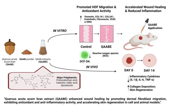
Quercus acuta Acorn Bran Extract Enhances Wound Healing by Promoting Human Dermal Fibroblast Migration and Antioxidant Activity

 , , , and
Abstract
1. Introduction
2. Results
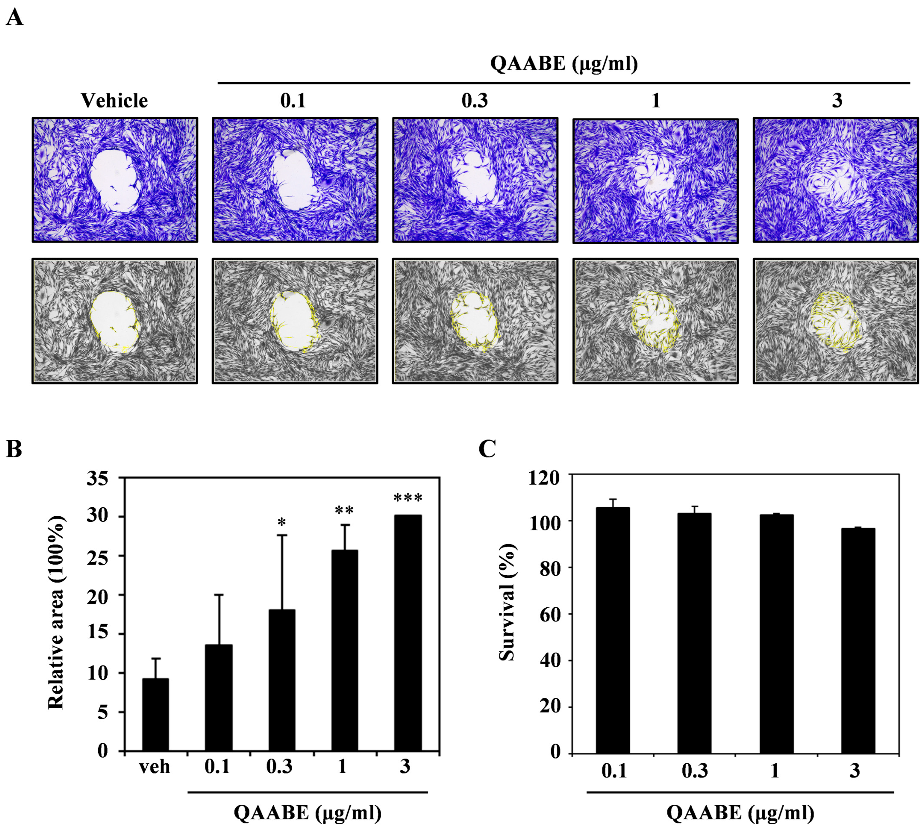
2.1. Q. acuta Acorn Bran Extract (QAABE) Enhances Migration Activity in HDF Cells
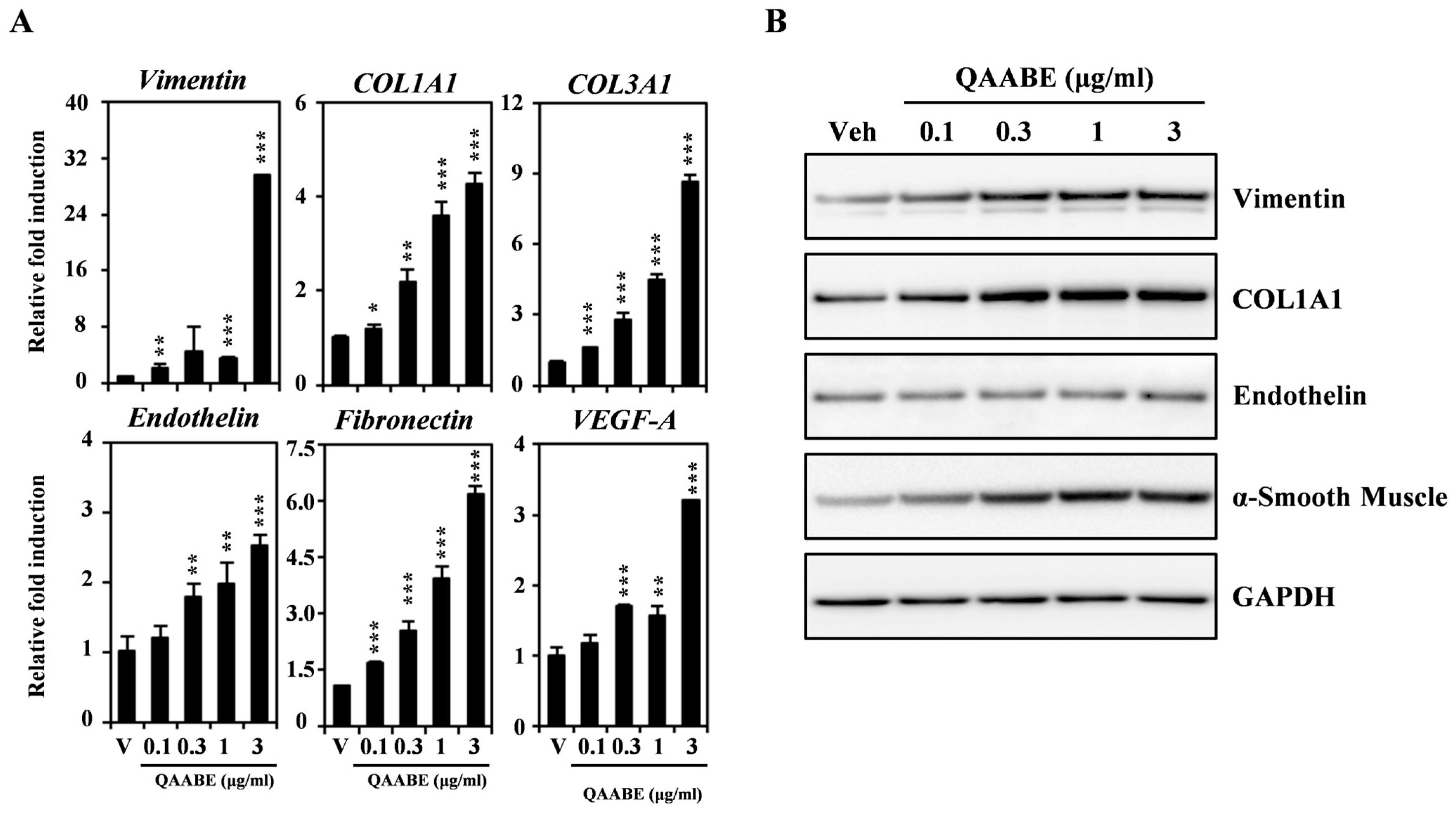
2.2. QAABE Upregulates Migration-Mediated Molecules in HDF Cells
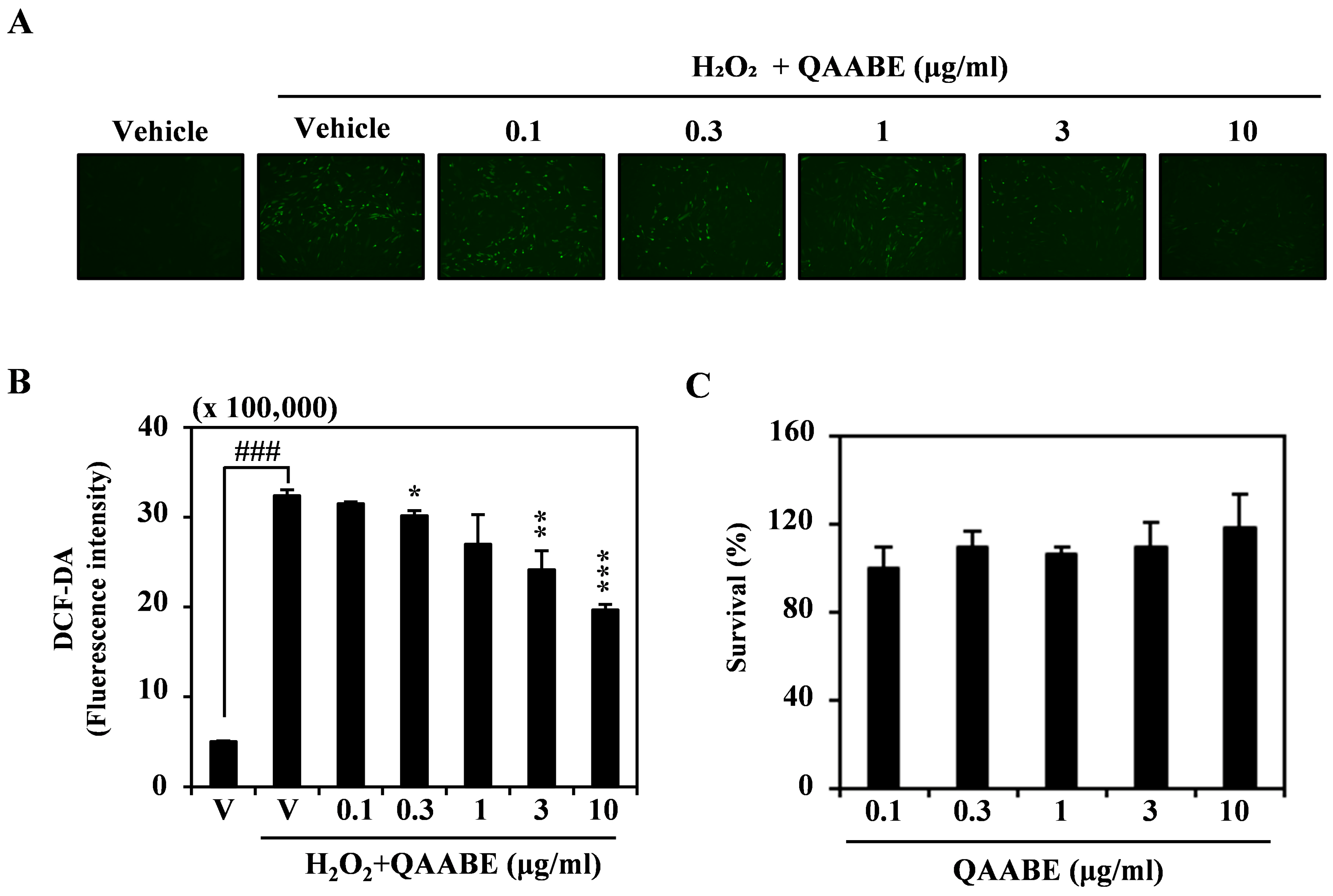
2.3. QAABE Mitigates ROS Induced by H2O2 in HDF Cells
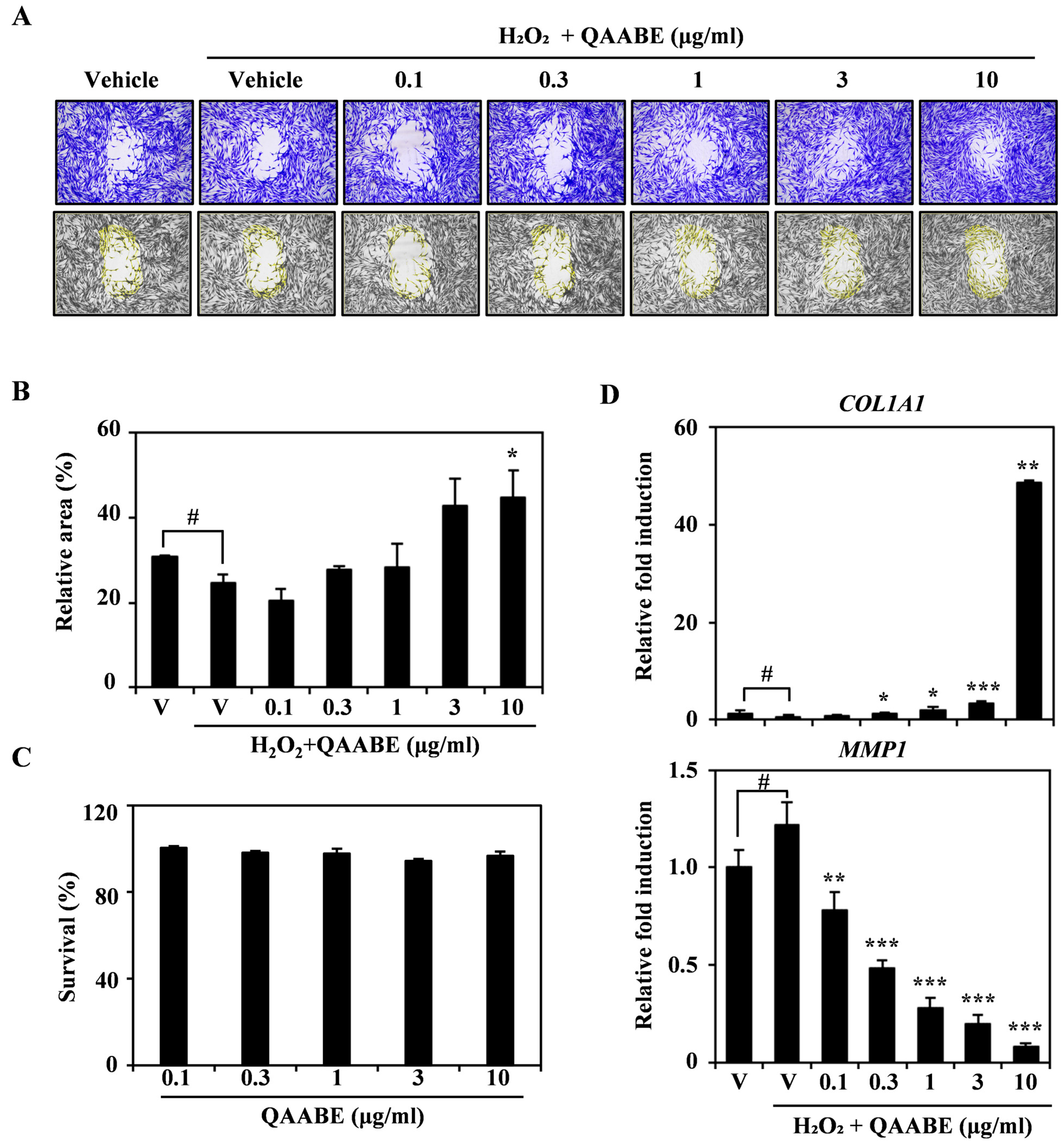
2.4. QAABE Ameliorates H2O2-Induced Impairment of Wound Healing in HDF Cells
2.5. QAABE Facilitates Wound Recovery In Vivo in Mouse Injury Model
2.6. QAABE Contains a Diverse Array of Polyphenolic Compounds
3. Discussion
4. Materials and Methods
4.1. Q. acuta Acorn Bran (QAAB) Preparation
4.2. QAAB Extraction
4.3. Reagents and Antibodies
4.4. Cell Culture
4.5. Migration Assay
4.6. Cell Cytotoxicity Assay
4.7. RNA Isolation and Quantitative Reverse Transcription Polymerase Chain Reaction (qRT-PCR)
4.8. Western Blotting
4.9. 2,7-Dichlorofluorescein Diacetate (DCF-DA) Staining
4.10. Cell-Free DCFH Oxidation Assay
4.11. Ethical Considerations in Animal Testing
4.12. Arrive Guidelines
4.13. Murine Cutaneous Wound Model
4.14. Wound Area Histopathological Staining
4.15. Liquid Chromatography-Tandem Mass Spectrometry (LC-MS/MS)
4.16. Statistical Analysis
5. Conclusions
Supplementary Materials
Author Contributions
Funding
Institutional Review Board Statement
Informed Consent Statement
Data Availability Statement
Conflicts of Interest
Abbreviations
| DCF-DA | 2,7-dichlorofluorescein diacetate |
| ANOVA | Analysis of variance |
| CCK-8 | Cell Counting Kit-8 |
| cDNA | complementary DNA |
| DMSO | Dimethyl sulfoxide |
| DDW | Double-distilled water |
| DMEM | Dulbecco’s Modified Eagle’s Medium |
| ET-1 | Endothelin-1 |
| FBS | Fetal bovine serum |
| H&E | Hematoxylin and eosin |
| HPLC | High-performance liquid chromatography |
| HRP | Horseradish peroxidase |
| HDF | Human dermal fibroblast |
| H2O2 | Hydrogen peroxide |
| Hprt1 | Hypoxanthine phosphoribosyltransferase 1 |
| ICR | Institute for Cancer Research |
| KFS | Korea Forest Service |
| LC-MS/MS | Liquid Chromatography-Tandem Mass Spectrometry |
| MMP1 | Matrix metalloproteinase-1 |
| NIFoS | National Institute of Forest Science |
| NO | Nitric oxide |
| QAAB | Quercus acuta Acorn Bran |
| QAABE | Quercus acuta acorn bran extract |
| qRT-PCR | Quantitative Reverse Transcription Polymerase Chain Reaction |
| ROS | Reactive oxygen species |
| TGF | Transforming growth factor |
| TNF-α | Tumor necrosis factor-alpha |
| VEGF | Vascular endothelial growth factor |
| α-SMA | α-smooth muscle actin |
References
- Lin, T.K.; Zhong, L.; Santiago, J.L. Anti-Inflammatory and Skin Barrier Repair Effects of Topical Application of Some Plant Oils. Int. J. Mol. Sci. 2017, 19, 70. [Google Scholar] [CrossRef] [Scilit]
- Ibrahim, N.; Wong, S.K.; Mohamed, I.N.; Mohamed, N.; Chin, K.Y.; Ima-Nirwana, S.; Shuid, A.N. Wound Healing Properties of Selected Natural Products. Int. J. Environ. Res. Public Health 2018, 15, 2360. [Google Scholar] [CrossRef] [Scilit]
- Wallace, H.A.; Basehore, B.M.; Zito, P.M. Wound Healing Phases. In StatPearls; StatPearls Ineligible Companies: Treasure Island, FL, USA, 2025. [Google Scholar]
- Karppinen, S.M.; Heljasvaara, R.; Gullberg, D.; Tasanen, K.; Pihlajaniemi, T. Toward understanding scarless skin wound healing and pathological scarring. F1000Research 2019, 8, F1000-Faculty. [Google Scholar] [CrossRef] [Scilit]
- Singer, A.J. Healing Mechanisms in Cutaneous Wounds: Tipping the Balance. Tissue Eng. Part B Rev. 2022, 28, 1151–1167. [Google Scholar] [CrossRef] [Scilit] [PubMed]
- Scialò, F.; Fernández-Ayala, D.J.; Sanz, A. Role of Mitochondrial Reverse Electron Transport in ROS Signaling: Potential Roles in Health and Disease. Front. Physiol. 2017, 8, 428. [Google Scholar] [CrossRef] [Scilit] [PubMed]
- Rodriguez, P.G.; Felix, F.N.; Woodley, D.T.; Shim, E.K. The Role of Oxygen in Wound Healing: A Review of the Literature. Dermatol. Surg. 2008, 34, 1159–1169. [Google Scholar] [CrossRef] [Scilit]
- Cano Sanchez, M.; Lancel, S.; Boulanger, E.; Neviere, R. Targeting Oxidative Stress and Mitochondrial Dysfunction in the Treatment of Impaired Wound Healing: A Systematic Review. Antioxidants 2018, 7, 98. [Google Scholar] [CrossRef] [Scilit]
- Ponugoti, B.; Xu, F.; Zhang, C.; Tian, C.; Pacios, S.; Graves, D.T. FOXO1 promotes wound healing through the up-regulation of TGF-β1 and prevention of oxidative stress. J. Cell Biol. 2013, 203, 327–343. [Google Scholar] [CrossRef] [Scilit]
- Dunnill, C.; Patton, T.; Brennan, J.; Barrett, J.; Dryden, M.; Cooke, J.; Leaper, D.; Georgopoulos, N.T. Reactive oxygen species (ROS) and wound healing: The functional role of ROS and emerging ROS-modulating technologies for augmentation of the healing process. Int. Wound J. 2017, 14, 89–96. [Google Scholar] [CrossRef] [Scilit] [PubMed]
- Comino-Sanz, I.M.; Lopez-Franco, M.D.; Castro, B.; Pancorbo-Hidalgo, P.L. The Role of Antioxidants on Wound Healing: A Review of the Current Evidence. J. Clin. Med. 2021, 10, 3558. [Google Scholar] [CrossRef] [Scilit]
- Woodruff, L.D.; Bounkeo, J.M.; Brannon, W.M.; Dawes, K.S.; Barham, C.D.; Waddell, D.L.; Enwemeka, C.S. The efficacy of laser therapy in wound repair: A meta-analysis of the literature. Photomed. Laser Surg. 2004, 22, 241–247. [Google Scholar] [CrossRef] [Scilit]
- Chester, D.; Marrow, E.A.; Daniele, M.A.; Brown, A.C. Wound Healing and the Host Response in Regenerative Engineering. In Encyclopedia of Biomedical Engineering; Narayan, R., Ed.; Elsevier: Oxford, UK, 2019; pp. 1–12. [Google Scholar] [CrossRef] [Scilit]
- Yoon, I.S.; Park, D.H.; Bae, M.S.; Oh, D.S.; Kwon, N.H.; Kim, J.E.; Choi, C.Y.; Cho, S.S. In Vitro and In Vivo Studies on Quercus acuta Thunb. (Fagaceae) Extract: Active Constituents, Serum Uric Acid Suppression, and Xanthine Oxidase Inhibitory Activity. Evid. Based Complement. Alternat. Med. 2017, 2017, 4097195. [Google Scholar] [CrossRef] [Scilit]
- Kim, M.H.; Park, D.H.; Bae, M.S.; Song, S.H.; Seo, H.J.; Han, D.G.; Oh, D.S.; Jung, S.T.; Cho, Y.C.; Park, K.M.; et al. Analysis of the Active Constituents and Evaluation of the Biological Effects of Quercus acuta Thunb. (Fagaceae) Extracts. Molecules 2018, 23, 1772. [Google Scholar] [CrossRef] [Scilit]
- Cho, S.H.; Kim, K.J.; Park, C.W.; Sun, B.Y.; Chung, M.G.; Pak, J.H. Cuticle micromorphology of Leaves of Quercus L. (Fagaceae) and its taxonomic implications. Korean J. Plant Taxon. 2008, 38, 405–431. [Google Scholar] [CrossRef] [Scilit]
- Denk, T.; Grimm, G.W.; Manos, P.S.; Deng, M.; Hipp, A.L. An Updated Infrageneric Classification of the Oaks: Review of Previous Taxonomic Schemes and Synthesis of Evolutionary Patterns. In Oaks Physiological Ecology. Exploring the Functional Diversity of Genus Quercus L.; Gil-Pelegrín, E., Peguero-Pina, J.J., Sancho-Knapik, D., Eds.; Springer International Publishing: Cham, Germany, 2017; pp. 13–38. [Google Scholar] [CrossRef] [Scilit]
- Uyar, A.; Jhangir, G.M.; Keleş, Ö.F.; Yener, Z. The effects of Quercus (Oak) acorn on cutaneous wound healing in rats. Int. J. Plant Based Pharm. 2023, 3, 148–155. [Google Scholar] [CrossRef] [Scilit]
- Kim, S.H.; Shin, H.L.; Son, T.H.; Lim, S.A.; Kim, D.; Yoon, J.H.; Choi, H.; Kim, H.G.; Choi, S.W. Quercus glauca Acorn Seed Coat Extract Promotes Wound Re-Epithelialization by Facilitating Fibroblast Migration and Inhibiting Dermal Inflammation. Biology 2024, 13, 775. [Google Scholar] [CrossRef] [Scilit]
- Cheng, F.; Shen, Y.; Mohanasundaram, P.; Lindstrom, M.; Ivaska, J.; Ny, T.; Eriksson, J.E. Vimentin coordinates fibroblast proliferation and keratinocyte differentiation in wound healing via TGF-beta-Slug signaling. Proc. Natl. Acad. Sci. USA 2016, 113, E4320–E4327. [Google Scholar] [CrossRef] [Scilit]
- Hochstein, A.O.; Bhatia, A. Collagen: Its role in wound healing. Wound Manag. 2014, 4, 104–109. [Google Scholar]
- Barton, M.; Yanagisawa, M. Endothelin: 30 Years From Discovery to Therapy. Hypertension 2019, 74, 1232–1265. [Google Scholar] [CrossRef] [Scilit]
- Shinde, A.V.; Humeres, C.; Frangogiannis, N.G. The role of α-smooth muscle actin in fibroblast-mediated matrix contraction and remodeling. Biochim. Biophys. Acta (BBA)—Mol. Basis Dis. 2017, 1863, 298–309. [Google Scholar] [CrossRef] [Scilit]
- Wang, G.; Yang, F.; Zhou, W.; Xiao, N.; Luo, M.; Tang, Z. The initiation of oxidative stress and therapeutic strategies in wound healing. Biomed. Pharmacother. 2023, 157, 114004. [Google Scholar] [CrossRef] [Scilit] [PubMed]
- Lenselink, E.A. Role of fibronectin in normal wound healing. Int. Wound J. 2015, 12, 313–316. [Google Scholar] [CrossRef] [Scilit]
- Bao, P.; Kodra, A.; Tomic-Canic, M.; Golinko, M.S.; Ehrlich, H.P.; Brem, H. The role of vascular endothelial growth factor in wound healing. J. Surg. Res. 2009, 153, 347–358. [Google Scholar] [CrossRef] [Scilit] [PubMed]
- Guo, S.; Dipietro, L.A. Factors affecting wound healing. J. Dent. Res. 2010, 89, 219–229. [Google Scholar] [CrossRef] [Scilit]
- Dunn, L.; Prosser, H.C.; Tan, J.T.; Vanags, L.Z.; Ng, M.K.; Bursill, C.A. Murine model of wound healing. J. Vis. Exp. 2013, 75, e50265. [Google Scholar] [CrossRef] [Scilit] [PubMed]
- Said, A.; Wahid, F.; Bashir, K.; Rasheed, H.M.; Khan, T.; Hussain, Z.; Siraj, S. Sauromatum guttatum extract promotes wound healing and tissue regeneration in a burn mouse model via up-regulation of growth factors. Pharm. Biol. 2019, 57, 736–743. [Google Scholar] [CrossRef] [Scilit] [PubMed]
- Wu, J.; Landen, N.X. Investigation of Skin Wound Healing Using a Mouse Model. Methods Mol. Biol. 2020, 2154, 239–247. [Google Scholar] [CrossRef] [Scilit] [PubMed]
- Yadav, E.; Singh, D.; Yadav, P.; Verma, A. Attenuation of dermal wounds via downregulating oxidative stress and inflammatory markers by protocatechuic acid rich n-butanol fraction of Trianthema portulacastrum Linn. in wistar albino rats. Biomed. Pharmacother. 2017, 96, 86–97. [Google Scholar] [CrossRef] [Scilit]
- Baek, S.; Park, H.; Kim, M.; Lee, D. Preparation of PCL/(+)-catechin/gelatin film for wound healing using air-jet spinning. Appl. Surf. Sci. 2020, 509, 145033. [Google Scholar] [CrossRef] [Scilit]
- Rozen, S.; Skaletsky, H. Primer3 on the WWW for general users and for biologist programmers. Methods Mol. Biol. 2000, 132, 365–386. [Google Scholar] [CrossRef] [Scilit]
- Livak, K.J.; Schmittgen, T.D. Analysis of relative gene expression data using real-time quantitative PCR and the 2−ΔΔCT Method. Methods 2001, 25, 402–408. [Google Scholar] [CrossRef] [Scilit] [PubMed]
- Zaini, A.A.; Khaza’ai, H.; Ali, R.M.; Abdul Mutalib, M.S.; Baharuddin, A.A. Topical Treatment of Tocotrienol-Rich Fraction ( TRF ) on Deep Partial-Thickness Burn Wounds in Rats. J. Dermatol. Clin. Res. 2016, 4, 1063. [Google Scholar]


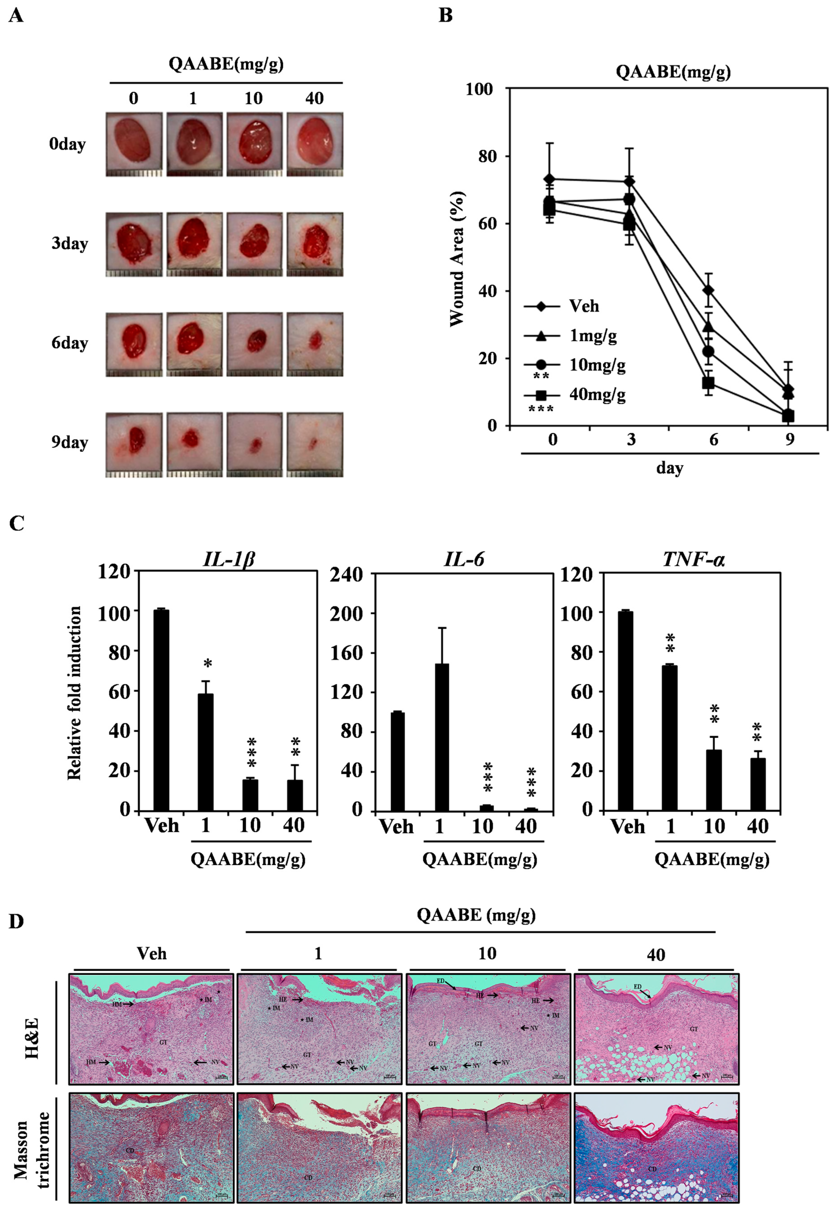

| Group | Vehicle (0 mg/g) | QAABE (1 mg/g) | QAABE (10 mg/g) | QAABE (40 mg/g) | |
|---|---|---|---|---|---|
| Score | |||||
| Epidermis | |||||
| – Epithelialization | 0 | 0 | 1 | 4 | |
| – Hemorrhage | 3 | 2 | 1 | 0 | |
| – Inflammation | 3 | 3 | 2 | 0 | |
| – Granulation tissue formation | 2 | 2 | 3 | 4 | |
| – Neovascularization | 1 | 2 | 2 | 3 | |
| Dermis | |||||
| – Collagen deposition | 1 | 1 | 2 | 4 | |
| No. | Identification | Formula | Molar Mass | mg/kg |
|---|---|---|---|---|
| 1 | Epicatechin gallate | C22H18O10 | 442.37 g/mol | 4.0 ± 0.57 |
| 2 | Protocatechuic acid | C7H6O4 | 154.12 g/mol | 543.3 ± 104.77 |
| 3 | Ellagic acid | C14H6O8 | 302.197 g/mol | 22.9 ± 5.34 |
| 4 | Gallic acid | C7H6O5 | 170.12 g/mol | 142.6 ± 18.69 |
| 5 | Isoquercitrin | C21H20O12 | 464.0955 g/mol | 0.2 ± 0.03 |
| 6 | Kaempferol-3-O-(2′6′-di-O-trans-p-coumaroyl)-beta-D-glucopyranoside | C39H32O15 | 740.7 g/mol | 0.2 ± 0.05 |
| 7 | Myricitrin | C21H20O12 | 464.37 g/mol | 0.7 ± 0.12 |
| 8 | Tiliroside | C30H26O13 | 594.5 g/mol | 6.4 ± 1.26 |
| 9 | Catechin | C15H14O6 | 290.26 g/mol | 179.8 ± 28.88 |
| 10 | Epicatechin | C15H14O6 | 290.27 g/mol | 24.9 ± 1.46 |
| 11 | Quercetin | C15H10O7 | 302.236 g/mol | 23.6 ± 4.65 |
| 12 | Rutin | C27H30O16 | 610.517 g/mol | 3.8 ± 0.32 |
| Target Gene | Forward Primer (5′–3′) | Reverse Primer (5′–3′) |
|---|---|---|
| COL1A1 | CCGTGCCCTGCCAGATC | CAGTTCTTGATTTCGTCGCAGATC |
| COL3A1 | TGGAGGATGGTTGCACGAAA | AAAAGCAAACAGGGCCAACG |
| VEGF-A | ATAAGTCCTGGAGCGTTCCCT | GGCAGCGTGGTTTCTGTATC |
| Vimentin | AACTTAGGGGCGCTCTTGTC | TGAGGGCTCCTAGCGGTTTA |
| Fibronectin | ACAAGCATGTCTCTCTGCCA | TTTGCATCTTGGTTGGCTGC |
| Endothelin | CTGCCTTTTCTCCCCGTTAAA | GGACTGGGAGTGGGTTTCTC |
| MMP1 | ATGCACAGCTTTCCTCCACT | GGGCCACTATTTCTCCGCTT |
| TNF-α | TAGCCCACGTCGTAGCAAAC | CTCAAAGTAGACCTGCCC |
| IL-1β | TGCCACCTTTTGACAGTGATG | AAGGTCCACGGGAAAGACAC |
| IL-6 | CAACGATGATGCACTTGCAGA | TGGAAATTGGGGTAGGAAGGAC |
| HPRT1 | GACCAGTCAACAGGGGACAT | GCTTGCGACCTTGACCATCT |
| β-actin(ACTB) | AAGGATTCCTATGTGGGCGAC | CGTACAGGGATAGCACAGCC |
| Score | 0 | 1 | 2 | 3 | 4 |
|---|---|---|---|---|---|
| Epithelialization | Absence of epithelial proliferation in 70% of tissue | Poor epidermal organization in 60% of tissue | Incomplete epidermal organization in 40% of tissue | Moderate epithelial proliferation in 60% of tissue | Complete epidermal remodeling in 80% of tissue |
| Hemorrhage | Absence of hemorrhage | 1–2 per site of hemorrhage | 3–4 per site of hemorrhage | 5–6 per site of hemorrhage | >7 per site of hemorrhage |
| Granulation tissue formation | Immature and inflammatory tissue in 70% of tissue | Thin, immature, and inflammatory tissue in 60% of tissue | Moderate remodeling in 40% of tissue | Thick granulation layer in 60% of tissue | Complete tissue organization in 80% of tissue |
| Neovascularization | Absence of angiogenesis | 1–2 vessels per site | 3–4 vessels per site | 5–6 vessels per site | >7 vessels per site |
| Collagen deposition | Absence of collagen deposition | Focal presence in fibroblasts around new capillaries | Moderate amount in repaired tissue | Dominant feature | |
| Inflammation | Inflammatory cell infiltration was scored as: - mild - moderate - severe | ||||
Disclaimer/Publisher’s Note: The statements, opinions and data contained in all publications are solely those of the individual author(s) and contributor(s) and not of MDPI and/or the editor(s). MDPI and/or the editor(s) disclaim responsibility for any injury to people or property resulting from any ideas, methods, instructions or products referred to in the content. |
© 2026 by the authors. Licensee MDPI, Basel, Switzerland. This article is an open access article distributed under the terms and conditions of the Creative Commons Attribution (CC BY) license.
Share and Cite
Lim, S.-A.; Son, T.H.; Shin, H.-L.; Kim, D.; Yoon, J.-H.; Kim, H.-G.; Choi, H.; Kim, S.-H.; Choi, S.-W. Quercus acuta Acorn Bran Extract Enhances Wound Healing by Promoting Human Dermal Fibroblast Migration and Antioxidant Activity. Pharmaceuticals 2026, 19, 481. https://doi.org/10.3390/ph19030481
Lim S-A, Son TH, Shin H-L, Kim D, Yoon J-H, Kim H-G, Choi H, Kim S-H, Choi S-W. Quercus acuta Acorn Bran Extract Enhances Wound Healing by Promoting Human Dermal Fibroblast Migration and Antioxidant Activity. Pharmaceuticals. 2026; 19(3):481. https://doi.org/10.3390/ph19030481
Chicago/Turabian StyleLim, So-An, Tae Hyun Son, Hye-Lim Shin, Dongsoo Kim, Jun-Hyuck Yoon, Hwan-Gyu Kim, Hyunmo Choi, Shin-Hye Kim, and Sik-Won Choi. 2026. "Quercus acuta Acorn Bran Extract Enhances Wound Healing by Promoting Human Dermal Fibroblast Migration and Antioxidant Activity" Pharmaceuticals 19, no. 3: 481. https://doi.org/10.3390/ph19030481
APA StyleLim, S.-A., Son, T. H., Shin, H.-L., Kim, D., Yoon, J.-H., Kim, H.-G., Choi, H., Kim, S.-H., & Choi, S.-W. (2026). Quercus acuta Acorn Bran Extract Enhances Wound Healing by Promoting Human Dermal Fibroblast Migration and Antioxidant Activity. Pharmaceuticals, 19(3), 481. https://doi.org/10.3390/ph19030481