Review

Physiological Conditions, Bioactive Ingredients, and Drugs Stimulating Non-Shivering Thermogenesis as a Promising Treatment Against Diabesity

by
Diego Salagre
1,2,3,
Ciskey V. Ayala-Mosqueda
1,
Samira Aouichat
1,4 and
Ahmad Agil
1,2,3,*
1
Department of Pharmacology, School of Medicine, University of Granada, 18016 Granada, Spain
2
Nutrition, Metabolism, Growth and Development Group, BioHealth Institute Granada (ibs.GRANADA), 18012 Granada, Spain
3
Neuroscience Institute “Federico Olóriz”, Biomedical Research Center (CIBM), University of Granada, 18016 Granada, Spain
4
Team of Cellular and Molecular Physiopathology, Faculty of Biological Sciences, University of Sciences and Technology Houari Boumediene, El Alia, Algiers 16111, Algeria
*
Author to whom correspondence should be addressed.
Pharmaceuticals 2025, 18(9), 1247; https://doi.org/10.3390/ph18091247
Submission received: 17 July 2025 / Revised: 13 August 2025 / Accepted: 20 August 2025 / Published: 22 August 2025

Abstract

Obesity (lipotoxicity) results from a chronic imbalance between energy intake and expenditure. It is strongly associated with type 2 diabetes mellitus (T2DM, glucotoxicity) and considered a major risk factor for the development of metabolic complications. Their convergence constitutes “diabesity”, representing a major challenge for public health worldwide. Limited treatment efficacy highlights the need for novel, multi-targeted therapies. Non-shivering thermogenesis (NST), mediated by brown and beige adipose tissue and skeletal muscle, has emerged as a promising therapy due to its capacity to increase energy expenditure and improve metabolic health. Also, skeletal muscle plays a central role in glucose uptake and lipid oxidation, further highlighting its relevance in diabesity. This review explores current and emerging knowledge on physiological stimuli, including cold exposure, physical activity, and fasting, as well as bioactive ingredients and drugs that stimulate NST in thermogenic tissues. Special emphasis is placed on melatonin as a potential regulator of mitochondrial function and energy balance. The literature search was conducted using MEDLINE and Web of Science. Studies were selected based on scientific relevance, novelty, and mechanistic insight; prioritizing human and high-quality rodent research published in peer-reviewed journals. Evidence shows that multiple interventions enhance NST, leading to improved glucose metabolism, reduced fat accumulation, and increased energy expenditure in humans and/or rodents. Melatonin, in particular, shows promise in modulating thermogenesis through organelle-molecular pathways and mitochondrial protective effects. In conclusion, a multi-target approach through the activation of NST by physiological, nutritional, and pharmacological agents offers an effective and safe treatment for diabesity. Further research is needed to confirm these effects in clinical practice and support their use as effective therapeutic strategies.

Graphical Abstract

1. Introduction

Modern lifestyle trends in our current society, characterized by abundant calorie intake, lower levels of physical activity, and longer life expectancy, have contributed to a higher prevalence of obesity (mainly associated with visceral fat accumulation), propelling it to an alarming worldwide expansion [1]. According to recent data published by the World Health Organization, the global prevalence of obesity has tripled. In 2022, more than 2.5 billion adults (aged 18 years and older, 43%) were overweight (body mass index (BMI) ≥ 25 kg/m2), and 890 million among these (16%) were obese (BMI ≥ 30 kg/m2) [2]. Obesity causes numerous physiological dysfunctions that affect every organ and system, producing multiple comorbidities such as type 2 diabetes mellitus (T2DM), also called diabesity (obesity and its associated T2DM). Diabesity increases the risk of cardiovascular diseases, hepatic steatosis, nephropathy, arthritis, musculoskeletal disorders, neurodegenerative diseases, certain types of cancers, and reproductive abnormalities (Figure 1). Furthermore, obese people have a lower quality of life, indicating a psychosocial impact as well. The etiology of obesity is multifactorial, involving a complex interaction among genetics, physiology, behavior, and the environment. Environmental factors lead to the prevalence of poor eating habits, such as having facilities where one can obtain unhealthy food in close proximity with very reduced prices, and the sedentary lifestyle that prevails in a great part of the population [3]. In line with the previously mentioned facts, developing efficient treatment approaches to address obesity is crucial. However, current interventions based on lifestyle changes and increased physical activity often fail to produce consistent results, with success achieved in only a small proportion of individuals [3,4]. Hence, effective and safe therapeutic strategies are urgently required to curb the rising prevalence of obesity. Targeting energy expenditure through thermogenesis activation is considered a new, promising approach against diabesity and its related metabolic disorders [4].
Thermogenesis is a physiological mechanism where energy is dissipated as heat for whole-body temperature maintenance in mammals [5]. It is divided into shivering thermogenesis, induced by repetitive muscle contraction, and non-shivering thermogenesis (NST) in skeletal muscle and thermogenic adipose tissues [4]. Brown adipose tissue (BAT) and beige adipose tissue (bAT) are rich in multilocular lipid droplets and abundant mitochondria expressing uncoupling protein 1 (UCP1) with great thermogenic function, unlike white adipose tissue (WAT), which is characterized by lipid storage for times of energy need [6]. Both BAT and bAT have been given greater importance as thermogenic organs, despite representing only 0.2% of body weight. However, thermogenic activity in skeletal muscle has been emerging as an attractive strategy, given that this organ accounts for one-third of body weight and has a greater contribution to basal metabolic rate [4]. Indeed, skeletal muscle is the organ that contributes the most to increasing energy expenditure by NST through the uncoupling of the sarcoendoplasmic reticulum calcium ATPase (SERCA) pump induced by sarcolipin (SLN) binding [4,5].
Several reviews have previously addressed thermogenesis as a therapeutic strategy against obesity and related metabolic disorders. For example, Cheng et al. [6] focused primarily on the role of brown and beige adipose tissue, while Li et al. [4] explored NST specifically in skeletal muscle, but without integrating both tissues’ thermogenesis. Betz and Enerbäck [5] emphasized physiological thermogenic stimuli such as cold and the thermogenic molecular mechanisms in adipose and muscle tissue, offering neither a multi-target therapeutic perspective nor a translational point of view. Although these studies have advanced our understanding, none has provided a comprehensive and complete analysis encompassing physiological stimuli (cold exposure, exercise, and fasting), bioactive compounds (many of which come from foods and spices that can be integrated into the diet), and pharmacological agents—including the emerging role of melatonin—while taking into account the thermogenic molecular pathways involved in both adipose and muscle tissues in the context of diabesity. This review addresses this gap by combining physiological, nutritional, and pharmacological approaches from a clinical perspective, proposing their combined use, and encouraging further research into their possible synergistic effects in improving glucose and lipid metabolism. By unifying these perspectives, the current review aims to summarize both established and emerging findings regarding the physiological conditions, bioactive agents, and drugs that stimulate NST with potential implications for the control of obesity and diabetes management. Particular emphasis is placed on melatonin as a novel and promising thermogenic agent to combat diabesity.

2. Methods

To provide a comprehensive overview of current and emerging strategies that stimulate NST with potential anti-diabesity effects, this review focused on physiological conditions, bioactive compounds, and pharmacological agents that promote thermogenic activation in brown/beige adipose tissue and skeletal muscle. The literature search was conducted in May 2025 using MEDLINE via PubMed and Web of Science databases. The following keywords and MeSH terms were used in various combinations: Obesity; Diabetes Mellitus, Type 2; Thermogenesis; Adipose Tissue, Brown; Muscle, Skeletal; Food Bioactive Ingredients; Melatonin; Cold Exposure; Exercise; Fasting; Capsaicin; Paradol; Gingerol; Zingerone; Cinnamaldehyde; Cinnamic acid; Curcumin; Allicin; Quercetin; Caffeine; Catechin; Theaflavin; Flavanol; Berberine; Docosahexaenoic acid; Omega-3 polyunsaturated fatty acid; Eicosapentaenoic acid; Eriocitrin; Menthol; Thymol; Resveratrol; Proanthocyanidin; Anthocyanin; Mirabegron; Levothyroxine; Liothyronine; Triiodothyronine; Resmetirom; Fexaramine; Farnesol; Chenodeoxycholic acid; Growth Hormone; Somatotropin; Tesamorelin; Liraglutide; Semaglutide; Exedin-4; Tirzepatide; Oxyntomodulin; Mazdutide; Retatrutide; Dapagliflozin; Empagliflozin; and Canagliflozin.
Additional relevant articles were identified by manually screening the reference lists of key papers and recent reviews. No date restrictions were applied to allow inclusion of pioneering studies and foundational discoveries, regardless of publication year. Articles were selected based on their scientific relevance, novelty, methodological robustness, and depth of mechanistic insight, with priority given to studies in humans and, when not available, to well-designed rodent studies. Only peer-reviewed original articles, systematic reviews, and narrative reviews written in English were considered. Studies in both in vivo and in vitro settings were included if they provided significant insight into NST activation. Exclusion criteria included non-peer-reviewed materials, case reports, conference abstracts, and older articles that lack mechanical relevance to the scope of the review or contain data corroborated in more recent studies. All selected papers were independently screened and discussed by the authors to ensure inclusion of the most informative and impactful literature. No formal quality assessment tool was applied, as this was a narrative review aiming to synthesize high-quality, conceptually relevant evidence across a broad timespan.
All figures and illustrations were created by the authors using the Biorender graphic tool (https://www.biorender.com/, accessed on 15 June 2025), and the molecular structures of the compounds were obtained using the PubChem tool (https://pubchem.ncbi.nlm.nih.gov/, accessed on 12 August 2025).

3. White and Thermogenic Adipose Tissues

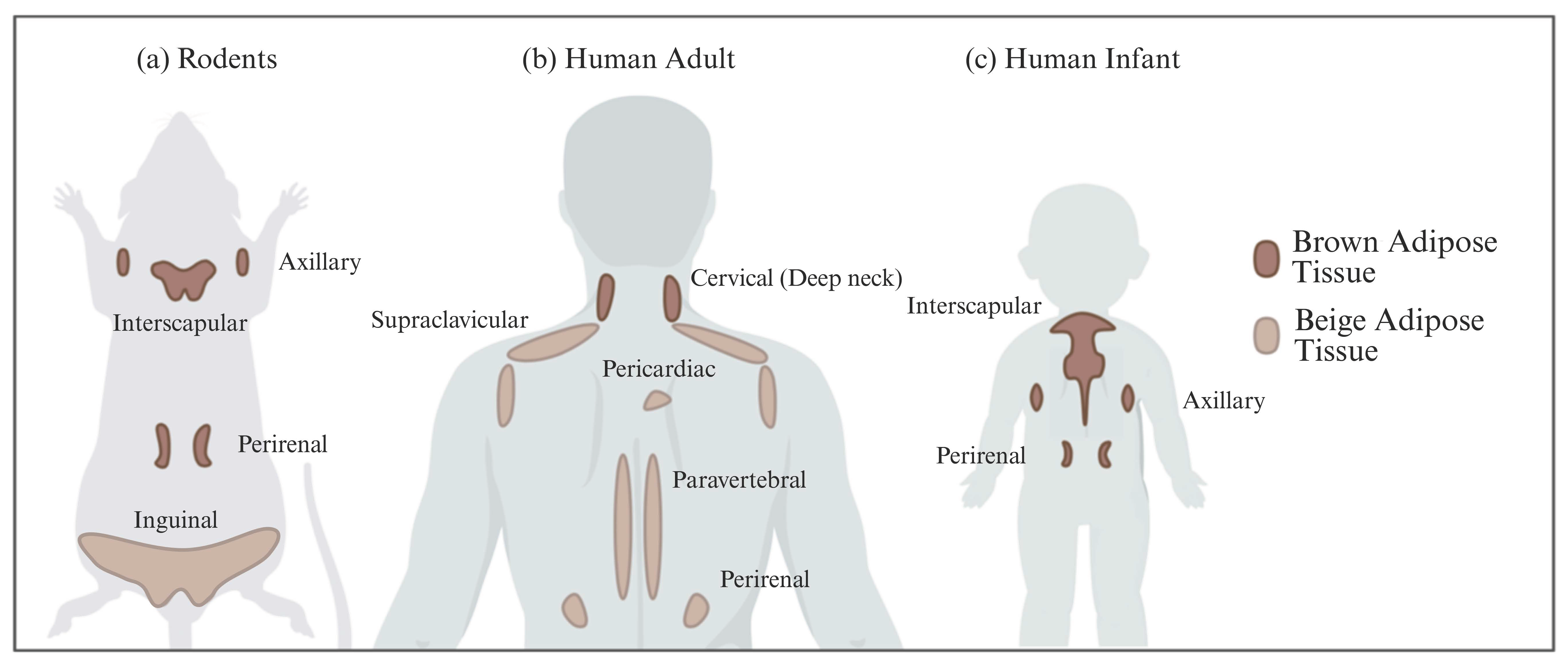
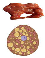
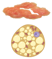
In all mammals, including humans, three different types of adipose depots can be found, known as white, brown, and beige adipose tissues. They have unique characteristics that differentiate them from each other in terms of function, morphology, structure, protein expression profile, and developmental origin. WAT can also be differentiated into visceral WAT (vWAT), or abdominal fat, which is distributed around internal organs and is located inside the peritoneum; and subcutaneous WAT (sWAT), which is located underneath the skin. vWAT tends to cause low-grade inflammation that promotes insulin resistance and is responsible for the morbidity associated with obesity, whereas sWAT is metabolically active with less inflammatory activity, and could favor metabolic insulin action [7]. The function of WAT is to store excess energy in the form of triglycerides, which can be released to fuel other tissues during fasting periods, whereas BAT plays a crucial role in whole-body energy homeostasis through NST. White adipocytes are unilocular cells with a large lipid droplet and a few mitochondria that are devoid of UCP1 (Table 1). bAT, also known as “inducible” or “recruitable” beige fat, is defined as a cluster of UCP1-expressing and mitochondria-rich adipocytes that, in response to various external stimuli, are developed within the WAT depots. Beige/brite adipocytes, similarly to BAT adipocytes, are recognized for their characteristic lipid morphology consisting of small and multilocular lipid droplets, their high mitochondrial content, and their positive expression of UCP1 (Table 1) [8]. The switch from white adipocyte phenotype to brown-like phenotype and thus bAT recruitment in response to external stimuli is called browning, britening, and/or beiging. Two theories have been proposed by the scientific community regarding the origin of beige adipocytes: (i) de novo differentiation from progenitor cells that reside in WAT, and (ii) reversible transdifferentiation, a bidirectional mechanism that involves interconversion of mature white adipocytes into mature beige/brite adipocytes and vice versa [9]. The most common anatomical location of BAT and bAT in rodents and humans is shown in Figure 2.
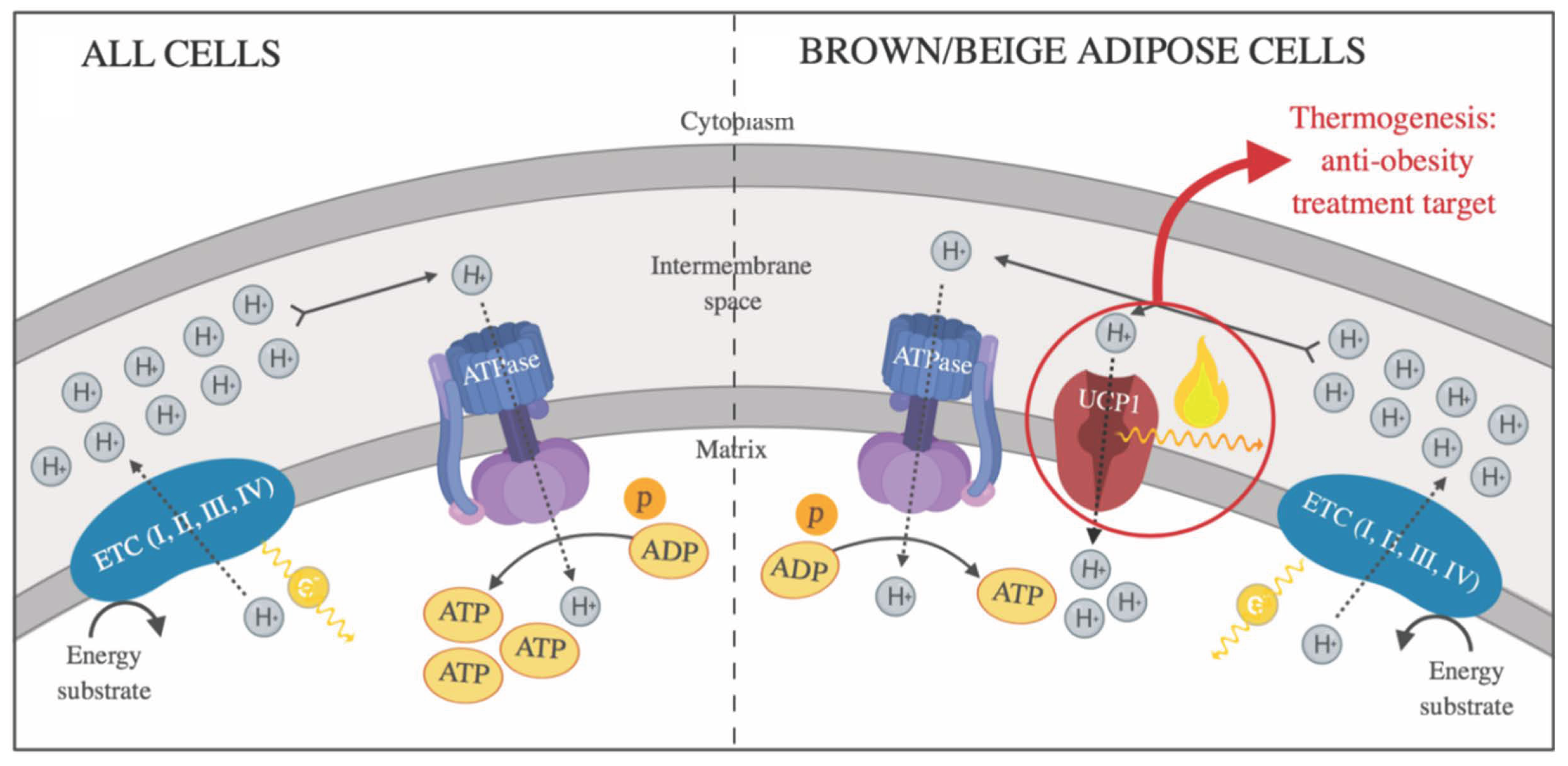
In mitochondria from BAT and bAT, the transmembrane protein UCP1 is responsible for thermogenic adipose tissue’s contribution to NST. Under certain stimuli, UCP1 is activated and generates heat production by allowing protons to re-enter the mitochondrial matrix, uncoupling substrate oxidation and oxidative phosphorylation (OXPHOS) from the ATP synthesis [10], from which the energy produced is released as heat (Figure 3).

4. Skeletal Muscle and NST

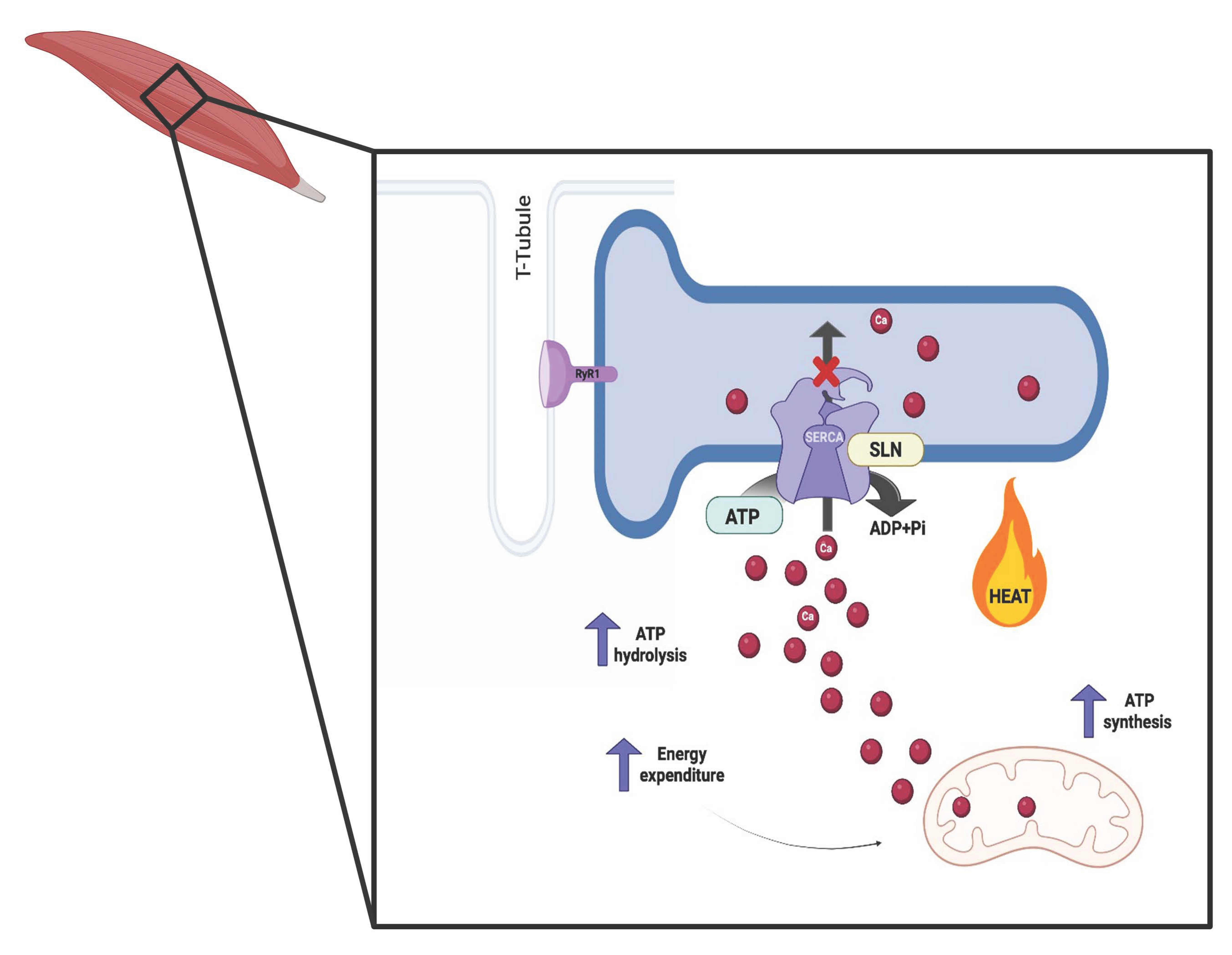
Skeletal muscle plays a key role in thermoregulation through the production of heat by non-shivering mechanisms. This organ represents around 50% of total body weight and is responsible for 70–80% insulin-stimulated glucose uptake, fatty acids metabolism, and whole-body energy expenditure through futile cycles [5,11]. In skeletal muscle, NST is activated by the uncoupling of SERCA. The SERCA pump is a transmembrane protein belonging to the family of P-type ATPases, whose main function is the transport of cytosolic calcium back to the sarcoplasmic reticulum using the energy generated from ATP hydrolysis (Figure 4), thus regulating calcium concentrations in both spaces following muscle contraction [12]. SERCA function is regulated by endogenous molecules such as SLN [11]. SLN is a single-spanning membrane protein, the expression of which predominates in skeletal muscle, and its expression increases with overloading and exercise performance in mice [13]. By dissociating calcium (Ca2+) ions from SERCA activity, it regulates muscle NST, allowing SERCA to repeatedly run a futile cycle of ATP hydrolysis into ADP without efficiently pumping Ca2+ ions into the endoplasmic reticulum. This results in the dissipation of the excess of energy not used for Ca2+ transport in the form of heat (Figure 4), playing an essential role in maintaining body temperature and representing another critical function of muscle beyond contraction [12]. This thermogenic mechanism used by skeletal muscle is indirectly associated with mitochondrial metabolism (Figure 4), unlike BAT, where thermogenic activity directly depends on mitochondrial respiration and metabolism [14].

5. Physiological Conditions Stimulating NST

5.1. Cold Exposure

Cold is a powerful natural stimulus for the activation of adaptive thermogenesis, thus generating heat production in BAT and skeletal muscle. It is well known that cold exposure increases energy expenditure and reduces insulin resistance, making it an interesting strategy during diabesity treatment [15]. Several studies in rodents have demonstrated that both acute and chronic cold activate thermogenic activity in BAT and lead to the emergence of UCP1-positive cells, as well as morphological changes in rodents’ white fat pads [16]. For example, Lim et al. [17] demonstrated an increase in UCP1-positive mitochondria in mice’s sWAT in response to prolonged cold exposure under 4 °C. In addition to molecular changes, the acquisition of brown-like adipose phenotype in WAT, simultaneously with increased vascular density, owing to activation of angiogenesis, was markedly observed. Activation of BAT thermogenesis was also noted in this study. In humans, BAT was shown to be effectively recruited and activated upon cold acclimation. For instance, one study showed that mild cold acclimation for 10 days in healthy subjects performing sedentary activities was enough to recruit BAT and increase NST but failed to promote browning of abdominal WAT [18]. Even in an obese man with low BAT activity, short-term cold acclimation increased cold-induced glucose uptake in BAT [18]. A study conducted in humans has shown that BAT can be successfully activated and recruited through cold acclimation; however, only one of the human studies with 4 °C cold chronically found WAT browning [19], suggesting that moderate acute cold exposure may not be sufficient to promote WAT browning in humans. In muscle tissue, studies in mice have shown that cold upregulates SLN expression and decreases SERCA pump activity, thus producing heat [20]. Hanssen et al. [21] showed in a ten-day study that acute cold exposure increased insulin sensitivity in the skeletal muscle of patients with type 2 diabetes through GLUT4 upregulation in the sarcolemma responding to cold acclimation. Changes in the mitochondrial architecture of muscle from mice exposed to mild cold (16 °C) and severe cold (4 °C) were also observed with ultrastructural remodeling of mitochondria cristae, especially in severe cold [14]. In addition, skeletal muscle and BAT thermogenesis have been proposed to be regulated by circulating myokines, as a close crosstalk between both processes was identified. Rowland et al. used SLN-Knockout (KO), UCP1-KO, and double-knockout (DKO) mice and exposed them gradually to cold. The lack of SLN or UCP1 was compensated for with an increase in the expression of the other protein after cold adaptation. DKO mice were affected by acute cold, developing hypothermia, consequently increasing food intake, and mobilizing almost all of their white fat. Therefore, cold exposure without SLN and UCP1 represented a high energy cost and the use of unsustainable thermogenic mechanisms [22].
The mechanism of cold-induced BAT and skeletal muscle non-shivering thermogenesis is mostly elucidated in small rodents. Briefly, cold is sensed by thermal transient receptor potential channels (TRPs), which are proteins that detect environmental changes, such as temperature, pain, touch, osmolarity, and certain natural compounds. When cold-activated TRPs in skin sensory neurons send signals to the brain, sympathetic nerve activity increases. Noradrenaline released from sympathetic terminals then triggers NST activation in BAT and skeletal muscle, as well as brown-like phenotype in WAT or browning through β3-adrenergic receptor (β3-ARs) activation and peroxisome proliferator-activated receptor gamma (PPARγ) coactivator 1-alpha (PGC1α) expression (the key regulator of thermogenesis) [12,16].

5.2. Physical Exercise

Physical exercise itself generates heat through fiber contraction, like cold-induced shivering thermogenesis. However, there is also a close relationship between exercise and non-shivering thermogenesis due to circulating myokines released after exercise. Irisin is the most important exercise-induced adipomyokine, which is involved in the activation of NST by inducing BAT activation and browning of white fat cells in both rodents and humans [23]. When performing physical exercise, PGC1α is activated in skeletal muscle, enhancing mitochondrial biogenesis and inducing the synthesis of a membrane protein named fibronectin-type III domain-containing 5 (FNDC5) expressed in skeletal muscle as well. The FNDC5 N-terminal end is released into the blood, and this product is known as irisin. Thus, sedentary people have lower levels of irisin in their blood compared to athletes or individuals engaged in exercise [23,24]. Nonetheless, Stengel et al. reported that in obese patients, irisin levels are altered as a possible physiological response to improve insulin resistance in these patients [25]. Transgenic mice overexpressing PGC1α in skeletal muscle were studied, and the results showed not only an enhanced and preserved skeletal muscle mitochondrial function but also an improvement in insulin sensitivity and glucose homeostasis [26].
In humans, combined training increases mitochondrial enzyme activity and activates BAT in obese and diabetic patients [27], and Boström’s group showed that irisin, the exercise-induced muscle-derived protein, plays a critical role in browning of WAT [24]. In their study, they found that irisin acts to stimulate UCP1 expression and a wide program of brown fat-like development. Similar effects were found in rodents subjected to an exercise program, after which elevated irisin levels in blood and adipose tissue, and subsequently increased expression of WAT browning markers, were observed [28]. Moreover, a recent study in a rat model of postmenopausal obesity showed that irisin supplementation not only improved the adiposity of vWAT and increased BAT and bAT activation and recruitment by browning but also increased SERCA1 and SLN expression, showing that irisin also activates skeletal muscle NST [29]. In addition, irisin was shown to play an important role in glucose sensitivity and lipid metabolism in skeletal muscle [23]. Also, irisin release after fiber contraction increased the expression of UCP3 mRNA in skeletal muscle [30] and was proposed to be essential for adaptive thermogenesis to extreme cold by promoting skeletal muscle mitochondrial biogenesis through UCP3 regulation and BAT activation.

5.3. Fasting

Fasting is an increasingly popular dietary approach in which the consumption of energy-providing foods and beverages is restricted for a certain number of hours. A wealth of studies highlight the efficacy of fasting in the prevention of obesity, which goes beyond thermogenesis activation and weight loss [31], as fasting also provides numerous health benefits by improving insulin sensitivity and metabolic alterations [32]. Although the results are promising for the use of fasting to reduce body weight and improve metabolic health, studies aimed at understanding the mechanism underlying these benefits in humans are scarce, with the gut microbiome being proposed as the main axis orchestrating the whole process [33]. Putri et al. also proposed CD36, a protein that participates in the transport of fatty acids in skeletal muscle and adipose tissue, as the key protein involved in thermogenesis and fasting regulation. CD36-KO mice subjected to fasting and cold exposure showed marked hypothermia due to energy depletion in BAT and skeletal muscle, and limited nutrient supply to these tissues [34].
Li et al. [33] demonstrated for the first time that intermittent fasting (IF) increases the total energy expenditure and selectively induces WAT browning by re-shaping gut microbiota, which led to the elevation of browning stimuli via increased short-chain fatty acid (SCFA) release, such as acetate and lactate. Similar results were obtained in rodents in which fasting was shown to activate PGC1α and UCP1 expression in WAT, reducing the adipocyte size and switching its morphology and function (browning), and in BAT [35,36]. In addition, IF increases energy expenditure and promotes BAT activation and sWAT browning in rodents under high-fat diet (HFD)-induced obesity [37]. Beyond the direct effects of fasting on thermogenesis, IF has been shown to induce changes in the gut microbiota, restoring it in obese patients, who had clear dysbiosis compared to healthy subjects [38]. The gut microbiota plays a crucial role in metabolic homeostasis by influencing nutrient sensing and absorption, hormonal regulation, and redox balance, thereby modulating key metabolic signaling pathways and overall energy balance [39]. Environmental factors such as ambient temperature have been associated with modifications in the microbiota of rodents, highlighting the close relationship between adaptive thermogenesis and the gut microbiota [40]. Depletion of the gut microbiota via antibiotics and germ-free mice was found to present reduced thermogenic capacity and UCP1 expression [41]. Butyrate supplementation, another SCFA produced by the gut microbiota, reversed these effects, increasing the thermogenic capacity and heat production and restoring the UCP1 expression in WAT and BAT in mice [41], underscoring the role of microbial metabolites in adaptive thermogenesis. IF also improved insulin resistance by modulating the gut microbiota and bile acid metabolism in diet-induced obese mice [42]. Furthermore, it has been proposed that the gut microbiota itself acts as a “thermogenic organ”, contributing up to 8% of resting energy expenditure through anaerobic fermentation processes [43]. These findings highlight thermogenic microbiota as an emerging and underexplored pathway in energy balance regulation, potentially acting as an indirect modulator of fasting-induced thermogenesis. Further studies will help to determine its translational relevance and potential use in clinical practice in the fight against diabesity.
The molecular mechanisms underlying the effect of IF on BAT thermogenesis and WAT browning are still unclear, but activation of β3-ARs mediated by Sirtuin 6 (SIRT6) may be essential in the regulation of these processes [44]. In humans, IF failed to alter UCP1 mRNA levels in sWAT from obese and overweight women [37]. However, an increase in skeletal muscle UCP3 gene expression was observed after 40 h of fasting [45]. Data from rodents propose IF as a novel strategy for BAT activation, beige fat development, and treatment of metabolic diseases, although further clinical investigations with well-designed protocols are needed to clarify its effectiveness in humans.
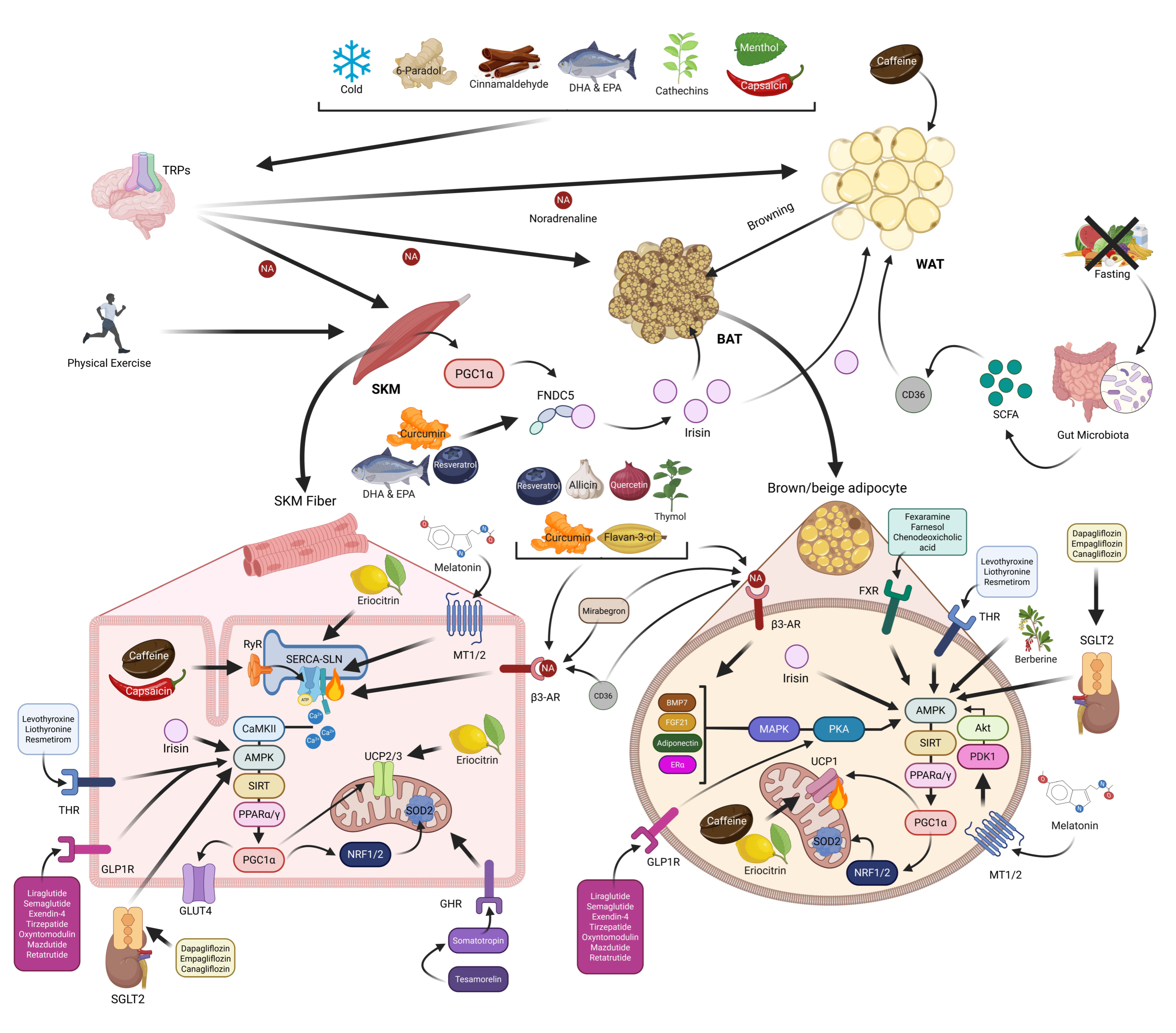
This section highlights the significant influence of physiological conditions such as cold exposure, physical exercise, and fasting on the activation of NST through mechanisms involving BAT, skeletal muscle, and WAT browning (Table 2 and Table 3 and Figure 5). While promising results have been observed, particularly in rodent models, the translation to human physiology remains inconsistent and requires further investigation.

6. Bioactive Ingredients and Their Role in Promoting NST

Several bioactive compounds have been shown to promote NST through BAT activation/WAT browning and/or skeletal muscle SERCA–SLN uncoupling. Food-derived bioactive agents have been extensively studied for their potential in treating diabesity due to their positive health effects.
Capsaicin (C18H27NO3), the primary capsaicinoid from chili peppers and their major pungent compound, is widely known for its thermogenic and anti-obesity effects. In skeletal muscle, capsaicin has been proposed to be a SERCA activity regulator and, thus, an activator of skeletal muscle NST, as it increases SERCA, the ryanodine receptor (RyR), and UCP2/3 expression in C2C12 myotubes [46]. Regarding thermogenic adipose tissue activation, oral ingestion of capsinoids has been shown to increase BAT activity, abdominal fat loss, and fat oxidation in humans [47]. In rodents, capsinoids also stimulate WAT browning through TRP Vanilloid 1 (TRPV1) activation in the gastrointestinal tract [48], suggesting an activation of the vagal afferent nerves that project into the ventromedial hypothalamus, stimulating the sympathetic nervous system and catecholamine secretion via β3-ARs. This enhances whole-body energy expenditure and reduces fat content via calcium/calmodulin-activated protein kinase II (CaMKII)/AMP-activated kinase (AMPK)/SIRT1/PGC1α signaling pathway activation, leading to the expression of thermogenic markers like UCP1 and PRDM16 [48].
6-Paradol (C17H26O3), found in ginger and the Grain of Paradise (GP), is another TRPV1 agonist with thermogenic properties. In humans, the GP extract increased whole-body energy expenditure and BAT function, thereby reducing visceral fat [49,50]; however, ginger’s thermogenic effects remain limited, with no evidence regarding energy expenditure. In obese mice, diets enriched in different ginger extracts promoted energy expenditure and lipolysis, suppressed adipogenesis in WAT, and stimulated BAT activity, increasing UCP1 expression and oxygen consumption [51]. In mice, ginger and its major components, 6-Gingerol (C17H26O4) and Zingerone (C11H14O3), were also found to increase heat production by WAT browning [52,53] and muscle mitochondrial biogenesis [54]. The mechanisms underlying the thermogenic effects of ginger may be partly regulated by the SIRT1/AMPK/PPARα/PGC1α pathway [52,54], possibly via β3-ARs activation.
Cinnamaldehyde (C9H8O), the pungent compound in cinnamon, has been shown to protect rodents against HFD-induced obesity, enhancing body temperature, BAT function, and WAT browning through increased mitochondrial ATP production and UCP1, PGC1α, PPARγ, PRDM16, and fibroblast growth factor 21 (FGF21) expression [55,56]. The effects were attributed to cinnamaldehyde-induced adrenaline secretion via TRPA1 and thus β3-ARs activation [57]. In adipocytes derived from human subcutaneous fat [57] and in healthy subjects, cinnamaldehyde increased energy expenditure and postprandial fat oxidation [58]; however, more clinical trials are needed to further elucidate the thermogenic effects. Other cinnamon-derived compounds, like Cinnamic Acid (C9H8O2), also promote fat browning and thermogenic activity by activating β3-ARs and AMPK signaling in mice [59].
Curcumin (C21H20O6), a polyphenol from turmeric, has been linked to anti-obesity effects, increasing energy expenditure and reducing fat mass and waist/hip circumference in overweight individuals [60]. It modulates SERCA activity in dynamic muscle cells from rodents [61], regulating muscle function and NST. In addition, in mice, curcumin improves plasma myokine levels of FNDC5 and irisin, insulin sensitivity, energy expenditure, oxygen consumption, and heat production, accompanied by white adipocyte and WAT browning, BAT and sWAT metabolic activity enhancement, and increased UCP1 expression [62]. These thermogenic effects are regulated via AMPK upregulation and noradrenaline secretion, suggesting the activation of a β3-ARs-dependent pathway [63].
Allicin (C6H10OS2), an organosulfur compound found in garlic, has antioxidant and immunomodulatory properties with strong potential in the treatment of several diseases, such as diabetes mellitus, cardiovascular diseases, and cancer. In obese mice, allicin administration enhances weight loss and thermogenesis by promoting lipolysis and increasing UCP1, PGC1α, and PRDM16 expression in both BAT and WAT, possibly through Sirtuins-mediated PPARα cascade activation [64]. Garlic administration to obese rodents also showed enhanced UCP expression in BAT, WAT, and skeletal muscle via β3-AR and AMPK-dependent pathway activation [65]. In addition, a fermented garlic extract was also found to increase UCP1 expression in human adipose-derived stem cells, increasing cell metabolism, fat oxidation, and mitochondrial oxygen consumption [66].
Quercetin (C15H10O7) is a polyphenol flavonoid found mainly in onions. In rodents, quercetin was also found to increase weight loss, energy expenditure, and heat production, not only by enhancing BAT mass and function but also by increasing WAT browning and modulating UCP1, PGC1α, and PPARγ expression [67]. In vitro, quercetin-rich extracts enhanced brown and beige-specific gene expression and increased lipolysis, fat β-oxidation, and glucose uptake through PPARα, FGF21, AMPK [68], and β3-ARs stimulation [67]. Moreover, quercetin and its derivatives were also known to modulate SERCA activity and function in muscle from rodents, showing its potential role in skeletal muscle NST directly binding to the ATPase domain [69], which may modulate mitochondriogenesis, energy expenditure, fat oxidation, mitochondrial function, adiposity, and insulin sensitivity.
Caffeine (C8H10N4O2), a psychostimulant consumed mainly in coffee, tea, and cocoa beans, has thermogenic effects on skeletal muscle, increasing energy expenditure and muscle heat production. In addition, a study suggested that 60 mg/kg of caffeine upregulates the expression of UCP1 in BAT and UCP3 in skeletal muscle of obese mice [70]. In humans, supraclavicular BAT was activated after acute caffeine administration, enhancing heat production and energy expenditure [71]. Also, in mice, caffeine promotes browning of white adipocytes to beige-like adipocytes and de novo differentiation of brown adipocytes [71], increasing UCP1, PPARγ, PGC1α, and PRDM16 expression and mitochondrial biogenesis [72]. The mechanisms underlying caffeine-induced skeletal muscle NST are still unclear, whereas the observed thermogenic actions are suggested to be mediated by calcium transport uncoupling, as caffeine is a RyR agonist.
Similar to caffeine, Catechins from tea, such as epigallocatechin-3-gallate (C22H18O11), are associated with weight loss and increased energy expenditure. In white adipocytes from mice, catechins increased lipid metabolism and heat production through UCP1 upregulation, promoting direct white-to-brown adipocyte transformation [73]. In rodents, green tea extracts and catechins promote body weight reduction and enhanced energy expenditure by BAT activation and/or WAT browning, increasing UCP1 expression through adiponectin-dependent PPARγ, PGC1α, PRDM16, bone morphogenetic protein 7 (BMP7), and FGF21 activation [74]. In humans, catechin beverages increased energy expenditure through direct activation of TRP Anquirina 1 (TRPA1) and TRPV1 in the sensory neurons of the gastrointestinal tract, and consequently activation and/or recruitment of BAT [75]. In addition, black tea Theaflavins (C29H24O12) in mice also increased muscle UCP3 through the AMPK/PGC1α activation pathway [76].
Flavan-3-Ols (C15H14O2), flavonoids found in cocoa beans, nuts, berries, stone fruits, apples, pears, and also green tea, increase UCP3 and PGC1α expression in the skeletal muscle from mice, enhancing total energy expenditure [77,78]. In rodents, cocoa flavonols increased BAT activation through AMPK/β3-ARs activation, increasing UCP1 and PGC1α expression [78].
Berberine (C20H18NO4+), a polyphenolic compound from herbs like Coptis chinensis (Chinese goldthread) and Hydrastis canadensis (goldenseal), has been reported to exert anti-diabetic and anti-hyperlipidemia effects in humans. In obese and diabetic mice, berberine increased BAT activity, WAT browning, and mitochondrial content through AMPK/SIRT1/PPARγ pathway activation and thermogenic marker upregulation (UCP1 and PGC1α), as recently reviewed in [79]. Berberine also promoted brown adipocyte differentiation in both mice and human primary preadipocytes [80]. In humans, chronic berberine treatment promoted weight loss and increased BAT mass and activity in mildly overweight subjects [80].
Docosahexaenoic acid (DHA, C22H32O2), an Omega-3 polyunsaturated fatty acid (PUFA) primarily found in fatty fish and marine algae, has been shown to reduce the efficiency of SERCA activity in rats, suggesting an uncoupling effect similar to SLN [81]. Moreover, DHA increased UCP1 expression in BAT and sWAT, enhancing mitochondrial functionality and energy expenditure in rodents [82]. Similar to DHA, Eicosapentaenoic acid (EPA, C20H30O2) in mice also enhances BAT thermogenic properties, increasing PRDM16, PGC1α, and UCP1 expression, and also promotes sWAT browning [82] and muscle NST through SERCA2b and FGF21 upregulation [83], supporting the idea that DHA and EPA could be attractive food-derived bioactive agents to curb obesity. In cultured subcutaneous adipocytes from overweight subjects, EPA induced beige-like adipocytes through AMPK/SIRT1/PGC1α thermogenic pathway activation [84]. The thermogenic effects of PUFAs are proposed to be mediated through the activation of TRPV1 and β3-ARs, as well as browning-related myokines (irisin and FGF21), via muscle calcium signaling [85,86]. All together, these findings suggest a novel and promising role of fish oils (DHA and EPA) in preventing obesity via BAT activation, browning, and skeletal muscle calcium SERCA uncoupling, although their effects on humans are still inconclusive.
Eriocitrin (C27H32O15), a flavonoid found in the skin of limes and lemons, has been linked to health benefits. Eriocitrin boosted several thermogenic genes’ expression in mice, such as UCP1 in BAT and UCP3, SLN, SERCA1, and SERCA2 in skeletal muscle, enhancing insulin sensitivity, fatty acid oxidation, and energy expenditure and reducing hepatic gluconeogenesis and steatosis [87].
Menthol (C10H20O), a cooling and flavoring alcohol found in mint oils, is a TRPM8 receptor agonist (β3-ARs activation) that has been shown to promote UCP1-dependent BAT thermogenesis, sWAT browning, glucose metabolism, and muscle energy expenditure in mice after chronic supplementation [88,89]. In rodents, menthol administration enhanced hyperthermia through increased UCP1, PGC1α, PRDM16, FGF21, AMPK, and Protein kinase A (PKA) expression [88]. In humans, menthol increases hyperthermia and energy expenditure, showing its potential to promote thermogenesis [90].
Thymol (C10H14O), a monoterpene polyphenolic compound found in thyme, enhances mitochondrial biogenesis and UCP1 expression through β3-ARs and PKA/AMPK signaling pathway activation in mice-derived white adipocytes [91]. These results suggest that thymol may act as a selective β3-sympathomimetic; however, in vivo studies are lacking.
Resveratrol (C14H12O3), a polyphenol primarily found in the skin of grapes and other berries, has anti-obesity effects, enhancing thermogenic tissue function. In vitro studies showed that resveratrol upregulates FNDC5/irisin expression in mice [92]. In rodents, resveratrol increased UCP3 expression in skeletal muscle, UCP1 expression in BAT and sWAT, and promoted mitochondrial dynamics, insulin sensitivity, glucose uptake, weight loss, and visceral fat mass loss [93,94]. In rodents, resveratrol decreased adipogenesis and enhanced angiogenesis, mitochondrial activity, and brown-like adipocyte marker expression via BMP7/Estrogen Receptor α (ERα)/AMPK/PPARα/γ/PGC1α activation [93,95]. In humans, resveratrol-enriched diets increased UCP1, PRDM16, PGC1α, and SIRT1 expression in sWAT biopsies, improving the glycemic and lipid profiles of individuals through FNDC5 activation [93]. Additionally, other dietary sources of resveratrol, like berry extracts [96,97], also showed similar thermogenic effects on BAT activity and/or WAT browning in rodents. Similarly, Proanthocyanidin (C31H28O12) and Anthocyanin (C15H11O+), other grape-derived bioactive compounds, also were shown to promote energy expenditure through increased skeletal muscle and BAT mitochondrial function [98], BAT activity, and sWAT browning in rodents [99]. All these compounds activate the β3-ARs pathway and increase the expression of thermogenic markers such as UCP1, PRDM16, and PGC1α, thereby limiting obesity [100].
Despite compelling evidence supporting the thermogenic effects of these food-derived bioactive compounds (Table 2 and Table 3 and Figure 5), human studies remain limited or very scarce, and further research is needed to elucidate their potential in anti-diabesity therapies.

7. Drugs Stimulating NST

7.1. FDA-Approved Drugs

Although numerous drugs exist for obesity-related conditions such as diabetes and cardiovascular diseases, pharmacological interventions specifically targeting obesity remain limited. Currently, only a few FDA-approved drugs are available for long-term obesity management, primarily acting by suppressing appetite or inhibiting lipid absorption. In contrast, therapies that enhance adaptive thermogenesis—particularly via BAT activation or WAT browning—are still underexplored [101].
β3-ARs agonists have emerged as good pharmacological tools to fight obesity and its complications due to their thermogenic effects. β3-ARs are abundant in thermogenic tissues such as BAT, regulating NST through sympathetic nerve stimulation and enhancing lipolysis, energy expenditure, and weight loss via AMPK/SIRT1/PGC1α pathway activation and UCP1 upregulation [101]. The most studied β3-ARs agonist is Mirabegron (C21H24N4O2S), an FDA-approved drug for overactive bladder syndrome. In rodents, mirabegron was shown to enhance BAT activity and WAT browning, increasing UCP1 expression in a dose-dependent manner [102]. In a pioneering human study, Cypess et al. showed that a dose (200 mg/day, orally) of mirabegron leads to higher BAT metabolic activity and further increased energy expenditure in healthy lean men [103]. In addition, in young healthy women, mirabegron acutely increased BAT activity, also increasing their metabolic rate and energy expenditure [104]. In the same study, the effects of mirabegron were more prominent in subjects with low BAT amount at baseline after 4 weeks of therapy, highly increasing BAT mass and ameliorating the lipid profile, insulin sensitivity, and secretion in the subjects, with no changes in body weight and composition [104]. An increase in skin temperature and improved glucose homeostasis were also reported after mirabegron administration in humans [105]. Further studies in obese insulin-resistant patients demonstrated that chronic mirabegron treatment induces UCP1 expression in sWAT and thermogenic PGC1α marker in skeletal muscle, while also increasing the number of oxidative fibers [19]. However, no effects on body weight and/or fat mass reduction were found. Although mirabegron has great thermogenic potential in anti-obesity therapies, the lack of significant weight-lowering effects and the cardiovascular side effects reported in diverse studies may limit its broad use in humans. Further long-term human studies are also needed to shed light on the applicability of mirabegron in limiting obesity.
Thyroid hormone receptor (THR) agonists, such as levothyroxine (C15H11I4NO4) and liothyronine (C15H12I3NO4), have also been investigated for their roles in metabolic regulation. While most evidence is derived from observational data linking thyroid disorders with weight changes, hyperthyroid patients and subjects under TH replacement therapy reported increased energy expenditure and body temperature, also showing reduced body weight and cholesterol levels associated with enhanced BAT activation and increased muscle glucose uptake [106]. Triiodothyronine (T3) has shown browning potential by promoting a brown-like adipocyte phenotype in human adipose-derived stem cells, increasing UCP1 expression, mitochondrial biogenesis, and oxidative metabolism [107]. In rodents, T3 also stimulates thermogenesis in BAT, WAT, and skeletal muscle, regulating fiber composition and SERCA isoform expression through AMPK and UCP1 activation after THR β (THRB) activation [106]. Selective THRB agonists like Resmetirom (C17H12Cl2N6O4), recently approved for nonalcoholic steatohepatitis, improve cholesterol and triglyceride levels and present strong anti-obesity potential [108], though human studies remain limited.
Farnesoid X receptor (FXR) agonists, known for regulating bile acid and lipid metabolism, also show promising thermogenic effects. In obese mice, fexaramine (C32H36N2O3) enhanced thermogenic gene expression in BAT and WAT, promoting mitochondriogenesis and fat oxidation [109]. Moreover, farnesol (C15H26O) treatment also activates the AMPK/PGC1α/UCP1 thermogenic pathway in both mice and human adipocytes [110], and oral supplementation of chenodeoxycholic acid (C24H40O4), a bile acid that stimulates the farnesoid X receptor, increases basal energy expenditure, promoting BAT activity and cold-induced thermogenesis in women [111]. While some FXR agonists are under clinical investigation for metabolic syndrome, their direct role in thermogenesis and WAT browning in humans is still unclear.
Growth hormone (GH) deficiency, such as somatotropin (C990H1529N263O299S7) deficiency, is associated with increased body fat and altered distribution. GH replacement therapy in humans restores basal metabolic rate, reduces visceral fat and waist circumference, and improves lipid profiles, glucose tolerance, and insulin sensitivity, recovering body composition [112] and highlighting GH’s link with central obesity and diabetes. Tesamorelin (C221H366N72O67S), a synthetic analogue of GH-releasing factor, also enhanced phosphocreatine recovery post-exercise in obese subjects [113], though no direct effects on BAT or WAT-mediated NST have been confirmed in humans. In rodents, GH therapy has controversial effects on BAT activity and WAT browning (as reviewed in [114]).
Glucagon-like peptide 1 (GLP-1) receptor agonists have emerged as pivotal tools in anti-diabesity therapy due to their ability to enhance insulin secretion, inhibit glucagon release, reduce appetite, and promote sustained weight loss [101]. Despite the absence of GLP-1 receptor expression in WAT, in vitro studies showed that GLP-1 analogs like exendin-4 (C184H282N50O60S) promote adipocyte browning with an increase in thermogenic gene expression via AMPK/SIRT1/PGC1α/UCP1 pathway stimulation [115]. In rodents, liraglutide (C172H265N43O51), a long-acting GLP-1 receptor agonist, stimulated BAT thermogenesis and promoted WAT browning, resulting in reduced body weight and food intake [116]. In humans, liraglutide effects were also confirmed, showing significant weight loss, improved metabolic markers, and increased energy expenditure in obese and diabetic patients [116]. Once-weekly semaglutide (C187H291N45O59), a newer and oral GLP-1 analog, demonstrated even greater weight loss and appetite suppression effects, reducing visceral fat mass and enhancing metabolic control in humans [117], making it the most effective GLP-1 analog to date. The thermogenic role of GLP-1 receptor agonists in humans remains scarce; however, enhanced BAT activity and WAT browning after semaglutide treatment have been reported in obese mice [118]. Tirzepatide (C225H348N48O68), a dual glucose-dependent insulinotropic polypeptide receptor (GIPR) and GLP-1 receptor agonist, has recently gained importance for its synergistic effects on energy metabolism. GIPR stimulation in human adipose-derived stem cells promotes browning, UCP1 upregulation, and lipid metabolism via PKA signaling [119]. Clinical studies, like SURMOUNT trials, show that tirzepatide has greater anti-obesity effects than semaglutide, promoting weight and fat mass reduction, appetite suppression, energy expenditure, insulin sensitivity, and glycemic parameters in obese [120] and diabetic patients [121]. Although thermogenesis was not evaluated in SURMOUNT studies, an ongoing clinical trial (NCT04081337) will elucidate tirzepatide’s role in energy expenditure and NST. Tirzepatide was recently approved not just for T2DM treatment but also for the treatment of obesity and overweight, particularly in people with related metabolic conditions [120]. Additionally, dual GLP-1/glucagon agonists such as oxyntomodulin (C192H295N59O60S) [122] and mazdutide (C207H317N45O65) have shown encouraging outcomes in promoting weight loss, reducing body mass index, waist circumference, and food intake, increasing energy expenditure, and improving glycemic control in obese and/or overweight subjects [123]. Retatrutide (C221H342N46O68), a triple GLP-1/GIP/glucagon receptor agonist, is currently under study to find its potential in anti-diabesity therapies; however, clinical relevance is not well established. In humans, retatrutide reduces body weight while improving glucose homeostasis, insulin sensitivity, and lipid profile [124]. Despite the promising clinical effects of these agonists, their precise impact on human thermogenesis, including BAT activation and WAT browning, remains insufficiently characterized. Future studies measuring energy expenditure, thermogenic gene expression, and adipose tissue remodeling are essential to fully define their roles in thermogenesis-based anti-diabesity therapies.
Sodium-glucose cotransporter 2 (SGLT2) inhibitors are emerging as promising anti-diabesity drugs due to their anti-hyperglycemic and body weight regulatory effects. In obese rodents, these drugs have demonstrated the ability to limit weight gain, enhance energy expenditure, and promote thermogenesis via upregulation of UCP1 in both BAT and WAT and increased muscle fat oxidation by activating the AMPK/SIRT1 signaling axis [125]. In humans, dapagliflozin (C21H25ClO6) reduced body weight, waist circumference, insulin resistance, and fat mass and improved glycemia, insulin sensitivity, respiratory exchange ratio, and the lipid profile of diabetic patients [126]. Empagliflozin (C23H27ClO7) and canagliflozin (C24H25FO5S) similarly reduced body weight and fat mass, and both restored glycemic parameters in diabetic subjects [127]. While these agents were designed to flush out glucose, they may also ignite dormant thermogenic circuits, offering a twofold strike against diabesity. More mechanistic human studies are needed to uncover the full thermogenic potential of SGLT2 inhibitors and validate their role in targeting energy balance through adipose tissue and skeletal muscle NST.

7.2. Melatonin

Melatonin (N-acetyl-5-methoxytryptamine, C13H16N2O2) is a highly conserved hormone present across the plant and animal kingdoms. In mammals, melatonin is predominantly synthesized at night by the pineal gland, but also in many other organs [128], and is widely exogenously administered in humans in Europe and the USA both as a drug and as a supplement [129]. Melatonin levels naturally decline with age, shift work, exposure to artificial light, and in metabolic diseases such as diabesity [130]. Beyond its well-established antioxidant and anti-inflammatory properties, melatonin is gaining importance for its metabolic and thermogenic roles [128].
In humans, however, data from several small-scale (<100 participants) studies on the use of melatonin in patients with metabolic syndrome or obesity are conflicting and inconclusive, ranging from a modest effect to no effect, depending on the protocol applied (melatonin dosage and duration) (reviewed in [129]). In this scenario, a systematic review and meta-analysis of 20 human studies proved that melatonin significantly reduced body weight, and in more than 50% of the studies, decreased body mass index, weight gain, or waist circumference in men and women, showing better outcomes with higher melatonin doses (8 mg/day) and longer treatment durations (48 weeks) [131]. A recent systematic review and dose–response meta-analysis of 28 randomized controlled clinical trials also exhibited that melatonin supplementation reduces hip circumference in both sexes and waist circumference in interventions longer than 8 weeks in overweight and/or diabetic subjects, also improving the lipid profile and reducing fat accumulation. Body weight in diabetic patients and body mass index in obese individuals were also lower after melatonin treatment compared to placebo [132]. However, numerous clinical trials show that melatonin has no effect on body weight loss in obese and/or diabetic subjects, results not comparable to those obtained in rodents, perhaps because of the great genetic variability of melatonin receptors in humans, especially the membrane melatonin receptor 2 (MT2) [129].
A substantial amount of promising data regarding the potential role of melatonin-based therapy in the prevention of obesity and its related metabolic disorders has been reported in preclinical studies. Various mechanisms have been proposed to explain the beneficial effects of melatonin on body weight and metabolism, and several receptors have been proposed, including the membrane receptors MT1 and MT2, the main receptors studied regulating melatonin’s effects on obesity [133]. (i) Since increased core body temperature and no changes in food intake have been found, and locomotor activity data in rodents are inconsistent, one mechanism may involve the increase in energy expenditure by NST activating BAT and/or WAT browning through UCP1 upregulation [133], and/or skeletal muscle NST through SERCA/SLN uncoupling and CaMKII/AMPK/PGC1α pathway activation [134]. (ii) Another mechanism is through its well-known chronobiotic effects in both humans and rodents, participating in the control of metabolism and energy balance of the organism over 24 h cycles, increasing insulin sensitivity and the energy intake use and/or storage during the active phase (day), and reducing it during the rest/sleep phase (night), accentuating adipose tissue lipolysis [130,135]. (iii) Also, melatonin improves diabesity through its antioxidant and anti-inflammatory properties and its potential to maintain mitochondrial homeostasis and function in adipose tissue and skeletal muscle [133,136]. (iv) Recently, studies showed that melatonin is also produced by microbiota, and exogenous supplementation can improve microbiota dysbiosis, alleviating weight gain and fat accumulation, reducing low-grade inflammation, and promoting lipid metabolism and energy expenditure [133]. In the following section, we will focus on the effect of melatonin on the recovery of mitochondrial function in obesity.
Our previous studies showed that chronic melatonin treatment reduced body weight gain in a preclinical animal model of diabesity, assembling human obesity-related type 2 diabetes, browned subcutaneous fat, increased thermogenic BAT function and mass, enhanced skeletal muscle NST and oxidative fiber phenotype, and improved lipid profile, glucose homeostasis, low-grade inflammation, and mitochondrial biogenesis and functionality [134,137,138,139,140,141,142,143,144]. These changes took place in the absence of changes in locomotor activity or feeding patterns, suggesting a thermogenic effect. These outstanding findings encourage further research on the translation of these findings on melatonin effects to humans. The beneficial effect of melatonin and its analogs against a wide range of diseases prompted many decades of studies to evaluate the safety aspect of this substance. A substantial number of both human and animal studies reported that short-term use of melatonin possesses no adverse effects, even in high doses, whereas long-term melatonin treatment in humans causes only mild adverse effects such as dizziness, headache, nausea, and sleepiness. However, due to a lack of studies, it is difficult to exclude any possible adverse effects in pregnant and breastfeeding women, as well as in children and adolescents. In experimental animal studies, the lethal dose has not been estimated; even high doses up to 800 mg/kg are without any acute toxic effects. Also, studies in healthy subjects indicate that a dose of 10 mg for 28 days shows no toxicity [145].
Melatonin works via multiple means to limit oxidative stress: (i) One mechanism is by acting as a direct scavenger to detoxify free oxygen and nitrogen species and stimulating the expression and activity of some antioxidant enzymes (e.g., NRF1/2, glutathione, GPx, SOD2, and SIRT1/3) [133,146]. (ii) Another mechanism is by stabilizing mitochondrial membrane integrity, thereby preventing the opening of the mitochondrial permeability transition pore (mPTP) and cytochrome C release into the cytosol, inhibiting pro-apoptotic pathways, regulating mitochondrial dynamics and autophagy, and promoting mitochondrial quality [136,147,148]. (iii) Also, it works by improving electron transport chain activity and mitochondrial function (increasing ATP production and the activities of respiratory chain Complex I-IV) [147,148,149]. (iv) In addition, melatonin can also modify the metabolic fuel used, regulating energy balance and increasing lipolysis and metabolism switching into a fat oxidative one, therefore reducing body intra-abdominal fat [133,142,150,151]. (v) Finally, melatonin also shows cell protective effects, preventing ER-stress mediated apoptosis [152]. Several of these melatonin effects have already been demonstrated in thermogenic tissues such as BAT [137], WAT [144,153,154], and skeletal muscle [142,150,155,156], being additional mechanisms to NST that regulate the energy balance and limit diabesity. Furthermore, melatonin positively affects muscle metabolism, enhancing insulin sensitivity and glucose homeostasis; preserving its structure; increasing myofiber size, mitochondrial function, membrane integrity, and dynamics; promoting myofiber differentiation; reducing oxidative stress, inflammation, and apoptosis in both human and preclinical models of diabesity; and preventing muscle loss and atrophy known as sarcopenia [133].
Based on all of melatonin’s aforementioned effects concerning its beneficial impact on body weight, the potential thermogenic effect, the safety aspect, and the powerful antioxidant properties, melatonin should be considered as a promising therapeutic option in addition to the drugs mentioned above for obesity management by activating thermogenic mechanisms (Table 2 and Table 3 and Figure 5).
Table 2. Summary of the physiological conditions, bioactive agents, and drugs stimulating non-shivering thermogenesis (NST) in rodents with their anti-diabesity effect, thermogenic mechanism, and molecular pathway involved.
StimuliAnti-Diabesity EffectMechanismMolecular PathwayReferences
Physiological conditionsCold exposure↑ Energy expenditure
↑ Insulin sensitivity
↑ BAT activity and recruitment
↑ BAT & WAT UCP1 expression
↑ WAT browning & angiogenesiss
↑ SKM SLN expression
↓ SKM SERCA activity
↑ SKM mitochondrial function
TRPs/β3-ARs/PPARγ/PGC1α[14,15,17,20]
Physical exercise↓ Insulin resistance
↑ Glucose sensitivity
↑ Lipid metabolism
↓ Fat mass
↑ BAT activity and recruitment
↑ WAT browning
& mitochondrial function
↑ WAT UCP1 expression
↓ vWAT adiposity
↑ SKM UCP3, SERCA1
& SLN expression
↑ SKM mitochondrial
biogenesis and function
PGC1α/FNDC5/Irisin[26,28,29,30]
Fasting↓ Body weight
↑ Energy expenditure
↑ BAT activation
↑ BAT & SKM energy
expenditure
↑ WAT browning
↑ BAT & WAT UCP1 expression
SCFA (Gut microbiota)/CD36/
β3-ARs/SIRT6/PGC1α
[31,33,34,35,37,44]
Bioactive ingredientsCapsaicin &
Capsinoids
Pharmaceuticals 18 01247 i004
(Pepper)
↓ Weight gain
↑ Energy expenditure
↓ Fat accumulation
Improved glucose levels
↑ BAT activity and recruitment
↑ WAT browning
↑ BAT & WAT UCP1 expression
↑ SKM UCP2/3, SERCA1/2
& RyR1/2 expression
↑ SKM SERCA activity & ATP hydrolysis
TRPV1/β3-ARs/CaMKII/AMPK/
SIRT1/PGC1α
[46,48]
6-Paradol,
Pharmaceuticals 18 01247 i005
Gingerol
Pharmaceuticals 18 01247 i006
& Zingerone
Pharmaceuticals 18 01247 i007
(Ginger)
↓ Body weight
↓ Fat mass
↑ Energy expenditure
↑ BAT activity & function
↑ vWAT, sWAT & white
adipocytes (in vitro) browning
↑ BAT & WAT UCP1 expression
↓ WAT adipogenesis & ↑ lipolysis
↑ SKM mitochondrial biogenesis
TRPV1/β3-ARs/AMPK/SIRT1/
PPARα/PGC1α
[51,52,53,54]
Cinnamaldehyde
Pharmaceuticals 18 01247 i008
& Cinnamic acid
Pharmaceuticals 18 01247 i009
(Cinnamon)
↓ Body weight
↑ Energy expenditure
↑ Body temperature
↑ BAT activity & brown
adipocytes (in vitro) function
↑ BAT mitochondrial ATP
production
↑ vWAT, sWAT & white
adipocytes (in vitro) browning
↑ BAT & WAT UCP1 expression
↓ Adipocytes size &
lipogenesis (in vitro)
TRPA1/β3-ARs/FGF21/AMPK/
PPARγ/PGC1α
[55,56,57,59]
Curcumin
Pharmaceuticals 18 01247 i010
(Turmeric)
↓ Body weight
↑ Energy expenditure
↑ Insulin sensitivity
↑ BAT activation
↑ sWAT & white adipocytes
(in vitro) browning
↑ BAT & sWAT UCP1 expression
↑ Muscle cells mitochondrial function & energy expenditure (in vitro)
↑ SKM SERCA1 expression
ATP hydrolysis & fiber type composition modulation
FNDC5/Irisin/β3-ARs/AMPK/
PPARγ/PGC1α
[61,62,63]
Allicin
Pharmaceuticals 18 01247 i011
(Garlic)
↓ Weight gain
↑ Energy expenditure
Improved glucose
homeostasis
↑ BAT activation & fat oxidation
↑ WAT & white adipocytes
(in vitro) browning
↑ BAT & sWAT UCP1 expression
↑ SKM UCP3 expression
↑ Lipolysis
β3-ARs/AMPK/SIRTs/
PPARα/PGC1α
[65]
Quercetin
Pharmaceuticals 18 01247 i012
(Onion)
↑ Weight loss
↑ Energy expenditure
↓ Fat accumulation
Improved glucose
homeostasis
↑ BAT mass & function
↑ sWAT & white adipocytes
(in vitro) browning
↑ BAT & WAT UCP1 expression
↑ Fat oxidation & ↓ adipogenesis
↑ SKM mitochondrial function
& biogenesis
SKM SERCA1/2 activity
& function modulation
SKM SERCAs conformational regulation
↑ SKM glucose uptake
β3-ARs/FGF21/PKA/AMPK/
SIRT1/PPARα/γ/PGC1α
[67,68,69]
Caffeine
Pharmaceuticals 18 01247 i013
(Coffee)
↑ Weight loss
↑ Energy expenditure
↑ White adipocytes browning
& UCP1 expression (in vitro)
↑ BAT UCP1 expression
↑ SKM UCP3 expression
SKM RyR agonist
& SERCA activity uncoupler
↑ Mitochondriogenesis
RyR/SERCA/PPARγ/PGC1α[70,71,72]
Catechins
Pharmaceuticals 18 01247 i014
& Theaflavins
Pharmaceuticals 18 01247 i015
(Tea)
↓ Body weight
↑ Energy expenditure
↑ BAT activity & function
↑ BAT & WAT UCP1 expression
↑ WAT & white adipocytes
(in vitro) browning
=/↑ SKM UCP3 expression
=SKM FNDC5/Irisin
& SLN expression
TRPA/V1/β3-ARs/BMP7/FGF21/
Adiponectin/AMPK/SIRT1/
PPARγ/PGC1α
[73,74,76]
Flavan-3-ols
Pharmaceuticals 18 01247 i016
(Cocoa)
↑ Energy expenditure↑ BAT activation & UCP1
expression
↑ SKM UCP3 expression
↑ Mitochondriogenesis
β3-ARs/AMPK/PGC1α[77,78]
Berberine
Pharmaceuticals 18 01247 i017
(Chinese goldthread, goldenseal)
↓ Body weight
↑ Energy expenditure
Improved glucose
homeostasis
↑ BAT activity
↑ sWAT & white adipocytes
(in vitro) browning
↑ Brown adipocytes
differentiation (in vitro)
↑ BAT & WAT UCP1 expression
↑ Mitochondrial biogenesis
& function
AMPK/SIRT1/PPARγ/PGC1α[79,80]
DHA
Pharmaceuticals 18 01247 i018
& EPA
Pharmaceuticals 18 01247 i019
(Oily fish)
↓ Weight gain
↑ Energy expenditure
Improved glucose
metabolism
↑ BAT mass & activity
↑ WAT browning
↑ BAT, sWAT & brown and white adipocytes (in vitro) UCP1
expression
↑ Mitochondrial function
& biogenesis
↑ Fat & glucose oxidation
↑ SKM SERCA activity
uncoupling
↑ SKM SERCA2b & SLN
expression
↑ SKM development
TRPV1/β3-ARs/FGF21/
Irisin/AMPK/SIRT1/PGC1α
[81,82,83,85,86]
Eriocitrin
Pharmaceuticals 18 01247 i020
(Lemon)
↑ Energy expenditure
↑ Insulin sensitivity
↑ BAT UCP1 expression
↑ SKM UCP3, SERCA1/2 & SLN expression
↑ Fat oxidation
?[87]
Menthol
Pharmaceuticals 18 01247 i021
(Mint)
↑ Weight loss
↑ Energy expenditure
Improved glucose
metabolism
↑ BAT activity & fat oxidation
↑ WAT & white
adipocyte (in vitro) browning
↑ BAT, WAT & brown and white adipocytes (in vitro) UCP1
expression
↑ Mitochondrial activity
& metabolic rate
↑ SKM energy expenditure
↑ Skin temperature
TRPM8/β3-ARs/FGF21/
Calcium/PKA/AMPK/PGC1α
[88,89]
Thymol
Pharmaceuticals 18 01247 i022
(Thyme)
?↑ White adipocytes browning
& UCP1 expression (in vitro)
↑ Mitochondriogenesis (in vitro)
β3-ARs/PKA/AMPK[91]
Resveratrol,
Pharmaceuticals 18 01247 i023
Proanthocyanidin
Pharmaceuticals 18 01247 i024
& Anthocyanin
Pharmaceuticals 18 01247 i025
(Grapes & Berries)
↓ Body weight
↑ Energy expenditure
↓ Fat mass
↓ Insulin resistance
Improved glycemic and
lipid profile
↑ BAT activity & recruitment
↑ WAT & white adipocytes
(in vitro) browning
↑ BAT, WAT & brown and white adipocytes (in vitro) UCP1
expression
↑ Fat oxidation & ↓ vWAT
adipogenesis
↑ SKM UCP3 expression
↑ SKM glucose uptake
↑ SKM, BAT, WAT, brown and white adipocytes (in vitro)
mitochondrial dynamic & function
β3-ARs/BMP7/FNDC5/Irisin/ERα/
AMPK/SIRT1/3/PPARα/γ/PGC1α
[92,93,95,96,97,98,99,100]
Drugsβ3-AR agonists
Mirabegron
Pharmaceuticals 18 01247 i026
↑ Weight loss
↑ Energy expenditure
↑ Insulin sensitivity
and secretion
Improved lipid
profile and glucose
homeostasis
↑ BAT mass & activity
↑ WAT & white adipocytes
(in vitro) browning
& UCP1 expression
β3-ARs/PGC1α[102]
THR agonist
Levothyroxine
Pharmaceuticals 18 01247 i027
Liothyronine
Pharmaceuticals 18 01247 i028
Resmetirom
Pharmaceuticals 18 01247 i029
↓ Body weight
↑ Energy expenditure
Improved lipid
profile
↑ BAT activity & function
↑ WAT browning
↑ BAT & WAT UCP1 expression
THRB/AMPK[106]
FXR agonist
Fexaramine
Pharmaceuticals 18 01247 i030
Farnesol
Pharmaceuticals 18 01247 i031
CDCA
Pharmaceuticals 18 01247 i032
↑ Energy expenditure↑ BAT mitochondriogenesis
& fat oxidation
↑ WAT browning
↑ BAT, WAT & white and brown adipocytes (in vitro) UCP1
expression
Adipogenesis modulation
FXR/AMPK/PPARγ/PGC1α[109,110]
Growth Hormone
Tesamorelin
Pharmaceuticals 18 01247 i033
↓ Fat mass
Improved lipid
profile, glucose
tolerance and insulin
sensitivity
=/↑ BAT activity
=/↑ WAT browning
GHR[114]
GLP1R agonists
Liraglutide
Pharmaceuticals 18 01247 i034
Semaglutide
Pharmaceuticals 18 01247 i035
Exedin-4
Pharmaceuticals 18 01247 i036
GLP1R/GIPR dual agonist
Tirzepatide
Pharmaceuticals 18 01247 i037
GLP1R/GR dual
agonist
Oxyntomodulin
Pharmaceuticals 18 01247 i038
Mazdutide
Pharmaceuticals 18 01247 i039
GLP1R/GR/GIPR
triple agonist
Retatrutide
Pharmaceuticals 18 01247 i040
↑ Weight loss
↑ Energy expenditure
↓ Food intake
↓ Fat mass
Improved insulin
sensitivity and
secretion, and
glycemic control
↑ BAT activity & function
↑ WAT & white adipocytes
(in vitro) browning
↑ BAT, WAT & white adipocytes (in vitro) UCP1 expression
↑ SKM mitochondrial function
& thermogenic genes expression
GLP1R/PKA/AMPK/SIRT1/PGC1α[115,116,118]
SGLT2 Inhibitors
Dapagliflozin
Pharmaceuticals 18 01247 i041
Empagliflozin
Pharmaceuticals 18 01247 i042
Canagliflozin
Pharmaceuticals 18 01247 i043
↓ Body weight
↑ Energy expenditure
↓ Fat mass
Improved lipid
profile, glucose
homeostasis and
insulin sensitivity
↑ BAT activity & WAT browning
↑ BAT & WAT UCP1 expression
↑ SKM fat oxidation
SGLT2/AMPK/SIRT1[125]
Melatonin
Pharmaceuticals 18 01247 i044
↑ Energy expenditure
↓ Body weight
↓ Visceral fat mass
Improved lipid
profile, glucose
homeostasis and
insulin sensitivity
↑ Body temperature
↑ BAT activity, mass & function
↑ sWAT browning
↑ BAT & WAT UCP1 expression
↑ SKM SERCA activity
& expression
↑ SKM SLN expression
↑ BAT, WAT & SKM
mitochondrial quality, function and biogenesis
↑ Mitochondrial membrane
integrity & dynamics
↓ Organellar stress & apoptosis
↓ Fat accumulation, oxidative stress & low-grade inflammation
↑ Lipid metabolism
Improved SKM fiber
composition, microbiota
dysbiosis, metabolism control and plasticity & energy balance over 24 h
MT1/2/PDK1/Akt/CaMKII/AMPK/
SIRT1/3/PGC1α/NRF1/2/SOD2
[134,137,138,139,140,141,142,143,144,150,151,153,154,155,156]
↑, increase in; ↓, decrease in; =, no changes in; Akt, protein kinase B; AMPK, 5′ adenosine monophosphate-activated protein kinase; ATP, adenosine triphosphate; β3-AR, β3 adrenergic receptor; BAT, brown adipose tissue; BMP7, bone morphogenetic protein 7; CaMKII, Ca2+/calmodulin-dependent protein kinase II; CD36, cluster of differentiation 36; CDCA, Chenodeoxycholic acid; DHA, docosahexaenoic acid; EPA, eicosapentaenoic acid; ERα, estrogen receptor α; FGF21, fibroblast growth factor 21; FNDC5, fibronectin type III domain-containing protein 5; FXR, farnesoid X receptor; GHR, growth hormone receptor; GIPR, glucose-dependent insulinotropic polypeptide receptor; GLP1R, glucagon-like peptide-1 receptor; GR, glucagon receptor; MT1/2, melatonin receptor 1/2; NRF1/2, nuclear respiratory factor 1/2; PDK1, pyruvate dehydrogenase kinase 1; PGC1α, peroxisome proliferator-activated receptor gamma co-activator 1α; PKA, protein kinase A; PPARα/γ, peroxisome proliferator-activated receptor α/γ; RyR, ryanodine receptor; SCFA, short-chain fatty acids; SERCA, sarcoplasmic/endoplasmic reticulum Ca2+-ATPase; SGLT2, sodium-glucose linked transporter 2; SIRT1/3/6, sirtuin 1/3/6; SKM, skeletal muscle; SLN, sarcolipin; SOD2, superoxide dismutase 2; sWAT, subcutaneous white adipose tissue; TH, thyroid hormone; THR, thyroid hormone receptor; THRB, thyroid hormone receptor β; TRP, transient receptor potential; TRPA1, transient receptor potential A1; TRPM8, transient receptor potential cation channel melastatin 8; TRPV1, transient receptor potential vanilloid 1; UCP1, uncoupling protein 1; UCP2, uncoupling protein 2; UCP3, uncoupling protein 3; vWAT, visceral white adipose tissue; WAT, white adipose tissue.
Table 3. Summary of human studies and clinical trials on non-shivering thermogenesis (NST) stimulation through physiological conditions, bioactive agents, and thermogenic drugs in diabesity.
StimuliType of StudyAnti-Diabesity EffectMechanismReferences
Physiological conditionsCold exposureClinical Trial↑ Energy expenditure
↑ Insulin sensitivity
↑ BAT activity and recruitment
↑ sWAT UCP1 expression
& mitochondrial function
↑ SKM GLUT4 expression
[18,19,21]
Physical exerciseObservational Human study↓ Insulin resistance
↑ Glucose sensitivity
↑ Lipid metabolism
↓ Visceral fat mass
↑ BAT activation
↑ WAT browning
↑ BAT & WAT UCP1 expression
↑ SKM UCP3 expression
[24,25,27]
FastingClinical Trial↓ Body weight
↑ Energy expenditure
= sWAT UCP1 expression[32,33]
In vivo Human studyImproved metabolic health↑ SKM UCP3 expression[45]
Bioactive ingredientsCapsaicin &
Capsinoids
Pharmaceuticals 18 01247 i045
(Pepper)
Clinical Trial↓ Weight gain
↑ Energy expenditure
↓ Abdominal fat
accumulation
Restored glucose levels
↑ BAT activity
↑ Fat oxidation
[47]
6-Paradol
Pharmaceuticals 18 01247 i046
(Grain of Paradise)
Clinical Trial↓ Body weight
↓ Visceral fat mass
↑ Energy expenditure
↑ BAT activation and recruitment[49,50]
Cinnamaldehyde
Pharmaceuticals 18 01247 i047
& Cinnamic acid
Pharmaceuticals 18 01247 i048
(Cinnamon)
In vitro Human study↑ Metabolic response↑ Adipocytes browning
& UCP1 expression
↑ Fat oxidation
[57]
Clinical Trial↓ Body weight
↑ Energy expenditure
↑ Facial skin temperature[58]
Curcumin
Pharmaceuticals 18 01247 i049
(Turmeric)
Clinical Trial↓ Body weight
↓ Waist/hip
circumference
↑ Energy expenditure
↑ Insulin sensitivity
↓ Fat mass & anthropometric measurements[60]
Allicin
Pharmaceuticals 18 01247 i050
(Garlic)
In vitro Human study↓ Weight gain
↑ Energy expenditure
Improved glucose
tolerance
↑ White adipocytes browning
& UCP1 expression
[66]
Caffeine
Pharmaceuticals 18 01247 i051
(Coffee)
In vitro & in vivo Human study↑ Weight loss
↑ Energy expenditure
↑ BAT activity & function
↑ Adipocytes browning
& UCP1 expression
= SKM UCP3 expression
[71]
Catechins
Pharmaceuticals 18 01247 i052
(Tea)
Clinical Trial↓ Body weight
↑ Energy expenditure
↑ BAT activation and recruitment[75]
Berberine
Pharmaceuticals 18 01247 i053
(Chinese goldthread, goldenseal)
In vitro & in vivo Human study↓ Body weight
↑ Energy expenditure
Improved glucose
homeostasis
Restored metabolic health
↑ BAT mass & function
↑ BAT recruitment
& brown adipogenesis
[80]
EPA
Pharmaceuticals 18 01247 i054
(Oily fish)
In vitro & in vivo Human study↓ Weight gain
↑ Energy expenditure
Improved glucose
metabolism
↑ Subcutaneous white
adipocytes browning
= SKM SERCA activity
& function
[84]
Menthol
Pharmaceuticals 18 01247 i055
(Mint)
Clinical Trial↑ Weight loss
↑ Energy expenditure
Improved glucose
metabolism
↑ Skin temperature
& metabolic rate
[90]
Resveratrol
Pharmaceuticals 18 01247 i056
(Grapes & Berries)
In vitro & in vivo Human study↓ Body weight
↑ Energy expenditure
↓ Visceral fat mass
↓ Insulin resistance
Improved glycemic
and lipid profile
↑ sWAT & white adipocytes browning
↑ sWAT & white adipocytes UCP1 expression
↑ SERCA activity and expression & calcium modulation
[93,94]
Drugsβ3-AR agonists
Mirabegron
Pharmaceuticals 18 01247 i057
Clinical Trial↑ Weight loss
↑ Energy expenditure
↑ Insulin sensitivity
and secretion
Improved lipid profile
and glucose homeostasis
↑ BAT mass & activity
↑ BAT energy expenditure
& metabolic rate
↑ sWAT browning & UCP1
expression
↑ SKM PGC1α expression
↑ SKM oxidative fibers
↑ Supraclavicular skin
temperature
[19,103,104,105]
THR agonist
Levothyroxine
Pharmaceuticals 18 01247 i058
Liothyronine
Pharmaceuticals 18 01247 i059
Resmetirom
Pharmaceuticals 18 01247 i060
In vitro & in vivo Human studyImproved metabolism
homeostasis
↑ BAT activity & function
↑ Adipocytes browning & UCP1 expression
↑ SKM glucose uptake & function
↑ SKM SERCA activity and
expression
SKM fiber composition
modulation
↑ Mitochondrial biogenesis
& oxidative metabolism
[106,107]
Clinical Trial↓ Body weight
↑ Energy expenditure
Improved lipid profile
↑ Body temperature[108]
FXR agonist
Farnesol
Pharmaceuticals 18 01247 i061
CDCA
Pharmaceuticals 18 01247 i062
In vitro Human study↓ Fat accumulation
↑ Metabolic response
↑ Adipocytes browning
& UCP1 expression
[110]
Clinical Trial↑ Energy expenditure↑ BAT activity & function[111]
Growth Hormone
Tesamorelin
Pharmaceuticals 18 01247 i063
Clinical Trial↑ Energy expenditure
↓ BMI, waist circumference
↓ Visceral fat mass
Improved lipid profile,
glucose tolerance
and insulin sensitivity
↑ SKM mitochondrial function
& phosphocreatine recovery
[112,113]
GLP1R agonists
Liraglutide
Pharmaceuticals 18 01247 i064
Semaglutide
Pharmaceuticals 18 01247 i065
GLP1R/GIPR dual agonist
Tirzepatide
Pharmaceuticals 18 01247 i066
GLP1R/GR dual
agonist
Oxyntomodulin
Pharmaceuticals 18 01247 i067
Mazdutide
Pharmaceuticals 18 01247 i068
GLP1R/GR/GIPR
triple agonist
Retatrutide
Pharmaceuticals 18 01247 i069
In vitro Human study↓ Fat accumulation↑ Adipocytes browning
↑ Lipid metabolism
[119]
Clinical Trial↑ Weight loss
↑ Energy expenditure
↓ BMI, waist circumference, and food intake
↓ Visceral fat mass
Improved insulin sensitivity and secretion, and glycemic control
↑ Supraclavicular
& neck BAT activity
↑ Lipid metabolism
[116,117,120,121,122,123,124]
SGLT2 Inhibitors
Dapagliflozin
Pharmaceuticals 18 01247 i070
Empagliflozin
Pharmaceuticals 18 01247 i071
Canagliflozin
Pharmaceuticals 18 01247 i072
Clinical Trial↓ Body weight
↑ Energy expenditure
↓ Waist circumference,
waist-to-height ratio
↓ Fat mass
Improved lipid profile,
glucose homeostasis and
insulin sensitivity
↑ SKM fat oxidative metabolic rate
Improved respiratory exchange ratio
[126,127]
Melatonin
Pharmaceuticals 18 01247 i073
Clinical Trial↑ Energy expenditure
↓/=/↑ Body weight
↓/= BMI & waist
and hip circumference
↓ Visceral fat mass
Improved lipid profile,
glucose homeostasis and
insulin sensitivity
↑ BAT activity, mass & function
↓ Oxidative stress
[131,132,145]
↑, increase in; ↓, decrease in; =, no changes in; β3-AR, β3 adrenergic receptor; BAT, brown adipose tissue; BMI, body mass index; CDCA, Chenodeoxycholic acid; EPA, eicosapentaenoic acid; FXR, farnesoid X receptor; GIPR, glucose-dependent insulinotropic polypeptide receptor; GLP1R, glucagon-like peptide-1 receptor; GLUT4, glucose transporter type 4; GR, glucagon receptor; PGC1α, peroxisome proliferator-activated receptor gamma co-activator 1α; SERCA, sarcoplasmic/endoplasmic reticulum Ca2+-ATPase; SGLT2, sodium-glucose linked transporter 2; SKM, skeletal muscle; sWAT, subcutaneous white adipose tissue; THR, thyroid hormone receptor; UCP1, uncoupling protein 1; UCP3, uncoupling protein 3; WAT, white adipose tissue.

8. Limitations of the Study

This review has several inherent limitations that should be considered when interpreting its results. First, the literature search was conducted using only MEDLINE via PubMed and Web of Science and included only peer-reviewed original articles, systematic reviews, and narrative reviews published in English. Although no date restrictions were applied, the literature search was not exhaustive, and language and database limitations may have excluded relevant studies published in other languages or indexed elsewhere. In addition, the quality of the studies included varied, and many did not use standardized methods to evaluate the outcomes of interest. Furthermore, the review does not include a formal assessment of the quality of the studies included. In addition, the predominance of preclinical studies in certain sections also represents a limitation in terms of the applicability and translationality of these thermogenic agents. More human studies are needed in this field as the overall clinical evidence remains scarce, especially with regard to well-designed randomized controlled trials.
This review provides a comprehensive and detailed overview of the impact of thermogenesis in the fight against diabesity, while also serving as an update on the state of the art in this field of research. However, among clinical studies, which tend to have a more robust methodology, many of them are observational and mainly measure indirect thermogenesis through anthropometric outcomes, such as weight loss or fat reduction. Direct measurements of NST through thermogenic markers are rarely evaluated, with results based on indirect measurements. Therefore, the heterogeneity in the assessment criteria and methodologies used introduces a bias that could affect the comparability and validity of the conclusions, and it is important for the scientific community to adopt standard methodologies in future clinical studies on thermogenesis.
The narrative nature of this review offers a broad and comprehensive perspective on a complex topic, but as a result, the level of evidence presented is less definitive than that of meta-analyses focused on more specific research questions where quantitative analysis is possible, with this also being a limitation of the current review.
Therefore, future research should address these limitations by conducting randomized clinical trials with sufficient statistical power, standardized protocols, direct measures of NST, and incorporating diverse populations that include different genders, ages, and metabolic states. In addition, exploring multi-target interventions that combine physiological stimuli, bioactive compounds from the diet, and pharmacological treatments could provide more effective strategies against diabesity, a question that remains unexplored.

9. Conclusions and Future Perspectives

After the discovery that brown and beige fat cells exist in human adults and contribute to energy expenditure, white-fat browning and BAT activation/recruitment, together with skeletal muscle SERCA/SLN-mediated NST, are regarded as a promising alternative strategy to treat obesity and related consequences. In this context, the main challenge now is to identify molecules that will directly target brown and beige fat cells and SLN expression in muscle and produce the intended metabolic benefits in humans, with minimal side effects. In recent decades, tremendous efforts have been made towards testing the potential of many treatments and physiological manipulations to induce WAT browning or BAT activity/recruitment.
At first, food-sourced bioactive compounds are gaining high interest in obesity research, and extensive studies are being performed towards their possible thermogenic effect and metabolic benefit. Current evidence from animal models, cell culture models, and limited human studies highlights the promising potential of physiological stimuli, bioactive compounds, and pharmacological agents to activate the thermogenic program as a therapeutic strategy against diabesity. However, rigorous, well-designed randomized controlled trials in humans are urgently needed to validate these findings and assess their translational value in the current clinical practice. It is essential that these clinical trials incorporate comprehensive assessments of thermogenic activity using direct molecular markers, such as UCP1 and SLN expression, mitochondrial function, and respirometry, alongside traditional clinical endpoints, such as energy expenditure, heat production, anthropometric measurements, and glycemic control. Similarly, it is essential to include diverse populations, encompassing both sexes, various age groups (children, adults, and older adults), and metabolic conditions (obesity, type 2 diabetes, and diabesity), as well as considering other physiological states such as pregnant and/or breastfeeding women. This will improve the understanding of the efficacy and safety profile of these therapies in different patient subgroups and bring them closer to clinical implementation. Furthermore, future research must take into consideration (i) the optimal dosage and duration, (ii) the administration method, and (iii) the correct treatment time period.
Meanwhile, no plausible pharmacotherapy is currently available in clinical trials to directly target obesity through NST. Currently, although some potential candidate drugs are available, data on their thermogenic effect on humans is scarce; hence, translating basic data to clinical trials targeting different human populations is still needed.
Given that obesity is intimately linked to excessive ROS production, which plays a pivotal role in its pathogenesis, it could be of great interest to combine antioxidant therapies with weight loss strategies. Melatonin stands out among antioxidants due to its dual role, providing antioxidant and anti-inflammatory protection while also acting as a metabolic regulator. Further pharmacological strategies are needed to provide the expected metabolic benefits of increased energy expenditure, reduced body fat, and improved metabolic health, but also reduced oxidative stress damage, without clinically significant side effects. Seemingly, the dual thermogenic capacity of melatonin on muscle and adipose-mediated NST and its beneficial effects on body weight and metabolic health, along with its antioxidant properties and safety profile, make melatonin a plausible therapeutic target and the most optimal clinical candidate for treating obesity and its devastating complications in humans. Well-designed controlled clinical trials with long follow-up periods that take into consideration the dose and time of day of administration are required to assess the potential thermogenic effect of melatonin in obese/overweight individuals and its impact on body weight and metabolic profile, as well as its pharmacological safety in different human populations with various physiological conditions.
Practical challenges, such as standardizing drug doses, administration methods, and the correct treatment period, as well as standardizing nutritional interventions, need to be carefully addressed in order to optimize clinical applicability. In addition, melatonin stands out as a particularly promising candidate due to its multifaceted role in mitochondrial protection, metabolic regulation, antioxidant capacity, and sleep improvement, all of which are critical for patients with diabesity, who often suffer from sleep disorders.
Although the individual intervention trials of thermogenic agents reviewed in this paper are necessary to elucidate their efficacy, it would be interesting for future clinical trials to prioritize a multi-target approach, combining physiological interventions, such as physical exercise and fasting, with nutritional interventions based on hypocaloric diets enriched in thermogenic food-derived bioactive compounds, and pharmacological agents such as melatonin, which has both thermogenic and antioxidant properties. As each stimulus independently activates thermogenesis by different mechanisms, their combined effect could be greater than the sum of individual responses. This integrative approach could synergistically enhance NST activation and metabolic improvements beyond isolated interventions, representing an effective therapeutic approach in the struggle against obesity and related diseases.

Author Contributions

Conceptualization, D.S. and A.A.; methodology, D.S. and A.A.; software, D.S., C.V.A.-M. and S.A.; validation, A.A.; formal analysis, A.A.; investigation, A.A.; resources, A.A.; data curation, A.A.; writing—original draft preparation, D.S., C.V.A.-M. and S.A.; writing—review and editing, A.A.; visualization, A.A.; supervision, A.A.; project administration, A.A.; funding acquisition, A.A. All authors have read and agreed to the published version of the manuscript.

Funding

This research was supported by grant PID 2021-125900OB-I00 funded by MCIN/AEI/10.13039/501100011033/ and ERDF, EU.

Institutional Review Board Statement

Not applicable.

Informed Consent Statement

Not applicable.

Data Availability Statement

Not applicable.

Acknowledgments

The authors thank Vanessa Blanca and Antonio Tirado for their administrative support.

Conflicts of Interest

The authors declare no conflicts of interest.

Abbreviations

The following abbreviations are used in this manuscript:
ADPAdenosine diphosphate
AktProtein kinase B
AMPK5′ adenosine monophosphate-activated protein kinase
ATPAdenosine triphosphate
β3-ARβ3 adrenergic receptor
BATBrown adipose tissue
bATBeige adipose tissue
BMP7Bone morphogenetic protein 7
Ca2+Calcium
CaMKIICa2+/calmodulin-dependent protein kinase II
CD36Cluster of differentiation 36
DHADocosahexaenoic acid
DKODouble knockout
EPAEicosapentaenoic acid
ERαEstrogen receptor α
FGF21Fibroblast growth factor 21
FNDC5Fibronectin type III domain-containing protein 5
FXRFarnesoid X receptor
GHGrowth hormone
GHRGrowth hormone receptor
GIPRGlucose-dependent insulinotropic polypeptide receptor
GLP-1Glucagon-like peptide 1
GLUT4Glucose transporter type 4
GPGrain of Paradise
GPxGlutathione peroxidase
GRGlucagon receptor
HFDHigh-fat diet
IFIntermittent fasting
KOKnockout
mPTPMitochondrial permeability transition pore
MTMelatonin receptor
NRF1/2Nuclear respiratory factor 1/2
NSTNon-shivering thermogenesis
OXPHOSOxidative phosphorylation
PGC1αPeroxisome proliferator-activated receptor gamma coactivator 1-alpha
PKAProtein kinase A
PPARPeroxisome proliferator-activated receptor
PRDM16PR/SET domain 16
PUFAPolyunsaturated fatty acid
RyRRyanodine receptor
SCFAShort-chain fatty acids
SERCASarcoplasmic/endoplasmic reticulum Ca2+-ATPase
SGLT2Sodium-glucose linked transporter 2
SIRTSirtuin
SKMSkeletal muscle
SLNSarcolipin
SOD2Superoxide dismutase 2
sWATSubcutaneous white adipose tissue
T2DMType 2 diabetes mellitus
T3Triiodothyronine
THThyroid hormone
THRThyroid hormone receptor
TRPTransient receptor potential
TRPA1Transient receptor potential A1
TRPM8Transient receptor potential cation channel melastatin 8
TRPV1Transient receptor potential vanilloid 1
UCPUncoupling protein
vWATVisceral white adipose tissue
WATWhite adipose tissue

References

  1. Hummasti, S.; Hotamisligil, G.S. Endoplasmic Reticulum Stress and Inflammation in Obesity and Diabetes. Circ. Res. 2010, 107, 579–591. [Google Scholar] [CrossRef]
  2. World Health Organization (WHO). Obesity and Overweight. Available online: https://www.who.int/news-room/fact-sheets/detail/obesity-and-overweight (accessed on 21 March 2025).
  3. Blüher, M. Obesity: Global Epidemiology and Pathogenesis. Nat. Rev. Endocrinol. 2019, 15, 288–298. [Google Scholar] [CrossRef] [PubMed]
  4. Li, H.; Wang, C.; Li, L.; Li, L. Skeletal Muscle Non-Shivering Thermogenesis as an Attractive Strategy to Combat Obesity. Life Sci. 2021, 269, 119024. [Google Scholar] [CrossRef]
  5. Betz, M.J.; Enerbäck, S. Targeting Thermogenesis in Brown Fat and Muscle to Treat Obesity and Metabolic Disease. Nat. Rev. Endocrinol. 2018, 14, 77–87. [Google Scholar] [CrossRef]
  6. Cheng, L.; Wang, J.; Dai, H.; Duan, Y.; An, Y.; Shi, L.; Lv, Y.; Li, H.; Wang, C.; Ma, Q.; et al. Brown and Beige Adipose Tissue: A Novel Therapeutic Strategy for Obesity and Type 2 Diabetes Mellitus. Adipocyte 2021, 10, 48–65. [Google Scholar] [CrossRef]
  7. Hamdy, O.; Porramatikul, S.; Al-Ozairi, E. Metabolic Obesity: The Paradox between Visceral and Subcutaneous Fat. Curr. Diabetes Rev. 2006, 2, 367–373. [Google Scholar] [CrossRef]
  8. Harms, M.; Seale, P. Brown and Beige Fat: Development, Function and Therapeutic Potential. Nat. Med. 2013, 19, 1252–1263. [Google Scholar] [CrossRef] [PubMed]
  9. Shao, M.; Wang, Q.A.; Song, A.; Vishvanath, L.; Busbuso, N.C.; Scherer, P.E.; Gupta, R.K. Cellular Origins of Beige Fat Cells Revisited. Diabetes 2019, 68, 1874–1885. [Google Scholar] [CrossRef] [PubMed]
  10. Porter, C. Quantification of UCP1 Function in Human Brown Adipose Tissue. Adipocyte 2017, 6, 167–174. [Google Scholar] [CrossRef]
  11. Periasamy, M.; Herrera, J.L.; Reis, F.C.G. Skeletal Muscle Thermogenesis and Its Role in Whole Body Energy Metabolism. Diabetes Metab. J. 2017, 41, 327–336. [Google Scholar] [CrossRef]
  12. Periasamy, M.; Maurya, S.K.; Sahoo, S.K.; Singh, S.; Reis, F.C.G.; Bal, N.C. Role of SERCA Pump in Muscle Thermogenesis and Metabolism. Compr. Physiol. 2017, 7, 879–890. [Google Scholar] [CrossRef]
  13. Morales-Alamo, D.; Martinez-Canton, M.; Gelabert-Rebato, M.; Martin-Rincon, M.; de Pablos-Velasco, P.; Holmberg, H.C.; Calbet, J.A.L. Sarcolipin Expression in Human Skeletal Muscle: Influence of Energy Balance and Exercise. Scand. J. Med. Sci. Sports 2020, 30, 408–420. [Google Scholar] [CrossRef]
  14. Bal, N.C.; Maurya, S.K.; Pani, S.; Sethy, C.; Banerjee, A.; Das, S.; Patnaik, S.; Kundu, C.N. Mild Cold Induced Thermogenesis: Are BAT and Skeletal Muscle Synergistic Partners? Biosci. Rep. 2017, 37, BSR20171087. [Google Scholar] [CrossRef] [PubMed]
  15. Leiria, L.O.; Wang, C.H.; Lynes, M.D.; Yang, K.; Shamsi, F.; Sato, M.; Sugimoto, S.; Chen, E.Y.; Bussberg, V.; Narain, N.R.; et al. 12-Lipoxygenase Regulates Cold Adaptation and Glucose Metabolism by Producing the Omega-3 Lipid 12-HEPE from Brown Fat. Cell Metab. 2019, 30, P768–P783. [Google Scholar] [CrossRef]
  16. Sun, W.; Luo, Y.; Zhang, F.; Tang, S.; Zhu, T. Involvement of TRP Channels in Adipocyte Thermogenesis: An Update. Front. Cell Dev. Biol. 2021, 9, 686173. [Google Scholar] [CrossRef]
  17. Lim, S.; Honek, J.; Xue, Y.; Seki, T.; Cao, Z.; Andersson, P.; Yang, X.; Hosaka, K.; Cao, Y. Cold-Induced Activation of Brown Adipose Tissue and Adipose Angiogenesis in Mice. Nat. Protoc. 2012, 7, 606–615. [Google Scholar] [CrossRef]
  18. Van Der Lans, A.A.J.J.; Hoeks, J.; Brans, B.; Vijgen, G.H.E.J.; Visser, M.G.W.; Vosselman, M.J.; Hansen, J.; Jörgensen, J.A.; Wu, J.; Mottaghy, F.M.; et al. Cold Acclimation Recruits Human Brown Fat and Increases Nonshivering Thermogenesis. J. Clin. Investig. 2013, 123, 3395–3403. [Google Scholar] [CrossRef] [PubMed]
  19. Finlin, B.S.; Memetimin, H.; Confides, A.L.; Kasza, I.; Zhu, B.; Vekaria, H.J.; Harfmann, B.; Jones, K.A.; Johnson, Z.R.; Westgate, P.M.; et al. Human Adipose Beiging in Response to Cold and Mirabegron. JCI Insight 2018, 3, e121510. [Google Scholar] [CrossRef] [PubMed]
  20. Pant, M.; Bal, N.C.; Periasamy, M. Cold Adaptation Overrides Developmental Regulation of Sarcolipin Expression in Mice Skeletal Muscle: SOS for Muscle-Based Thermogenesis? J. Exp. Biol. 2015, 218, 2321–2325. [Google Scholar] [CrossRef] [PubMed]
  21. Hanssen, M.J.W.; Hoeks, J.; Brans, B.; Van Der Lans, A.A.J.J.; Schaart, G.; Van Den Driessche, J.J.; Jörgensen, J.A.; Boekschoten, M.V.; Hesselink, M.K.C.; Havekes, B.; et al. Short-Term Cold Acclimation Improves Insulin Sensitivity in Patients with Type 2 Diabetes Mellitus. Nat. Med. 2015, 21, 863–865. [Google Scholar] [CrossRef]
  22. Rowland, L.A.; Bal, N.C.; Kozak, L.P.; Periasamy, M. Uncoupling Protein 1 and Sarcolipin Are Required to Maintain Optimal Thermogenesis, and Loss of Both Systems Compromises Survival of Mice under Cold Stress. J. Biol. Chem. 2015, 290, 12282–12289. [Google Scholar] [CrossRef]
  23. Arhire, L.I.; Mihalache, L.; Covasa, M. Irisin: A Hope in Understanding and Managing Obesity and Metabolic Syndrome. Front. Endocrinol. 2019, 10, 524. [Google Scholar] [CrossRef] [PubMed]
  24. Boström, P.; Wu, J.; Jedrychowski, M.P.; Korde, A.; Ye, L.; Lo, J.C.; Rasbach, K.A.; Boström, E.A.; Choi, J.H.; Long, J.Z.; et al. A PGC1-α-Dependent Myokine That Drives Brown-Fat-like Development of White Fat and Thermogenesis. Nature 2012, 481, 463–468. [Google Scholar] [CrossRef] [PubMed]
  25. Stengel, A.; Hofmann, T.; Goebel-Stengel, M.; Elbelt, U.; Kobelt, P.; Klapp, B.F. Circulating Levels of Irisin in Patients with Anorexia Nervosa and Different Stages of Obesit—Correlation with Body Mass Index. Peptides 2013, 39, 125–130. [Google Scholar] [CrossRef]
  26. Summermatter, S.; Shui, G.; Maag, D.; Santos, G.; Wenk, M.R.; Handschin, C. PGC-1α Improves Glucose Homeostasis in Skeletal Muscle in an Activity-Dependent Manner. Diabetes 2013, 62, 85–95. [Google Scholar] [CrossRef]
  27. Bonfante, I.L.P.; Monfort-Pires, M.; Duft, R.G.; da Silva Mateus, K.C.; de Lima Júnior, J.C.; dos Santos Trombeta, J.C.; Finardi, E.A.R.; Brunelli, D.T.; Morari, J.; de Lima, J.A.B.; et al. Combined Training Increases Thermogenic Fat Activity in Patients with Overweight and Type 2 Diabetes. Int. J. Obes. 2022, 46, 1145–1154. [Google Scholar] [CrossRef] [PubMed]
  28. Shi, H.; Hao, X.; Sun, Y.; Zhao, Y.; Wang, Y.; Cao, X.; Gong, Z.; Ji, S.; Lu, J.; Yan, Y.; et al. Exercise-Inducible Circulating Extracellular Vesicle Irisin Promotes Browning and the Thermogenic Program in White Adipose Tissue. Acta Physiol. 2024, 240, e14103. [Google Scholar] [CrossRef]
  29. Gheit, R.E.A.E.; Younis, R.L.; El-Saka, M.H.; Emam, M.N.; Soliman, N.A.; El-Sayed, R.M.; Hafez, Y.M.; AbuoHashish, N.A.; Radwan, D.A.; Khaled, H.E.; et al. Irisin Improves Adiposity and Exercise Tolerance in a Rat Model of Postmenopausal Obesity through Enhancing Adipo-Myocyte Thermogenesis. J. Physiol. Biochem. 2022, 78, 897–913. [Google Scholar] [CrossRef]
  30. Tekin, S.; Erden, Y.; Ozyalin, F.; Onalan, E.E.; Cigremis, Y.; Colak, C.; Tekedereli, I.; Sandal, S. Central Irisin Administration Suppresses Thyroid Hormone Production but Increases Energy Consumption in Rats. Neurosci. Lett. 2018, 674, 136–141. [Google Scholar] [CrossRef]
  31. Kim, K.H.; Kim, Y.H.; Son, J.E.; Lee, J.H.; Kim, S.; Choe, M.S.; Moon, J.H.; Zhong, J.; Fu, K.; Lenglin, F.; et al. Intermittent Fasting Promotes Adipose Thermogenesis and Metabolic Homeostasis via VEGF-Mediated Alternative Activation of Macrophage. Cell Res. 2017, 27, 1309–1326. [Google Scholar] [CrossRef]
  32. Gabel, K.; Hoddy, K.K.; Haggerty, N.; Song, J.; Kroeger, C.M.; Trepanowski, J.F.; Panda, S.; Varady, K.A. Effects of 8-Hour Time Restricted Feeding on Body Weight and Metabolic Disease Risk Factors in Obese Adults: A Pilot Study. Nutr. Healthy Aging 2018, 4, 345–353. [Google Scholar] [CrossRef]
  33. Li, G.; Xie, C.; Lu, S.; Nichols, R.G.; Tian, Y.; Li, L.; Patel, D.; Ma, Y.; Brocker, C.N.; Yan, T.; et al. Intermittent Fasting Promotes White Adipose Browning and Decreases Obesity by Shaping the Gut Microbiota. Cell Metab. 2017, 26, 801. [Google Scholar] [CrossRef]
  34. Putri, M.; Syamsunarno, M.R.A.A.; Iso, T.; Yamaguchi, A.; Hanaoka, H.; Sunaga, H.; Koitabashi, N.; Matsui, H.; Yamazaki, C.; Kameo, S.; et al. CD36 Is Indispensable for Thermogenesis under Conditions of Fasting and Cold Stress. Biochem. Biophys. Res. Commun. 2015, 457, 520–525. [Google Scholar] [CrossRef] [PubMed]
  35. Mao, T.; Wei, Q.; Zhao, F.; Zhang, C. Short-Term Fasting Reshapes Fat Tissue. Endocr. J. 2021, 68, 387–398. [Google Scholar] [CrossRef]
  36. Wu, D.; Wang, H.; Xie, L.; Hu, F. Cross-Talk Between Gut Microbiota and Adipose Tissues in Obesity and Related Metabolic Diseases. Front. Endocrinol. 2022, 13, 908868. [Google Scholar] [CrossRef]
  37. Liu, B.; Page, A.J.; Hutchison, A.T.; Wittert, G.A.; Heilbronn, L.K. Intermittent Fasting Increases Energy Expenditure and Promotes Adipose Tissue Browning in Mice. Nutrition 2019, 66, 38–43. [Google Scholar] [CrossRef]
  38. Hu, X.; Xia, K.; Dai, M.; Han, X.; Yuan, P.; Liu, J.; Liu, S.; Jia, F.; Chen, J.; Jiang, F.; et al. Intermittent Fasting Modulates the Intestinal Microbiota and Improves Obesity and Host Energy Metabolism. NPJ Biofilms Microbiomes 2023, 9, 19. [Google Scholar] [CrossRef]
  39. Jyoti; Dey, P. Mechanisms and Implications of the Gut Microbial Modulation of Intestinal Metabolic Processes. NPJ Metab. Health Dis. 2025, 3, 24. [Google Scholar] [CrossRef]
  40. Hylander, B.L.; Repasky, E.A. Temperature as a Modulator of the Gut Microbiome: What Are the Implications and Opportunities for Thermal Medicine? Int. J. Hyperth. 2019, 36, 83–89. [Google Scholar] [CrossRef] [PubMed]
  41. Li, B.; Li, L.; Li, M.; Lam, S.M.; Wang, G.; Wu, Y.; Zhang, H.; Niu, C.; Zhang, X.; Liu, X.; et al. Microbiota Depletion Impairs Thermogenesis of Brown Adipose Tissue and Browning of White Adipose Tissue. Cell Rep. 2019, 26, 2720–2737.e5. [Google Scholar] [CrossRef] [PubMed]
  42. Lei, S.; Liu, G.; Wang, S.; Zong, G.; Zhang, X.; Pan, L.; Han, J. Intermittent Fasting Improves Insulin Resistance by Modulating the Gut Microbiota and Bile Acid Metabolism in Diet-Induced Obesity. Mol. Nutr. Food Res. 2024, 68, e2400451. [Google Scholar] [CrossRef] [PubMed]
  43. Riedl, R.A.; Burnett, C.M.L.; Pearson, N.A.; Reho, J.J.; Mokadem, M.; Edwards, R.A.; Kindel, T.L.; Kirby, J.R.; Grobe, J.L. Gut Microbiota Represent a Major Thermogenic Biomass. Function 2021, 2, zqab019. [Google Scholar] [CrossRef]
  44. Wu, D.; Bang, I.H.; Park, B.H.; Bae, E.J. Loss of Sirt6 in Adipocytes Impairs the Ability of Adipose Tissue to Adapt to Intermittent Fasting. Exp. Mol. Med. 2021, 53, 1298–1306. [Google Scholar] [CrossRef]
  45. Tunstall, R.J.; Mehan, K.A.; Hargreaves, M.; Spriet, L.L.; Cameron-Smith, D. Fasting Activates the Gene Expression of UCP3 Independent of Genes Necessary for Lipid Transport and Oxidation in Skeletal Muscle. Biochem. Biophys. Res. Commun. 2002, 294, 301–308. [Google Scholar] [CrossRef]
  46. Abdillah, A.M.; Lee, J.Y.; Lee, Y.R.; Yun, J.W. Modulatory Roles of Capsaicin on Thermogenesis in C2C12 Myoblasts and the Skeletal Muscle of Mice. Chem. Biol. Interact. 2025, 407, 111380. [Google Scholar] [CrossRef]
  47. Snitker, S.; Fujishima, Y.; Shen, H.; Ott, S.; Pi-Sunyer, X.; Furuhata, Y.; Sato, H.; Takahashi, M. Effects of Novel Capsinoid Treatment on Fatness and Energy Metabolism in Humans: Possible Pharmacogenetic Implications. Am. J. Clin. Nutr. 2009, 89, 45–50. [Google Scholar] [CrossRef]
  48. Baskaran, P.; Krishnan, V.; Ren, J.; Thyagarajan, B. Capsaicin Induces Browning of White Adipose Tissue and Counters Obesity by Activating TRPV1 Channel-Dependent Mechanisms. Br. J. Pharmacol. 2016, 173, 2369–2389. [Google Scholar] [CrossRef]
  49. Sugita, J.; Yoneshiro, T.; Hatano, T.; Aita, S.; Ikemoto, T.; Uchiwa, H.; Iwanaga, T.; Kameya, T.; Kawai, Y.; Saito, M. Grains of Paradise (Aframomum melegueta) Extract Activates Brown Adipose Tissue and Increases Whole-Body Energy Expenditure in Men. Br. J. Nutr. 2013, 110, 733–738. [Google Scholar] [CrossRef]
  50. Sugita, J.; Yoneshiro, T.; Sugishima, Y.; Ikemoto, T.; Uchiwa, H.; Suzuki, I.; Saito, M. Daily Ingestion of Grains of Paradise (Aframomum melegueta) Extract Increases Whole-Body Energy Expenditure and Decreases Visceral Fat in Humans. J. Nutr. Sci. Vitaminol. 2014, 60, 22–27. [Google Scholar] [CrossRef]
  51. Kim, S.P.; Jeong, I.; Kang, N.; Kim, M.; Kim, O.K. Black Ginger Extract Suppresses Fat Accumulation by Regulating Lipid Metabolism in High-Fat Diet-Fed Mice. J. Med. Food 2024, 27, 922–930. [Google Scholar] [CrossRef] [PubMed]
  52. Wang, J.; Li, D.; Wang, P.; Hu, X.; Chen, F. Ginger Prevents Obesity through Regulation of Energy Metabolism and Activation of Browning in High-Fat Diet-Induced Obese Mice. J. Nutr. Biochem. 2019, 70, 105–115. [Google Scholar] [CrossRef]
  53. Li, X.; Yao, Y.; Yu, C.; Wei, T.; Xi, Q.; Li, J.; Chen, F.; Deng, Z.Y.; Luo, T. Modulation of PPARα-Thermogenesis Gut Microbiota Interactions in Obese Mice Administrated with Zingerone. J. Sci. Food Agric. 2023, 103, 3065–3076. [Google Scholar] [CrossRef]
  54. Deng, X.; Zhang, S.; Wu, J.; Sun, X.; Shen, Z.; Dong, J.; Huang, J. Promotion of Mitochondrial Biogenesis via Activation of AMPK-PGC1ɑ Signaling Pathway by Ginger (Zingiber Officinale Roscoe) Extract, and Its Major Active Component 6-Gingerol. J. Food Sci. 2019, 84, 2101–2111. [Google Scholar] [CrossRef] [PubMed]
  55. Neto, J.G.O.; Boechat, S.K.; Romão, J.S.; Kuhnert, L.R.B.; Pazos-Moura, C.C.; Oliveira, K.J. Cinnamaldehyde Treatment during Adolescence Improves White and Brown Adipose Tissue Metabolism in a Male Rat Model of Early Obesity. Food Funct. 2022, 13, 3405–3418. [Google Scholar] [CrossRef]
  56. Zuo, J.; Zhao, D.; Yu, N.; Fang, X.; Mu, Q.; Ma, Y.; Mo, F.; Wu, R.; Ma, R.; Wang, L.; et al. Cinnamaldehyde Ameliorates Diet-Induced Obesity in Mice by Inducing Browning of White Adipose Tissue. Cell. Physiol. Biochem. 2017, 42, 1514–1525. [Google Scholar] [CrossRef]
  57. Jiang, J.; Emont, M.P.; Jun, H.; Qiao, X.; Liao, J.; Kim, D.I.; Wu, J. Cinnamaldehyde Induces Fat Cell-Autonomous Thermogenesis and Metabolic Reprogramming. Metabolism 2017, 77, 58–64. [Google Scholar] [CrossRef]
  58. Michlig, S.; Merlini, J.M.; Beaumont, M.; Ledda, M.; Tavenard, A.; Mukherjee, R.; Camacho, S.; Le Coutre, J. Effects of TRP Channel Agonist Ingestion on Metabolism and Autonomic Nervous System in a Randomized Clinical Trial of Healthy Subjects. Sci. Rep. 2016, 6, 20795. [Google Scholar] [CrossRef]
  59. Kang, N.H.; Mukherjee, S.; Yun, J.W. Trans-Cinnamic Acid Stimulates White Fat Browning and Activates Brown Adipocytes. Nutrients 2019, 11, 577. [Google Scholar] [CrossRef] [PubMed]
  60. Dipierro, F.; Bressan, A.; Ranaldi, D.; Rapacioli, G.; Giacomelli, L.; Bertuccioli, A. Potential Role of Bioavailable Curcumin in Weight Loss and Omental Adipose Tissue Decrease: Preliminary Data of a Randomized, Controlled Trial in Overweight People with Metabolic Syndrome. Preliminary Study. Eur. Rev. Med. Pharmacol. Sci. 2015, 19, 4195–4202. [Google Scholar]
  61. Bilmen, J.G.; Khan, S.Z.; Javed, M.U.H.; Michelangeli, F. Inhibition of the SERCA Ca2+ Pumps by Curcumin. Curcumin Putatively Stabilizes the Interaction between the Nucleotide-Binding and Phosphorylation Domains in the Absence of ATP. Eur. J. Biochem. 2001, 268, 6318–6327. [Google Scholar] [CrossRef]
  62. Zou, T.; Li, S.; Wang, B.; Wang, Z.; Liu, Y.; You, J. Curcumin Improves Insulin Sensitivity and Increases Energy Expenditure in High-Fat-Diet-Induced Obese Mice Associated with Activation of FNDC5/Irisin. Nutrition 2021, 90, 111263. [Google Scholar] [CrossRef] [PubMed]
  63. Wang, S.; Wang, X.; Ye, Z.; Xu, C.; Zhang, M.; Ruan, B.; Wei, M.; Jiang, Y.; Zhang, Y.; Wang, L.; et al. Curcumin Promotes Browning of White Adipose Tissue in a Norepinephrine-Dependent Way. Biochem. Biophys. Res. Commun. 2015, 466, 247–253. [Google Scholar] [CrossRef]
  64. Zhang, C.; He, X.; Sheng, Y.; Xu, J.; Yang, C.; Zheng, S.; Liu, J.; Li, H.; Ge, J.; Yang, M.; et al. Allicin Regulates Energy Homeostasis through Brown Adipose Tissue. iScience 2020, 23, 101113. [Google Scholar] [CrossRef] [PubMed]
  65. Lee, M.S.; Kim, I.H.; Kim, C.T.; Kim, Y. Reduction of Body Weight by Dietary Garlic Is Associated with an Increase in Uncoupling Protein MRNA Expression and Activation of AMP-Activated Protein Kinase in Diet-Induced Obese Mice. J. Nutr. 2011, 141, 1947–1953. [Google Scholar] [CrossRef] [PubMed]
  66. Park, E.; Baek, S.H.; Bang, K.S.; Kim, N.H.; Takimoto, K. Fermented Garlic Extract Increases Oxygen Consumption and UCP-1 MRNA Expression in Human Adipose-Derived Stem Cells. Cell J. 2019, 21, 357–362. [Google Scholar] [CrossRef]
  67. Choi, H.; Kim, C.S.; Yu, R. Quercetin Upregulates Uncoupling Protein 1 in White/Brown Adipose Tissues through Sympathetic Stimulation. J. Obes. Metab. Syndr. 2018, 27, 102–109. [Google Scholar] [CrossRef]
  68. Lee, S.G.; Parks, J.S.; Kang, H.W. Quercetin, a Functional Compound of Onion Peel, Remodels White Adipocytes to Brown-like Adipocytes. J. Nutr. Biochem. 2017, 42, 62–71. [Google Scholar] [CrossRef]
  69. Rezbarikova, P.; Viskupicova, J.; Majekova, M.; Horakova, L. Interaction of Quercetin and Its Derivatives with Ca2+-ATPase from Sarcoplasmic Reticulum: Kinetic and Molecular Modeling Studies. Gen. Physiol. Biophys. 2023, 42, 457–468. [Google Scholar] [CrossRef]
  70. Kogure, A.; Sakane, N.; Takakura, Y.; Umekawa, T.; Yoshioka, K.; Nishino, H.; Yamamoto, T.; Kawada, T.; Yoshikawa, T.; Yoshida, T. Effects of Caffeine on the Uncoupling Protein Family in Obese Yellow KK Mice. Clin. Exp. Pharmacol. Physiol. 2002, 29, 391–394. [Google Scholar] [CrossRef]
  71. Velickovic, K.; Wayne, D.; Leija, H.A.L.; Bloor, I.; Morris, D.E.; Law, J.; Budge, H.; Sacks, H.; Symonds, M.E.; Sottile, V. Caffeine Exposure Induces Browning Features in Adipose Tissue in Vitro and in Vivo. Sci. Rep. 2019, 9, 9104. [Google Scholar] [CrossRef]
  72. Martins, B.C.; Soares, A.C.; Martins, F.F.; Resende, A.d.C.; Inada, K.O.P.; Souza-Mello, V.; Nunes, N.M.; Daleprane, J.B. Coffee Consumption Prevents Obesity-Related Comorbidities and Attenuates Brown Adipose Tissue Whitening in High-Fat Diet-Fed Mice. J. Nutr. Biochem. 2023, 117, 109336. [Google Scholar] [CrossRef]
  73. Wang, Y.; Li, C.; Peng, W.; Sheng, J.; Zi, C.; Wu, X. EGCG Suppresses Adipogenesis and Promotes Browning of 3T3-L1 Cells by Inhibiting Notch1 Expression. Molecules 2024, 29, 2555. [Google Scholar] [CrossRef] [PubMed]
  74. Chen, L.H.; Chien, Y.W.; Liang, C.T.; Chan, C.H.; Fan, M.H.; Huang, H.Y. Green Tea Extract Induces Genes Related to Browning of White Adipose Tissue and Limits Weight-Gain in High Energy Diet-Fed Rat. Food Nutr. Res. 2017, 61, 1347480. [Google Scholar] [CrossRef] [PubMed]
  75. Yoneshiro, T.; Matsushita, M.; Hibi, M.; Tone, H.; Takeshita, M.; Yasunaga, K.; Katsuragi, Y.; Kameya, T.; Sugie, H.; Saito, M. Tea Catechin and Caffeine Activate Brown Adipose Tissue and Increase Cold-Induced Thermogenic Capacity in Humans. Am. J. Clin. Nutr. 2017, 105, 873–881. [Google Scholar] [CrossRef]
  76. Kudo, N.; Arai, Y.; Suhara, Y.; Ishii, T.; Nakayama, T.; Osakabe, N. A Single Oral Administration of Theaflavins Increases Energy Expenditure and the Expression of Metabolic Genes. PLoS ONE 2015, 10, e0137809. [Google Scholar] [CrossRef] [PubMed]
  77. Matsumura, Y.; Nakagawa, Y.; Mikome, K.; Yamamoto, H.; Osakabe, N. Enhancement of Energy Expenditure Following a Single Oral Dose of Flavan-3-Ols Associated with an Increase in Catecholamine Secretion. PLoS ONE 2014, 9, e112180. [Google Scholar] [CrossRef]
  78. Kamio, N.; Suzuki, T.; Watanabe, Y.; Suhara, Y.; Osakabe, N. A Single Oral Dose of Flavan-3-Ols Enhances Energy Expenditure by Sympathetic Nerve Stimulation in Mice. Free Radic. Biol. Med. 2016, 91, 256–263. [Google Scholar] [CrossRef]
  79. Ağaçdiken, A.A.; Göktaş, Z. Berberine-Induced Browning and Energy Metabolism: Mechanisms and Implications. PeerJ 2025, 13, e18924. [Google Scholar] [CrossRef]
  80. Wu, L.; Xia, M.; Duan, Y.; Zhang, L.; Jiang, H.; Hu, X.; Yan, H.; Zhang, Y.; Gu, Y.; Shi, H.; et al. Berberine Promotes the Recruitment and Activation of Brown Adipose Tissue in Mice and Humans. Cell Death Dis. 2019, 10, 468. [Google Scholar] [CrossRef]
  81. Fajardo, V.A.; Bombardier, E.; Irvine, T.; Metherel, A.H.; Stark, K.D.; Duhamel, T.; Rush, J.W.E.; Green, H.J.; Tupling, A.R. Dietary Docosahexaenoic Acid Supplementation Reduces SERCA Ca2+ Transport Efficiency in Rat Skeletal Muscle. Chem. Phys. Lipids 2015, 187, 56–61. [Google Scholar] [CrossRef]
  82. Bargut, T.C.L.; Martins, F.F.; Santos, L.P.; Aguila, M.B.; Mandarim-de-Lacerda, C.A. Administration of Eicosapentaenoic and Docosahexaenoic Acids May Improve the Remodeling and Browning in Subcutaneous White Adipose Tissue and Thermogenic Markers in Brown Adipose Tissue in Mice. Mol. Cell Endocrinol. 2019, 482, 18–27. [Google Scholar] [CrossRef] [PubMed]
  83. Zu, Y.; Pahlavani, M.; Ramalingam, L.; Jayarathne, S.; Andrade, J.; Scoggin, S.; Festuccia, W.T.; Kalupahana, N.S.; Moustaid-Moussa, N. Temperature-Dependent Effects of Eicosapentaenoic Acid (EPA) on Browning of Subcutaneous Adipose Tissue in UCP1 Knockout Male Mice. Int. J. Mol. Sci. 2023, 24, 8708. [Google Scholar] [CrossRef] [PubMed]
  84. Laiglesia, L.M.; Lorente-Cebrián, S.; Prieto-Hontoria, P.L.; Fernández-Galilea, M.; Ribeiro, S.M.R.; Sáinz, N.; Martínez, J.A.; Moreno-Aliaga, M.J. Eicosapentaenoic Acid Promotes Mitochondrial Biogenesis and Beige-like Features in Subcutaneous Adipocytes from Overweight Subjects. J. Nutr. Biochem. 2016, 37, 76–82. [Google Scholar] [CrossRef] [PubMed]
  85. Kim, M.; Goto, T.; Yu, R.; Uchida, K.; Tominaga, M.; Kano, Y.; Takahashi, N.; Kawada, T. Fish Oil Intake Induces UCP1 Upregulation in Brown and White Adipose Tissue via the Sympathetic Nervous System. Sci. Rep. 2015, 5, 18013. [Google Scholar] [CrossRef]
  86. Wei, W.; Yu, S.; Zeng, H.; Tan, W.; Hu, M.; Huang, J.; Li, X.; Mao, L. Docosahexaenoic and Eicosapentaenoic Acids Promote the Accumulation of Browning-Related Myokines via Calcium Signaling in Insulin-Resistant Mice. J. Nutr. 2024, 154, 1271–1281. [Google Scholar] [CrossRef]
  87. Kwon, E.Y.; Choi, M.S. Eriocitrin Improves Adiposity and Related Metabolic Disorders in High-Fat Diet-Induced Obese Mice. J. Med. Food 2020, 23, 233–241. [Google Scholar] [CrossRef]
  88. Ma, S.; Yu, H.; Zhao, Z.; Luo, Z.; Chen, J.; Ni, Y.; Jin, R.; Ma, L.; Wang, P.; Zhu, Z.; et al. Activation of the Cold-Sensing TRPM8 Channel Triggers UCP1-Dependent Thermogenesis and Prevents Obesity. J. Mol. Cell Biol. 2012, 4, 88–96. [Google Scholar] [CrossRef]
  89. Sankina, P.; Lal, R.; Khare, P.; von Hörsten, S.; Fester, L.; Aggarwal, V.; Zimmermann, K.; Bishnoi, M. Topical Menthol, a Pharmacological Cold Mimic, Induces Cold Sensitivity, Adaptive Thermogenesis and Brown Adipose Tissue Activation in Mice. Diabetes Obes. Metab. 2024, 26, 4329–4345. [Google Scholar] [CrossRef]
  90. Valente, A.; Carrillo, A.E.; Tzatzarakis, M.N.; Vakonaki, E.; Tsatsakis, A.M.; Kenny, G.P.; Koutedakis, Y.; Jamurtas, A.Z.; Flouris, A.D. The Absorption and Metabolism of a Single L-Menthol Oral versus Skin Administration: Effects on Thermogenesis and Metabolic Rate. Food Chem. Toxicol. 2015, 86, 262–273. [Google Scholar] [CrossRef]
  91. Choi, J.H.; Kim, S.W.; Yu, R.; Yun, J.W. Monoterpene Phenolic Compound Thymol Promotes Browning of 3T3-L1 Adipocytes. Eur. J. Nutr. 2017, 56, 2329–2341. [Google Scholar] [CrossRef]
  92. Abedi-Taleb, E.; Vahabi, Z.; Sekhavati-Moghadam, E.; Khedmat, L.; Jazayeri, S.; Saboor-Yaraghi, A.A. Upregulation of FNDC5 Gene Expression in C2C12 Cells after Single and Combined Treatments of Resveratrol and ATRA. Lipids Health Dis. 2019, 18, 181. [Google Scholar] [CrossRef]
  93. Andrade, J.M.O.; Barcala-Jorge, A.S.; Batista-Jorge, G.C.; Paraíso, A.F.; de Freitas, K.M.; Lelis, D.d.F.; Guimarães, A.L.S.; de Paula, A.M.B.; Santos, S.H.S. Effect of Resveratrol on Expression of Genes Involved Thermogenesis in Mice and Humans. Biomed. Pharmacother. 2019, 112, 108634. [Google Scholar] [CrossRef]
  94. Aguirre, L.; Fernández-Quintela, A.; Arias, N.; Portillo, M.P. Resveratrol: Anti-Obesity Mechanisms of Action. Molecules 2014, 19, 18632–18655. [Google Scholar] [CrossRef]
  95. Sreekumar, S.; Gangaraj, K.P.; Kiran, M.S. Modulation of Angiogenic Switch in Reprogramming Browning and Lipid Metabolism in White Adipocytes. Biochim. Biophys. Acta Mol. Cell Biol. Lipids 2024, 1869, 159423. [Google Scholar] [CrossRef] [PubMed]
  96. Guo, J.; Han, X.; Tan, H.; Huang, W.; You, Y.; Zhan, J. Blueberry Extract Improves Obesity through Regulation of the Gut Microbiota and Bile Acids via Pathways Involving FXR and TGR5. iScience 2019, 19, 676–690. [Google Scholar] [CrossRef] [PubMed]
  97. Zou, T.; Wang, B.; Yang, Q.; de Avila, J.M.; Zhu, M.J.; You, J.; Chen, D.; Du, M. Raspberry Promotes Brown and Beige Adipocyte Development in Mice Fed High-Fat Diet through Activation of AMP-Activated Protein Kinase (AMPK) A1. J. Nutr. Biochem. 2018, 55, 157–164. [Google Scholar] [CrossRef]
  98. Pajuelo, D.; Díaz, S.; Quesada, H.; Fernández-Iglesias, A.; Mulero, M.; Arola-Arnal, A.; Salvadó, M.J.; Bladé, C.; Arola, L. Acute Administration of Grape Seed Proanthocyanidin Extract Modulates Energetic Metabolism in Skeletal Muscle and BAT Mitochondria. J. Agric. Food Chem. 2011, 59, 4279–4287. [Google Scholar] [CrossRef]
  99. Du, H.; Wang, Q.; Li, T.; Ren, D.; Yang, X. Grape Seed Proanthocyanidins Reduced the Overweight of C57BL/6J Mice through Modulating Adipose Thermogenesis and Gut Microbiota. Food Funct. 2021, 12, 8467–8477. [Google Scholar] [CrossRef] [PubMed]
  100. Choi, M.; Mukherjee, S.; Yun, J.W. Anthocyanin Oligomers Stimulate Browning in 3T3-L1 White Adipocytes via Activation of the Β3-Adrenergic Receptor and ERK Signaling Pathway. Phytother. Res. 2021, 35, 6281–6294. [Google Scholar] [CrossRef]
  101. Genchi, V.A.; Palma, G.; Sorice, G.P.; D’Oria, R.; Caccioppoli, C.; Marrano, N.; Biondi, G.; Caruso, I.; Cignarelli, A.; Natalicchio, A.; et al. Pharmacological Modulation of Adaptive Thermogenesis: New Clues for Obesity Management? J. Endocrinol. Investig. 2023, 46, 2213–2236. [Google Scholar] [CrossRef]
  102. Hao, L.; Scott, S.; Abbasi, M.; Zu, Y.; Khan, M.S.H.; Yang, Y.; Wu, D.; Zhao, L.; Wang, S. Beneficial Metabolic Effects of Mirabegron In Vitro and in High-Fat Diet-Induced Obese Mice. J. Pharmacol. Exp. Ther. 2019, 369, 419–427. [Google Scholar] [CrossRef] [PubMed]
  103. Cypess, A.M.; Weiner, L.S.; Roberts-Toler, C.; Elía, E.F.; Kessler, S.H.; Kahn, P.A.; English, J.; Chatman, K.; Trauger, S.A.; Doria, A.; et al. Activation of Human Brown Adipose Tissue by a Β3-Adrenergic Receptor Agonist. Cell Metab. 2015, 21, 33–38. [Google Scholar] [CrossRef] [PubMed]
  104. O’Mara, A.E.; Johnson, J.W.; Linderman, J.D.; Brychta, R.J.; McGehee, S.; Fletcher, L.A.; Fink, Y.A.; Kapuria, D.; Cassimatis, T.M.; Kelsey, N.; et al. Chronic Mirabegron Treatment Increases Human Brown Fat, HDL Cholesterol, and Insulin Sensitivity. J. Clin. Investig. 2020, 130, 2209–2219. [Google Scholar] [CrossRef]
  105. Ma, L.; Xiong, L.; Huang, G. Effects of Mirabegron on Brown Adipose Tissue and Metabolism in Humans: A Systematic Review and Meta-Analysis. Eur. J. Clin. Pharmacol. 2024, 80, 317–333. [Google Scholar] [CrossRef] [PubMed]
  106. Mullur, R.; Liu, Y.Y.; Brent, G.A. Thyroid Hormone Regulation of Metabolism. Physiol. Rev. 2014, 94, 355–382. [Google Scholar] [CrossRef]
  107. Lee, J.Y.; Takahashi, N.; Yasubuchi, M.; Kim, Y.I.; Hashizaki, H.; Kim, M.J.; Sakamoto, T.; Goto, T.; Kawada, T. Triiodothyronine Induces UCP-1 Expression and Mitochondrial Biogenesis in Human Adipocytes. Am. J. Physiol. Cell Physiol. 2012, 302, C463–C472. [Google Scholar] [CrossRef]
  108. Grover, G.; Mellstrom, K.; Malm, J. Therapeutic Potential for Thyroid Hormone Receptor-Beta Selective Agonists for Treating Obesity, Hyperlipidemia and Diabetes. Curr. Vasc. Pharmacol. 2007, 5, 141–154. [Google Scholar] [CrossRef]
  109. Fang, S.; Suh, J.M.; Reilly, S.M.; Yu, E.; Osborn, O.; Lackey, D.; Yoshihara, E.; Perino, A.; Jacinto, S.; Lukasheva, Y.; et al. Intestinal FXR Agonism Promotes Adipose Tissue Browning and Reduces Obesity and Insulin Resistance. Nat. Med. 2015, 21, 159–165. [Google Scholar] [CrossRef]
  110. Kim, H.L.; Jung, Y.; Park, J.; Youn, D.H.; Kang, J.; Lim, S.; Lee, B.S.; Jeong, M.Y.; Choe, S.K.; Park, R.; et al. Farnesol Has an Anti-Obesity Effect in High-Fat Diet-Induced Obese Mice and Induces the Development of Beige Adipocytes in Human Adipose Tissue Derived-Mesenchymal Stem Cells. Front. Pharmacol. 2017, 8, 654. [Google Scholar] [CrossRef]
  111. Broeders, E.P.M.; Nascimento, E.B.M.; Havekes, B.; Brans, B.; Roumans, K.H.M.; Tailleux, A.; Schaart, G.; Kouach, M.; Charton, J.; Deprez, B.; et al. The Bile Acid Chenodeoxycholic Acid Increases Human Brown Adipose Tissue Activity. Cell Metab. 2015, 22, 418–426. [Google Scholar] [CrossRef]
  112. Beauregard, C.; Utz, A.L.; Schaub, A.E.; Nachtigall, L.; Biller, B.M.K.; Miller, K.K.; Klibanski, A. Growth Hormone Decreases Visceral Fat and Improves Cardiovascular Risk Markers in Women with Hypopituitarism: A Randomized, Placebo-Controlled Study. J. Clin. Endocrinol. Metab. 2008, 93, 2063–2071. [Google Scholar] [CrossRef]
  113. Makimura, H.; Murphy, C.A.; Feldpausch, M.N.; Grinspoon, S.K. The Effects of Tesamorelin on Phosphocreatine Recovery in Obese Subjects with Reduced GH. J. Clin. Endocrinol. Metab. 2014, 99, 338–343. [Google Scholar] [CrossRef]
  114. de Winne, C.; Pascual, F.L.; Lopez-Vicchi, F.; Etcheverry-Boneo, L.; Mendez-Garcia, L.F.; Ornstein, A.M.; Lacau-Mengido, I.M.; Sorianello, E.; Becu-Villalobos, D. Neuroendocrine Control of Brown Adipocyte Function by Prolactin and Growth Hormone. J. Neuroendocrinol. 2024, 36, e13248. [Google Scholar] [CrossRef] [PubMed]
  115. Xu, F.; Lin, B.; Zheng, X.; Chen, Z.; Cao, H.; Xu, H.; Liang, H.; Weng, J. GLP-1 Receptor Agonist Promotes Brown Remodelling in Mouse White Adipose Tissue through SIRT1. Diabetologia 2016, 59, 1059–1069. [Google Scholar] [CrossRef]
  116. Beiroa, D.; Imbernon, M.; Gallego, R.; Senra, A.; Herranz, D.; Villarroya, F.; Serrano, M.; Fernø, J.; Salvador, J.; Escalada, J.; et al. GLP-1 Agonism Stimulates Brown Adipose Tissue Thermogenesis and Browning through Hypothalamic AMPK. Diabetes 2014, 63, 3346–3358. [Google Scholar] [CrossRef]
  117. Rubino, D.M.; Greenway, F.L.; Khalid, U.; O’Neil, P.M.; Rosenstock, J.; Sørrig, R.; Wadden, T.A.; Wizert, A.; Garvey, W.T. Effect of Weekly Subcutaneous Semaglutide vs Daily Liraglutide on Body Weight in Adults with Overweight or Obesity Without Diabetes: The STEP 8 Randomized Clinical Trial. JAMA 2022, 327, 138–150. [Google Scholar] [CrossRef] [PubMed]
  118. Martins, F.F.; Marinho, T.S.; Cardoso, L.E.M.; Barbosa-da-Silva, S.; Souza-Mello, V.; Aguila, M.B.; Mandarim-de-Lacerda, C.A. Semaglutide (GLP-1 Receptor Agonist) Stimulates Browning on Subcutaneous Fat Adipocytes and Mitigates Inflammation and Endoplasmic Reticulum Stress in Visceral Fat Adipocytes of Obese Mice. Cell Biochem. Funct. 2022, 40, 903–913. [Google Scholar] [CrossRef] [PubMed]
  119. Chen, X.; He, X.; Guo, Y.; Liu, L.; Li, H.; Tan, J.; Feng, W.; Guan, H.; Cao, X.; Xiao, H.; et al. Glucose-Dependent Insulinotropic Polypeptide Modifies Adipose Plasticity and Promotes Beige Adipogenesis of Human Omental Adipose-Derived Stem Cells. FASEB J. 2021, 35, e21534. [Google Scholar] [CrossRef]
  120. Look, M.; Dunn, J.P.; Kushner, R.F.; Cao, D.; Harris, C.; Gibble, T.H.; Stefanski, A.; Griffin, R. Body Composition Changes during Weight Reduction with Tirzepatide in the SURMOUNT-1 Study of Adults with Obesity or Overweight. Diabetes Obes. Metab. 2025, 27, 2720–2729. [Google Scholar] [CrossRef]
  121. Heise, T.; Devries, J.H.; Urva, S.; Li, J.; Pratt, E.J.; Thomas, M.K.; Mather, K.J.; Karanikas, C.A.; Dunn, J.; Haupt, A.; et al. Tirzepatide Reduces Appetite, Energy Intake, and Fat Mass in People with Type 2 Diabetes. Diabetes Care 2023, 46, 998–1004. [Google Scholar] [CrossRef]
  122. Wynne, K.; Park, A.J.; Small, C.J.; Meeran, K.; Ghatei, M.A.; Frost, G.S.; Bloom, S.R. Oxyntomodulin Increases Energy Expenditure in Addition to Decreasing Energy Intake in Overweight and Obese Humans: A Randomised Controlled Trial. Int. J. Obes. 2006, 30, 1729–1736. [Google Scholar] [CrossRef]
  123. Ji, L.; Jiang, H.; Cheng, Z.; Qiu, W.; Liao, L.; Zhang, Y.; Li, X.; Pang, S.; Zhang, L.; Chen, L.; et al. A Phase 2 Randomised Controlled Trial of Mazdutide in Chinese Overweight Adults or Adults with Obesity. Nat. Commun. 2023, 14, 8289. [Google Scholar] [CrossRef]
  124. Rosenstock, J.; Frias, J.; Jastreboff, A.M.; Du, Y.; Lou, J.; Gurbuz, S.; Thomas, M.K.; Hartman, M.L.; Haupt, A.; Milicevic, Z.; et al. Retatrutide, a GIP, GLP-1 and Glucagon Receptor Agonist, for People with Type 2 Diabetes: A Randomised, Double-Blind, Placebo and Active-Controlled, Parallel-Group, Phase 2 Trial Conducted in the USA. Lancet 2023, 402, 529–544. [Google Scholar] [CrossRef]
  125. Lee, J.Y.; Lee, M.; Lee, J.Y.; Bae, J.; Shin, E.; Lee, Y.H.; Lee, B.W.; Kang, E.S.; Cha, B.S. Ipragliflozin, an SGLT2 Inhibitor, Ameliorates High-Fat Diet-Induced Metabolic Changes by Upregulating Energy Expenditure through Activation of the AMPK/SIRT1 Pathway. Diabetes Metab. J. 2021, 45, 921–932. [Google Scholar] [CrossRef]
  126. Op den Kamp, Y.J.M.; de Ligt, M.; Dautzenberg, B.; Kornips, E.; Esterline, R.; Hesselink, M.K.C.; Hoeks, J.; Schrauwen-Hinderling, V.B.; Havekes, B.; Oscarsson, J.; et al. Effects of the SGLT2 Inhibitor Dapagliflozin on Energy Metabolism in Patients with Type 2 Diabetes: A Randomized, Double-Blind Crossover Trial. Diabetes Care 2021, 44, 1334–1343. [Google Scholar] [CrossRef]
  127. Pan, R.; Zhang, Y.; Wang, R.; Xu, Y.; Ji, H.; Zhao, Y. Effect of SGLT-2 Inhibitors on Body Composition in Patients with Type 2 Diabetes Mellitus: A Meta-Analysis of Randomized Controlled Trials. PLoS ONE 2022, 17, e0279889. [Google Scholar] [CrossRef]
  128. Navarro-Alarcón, M.; Ruiz-Ojeda, F.J.; Blanca-Herrera, R.M.; A-Serrano, M.M.; Acuña-Castroviejo, D.; Fernández-Vázquez, G.; Agil, A. Melatonin and Metabolic Regulation: A Review. Food Funct. 2014, 5, 2806–2832. [Google Scholar] [CrossRef]
  129. Karamitri, A.; Jockers, R. Melatonin in Type 2 Diabetes Mellitus and Obesity. Nat. Rev. Endocrinol. 2019, 15, 105–125. [Google Scholar] [CrossRef]
  130. Cipolla-Neto, J.; Amaral, F.G.; Afeche, S.C.; Tan, D.X.; Reiter, R.J. Melatonin, Energy Metabolism, and Obesity: A Review. J. Pineal Res. 2014, 56, 371–381. [Google Scholar] [CrossRef]
  131. Delpino, F.M.; Figueiredo, L.M. Melatonin Supplementation and Anthropometric Indicators of Obesity: A Systematic Review and Meta-Analysis. Nutrition 2021, 91–92, 111399. [Google Scholar] [CrossRef]
  132. Vajdi, M.; Moeinolsadat, S.; Noshadi, N.; Pourteymour Fard Tabrizi, F.; Khajeh, M.; Abbasalizad-Farhangi, M.; Alipour, B. Effect of Melatonin Supplementation on Body Composition and Blood Pressure in Adults: A Systematic Review and Dose-Response Meta-Analysis of Randomized Controlled Trial. Heliyon 2024, 10, e34604. [Google Scholar] [CrossRef]
  133. Guan, Q.; Wang, Z.; Cao, J.; Dong, Y.; Chen, Y. Mechanisms of Melatonin in Obesity: A Review. Int. J. Mol. Sci. 2021, 23, 218. [Google Scholar] [CrossRef]
  134. Salagre, D.; Navarro-Alarcón, M.; Villalón-Mir, M.; Alcázar-Navarrete, B.; Gómez-Moreno, G.; Tamimi, F.; Agil, A. Chronic Melatonin Treatment Improves Obesity by Inducing Uncoupling of Skeletal Muscle SERCA-SLN Mediated by CaMKII/AMPK/PGC1α Pathway and Mitochondrial Biogenesis in Female and Male Zücker Diabetic Fatty Rats. Biomed. Pharmacother. 2024, 172, 116314. [Google Scholar] [CrossRef]
  135. Amaral, F.G.D.; Andrade-Silva, J.; Kuwabara, W.M.T.; Cipolla-Neto, J. New Insights into the Function of Melatonin and Its Role in Metabolic Disturbances. Expert. Rev. Endocrinol. Metab. 2019, 14, 293–300. [Google Scholar] [CrossRef]
  136. Cardinali, D.P.; Vigo, D.E. Melatonin, Mitochondria, and the Metabolic Syndrome. Cell. Mol. Life Sci. 2017, 74, 3941–3954. [Google Scholar] [CrossRef]
  137. Agil, A.; Navarro-Alarcon, M.; Ali, F.A.Z.; Albrakati, A.; Salagre, D.; Campoy, C.; Elmahallawy, E.K. Melatonin Enhances the Mitochondrial Functionality of Brown Adipose Tissue in Obese-Diabetic Rats. Antioxidants 2021, 10, 1482. [Google Scholar] [CrossRef]
  138. Fernández Vázquez, G.; Reiter, R.J.; Agil, A. Melatonin Increases Brown Adipose Tissue Mass and Function in Zücker Diabetic Fatty Rats: Implications for Obesity Control. J. Pineal Res. 2018, 64, e12472. [Google Scholar] [CrossRef]
  139. Jiménez-Aranda, A.; Fernández-Vázquez, G.; Campos, D.; Tassi, M.; Velasco-Perez, L.; Tan, D.X.; Reiter, R.J.; Agil, A. Melatonin Induces Browning of Inguinal White Adipose Tissue in Zucker Diabetic Fatty Rats. J. Pineal Res. 2013, 55, 416–423. [Google Scholar] [CrossRef]
  140. Agil, A.; Rosado, I.; Ruiz, R.; Figueroa, A.; Zen, N.; Fernández-Vázquez, G. Melatonin Improves Glucose Homeostasis in Young Zucker Diabetic Fatty Rats. J. Pineal Res. 2012, 52, 203–210. [Google Scholar] [CrossRef] [PubMed]
  141. Agil, A.; Navarro-Alarcõn, M.; Ruiz, R.; Abuhamadah, S.; El-Mir, M.Y.; Vázquez, G.F. Beneficial Effects of Melatonin on Obesity and Lipid Profile in Young Zucker Diabetic Fatty Rats. J. Pineal Res. 2011, 50, 207–212. [Google Scholar] [CrossRef]
  142. Salagre, D.; Bajit, H.; Fernández-Vázquez, G.; Dwairy, M.; Garzón, I.; Haro-López, R.; Agil, A. Melatonin Induces Fiber Switching by Improvement of Mitochondrial Oxidative Capacity and Function via NRF2/RCAN/MEF2 in the Vastus Lateralis Muscle from Both Sex Zücker Diabetic Fatty Rats. Free Radic. Biol. Med. 2025, 227, 322–335. [Google Scholar] [CrossRef]
  143. Agil, A.; Reiter, R.J.; Jiménez-Aranda, A.; Ibán-Arias, R.; Navarro-Alarcón, M.; Marchal, J.A.; Adem, A.; Fernández-Vázquez, G. Melatonin Ameliorates Low-Grade Inflammation and Oxidative Stress in Young Zucker Diabetic Fatty Rats. J. Pineal Res. 2013, 54, 381–388. [Google Scholar] [CrossRef]
  144. Jimenéz-Aranda, A.; Fernández-Vázquez, G.; Mohammad A-Serrano, M.; Reiter, R.J.; Agil, A. Melatonin Improves Mitochondrial Function in Inguinal White Adipose Tissue of Zücker Diabetic Fatty Rats. J. Pineal Res. 2014, 57, 103–109. [Google Scholar] [CrossRef]
  145. Andersen, L.P.H.; Gögenur, I.; Rosenberg, J.; Reiter, R.J. The Safety of Melatonin in Humans. Clin. Drug Investig. 2016, 36, 169–175. [Google Scholar] [CrossRef]
  146. Reiter, R.J.; Tan, D.X.; Rosales-Corral, S.; Galano, A.; Zhou, X.J.; Xu, B. Mitochondria: Central Organelles for Melatonin’s Antioxidant and Anti-Aging Actions. Molecules 2018, 23, 509. [Google Scholar] [CrossRef] [PubMed]
  147. Kopustinskiene, D.M.; Bernatoniene, J. Molecular Mechanisms of Melatonin-Mediated Cell Protection and Signaling in Health and Disease. Pharmaceutics 2021, 13, 129. [Google Scholar] [CrossRef] [PubMed]
  148. Paradies, G.; Paradies, V.; Ruggiero, F.M.; Petrosillo, G. Protective Role of Melatonin in Mitochondrial Dysfunction and Related Disorders. Arch. Toxicol. 2015, 89, 923–939. [Google Scholar] [CrossRef]
  149. Hardeland, R. Melatonin and the Electron Transport Chain. Cell. Mol. Life Sci. 2017, 74, 3883–3896. [Google Scholar] [CrossRef] [PubMed]
  150. Salagre, D.; Raya Álvarez, E.; Cendan, C.M.; Aouichat, S.; Agil, A. Melatonin Improves Skeletal Muscle Structure and Oxidative Phenotype by Regulating Mitochondrial Dynamics and Autophagy in Zücker Diabetic Fatty Rat. Antioxidants 2023, 12, 1499. [Google Scholar] [CrossRef]
  151. Xu, L.; Li, D.; Li, H.; Zhang, O.; Huang, Y.; Shao, H.; Wang, Y.; Cai, S.; Zhu, Y.; Jin, S.; et al. Suppression of Obesity by Melatonin through Increasing Energy Expenditure and Accelerating Lipolysis in Mice Fed a High-Fat Diet. Nutr. Diabetes 2022, 12, 42. [Google Scholar] [CrossRef]
  152. de Almeida Chuffa, L.G.; Seiva, F.R.F.; Silveira, H.S.; Cesário, R.C.; da Silva Tonon, K.; Simão, V.A.; Zuccari, D.A.P.C.; Reiter, R.J. Melatonin Regulates Endoplasmic Reticulum Stress in Diverse Pathophysiological Contexts: A Comprehensive Mechanistic Review. J. Cell Physiol. 2024, 239, e31383. [Google Scholar] [CrossRef]
  153. Fernández-Mateos, P.; Cano-Barquilla, P.; Jiménez-Ortega, V.; Virto, L.; Pérez-Miguelsanz, J.; Esquifino, A.I. Effect of Melatonin on Redox Enzymes Daily Gene Expression in Perirenal and Subcutaneous Adipose Tissue of a Diet Induced Obesity Model. Int. J. Mol. Sci. 2023, 24, 960. [Google Scholar] [CrossRef]
  154. Wang, L.; McFadden, J.W.; Yang, G.; Zhu, H.; Lian, H.; Fu, T.; Sun, Y.; Gao, T.; Li, M. Effect of Melatonin on Visceral Fat Deposition, Lipid Metabolism and Hepatic Lipo-Metabolic Gene Expression in Male Rats. J. Anim. Physiol. Anim. Nutr. 2021, 105, 787–796. [Google Scholar] [CrossRef] [PubMed]
  155. Salagre, D.; Navarro-Alarcón, M.; González, L.G.; Elrayess, M.A.; Villalón-Mir, M.; Haro-López, R.; Agil, A. Melatonin Ameliorates Organellar Calcium Homeostasis, Improving Endoplasmic Reticulum Stress-Mediated Apoptosis in the Vastus Lateralis Muscle of Both Sexes of Obese Diabetic Rats. Antioxidants 2024, 14, 16. [Google Scholar] [CrossRef] [PubMed]
  156. Ge, X.; Wang, C.; Yang, G.; Maimaiti, D.; Hou, M.; Liu, H.; Yang, H.; Chen, X.; Xu, Y.; He, F. Enhancement of Mitochondrial Energy Metabolism by Melatonin Promotes Vascularized Skeletal Muscle Regeneration in a Volumetric Muscle Loss Model. Free Radic. Biol. Med. 2024, 210, 146–157. [Google Scholar] [CrossRef] [PubMed]
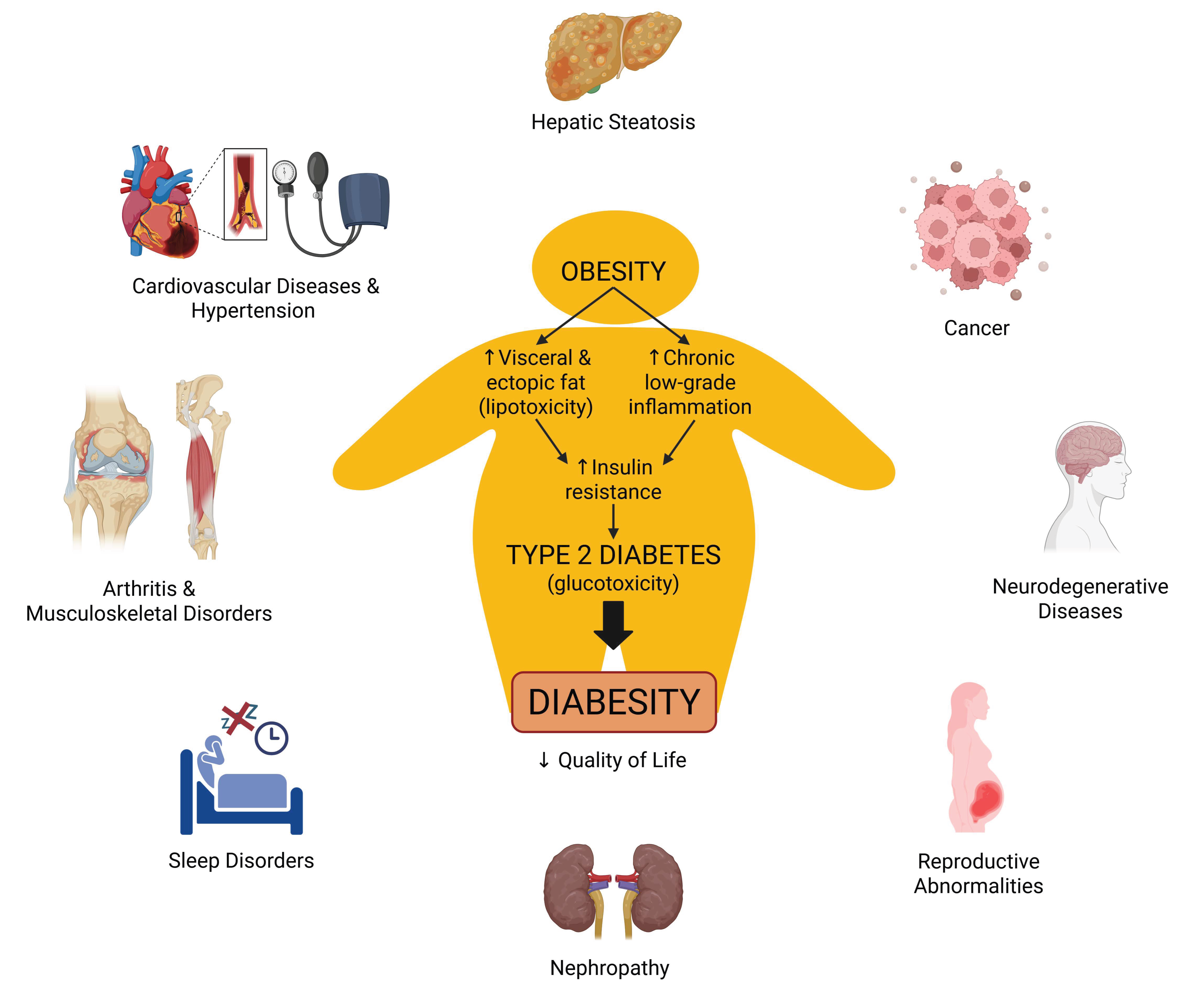
Figure 1. The most common clinical conditions that are associated with obesity and type 2 diabetes (diabesity), and their complex relationship, which directly impact the quality of life of patients.
Pharmaceuticals 18 01247 g001
Figure 2. Topographic anatomical locations of thermogenic adipose depots in (a) rodents, (b) human adults, and (c) human infants. Human infants classically have mainly brown adipose tissue (BAT), located in similar locations as rodents, such as axillary, perirenal, and most importantly, interscapular. Rodents additionally contain a large white adipose tissue (WAT) depot with recruitable beige fat in the inguinal region. Adult humans have few classical BAT depots, such as cervical (deep neck), but a larger proportion of beige adipose tissue (bAT) has been found in the supraclavicular, pericardiac, paravertebral, and perirenal regions.
Pharmaceuticals 18 01247 g002
Figure 3. Uncoupling OXPHOS mechanism by UCP1 in thermogenic adipose cells in the mitochondrial intermembrane space. On the left, ATPase pumps protons down their gradient to catalyze the synthesis of ATP in all cells, including WAT cells. On the right, UCP1 in BAT and bAT facilitates a proton leak from the intermembrane space back into the matrix, from which the energy produced is released as heat (thermogenesis). I, Complex I NADH dehydrogenase; II, Complex II succinate dehydrogenase; III, Complex III cytochrome bc1 complex; IV, cytochrome c oxidase; ADP, Adenosine diphosphate; ATP, Adenosine triphosphate; ATPase, Complex V Adenosine triphosphate synthase; ETC, Electron transport gradient; H+, Hydrogen ion/proton; P, phosphate; UCP-1, Uncoupling protein-1/Thermogenin.
Pharmaceuticals 18 01247 g003
Figure 4. Uncoupling of sarcoendoplasmic reticulum calcium ATPase (SERCA) by sarcolipin (SLN) and crosstalk between sarcoplasmic reticulum and mitochondria in skeletal muscle. Ca, Calcium; Pi, inorganic Phosphate; RyR1, Ryanodine Receptor 1; ADP, Adenosine diphosphate; ATP, Adenosine triphosphate.
Pharmaceuticals 18 01247 g004
Figure 5. Physiological conditions, bioactive agents, and drugs stimulating non-shivering thermogenesis (NST). Akt, protein kinase B; AMPK, 5′ adenosine monophosphate-activated protein kinase; ATP, adenosine triphosphate; β3-AR, β3 adrenergic receptor; BAT, brown adipose tissue; BMP7, bone morphogenetic protein 7; Ca2+, calcium; CaMKII, Ca2+/calmodulin-dependent protein kinase II; CD36, cluster of differentiation 36; DHA, docosahexaenoic acid; EPA, eicosapentaenoic acid; ERα, estrogen receptor α; FGF21, fibroblast growth factor 21; FNDC5, fibronectin type III domain-containing protein 5; FXR, farnesoid X receptor; GHR, growth hormone receptor; GLP1R, glucagon-like peptide-1 receptor; GLUT4, glucose transporter type 4; MAPK, mitogen-activated protein kinases; MT1/2, melatonin receptor 1/2; N, nitrogen; NA, noradrenaline; NRF1/2, nuclear respiratory factor 1/2; O, oxygen; PDK1, pyruvate dehydrogenase kinase 1; PGC1α, peroxisome proliferator-activated receptor gamma co-activator 1α; PKA, protein kinase A; PPARα/γ, peroxisome proliferator-activated receptor α/γ; RyR, ryanodine receptor; SCFA, short-chain fatty acids; SERCA, sarcoplasmic/endoplasmic reticulum Ca2+-ATPase; SGLT2, sodium-glucose linked transporter 2; SIRT, sirtuin; SKM, skeletal muscle; SLN, sarcolipin; SOD2, superoxide dismutase 2; T3, triiodothyronine; THR, thyroid hormone receptor; TRP, transient receptor potential; UCP1, uncoupling protein 1; UCP2/3, uncoupling protein 2/3; WAT, white adipose tissue.
Pharmaceuticals 18 01247 g005
Table 1. Differential characteristics of the white, brown, and beige adipose tissue types.
CharacteristicsWhite Adipose Tissue
Pharmaceuticals 18 01247 i001
Brown Adipose Tissue
Pharmaceuticals 18 01247 i002
Beige Adipose Tissue
Pharmaceuticals 18 01247 i003
Lipid morphologySingle large dropletSmall multilocular dropletsSmall/Medium multilocular droplets
Mitochondrial contentLowVery highHigh
UCP1 protein expression+++++
VascularityLowAbundantHigh
LocationRodents
& human
infants
Intra-abdominal (Visceral)
Subcutaneous
Interscapular
Axillary
Perirenal
Inguinal (rodents)
Adult humansIntra-abdominal (Visceral)
Subcutaneous
Cervical (Deep neck)Supraclavicular
Pericardiac
Paravertebral
Perirenal
FunctionEnergy storage as
triglycerides
Heat production
(Non-shivering thermogenesis)
Thermogenic potential
Adipose tissues’ images were taken from our experimental diabesity model, the Zücker Diabetic Fatty rat.
Disclaimer/Publisher’s Note: The statements, opinions and data contained in all publications are solely those of the individual author(s) and contributor(s) and not of MDPI and/or the editor(s). MDPI and/or the editor(s) disclaim responsibility for any injury to people or property resulting from any ideas, methods, instructions or products referred to in the content.