Review

Antimicrobial Nanoparticles Against Superbugs: Mechanistic Insights, Biomedical Applications, and Translational Frontiers

Department of Public Health, College of Applied Medical Sciences, Qassim University, P.O. Box 6666, Buraydah 51452, Saudi Arabia
*
Author to whom correspondence should be addressed.
Pharmaceuticals 2025, 18(8), 1195; https://doi.org/10.3390/ph18081195
Submission received: 15 July 2025 / Revised: 9 August 2025 / Accepted: 11 August 2025 / Published: 13 August 2025
(This article belongs to the Section Pharmaceutical Technology)

Abstract

The accelerating threat of antimicrobial resistance (AMR) demands transformative strategies that go beyond conventional antibiotic therapies. Nanoparticles (NPs) have emerged as versatile antimicrobial agents, offering a combination of physical, chemical, and immunological mechanisms to combat multidrug-resistant (MDR) pathogens. Their small size, surface tunability, and ability to disrupt microbial membranes, generate reactive oxygen species (ROS), and deliver antibiotics directly to infection sites position them as powerful tools for infection control. This narrative review explores the major classes, mechanisms of action, and biomedical applications of antimicrobial NPs—including their roles in wound healing, implant coatings, targeted drug delivery, inhalation-based therapies, and the treatment of intracellular infections. We also highlight the current landscape of clinical trials and evolving regulatory frameworks that govern the translation of these technologies into clinical practice. A distinctive feature of this review is its focus on the interplay between NPs and the human microbiota—an emerging frontier with significant implications for therapeutic efficacy and safety. Addressing this bidirectional interaction is essential for developing microbiota-informed, safe-by-design nanomedicines. Despite promising advances, challenges such as scalability, regulatory standardization, and long-term biosafety remain. With interdisciplinary collaboration and continued innovation, antimicrobial NPs could reshape the future of infectious disease treatment and help curb the growing tide of AMR.

Graphical Abstract

1. Introduction

Antimicrobial resistance (AMR) poses a major global health challenge, undermining the efficacy of antibiotics that have long served as the cornerstone of modern medicine. Multidrug-resistant (MDR) pathogens—particularly those in the ESKAPE group, including Enterococcus faecium (E. faecium), Staphylococcus aureus (S. aureus), Klebsiella pneumoniae K. pneumoniae), Acinetobacter baumannii (A. baumannii), Pseudomonas aeruginosa (P. aeruginosa), and Enterobacter species—are responsible for a significant proportion of hospital-acquired infections and exhibit resistance to many first-line treatments [1,2]. Global estimates indicate that AMR was directly responsible for 1.27 million deaths in 2019 and contributed to nearly 5 million fatalities overall [3]. Without effective intervention, AMR could result in 10 million deaths annually by 2050—surpassing cancer as the leading cause of mortality [4,5].
Exacerbating this crisis is the stagnation in antibiotic discovery, as most newly approved agents are structural derivatives of existing drug classes, offering limited innovation [6,7]. Treatment challenges are further compounded by infections involving biofilms and intracellular pathogens, which hinder the delivery and efficacy of conventional drugs [8,9,10]. These limitations underscore the urgent need for advanced antimicrobial platforms that not only exert direct bactericidal effects but also enhance targeted delivery, circumvent resistance mechanisms, and minimize host toxicity.
In response, nanoparticles (NPs) have emerged as promising antimicrobial agents. Their unique size-dependent properties, high surface area-to-volume ratio, and functional tunability enable multiple mechanisms of action [11,12]. Metallic NPs—such as silver (AgNPs), gold (AuNPs), zinc oxide (ZnO NPs), and copper oxide (CuO NPs)—exhibit broad-spectrum activity against multidrug-resistant (MDR) pathogens [13,14,15]. Their antimicrobial mechanisms include reactive oxygen species (ROS) generation, membrane disruption, DNA interaction, protein inactivation, and inhibition of biofilm formation [16].
Unlike conventional antibiotics, which typically target a single cellular pathway, NPs can disrupt membranes, interfere with intracellular signaling, and generate ROS simultaneously. This multimodal action makes it more difficult for pathogens to develop resistance through single-point mutations. In addition to their inherent antimicrobial activity, NP-based formulations are increasingly designed to improve pharmaceutical properties such as solubility, systemic circulation time, tissue-specific targeting, and sustained release. These features are especially valuable for treating persistent or localized infections—such as chronic wounds, implant-associated infections, and pulmonary diseases—where traditional antibiotics often fail to reach therapeutic levels. Moreover, multifunctional nanocarriers, including stimuli-responsive and theranostic systems, are being developed to integrate diagnostic imaging, controlled drug release, and immunomodulatory activity into a single therapeutic platform [17].
Despite the availability of various antibiotic classes, many formulations suffer from poor tissue penetration, rapid clearance, and nonspecific biodistribution—particularly in complex microenvironments like biofilms, intracellular niches, and hypoxic tissues. These pharmacokinetic limitations reduce therapeutic efficacy and facilitate the emergence of resistant strains due to sub-therapeutic exposure [18,19]. Furthermore, sub-therapeutic antibiotic exposure in these compartments contributes to the selection of resistant strains and exacerbates AMR spread [20]. To address these issues, advanced delivery systems—including liposomes, polymeric NPs, and ligand-targeted nanocarriers—have been developed to enhance tissue-specific accumulation, prolong circulation, and enable controlled or responsive drug release [21]. These technologies are particularly promising for treating chronic infections, reducing systemic toxicity, and overcoming barriers that compromise conventional therapy. Recent advances in nanomedicine also emphasize the use of biodegradable and biocompatible materials to support safer clinical translation and reduce long-term toxicity risks. Collectively, these innovations align with the goals of precision nanomedicine, where treatment can be tailored to patient-specific microbiota, immune profiles, and resistance patterns.
Progress in antimicrobial nanomaterials has led to innovative therapeutic approaches. Yathavan et al. [22] demonstrated that AgNPs embedded in silk elastin-like protein hydrogels effectively inhibited S. aureus and P. aeruginosa biofilms while promoting mucosal tissue regeneration. Alhosani et al. [23] developed ZnO–Ag nanocomposite coatings that exhibited strong antibiofilm activity against P. aeruginosa through ROS-mediated membrane damage. Additionally, AgNPs have shown synergistic effects when combined with conventional antibiotics. Kamer et al. [24] reported that biosynthesized AgNPs significantly enhanced the antibacterial activity of gentamicin, ceftazidime, and ciprofloxacin against MDR and XDR P. aeruginosa, reducing minimum inhibitory concentrations (MICs) by 2–10–fold and suppressing virulence gene expression.
A novel strategy by Szymczak et al. [25] involved conjugating lytic T7 bacteriophages with AgNPs, resulting in a “phage-armed” nanoplatform with superior antibiofilm efficacy against Escherichia coli (E. coli) compared to either agent alone. This formulation demonstrated low cytotoxicity in mammalian cells, supporting its translational potential. Similarly, Ahmad et al. [26] showed that green-synthesized AgNPs from Phoenix dactylifera extract suppressed P. aeruginosa biofilm formation and virulence factors—such as swarming motility and pyocyanin production—by interfering with quorum sensing (QS) pathways. These studies collectively highlight the multifaceted potential of antimicrobial NPs in overcoming drug resistance, eradicating biofilms, and maintaining host compatibility. In parallel, a 2025 study by Ahmad et al. [26] investigated green-synthesized AgNPs derived from Phoenix dactylifera extract and revealed that sub-inhibitory concentrations (MIC/5 to MIC/2) substantially suppressed P. aeruginosa biofilm formation and virulence factor expression, including swarming motility, protease activity, and pyocyanin production.
These effects were attributed to interference with QS signaling pathways. Mechanistically, such antimicrobial action is supported by numerous studies confirming that NPs—particularly Ag and ZnO NPs—generate ROS, which induce lipid peroxidation, disrupt bacterial membranes, and lead to cell death. Collectively, these findings underscore the multifaceted potential of antimicrobial NPs not only in overcoming conventional resistance mechanisms and eradicating biofilms but also in maintaining host biocompatibility, thereby advancing their translational value in infection-prone clinical environments.
Nonetheless, clinical adoption of NP-based antimicrobials remains constrained by regulatory, manufacturing, and safety challenges. Key barriers include the absence of standardized classification systems, inconsistent toxicological profiles, and limited data on long-term biosafety [27,28]. Regulatory approval is further complicated by the lack of harmonized testing protocols and variable definitions of nanomaterials across jurisdictions [29,30]. Additionally, scalability issues, complex designs, and batch-to-batch variability hinder clinical translation beyond preclinical stages [31,32]. Recognizing these gaps, global health organizations such as the WHO and Horizon Europe have prioritized nanotechnology in the fight against AMR, supporting dedicated funding for translational research and regulatory alignment.
While previous reviews have addressed the synthesis and applications of antimicrobial NPs, their interaction with the host microbiota remains underexplored. This review provides an integrated perspective, emphasizing not only the physicochemical mechanisms and biomedical applications of NPs but also their bidirectional relationship with the gut microbiota—a critical aspect influencing host immunity, therapeutic efficacy, and safety. By addressing this knowledge gap, we propose a framework for the development of microbiota-informed nanotherapeutics in the fight against resistant pathogens.
From a pharmaceutical perspective, the formulation, scale-up, and pharmacokinetic optimization of NP-based antimicrobials must meet both therapeutic and regulatory requirements. This review synthesizes recent advances in NP types, antimicrobial mechanisms, biomedical applications, and translational barriers. To support this discussion, Figure 1 presents a conceptual overview of NP classes, core mechanisms, microbiota interactions, and major challenges impeding clinical adoption. Together, these insights underscore the need for safe, precise, and microbiota-conscious nanotherapeutic strategies to combat MDR infections.

2. Materials and Methods

This narrative review is based on a comprehensive literature search of peer-reviewed articles published between 2008 and 2025. The databases consulted included PubMed, Scopus, and Web of Science. The following search terms were used individually and in combination: “antimicrobial nanoparticles (NPs),” “nanomaterials,” “drug-resistant bacteria,” “biofilm inhibition,” “nanoparticle drug delivery,” “microbiota–NP interaction,” and “translational nanomedicine.” Preference was given to articles published in high-impact journals, systematic reviews, and original research studies with clinical or translational relevance. Only English-language publications were considered. Priority was placed on studies that described well-characterized nanoparticle systems, provided mechanistic insights into antimicrobial action, and included translational or toxicological evaluations. Articles were selected based on their relevance to the core themes of this review: the classes and mechanisms of antimicrobial NPs, biomedical applications, interactions with the gut microbiota, and translational or regulatory challenges. No new experimental data were generated or analyzed for this review.

3. Classes and Characteristics of Antimicrobial NPs

NPs designed for antimicrobial applications encompass a broad range of core materials, surface chemistries, and functional architectures. Critical physicochemical attributes—such as particle size, surface charge, morphology, and chemical composition—profoundly influence their antimicrobial potency, biocompatibility, biodistribution, degradation kinetics, and formulation feasibility for therapeutic deployment [12,13]. The antimicrobial efficacy of NPs arises from multifactorial and often synergistic mechanisms, including disruption of microbial membranes, generation of ROS, interference with protein synthesis and nucleic acid replication, and inhibition of QS and biofilm development [16,33]. These multifaceted actions confer a significant advantage over conventional antibiotics, particularly in combating MDR and biofilm-associated pathogens.
From a pharmaceutical development perspective, the rational design of antimicrobial NPs requires careful consideration of their physicochemical stability, site-specific targeting ability, controlled or sustained drug release profiles, and overall biocompatibility. Lipid-based nanocarriers, particularly liposomes, have advanced into clinical use due to their versatility in encapsulating both hydrophilic and lipophilic therapeutics, extending systemic circulation time, and minimizing off-target toxicity [34]. Similarly, solid lipid NPs and polymer-based systems offer enhanced physical stability and scalability for large-scale manufacturing [35]. This section integrates recent experimental studies that validate not only the antibacterial action of various NP classes but also their safety in mammalian cell models, aligning with translational goals.

3.1. Metal and Metal Oxide NPs

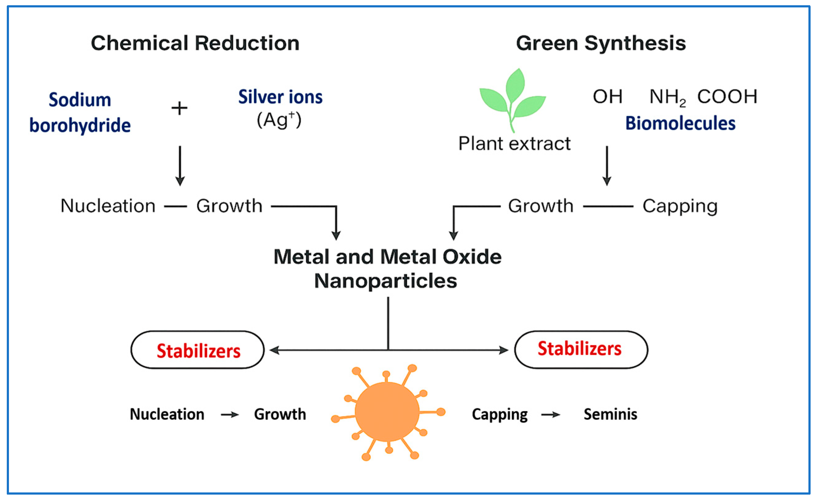
This subsection not only outlines diverse synthesis strategies for metal-based NPs but also critically compares their effects on antimicrobial performance and translational potential. Metal-based NPs are foundational to antimicrobial nanotechnology due to their potent microbicidal activity, broad-spectrum efficacy, and versatility in biomedical applications [36,37]. Their antimicrobial properties stem from both intrinsic physicochemical features and synthesis-dependent characteristics. For instance, in classical chemical reduction methods, AgNPs are synthesized through the nucleation of silver ions (Ag+), which are reduced by agents such as sodium borohydride or plant-derived polyphenols [38]. This process initiates seed formation, followed by growth and capping steps. Stabilizers such as citrate, gelatin, or chitosan are then added to prevent agglomeration and to define the final particle size, shape, and surface chemistry [39]. A schematic overview of chemical and green synthesis pathways is presented in Figure 2, highlighting key stages—nucleation, growth, and capping—as well as the reagents and surface functionalization techniques involved.
Synthesis parameters, including reaction time, temperature, and the concentration of reducing agents, significantly influence critical properties such as particle size (typically 10–50 nm), zeta potential, and crystallinity. These parameters directly affect silver ion release and ROS generation, which are key determinants of antimicrobial activity [40]. Green synthesis approaches, which utilize phytochemicals and proteins, produce biofunctional surface groups (e.g., flavonoids, terpenoids, amines) that promote bacterial adhesion via hydrogen bonding or electrostatic interactions, thereby enhancing antimicrobial efficacy [41]. These structural and surface features govern membrane permeabilization, enzyme inactivation, and intracellular DNA disruption, all of which contribute to the observed variability in antimicrobial potency and therapeutic outcomes [42].
Recent experimental studies have validated the dual performance and safety of metal and metal oxide NPs. El-Fallal et al. [43] biosynthesized ZnO NPs using kombucha extract and demonstrated >90 % bactericidal activity against E. coli and K. pneumoniae at concentrations under 100 µg/mL. Tazeen et al. [44] designed chitosan–ZnO nanocomposites exhibiting potent antibiofilm efficacy and low cytotoxicity in human keratinocytes. These data confirm that optimized synthesis and surface engineering can yield metal-based nanomaterials that are both therapeutically potent and cytocompatible—supporting their translation into medical applications such as wound dressings, coatings, and topical antimicrobials.
In addition to established agents such as Ag, ZnO, CuO, and TiO2 NPs, platinum NPs (PtNPs) have emerged as a compelling class of antimicrobial nanomaterials due to their catalytic and redox-active properties. PtNPs exhibit broad-spectrum antibacterial activity—and in some formulations also antibiofilm action—through multiple mechanisms, including membrane disruption, ROS generation, and oxidative damage to intracellular biomolecules (e.g., DNA and proteins). Studies have shown that PtNPs maintain their antimicrobial efficacy in low-oxygen environments and synergize with conventional antibiotics to reduce bacterial growth and biofilm resilience [45,46,47,48,49]. Moreover, green synthesis approaches and surface functionalization strategies have improved their biocompatibility profiles, making PtNPs promising candidates for translational applications such as wound dressings, implant coatings, and targeted antimicrobial delivery.
AgNPs exhibit multifaceted antimicrobial mechanisms. These include the sustained release of Ag+, which disrupts thiol-containing enzymes and structural proteins, leading to enzymatic inhibition and loss of membrane integrity. Simultaneously, AgNPs induce oxidative stress through ROS generation, impairing bacterial DNA and protein synthesis and ultimately triggering microbial apoptosis [16,50]. Moreover, AgNPs interfere with QS pathways, thereby reducing bacterial virulence and inhibiting biofilm formation [51,52].
Recent advances in nanomedicine have facilitated the clinical translation of these mechanisms. For example, Chen et al. [53] developed a carrier-free injectable hydrogel incorporating AgNPs, which successfully eradicated methicillin-resistant S. aureus (MRSA) in a diabetic wound model. In addition to its antimicrobial efficacy, the nanocomposite promoted tissue regeneration and attenuated pro-inflammatory cytokine production, demonstrating dual functionality for infection control and wound healing.
ZnO NPs also display broad-spectrum antimicrobial activity, primarily mediated through ROS generation and zinc ion (Zn2+) release. Unlike AgNPs, ZnO NPs possess intrinsic photocatalytic properties under ultraviolet (UV) or visible light, further enhancing their disinfection capabilities. Gao et al. [54] reported several key mechanisms, including membrane destabilization, intracellular oxidative damage, and interference with QS and biofilm development. Translationally, Selmani et al. [55] fabricated ZnO–calcium carbonate nanocomposite coatings for titanium implants, achieving over 90% reduction in the viability of S. aureus, S. epidermidis, and Candida albicans (C. albicans), while preserving osteoblast compatibility—highlighting their promise in orthopedic applications.
CuO and titanium dioxide (TiO2) NPs also demonstrate strong antimicrobial properties, primarily via ROS-mediated microbial damage. CuO NPs release copper ions (Cu2+) and catalyze redox reactions that compromise bacterial membrane integrity and denature intracellular proteins and nucleic acids. These effects have been validated against MDR strains including E. coli, K. pneumoniae, P. aeruginosa, and MRSA, with MICs ranging from 62.5 to 125 µg/mL [56]. TiO2 NPs—particularly in their anatase crystalline form—act as photocatalysts capable of generating hydroxyl radicals and singlet oxygen under UV or visible light activation. A recent study showed that visible light-activated TiO2 nanostructures enhanced the antibacterial efficacy of conventional antibiotics against P. aeruginosa and K. pneumoniae, while maintaining low cytotoxicity [57]. However, the need for light activation and limited pharmacokinetic data remain barriers to systemic clinical use.
From a formulation perspective, the therapeutic performance and biosafety of metal and metal oxide NPs are closely tied to their physicochemical characteristics—such as particle size, morphology, surface charge, and colloidal stability. Optimizing these properties enhances microbial membrane targeting and minimizes off-target toxicity. Nonetheless, issues like nanoparticle aggregation in physiological environments often require stabilizing strategies such as PEGylation or surfactant-based coatings [56].
These NPs are increasingly incorporated into advanced delivery systems—including hydrogels, wound dressings, orthopedic cements, implant coatings, and sprayable formulations—offering localized and sustained antimicrobial effects while reducing systemic exposure. Despite promising preclinical results, clinical translation still demands comprehensive toxicological evaluation, scalable synthesis protocols, and harmonized regulatory standards [12]. Given their potent antimicrobial activity, modular design flexibility, and multi-targeted mechanisms, metal-based NPs remain at the forefront of next-generation antimicrobial strategies.

3.2. Polymeric NPs

Polymeric NPs represent a versatile and promising class of antimicrobial nanostructures, offering both intrinsic antimicrobial properties (in specific cases) and advanced capabilities for controlled drug delivery. These nanosystems are typically fabricated from biocompatible and biodegradable polymers such as chitosan, polylactic-co-glycolic acid (PLGA), and polyethylene glycol (PEG). Their physicochemical characteristics support prolonged drug release, site-specific targeting, and reduced systemic toxicity—attributes that are critical for therapeutic success and clinical translation [58,59].
Among polymeric NPs, chitosan-based formulations are particularly notable for their inherent antimicrobial activity, which arises from chitosan’s cationic nature. This allows for electrostatic interactions with the negatively charged bacterial membrane, leading to membrane destabilization, increased permeability, and cell death. Tazeen et al. [44] demonstrated that chitosan–ZnO nanocomposites not only eradicated E. coli and P. aeruginosa biofilms but also exhibited excellent cytocompatibility in human keratinocyte assays, underscoring their clinical potential.
PLGA-based NPs, approved by regulatory authorities such as the U.S. FDA and EMA, are among the most investigated nanocarriers for antimicrobial therapy. Their degradation kinetics can be fine-tuned by altering the lactic/glycolic acid ratio, allowing precise control over drug release profiles. Türeli et al. [59] reported that ciprofloxacin-loaded PLGA nanoparticles significantly enhanced pulmonary delivery and biofilm penetration in P. aeruginosa-infected airway models, with negligible cytotoxicity to CFBE41o and Calu-3 lung epithelial cells. Shariati et al. [58] similarly encapsulated tobramycin in PEG–PLGA nanoparticles and found that the formulation exhibited enhanced antibiofilm efficacy in artificial sputum while maintaining high cell viability in bronchial epithelial cultures.
PEGylation further improves the physicochemical stability and in vivo behavior of polymeric NPs. By reducing opsonization and immune clearance, PEG–PLGA nanoparticles prolong systemic circulation time and improve tissue penetration. Tchatchiashvili et al. [60] developed PEG–PLGA carriers targeting P. aeruginosa and Burkholderia cenocepacia, demonstrating both antimicrobial efficacy and epithelial cell compatibility, thus reinforcing their translational suitability for respiratory infections.
Moreover, polymeric nanocarriers can be engineered with stimuli-responsive elements or targeting ligands to achieve site-specific drug release. These smart systems can respond to pH gradients, enzymatic activity (such as bacterial enzymes), and redox changes typically present in infected microenvironments—thereby enhancing therapeutic precision and minimizing off-target effects. For example, Cöksu et al. [61] developed PLGA NPs loaded with antimicrobial peptides and decorated with enzyme-cleavable linkers to achieve selective release in S. aureus-infected tissues, showing enhanced antibacterial efficacy in vitro and in vivo with minimal mammalian cytotoxicity. Similarly, Miranda Calderón et al. [62] formulated antibody-functionalized polymeric NPs that selectively target bacterial cells, leveraging ligand-mediated specificity to improve antimicrobial action while preserving host cell viability.
Although specific glutathione (redox)-responsive antibiotic release systems are still emerging in microbial infection models, broad evidence supports the following principle: stimuli-sensitive polymeric nanocarriers enable environment-triggered drug release, thereby increasing efficacy and avoiding sub-therapeutic exposure, which promotes resistance [63]. In summary, polymeric NPs offer adaptable delivery platforms across administration routes—including intravenous, pulmonary, transdermal, and mucosal—making them a cornerstone in next-generation antimicrobial nanotherapies.

3.3. Lipid-Based NPs

Lipid-based NPs—including liposomes, solid lipid NPs, and nanostructured lipid carriers (NLCs)—have emerged as promising vehicles for antimicrobial delivery due to their excellent biocompatibility, drug encapsulation versatility, and sustained release potential. Constructed from physiologically compatible lipids, these systems are inherently suited for both systemic and localized administration, offering favorable safety profiles and low immunogenicity. Liposomes, composed of phospholipid bilayers encapsulating aqueous cores, are extensively studied for antibiotic and antimicrobial peptide delivery due to their membrane-mimetic properties, which facilitate fusion with microbial cells and intracellular delivery. Afrasiabi et al. [64] developed a doxycycline-loaded liposome doped with curcumin (NL-Cur + Dox) for combination antibacterial photodynamic therapy against Aggregatibacter actinomycetemcomitans. Their formulation achieved an 82.7 % reduction in biofilm mass and 75 % decrease in metabolic activity, with no significant cytotoxicity to human gingival fibroblasts (cell viability remained at ~ 90%, p = 0.074), as well as negligible hemolysis (< 5%), under therapeutic conditions.
SLNs and NLCs, considered next-generation lipid carriers, provide enhanced stability and drug loading relative to conventional liposomes. Arabestani et al. [65] reported that cationic SLNs attached more rapidly to bacterial cells and delivered superior antibacterial outcomes compared to their non-ionic counterparts, aligning with improved interactions and efficacy in wound infection models. Motsoene et al. [66] investigated multifunctional lipid-based nanoparticles in wound healing applications and found that these formulations supported sustained antimicrobial release, accelerated skin regeneration, and maintained fibroblast viability above 85 %—highlighting the translational synergy between infection control and tissue repair. Overall, lipid-based nanocarriers overcome limitations of traditional antimicrobials, particularly in biofilm-associated infections and delivery of hydrophobic agents. Their utility spans pharmaceutical, food, and cosmetic domains. However, rigorous in vivo and toxicological evaluations remain necessary to support regulatory approval and further clinical translation.

3.4. Hybrid NPs (MOF-Based Nanozymes)

Hybrid nanoparticles, particularly those incorporating metal–organic frameworks (MOFs) with catalytic or enzymatic properties, represent an innovative and multifunctional approach in antimicrobial nanotherapy [67,68]. These systems integrate multiple bactericidal mechanisms—such as metal ion release, ROS generation, and enzyme-mimetic activity—into a single nanosystem, enabling broad-spectrum antimicrobial efficacy while maintaining low cytotoxicity to host tissues.
For instance, Omer et al. [69] developed polycaprolactone–lipid hybrid NPs encapsulating ciprofloxacin, which provided sustained antibiotic release over seven days and significantly enhanced antibacterial activity against E. coli compared to free drug formulations [70]. This design demonstrated the ability of hybrid NPs to improve drug retention and therapeutic outcomes.
A notable example includes molybdenum-doped zeolitic imidazolate framework-8 nanozymes (Mo@ZIF-8), which possess intrinsic peroxidase-like activity. In the presence of trace hydrogen peroxide, Mo@ZIF-8 catalyzes the generation of hydroxyl radicals, producing potent bactericidal effects. Lian et al. [71] reported ≥ 99 % reduction in the viability of E. coli and S. aureus at concentrations as low as 10 µg/mL, with negligible cytotoxicity to mammalian cells.
Similarly, Shen et al. [72] designed a silk fibroin–lysozyme/ZIF-8 composite coating for titanium implants that combined enzymatic degradation of bacterial cell walls with controlled Zn2+ ion release. The coating achieved over 90% bacterial inhibition against S. aureus and E. coli in vitro and promoted anti-inflammatory macrophage polarization, osteogenic differentiation, and neovascularization in murine bone defect models—underscoring its dual function in infection control and tissue regeneration.
In another example, Cai et al. [73] engineered pH-responsive Fe3O4@ZIF-8 magnetic NPs loaded with norfloxacin, which exhibited enhanced antimicrobial efficacy under acidic conditions typical of infected sites. The system also demonstrated excellent biocompatibility, supporting its suitability for localized drug delivery. Additionally, Wang et al. [74] developed a glucose-responsive hollow molybdenum-based nanozyme (HMMo/GOx@P) that converts endogenous glucose into hydrogen peroxide, triggering hydroxyl radical formation for effective bacterial eradication. This platform successfully eliminated E. coli and S. aureus in diabetic wound models, accelerated tissue regeneration, and showed low systemic toxicity.
These MOF-based nanozymes exemplify the potential of hybrid platforms to integrate multiple antimicrobial strategies within a single therapeutic system. However, key challenges remain, including synthesis scalability, batch-to-batch reproducibility, regulatory compliance, and comprehensive biocompatibility assessments for clinical translation. To support comparative analysis, Table 1 summarizes recent studies describing the synthesis, structural features, and antimicrobial applications of metal and metal oxide NPs. Key parameters include synthesis methods, particle size, surface area, synthesis conditions, morphology, and target pathogens or biofilm activity—offering a broad overview of design strategies and translational relevance. This comparative table enhances clarity and complements the diversity of the nanomaterials discussed in Section 3.1, Section 3.2, Section 3.3 and Section 3.4. All entries are drawn from recent peer-reviewed studies to ensure scientific accuracy and current relevance.

4. Mechanisms of Antimicrobial Action of NPs

NPs exhibit potent antimicrobial activity through a variety of synergistic and multifactorial mechanisms [95]. Unlike conventional antibiotics, which typically target a single biochemical pathway, NPs can simultaneously disrupt multiple microbial processes, thereby reducing the likelihood of resistance development [96,97]. These mechanisms include membrane disruption, generation of ROS, release of antimicrobial metal ions, interference with protein and nucleic acid synthesis, inhibition of biofilm formation, suppression of QS, and modulation of host immune responses. The physicochemical properties of NPs—such as particle size, shape, surface charge, and chemical composition—play a critical role in mediating their interactions with microbial cells [12].
A primary mode of action involves the disruption of bacterial cell membranes. Metallic NPs such as AgNPs, ZnO NPs, and CuO NPs exhibit strong affinity for the negatively charged bacterial surface, where they adhere and penetrate the lipid bilayer. This results in increased membrane permeability, leakage of cellular contents, membrane depolarization, and eventual cell lysis [13,15]. Chitosan-based polymeric NPs also disrupt microbial membranes through their polycationic nature, destabilizing the envelope without the need for metal ion release [98].
Another key mechanism is the intracellular generation of ROS, including hydroxyl radicals, superoxide anions, and hydrogen peroxide. These reactive species damage essential biomolecules such as lipids, proteins, and DNA, leading to oxidative stress and cell death [14]. AgNPs and ZnO NPs are particularly known for their ROS-generating capabilities. Additionally, TiO2 NPs exhibit photocatalytic activity that enhances ROS production upon exposure to UV or visible light, making them suitable for environmental disinfection and sterilization of medical devices [54,57].
Controlled metal ion release also contributes to the antimicrobial activity of NPs. For instance, Ag+ ions released from AgNPs interact with thiol groups in enzymes and structural proteins, disrupting key metabolic pathways. These ions can also impair DNA replication and transcription, leading to genotoxic stress [15,50]. Similarly, Zn2+ and Cu2+ ions exhibit strong bactericidal effects by binding to bacterial proteins, nucleic acids, and components of the cell envelope [12].
Biofilm inhibition is another critical mechanism by which NPs enhance infection control. Biofilms protect bacteria from antibiotics and immune responses by forming a dense extracellular polymeric matrix. AgNPs and ZnO NPs can prevent bacterial adhesion, degrade biofilm matrix components, and reduce the viability of embedded bacterial populations [26]. Additionally, NPs can interfere with QS, the bacterial communication system that regulates biofilm maturation and virulence. For example, biosynthesized AgNPs derived from Lactobacillus rhamnosus significantly downregulated QS-related genes responsible for swarming motility, protease production, and biofilm formation in P. aeruginosa [51,52].
Certain NPs also penetrate host cells, enabling intracellular antimicrobial activity and immune modulation. This is especially valuable for treating infections caused by intracellular pathogens or those associated with immune evasion. Mesoporous silica nanoparticles (MSNs), for example, are efficiently internalized by antigen-presenting cells such as macrophages and dendritic cells. Within endosomal compartments, MSNs deliver their therapeutic cargo while exhibiting minimal cytotoxicity. Functionalized MSNs have been shown to stimulate immune responses by enhancing cytokine release and antigen presentation, making them promising candidates for both antimicrobial therapy and vaccine adjuvant development [99].
In parallel, manganese–phosphorus nanosheets have demonstrated the ability to augment humoral immunity in implant-associated infections. These inorganic nanostructures promote macrophage polarization toward the M1 phenotype and enhance dendritic cell maturation, leading to robust memory B-cell responses and antibody production. Such immunomodulatory effects contribute to durable protection against biofilm-forming pathogens commonly linked to implanted medical devices [100].
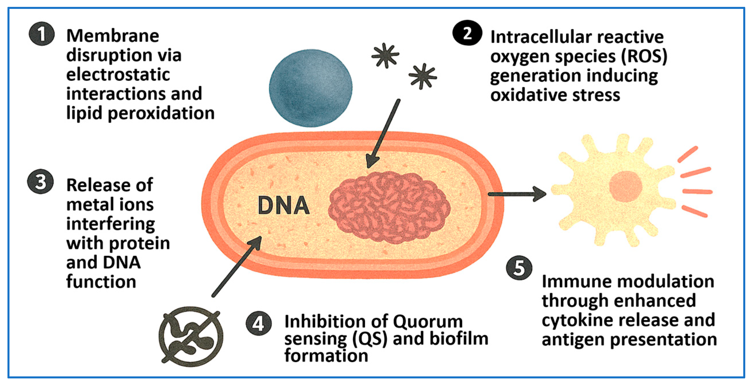
Figure 3 provides an integrated schematic summarizing the principal mechanisms of action of antimicrobial NPs. These include (1) microbial membrane disruption, (2) intracellular oxidative stress via ROS production, (3) metal ion release impairing enzyme function and DNA integrity, (4) inhibition of biofilm development and QS, and (5) immune activation through cytokine secretion, macrophage polarization, and antigen presentation. This multimodal strategy not only enhances pathogen clearance but also lowers the risk of resistance emergence.
To facilitate a comparative understanding of antimicrobial mechanisms, Table 2 summarizes the key features of various metal and metal oxide NPs—including their antimicrobial spectrum, MIC ranges, and cytotoxicity profiles. The data, derived from recent peer-reviewed studies, offer translational insights into their efficacy and biosafety, supporting informed decisions for clinical and biomedical applications.

5. Biomedical Applications of Antimicrobial NPs

The integration of antimicrobial nanoparticles (NPs) in biomedical applications has emerged as a transformative strategy to combat both persistent and emerging infectious diseases. This is especially relevant in the context of challenges posed by AMR, biofilm-associated infections, and intracellular bacterial persistence [120,121,122]. NPs offer several advantages over conventional antimicrobials, including their physicochemical tunability, high surface area-to-volume ratio, and the ability to be functionalized with both antimicrobial agents and targeting ligands. These properties enable site-specific drug delivery, controlled or sustained release, and multifaceted antimicrobial mechanisms—such as membrane disruption, ROS generation, and immune modulation. As a result, antimicrobial NPs are being increasingly employed across a broad range of biomedical applications, including wound dressings, implant coatings, targeted drug delivery systems, inhalation therapies, and immuno-engineered infection control strategies.

5.1. Wound Healing and Skin Infections

Wound management remains a significant challenge in infection control, particularly in chronic, non-healing wounds such as diabetic ulcers, pressure sores, and burn injuries [123]. These wounds are frequently colonized by MDR organisms such as S. aureus and P. aeruginosa, which delay tissue regeneration and increase the risk of systemic infections [124,125]. Antimicrobial NPs, especially AgNPs, have demonstrated strong efficacy in reducing microbial load while simultaneously promoting wound healing [126,127]. When incorporated into hydrogels, foams, or nanofibrous scaffolds, AgNPs release Ag+ ions that disrupt bacterial membranes, generate ROS, and downregulate virulence gene expression [128].
While AgNPs are well established for their antimicrobial and anti-inflammatory properties, AuNPs have recently gained attention as multifunctional agents in regenerative medicine. In a 2024 study, curcumin-loaded AuNPs (~ 42 nm) embedded in a Pluronic® F127 hydrogel significantly accelerated wound closure in diabetic rat models. These NPs enhanced collagen deposition, stimulated angiogenesis, and reduced oxidative stress—outperforming free curcumin through sustained release and improved tissue penetration [129].
Additional studies have reported that green-synthesized AuNPs stabilized with biomolecules such as collagen or chitosan enhance fibroblast proliferation, re-epithelialization, and tissue remodeling. For example, collagen-coated AuNPs promoted dermal regeneration by lowering inflammatory cytokines (e.g., IL-6 and TNF-α) and upregulating growth factors such as basic fibroblast growth factor (bFGF) and vascular endothelial growth factor (VEGF) in human skin fibroblast cultures [130]. Moreover, VEGF-functionalized AuNPs have been shown to stimulate angiogenesis and accelerate healing in full-thickness skin wounds by modulating endothelial and fibroblast activity [131].
These findings underscore the potential of AuNP-based scaffolds and hydrogels as multifunctional platforms for wound healing—capable of integrating antimicrobial action, tissue regeneration, and controlled drug delivery. As hybrid nanomaterials continue to evolve by incorporating antibiotics, bioactive molecules, and anti-inflammatory agents, they are poised to revolutionize the management of chronic wounds.

5.2. Implant-Associated and Nosocomial Infections

Implantable medical devices—including catheters, orthopedic prostheses, and dental implants—are highly susceptible to colonization by biofilm-forming bacteria, which exhibit tolerance to systemic antibiotics and can evade host immune responses [132,133]. Once established, biofilms on implant surfaces often result in persistent infections that are difficult to eradicate and may necessitate device removal [134]. In response, nanoparticle-based surface modifications have emerged as a promising strategy to provide durable antimicrobial protection while preserving implant biocompatibility and mechanical integrity.
Among the most studied materials, ZnO and CuO NPs have demonstrated effective integration into implant coatings. These NPs release metal ions and generate ROS, producing bactericidal effects while simultaneously disrupting QS and preventing microbial adhesion. Alhosani et al. [23] reported that ZnO–Ag nanocomposite coatings successfully prevented P. aeruginosa biofilm formation and disrupted existing biofilms through both chemical (ROS generation) and physical (nanoscale sharpness) mechanisms.
Beyond metal oxides, gold nanostar (GNS) coatings have attracted interest for their dual role in antimicrobial photothermal therapy and promotion of osseointegration. Li et al. [135] demonstrated that titanium implants coated with GNSs and activated by near-infrared light achieved complete eradication of S. aureus while significantly enhancing osteoblast differentiation and bone regeneration in vivo. Similarly, gold-pre-deposited, ceramic-treated titanium showed antimicrobial activity against Fusobacterium nucleatum and S. aureus while maintaining corrosion resistance, making it suitable for dental applications [136].
Hybrid coatings combining NPs with antimicrobial peptides have also shown synergistic antimicrobial effects with reduced host cytotoxicity. For example, ZnO–CuS/F127 hydrogel coatings exhibited multienzyme-mimicking properties, significantly reduced biofilm biomass, and supported tissue regeneration without eliciting inflammatory responses [137].
Recent advances in nanostructured platforms have introduced multifunctional coatings that integrate graphene-based materials, chitosan, and AgNPs onto titanium surfaces. San et al. [138] developed reduced graphene oxide (rGO)/AgNP coatings using plasma electrolytic oxidation on 3D-printed porous titanium implants. These coatings eradicated over 99 % of S. aureus and MRSA biofilms while supporting osteoblast adhesion and proliferation—demonstrating both antimicrobial and osteoinductive potential.
Titanium nitride–silver (TiN–Ag) composite coatings have also demonstrated robust resistance to both Gram-positive and Gram-negative bacteria. Hojda et al. [139] showed that electrophoretically deposited TiN/Ag films significantly inhibited biofilm formation by E. coli, S. aureus, E. faecalis, and E. faecium while preserving critical implant properties such as hardness and corrosion resistance.
These innovative coatings underscore the translational potential of antimicrobial NPs in preventing implant-associated infections. By integrating potent bactericidal activity, biofilm inhibition, and host biocompatibility into durable surface materials, these nanostructures address one of the most persistent challenges in surgical and clinical settings. Notably, implant-coating formulations—including ZnO–Ag nanocomposites, Au nanostars, and graphene-based systems—have consistently demonstrated robust antibiofilm efficacy while preserving cytocompatibility with osteoblasts and epithelial cells, reinforcing their suitability for clinical translation.

5.3. Targeted Antibiotic Delivery Systems

NP-based antibiotic delivery platforms have revolutionized antimicrobial therapy by enhancing drug solubility, stability, and bioavailability while facilitating targeted delivery to sites of infection. These systems help overcome several limitations of conventional antibiotics that can lead to off-target toxicity, including poor tissue penetration, rapid degradation, and nonspecific distribution. By concentrating antimicrobial agents at the infection site and shielding them from inactivation, NPs offer a means of restoring antibiotic efficacy, particularly against MDR and persistent infections [97,140].
Lipid-based nanocarriers—including liposomes, solid lipid NPs, and NLCs—have been extensively used to encapsulate antibiotics such as rifampicin, ciprofloxacin, and vancomycin. These carriers extend systemic circulation times, enhance tissue accumulation, and reduce the systemic toxicity of their payloads. For example, maleimide-conjugated PEGylated liposomes have been engineered to deliver a wide range of β-lactam and aminoglycoside antibiotics, including ceftriaxone, doxycycline, cephalexin, and ampicillin. Ladva et al. [141] demonstrated that these liposomes significantly enhanced antibiotic uptake by bacterial cells, reduced the MICs by up to ninefold against E. coli and K. pneumoniae, and accelerated wound healing in vitro—all while maintaining low cytotoxicity.
Polymeric NPs, particularly those formulated from biodegradable and biocompatible polymers like PLGA and PEG, enable sustained and controlled antibiotic release [142,143]. These systems can be surface-functionalized with targeting moieties, such as folate or mannose, to direct drug-loaded NPs to infected cells, especially macrophages and dendritic cells. In a notable study, Maurya et al. [144] reported that folate–chitosan–decanoic acid-coated mesoporous silica NPs loaded with ethionamide effectively targeted alveolar macrophages in a Mycobacterium tuberculosis (M. tuberculosis) model. This approach enhanced intracellular drug concentrations and antimycobacterial activity over five days compared to non-targeted formulations.
Targeted delivery systems offer multiple clinical benefits. First, they minimize the required therapeutic dose by localizing antibiotic release at the site of infection, thereby reducing systemic toxicity and adverse effects. Second, they shield antibiotics from enzymatic degradation and efflux pumps—restoring efficacy against resistant pathogens. Third, ligand-mediated uptake by immune cells enables efficient treatment of intracellular infections caused by M. tuberculosis, S. enterica, and other intracellular pathogens.
Various nanocarriers—including liposomes, polymeric nanoparticles, dendrimers, and mesoporous silica NPs—are being actively optimized for translational antimicrobial applications. As these platforms continue to evolve, they offer considerable promise for precision therapy tailored to the infection site, pathogen profile, and resistance mechanism [145,146]. Importantly, both polymeric and lipid-based nanocarriers have demonstrated low cytotoxicity in mammalian cell models, with effective antimicrobial activity observed at concentrations well below cytotoxic thresholds—reinforcing their safety and clinical potential.

5.4. Inhalation-Based Therapies for Respiratory Infections

The respiratory tract is a major site of MDR infections, including hospital-acquired pneumonia, ventilator-associated pneumonia, and chronic respiratory conditions such as cystic fibrosis and bronchiectasis [147,148]. These infections present substantial therapeutic challenges due to biofilm formation, limited antibiotic penetration, and systemic toxicity associated with high-dose parenteral therapies. Inhalable NP-based delivery systems offer a promising strategy to overcome these limitations by providing localized, high-concentration drug delivery directly to the lungs while minimizing systemic exposure [149,150].
AgNPs have been widely investigated for inhalation therapy due to their broad-spectrum antimicrobial activity and immunomodulatory properties. In a murine model of K. pneumoniae pneumonia, aerosolized AgNPs significantly reduced bacterial loads in lung tissue, attenuated pro-inflammatory cytokine levels, and improved survival outcomes [151]. These findings underscore the potential of AgNPs to serve dual roles as antimicrobial and anti-inflammatory agents in the treatment of resistant pulmonary infections.
Beyond metallic NPs, polymer-based systems have also shown promise. Jalal et al. [152] developed methacrylated chitosan-coated ciprofloxacin nanoparticles designed for pulmonary delivery. These mucoadhesive particles adhered effectively to the lung mucosa and provided sustained drug release. In rat models of bronchopneumonia, the formulation enhanced antibiotic retention in the lungs and significantly improved bacterial clearance compared to conventional ciprofloxacin treatment. The strong mucoadhesive properties of the NPs also mitigated mucociliary clearance, which often limits the efficacy of inhaled drugs.
Liposomal formulations are another prominent class of inhalable nanocarriers. ARD-3150, a dry-powder liposomal ciprofloxacin formulation, has undergone evaluation in two Phase III clinical trials—ORBIT-3 and ORBIT-4. In patients with non-cystic fibrosis bronchiectasis chronically infected with P. aeruginosa, inhaled ARD-3150 significantly delayed the time to first pulmonary exacerbation and improved respiratory symptoms, confirming both the safety and therapeutic benefit of this platform.
Recent reviews have highlighted key advances in lipidic and polymeric nanocarriers tailored for pulmonary drug delivery. Lipid-based NPs enhance penetration through mucus and epithelial barriers and are compatible with inhalation devices such as nebulizers and dry-powder inhalers [153]. Concurrently, chitosan-based NPs offer robust mucoadhesion, enhanced drug stability, and extended lung retention—making them particularly suitable for managing chronic respiratory infections [154].
Inhalable NP systems enable targeted pulmonary delivery, extended therapeutic residence time, and reduced systemic exposure—establishing them as promising platforms for precision treatment of MDR respiratory infections. Preclinical inhalation studies consistently demonstrate high local antimicrobial efficacy with minimal off-target toxicity. Moreover, cytotoxicity assays confirm that these formulations preserve lung epithelial cell integrity, underscoring their potential for safe and effective clinical application in respiratory infection therapy.

5.5. Intracellular Infections and NP-Mediated Immunotherapy

Intracellular bacterial pathogens—such as M. tuberculosis, S. enterica, and L. monocytogenes—pose significant therapeutic challenges due to their ability to survive and replicate within host cells, effectively evading both immune surveillance and conventional antibiotics [146,155]. Many standard antimicrobials lack sufficient cell membrane permeability or are rapidly degraded in intracellular compartments, leading to therapeutic failure and persistent infection. NPs have emerged as powerful tools for overcoming these barriers, offering the capacity to deliver antimicrobial agents directly into infected cells and modulate host immune responses for improved treatment outcomes.
Surface-engineered NPs can be functionalized with targeting ligands that promote active uptake by phagocytic cells. For instance, Bera et al. [156] developed mannose-decorated solid lipid nanoparticles loaded with rifampicin to selectively target alveolar macrophages via mannose receptor-mediated endocytosis. These NPs achieved sustained intracellular drug release, significantly enhanced clearance of Mycobacterium intracellulare, and demonstrated minimal cytotoxicity to host cells—highlighting their potential for treating intracellular mycobacterial infections.
In a complementary approach, Dai et al. [157] designed an ROS-responsive nanosystem composed of vancomycin-loaded NPs cloaked in membranes derived from Staphylococcus aureus-infected macrophages. This biomimetic platform enabled targeted delivery to infection sites and triggered drug release in response to elevated ROS levels. The system exhibited potent bactericidal activity and accelerated wound healing in murine models, illustrating the therapeutic value of infection-responsive and immune-camouflaged nanocarriers.
These advanced platforms exemplify a dual-action therapeutic paradigm: facilitating the intracellular delivery of antibiotics while responding to infection-specific cues such as pH shifts and ROS accumulation for controlled drug release. Moreover, certain NPs can enhance host immune responses by promoting antigen presentation and stimulating cytokine production. For example, mesoporous silica NPs and manganese-based nanosheets have been shown to polarize macrophages toward an M1 phenotype, augment dendritic cell maturation, and foster robust memory B-cell and antibody responses—key elements of long-term immunity against intracellular pathogens.
Altogether, NP-mediated approaches to intracellular infections provide a multifaceted strategy that combines precise drug delivery, host cell targeting, microenvironment-responsive release, and immune system engagement. These innovations represent a promising frontier in the treatment of persistent, drug-resistant, and immune-evasive bacterial infections.

5.6. Clinical Trials and Regulatory Perspectives

Despite promising preclinical outcomes, only a limited number of antimicrobial nanoparticle (NP) formulations have advanced to human clinical trials. One of the most notable examples is ARD-3150 (Pulmaquin®), an inhalable liposomal ciprofloxacin developed for patients with non-cystic fibrosis bronchiectasis chronically infected with Pseudomonas aeruginosa. In two Phase III trials—ORBIT-3 and ORBIT-4—Haworth et al. [158] reported that while ORBIT-4 showed a significant delay in the time to first pulmonary exacerbation, ORBIT-3 failed to meet its primary endpoint. When pooled, the trials did not yield statistically significant results, underscoring the translational gap between preclinical efficacy and consistent clinical outcomes. Another example includes PLGA-based nanocarriers loaded with colistin or meropenem, currently undergoing early-phase evaluation for treating MDR Gram-negative respiratory infections [159]. These biodegradable systems offer sustained pulmonary retention, reduced nephrotoxicity, and improved antimicrobial penetration, making them attractive candidates for future translation.
In contrast, AgNP-based wound dressings have already received regulatory approval and are widely utilized, particularly in European clinical settings. Products such as Acticoat™ and AgNP-containing hydrogels are regulated as medical devices rather than pharmaceuticals, benefiting from less stringent approval pathways. In a randomized controlled trial, Pathi et al. [160] compared Kadermin, a topical AgNP-based cream, with mupirocin in 86 patients with infected wounds. Kadermin achieved higher bacterial clearance (86 % vs. 65.1 % by day 5; p = 0.023) and significantly improved wound healing at day 28 (81.4 % vs. 37.2 %; p < 0.001) without reported adverse effects—highlighting its promise as an antibiotic-sparing therapy.
Regulatory frameworks are evolving to accommodate the complexities of nanomedicine. Vass et al. [161] described how the EMA has introduced innovation-enabling platforms such as the Innovation Task Force and the Quality Innovation Group to support the development of advanced therapeutics. Notably, nanotechnology-based antimicrobials were prioritized in the EMA’s Enabling Technologies Roadmap. Complementing these efforts, Emily et al. [162] discussed the U.S. FDA’s finalized guide “Drug Products, Including Biological Products, That Contain Nanomaterials,” which emphasizes defining critical quality attributes, rigorous physicochemical characterization, and reproducible manufacturing processes. These guidelines are pivotal in ensuring the safety, efficacy, and quality of nanoparticle-based therapeutics.
However, regulatory challenges remain. Soares et al. [30] highlighted the absence of standardized classification systems for nanomedicines, particularly in nanoparticle characterization and toxicology. They called for harmonized protocols that consider nanoparticle size, surface charge, and surface functionalization—critical determinants of in vivo behavior. Patra et al. [163] further noted that global health agencies are incorporating nanotechnology into their AMR strategies, supporting translational research and clinical validation of nano-enabled antimicrobials. Collectively, these efforts reflect a convergence in international regulatory thinking—aimed at aligning innovation with scalable, safe, and effective nanotherapeutic development.
Despite progress, several barriers continue to limit the clinical adoption of antimicrobial NPs. These include the lack of standardized toxicity assays, ambiguous classification (drug vs. device vs. combination), and manufacturing hurdles under good manufacturing practice conditions. Soares et al. [30] also noted batch-to-batch variability and long-term biosafety concerns, including potential nanoparticle-induced immunogenicity and microbiome disruption, as significant challenges.
To address these hurdles, global initiatives such as the EU Nanomedicine Characterization Laboratory and the FDA–EMA Nanomedicines Working Group are striving to harmonize preclinical testing and regulatory expectations. These collaborations aim to streamline approval pathways without compromising safety or efficacy. Advancing antimicrobial NP therapies will require early regulatory engagement, robust pharmacokinetic/pharmacodynamic modeling, scalable production, and comprehensive safety evaluations—including host–microbiota interaction studies. Continued investment in regulatory science and interdisciplinary collaboration is essential for realizing the clinical potential of nanomedicine.
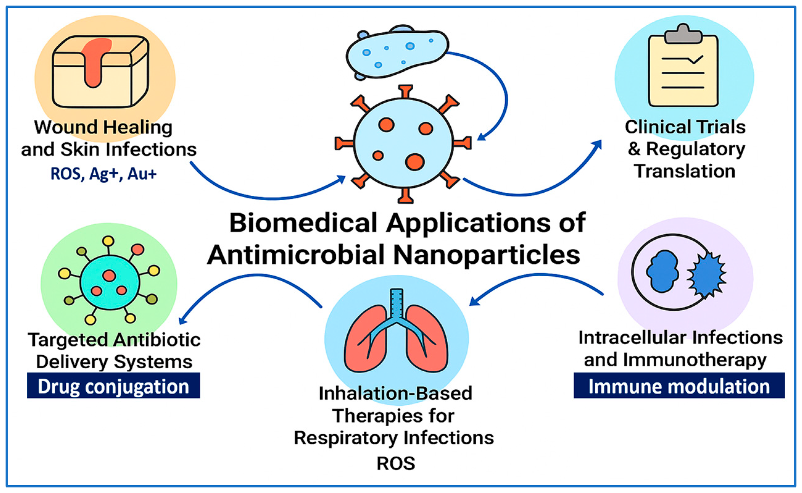
Figure 4 provides a visual summary of the key biomedical applications of antimicrobial NPs, illustrating their multifunctional roles in modern infection control strategies. These applications span six major areas: (1) wound healing and skin infections, where NPs such as Ag+, Au+, and ROS-generating agents facilitate antimicrobial action and tissue regeneration; (2) implantable device-related infections and clinical translation, emphasizing regulatory progress and therapeutic implementation; (3) targeted antibiotic delivery systems, leveraging nanoparticle–drug conjugation for enhanced site-specific therapy; (4) inhalation-based therapies for respiratory infections, utilizing NP-mediated ROS generation to eradicate pulmonary pathogens; (5) intracellular infections and immunotherapy, where immune-modulating NPs deliver antibiotics to phagocytic cells and stimulate host defense mechanisms; and (6) ongoing clinical trials and regulatory efforts, representing the pathway toward translational adoption. Together, these advances position antimicrobial NPs at the forefront of next-generation therapeutic interventions against multidrug-resistant and biofilm-associated infections.

5.7. Price and Affordability Constraints

Despite the therapeutic promise of antimicrobial NPs, their widespread clinical adoption is constrained by high production costs and scale-up limitations. Many synthesis processes—particularly those involving chemical or physical reduction methods—require expensive precursors, energy-intensive protocols, and sophisticated instrumentation, making them cost-prohibitive for large-scale pharmaceutical deployment. Sati et al. [164] emphasized that industrial-scale production of AgNPs remains economically challenging due to strict requirements for purity, particle uniformity, and environmental safety, which increase operational costs.
In response, green synthesis methods have gained traction as cost-effective and eco-friendly alternatives. By utilizing plant extracts and biological reducing agents, these approaches minimize reagent expenses and reduce environmental liabilities. Shahzadi et al. [41] demonstrated that biosynthetic routes offer a lower-cost, scalable pathway for AgNP production, particularly when optimized for plant metabolite content and reaction conditions. Similarly, Jebril et al. [165] reported that the use of multiple plant extract systems not only improved nanoparticle yield and stability but also significantly lowered overall synthesis costs, paving the way for broader clinical and industrial feasibility.
To facilitate equitable global access to nano-enabled antimicrobial therapies, future development must prioritize affordability alongside efficacy and safety. This includes standardizing green synthesis protocols, reducing batch-to-batch variability, and investing in scalable manufacturing platforms. Addressing cost barriers will be essential for integrating NPs into mainstream infection management strategies, particularly in low- and middle-income healthcare systems.

6. Translational Challenges and Safety Considerations of Antimicrobial NPs

Despite their therapeutic promise, antimicrobial NPs face numerous translational hurdles, including dose-dependent toxicity, immunogenicity, unpredictable biodistribution, manufacturing inconsistencies, and insufficient regulatory harmonization. Addressing these challenges through robust preclinical studies and standardized evaluation protocols is essential for their safe clinical implementation.

6.1. Toxicological Concerns and Dose-Dependent Toxicity

While metal-based NPs such as AgNPs and ZnO NPs demonstrate potent antimicrobial activity, they may also accumulate in organs and induce oxidative stress in vivo. In a good laboratory practice-compliant 90 day oral toxicity study in F344 rats, Kim et al. [166] reported that repeated exposure to ~56 nm AgNPs (30–500 mg/kg) led to dose-dependent silver accumulation in the liver and kidneys, bile duct hyperplasia, elevated liver enzymes, and weight loss—establishing a no observed adverse effect level (NOAEL) of ~30 mg/kg and a lowest observed adverse effect level (LOAEL) of ~ 125 mg/kg.
Cunningham et al. [167] showed that ultrasmall AgNPs (< 10 nm) induced more severe embryotoxicity in zebrafish, independent of silver ion release, emphasizing the size-dependent toxicity associated with nanoscale particles. Furthermore, Wen et al. [168] demonstrated that PEGylation of iron oxide NPs in rats significantly reduced their accumulation in the liver, spleen, and kidneys and attenuated inflammatory cytokine responses—highlighting the importance of surface modification in mitigating systemic toxicity.

6.2. Immune Activation and Inflammatory Risks

Cationic and lipid-based NPs—commonly employed in vaccines and antimicrobial formulations—can inadvertently activate innate immune pathways. Guo et al. [169] showed that intravenous administration of AgNPs in mice resulted in endothelial uptake, ROS overproduction, and disruption of VE–cadherin junctions, ultimately causing inflammation in the liver, lungs, and kidneys. Notably, the inflammatory response was size-dependent, with 75 nm and 110 nm AgNPs inducing more pronounced cellular infiltration than 10 nm particles.
Additionally, lipid-based NP components used in mRNA vaccine platforms have been implicated in immune activation. Connors et al. [170] found that empty lipid NPs enhanced dendritic cell maturation and cytokine production, with notable age-dependent effects in human peripheral blood cells. Complementary in vitro studies by Hanafy et al. [171] demonstrated that DOTAP-containing cationic lipids triggered IL-6 secretion and vascular permeability, consistent with the activation of the TLR/NLRP3 inflammasome pathways. Similarly, Lonez et al. [172] confirmed that ionizable cationic lipids can activate TLR2 and NLRP3 inflammasome signaling, resulting in IL-1β release—even in the absence of nucleic acids.

6.3. Protein Corona Formation and Biodistribution

When NPs encounter biological fluids such as blood or alveolar surfactant, they rapidly acquire a protein corona that reshapes their physicochemical identity. This layer significantly influences cellular uptake, organ distribution, clearance kinetics, and immunogenicity. Bai et al. [173] demonstrated that the in vivo protein corona—formed on engineered NPs following exposure to animal sera—profoundly altered biodistribution and toxicity profiles compared to in vitro predictions.
González-Vega et al. [174] further linked the composition of the protein corona to pulmonary deposition and inflammation. In rodent inhalation models, corona-coated AgNPs accumulated in alveolar macrophages and lung parenchyma, promoting neutrophil infiltration and cytokine elevation. Importantly, these effects varied depending on whether the NPs were inhaled as-is or pre-coated with coronas ex vivo.

6.4. Manufacturing, Scalability, and Standardization

The clinical scalability of antimicrobial NPs is hindered by batch-to-batch variability in synthesis, especially with green or hybrid methods. Small alterations in parameters such as pH, temperature, or extract concentration can result in significant differences in particle size, surface charge, stability, and antimicrobial efficacy. Liaqat et al. [175] compared various green synthesis protocols for AgNPs and found that these minor variations caused shifts in zeta potential (−15 mV to −5 mV), particle size (20–60 nm), and MIC values—emphasizing the need for standardization.
Herdiana et al. [176] demonstrated scale-up challenges using polymeric NP systems, where batch yield, monodispersity, and drug release profiles were inconsistent between lab-scale and continuous manufacturing platforms. To meet GMP requirements and ensure therapeutic reproducibility, manufacturing must define critical quality attributes, including polydispersity index, endotoxin levels, particle uniformity, and drug loading efficiency.

6.5. Green Synthesis and Biocompatibility

NPs synthesized via green chemistry—employing plant extracts or microbial agents—tend to exhibit enhanced biocompatibility while retaining antimicrobial efficacy. Liaqat et al. [175] tested over a dozen green-synthesized AgNPs and observed negligible cytotoxicity toward mammalian cells (e.g., fibroblasts, keratinocytes) at concentrations up to 50 µg/mL, with MICs ranging from 4 to 16 µg/mL. Naveen et al. [177] synthesized AgNPs using Aloe vera gel and found < 5 % hemolysis at 0.3 mg/mL, indicating excellent blood compatibility. Dudhagara et al. [178] reported similar results for lysozyme-stabilized AgNPs, which combined antibacterial and antiplatelet activity with minimal hemolytic effects (~ 6 %) at therapeutic doses.

6.6. PEGylation and Reduced Toxicity

Surface PEGylation improves NP safety by enhancing colloidal stability, reducing protein binding, and evading immune detection. Patlolla et al. [179] showed that oral administration of PEG-coated gold NPs in rats (12.5–100 µg/kg for 5 days) significantly lowered hepatic oxidative stress and histopathological alterations compared to uncoated controls. Cho et al. [180] found that intravenously administered PEGylated AuNPs (10 to 30 nm) circulated longer and accumulated less in the liver and spleen than their uncoated counterparts. These findings affirm PEGylation as a robust strategy for reducing toxicity while maintaining therapeutic efficacy.

6.7. Regulatory Frameworks and Clinical Translation

Clinical adoption of antimicrobial NPs is limited by fragmented regulatory guidelines. To date, only a few formulations—mainly topical AgNP-based gels—have entered early-phase trials (e.g., NCT03752424, NCT04894409, NCT04775238) [181]. Regulatory agencies like the FDA have issued generalized guidance on nanotechnology, but lack NP-specific criteria for pharmacokinetics, toxicity, and long-term safety assessments [182,183].
Kumari et al. [184] emphasized that divergent standards across regions complicate the translation of nanomedicine products. Nadar et al. [185] highlighted barriers to AgNP commercialization, including inconsistent classification as drugs or devices and a lack of standardized toxicity profiles. A 2024 review by Kumarasamy et al. [186] found that most clinical NP trials focus on antiviral or antifungal applications, with very few addressing antibacterial therapy—underscoring a translational gap.
A recent comparative regulatory review by Rodríguez-Gómez et al. [187] confirmed that both the EU and US frameworks still lack nanoparticle-specific approval pathways and exhibit inconsistencies in classification, safety testing requirements, and quality attribute definitions for nanotechnology-enabled health products. Likewise, Desai et al. [188] provide a global clinical perspective, identifying critical bottlenecks in pharmacokinetics characterization, immunotoxicity assessments, and standardization protocols—barriers that continue to slow the translation of nanoparticle therapeutics to human trials.

6.8. Microbiota–NP Interactions

Interactions between NPs and the host microbiota are increasingly recognized as critical to therapeutic safety and efficacy. Orally and intravenously administered antimicrobial NPs—particularly AgNPs, ZnO NPs, and TiO2 NPs—have been shown to induce gut dysbiosis and epithelial barrier disruption.
Van den Brule et al. [189] reported that AgNP exposure in mice led to reduced microbial diversity and a dose-dependent shift in the Firmicutes/Bacteroidetes ratio. Sulfidated AgNPs exhibited attenuated effects, suggesting environmental transformations may mitigate microbiota disruption. Lyu et al. [190] demonstrated that prenatal AgNP exposure caused long-term changes in microbiota composition and metabolic function in offspring, with implications for neurodevelopment and obesity.
Ren et al. [191] observed intestinal injury and leukocyte infiltration following AgNP exposure, while Wang et al. [192] noted decreased Gram-negative taxa and elevated serotonin levels in NP-treated mice—indicating potential neuroendocrine and metabolic consequences. Lamas et al. [193] provided a comprehensive mechanistic review showing that chronic dietary exposure to inorganic NPs—including Ag, TiO2, ZnO, and SiO2—frequently induces gut microbial imbalance, pro-inflammatory shifts (e.g., Proteobacteria overabundance), and reductions in beneficial taxa (e.g., Lactobacillus, Bifidobacterium), thereby promoting dysbiosis linked to metabolic disorders and inflammatory diseases.
Wu et al. [194] conducted a controlled rodent study demonstrating that oral TiO2 nanoparticle administration disrupted the gut bacterial community, reduced the abundance of short-chain fatty acid-producing probiotics, triggered intestinal inflammation and oxidative stress, and altered host lipid metabolism. An in vivo study by Kumar et al. [195] demonstrated that exposure to Ag, SiO2, and TiO2 nanoparticles in C57BL/6J mice induced dose-dependent alterations in gut microbiota composition, ranging from mild community shifts at lower doses to pronounced dysbiosis at higher exposures, characterized by elevated pro-inflammatory markers and reduced microbial diversity.
A recent study by Luo et al. [196] investigated the effects of dietary exposure to inorganic nanoparticles—including AgNPs, TiO2, and SiO2—on gut epithelial integrity in murine models. The findings revealed a dose-dependent downregulation of critical tight junction proteins (ZO-1, occludin, and claudin-1), along with mucosal thinning and increased intestinal permeability. These structural disruptions were accompanied by pronounced dysbiosis, characterized by significant reductions in beneficial genera such as Lactobacillus and Bifidobacterium, and elevated levels of colonic pro-inflammatory cytokines TNF-α and IL-6. Collectively, the results establish a mechanistic link between NP exposure, epithelial barrier impairment, intestinal inflammation, and microbial imbalance.
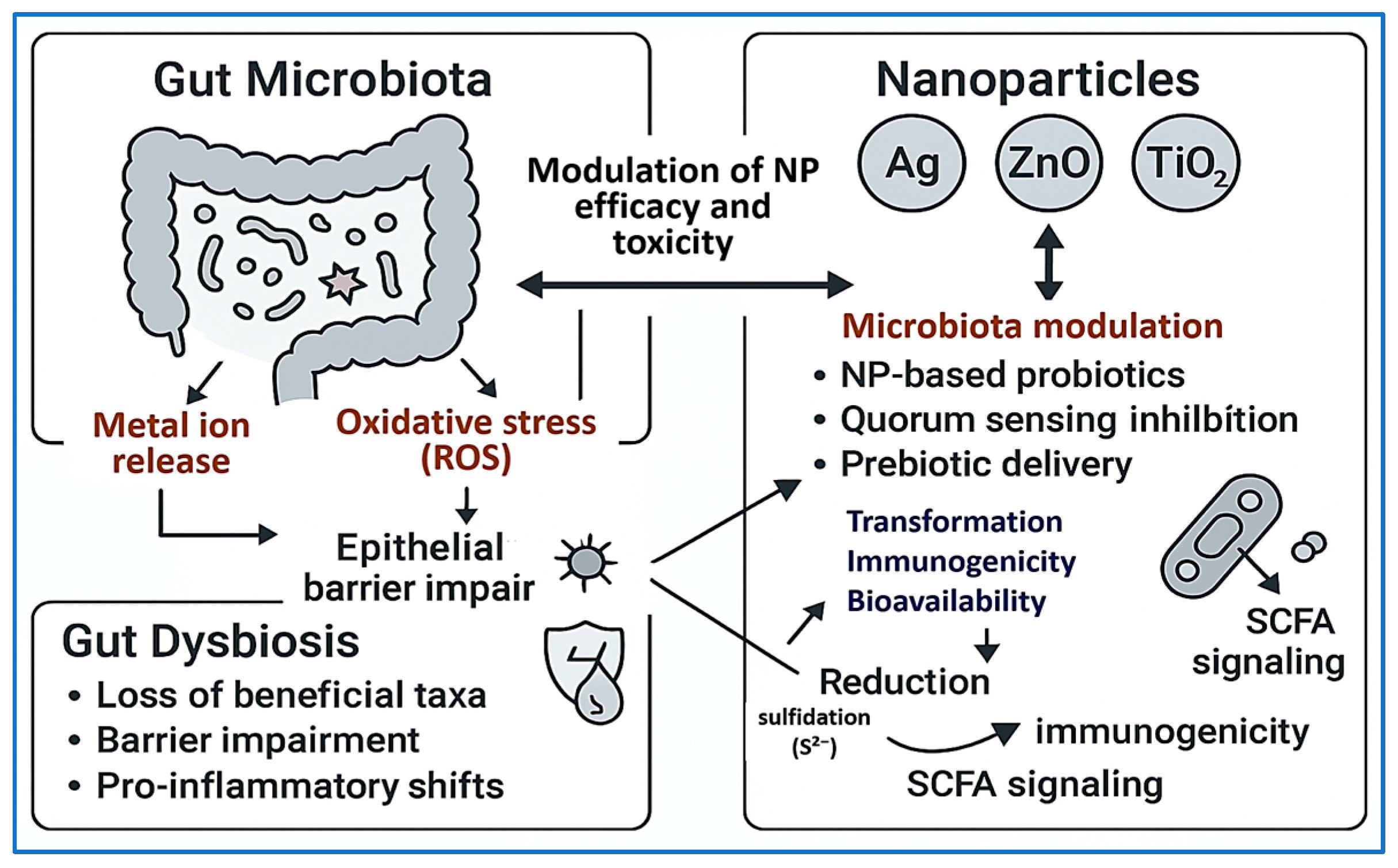
These emerging findings underscore the importance of incorporating microbiome-informed strategies into the development, safety evaluation, and regulatory assessment of antimicrobial nanotherapeutics—especially for formulations intended for oral or systemic administration. Figure 5 schematically illustrates the bidirectional interactions between antimicrobial nanoparticles and the gut microbiota, highlighting both microbiota-mediated NP transformation and NP-induced microbial and epithelial alterations.

7. Future Directions and Research Gaps

The advent of antimicrobial NPs has introduced transformative opportunities in combating MDR pathogens. However, several critical research gaps (Table 3) and translational challenges must be addressed to fully harness their clinical potential. A key area of innovation involves the development of stimuli-responsive or “smart” NPs that release their antimicrobial payloads selectively at infection sites—thereby minimizing systemic toxicity and enhancing therapeutic precision. For example, Li et al. [197] designed pH-sensitive PAMAM–HA–SNO “megamer” NPs co-loaded with nitric oxide and ciprofloxacin. These NPs disassembled in the acidic microenvironments characteristic of infected tissues, efficiently clearing Staphylococcus aureus infections in vivo while sparing healthy tissues. Similarly, Kalhapure et al. [198] engineered vancomycin-loaded solid lipid NPs that preferentially released the drug under acidic conditions in MRSA-infected murine skin, resulting in enhanced bacterial clearance and improved therapeutic outcomes.
Another prominent research frontier is biofilm eradication, which remains a major obstacle due to the protective nature of the extracellular polymeric matrix. Tasia et al. [199] developed DNase I-functionalized, silver-doped mesoporous silica NPs capable of enzymatically degrading extracellular DNA and delivering Ag+. These multifunctional NPs significantly disrupted both Gram-positive and Gram-negative biofilms in vitro. Other studies support the synergistic use of enzymatic disruption and targeted antimicrobial delivery to enhance biofilm penetration and eradication.
Despite growing preclinical evidence, the long-term biodistribution and toxicity of antimicrobial NPs remain under-investigated. Most studies focus on short-term efficacy, while chronic exposure—particularly in patients requiring repeated or prolonged therapy—raises safety concerns. Radiolabeled NP tracking in non-infected models has shown prolonged accumulation in the liver and spleen, emphasizing the need for long-term monitoring in relevant infection models. Future research should prioritize the development of advanced imaging protocols, pharmacokinetic modeling, and toxicity assays tailored to infectious disease contexts.
Environmental safety is another emerging concern as the widespread use of NPs in consumer and medical products may impact ecosystems and commensal microbial communities. Green synthesis approaches, such as those employing Azadirachta indica leaf extract, have produced AgNPs with potent antimicrobial activity and reduced toxicity toward soil and aquatic microbiota. These environmentally friendly methods warrant further exploration, and longitudinal environmental surveillance is essential to ensure sustainable deployment.
The integration of diagnostics and therapeutics in nanotheranostic platforms represents a promising avenue for personalized infection management. Hajfathalian et al. [200] developed dextran-coated gold-in-gold cage NPs capable of both photoacoustic imaging and photothermal ablation of biofilms in vivo, exemplifying dual-functionality systems. Likewise, enzyme-responsive fluorescent–antibiotic nanoshells offer real-time infection sensing with site-specific drug release [201]. These innovations align with precision medicine goals and may enable dynamic, patient-tailored interventions.
Lastly, international collaboration and regulatory harmonization are vital for advancing safe and effective clinical translation. Initiatives such as the EU’s Safe-by-Design framework and ISO/TR 10993-22 [202] guidelines for nanomaterial-containing medical devices are critical milestones. However, broader global adoption and cross-agency coordination remain urgently needed to accelerate clinical development, ensure safety, and streamline approval pathways.
Table 3. Key research gaps and future directions in antimicrobial NPs.
Research GapDescriptionProposed Future DirectionsKey References
Lack of stimuli-responsive drug delivery systemsCurrent NPs often release drugs passively, leading to off-target effects and reduced therapeutic precision.Design infection-triggered or pH-/enzyme-responsive systems to enable site-specific activation.[197]
Poor efficacy against biofilmsBiofilm matrices hinder NP penetration and antimicrobial activity, reducing treatment effectiveness.Develop enzyme-functionalized NPs (e.g., DNase, protease) or ROS-generating NPs to disrupt biofilms.[199,203]
Limited long-term safety and biodistribution dataMost studies assess short-term efficacy, lacking insight into chronic toxicity and organ accumulation.Utilize radiolabeling or imaging-guided platforms to evaluate NP clearance, persistence, and tissue-specific effects over time.[204,205]
Environmental impact and microbiome disruptionPersistent NPs, especially metal-based ones, may harm beneficial microbes in the environment or gut microbiota.Prioritize green synthesis methods and biodegradable nanomaterials that limit ecological toxicity.[206]
Fragmented regulatory frameworksLack of harmonized guidelines delays clinical translation and hinders global acceptance of NP therapeutics.Develop unified regulatory protocols addressing NP characterization, manufacturing, and pharmacokinetics.[29,207]
Insufficient integration with diagnostics (theranostics)Few platforms allow simultaneous infection detection and treatment, limiting real-time precision therapy.Create dual-function NPs combining antimicrobial delivery with imaging or biosensing elements.[208]

8. Conclusions

NPs represent a powerful and versatile tool in the global effort to combat MDR pathogens. Their unique ability to disrupt bacterial membranes, penetrate biofilms, enable targeted drug delivery, and modulate host immune responses offers clear advantages over conventional antimicrobial therapies. Throughout this review, we have highlighted the broad spectrum of biomedical applications for NPs—ranging from wound healing and implant coatings to inhalation therapies and intracellular infection control—while emphasizing their mechanistic diversity and clinical potential. A particularly novel aspect addressed in this review is the bidirectional interaction between NPs and the host microbiota. While NPs provide enhanced antimicrobial efficacy, they also have the capacity to disrupt microbial homeostasis and influence host physiology, raising important concerns about long-term safety and unintended immunological consequences. These findings underscore the importance of considering not only efficacy but also the biological context in which NPs operate. As the field advances, it is imperative that future antimicrobial NP platforms embrace “safe-by-design” principles, integrating microbiota-informed approaches, precision pharmacokinetic profiling, and rigorous toxicological evaluation. Translating laboratory breakthroughs into clinical success will require robust regulatory frameworks, harmonized safety standards, and proactive engagement with regulatory agencies. Most importantly, progress in this domain will depend on interdisciplinary collaboration among microbiologists, nanotechnologists, clinicians, toxicologists, and policymakers. By aligning innovation with clinical and regulatory realities, antimicrobial NPs have the potential to transform the landscape of infectious disease prevention and treatment—ultimately serving as a cornerstone in the post-antibiotic era.

Author Contributions

Conceptualization, A.E. and A.A.; writing—original draft preparation, A.E. and A.A.; writing—review and editing, A.E. and A.A.; visualization, A.E. and A.A.; supervision, A.E. and A.A.; project administration, A.E. and A.A.; funding acquisition, A.A. All authors have read and agreed to the published version of the manuscript.

Funding

This research received no external funding.

Institutional Review Board Statement

Not applicable. This article does not involve studies with human participants or animals.

Informed Consent Statement

Not applicable.

Data Availability Statement

Not applicable. No new data were created or analyzed in this study.

Acknowledgments

The researchers would like to thank the Deanship of Graduate Studies and Scientific Research at Qassim University for financial support (QU-APC-2025).

Conflicts of Interest

The authors declare no conflicts of interest.

Abbreviations

The following abbreviations are used in this manuscript:
AbbreviationFull Term
AMRAntimicrobial Resistance
NPsNanoparticles
MDRMultidrug-resistant
AgNPsSilver Nanoparticles
AIArtificial Intelligence
AuNPsGold Nanoparticles
CFUColony-Forming Units
CuOCopper Oxide
CuO NPsCopper Oxide Nanoparticles
DNADeoxyribonucleic Acid
EMAEuropean Medicines Agency
FDAU.S. Food and Drug Administration
GNSGold Nanostar
IFN-γInterferon Gamma
IL-1βInterleukin-1 Beta
MICMinimum Inhibitory Concentration
Mo@ZIF-8Molybdenum-Doped Zeolitic Imidazolate Framework-8
MOFMetal–Organic Framework
MSNsMesoporous Silica Nanoparticles
NLCsNanostructured Lipid Carriers
NP(s)Nanoparticle(s)
PEGPolyethylene Glycol
PLGAPoly(lactic-co-glycolic acid)
QSQuorum Sensing
rGOReduced Graphene Oxide
RNARibonucleic Acid
ROSReactive Oxygen Species
SCFAsShort-Chain Fatty Acids
S. aureusStaphylococcus aureus
E. coliEscherichia coli
P. aeruginosaPseudomonas aeruginosa
K. pneumoniaeKlebsiella pneumoniae
S. typhimuriumSalmonella typhimurium
TiO2Titanium Dioxide
TiO2 NPsTitanium Dioxide Nanoparticles
TiN–AgTitanium Nitride–Silver
ZnOZinc Oxide
ZnO NPsZinc Oxide Nanoparticles
ZnO–AgZinc Oxide–Silver
ZIF-8Zeolitic Imidazolate Framework-8

References

  1. Rice, L.B. Federal funding for the study of antimicrobial resistance in nosocomial pathogens: No ESKAPE. J. Infect. Dis. 2008, 197, 1079–1081. [Google Scholar] [CrossRef]
  2. Tacconelli, E.; Carrara, E.; Savoldi, A.; Harbarth, S.; Mendelson, M.; Monnet, D.L.; Pulcini, C.; Kahlmeter, G.; Kluytmans, J.; Carmeli, Y. Discovery, research, and development of new antibiotics: The WHO priority list of antibiotic-resistant bacteria and tuberculosis. Lancet Infect. Dis. 2018, 18, 318–327. [Google Scholar] [CrossRef] [PubMed]
  3. Murray, C.J.; Ikuta, K.S.; Sharara, F.; Swetschinski, L.; Aguilar, G.R.; Gray, A.; Han, C.; Bisignano, C.; Rao, P.; Wool, E. Global burden of bacterial antimicrobial resistance in 2019: A systematic analysis. Lancet 2022, 399, 629–655. [Google Scholar] [CrossRef] [PubMed]
  4. O’Neill, J. Tackling Drug-Resistant Infections Globally: Final Report and Recommendations; 2016. Available online: https://amr-review.org/sites/default/files/160518_Final%20paper_with%20cover.pdf (accessed on 8 July 2025).
  5. WHO. Global Action Plan on Antimicrobial Resistance; 2015. Available online: https://www.emro.who.int/health-topics/drug-resistance/global-action-plan.html (accessed on 8 July 2025).
  6. Theuretzbacher, U.; Piddock, L.J. Non-traditional antibacterial therapeutic options and challenges. Cell Host Microbe 2019, 26, 61–72. [Google Scholar] [CrossRef] [PubMed]
  7. De Oliveira, D.M.; Forde, B.M.; Kidd, T.J.; Harris, P.N.; Schembri, M.A.; Beatson, S.A.; Paterson, D.L.; Walker, M.J. Antimicrobial resistance in ESKAPE pathogens. Clin. Microbiol. Rev. 2020, 33, e00181-19. [Google Scholar] [CrossRef]
  8. Flemming, H.-C.; Wingender, J.; Szewzyk, U.; Steinberg, P.; Rice, S.A.; Kjelleberg, S. Biofilms: An emergent form of bacterial life. Nat. Rev. Microbiol. 2016, 14, 563–575. [Google Scholar] [CrossRef]
  9. Hall, C.W.; Mah, T.-F. Molecular mechanisms of biofilm-based antibiotic resistance and tolerance in pathogenic bacteria. FEMS Microbiol. Rev. 2017, 41, 276–301. [Google Scholar] [CrossRef]
  10. Koo, H.; Allan, R.N.; Howlin, R.P.; Stoodley, P.; Hall-Stoodley, L. Targeting microbial biofilms: Current and prospective therapeutic strategies. Nat. Rev. Microbiol. 2017, 15, 740–755. [Google Scholar] [CrossRef]
  11. Hajipour, M.J.; Fromm, K.M.; Ashkarran, A.A.; de Aberasturi, D.J.; de Larramendi, I.R.; Rojo, T.; Serpooshan, V.; Parak, W.J.; Mahmoudi, M. Antibacterial properties of nanoparticles. Trends Biotechnol. 2012, 30, 499–511. [Google Scholar] [CrossRef]
  12. Lemire, J.A.; Harrison, J.J.; Turner, R.J. Antimicrobial activity of metals: Mechanisms, molecular targets and applications. Nat. Rev. Microbiol. 2013, 11, 371–384. [Google Scholar] [CrossRef]
  13. Franci, G.; Falanga, A.; Galdiero, S.; Palomba, L.; Rai, M.; Morelli, G.; Galdiero, M. Silver nanoparticles as potential antibacterial agents. Molecules 2015, 20, 8856–8874. [Google Scholar] [CrossRef]
  14. Durán, N.; Durán, M.; De Jesus, M.B.; Seabra, A.B.; Fávaro, W.J.; Nakazato, G. Silver nanoparticles: A new view on mechanistic aspects on antimicrobial activity. Nanomed. Nanotechnol. Biol. Med. 2016, 12, 789–799. [Google Scholar] [CrossRef] [PubMed]
  15. Shaikh, S.; Nazam, N.; Rizvi, S.M.D.; Ahmad, K.; Baig, M.H.; Lee, E.J.; Choi, I. Mechanistic insights into the antimicrobial actions of metallic nanoparticles and their implications for multidrug resistance. Int. J. Mol. Sci. 2019, 20, 2468. [Google Scholar] [CrossRef] [PubMed]
  16. Rai, M.; Yadav, A.; Gade, A. Silver nanoparticles as a new generation of antimicrobials. Biotechnol. Adv. 2009, 27, 76–83. [Google Scholar] [CrossRef]
  17. Anselmo, A.C.; Mitragotri, S. Nanoparticles in the clinic: An update. Bioeng. Transl. Med. 2019, 4, e10143. [Google Scholar] [CrossRef]
  18. Høiby, N.; Ciofu, O.; Johansen, H.K.; Song, Z.j.; Moser, C.; Jensen, P.Ø.; Molin, S.; Givskov, M.; Tolker-Nielsen, T.; Bjarnsholt, T. The clinical impact of bacterial biofilms. Int. J. Oral Sci. 2011, 3, 55–65. [Google Scholar] [CrossRef]
  19. Flemming, H.-C.; Wuertz, S. Bacteria and archaea on Earth and their abundance in biofilms. Nat. Rev. Microbiol. 2019, 17, 247–260. [Google Scholar] [CrossRef]
  20. Tängdén, T.; Giske, C. Global dissemination of extensively drug-resistant carbapenemase-producing E nterobacteriaceae: Clinical perspectives on detection, treatment and infection control. J. Intern. Med. 2015, 277, 501–512. [Google Scholar] [CrossRef]
  21. Sercombe, L.; Veerati, T.; Moheimani, F.; Wu, S.Y.; Sood, A.K.; Hua, S. Advances and challenges of liposome assisted drug delivery. Front. Pharmacol. 2015, 6, 286. [Google Scholar] [CrossRef]
  22. Yathavan, B.; Chhibber, T.; Steinhauff, D.; Pulsipher, A.; Alt, J.A.; Ghandehari, H.; Jafari, P. Matrix-mediated delivery of silver nanoparticles for prevention of Staphylococcus aureus and Pseudomonas aeruginosa biofilm formation in chronic rhinosinusitis. Pharmaceutics 2023, 15, 2426. [Google Scholar] [CrossRef]
  23. Alhosani, F.; Islayem, D.; Almansoori, S.; Zaka, A.; Nayfeh, L.; Rezk, A.; Yousef, A.F.; Pappa, A.M.; Nayfeh, A. Antibiofilm activity of ZnO–Ag nanoparticles against Pseudomonas aeruginosa. Sci. Rep. 2025, 15, 17321. [Google Scholar] [CrossRef] [PubMed]
  24. Kamer, A.M.A.; El Maghraby, G.M.; Shafik, M.M.; Al-Madboly, L.A. Silver nanoparticle with potential antimicrobial and antibiofilm efficiency against multiple drug resistant, extensive drug resistant Pseudomonas aeruginosa clinical isolates. BMC Microbiol. 2024, 24, 277. [Google Scholar] [CrossRef]
  25. Szymczak, M.; Pankowski, J.A.; Kwiatek, A.; Grygorcewicz, B.; Karczewska-Golec, J.; Sadowska, K.; Golec, P. An effective antibiofilm strategy based on bacteriophages armed with silver nanoparticles. Sci. Rep. 2024, 14, 9088. [Google Scholar] [CrossRef]
  26. Ahmad, I.; Khan, S.A.; Naseer, A.; Nazir, A. Green synthesized silver nanoparticles from Phoenix dactylifera synergistically interact with bioactive extract of Punica granatum against bacterial virulence and biofilm development. Microb. Pathog. 2024, 192, 106708. [Google Scholar] [CrossRef]
  27. Manke, A.; Wang, L.; Rojanasakul, Y. Mechanisms of nanoparticle-induced oxidative stress and toxicity. BioMed Res. Int. 2013, 2013, 942916. [Google Scholar] [CrossRef]
  28. Kah, M.; Tufenkji, N.; White, J.C. Nano-enabled strategies to enhance crop nutrition and protection. Nat. Nanotechnol. 2019, 14, 532–540. [Google Scholar] [CrossRef]
  29. Foulkes, R.; Man, E.; Thind, J.; Yeung, S.; Joy, A.; Hoskins, C. The regulation of nanomaterials and nanomedicines for clinical application: Current and future perspectives. Biomater. Sci. 2020, 8, 4653–4664. [Google Scholar] [CrossRef]
  30. Soares, S.; Sousa, J.; Pais, A.; Vitorino, C. Nanomedicine: Principles, properties, and regulatory issues. Front. Chem. 2018, 6, 360. [Google Scholar] [CrossRef]
  31. Younis, M.A.; Tawfeek, H.M.; Abdellatif, A.A.; Abdel-Aleem, J.A.; Harashima, H. Clinical translation of nanomedicines: Challenges, opportunities, and keys. Adv. Drug Deliv. Rev. 2022, 181, 114083. [Google Scholar] [CrossRef]
  32. Đorđević, S.; Gonzalez, M.M.; Conejos-Sánchez, I.; Carreira, B.; Pozzi, S.; Acúrcio, R.C.; Satchi-Fainaro, R.; Florindo, H.F.; Vicent, M.J. Current hurdles to the translation of nanomedicines from bench to the clinic. Drug Deliv. Transl. Res. 2022, 12, 500–525. [Google Scholar] [CrossRef]
  33. Wang, L.; Hu, C.; Shao, L. The antimicrobial activity of nanoparticles: Present situation and prospects for the future. Int. J. Nanomed. 2017, 12, 1227–1249. [Google Scholar] [CrossRef] [PubMed]
  34. Allen, T.M.; Cullis, P.R. Liposomal drug delivery systems: From concept to clinical applications. Adv. Drug Deliv. Rev. 2013, 65, 36–48. [Google Scholar] [CrossRef] [PubMed]
  35. Mehnert, W.; Mäder, K. Solid lipid nanoparticles: Production, characterization and applications. Adv. Drug Deliv. Rev. 2012, 64, 83–101. [Google Scholar] [CrossRef]
  36. Jiang, H.; Li, L.; Li, Z.; Chu, X. Metal-based nanoparticles in antibacterial application in biomedical field: Current development and potential mechanisms. Biomed. Microdevices 2024, 26, 12. [Google Scholar] [CrossRef]
  37. Moradialvand, M.; Asri, N.; Jahdkaran, M.; Beladi, M.; Houri, H. Advancements in nanoparticle-based strategies for enhanced antibacterial interventions. Cell Biochem. Biophys. 2024, 82, 3071–3090. [Google Scholar] [CrossRef]
  38. Okka, E.Z.; Tongur, T.; Aytas, T.T.; Yılmaz, M.; Topel, Ö.; Sahin, R. Green Synthesis and the formation kinetics of silver nanoparticles in aqueous Inula viscosa extract. Optik 2023, 294, 171487. [Google Scholar] [CrossRef]
  39. Gur, T. Green synthesis, characterizations of silver nanoparticles using sumac (Rhus coriaria L.) plant extract and their antimicrobial and DNA damage protective effects. Front. Chem. 2022, 10, 968280. [Google Scholar] [CrossRef]
  40. Ershov, V.A.; Ershov, B.G. Effect of silver nanoparticle size on antibacterial activity. Toxics 2024, 12, 801. [Google Scholar] [CrossRef]
  41. Shahzadi, S.; Fatima, S.; Shafiq, Z.; Janjua, M.R.S.A. A review on green synthesis of silver nanoparticles (SNPs) using plant extracts: A multifaceted approach in photocatalysis, environmental remediation, and biomedicine. RSC Adv. 2025, 15, 3858–3903. [Google Scholar] [CrossRef]
  42. Aguilar-Garay, R.; Lara-Ortiz, L.F.; Campos-López, M.; Gonzalez-Rodriguez, D.E.; Gamboa-Lugo, M.M.; Mendoza-Pérez, J.A.; Anzueto-Ríos, Á.; Nicolás-Álvarez, D.E. A comprehensive review of silver and gold nanoparticles as effective antibacterial agents. Pharmaceuticals 2024, 17, 1134. [Google Scholar] [CrossRef]
  43. El-Fallal, A.A.; Elfayoumy, R.A.; El-Zahed, M.M. Antibacterial activity of biosynthesized zinc oxide nanoparticles using Kombucha extract. SN Appl. Sci. 2023, 5, 332. [Google Scholar] [CrossRef]
  44. Tazeen, S.K.; Niazi, M.B.K.; Binobead, M.A.; Ahmed, T.; Shahid, M. Characterization and toxicity evaluation of chitosan/ZnO nanocompoite as promising nano-biopolymer for treatment of synthetic wastewater. J. King Saud Univ.-Sci. 2024, 36, 103432. [Google Scholar]
  45. Azad, A.; Hussain, S.; Akram, H.; Fida, H.; Iqbal, M.A.; Butt, T.E. Environmentally-friendly synthesis of platinum nanoparticles: Phytochemical, antioxidant, and antimicrobial properties of Cichorium intybus in different solvents. Discov. Plants 2024, 1, 24. [Google Scholar] [CrossRef]
  46. Prashanth, G.; Lalithamba, H.; Rao, S.; Rashmi, K.; Bhagya, N.; Dileep, M.; Gadewar, M.; Ghosh, M.K. Green synthesized Pt-based nanoparticles redefining biomedical frontiers: A brief review. Next Mater. 2025, 8, 100613. [Google Scholar] [CrossRef]
  47. Su, L.; Gong, X.; Fan, R.; Ni, T.; Yang, F.; Zhang, X.; Li, X. Mechanism of action of platinum nanoparticles implying from antioxidant to metabolic programming in light-induced retinal degeneration model. Redox Biol. 2023, 65, 102836. [Google Scholar] [CrossRef]
  48. Belloso Daza, M.V.; Scarsi, A.; Gatto, F.; Rocchetti, G.; Pompa, P.P.; Cocconcelli, P.S. Role of Platinum Nanozymes in the Oxidative Stress Response of Salmonella typhimurium. Antioxidants 2023, 12, 1029. [Google Scholar] [CrossRef]
  49. Gatto, F.; Moglianetti, M.; Pompa, P.P.; Bardi, G. Platinum nanoparticles decrease reactive oxygen species and modulate gene expression without alteration of immune responses in THP-1 monocytes. Nanomaterials 2018, 8, 392. [Google Scholar] [CrossRef]
  50. Salleh, A.; Naomi, R.; Utami, N.D.; Mohammad, A.W.; Mahmoudi, E.; Mustafa, N.; Fauzi, M.B. The potential of silver nanoparticles for antiviral and antibacterial applications: A mechanism of action. Nanomaterials 2020, 10, 1566. [Google Scholar] [CrossRef]
  51. Shah, S.; Gaikwad, S.; Nagar, S.; Kulshrestha, S.; Vaidya, V.; Nawani, N.; Pawar, S. Biofilm inhibition and anti-quorum sensing activity of phytosynthesized silver nanoparticles against the nosocomial pathogen Pseudomonas aeruginosa. Biofouling 2019, 35, 34–49. [Google Scholar] [CrossRef]
  52. Awadelkareem, A.M.; Siddiqui, A.J.; Noumi, E.; Ashraf, S.A.; Hadi, S.; Snoussi, M.; Badraoui, R.; Bardakci, F.; Ashraf, M.S.; Danciu, C. Biosynthesized silver nanoparticles derived from probiotic Lactobacillus rhamnosus (AgNPs-LR) targeting biofilm formation and quorum sensing-mediated virulence factors. Antibiotics 2023, 12, 986. [Google Scholar] [CrossRef]
  53. Chen, S.; Yao, J.; Huo, S.; Xu, C.; Yang, R.; Tao, D.; Fang, B.; Ma, G.; Zhu, Z.; Zhang, Y. Designing injectable dermal matrix hydrogel combined with silver nanoparticles for methicillin-resistant Staphylococcus aureus infected wounds healing. Nano Converg. 2024, 11, 41. [Google Scholar] [CrossRef] [PubMed]
  54. Gao, Q.; Hu, F.; Chai, Z.; Zheng, C.; Zhang, W.; Pu, K.; Yang, Z.; Zhang, Y.; Ramrkrishna, S.; Wu, X. Multifunctional hydrogel with mild photothermal properties enhances diabetic wound repair by targeting MRSA energy metabolism. J. Nanobiotechnol. 2025, 23, 380. [Google Scholar] [CrossRef] [PubMed]
  55. Selmani, A.; Zeiringer, S.; Šarić, A.; Stanković, A.; Učakar, A.; Vidmar, J.; Abram, A.; Njegić Džakula, B.; Kontrec, J.; Zore, A. ZnO Nanoparticle-Infused Vaterite Coatings: A Novel Approach for Antimicrobial Titanium Implant Surfaces. J. Funct. Biomater. 2025, 16, 108. [Google Scholar] [CrossRef]
  56. Serov, D.A.; Gritsaeva, A.V.; Yanbaev, F.M.; Simakin, A.V.; Gudkov, S.V. Review of antimicrobial properties of titanium dioxide nanoparticles. Int. J. Mol. Sci. 2024, 25, 10519. [Google Scholar] [CrossRef]
  57. Najm, M.A.; Shakir, H.A.; Hasen, S.T.; Jawad, K.H.; Hasoon, B.A.; Jabir, M.S.; Issa, A.A.; Albukhaty, S.; Gatasheh, M.K.; Molla, M.H. Titanium dioxide nanoparticles augment Ciprofloxacin activity via Inhibition of biofilm formation for multidrug resistance bacteria in-vitro and insilco prediction study. Sci. Rep. 2025, 15, 18014. [Google Scholar] [CrossRef]
  58. Shariati, A.; Chegini, Z.; Ghaznavi-Rad, E.; Zare, E.N.; Hosseini, S.M. PLGA-based nanoplatforms in drug delivery for inhibition and destruction of microbial biofilm. Front. Cell. Infect. Microbiol. 2022, 12, 926363. [Google Scholar] [CrossRef]
  59. Türeli, N.G.; Torge, A.; Juntke, J.; Schwarz, B.C.; Schneider-Daum, N.; Türeli, A.E.; Lehr, C.-M.; Schneider, M. Ciprofloxacin-loaded PLGA nanoparticles against cystic fibrosis P. Eur. J. Pharm. Biopharm. 2017, 117, 363–371. [Google Scholar] [CrossRef]
  60. Tchatchiashvili, T.; Duering, H.; Mueller-Boetticher, L.; Grune, C.; Fischer, D.; Pletz, M.W.; Makarewicz, O. PEG-PLGA nanoparticles deposited in Pseudomonas aeruginosa and Burkholderia cenocepacia. J. Pharm. Anal. 2024, 14, 100939. [Google Scholar] [CrossRef]
  61. Coksu, I.; Dokuz, S.; Akgul, B.; Ozbek, T.; Abamor, E.S.; Duranoglu, D.; Acar, S. Enhancing the treatment of Staphylococcus aureus infections: A nanosystem with including dual antimicrobial peptide. J. Drug Deliv. Sci. Technol. 2024, 97, 105830. [Google Scholar] [CrossRef]
  62. Miranda Calderon, L.G.; Alejo, T.; Santos, S.; Mendoza, G.; Irusta, S.; Arruebo, M. Antibody-functionalized polymer nanoparticles for targeted antibiotic delivery in models of pathogenic bacteria infecting human macrophages. ACS Appl. Mater. Interfaces 2023, 15, 40213–40227. [Google Scholar] [CrossRef]
  63. Ding, M.; Zhao, W.; Song, L.-J.; Luan, S.-F. Stimuli-responsive nanocarriers for bacterial biofilm treatment. Rare Met. 2022, 41, 482–498. [Google Scholar] [CrossRef]
  64. Afrasiabi, S.; Partoazar, A.; Chiniforush, N. In vitro study of nanoliposomes containing curcumin and doxycycline for enhanced antimicrobial photodynamic therapy against Aggregatibacter actinomycetemcomitans. Sci. Rep. 2023, 13, 11552. [Google Scholar] [CrossRef]
  65. Arabestani, M.R.; Bigham, A.; Kamarehei, F.; Dini, M.; Gorjikhah, F.; Shariati, A.; Hosseini, S.M. Solid lipid nanoparticles and their application in the treatment of bacterial infectious diseases. Biomed. Pharmacother. 2024, 174, 116433. [Google Scholar] [CrossRef]
  66. Motsoene, F.; Abrahamse, H.; Kumar, S.S.D. Multifunctional lipid-based nanoparticles for wound healing and antibacterial applications: A review. Adv. Colloid Interface Sci. 2023, 321, 103002. [Google Scholar] [CrossRef] [PubMed]
  67. Li, R.; Chen, T.; Pan, X. Metal–organic-framework-based materials for antimicrobial applications. ACS Nano 2021, 15, 3808–3848. [Google Scholar] [CrossRef]
  68. Hu, W.; Ouyang, Q.; Jiang, C.; Huang, S.; Alireza, N.-E.; Guo, D.; Liu, J.; Peng, Y. Biomedical Metal–Organic framework materials on antimicrobial therapy: Perspectives and challenges. Mater. Today Chem. 2024, 41, 102300. [Google Scholar] [CrossRef]
  69. Omer, M.E.; Halwani, M.; Alenazi, R.M.; Alharbi, O.; Aljihani, S.; Massadeh, S.; Al Ghoribi, M.; Al Aamery, M.; Yassin, A.E. Novel self-assembled polycaprolactone–lipid hybrid nanoparticles enhance the antibacterial activity of ciprofloxacin. SLAS Technol. Transl. Life Sci. Innov. 2020, 25, 598–607. [Google Scholar] [CrossRef]
  70. Rajak, K.K.; Pahilani, P.; Patel, H.; Kikani, B.; Desai, R.; Kumar, H. Green synthesis of silver nanoparticles using Curcuma longa flower extract and antibacterial activity. arXiv 2023, arXiv:2304.04777. [Google Scholar] [CrossRef]
  71. Lian, Z.; Lu, C.; Zhu, J.; Zhang, X.; Wu, T.; Xiong, Y.; Sun, Z.; Yang, R. Mo@ ZIF-8 nanozyme preparation and its antibacterial property evaluation. Front. Chem. 2022, 10, 1093073. [Google Scholar] [CrossRef]
  72. Shen, Z.-Y.; Sadiq, S.; Xu, T.; Wu, P.; Khan, I.; Jiao, X.; Khan, A.; Wang, L.; Lin, S. Inhibitory effect of organometallic framework composite nanomaterial ZIF8@ ZIF67 on different pathogenic microorganisms of silkworms. Pestic. Biochem. Physiol. 2025, 208, 106307. [Google Scholar] [CrossRef]
  73. Cai, W.; Zhang, W.; Chen, Z. Magnetic Fe3O4@ ZIF-8 nanoparticles as a drug release vehicle: pH-sensitive release of norfloxacin and its antibacterial activity. Colloids Surf. B Biointerfaces 2023, 223, 113170. [Google Scholar] [CrossRef] [PubMed]
  74. Wang, J.; Yu, Y.; Chen, L.; Yu, J.; Jin, X.; Zeng, R.; Luo, X.; Cong, Y.; Xu, G.; Zhang, J. NIR-triggered and Glucose-Powered Hollow mesoporous Mo-based single-atom nanozymes for cascade chemodynamic diabetic infection therapy. Mater. Today Bio 2025, 31, 101557. [Google Scholar] [CrossRef] [PubMed]
  75. Aggarwal, D.; Choudhary, M. Recent Updates on Green Synthesized Silver Nanoparticles: Preparation Technologies, Properties, and Applications In Biomedical Sector. Int. J. Environ. Sci. 2025, 11, 968–982. [Google Scholar] [CrossRef]
  76. Sharma, R.; Basist, P.; Alhalmi, A.; Khan, R.; Noman, O.M.; Alahdab, A. Synthesis of quercetin-loaded silver nanoparticles and assessing their anti-bacterial potential. Micromachines 2023, 14, 2154. [Google Scholar] [CrossRef]
  77. Quintero-Quiroz, C.; Acevedo, N.; Zapata-Giraldo, J.; Botero, L.E.; Quintero, J.; Zárate-Triviño, D.; Saldarriaga, J.; Pérez, V.Z. Optimization of silver nanoparticle synthesis by chemical reduction and evaluation of its antimicrobial and toxic activity. Biomater. Res. 2019, 23, 27. [Google Scholar] [CrossRef]
  78. Swidan, N.S.; Hashem, Y.A.; Elkhatib, W.F.; Yassien, M.A. Antibiofilm activity of green synthesized silver nanoparticles against biofilm associated enterococcal urinary pathogens. Sci. Rep. 2022, 12, 3869. [Google Scholar] [CrossRef]
  79. El-Habib, I.; Maatouk, H.; Lemarchand, A.; Dine, S.; Roynette, A.; Mielcarek, C.; Traoré, M.; Azouani, R. Antibacterial size effect of ZnO nanoparticles and their role as additives in emulsion waterborne paint. J. Funct. Biomater. 2024, 15, 195. [Google Scholar] [CrossRef]
  80. El-Beltagi, H.S.; Rageb, M.; El-Saber, M.M.; El-Masry, R.A.; Ramadan, K.M.; Kandeel, M.; Alhajri, A.S.; Osman, A. Green synthesis, characterization, and hepatoprotective effect of zinc oxide nanoparticles from Moringa oleifera leaves in CCl4-treated albino rats. Heliyon 2024, 10, e30627. [Google Scholar] [CrossRef]
  81. Khamis, M.; Gouda, G.A.; Nagiub, A.M. Biosynthesis approach of zinc oxide nanoparticles for aqueous phosphorous removal: Physicochemical properties and antibacterial activities. BMC Chem. 2023, 17, 99. [Google Scholar] [CrossRef]
  82. Ali, S.A.; Ali, E.; Hamdy, G.; Badawy, M.S.E.; Ismail, A.R.; El-Sabbagh, I.A.; El-Fass, M.M.; Elsawy, M.A. Enhancing physical characteristics and antibacterial efficacy of chitosan through investigation of microwave-assisted chemically formulated chitosan-coated ZnO and chitosan/ZnO physical composite. Sci. Rep. 2024, 14, 9348. [Google Scholar] [CrossRef]
  83. Al-Mohaimeed, A.M.; Al-Onazi, W.A.; El-Tohamy, M.F. Multifunctional eco-friendly synthesis of ZnO nanoparticles in biomedical applications. Molecules 2022, 27, 579. [Google Scholar] [CrossRef]
  84. Neiva, J.; Benzarti, Z.; Carvalho, S.; Devesa, S. Green Synthesis of CuO Nanoparticles—Structural, Morphological, and Dielectric Characterization. Materials 2024, 17, 5709. [Google Scholar] [CrossRef]
  85. Chen, N.-F.; Liao, Y.-H.; Lin, P.-Y.; Chen, W.-F.; Wen, Z.-H.; Hsieh, S. Investigation of the characteristics and antibacterial activity of polymer-modified copper oxide nanoparticles. Int. J. Mol. Sci. 2021, 22, 12913. [Google Scholar] [CrossRef]
  86. Palani, M.; Kalaiselvan, S.; Mark, J.A.M.; Chandran, K.; Ekhambaram, V. Green synthesis of CuO nanoparticles: A promising role of antioxidant and antimicrobial activity by using Tribulus terrestris L. Asp. Mol. Med. 2024, 4, 100049. [Google Scholar] [CrossRef]
  87. Lone, A.L.; Rehman, S.U.; Haq, S.; Shahzad, N.; Al-Sadoon, M.K.; Shahzad, M.I.; Razzokov, J.; Shujaat, S.; Samad, A. Unveiling the physicochemical, photocatalytic, antibacterial and antioxidant properties of MWCNT-modified Ag2O/CuO/ZnO nanocomposites. RSC Adv. 2025, 15, 1323–1334. [Google Scholar] [CrossRef] [PubMed]
  88. Jeevarathinam, M.; Asharani, I. Synthesis of CuO, ZnO nanoparticles, and CuO-ZnO nanocomposite for enhanced photocatalytic degradation of Rhodamine B: A comparative study. Sci. Rep. 2024, 14, 9718. [Google Scholar] [CrossRef] [PubMed]
  89. Jabber, A.A.; Abdulridha, A.R. Preparation and characterization of CuO/ZnO nanostructures thin films using thermal evaporation for advanced gas sensing applications. Trends Sci. 2025, 22, 9002. [Google Scholar] [CrossRef]
  90. Younis, A.B.; Haddad, Y.; Kosaristanova, L.; Smerkova, K. Titanium dioxide nanoparticles: Recent progress in antimicrobial applications. Wiley Interdiscip. Rev. Nanomed. Nanobiotechnol. 2023, 15, e1860. [Google Scholar] [CrossRef]
  91. Aravind, M.; Amalanathan, M.; Mary, M. Synthesis of TiO2 nanoparticles by chemical and green synthesis methods and their multifaceted properties. SN Appl. Sci. 2021, 3, 409. [Google Scholar] [CrossRef]
  92. Amiri, M.R.; Alavi, M.; Taran, M.; Kahrizi, D. Antibacterial, antifungal, antiviral, and photocatalytic activities of TiO2 nanoparticles, nanocomposites, and bio-nanocomposites: Recent advances and challenges. J. Public Health Res. 2022, 11, 22799036221104151. [Google Scholar] [CrossRef]
  93. Zhou, B.; Zhao, X.; Liu, Y. The latest research progress on the antibacterial properties of TiO2 nanocomposites. J. Text. Inst. 2025, 116, 634–660. [Google Scholar] [CrossRef]
  94. Mohammadi, H.; Moradpoor, H.; Beddu, S.; Mozaffari, H.R.; Sharifi, R.; Rezaei, R.; Fallahnia, N.; Ebadi, M.; Mazlan, S.A.; Safaei, M. Current trends and research advances on the application of TiO2 nanoparticles in dentistry: How far are we from clinical translation? Heliyon 2025, 11, e42169. [Google Scholar] [CrossRef]
  95. Ribeiro, A.I.; Dias, A.M.; Zille, A. Synergistic effects between metal nanoparticles and commercial antimicrobial agents: A review. ACS Appl. Nano Mater. 2022, 5, 3030–3064. [Google Scholar] [CrossRef]
  96. Mubeen, B.; Ansar, A.N.; Rasool, R.; Ullah, I.; Imam, S.S.; Alshehri, S.; Ghoneim, M.M.; Alzarea, S.I.; Nadeem, M.S.; Kazmi, I. Nanotechnology as a novel approach in combating microbes providing an alternative to antibiotics. Antibiotics 2021, 10, 1473. [Google Scholar] [CrossRef] [PubMed]
  97. AlQurashi, D.M.; AlQurashi, T.F.; Alam, R.I.; Shaikh, S.; Tarkistani, M.A.M. Advanced nanoparticles in combating antibiotic resistance: Current innovations and future directions. J. Nanotheranostics 2025, 6, 9. [Google Scholar] [CrossRef]
  98. Chandrasekaran, M.; Kim, K.D.; Chun, S.C. Antibacterial activity of chitosan nanoparticles: A review. Processes 2020, 8, 1173. [Google Scholar] [CrossRef]
  99. Heidegger, S.; Gößl, D.; Schmidt, A.; Niedermayer, S.; Argyo, C.; Endres, S.; Bein, T.; Bourquin, C. Immune response to functionalized mesoporous silica nanoparticles for targeted drug delivery. Nanoscale 2016, 8, 938–948. [Google Scholar] [CrossRef]
  100. Yang, C.; Luo, Y.; Shen, H.; Ge, M.; Tang, J.; Wang, Q.; Lin, H.; Shi, J.; Zhang, X. Inorganic nanosheets facilitate humoral immunity against medical implant infections by modulating immune co-stimulatory pathways. Nat. Commun. 2022, 13, 4866. [Google Scholar] [CrossRef]
  101. Wypij, M.; Jędrzejewski, T.; Trzcińska-Wencel, J.; Ostrowski, M.; Rai, M.; Golińska, P. Green synthesized silver nanoparticles: Antibacterial and anticancer activities, biocompatibility, and analyses of surface-attached proteins. Front. Microbiol. 2021, 12, 632505. [Google Scholar] [CrossRef]
  102. Srećković, N.Z.; Nedić, Z.P.; Monti, D.M.; D’Elia, L.; Dimitrijević, S.B.; Mihailović, N.R.; Katanić Stanković, J.S.; Mihailović, V.B. Biosynthesis of silver nanoparticles using Salvia pratensis L. aerial part and root extracts: Bioactivity, biocompatibility, and catalytic potential. Molecules 2023, 28, 1387. [Google Scholar] [CrossRef]
  103. Chahardoli, A.; Qalekhani, F.; Hajmomeni, P.; Shokoohinia, Y.; Fattahi, A. Enhanced hemocompatibility, antimicrobial and anti-inflammatory properties of biomolecules stabilized AgNPs with cytotoxic effects on cancer cells. Sci. Rep. 2025, 15, 1186. [Google Scholar] [CrossRef]
  104. Vijaya, P.; Rekha, B.; Mathew, A.T.; Syed Ali, M.; Yogananth, N.; Anuradha, V.; Kalitha Parveen, P. Antigenotoxic effect of green-synthesised silver nanoparticles from Ocimum sanctum leaf extract against cyclophosphamide induced genotoxicity in human lymphocytes—In vitro. Appl. Nanosci. 2014, 4, 415–420. [Google Scholar] [CrossRef]
  105. Salem, S.S.; El-Belely, E.F.; Niedbała, G.; Alnoman, M.M.; Hassan, S.E.-D.; Eid, A.M.; Shaheen, T.I.; Elkelish, A.; Fouda, A. Bactericidal and in-vitro cytotoxic efficacy of silver nanoparticles (Ag-NPs) fabricated by endophytic actinomycetes and their use as coating for the textile fabrics. Nanomaterials 2020, 10, 2082. [Google Scholar] [CrossRef] [PubMed]
  106. Khan, A.K.; Renouard, S.; Drouet, S.; Blondeau, J.-P.; Anjum, I.; Hano, C.; Abbasi, B.H.; Anjum, S. Effect of UV irradiation (A and C) on Casuarina equisetifolia-mediated biosynthesis and characterization of antimicrobial and anticancer activity of biocompatible zinc oxide nanoparticles. Pharmaceutics 2021, 13, 1977. [Google Scholar] [CrossRef] [PubMed]
  107. Dobrucka, R.; Dlugaszewska, J.; Kaczmarek, M. Cytotoxic and antimicrobial effects of biosynthesized ZnO nanoparticles using of Chelidonium majus extract. Biomed. Microdevices 2018, 20, 5. [Google Scholar] [CrossRef]
  108. Zhou, Y.; Wang, S.; Hu, Y. Zinc oxide nanoparticle-reinforced sodium alginate/hydroxyapatite scaffolds for osteoporosis treatment in fragility fracture patients: Development and characterization using artificial neural networks (ANNs) modeling. Iran. J. Basic Med. Sci. 2024, 27, 1592. [Google Scholar]
  109. Al-Hamad, K.A.; Asiri, A.; Alqahtani, A.M.; Alotaibi, S.; Almalki, A. Monitoring the Antibacterial Activity of the Green Synthesized ZnO Nanoparticles on the Negative and Positive Gram Bacteria Mimicking Oral Environment by Using a Quartz Tuning Fork (QTF) Micromechanical Sensor. Int. J. Nanomed. 2025, 20, 7975–7985. [Google Scholar] [CrossRef]
  110. Sarfraz, M.H.; Zubair, M.; Aslam, B.; Ashraf, A.; Siddique, M.H.; Hayat, S.; Cruz, J.N.; Muzammil, S.; Khurshid, M.; Sarfraz, M.F. Comparative analysis of phyto-fabricated chitosan, copper oxide, and chitosan-based CuO nanoparticles: Antibacterial potential against Acinetobacter baumannii isolates and anticancer activity against HepG2 cell lines. Front. Microbiol. 2023, 14, 1188743. [Google Scholar] [CrossRef]
  111. Badawy, A.A.; Abdelfattah, N.A.; Salem, S.S.; Awad, M.F.; Fouda, A. Efficacy assessment of biosynthesized copper oxide nanoparticles (CuO-NPs) on stored grain insects and their impacts on morphological and physiological traits of wheat (Triticum aestivum L.). Plant Biol. 2021, 10, 233. [Google Scholar] [CrossRef]
  112. Nassar, A.-R.A.; Atta, H.M.; Abdel-Rahman, M.A.; El Naghy, W.S.; Fouda, A. Myco-synthesized copper oxide nanoparticles using harnessing metabolites of endophytic fungal strain Aspergillus terreus: An insight into antibacterial, anti-Candida, biocompatibility, anticancer, and antioxidant activities. BMC Complement. Med. Ther. 2023, 23, 261. [Google Scholar] [CrossRef]
  113. El-Sherbiny, G.M.; Kalaba, M.H.; Sharaf, M.H.; Moghannem, S.A.; Radwan, A.A.; Askar, A.A.; Ismail, M.K.; El-Hawary, A.S.; Abushiba, M.A. Biogenic synthesis of CuO-NPs as nanotherapeutics approaches to overcome multidrug-resistant Staphylococcus aureus (MDRSA). Artif. Cells Nanomed. Biotechnol. 2022, 50, 260–274. [Google Scholar] [CrossRef]
  114. Shehabeldine, A.M.; Badr, B.M.; Elkady, F.M.; Watanabe, T.; Abdel-Maksoud, M.A.; Alamri, A.M.; Alrokayan, S.; Abdelaziz, A.M. Anti-Virulence Properties of Curcumin/CuO-NPs and Their Role in Accelerating Wound Healing In Vivo. Medicina 2025, 61, 515. [Google Scholar] [CrossRef]
  115. Hasanin, M.S.; Elhenawy, Y.; Abdel-Hamid, S.M.; Fouad, Y.; Monica, T.; Al-Qabandi, O.; El Fray, M.; Bassyouni, M. New eco-friendly, biocompatible, bactericidal, fungicidal and anticancer-activity-exhibiting nanocomposites based on bimetallic TiO2@ Cr2O3 nanoparticle core and biopolymer shells. J. Compos. Sci. 2023, 7, 426. [Google Scholar] [CrossRef]
  116. Al-Shabib, N.A.; Husain, F.M.; Qais, F.A.; Ahmad, N.; Khan, A.; Alyousef, A.A.; Arshad, M.; Noor, S.; Khan, J.M.; Alam, P. Phyto-mediated synthesis of porous titanium dioxide nanoparticles from Withania somnifera root extract: Broad-spectrum attenuation of biofilm and cytotoxic properties against HepG2 cell lines. Front. Microbiol. 2020, 11, 1680. [Google Scholar] [CrossRef] [PubMed]
  117. Akhtar, S.; Shahzad, K.; Mushtaq, S.; Ali, I.; Rafe, M.H.; Fazal-ul-Karim, S.M. Antibacterial and antiviral potential of colloidal Titanium dioxide (TiO2) nanoparticles suitable for biological applications. Mater. Res. Express 2019, 6, 105409. [Google Scholar] [CrossRef]
  118. Ghareeb, A.; Fouda, A.; Kishk, R.M.; El Kazzaz, W.M. Multifaceted biomedical applications of biogenic titanium dioxide nanoparticles fabricated by marine actinobacterium Streptomyces vinaceusdrappus AMG31. Sci. Rep. 2025, 15, 20244. [Google Scholar] [CrossRef]
  119. Rafe Hatshan, M.; Perianaika Matharasi Antonyraj, A.; Marunganathan, V.; Rafi Shaik, M.; Deepak, P.; Thiyagarajulu, N.; Manivannan, C.; Jain, D.; Melo Coutinho, H.D.; Guru, A. Synergistic Action of Vanillic Acid-Coated Titanium Oxide Nanoparticles: Targeting Biofilm Formation Receptors of Dental Pathogens and Modulating Apoptosis Genes for Enhanced Oral Anticancer Activity. Chem. Biodivers. 2025, 22, e202402080. [Google Scholar] [CrossRef]
  120. Bharti, S. Harnessing the potential of bimetallic nanoparticles: Exploring a novel approach to address antimicrobial resistance. World J. Microbiol. Biotechnol. 2024, 40, 89. [Google Scholar] [CrossRef]
  121. Solanki, R.; Makwana, N.; Kumar, R.; Joshi, M.; Patel, A.; Bhatia, D.; Sahoo, D.K. Nanomedicines as a cutting-edge solution to combat antimicrobial resistance. RSC Adv. 2024, 14, 33568–33586. [Google Scholar] [CrossRef]
  122. Singh, R. Revolutionizing Antimicrobial Therapies Through Biofilm-Targeted Nanomedicine. Curr. Pharm. Res. 2025, 1, 78–97. [Google Scholar] [CrossRef]
  123. Boersema, G.C.; Smart, H.; Giaquinto-Cilliers, M.; Mulder, M.; Weir, G.R.; Bruwer, F.A.; Idensohn, P.J.; Sander, J.E.; Stavast, A.; Swart, M. Management of non-healable and maintenance wounds: A systematic integrative review and referral pathway. Wound Heal. S. Afr. 2021, 14, 8–17. [Google Scholar]
  124. Serra, R.; Grande, R.; Butrico, L.; Rossi, A.; Settimio, U.F.; Caroleo, B.; Amato, B.; Gallelli, L.; De Franciscis, S. Chronic wound infections: The role of Pseudomonas aeruginosa and Staphylococcus aureus. Expert Rev. Anti-Infect. Ther. 2015, 13, 605–613. [Google Scholar] [CrossRef]
  125. Rahim, K.; Saleha, S.; Zhu, X.; Huo, L.; Basit, A.; Franco, O.L. Bacterial contribution in chronicity of wounds. Microb. Ecol. 2017, 73, 710–721. [Google Scholar] [CrossRef] [PubMed]
  126. Paladini, F.; Pollini, M. Antimicrobial silver nanoparticles for wound healing application: Progress and future trends. Materials 2019, 12, 2540. [Google Scholar] [CrossRef] [PubMed]
  127. Nandhini, J.; Karthikeyan, E.; Rani, E.E.; Karthikha, V.; Sanjana, D.S.; Jeevitha, H.; Rajeshkumar, S.; Venugopal, V.; Priyadharshan, A. Advancing engineered approaches for sustainable wound regeneration and repair: Harnessing the potential of green synthesized silver nanoparticles. Eng. Regen. 2024, 5, 306–325. [Google Scholar] [CrossRef]
  128. Rigo, C.; Ferroni, L.; Tocco, I.; Roman, M.; Munivrana, I.; Gardin, C.; Cairns, W.R.; Vindigni, V.; Azzena, B.; Barbante, C. Active silver nanoparticles for wound healing. Int. J. Mol. Sci. 2013, 14, 4817–4840. [Google Scholar] [CrossRef]
  129. Salama, A.; Elsherbiny, N.; Hetta, H.F.; Safwat, M.A.; Atif, H.M.; Fathalla, D.; Almanzalawi, W.S.; Almowallad, S.; Soliman, G.M. Curcumin-loaded gold nanoparticles with enhanced antibacterial efficacy and wound healing properties in diabetic rats. Int. J. Pharm. 2024, 666, 124761. [Google Scholar] [CrossRef]
  130. Poomrattanangoon, S.; Pissuwan, D. Gold nanoparticles coated with collagen-I and their wound healing activity in human skin fibroblast cells. Heliyon 2024, 10, e33302. [Google Scholar] [CrossRef]
  131. Chen, Y.; Wu, Y.; Gao, J.; Zhang, Z.; Wang, L.; Chen, X.; Mi, J.; Yao, Y.; Guan, D.; Chen, B. Transdermal vascular endothelial growth factor delivery with surface engineered gold nanoparticles. ACS Appl. Mater. Interfaces 2017, 9, 5173–5180. [Google Scholar] [CrossRef]
  132. Veerachamy, S.; Yarlagadda, T.; Manivasagam, G.; Yarlagadda, P.K. Bacterial adherence and biofilm formation on medical implants: A review. Proc. Inst. Mech. Eng. Part H J. Eng. Med. 2014, 228, 1083–1099. [Google Scholar] [CrossRef]
  133. Di Domenico, E.G.; Oliva, A.; Guembe, M. The current knowledge on the pathogenesis of tissue and medical device-related biofilm infections. Microorganisms 2022, 10, 1259. [Google Scholar] [CrossRef] [PubMed]
  134. Arciola, C.R.; Campoccia, D.; Montanaro, L. Implant infections: Adhesion, biofilm formation and immune evasion. Nat. Rev. Microbiol. 2018, 16, 397–409. [Google Scholar] [CrossRef] [PubMed]
  135. Li, L.; Wu, J.; Liu, L.; Zhang, P.; Zhang, Y.; Zhou, Z.; Gao, X.; Sun, S. Photothermal Antibacterial Effect of Gold Nanostars Coating on Titanium Implant and Its Osteogenic Performance. Int. J. Nanomed. 2025, 5983–5999. [Google Scholar] [CrossRef] [PubMed]
  136. Aly, Y.M.; Zhang, Z.; Ali, N.; Milward, M.R.; Poologasundarampillai, G.; Dong, H.; Kuehne, S.A.; Camilleri, J. Ceramic conversion treated titanium implant abutments with gold for enhanced antimicrobial activity. Dent. Mater. 2024, 40, 1199–1207. [Google Scholar] [CrossRef]
  137. Sun, Y.; Zhang, W.; Luo, Z.; Zhu, C.; Zhang, Y.; Shu, Z.; Shen, C.; Yao, X.; Wang, Y.; Wang, X. ZnO-CuS/F127 hydrogels with multienzyme properties for implant-related infection therapy by inhibiting bacterial arginine biosynthesis and promoting tissue repair. Adv. Funct. Mater. 2025, 35, 2415778. [Google Scholar] [CrossRef]
  138. San, H.; Paresoglou, M.; Minneboo, M.; van Hengel, I.A.; Yilmaz, A.; Gonzalez-Garcia, Y.; Fluit, A.C.; Hagedoorn, P.-L.; Fratila-Apachitei, L.E.; Apachitei, I. Fighting antibiotic-resistant bacterial infections by surface biofunctionalization of 3D-printed porous titanium implants with reduced graphene oxide and silver nanoparticles. Int. J. Mol. Sci. 2022, 23, 9204. [Google Scholar] [CrossRef]
  139. Hojda, S.; Biegun-Żurowska, M.; Skórkowska, A.; Klesiewicz, K.; Ziąbka, M. A Weapon Against Implant-Associated Infections: Antibacterial and Antibiofilm Potential of Biomaterials with Titanium Nitride and Titanium Nitride-Silver Nanoparticle Electrophoretic Deposition Coatings. Int. J. Mol. Sci. 2025, 26, 1646. [Google Scholar] [CrossRef]
  140. Parvin, N.; Joo, S.W.; Mandal, T.K. Nanomaterial-based strategies to combat antibiotic resistance: Mechanisms and applications. Antibiotics 2025, 14, 207. [Google Scholar] [CrossRef]
  141. Ladva, D.N.; Selvadoss, P.P.; Chitroda, G.K.; Dhanasekaran, S.; Nellore, J.; Tippabathani, J.; Solomon, S.M. Maleimide conjugated PEGylated liposomal antibiotic to combat multi-drug resistant Escherichia coli and Klebsiella pneumoniae with enhanced wound healing potential. Sci. Rep. 2024, 14, 18361. [Google Scholar] [CrossRef]
  142. Shakya, A.K.; Al-Sulaibi, M.; Naik, R.R.; Nsairat, H.; Suboh, S.; Abulaila, A. Review on PLGA polymer based nanoparticles with antimicrobial properties and their application in various medical conditions or infections. Polymers 2023, 15, 3597. [Google Scholar] [CrossRef]
  143. Spirescu, V.A.; Chircov, C.; Grumezescu, A.M.; Andronescu, E. Polymeric nanoparticles for antimicrobial therapies: An up-to-date overview. Polymers 2021, 13, 724. [Google Scholar] [CrossRef] [PubMed]
  144. Maurya, P.; Saklani, R.; Singh, S.; Nisha, R.; Mishra, N.; Singh, P.; Pal, R.R.; Kumar, A.; Chourasia, M.K.; Saraf, S.A. Effective uptake of folate-functionalized ethionamide-loaded hybrid system: Targeting alveolar macrophages. Nanomedicine 2022, 17, 1819–1831. [Google Scholar] [CrossRef] [PubMed]
  145. Makabenta, J.M.V.; Nabawy, A.; Li, C.-H.; Schmidt-Malan, S.; Patel, R.; Rotello, V.M. Nanomaterial-based therapeutics for antibiotic-resistant bacterial infections. Nat. Rev. Microbiol. 2021, 19, 23–36. [Google Scholar] [CrossRef] [PubMed]
  146. Wang, C.; Yang, Y.; Cao, Y.; Liu, K.; Shi, H.; Guo, X.; Liu, W.; Hao, R.; Song, H.; Zhao, R. Nanocarriers for the delivery of antibiotics into cells against intracellular bacterial infection. Biomater. Sci. 2023, 11, 432–444. [Google Scholar] [CrossRef]
  147. Raj, K.; Attavar, P.C. Multidrug resistance in microorganisms causing ventilator-associated pneumonia: A literature review. Int. J. Res. Publ. Rev. Internet 2024, 5, 5965–5974. [Google Scholar] [CrossRef]
  148. Reynolds, D.; Burnham, J.P.; Guillamet, C.V.; McCabe, M.; Yuenger, V.; Betthauser, K.; Micek, S.T.; Kollef, M.H. The threat of multidrug-resistant/extensively drug-resistant Gram-negative respiratory infections: Another pandemic. Eur. Respir. Rev. 2022, 31, 220068. [Google Scholar] [CrossRef]
  149. Pramanik, S.; Mohanto, S.; Manne, R.; Rajendran, R.R.; Deepak, A.; Edapully, S.J.; Patil, T.; Katari, O. Nanoparticle-based drug delivery system: The magic bullet for the treatment of chronic pulmonary diseases. Mol. Pharm. 2021, 18, 3671–3718. [Google Scholar] [CrossRef]
  150. Pei, J.; Yan, Y.; Palanisamy, C.P.; Jayaraman, S.; Natarajan, P.M.; Umapathy, V.R.; Gopathy, S.; Roy, J.R.; Sadagopan, J.C.; Thalamati, D. Materials-based drug delivery approaches: Recent advances and future perspectives. Green Process. Synth. 2024, 13, 20230094. [Google Scholar] [CrossRef]
  151. Madkour, L.H. Eco-friendly green biosynthesized metallic nanoparticles and biotechnological applications in pharmaceuticals sciences. J. Mater. Sci. Eng. B 2023, 13, 1–69. [Google Scholar] [CrossRef]
  152. Jalal, R.R.; Ways, T.M.M.; Elella, M.H.A.; Hassan, D.A.; Khutoryanskiy, V.V. Preparation of mucoadhesive methacrylated chitosan nanoparticles for delivery of ciprofloxacin. Int. J. Biol. Macromol. 2023, 242, 124980. [Google Scholar] [CrossRef]
  153. Fernández-García, R.; Fraguas-Sánchez, A.I. Nanomedicines for Pulmonary Drug Delivery: Overcoming Barriers in the Treatment of Respiratory Infections and Lung Cancer. Pharmaceutics 2024, 16, 1584. [Google Scholar] [CrossRef]
  154. Zacaron, T.M.; Silva, M.L.S.e.; Costa, M.P.; Silva, D.M.e.; Silva, A.C.; Apolônio, A.C.M.; Fabri, R.L.; Pittella, F.; Rocha, H.V.A.; Tavares, G.D. Advancements in chitosan-based nanoparticles for pulmonary drug delivery. Polymers 2023, 15, 3849. [Google Scholar] [CrossRef]
  155. Van Giau, V.; An, S.S.A.; Hulme, J. Recent advances in the treatment of pathogenic infections using antibiotics and nano-drug delivery vehicles. Drug Des. Dev. Ther. 2019, 13, 327–343. [Google Scholar] [CrossRef] [PubMed]
  156. Bera, H.; Zhao, C.; Tian, X.; Cun, D.; Yang, M. Mannose-decorated solid-lipid nanoparticles for alveolar macrophage targeted delivery of rifampicin. Pharmaceutics 2024, 16, 429. [Google Scholar] [CrossRef] [PubMed]
  157. Dai, X.; Li, Y.; Liu, X.; Zhang, Y.; Gao, F. Intracellular infection-responsive macrophage-targeted nanoparticles for synergistic antibiotic immunotherapy of bacterial infection. J. Mater. Chem. B 2024, 12, 5248–5260. [Google Scholar] [CrossRef] [PubMed]
  158. Haworth, C.S.; Bilton, D.; Chalmers, J.D.; Davis, A.M.; Froehlich, J.; Gonda, I.; Thompson, B.; Wanner, A.; O’Donnell, A.E. Inhaled liposomal ciprofloxacin in patients with non-cystic fibrosis bronchiectasis and chronic lung infection with Pseudomonas aeruginosa (ORBIT-3 and ORBIT-4): Two phase 3, randomised controlled trials. Lancet Respir. Med. 2019, 7, 213–226. [Google Scholar] [CrossRef]
  159. Singh, A.V.; Varma, M.; Laux, P.; Choudhary, S.; Datusalia, A.K.; Gupta, N.; Luch, A.; Gandhi, A.; Kulkarni, P.; Nath, B. Artificial intelligence and machine learning disciplines with the potential to improve the nanotoxicology and nanomedicine fields: A comprehensive review. Arch. Toxicol. 2023, 97, 963–979. [Google Scholar] [CrossRef]
  160. Pathi, B.K.; Mishra, S.; Moharana, N.; Kanungo, A.; Mishra, A.; Sahu, S.; Dash, R.K.; Dubey, R.; Das, M.K.; Pathi, B.K. Effect of topical silver nanoparticle formulation on wound bacteria clearance and healing in patients with infected wounds compared to standard topical antibiotic application: A randomized open-label parallel clinical trial. Cureus 2024, 16, e60569. [Google Scholar] [CrossRef]
  161. Vass, P.; Akdag, D.S.; Broholm, G.E.; Kjaer, J.; Humphreys, A.J.; Ehmann, F. Enabling technologies driving drug research and development. Front. Med. 2023, 10, 1122405. [Google Scholar] [CrossRef]
  162. Emily, M.; Ioanna, N.; Scott, B.; Beat, F. Reflections on FDA draft guidance for products containing nanomaterials: Is the abbreviated new drug application (ANDA) a suitable pathway for nanomedicines? AAPS J. 2018, 20, 92. [Google Scholar] [CrossRef]
  163. Patra, J.K.; Das, G.; Fraceto, L.F.; Campos, E.V.R.; Rodriguez-Torres, M.d.P.; Acosta-Torres, L.S.; Diaz-Torres, L.A.; Grillo, R.; Swamy, M.K.; Sharma, S. Nano based drug delivery systems: Recent developments and future prospects. J. Nanobiotechnol. 2018, 16, 71. [Google Scholar] [CrossRef]
  164. Sati, A.; Ranade, T.N.; Mali, S.N.; Ahmad Yasin, H.K.; Pratap, A. Silver nanoparticles (AgNPs): Comprehensive insights into bio/synthesis, key influencing factors, multifaceted applications, and toxicity─ A 2024 update. ACS Omega 2025, 10, 7549–7582. [Google Scholar] [CrossRef]
  165. Jebril, S.; Fdhila, A.; Dridi, C. Nanoengineering of eco-friendly silver nanoparticles using five different plant extracts and development of cost-effective phenol nanosensor. Sci. Rep. 2021, 11, 22060. [Google Scholar] [CrossRef] [PubMed]
  166. Kim, Y.S.; Song, M.Y.; Park, J.D.; Song, K.S.; Ryu, H.R.; Chung, Y.H.; Chang, H.K.; Lee, J.H.; Oh, K.H.; Kelman, B.J. Subchronic oral toxicity of silver nanoparticles. Part. Fibre Toxicol. 2010, 7, 20. [Google Scholar] [CrossRef] [PubMed]
  167. Cunningham, B.; Engstrom, A.M.; Harper, B.J.; Harper, S.L.; Mackiewicz, M.R. Silver nanoparticles stable to oxidation and silver ion release show size-dependent toxicity in vivo. Nanomaterials 2021, 11, 1516. [Google Scholar] [CrossRef] [PubMed]
  168. Wen, H.; Huo, G.; Qin, C.; Wu, H.; Wang, D.; Dan, M.; Geng, X.; Liu, S. Safety evaluation of PEGylated MNPs and p-PEGylated MNPs in SD rats. Sci. Rep. 2023, 13, 21501. [Google Scholar] [CrossRef]
  169. Guo, H.; Zhang, J.; Boudreau, M.; Meng, J.; Yin, J.-J.; Liu, J.; Xu, H. Intravenous administration of silver nanoparticles causes organ toxicity through intracellular ROS-related loss of inter-endothelial junction. Part. Fibre Toxicol. 2015, 13, 21. [Google Scholar] [CrossRef]
  170. Connors, J.; Joyner, D.; Mege, N.J.; Cusimano, G.M.; Bell, M.R.; Marcy, J.; Taramangalam, B.; Kim, K.M.; Lin, P.J.; Tam, Y.K. Lipid nanoparticles (LNP) induce activation and maturation of antigen presenting cells in young and aged individuals. Commun. Biol. 2023, 6, 188. [Google Scholar] [CrossRef]
  171. Hanafy, M.S.; Dao, H.M.; Xu, H.; Koleng, J.J.; Sakran, W.; Cui, Z. Effect of the amount of cationic lipid used to complex siRNA on the cytotoxicity and proinflammatory activity of siRNA-solid lipid nanoparticles. Int. J. Pharm. X 2023, 6, 100197. [Google Scholar] [CrossRef]
  172. Lonez, C.; Bessodes, M.; Scherman, D.; Vandenbranden, M.; Escriou, V.; Ruysschaert, J.-M. Cationic lipid nanocarriers activate Toll-like receptor 2 and NLRP3 inflammasome pathways. Nanomed. Nanotechnol. Biol. Med. 2014, 10, 775–782. [Google Scholar] [CrossRef]
  173. Bai, X.; Wang, J.; Mu, Q.; Su, G. In vivo protein corona formation: Characterizations, effects on engineered nanoparticles’ biobehaviors, and applications. Front. Bioeng. Biotechnol. 2021, 9, 646708. [Google Scholar] [CrossRef]
  174. González-Vega, J.G.; García-Ramos, J.C.; Chavez-Santoscoy, R.A.; Castillo-Quiñones, J.E.; Arellano-Garcia, M.E.; Toledano-Magaña, Y. Lung models to evaluate silver nanoparticles’ toxicity and their impact on human health. Nanomaterials 2022, 12, 2316. [Google Scholar] [CrossRef] [PubMed]
  175. Liaqat, N.; Jahan, N.; Anwar, T.; Qureshi, H. Green synthesized silver nanoparticles: Optimization, characterization, antimicrobial activity, and cytotoxicity study by hemolysis assay. Front. Chem. 2022, 10, 952006. [Google Scholar] [CrossRef] [PubMed]
  176. Herdiana, Y.; Wathoni, N.; Shamsuddin, S.; Muchtaridi, M. Scale-up polymeric-based nanoparticles drug delivery systems: Development and challenges. OpenNano 2022, 7, 100048. [Google Scholar] [CrossRef]
  177. Naveen, K.V.; Saravanakumar, K.; Sathiyaseelan, A.; Wang, M.-H. Eco-friendly synthesis and characterization of Aloe vera/Gum Arabic/silver nanocomposites and their antibacterial, antibiofilm, and wound healing properties. Colloid Interface Sci. Commun. 2022, 46, 100566. [Google Scholar] [CrossRef]
  178. Dudhagara, P.; Alagiya, J.; Bhagat, C.; Dudhagara, D.; Ghelani, A.; Desai, J.; Patel, R.; Vansia, A.; Nhiem, D.N.; Chen, Y.-Y. Biogenic synthesis of antibacterial, hemocompatible, and antiplatelets lysozyme functionalized silver nanoparticles through the one-step process for therapeutic applications. Processes 2022, 10, 623. [Google Scholar] [CrossRef]
  179. Patlolla, A.K.; Kumari, S.A.; Tchounwou, P.B. A comparison of poly-ethylene-glycol-coated and uncoated gold nanoparticle-mediated hepatotoxicity and oxidative stress in Sprague Dawley rats. Int. J. Nanomed. 2019, 14, 639–647. [Google Scholar] [CrossRef]
  180. Cho, W.-S.; Cho, M.; Jeong, J.; Choi, M.; Han, B.S.; Shin, H.-S.; Hong, J.; Chung, B.H.; Jeong, J.; Cho, M.-H. Size-dependent tissue kinetics of PEG-coated gold nanoparticles. Toxicol. Appl. Pharmacol. 2010, 245, 116–123. [Google Scholar] [CrossRef]
  181. Mondal, S.K.; Chakraborty, S.; Manna, S.; Mandal, S.M. Antimicrobial nanoparticles: Current landscape and future challenges. RSC Pharm. 2024, 1, 388–402. [Google Scholar] [CrossRef]
  182. FDA. Considering Whether an FDA-Regulated Product Involves the Application of Nanotechnology. Available online: https://www.fda.gov/regulatory-information/search-fda-guidance-documents/considering-whether-fda-regulated-product-involves-application-nanotechnology?utm_source = chatgpt.com (accessed on 3 August 2025).
  183. Law, H. The Role of FDA in the Regulation of Manufactured Nanomaterials. Available online: https://www.steptoe.com/a/web/2497/4145.pdf?utm_source = chatgpt.com (accessed on 3 August 2025).
  184. Kumari, R.; Suman, K.; Karmakar, S.; Mishra, V.; Lakra, S.G.; Saurav, G.K.; Mahto, B.K. Regulation and safety measures for nanotechnology-based agri-products. Front. Genome Ed. 2023, 5, 1200987. [Google Scholar] [CrossRef]
  185. Nadar, S.; Safwat, A.; Qin, N.; Czyż, D.M. An uphill path to commercialization of silver nanoparticle antimicrobials: From bench to market. Nanomedicine 2025, 1–4. [Google Scholar] [CrossRef]
  186. Kumarasamy, R.V.; Natarajan, P.M.; Umapathy, V.R.; Roy, J.R.; Mironescu, M.; Palanisamy, C.P. Clinical applications and therapeutic potentials of advanced nanoparticles: A comprehensive review on completed human clinical trials. Front. Nanotechnol. 2024, 6, 1479993. [Google Scholar] [CrossRef]
  187. Rodríguez-Gómez, F.D.; Monferrer, D.; Penon, O.; Rivera-Gil, P. Regulatory pathways and guidelines for nanotechnology-enabled health products: A comparative review of EU and US frameworks. Front. Med. 2025, 12, 1544393. [Google Scholar] [CrossRef] [PubMed]
  188. Desai, N.; Rana, D.; Patel, M.; Bajwa, N.; Prasad, R.; Vora, L.K. Nanoparticle Therapeutics in Clinical Perspective: Classification, Marketed Products, and Regulatory Landscape. Small 2025, 21, 2502315. [Google Scholar] [CrossRef] [PubMed]
  189. van Den Brule, S.; Ambroise, J.; Lecloux, H.; Levard, C.; Soulas, R.; De Temmerman, P.-J.; Palmai-Pallag, M.; Marbaix, E.; Lison, D. Dietary silver nanoparticles can disturb the gut microbiota in mice. Part. Fibre Toxicol. 2015, 13, 38. [Google Scholar] [CrossRef] [PubMed]
  190. Lyu, Z.; Ghoshdastidar, S.; Rekha, K.R.; Suresh, D.; Mao, J.; Bivens, N.; Kannan, R.; Joshi, T.; Rosenfeld, C.S.; Upendran, A. Developmental exposure to silver nanoparticles leads to long term gut dysbiosis and neurobehavioral alterations. Sci. Rep. 2021, 11, 6558. [Google Scholar] [CrossRef]
  191. Ren, Q.; Ma, J.; Li, X.; Meng, Q.; Wu, S.; Xie, Y.; Qi, Y.; Liu, S.; Chen, R. Intestinal toxicity of metal nanoparticles: Silver nanoparticles disorder the intestinal immune microenvironment. ACS Appl. Mater. Interfaces 2023, 15, 27774–27788. [Google Scholar] [CrossRef]
  192. Wang, X.; Cui, X.; Wu, J.; Bao, L.; Chen, C. Oral administration of silver nanomaterials affects the gut microbiota and metabolic profile altering the secretion of 5-HT in mice. J. Mater. Chem. B 2023, 11, 1904–1915. [Google Scholar] [CrossRef]
  193. Lamas, B.; Martins Breyner, N.; Houdeau, E. Impacts of foodborne inorganic nanoparticles on the gut microbiota-immune axis: Potential consequences for host health. Part. Fibre Toxicol. 2020, 17, 19. [Google Scholar] [CrossRef]
  194. Wu, Y.; Cao, X.; Du, H.; Guo, X.; Han, Y.; McClements, D.J.; Decker, E.; Xing, B.; Xiao, H. Adverse effects of titanium dioxide nanoparticles on beneficial gut bacteria and host health based on untargeted metabolomics analysis. Environ. Res. 2023, 228, 115921. [Google Scholar] [CrossRef]
  195. Kumar, A.; Pramanik, J.; Batta, K.; Bamal, P.; Gaur, M.; Rustagi, S.; Prajapati, B.G.; Bhattacharya, S. Impact of metallic nanoparticles on gut microbiota modulation in colorectal cancer: A review. Cancer Innov. 2024, 3, e150. [Google Scholar] [CrossRef]
  196. Luo, D.; Luo, G.; Xu, H.; Li, K.; Li, Z.; Zhang, C. Inorganic dietary nanoparticles in intestinal barrier function of inflammatory bowel disease: Allies or adversaries? Front. Immunol. 2025, 16, 1563504. [Google Scholar] [CrossRef] [PubMed]
  197. Li, G.; Lv, K.; Cheng, Q.; Xing, H.; Xue, W.; Zhang, W.; Lin, Q.; Ma, D. Enhanced bacterial-infected wound healing by nitric oxide-releasing topological supramolecular nanocarriers with self-optimized cooperative multi-point anchoring. Adv. Sci. 2023, 10, 2206959. [Google Scholar] [CrossRef] [PubMed]
  198. Kalhapure, R.S.; Sikwal, D.R.; Rambharose, S.; Mocktar, C.; Singh, S.; Bester, L.; Oh, J.K.; Renukuntla, J.; Govender, T. Enhancing targeted antibiotic therapy via pH responsive solid lipid nanoparticles from an acid cleavable lipid. Nanomed. Nanotechnol. Biol. Med. 2017, 13, 2067–2077. [Google Scholar] [CrossRef] [PubMed]
  199. Tasia, W.; Lei, C.; Cao, Y.; Ye, Q.; He, Y.; Xu, C. Enhanced eradication of bacterial biofilms with DNase I-loaded silver-doped mesoporous silica nanoparticles. Nanoscale 2020, 12, 2328–2332. [Google Scholar] [CrossRef]
  200. Hajfathalian, M.; de Vries, C.R.; Hsu, J.C.; Amirshaghaghi, A.; Dong, Y.C.; Ren, Z.; Liu, Y.; Huang, Y.; Li, Y.; Knight, S.A. Theranostic gold-in-gold cage nanoparticles enable photothermal ablation and photoacoustic imaging in biofilm-associated infection models. J. Clin. Investig. 2023, 133, e168485. [Google Scholar] [CrossRef]
  201. Athauda, I.; Shetty, M.; Pai, P.; Hegde, M.; Gurumurthy, S.; Babitha, K. Enhanced bactericidal effects and drug delivery with gentamicin-conjugated nanoparticles. J. Clust. Sci. 2024, 35, 371–390. [Google Scholar] [CrossRef]
  202. International Organization for Standardization. Biological Evaluation of Medical Devices—Part 22: Guidance on Nanomaterials. ISO/TR 10993-22:2017. 2017; International Organization for Standardization: Geneva, Switzerland. [Google Scholar]
  203. Li, M.; Liu, Y.; Gong, Y.; Yan, X.; Wang, L.; Zheng, W.; Ai, H.; Zhao, Y. Recent advances in nanoantibiotics against multidrug-resistant bacteria. Nanoscale Adv. 2023, 5, 6278–6317. [Google Scholar] [CrossRef]
  204. Luo, L.; Huang, W.; Zhang, J.; Yu, Y.; Sun, T. Metal-based nanoparticles as antimicrobial agents: A review. ACS Appl. Nano Mater. 2024, 7, 2529–2545. [Google Scholar] [CrossRef]
  205. Gatto, M.S.; Najahi-Missaoui, W. Lyophilization of nanoparticles, does it really work? Overview of the current status and challenges. Int. J. Mol. Sci. 2023, 24, 14041. [Google Scholar] [CrossRef]
  206. Zhang, H.; Zheng, T.; Wang, Y.; Li, T.; Chi, Q. Multifaceted impacts of nanoparticles on plant nutrient absorption and soil microbial communities. Front. Plant Sci. 2024, 15, 1497006. [Google Scholar] [CrossRef]
  207. Rosenholm, J.M.; Sahlgren, C.; Lindén, M. Towards multifunctional, targeted drug delivery systems using mesoporous silica nanoparticles–opportunities & challenges. Nanoscale 2010, 2, 1870–1883. [Google Scholar]
  208. Jiang, L.; Ding, L.; Liu, G. Nanoparticle formulations for therapeutic delivery, pathogen imaging and theranostic applications in bacterial infections. Theranostics 2023, 13, 1545. [Google Scholar] [CrossRef]
Figure 1. Schematic overview of antimicrobial NPs against superbugs, highlighting key mechanisms (ROS, membrane disruption, metal ion release, biofilm inhibition), major NP classes (metallic, lipid-based, hybrid), gut microbiota interactions, and translational challenges (toxicity, immunogenicity, protein corona, regulatory barriers).
Pharmaceuticals 18 01195 g001
Figure 2. Schematic of chemical vs. green synthesis of metal/metal oxide NPs. Chemical routes use synthetic reducers (e.g., sodium borohydride), while green synthesis employs plant biomolecules for eco-friendly reduction and capping. Stabilizers (e.g., citrate, chitosan) prevent aggregation and tune surface properties.
Pharmaceuticals 18 01195 g002
Figure 3. Mechanisms of antimicrobial action of NPs. This schematic illustrates the multifaceted bactericidal actions of NPs, including membrane disruption, ROS generation, metal ion release, quorum sensing inhibition, and immune modulation.
Pharmaceuticals 18 01195 g003
Figure 4. Biomedical applications of antimicrobial NPs. Key areas include wound healing, implant infection prevention, targeted drug delivery, inhalation therapies, intracellular infection treatment, and clinical/regulatory progress—underscoring their potential in combating resistant infections.
Pharmaceuticals 18 01195 g004
Figure 5. Gut microbiota–nanoparticle interplay. Antimicrobial NPs (Ag, ZnO, TiO2) disrupt gut homeostasis via ROS, metal ions, and barrier damage, while the microbiota modulates NP transformation, bioavailability, and immune effects—shaping both toxicity and therapeutic outcomes.
Pharmaceuticals 18 01195 g005
Table 1. Comparative overview of recent metal and metal oxide NPs used for antimicrobial applications: synthesis methods, physicochemical characteristics, and functional outcomes.
Synthesis MethodSize (nm)Surface Area (m2/g)Temp/TimePrecursorsMorphologyApplicationReference
AgNPs
Green synthesis using sumac (Rhus coriaria) extract ~441.825 °C/12 hSumac extract + silver nitrate (AgNO3)Spherical, homogeneous distribution, face-centered cubic crystalline structure Antibacterial activity against B. cereus, B. subtilis, Enterococcus faecalis, P. aeruginosa, and C. albicans; protection against plasmid DNA damage[39]
Green synthesis using Ocimum sanctum (Tulsi) extract~18–25 35.660–70 °C/2 hAgNO3, aqueous tulsi leaf extractSpherical (TEM, SEM)Antibacterial and anticancer potential[75]
Green synthesis using Candida parapsilosis (yeast) isolated from Sudanese soil ~10–25 48Not specifiedYeast biomass extract + AgNO3Spherical (HRTEM-confirmed)Antibacterial against MDR bacteria; inhibition zones up to 29 mm; MIC/MBC 0.3125 mg/mL; synergistic antibiotic enhancement (up to 9.84-fold)[76]
Chemical reduction optimized via Face-Centered Central Composite Design (FCCCD) with varying pH, AgNO3, sodium citrate (TSC), and NaBH4 concentrations Majority < 10.30 nm (67.66 % of particles) 53Ambient conditions/Reaction time not explicitly stated AgNO3, TSC, NaBH4Spherical and hemispherical Antimicrobial and antifungal activity against S. aureus, E. coli, E. coli AmpC-resistant, and C. albicans; reduced cytotoxicity profile[77]
Green synthesis using Zingiber officinale (ginger) extract41.98 34.8Ambient/Not specifiedAgNO3, ginger extract (gingerol source) Spherical Antibacterial and antibiofilm activity against biofilm-associated Enterococcus spp. from urinary tract clinical isolates[78]
ZnO NPs
Sol–gel method16–2852.280 °C/4 hZinc acetate, NaOHHexagonal rodsAntibacterial textiles[79]
Green synthesis (Moringa oleifera leaf extract)5515.43~Room Temp/unspecified durationAqueous extract of Moringa oleifera leavesSphericalHepatoprotective effect in CCl4-treated albino rats[80]
Green synthesis (using Scenedesmus obliquus algae extract)17–34 34.970 °C/2 hZinc nitrate hexahydrate, algae extractSphericalPhosphorus adsorption; antibacterial vs. E. coli, S. aureus[81]
Green synthesis (Allium cepa (onion) extract)8.13 (avg.)33.360 °C/1 h (plus annealing at 400 °C for 2 h)ZnSO4·7H2O, onion extract, NaOHSpherical, aggregated (1–16 nm TEM)Phosphorus adsorption (opt. at pH 3), antibacterial activity vs. E. coli and S. aureus[82]
Green synthesis using Trifolium pratense (red clover) extract20–80 14.29 60 °C/3 h (stirring) + drying and calcination at 400 °CZinc acetate dihydrate, red clover extract, NaOHSpherical to irregularAntibacterial activity against E. coli, S. aureus, and C. albicans[83]
CuO NPs/CuO/ZnO NPs
Green synthesis using Syzygium aromaticum (clove) extract14.8 Not reported80 °C/3 h (stirring) + drying at 100 °C, annealed at 400 °CCuCl2·2H2O, clove extractSpherical to aggregatedStructural and dielectric material development[84]
Precipitation + polymer surface modification using PEG, PVP, and chitosan13–50 Not reportedRoom temp/24 h + drying at 60 °CCuCl2·2H2O, NaOH, PEG/PVP/Chitosan (modifier)Spherical and quasi-sphericalAntibacterial activity against E. coli, S. aureus, B. subtilis, C. albicans[85]
Green synthesis using Tribulus terrestris leaf extract18.9 Not re-ported80 °C/2 h (stirring) + calcination at 500 °CCuSO4·5H2O, plant extractSphericalAntimicrobial activity against S. aureus, E. coli; strong antioxidant activity (DPPH, ABTS)[86]
Co-precipitation + multi-walled carbon nanotube (MWCNT) surface modification13.44–21.96 33.8370 °C/3 h + calcination at 400 °CAgNO3, Cu(NO3)2·3H2O, Zn(NO3)2·6H2O, NaOH, MWCNT (5%)Irregular, spherical, and semi-porousPhotocatalytic degradation of methylene blue (96.2 %) under sunlight; antibacterial activity vs. S. aureus, E. coli[87]
Co-precipitation + annealing15–3041.7500 °C/2 hCu(NO3)2, Zn(NO3)2, NH4OHAggregated clustersWater disinfection[88]
Physical vapor deposition (thermal evaporation) + post-annealing~42 (CuO)/~39 (ZnO)Not re-portedAnnealing at 350 °C for 1 hHigh-purity Cu and Zn metals evaporated on glass substrateUniform thin film with granular nanostructureGas sensing (ammonia and acetone detection)[89]
TiO2 NPs
Sol–gel method15–25~11280 °C/6 hTitanium isopropoxide, ethanol, water, HClSpherical anataseAntibacterial activity against E. coli, S. aureus, and C. albicans under UV illumination[90]
Green synthesis using Carica papaya leaves~5078.9Calcined at 400 °C/3 hTitanium isopropoxide (TTIP), Carica papaya leaf extractSpherical, smooth anatase NPsEffective antibacterial activity against E. coli, S. aureus, and B. subtilis[91]
Sol–gel (chemical)~14–25Not reportedCalcination at 400 °CTitanium isopropoxide, ethanolSphericalStrong antibacterial activity against E. coli and S. aureus, and antifungal against C. albicans. Also effective against HSV-1 virus and in photocatalysis under UV light[92]
Sol–gel synthesis (TiO2/Ag nanocomposite)~10–20Not specified450 °C/2 h (calcination)Titanium isopropoxide, AgNO3Spherical with Ag clustersBroad-spectrum antibacterial activity against E. coli and S. aureus[93]
Sol–gel synthesis (commonly used in cited dental applications)~15–30Not reported~400–500 °C (calcination)Titanium isopropoxide, solventsSpherical, anataseAntibacterial coatings on dental implants, restorative materials, and endodontic sealers[94]
Table 2. Comparison of metal and metal oxide NPs: target pathogens, MIC ranges, and biocompatibility profiles.
NP TypeTarget PathogensMIC Range (µg/mL)Cytotoxicity/BiocompatibilityReference
AgNPs
Actinobacteria-mediated, protein-cappedE. coli, K. pneumoniae, P. aeruginosa, S. aureus8–128; 64–256Dose-dependent cytotoxicity: IC50 (MTT) 16.3 µg/mL (RAW 264.7), 12.0 µg/mL (MCF-7); higher toxicity to cancer cells; ROS increase (MCF-7: 1.47–3.13×, RAW 264.7: 1.02–2.58×); LDH leakage up to ~45 %[101]
Salvia pratensis aerial and root extractsBroad-spectrum antibacterial and antifungal activity; strongest against Penicillium spp.Bacteria: < 0.0039; Fungi (Penicillium): < 0.0391Fully biocompatible with tested eukaryotic cells; no hemolysis at ≤150 µg/mL[102]
Green-synthesized using Achillea millefolium extractE. coli, P. aeruginosa, S. aureus, C. albicansBacteria: 3.12–25; Fungi: 6.25–50No significant cytotoxicity against Vero and HaCaT cell lines at concentrations ≤100 µg/mL; >90% cell viability maintained[103]
Green-synthesized using Ocimum sanctum leaf extract)Not primarily antimicrobial-focused; study evaluated genotoxicity protection—AgNPs generally active against Gram-positive bacteria, Gram-negative bacteria, and fungi per the phytosynthetic AgNP literatureNot determined in this studyShowed protective effect against cyclophosphamide-induced DNA damage in human lymphocytes; no acute cytotoxicity reported in vitro[104]
Green-synthesized using Streptomyces antimycoticus L-1S. aureus, B. subtilis, P. aeruginosa, E. coli, S. typhimurium6.25–100 ppm (zone of inhibition: 9.5–21.7 mm)IC50 (Caco-2 cells) = 5.7 ± 0.2 ppm; 100 ppm considered safe for fabric coating; retained antibacterial activity after 10 wash cycles[105]
ZnO NPs
ZnO NPs immobilized in poly(allylamine hydrochloride)/alginate multilayers with vaterite on titanium coatingS. aureus, S. epidermidis, C. albicansNot specified; >90% microbial viability reductionZn2+ release below cytotoxic limit for MC3T3-E1 preosteoblast cells; biocompatible for implant applications[55]
Green-synthesized ZnO NPs using Casuarina equisetifolia leaf extract under UV-A and UV-C lightB. subtilis, P. fluorescens, P. aeruginosa3.12–25Reduced HepG2 cell viability to 36.97 %; highly biocompatible toward brine shrimp and human RBCs[106]
Green-synthesized ZnO NPs using Chelidonium majus extractS. aureus (NCTC 4163, clinical), P. aeruginosa (NCTC 6749, clinical), E. coli (ATCC 25922, clinical), C. albicans (ATCC 10231, clinical), Aspergillus niger (ATCC 16404), Trichophyton rubrum (ATCC 28188)Bacteria: 3.12–25; Fungi: 6.25–50Demonstrated high efficiency against human non-small cell lung cancer A549 cells; no specific normal cell cytotoxicity data reported in Abstract[107]
ZnO-NPs reinforced in sodium alginate/hydroxyapatite scaffoldsE. coli, S. aureus6.25–50>70 % porosity; neutral pH; enhanced apatite deposition; high bioactivity; suitable for bone regeneration; no cytotoxicity reported[108]
Green-synthesized ZnO nanoparticles (Rosmarinus officinalis L. extract; size 53–67 nm)S. aureus (Gram-positive), E. coli (Gram-negative)~81.4, 40.7, 20.35, and 10.17 µg/mLReported as biocompatible and non-toxic to humans in prior studies; no direct cytotoxicity assay performed in this study[109]
CuO NPs
Green-synthesized using Trichoderma harzianum; ~15–40 nmA. baumannii clinical isolates12.5–50Low cytotoxicity toward human fibroblast cells at ≤50 µg/mL; also showed anticancer activity against HepG2 cells[110]
Green-synthesized using metabolites of Aspergillus niger G3-1, spherical, 14–47.4 nmSitophilus granarius, Rhyzopertha dominica50–100 ppm (dose-dependent mortality: 55–94.4 % for S. granarius, 70–90% for R. dominica)Low phytotoxicity; 50 ppm promoted wheat growth, photosynthetic pigments, and antioxidant enzyme activity without affecting carbohydrate or protein content[111]
Biosynthesized via Aspergillus terreus BR.1 from Allium sativum rootDifferent pathogenic bacteria and Candida species25–50Selective cytotoxicity: IC50 for MCF7 (159.2 µg/mL) and PC3 (116.2 µg/mL) cancer cells; higher IC50 for normal cells (Vero: 220.6 µg/mL, Wi38: 229.5 µg/mL); stable and non-toxic at lower concentrations[112]
Biosynthesized via Streptomyces rochei cell-free filtrateMultidrug-resistant S. aureus, including MRSA clinical isolates6.5 (CuO-NPs, in combination with cefoxitin)Non-toxic to human HFB-4 cells at ≤8 µg/mL (100 % viability); normal cell morphology observed[113]
Mycosynthesized CuO-NPsP. aeruginosaMIC: 25 MBC: 50 Minimal toxicity toward normal human skin cells; selective cytotoxicity against HepG2 cancer cells; enhanced wound healing efficacy when combined with curcumin (CUR)[114]
TiO2 NPs
Aloe vera-mediated in PVA:SA nanocomposite filmsB. cereus, S. aureus, E. coli12.5–50Biocompatible with human skin fibroblasts; suitable for wound dressing applications[115]
Green-synthesized using Withania somnifera root extractE. coli, P. aeruginosa, MRSA, L. monocytogenes, Serratia marcescens, C. albicans6.25–50Significant biofilm inhibition (43–71 % at 0.5× MIC for formation; 24–64 % for mature biofilms); cytotoxic against HepG2 liver cancer cells in vitro[116]
TiO2 nano-colloids (sonochemically synthesized)P. aeruginosa, other Gram-positive and Gram-negative bacteria; Newcastle disease virus (NDV)2.24–21.21Not specified; demonstrated antiviral and antibacterial activity at non-toxic doses in assays[117]
Biosynthesized using Streptomyces vinaceusdrappus AMG31)Enterococcus faecalis, E. coli, Penicillium glabrum, Aspergillus niger, C. albicansGram-positive: MIC 12.5; MBC 25; Gram-negative (E. coli): MIC 6.25; MBC 12.5Minimal hemolysis (1.9 % at 1000 µg/mL); selective cytotoxicity toward cancer cells (Caco-2 IC50 74.1 µg/mL, PANC-1 IC50 71.04 µg/mL) with lower toxicity to normal WI38 cells (IC50 153.1 µg/mL); hemocompatible and moderate wound healing effect[118]
Vanillic acid–conjugated TiO2 NPs (VA–TiO2 NPs)S. aureus, Streptococcus mutans, Enterococcus faecalis, C. albicans60 Concentration-dependent apoptosis in human oral carcinoma KB cells at 15–120 µg/mL; promising for dental applications; not explicitly stated for normal cell safety[119]
Disclaimer/Publisher’s Note: The statements, opinions and data contained in all publications are solely those of the individual author(s) and contributor(s) and not of MDPI and/or the editor(s). MDPI and/or the editor(s) disclaim responsibility for any injury to people or property resulting from any ideas, methods, instructions or products referred to in the content.