Article

In Silico Mining of the Streptome Database for Hunting Putative Candidates to Allosterically Inhibit the Dengue Virus (Serotype 2) RdRp

by
Alaa H. M. Abdelrahman
1,*,
Gamal A. H. Mekhemer
1,
Peter A. Sidhom
2,
Tarad Abalkhail
3,
Shahzeb Khan
4 and
Mahmoud A. A. Ibrahim
1,5,6,*
1
Computational Chemistry Laboratory, Chemistry Department, Faculty of Science, Minia University, Minia 61519, Egypt
2
Department of Pharmaceutical Chemistry, Faculty of Pharmacy, Tanta University, Tanta 31527, Egypt
3
Department of Botany and Microbiology, College of Science, King Saud University, P.O. Box 2455, Riyadh 11451, Saudi Arabia
4
Centre for Pharmaceutical Engineering Science, Faculty of Life Science, School of Pharmacy and Medical Sciences, University of Bradford, Bradford BD7 1DP, UK
5
Department of Engineering, College of Engineering and Technology, University of Technology and Applied Sciences, Nizwa 611, Oman
6
School of Health Sciences, University of KwaZulu-Natal, Westville Campus, Durban 4000, South Africa
*
Authors to whom correspondence should be addressed.
Pharmaceuticals 2025, 18(8), 1135; https://doi.org/10.3390/ph18081135
Submission received: 11 June 2025 / Revised: 15 July 2025 / Accepted: 28 July 2025 / Published: 30 July 2025

Abstract

Background/Objectives: In the last few decades, the dengue virus, a prevalent flavivirus, has demonstrated various epidemiological, economic, and health impacts around the world. Dengue virus serotype 2 (DENV2) plays a vital role in dengue-associated mortality. The RNA-dependent RNA polymerase (RdRp) of DENV2 is a charming druggable target owing to its crucial function in viral reproduction. In recent years, streptomycetes natural products (NPs) have attracted considerable attention as a potential source of antiviral drugs. Methods: Seeking prospective inhibitors that inhibit the DENV2 RdRp allosteric site, in silico mining of the Streptome database was executed. AutoDock4.2.6 software performance in predicting docking poses of the inspected inhibitors was initially conducted according to existing experimental data. Upon the assessed docking parameters, the Streptome database was virtually screened against DENV2 RdRp allosteric site. The streptomycetes NPs with docking scores less than the positive control (68T; calc. −35.6 kJ.mol−1) were advanced for molecular dynamics simulations (MDS), and their binding affinities were computed by employing the MM/GBSA approach. Results: SDB9818 and SDB4806 unveiled superior inhibitor activities against DENV2 RdRp upon MM/GBSA//300 ns MDS than 68T with ΔGbinding values of −246.4, −242.3, and −150.6 kJ.mol−1, respectively. A great consistency was found in both the energetic and structural analyses of the identified inhibitors within the DENV2 RdRp allosteric site. Furthermore, the physicochemical characteristics of the identified inhibitors demonstrated good oral bioavailability. Eventually, quantum mechanical computations were carried out to evaluate the chemical reactivity of the identified inhibitors. Conclusions: As determined by in silico computations, the identified streptomycetes NPs may act as DENV2 RdRp allosteric inhibitors and mandate further experimental assays.

Graphical Abstract

1. Introduction

The dengue virus (DENV) is categorized within the Flaviviridae family and is part of the Orthoflavivirus genus [1]. The Orthoflavivirus genus also involves other viruses, like Zika and West Nile, that transmit human diseases through ticks and mosquitoes [2,3]. Typically, DENV is transferred to individuals by bites from female mosquitoes, especially A. aegypti [4]. It has been recently reported that DENV is responsible for more than 25,000 fatalities and 100 million sicknesses every year throughout the world, indicating that the DENV catastrophe is having a significant impact on global healthcare [5,6,7]. The earliest known serotype of DENV was DENV1, which was isolated for the first time in 1943 [8]. Dengue contagion is induced by four types of viruses (DENV1–4) that participate in approximately 65% of their genome [8]. In spite of this genetic resemblance, each serotype still exhibits significant genetic discrepancy [9]. Among the serotypes, DENV2 plays a key function in dengue-associated mortality, as it is not only described by its specific antigenic properties but also shares similarities with other types [10]. The genetic makeup of DENV2 consists of an 11-kilobase RNA molecule [11]. DENV2 RNA is surrounded by open reading frames (ORFs) as well as 5′ and 3′ untranslated regions (UTRs) that are important for translation, replication, and packaging [12]. In spite of the serious repercussions and high mortality rates of the DENV2 contagion, particular therapeutic choices other than symptomatic relief remain scarce. This challenge is caused by a variety of factors, including DENV2’s rapid replication, repeated alterations, and the utilization of multiple strategies to pervert the host immune system [13,14,15]. Considering these challenges, continuous research on curative techniques for DENV2 contagion is specifically complicated.
Furthermore, about three structural proteins, including envelope, membrane, and capsid protein, as well as seven nonstructural proteins (NSP1–5), are encoded by the DENV2 genomic constituent [16]. These components make up the external sheath of DENV2, which is essential for interaction with the host cell and the escape of immune ripostes [17,18]. Of note, each of the seven NSPs plays a distinct role. In addition to viral repetition, NSP1 also contributes to immune evasion, while NSP2A participates in viral congregation and RNA replication [19]. The NSP2B works as a cofactor and is necessary for the protease activity and viral replication of NSP3 [19]. NSP4A expedites host cell membrane realignment and viral reproduction, whereas NSP4B pitches in forming viral repetition complexes and regulates the host immune retorts [20]. NSP5 acts as an RNA-dependent RNA polymerase (RdRp) and methyltransferase and is crucial for viral RNA synthesis and capping [21]. RdRp is in charge of both positive- and negative-strand RNA synthesis through the replication and transcription of its genome [22,23,24]. There is 65–70% sequence homology among the four DENV serotypes [25]. Due to the absence of a mammalian equivalent for DENV RdRp, this offers an appealing chance for discovering effective and novel antiviral medicines [26,27]. Recently, DENV2 RdRp has attracted increasing attention, and many RdRp inhibitors have been proposed and investigated [28,29,30,31,32]. RdRp inhibitors typically target two fundamental kinds of binding sites, namely, the allosteric and the catalytic binding sites [33]. Nucleoside inhibitors (NIs), including remdesivir, favipiravir, BCX4430, and ribavirin, demonstrate wide-spectrum antiviral activity via inhibiting the RdRp binding site [34,35,36,37]. NIs are phosphorylated into triphosphate analogs, which causes them to integrate into the growing viral RNA strand, which, in turn, terminates the RNA replication process. Nevertheless, the inhibition mechanism employed by NIs frequently results in off-target side effects [38]. Conversely, non-nucleoside inhibitors (NNIs) bind to the RdRp allosteric site and demonstrate antiviral activity by inhibiting the configurational changes necessary for viral RNA transcription [39]. NNIs have attracted significant interest in antiviral medication evolution because of their low toxicity and low side effects [40]. A promising allosteric inhibitor of DENV2 RdRp has been identified by the Novartis Institute for Tropical Diseases, namely an 8-quinolyl sulfonamide (27/68T), demonstrating significant activity against all DENV serotypes with an average IC50 value ranging from 0.013 to 0.074 μM [41]. However, this compound did not succeed in safety or effectiveness evaluations in clinical trials [42]. Consequently, the discovery of effective drug candidates that inhibit the DENV2 RdRp remains a formidable challenge.
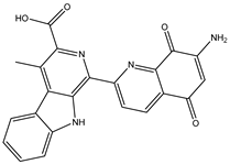
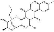
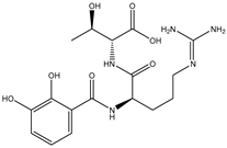
Streptomyces have been recognized as an important source of medicinal drugs [43]. Precisely, the Streptomyces species is known to produce over two-thirds of clinically used antibiotics and other pharmacologically important compounds [44]. Streptomycetes have demonstrated potential antiviral activity by targeting various viral enzymes and proteins, including virantmycin and alanosine [45]. Narasin, an ionophore isolated from Streptomyces aureofaciens, has been found to inhibit the replication of DENV [46]. It has been documented that the use of narasin as a treatment for DENV2-infected cells from 12 to 48 h after infection did not decrease the levels of both positive-strand and negative-strand DENV2 RNA, suggesting that narasin does not impede DENV2 RNA replication [46,47]. Unlike broader natural product libraries (e.g., ZINC Natural Products, NPASS, or SuperNatural II) that include a wide range of compounds from diverse sources, the Streptome database uniquely provides the most comprehensive collection of streptomyces-derived natural products [48]. This enhances the likelihood of identifying biologically active scaffolds with established antimicrobial or therapeutic potential. Seeking effective anti-DENV2 drug candidates, the Streptome database, containing > 6500 streptomycetes natural products (NPs), was mined to hunt novel allosteric inhibitors toward DENV2 RdRp with a higher potency than 68T. Upon the docking computations, the most potent streptomycetes NPs were picked up and advanced for molecular dynamics simulations (MDS) over 300 ns, accompanied by binding energies utilizing the MM/GBSA approach. Post-MD analyses were executed on the most promising streptomycetes NPs bound to the DENV2 RdRp. Additionally, the drug-like features of the identified streptomycetes NPs were predicted. Furthermore, quantum mechanical computations were employed to obtain a deeper understanding of the geometrical and electronic properties of the identified streptomycetes NPs. A schematic diagram of the applied computational approaches to filter the Streptome database is portrayed in Figure 1. The obtained results highlight the potential of the identified streptomycetes NPs as allosteric DENV2 RdRp inhibitors and offer viable curative candidates for future experimental assays.

2. Results and Discussion

2.1. Docking Protocol Assessment

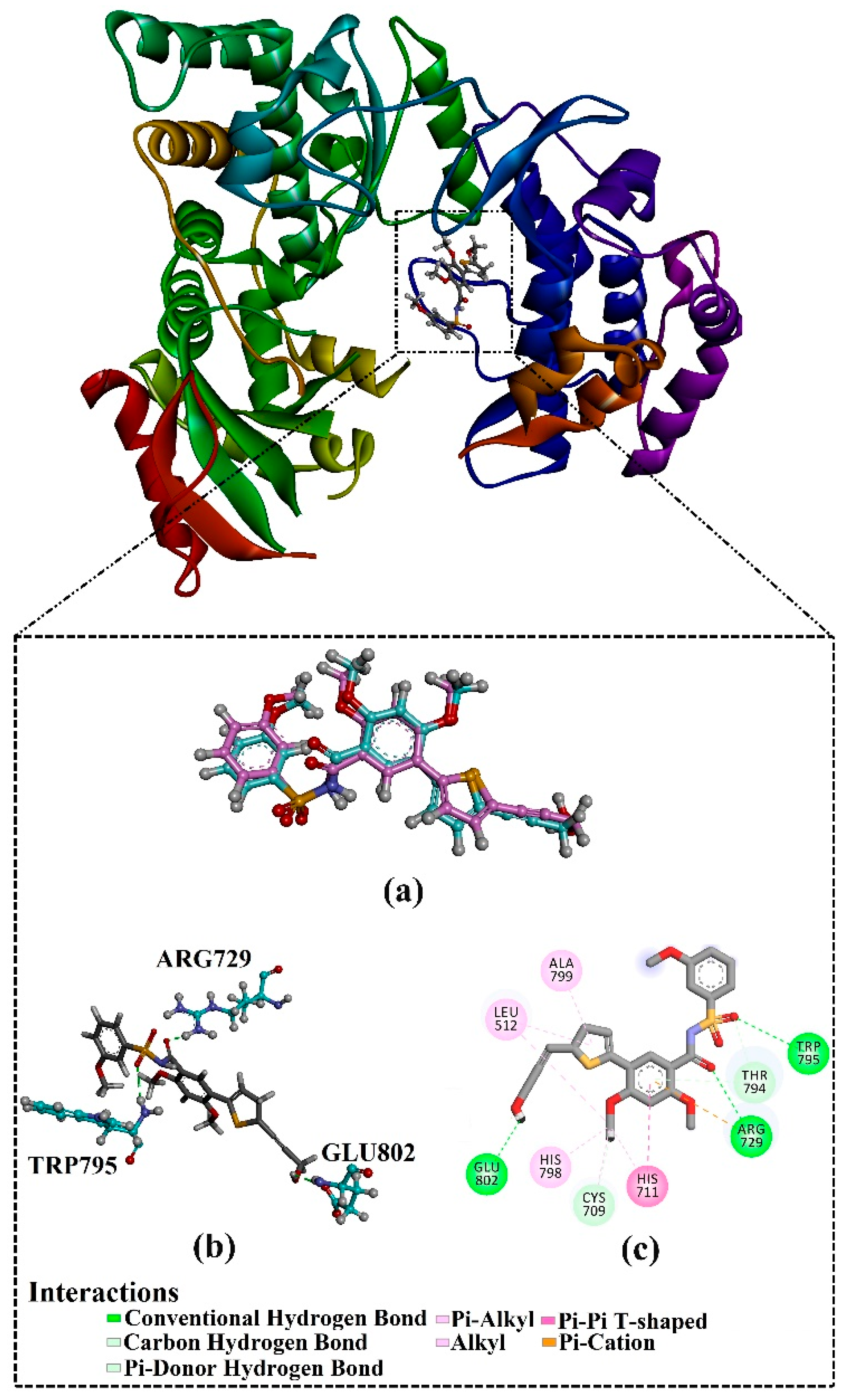
Prior to data generation, the AutoDock4.2.6 software’s performance in predicting the binding pose of the co-crystallized compound 27/68T inside the DENV2 RdRp allosteric site was validated based on the available experimental data. The anticipated binding pose of compound 27/68T was compared to its original binding pose (PDB entry: 5K5M [41]) (Figure 2). According to the literature, the RMSD values between the anticipated binding pose and the resolved original binding pose should be <2.0 Å [49,50,51]. From Figure 2, the predicted pose closely matched the experimental structure with an RMSD value of 0.79 Å. Examining the binding pose of 68T demonstrated three fundamental H-bonds with ARG729 (3.11 Å), TRP795 (3.40 Å), and GLU802 (2.67 Å). As well, the 68T exhibited a pi-pi T-shaped interaction with HIS711 and a pi-cation interaction with ARG729. Re-docking results showed that the AutoDock2.4.6 software correctly predicted the binding pose of ligand-RdRp complexes. As a result, the AutoDock4.2.6 software was used to mine the Streptome database for hunting putative DENV2 RdRp ligands.

2.2. Virtual Screening of the Streptome Database

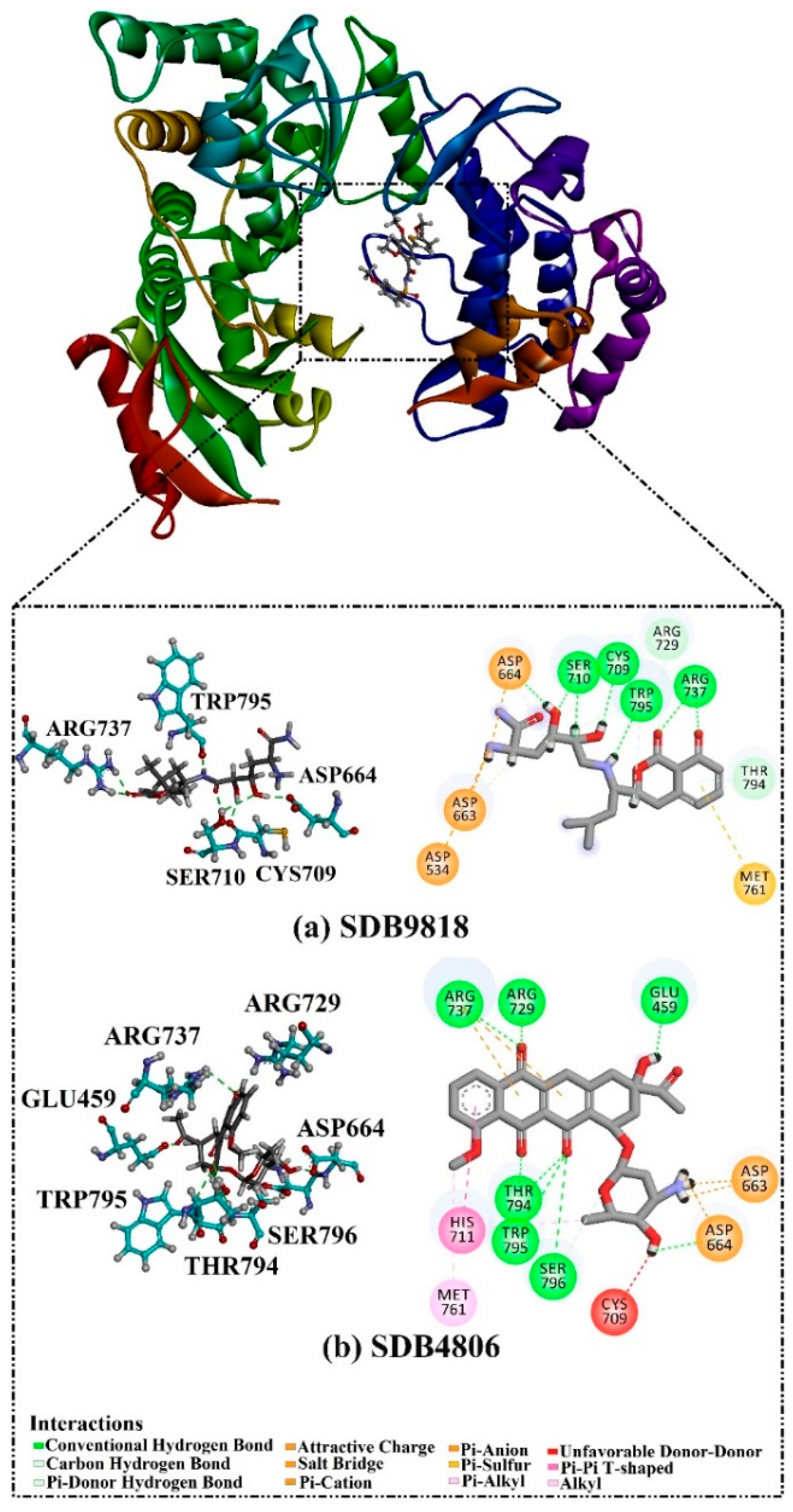
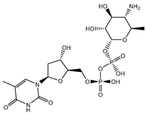
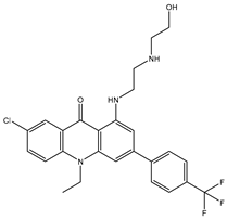
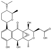
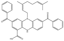
A virtual screening technique can be used to hunt prospective bioactive inhibitors at the early stages of the drug design process [52]. Herein, the Streptome database encompassing > 6500 NPs was mined using standard docking parameters of eval = 5 million and GA = 50. On the basis of standard docking computations, only 151 streptomycetes NPs unveiled docking scores equal to or lower than that of 68T (calc. −35.6 kJ.mol−1). As a result, these 151 streptomycetes NPs were re-docked toward the DENV2 RdRp utilizing expensive parameters (i.e., eval = 25 million and GA = 250) (Table S1). As reported in Table S1, only 39 streptomycetes NPs revealed lower docking scores in comparison with 68T (calc. −35.6 kJ.mol−1). Figure S1 depicts the 2D illustrations for the anticipated binding poses of these 39 streptomycetes NPs within the DENV2 RdRp allosteric site. As shown in Figure S1, all investigated streptomycetes NPs within the DENV2 RdRp allosteric site displayed a fundamental H-bond with ARG729 and TRP795. Pi-Sigma, pi-pi T-shaped, amide-pi stacked, carbon H-bond, and pi-cation interactions were also monitored between the inspected streptomycetes NPs and the main residues in the DENV2 RdRp allosteric site. 2D Chemical structures, computed standard and expensive docking scores, and the intramolecular H-bond of the top 10 scoring streptomycetes NPs toward DENV2 RdRp are registered in Table 1. As well, the 3D and 2D depictions for the predicted binding poses of two outstanding streptomycetes NPs within the DENV2 RdRp allosteric site are illustrated in Figure 3. Furthermore, the most potent two streptomycetes NPs were re-docked using GA = 1000 against the DENV2 RdRp allosteric site. Comparing results obtained with a GA value of 250 to those using a GA value of 1000, no notable differences were observed in the docking poses and scores of these two streptomycetes NPs within the DENV2 RdRp allosteric site (Figure S2). Of note, these two promising streptomycetes NPs were selected according to the evaluated binding affinity throughout 300 ns MDS, as evidenced in the MDS section.
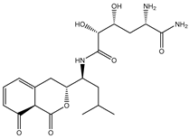
According to data listed in Table 1, SDB9818 manifested a significantly favorable docking score of −46.9 kJ.mol−1 against the DENV2 RdRp allosteric site. SDB9818 is a natural product isolated from Streptomyces sp. VITBRK2, which shows a high activity against MRSA [53]. Structural insights into the docking pose of SDB9818 against the DENV2 RdRp allosteric site showed that the CO of SDB9818 formed three H-bonds with the NH2 of ARG737 (2.04, 2.62, and 1.83 Å). Moreover, the two OH groups of (2R,3S)-butane-2,3-diol exhibited three H-bonds with the OH of SER796 (3.01 Å), CO of CYS709 (1.83 Å), and carboxylate of ASP664 (1.74 Å). Ultimately, the NH and CO groups of formamide showed two H-bonds with the CO of TRP795 (1.80 Å) and the OH of SER710 (1.70 Å) (Figure 3).
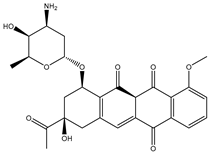
SDB4806 is an anthracycline antibiotic extracted from Streptomyces peucetius var. aureus, which demonstrates anticarcinoma activity by impeding RNA and DNA synthesis [54]. SDB4806 revealed the second-lowest docking score of −45.6 kJ.mol−1 against the DENV2 RdRp allosteric site. According to Figure 3, SDB4806 displayed eight H-bonds with the essential residues of the DENV2 RdRp allosteric site. More precisely, the OH groups of SDB4806 established two H-bonds with the carboxylate of GLU459 (1.63 Å) and ASP664 (1.84 Å). Moreover, the CO groups of SDB4806 exhibited six H-bonds with the NH2 of ARG729 (2.16 Å), NH2 of ARG737 (2.84 Å), OH of THR794 (1.97 and 2.44 Å), NH of TRP795 (2.54 Å), and NH of SER796 (2.83 Å).
According to sequence and structure comparisons, the DENV2 RdRp allosteric site is not homologous to any known domains in human DNA/RNA polymerases or bacterial polymerases, reducing off-target interactions [55]. Moreover, SDB9818 and SDB4806 often do not exhibit significant activity against eukaryotic polymerases unless they are specifically selected or designed to have this effect.

2.3. Molecular Dynamics Simulations (MDS)

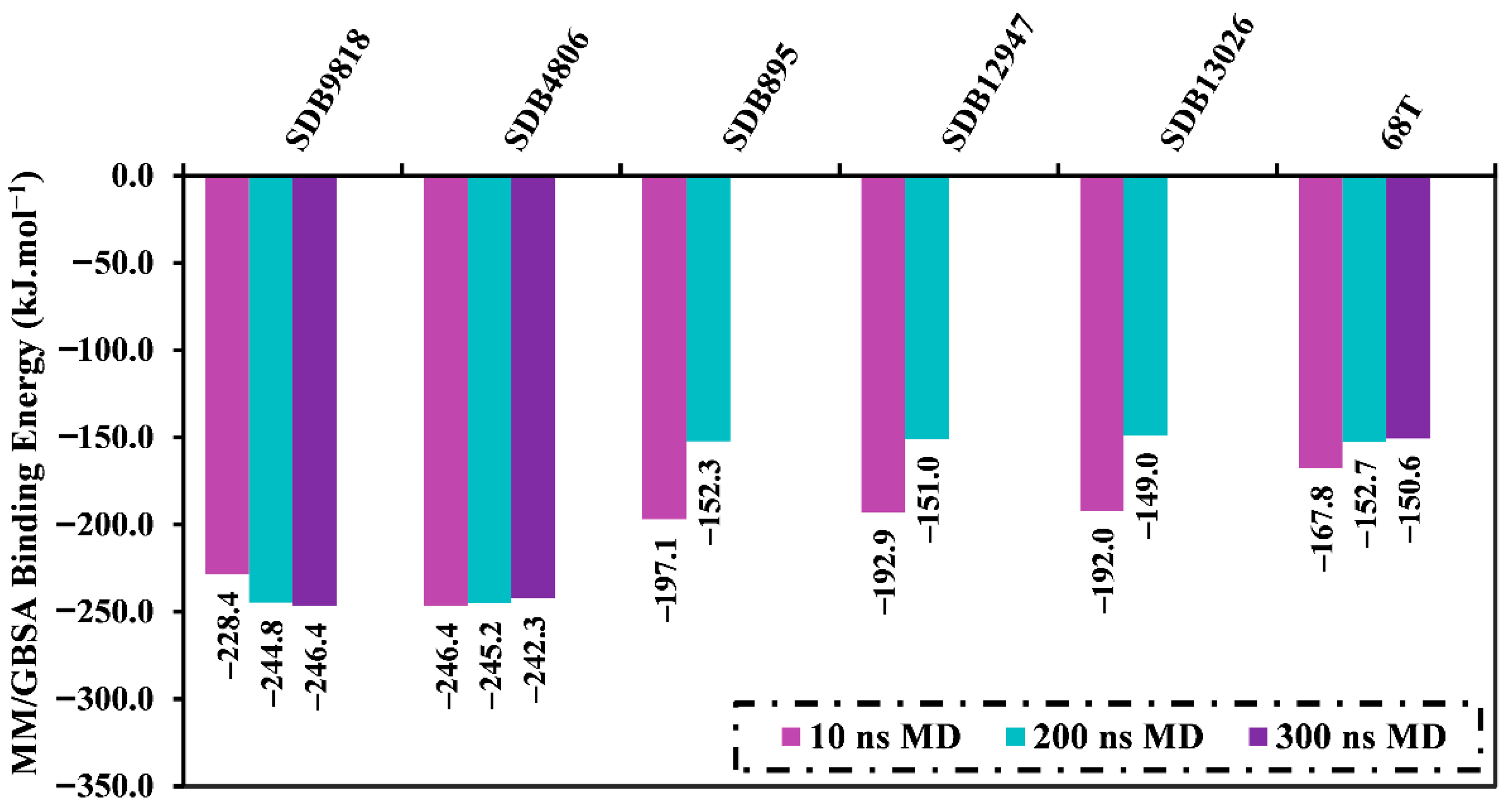
To examine receptor–ligand stability and conformational variations, MDS has been widely used to analyze the dynamic properties of the inhibitor complexed with receptors. The most promising 39 streptomycetes NPs with docking scores less than 68T (calc. −35.6 kJ.mol−1) in the complex with the DENV2 RdRp allosteric site were advanced for MDS over 10 ns. Table S2 lists the corresponding MM/GBSA binding energies over the 10 ns MDS. As found in Table S2, only five streptomycetes NPs unveiled higher binding affinities than 68TGbinding = −167.8 kJ.mol−1) toward the DENV2 RdRp allosteric site. In order to achieve more reliable outcomes, these five streptomycetes NPs in complex with the DENV2 RdRp allosteric site were introduced to longer MDS throughout 200 ns, accompanied by MM/GBSA binding energy estimations (Figure 4). As depicted in Figure 4, two streptomycetes NPs, namely SDB9818 and SDB4806, demonstrated lower binding energies compared to 68TGbinding = −152.7 kJ.mol−1). Moreover, a 300 ns MDS was executed for these two streptomycetes NPs bound to the DENV2 RdRp allosteric site, followed by binding affinity computations (Figure 4). From Figure 4, it is obvious that no significant variations were observed in the evaluated binding energies over 200 and 300 ns MDS for the identified two streptomycetes NPs complexed with the DENV2 RdRp allosteric site. In comparison with 68TGbinding = −150.6 kJ.mol−1), SDB9818 and SDB4806 unveiled superior binding affinities toward the DENV2 RdRp allosteric site throughout 300 ns MDS with ΔGbinding values of −246.4 and −242.3 kJ.mol−1, respectively. Moreover, the standard deviation and standard error of the mean for the binding energy (ΔGbinding) of the two identified streptomycetes NPs and 68T complexed with the DENV2 allosteric site over 300 ns MDS were computed (Table S3). As registered in Table S3, the low standard deviation and standard error of mean values indicated that the binding energy remained relatively stable throughout the simulation time. These outcomes illuminated the efficiency of the identified streptomycetes NPs as promising anti-DENV2 drug candidates.
The main limitation of the current research is the lack of MDS runs replication for the identified streptomycetes NPs as promising anti-DENV2 drug candidates. As a consequence, the future study will consider multiple independent MDS with a variety of initial velocities in order to verify the consistency of the findings.
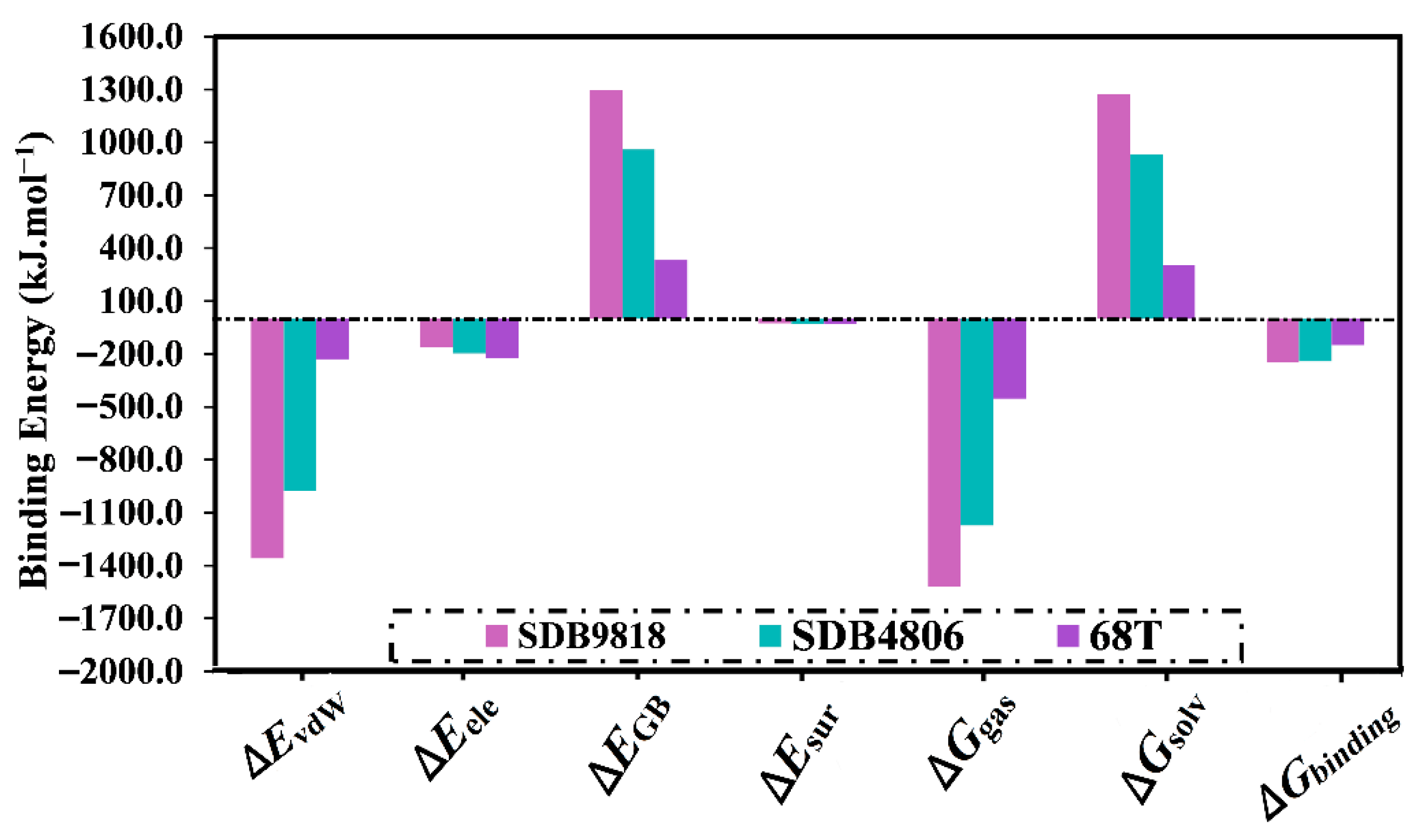
The decomposition of the computed binding energies into separate components was then executed and is depicted in Figure 5. As depicted in Figure 5, the EvdW was the predominant participation in the ΔGbinding of SDB9818, SDB4806, and 68T complexed with the DENV2 RdRp allosteric site, with mean values of −1358.5, −977.4, and −228.9 kJ.mol−1, respectively. Moreover, the Eele demonstrated a favorable contribution to ΔGbinding with the mean values of −160.7, −196.2, and −224.3 kJ.mol−1 for SDB4806, SDB9818, and 68T complexed with the DENV2 RdRp allosteric site, respectively.
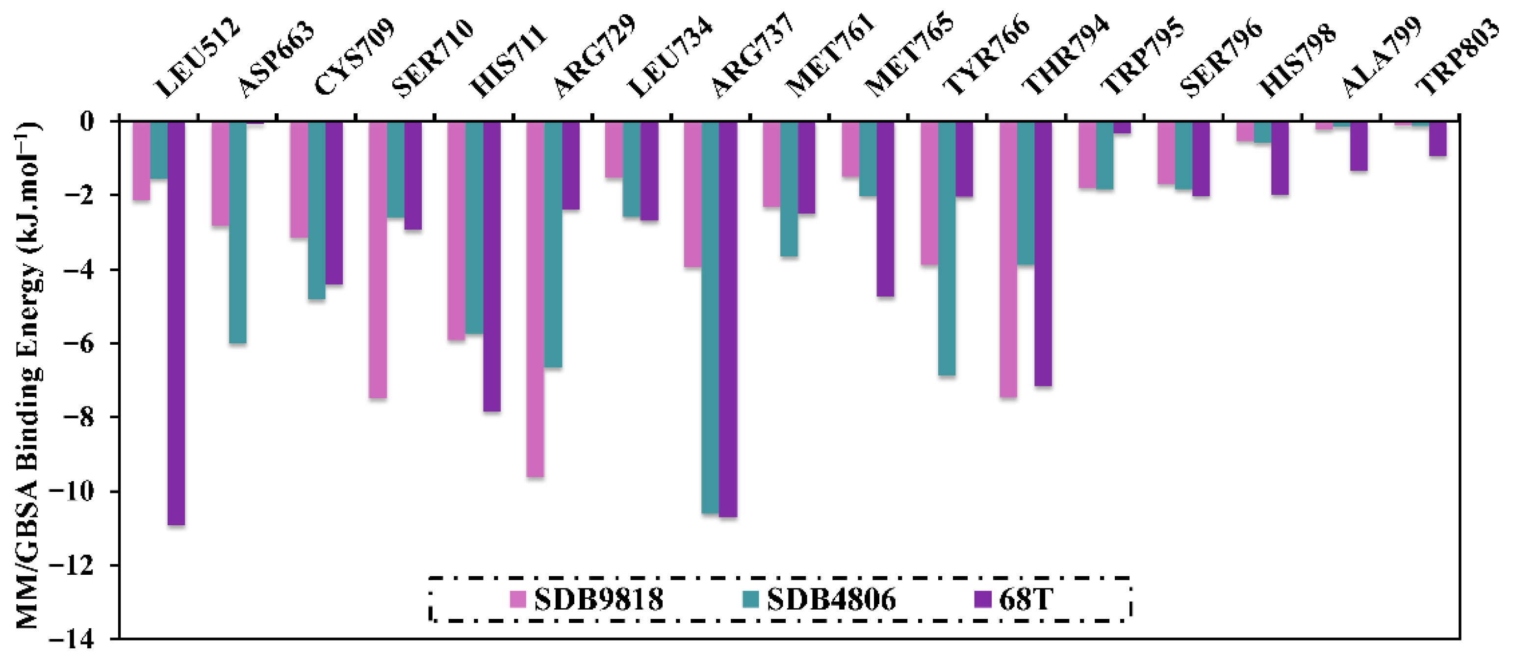
To obtain more insights into the fundamental amino acids included in the inhibition of the DENV2 RdRp allosteric site, the per-residue energy decomposition was performed and plotted in Figure 6. It is worth noting that only residues with ΔGbinding < −2.1 kJ.mol−1 were taken into account. From the delineated per-residue energy decomposition in Figure 6, ARG729, THR794, TRP795, SER796, and ARG737 disclosed promising participations in the binding of SDB9818, SDB4806, SDB895, and 68T with the allosteric site of the DENV2 RdRp. The significant participation of ARG729 in the allosteric site of DENV RdRp evinced −9.6, −6.7, −7.1, and −2.5 kJ.mol−1 for SDB9818-, SDB4806-, and 68T-RdRp complexes, respectively.

2.4. Post-MD Analyses

In order to further investigate the steadiness and behavior of SDB9818-, SDB4806-, and 68T-RdRp, post-MD analyses were accomplished over the course of 300 ns MDS. Six properties were evaluated, including root-mean-square fluctuation and deviation (RMSF and RMSD), radius of gyration (Rg), binding energy per trajectory, H-bond analysis, and solvent-accessible surface area (SASA).

2.4.1. Binding Energy per Trajectory

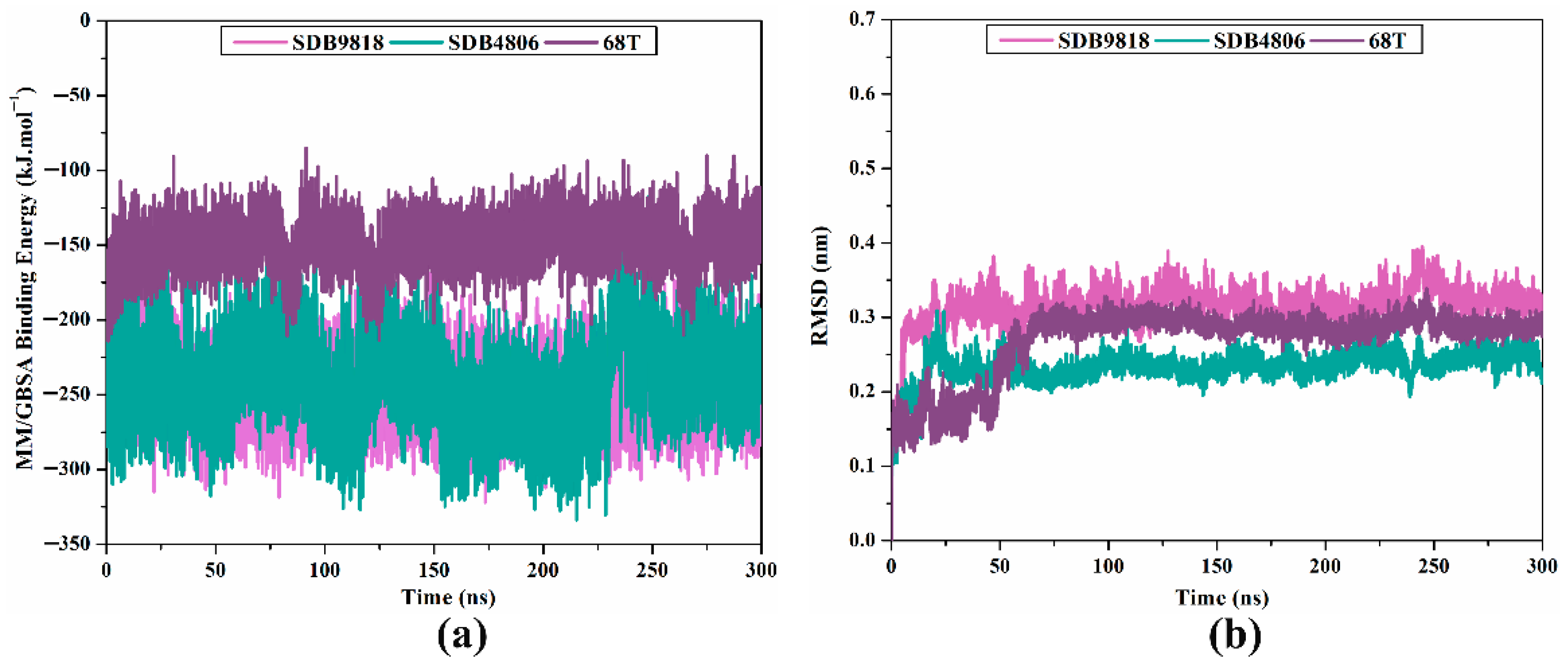
The energetic persistence of SDB9818, SDB4806, and 68T bound to the DENV2 RdRp allosteric site was evaluated by measuring the binding energy versus time correlation (Figure 7a). As illustrated in Figure 7a, the comprehensive constancy for SDB9818, SDB4806, and 68T bound to the DENV2 RdRp allosteric site was observed with average ΔGbinding values of −246.4, −242.3, and −150.6 kJ.mol−1, respectively. The most striking outcome to emerge from this graph is that all inspected complexes conserved steadiness throughout 300 ns MDS.

2.4.2. RMSD Analysis

In order to supply valuable insights into ligand–receptor structural steadiness, RMSD for the backbone atoms relative to the initial position was measured over 300 ns MDS (Figure 7b). Based on Figure 7b, the mean RMSD values were 0.32, 0.23, and 0.27 nm for SDB9818-, SDB4806-, and 68T-RdRp complexes, respectively. These results demonstrated that these inspected streptomycetes NPs were bound tightly and were overall stable within the RdRp allosteric site. Furthermore, multiple snapshots taken at consistent intervals throughout the MDS were extracted to further assess the stability of the identified streptomycetes NPs within the DENV2 RdRp allosteric site. The 3D binding patterns of the identified streptomycetes NPs inside the allosteric site of DENV2 RdRp are depicted in Figure S3. As illustrated in Figure S3, the identified streptomycetes NPs sustained stable interactions at the allosteric site of streptomycetes NPs, resulting in the formation of a stable RdRp-inhibitor complex.

2.4.3. Rg Analysis

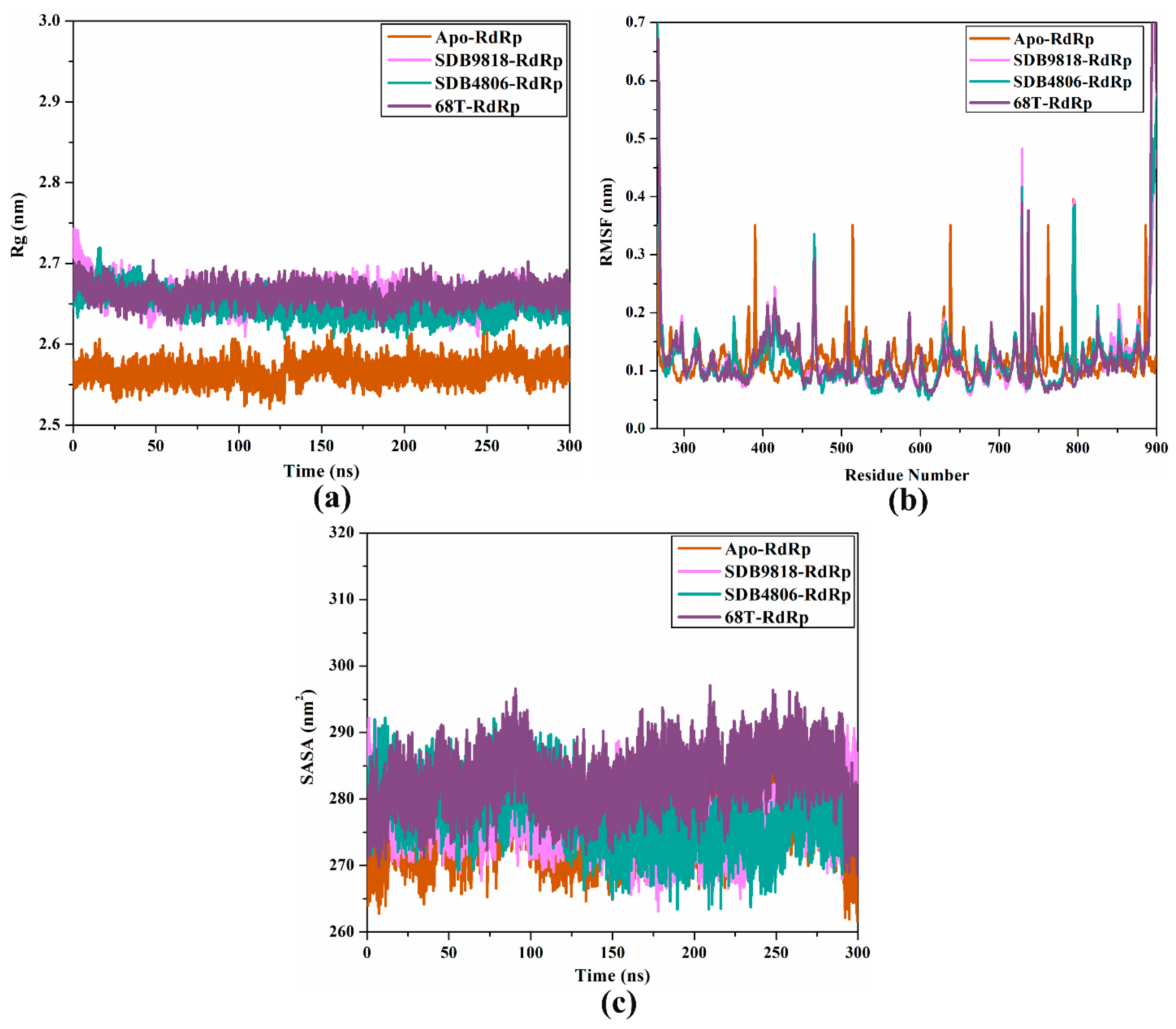
The Rg is one of the parameters utilized to gauge the equilibrium configurations of the whole system, and this assists in determining receptor compactness during MDS. Protein compactness is dependent on their residue sequence constitution, and it changes during receptor–ligand interactions [56]. As a result, the Rg for SDB4806, SDB9818, and 68T complexed with the DENV2 RdRp allosteric site was estimated throughout 300 ns MDS in order to investigate how residue flexibility affects enzyme compactness (Figure 8a). The measured average Rg values were 2.57, 2.66, 2.65, and 2.66 nm for apo-, SDB4806-, SDB9818-, and 68T-RdRp, respectively. These results implied that the RdRp structure was well-compacted and more steady after the complexation with SDB4806 and SDB9818.

2.4.4. RMSF Analysis

RMSF is a parameter utilized for determining the elasticity of every residue of the target during the MDS. RMSF explains variations in the structural configurations of the target from the starting position until the termination of the simulations according to the residue fluctuations [57]. Thus, the RMSF was evaluated over 300 ns MDS to grasp how structural deviations affect target elasticity (Figure 8b). It is known that greater RMSF demonstrates higher elasticity of the receptor during simulations. As illustrated in Figure 8b, it was noticed that the 68T-RdRp had a greater appearance of oscillating residues compared to the SDB4806 and SDB9818 complexed with the DENV2 RdRp allosteric site. The average RMSF values were 0.117, 0.118, 0.124, and 0.122 nm for apo-, SDB9818-, SDB4806-, and 68T-RdRp, respectively. Based on RMSF results, most of the residues of the DENV2 RdRp allosteric site exhibited higher stability after complexation with SDB4806, SDB9818, and 68T.

2.4.5. SASA Analysis

Water plays a substantial role in the determination of the stability, structure, role, and dynamics of targets. SASA is an indicator that is utilized for measuring the attainability of protein amino acids to encompass water molecules, and any alterations in the attainability could impact protein functions, dynamics, and structure [58]. Herein, SASA was employed to gauge the binding impact of the investigated inhibitor on the configurational behavior of the DENV2 RdRp allosteric site. Figure 8c illustrates the SASA of the DENV2 RdRp allosteric site upon binding with SDB4806, SDB9818, and 68T throughout 300 ns MDS. As can be seen from the SASA plots, SDB4806, SDB9818, and 68T demonstrated similar frame patterns after binding to the RdRp allosteric site. Nevertheless, the SASA of RdRp increased upon binding with the 68T (Figure 8c). In contrast, as SDB4806 and SDB9818 were bound to the RdRp allosteric site, the SASA value slightly decreased. The average SASA values were 275.9, 277.0, 277.8, and 282.9 nm2 for apo-, SDB4806-, SDB9818-, and 68T-RdRp, respectively. SASA outcomes indicated that the streptomycetes NPs and 68T had little impact on the solvent accessibility of the DENV2 RdRp enzyme.

2.4.6. H-Bond Analysis

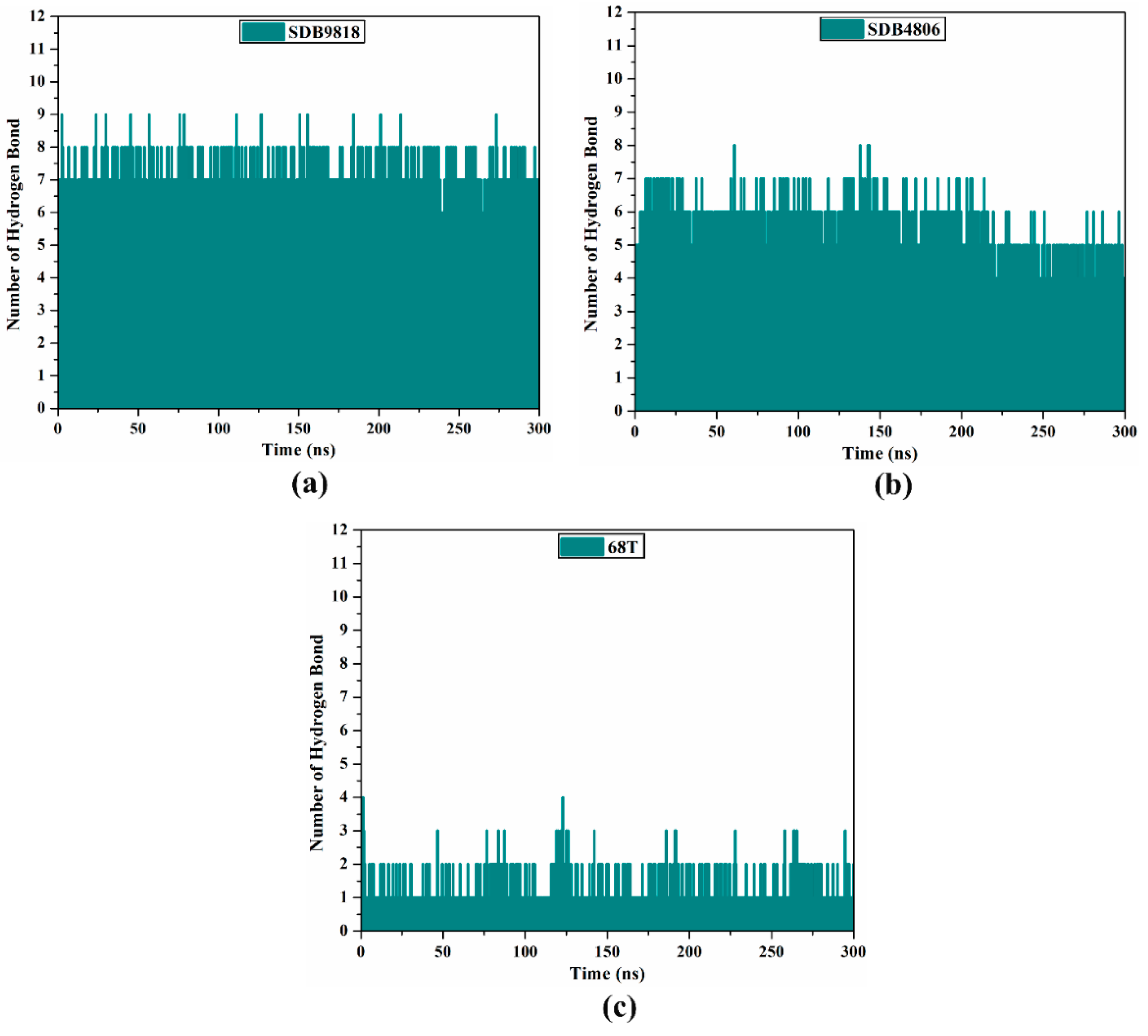
The steadiness and binding affinity of inhibitors with the receptor were investigated by gauging the number of conventional H-bonds established during the MDS. Figure 9 portrays the number of H-bonds between the investigated streptomycetes NPs and the key residues of the DENV2 RdRp allosteric site throughout 300 ns MDS. As shown in Figure 9, the average number of H-bonds was 7, 6, and 2 for SDB9818-, SDB4806-, and 68T-RdRp complexes, respectively. Notably, the average number of H-bonds for the identified streptomycetes NPs was more remarkable compared to the co-crystallized 68T complexed with the DENV2 RdRp, indicating the higher stability of these streptomycetes NPs than 68T within the DENV2 RdRp allosteric site over 300 ns MDS. These results firmly endorsed the anticipated H-bond interactions between the investigated streptomycetes NPs and the allosteric site amino acids of RdRp via molecular docking computations.

2.5. Physicochemical Characteristics

The drug-likeness of a molecule is estimated in accordance with Lipinski’s rule of five (Ro5), which detects whether the compound is orally active or not. Table 2 lists the drug-likeness features of the identified streptomycetes NPs and 68T. According to Ro5, a molecule failing to meet more than two of its criteria is likely to have poor absorption in preclinical testing [59]. According to data listed in Table 2, SDB9818 violates only the HBD limit (6 vs. 5), which may marginally impact permeability but not necessarily disqualify it as a lead, especially given its favorable solubility (LogP = 0.40). SDB4806 slightly exceeds the MW threshold (511.52 g/mol) but remains within acceptable bounds for lead optimization. The LogP and H-bond of SDB4806 counts were favorable. 68T complied fully with Ro5, supporting its drug-like profile. More precisely, the MW was in the range of 423.46 to 511.52 g/mol for the investigated streptomycetes NPs and 68T. Moreover, the HBD was 6, 4, and 2 for SDB9818, SDB4806, and 68T, respectively. SDB4806, SDB9818, and 68T were found to have a number of HBA with values ranging from 7 to 10. The logP was less than 5 for the investigated streptomycetes NPs and 68T. Notably, these results underscore the promise of the identified streptomycetes NPs as potential allosteric inhibitors of DENV2 RdRp and present them as viable candidates for future experimental validation.
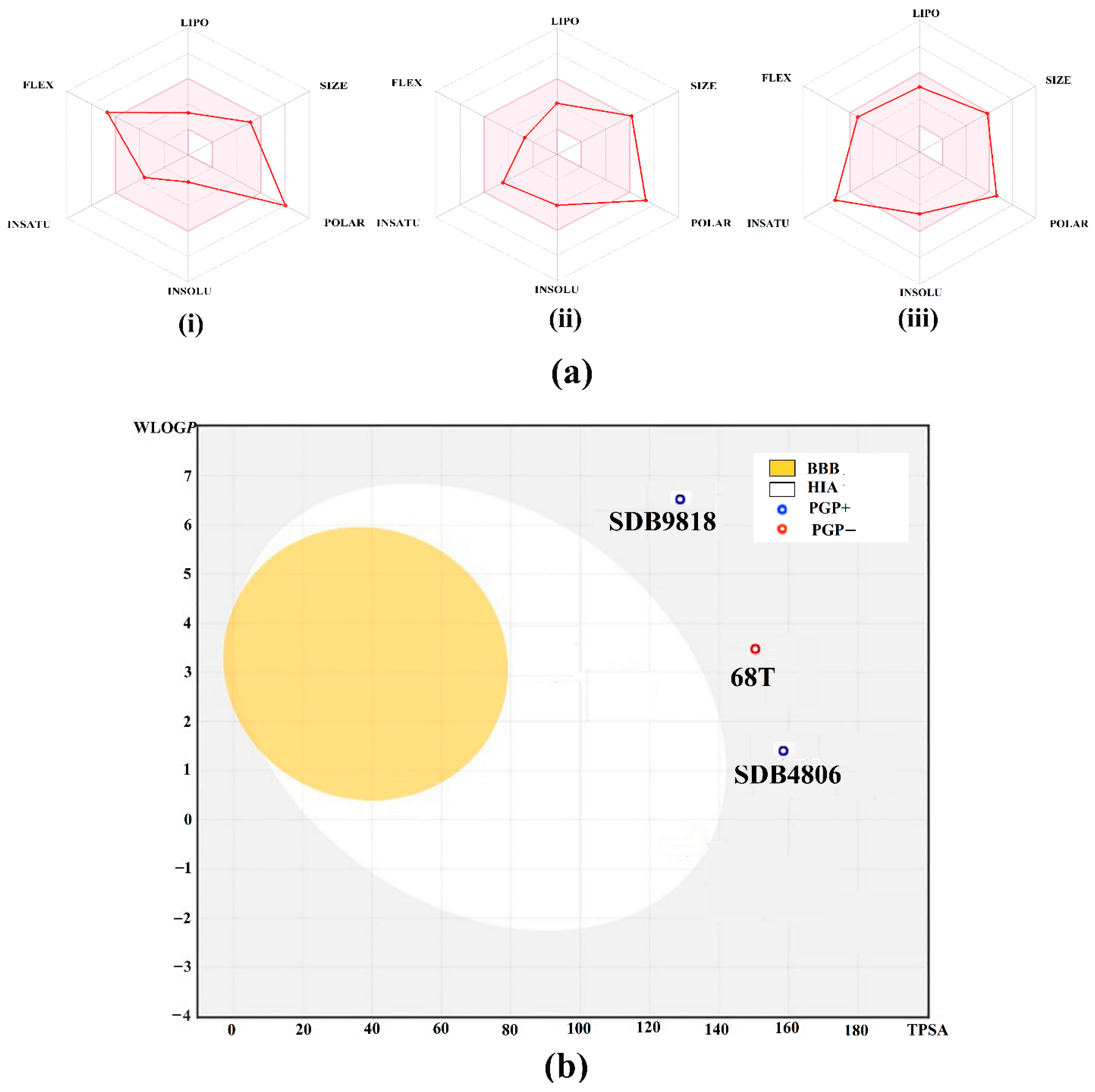
Figure 10a displays the bioavailability radar plots of the identified streptomycetes NPs and 68T and their drug-likeness characteristics. The pink area inside the hexagonal shape represents the ideal range for the investigated compounds. The recommended parameters for the drug-likeness of a small molecule are as follows: (i) insaturation (INSITU): a fraction of carbons exhibiting sp3 hybridization of at least 0.25; (ii) insolubility (INSOLE): a log S not exceeding 6; (iii) hydrophobicity (LIPO): ranging from −0.7 to +5.0; (iv) rotatable bonds (FLEXI): a maximum of 9 rotatable bonds; (v) molecular weight (SIZE): between 150 and 500 g/mol; and (vi) polar surface area (POLAR): between 20 and 130 g/mol, along with polar surface area (POLAR): within the range of 20 and 130 Å2. According to data presented in Figure 10a, the investigated streptomycetes NPs and 68T lie within the passable range, indicating their favorable drug-like characteristics.
In addition, the pharmacokinetic features of the identified streptomycetes NPs and 68T were investigated using a BOILED-Egg model (Figure 10b). The BOILED-Egg model demonstrates its utility in simultaneously forecasting two significant pharmacokinetic properties, specifically, passive gastrointestinal absorption (HIA) and penetration through the blood–brain barrier (BBB). As shown in Figure 10b, SDB9818, SDB4806, and 68T were detected in the grey region, indicating that these compounds demonstrated low passive HIA and BBB. Consequently, the investigated compounds would gain better bioavailability profiles during a drug development process.

2.6. QM Computations

ESP analysis is a reliable approach to demonstrate the negative and positive potentials on the surface of a chemical molecule. For the optimized last snapshot of streptomycetes NPs and 68T retrieved from MDS, the MEP maps were generated and are graphed in Figure 11. As delineated in Figure 11, red regions were noticed above the N and O atoms of the investigated streptomycetes NPs and 68T, indicating their nucleophilic nature. Additionally, blue regions were marked above the H atoms of the inspected streptomycetes NPs and 68T, implying their electrophilic nature. According to the MEP maps, streptomycetes NPs and 68T displayed the capability of forming H-bonds with fundamental residues within the allosteric site of DENV2 RdRp.
Based on Figure 12, HOMO levels were mainly found around the electron-rich zones of the investigated compounds (e.g., O and N atoms). In addition, LUMO levels were concentrated around the electron-deficient regions in the investigated streptomycetes NPs and 68T (e.g., H atoms). As numerical evidence from Table 3, the EHOMO/ELUMO values were −7.99/−1.34, −8.06/−2.33, and −6.88/−0.83 eV for SDB9818, SDB4806, and 68T, respectively. Moreover, SDB9818, SDB4806, and 68T revealed EFL values in the range of −3.85 to −5.19 eV. SDB9818, SDB4806, and 68T displayed low Egap with values of 6.65, 5.74, and 6.05 eV, respectively. Of note, the low Egap values indicated the considerable chemical reactivity of the investigated streptomycetes NPs and 68T.
According to the indisputable role of electronic parameters, global descriptors were computed for SDB9818, SDB4806, and 68T (Table 4). As reported in Table 4, SDB9818, SDB4806, and 68T demonstrated IP values ranging from 6.88 to 8.71 eV. In addition, the EA values were found to be 1.34, 2.33, and 0.83 eV for SDB9818, SDB4806, and 68T, respectively. The η and S of the investigated compounds can serve as an indicator for their stability and chemical reactivity. The η values for SDB9818, SDB4806, and 68T ranged from 2.87 to 3.32 eV. SDB9818, SDB4806, and 68T unveiled outstanding S values of 0.30, 0.35, and 0.33 eV−1, respectively. Among the three compounds, SDB4806 exhibited the highest softness (0.35 eV−1), suggesting a greater capacity for electronic adaptability. Generally, the obtained η and S values of SDB9818, SDB4806, and 68T indicated promising polarizability and electron-donating/accepting flexibility, which facilitated effective interactions with DENV RdRp allosteric site through non-covalent interactions (e.g., H-bonds, electrostatic interactions, and π–π stacking). Moreover, the computed global descriptors provided an electronic rationale for the biological activity of the investigated compounds and reinforced their potential as effective inhibitors for the DENV2 RdRp allosteric site.

3. Computational Methodology

3.1. RdRp Preparation

The X-ray crystallographic structure of DENV2 RdRp (PDB entry: 5K5M, resolution: 2.1 Å) was chosen and employed as a template for all in silico computations [41]. All heteroatoms, including ions, inhibitors, and water molecules, were extracted for preparation purposes. The ionization status of titratable residues at a pH of 7.4 was determined utilizing the PropKa3 software [60]. All missing H-atoms were consequently inserted.

3.2. Streptome Database Preparation

The Streptome database, involving > 6500 NPs derived from approximately 3300 streptomycetes strains, was retrieved in SDF format [61]. Duplicates were removed in accordance with the International Chemical Identifier (InChIKey) [62]. Omega2 software (version 4.1.1.0) was utilized for generating 3D structures for each streptomycete NP [63,64]. All generated 3D structures were then adopted for an optimization process utilizing the MMFF94S force field implemented in SZYBKI software (version 2.4.0.0) [65,66]. The FixPka tool within the QUACPAC program (version 2.1.3.0) was used to determine the dominant ionization state at a pH of 7.4 [67]. The atomic charges of the streptomycetes NPs were computed with the assistance of the Gasteiger-Marsili method [68]. At www.compchem.net/ccdb, accessed on 13 February 2024, all prepared streptomycetes NPs can be accessed.

3.3. Docking Computation

AutoDock4.2.6 software was applied to accomplish molecular docking computations [69]. For docking calculations, the DENV2 RdRp enzyme was saved in the pdbqt format with the help of MGLTools 1.5.7 [70]. This research included two stages of docking calculations, namely standard and expensive docking computations. For standard and expensive docking computations, the genetic algorithm (GA) runs were set to 50 and 250 iterations, respectively. Moreover, 5 and 25 million energy evaluations (eval) were adjusted to standard and expensive docking computations, respectively. Other docking parameters were left at their defaults. The grid was designed to include the entire allosteric site of the DENV2 RdRP, measuring 40 × 40 × 40 Å3, with a spacing value of 0.375 Å. Grid coordinates were located at x = −15.551, y = −44.03, and z = −19.247. The docking conformation with the highest binding affinity was selected from the largest cluster as a representative docking mode.

3.4. MD Simulations (MDS)

All MDS for the most promising streptomycetes NPs complexed with DENV2 RdRp allosteric site were accomplished utilizing AMBER20 software [71]. Parameters for conducting MDS are more fully described elsewhere [72,73,74]. Briefly, DENV2 RdRp was characterized by the AMBER force field 14SB [75]. The general AMBER Force Field (GAFF2) was utilized for the parameterization of the inspected streptomycetes NPs [76]. Using Gaussian09 software, the geometrical optimization for the inspected streptomycetes NPs was executed at the HF/6-31G* level [77]. The restrained electrostatic potential (RESP) was then used for computing the atomic charges for the optimized streptomycetes NPs [78]. In a truncated octahedral periodic box, each RdRp-NP complex was solvated with the TIP3P water model [79]. In order to preserve the electroneutrality of the inspected RdRp-NP complexes, sodium or chloride counterions were added. The isosmotic salt environment was also maintained by the insertion of 0.15 M NaCl. The inspected complexes were advanced for 5000 iterations of energy minimization. Following that, the investigated complexes were gently annealed up to 310 K throughout 50 ps. The heated complexes were submitted to the equilibration stage for 10 ns. Finally, the production runs were carried out on the equilibrated complexes throughout 10, 200, and 300 ns. MD trajectories were recorded every 10 ps for post-MD analyses, which were performed using the CPPTRAJ tool [80]. MDS was accelerated using the PMEMD version of CUDA on GPU cores. All molecular interactions were displayed utilizing the Discovery Studio module of Biovia software (version 2019) [81].

3.5. Binding Energy Computations

To evaluate the binding energy between DENV2 RdRp and streptomycetes NPs, the molecular mechanics/generalized Born surface area (MM/GBSA) approach was used [82]. The binding energy was estimated by the following equation:
Δ G b i n d i n g = G R d R p N P   G R d R p   G N P
where the energy term (G) was numerically assessed as follows:
G = E v d W + G S A + G G B   + E e l e T S
EvdW points out van der Waals energy. GSA and GGB imply non-polar and polar contributions of the desolvation energy, respectively. Using the modified GB model (iGB = 2) developed by Onufriev et al., the GGB was computed [83]. Eele stands for electrostatic energy. T indicates absolute temperature, and S is the entropy participation. Entropic participation was not considered due to its high computation costs [84,85]. Notably, the exclusion of the entropic component had no significant effect on the MM/GBSA binding energy estimations [86].

3.6. Physicochemical Features

The SwissADME web server was employed to estimate the physicochemical characteristics of the identified streptomycetes NPs based on Lipinski’s rule of five (Ro5) conditions [87]. Lipinski’s rule states that (i) the number of H-bond acceptors (HBA) of the investigated compound should be <10, (ii) the molecular weight (MW) should not be >500 daltons, (iii) the number of H-bond donors (HBD) should be <5, and (iv) the partition coefficient (logP) should not be >5. In addition, the BOILED-Egg method was utilized for predicting gastrointestinal absorption and brain permeation of the identified streptomycetes NPs [88]. In the shape of a boiled egg, the plot is composed of white, grey, and yolk regions. More exactly, compounds in white zones are those that are more likely to be absorbed by the gastrointestinal tract, while compounds in yolk zones are those that are more likely to permeate the brain. Meanwhile, compounds in grey zones are those that are less likely to be non-absorbed by the gastrointestinal tract or non-brain permeating. For dot coloring, the blue dots indicate that the compound could be effluxed from the Central Nervous System (CNS) by P-Glycoprotein (PGP), whereas the red dots mean that compounds could not be effluxed from CNS by PGP [88].

3.7. Quantum Mechanical (QM) Computations

In the realm of QM computations, the final frame of the identified streptomycetes NPs elicited from MDS was geometrically optimized utilizing the M062X/6-311+G** level of theory with the assistance of Gaussian09 software [77]. For the optimized streptomycetes NPs, electrostatic potential (ESP) analysis was performed, and the corresponding molecular electrostatic potential (MEP) maps were generated at an electron density envelope of 0.002 au [89]. The frontier molecular orbitals (FMOs) theory was implemented for a deeper understanding of the electronic features of the optimized streptomycetes NPs. As a result, the energetic values and electronic patterns of the highest occupied/lowest unoccupied molecular orbital (i.e., HOMO/LUMO) were generated. Furthermore, the Fermi level (EFL) energy and energy gap (Egap) were computed on the basis of ELUMO and EHOMO, as shown in Equations (3) and (4), respectively.
E F L = E H O M O + E L U M O E H O M O   2
E g a p = E L U M O E H O M O
As well, further electronic parameters such as global hardness (η), global softness (S), ionization potential (IP), and electron affinity (EA) were computed as follows:
η = E L U M O E H O M O   2
I P = E H O M O
S = 1 η
E A = E L U M O

4. Conclusions

Recently, the dengue virus, a commonly encountered flavivirus, has shown a range of epidemiological, economic, and health effects globally. DENV2 is a significant factor in mortality associated with dengue. DENV2 RdRp is an attractive druggable target owing to its essential role in the replication of the virus. In the present work, the Streptome database, containing > 6500 compounds, was mined to hunt the most prospective inhibitors against the allosteric site of RdRp, utilizing advanced in silico approaches. Based on docking computations and MDS coupled with the MM/GBSA binding energy evaluations, SDB4806 and SDB9818 revealed promising binding affinities with ΔGbinding < −188.3 kJ.mol−1 against RdRp. The constancy of the identified streptomycetes NPs bound to the DENV2 RdRp allosteric site was confirmed upon the post-MD analyses throughout 300 ns. Based on the drug-likeness features, the identified streptomycetes NPs unveiled good oral bioavailability. Quantum mechanical computations were also performed for the identified streptomycetes NPs, and the outcomes demonstrated their stability and chemical reactivity. These findings provide a solid basis for future experimental validations and in-vitro studies aimed at combating DENV2.

Supplementary Materials

The following supporting information can be downloaded at https://www.mdpi.com/article/10.3390/ph18081135/s1: Figure S1: 2D molecular interactions of the anticipated binding modes for the top 39 streptomycetes NPs against the allosteric site of DENV2 RdRp; Figure S2: Overlapping between the predicted docking poses using a GA of 250 (pink) and 1000 (cyan) of (a) SDB9818 and (b) SDB4806 inside the DENV2 RdRp allosteric site. The computed docking score is displayed in kJ.mol−1; Figure S3: 3D binding patterns of (a) SDB9818 and (b) SDB4806 inside the DENV2 RdRp allosteric site at time intervals of 50, 100, 150, 200, 250, and 300 ns MDS; Table S1: The anticipated standard and expensive docking scores (in kJ.mol−1) for the top 151 streptomycetes NPs and 68T toward DENV2RdRp allosteric site; Table S2: Estimated standard and expensive docking scores and MM/GBSA binding energies (in kJ.mol−1) over 10 ns MD simulations of the promising 39 streptomycetes NPs toward DENV2 RdRp allosteric site; Table S3: Estimated MM/GBSA binding energies, standard deviation, and standard error of the mean (in kJ.mol−1) over 300 ns MDS of the most promising two streptomycetes NPs and 68T toward the DENV2 RdRp allosteric site.

Author Contributions

Conceptualization, M.A.A.I.; Methodology, T.A. and M.A.A.I.; Software, M.A.A.I.; Formal analysis, A.H.M.A.; Investigation, A.H.M.A. and P.A.S.; Resources, T.A. and M.A.A.I.; Data curation, A.H.M.A.; Writing—original draft preparation, A.H.M.A.; Visualization, A.H.M.A. and P.A.S.; Supervision, G.A.H.M. and M.A.A.I.; Project administration, M.A.A.I. and S.K.; Writing—review and editing, G.A.H.M., P.A.S., T.A., S.K. and M.A.A.I.; All authors have read and agreed to the published version of the manuscript.

Funding

This research received no external funding.

Institutional Review Board Statement

Not applicable.

Informed Consent Statement

Not applicable.

Data Availability Statement

The data presented in this study are available in the Supplementary Material.

Acknowledgments

The authors extend their appreciation to the Ongoing Research Funding Program (ORF-2025-1438), King Saud University, Riyadh, Saudi Arabia, for funding this work. A.H.M.A. and M.A.A.I. would like to thank Shaban R. M. Sayed for his discussion of the research and manuscript. The computational work was completed with resources provided by the Center for High-Performance Computing (Cape Town, South Africa, http://www.chpc.ac.za) and Bibliotheca Alexandrina (http://hpc.bibalex.org).

Conflicts of Interest

The authors declare no conflict of interest.

References

  1. Postler, T.S.; Beer, M.; Blitvich, B.J.; Bukh, J.; de Lamballerie, X.; Drexler, J.F.; Imrie, A.; Kapoor, A.; Karganova, G.G.; Lemey, P.; et al. Renaming of the genus Flavivirus to Orthoflavivirus and extension of binomial species names within the family Flaviviridae. Arch. Virol. 2023, 168, 224. [Google Scholar] [CrossRef]
  2. Guzman, M.G.; Halstead, S.B.; Artsob, H.; Buchy, P.; Farrar, J.; Gubler, D.J.; Hunsperger, E.; Kroeger, A.; Margolis, H.S.; Martinez, E.; et al. Dengue: A continuing global threat. Nat. Rev. Microbiol. 2010, 8, S7–S16. [Google Scholar] [CrossRef]
  3. Guzman, M.G.; Vazquez, S. The complexity of antibody-dependent enhancement of dengue virus infection. Viruses 2010, 2, 2649–2662. [Google Scholar] [CrossRef]
  4. Ferreira-de-Lima, V.H.; Lima-Camara, T.N. Natural vertical transmission of dengue virus in Aedes aegypti and Aedes albopictus: A systematic review. Parasites Vectors 2018, 11, 77. [Google Scholar] [CrossRef]
  5. Bhatt, S.; Gething, P.W.; Brady, O.J.; Messina, J.P.; Farlow, A.W.; Moyes, C.L.; Drake, J.M.; Brownstein, J.S.; Hoen, A.G.; Sankoh, O.; et al. The global distribution and burden of dengue. Nature 2013, 496, 504–507. [Google Scholar] [CrossRef]
  6. Beatty, M.E.; Stone, A.; Fitzsimons, D.W.; Hanna, J.N.; Lam, S.K.; Vong, S.; Guzman, M.G.; Mendez-Galvan, J.F.; Halstead, S.B.; Letson, G.W.; et al. Best practices in dengue surveillance: A report from the Asia-Pacific and Americas Dengue Prevention Boards. PLoS Negl. Trop. Dis. 2010, 4, e890. [Google Scholar] [CrossRef] [PubMed]
  7. Balasubramanian, A.; Pilankatta, R.; Teramoto, T.; Sajith, A.M.; Nwulia, E.; Kulkarni, A.; Padmanabhan, R. Inhibition of dengue virus by curcuminoids. Antivir. Res. 2019, 162, 71–78. [Google Scholar] [CrossRef]
  8. Normile, D. Tropical medicine. Surprising new dengue virus throws a spanner in disease control efforts. Science 2013, 342, 415. [Google Scholar] [CrossRef] [PubMed]
  9. Khetarpal, N.; Khanna, I. Dengue Fever: Causes, Complications, and Vaccine Strategies. J. Immunol. Res. 2016, 2016, 6803098. [Google Scholar] [CrossRef] [PubMed]
  10. Vaughn, D.W.; Green, S.; Kalayanarooj, S.; Innis, B.L.; Nimmannitya, S.; Suntayakorn, S.; Endy, T.P.; Raengsakulrach, B.; Rothman, A.L.; Ennis, F.A.; et al. Dengue viremia titer, antibody response pattern, and virus serotype correlate with disease severity. J. Infect. Dis. 2000, 181, 2–9. [Google Scholar] [CrossRef]
  11. Murugesan, A.; Manoharan, M. Dengue Virus. In Emerging and Reemerging Viral Pathogens; Ennaji, M.M., Ed.; Academic Press: London, UK, 2020; pp. 281–359. [Google Scholar]
  12. Wei, Y.; Qin, C.; Jiang, T.; Li, X.; Zhao, H.; Liu, Z.; Deng, Y.; Liu, R.; Chen, S.; Yu, M.; et al. Translational regulation by the 3’ untranslated region of the dengue type 2 virus genome. Am. J. Trop. Med. Hyg. 2009, 81, 817–824. [Google Scholar] [CrossRef]
  13. Carocci, M.; Yang, P.L. Lactimidomycin is a broad-spectrum inhibitor of dengue and other RNA viruses. Antivir. Res. 2016, 128, 57–62. [Google Scholar] [CrossRef] [PubMed]
  14. Guo, C.; Zhou, Z.; Wen, Z.; Liu, Y.; Zeng, C.; Xiao, D.; Ou, M.; Han, Y.; Huang, S.; Liu, D.; et al. Global Epidemiology of Dengue Outbreaks in 1990–2015: A Systematic Review and Meta-Analysis. Front. Cell Infect. Microbiol. 2017, 7, 317. [Google Scholar] [CrossRef] [PubMed]
  15. Soe, A.M.; Ngwe Tun, M.M.; Nabeshima, T.; Myat, T.W.; Htun, M.M.; Lin, H.; Hom, N.S.; Inoue, S.; Nwe, K.M.; Aye, L.P.P.; et al. Emergence of a Novel Dengue Virus 3 (DENV-3) Genotype-I Coincident with Increased DENV-3 Cases in Yangon, Myanmar between 2017 and 2019. Viruses 2021, 13, 1152. [Google Scholar] [CrossRef] [PubMed]
  16. Shrivastava, G.; Visoso-Carvajal, G.; Garcia-Cordero, J.; Leon-Juarez, M.; Chavez-Munguia, B.; Lopez, T.; Nava, P.; Villegas-Sepulveda, N.; Cedillo-Barron, L. Dengue Virus Serotype 2 and Its Non-Structural Proteins 2A and 2B Activate NLRP3 Inflammasome. Front. Immunol. 2020, 11, 352. [Google Scholar] [CrossRef]
  17. da Fonseca, N.J., Jr.; Lima Afonso, M.Q.; Pedersolli, N.G.; de Oliveira, L.C.; Andrade, D.S.; Bleicher, L. Sequence, structure and function relationships in flaviviruses as assessed by evolutive aspects of its conserved non-structural protein domains. Biochem. Biophys. Res. Commun. 2017, 492, 565–571. [Google Scholar] [CrossRef]
  18. Verhaegen, M.; Vermeire, K. The endoplasmic reticulum (ER): A crucial cellular hub in flavivirus infection and potential target site for antiviral interventions. npj Viruses 2024, 2, 24. [Google Scholar] [CrossRef]
  19. Dey, D.; Poudyal, S.; Rehman, A.; Hasan, S.S. Structural and biochemical insights into flavivirus proteins. Virus Res. 2021, 296, 198343. [Google Scholar] [CrossRef]
  20. Cortese, M.; Mulder, K.; Chatel-Chaix, L.; Scaturro, P.; Cerikan, B.; Plaszczyca, A.; Haselmann, U.; Bartenschlager, M.; Neufeldt, C.J.; Bartenschlager, R. Determinants in Nonstructural Protein 4A of Dengue Virus Required for RNA Replication and Replication Organelle Biogenesis. J. Virol. 2021, 95, e0131021. [Google Scholar] [CrossRef]
  21. Li, Q.; Kang, C. Structures and Dynamics of Dengue Virus Nonstructural Membrane Proteins. Membranes 2022, 12, 231. [Google Scholar] [CrossRef]
  22. Acosta, E.G.; Kumar, A.; Bartenschlager, R. Chapter One—Revisiting Dengue Virus–Host Cell Interaction: New Insights into Molecular and Cellular Virology. In Advances in Virus Research; Maramorosch, K., Murphy, F.A., Eds.; Academic Press: London, UK, 2014; Volume 88, pp. 1–109. [Google Scholar]
  23. Selisko, B.; Wang, C.; Harris, E.; Canard, B. Regulation of Flavivirus RNA synthesis and replication. Curr. Opin. Virol. 2014, 9, 74–83. [Google Scholar] [CrossRef]
  24. Bollati, M.; Milani, M.; Mastrangelo, E.; Ricagno, S.; Tedeschi, G.; Nonnis, S.; Decroly, E.; Selisko, B.; de Lamballerie, X.; Coutard, B.; et al. Recognition of RNA cap in the Wesselsbron virus NS5 methyltransferase domain: Implications for RNA-capping mechanisms in Flavivirus. J. Mol. Biol. 2009, 385, 140–152. [Google Scholar] [CrossRef]
  25. Holmes, E.C.; Twiddy, S.S. The origin, emergence and evolutionary genetics of dengue virus. Infect. Genet. Evol. 2003, 3, 19–28. [Google Scholar] [CrossRef]
  26. Lim, S.P.; Noble, C.G.; Shi, P.Y. The dengue virus NS5 protein as a target for drug discovery. Antivir. Res. 2015, 119, 57–67. [Google Scholar] [CrossRef] [PubMed]
  27. Koonin, E.V. Computer-assisted identification of a putative methyltransferase domain in NS5 protein of flaviviruses and lambda 2 protein of reovirus. J. Gen. Virol. 1993, 74, 733–740. [Google Scholar] [CrossRef]
  28. Xu, H.T.; Colby-Germinario, S.P.; Hassounah, S.; Quashie, P.K.; Han, Y.; Oliveira, M.; Stranix, B.R.; Wainberg, M.A. Identification of a Pyridoxine-Derived Small-Molecule Inhibitor Targeting Dengue Virus RNA-Dependent RNA Polymerase. Antimicrob. Agents Chemother. 2016, 60, 600–608. [Google Scholar] [CrossRef]
  29. Cannalire, R.; Tarantino, D.; Astolfi, A.; Barreca, M.L.; Sabatini, S.; Massari, S.; Tabarrini, O.; Milani, M.; Querat, G.; Mastrangelo, E.; et al. Functionalized 2,1-benzothiazine 2,2-dioxides as new inhibitors of Dengue NS5 RNA-dependent RNA polymerase. Eur. J. Med. Chem. 2018, 143, 1667–1676. [Google Scholar] [CrossRef]
  30. Noble, C.G.; Lim, S.P.; Chen, Y.L.; Liew, C.W.; Yap, L.; Lescar, J.; Shi, P.Y. Conformational flexibility of the Dengue virus RNA-dependent RNA polymerase revealed by a complex with an inhibitor. J. Virol. 2013, 87, 5291–5295. [Google Scholar] [CrossRef]
  31. Manvar, D.; Kucukguzel, I.; Erensoy, G.; Tatar, E.; Deryabasogullari, G.; Reddy, H.; Talele, T.T.; Cevik, O.; Kaushik-Basu, N. Discovery of conjugated thiazolidinone-thiadiazole scaffold as anti-dengue virus polymerase inhibitors. Biochem. Biophys. Res. Commun. 2016, 469, 743–747. [Google Scholar] [CrossRef] [PubMed]
  32. Anusuya, S.; Velmurugan, D.; Gromiha, M.M. Identification of dengue viral RNA-dependent RNA polymerase inhibitor using computational fragment-based approaches and molecular dynamics study. J. Biomol. Struct. Dyn. 2016, 34, 1512–1532. [Google Scholar] [CrossRef] [PubMed]
  33. Brown, J.A.; Espiritu, M.V.; Abraham, J.; Thorpe, I.F. Computational predictions suggest that structural similarity in viral polymerases may lead to comparable allosteric binding sites. Virus Res. 2016, 222, 80–93. [Google Scholar] [CrossRef] [PubMed]
  34. Crotty, S.; Maag, D.; Arnold, J.J.; Zhong, W.; Lau, J.Y.; Hong, Z.; Andino, R.; Cameron, C.E. The broad-spectrum antiviral ribonucleoside ribavirin is an RNA virus mutagen. Nat. Med. 2000, 6, 1375–1379. [Google Scholar] [CrossRef]
  35. Warren, T.K.; Wells, J.; Panchal, R.G.; Stuthman, K.S.; Garza, N.L.; Van Tongeren, S.A.; Dong, L.; Retterer, C.J.; Eaton, B.P.; Pegoraro, G.; et al. Protection against filovirus diseases by a novel broad-spectrum nucleoside analogue BCX4430. Nature 2014, 508, 402–405. [Google Scholar] [CrossRef]
  36. Furuta, Y.; Takahashi, K.; Shiraki, K.; Sakamoto, K.; Smee, D.F.; Barnard, D.L.; Gowen, B.B.; Julander, J.G.; Morrey, J.D. T-705 (favipiravir) and related compounds: Novel broad-spectrum inhibitors of RNA viral infections. Antivir. Res. 2009, 82, 95–102. [Google Scholar] [CrossRef]
  37. Brown, A.J.; Won, J.J.; Graham, R.L.; Dinnon, K.H., 3rd; Sims, A.C.; Feng, J.Y.; Cihlar, T.; Denison, M.R.; Baric, R.S.; Sheahan, T.P. Broad spectrum antiviral remdesivir inhibits human endemic and zoonotic deltacoronaviruses with a highly divergent RNA dependent RNA polymerase. Antivir. Res. 2019, 169, 104541. [Google Scholar] [CrossRef] [PubMed]
  38. Winkler, J.; Stessl, M.; Amartey, J.; Noe, C.R. Off-target effects related to the phosphorothioate modification of nucleic acids. ChemMedChem 2010, 5, 1344–1352. [Google Scholar] [CrossRef] [PubMed]
  39. Sofia, M.J.; Chang, W.; Furman, P.A.; Mosley, R.T.; Ross, B.S. Nucleoside, nucleotide, and non-nucleoside inhibitors of hepatitis C virus NS5B RNA-dependent RNA-polymerase. J. Med. Chem. 2012, 55, 2481–2531. [Google Scholar] [CrossRef]
  40. Denel-Bobrowska, M.; Olejniczak, A.B. Non-nucleoside structured compounds with antiviral activity-past 10 years (2010-2020). Eur. J. Med. Chem. 2022, 231, 114136. [Google Scholar] [CrossRef]
  41. Lim, S.P.; Noble, C.G.; Seh, C.C.; Soh, T.S.; El Sahili, A.; Chan, G.K.; Lescar, J.; Arora, R.; Benson, T.; Nilar, S.; et al. Potent Allosteric Dengue Virus NS5 Polymerase Inhibitors: Mechanism of Action and Resistance Profiling. PLoS Pathog. 2016, 12, e1005737. [Google Scholar] [CrossRef]
  42. Yao, X.; Guo, S.; Wu, W.; Wang, J.; Wu, S.; He, S.; Wan, Y.; Nandakumar, K.S.; Chen, X.; Sun, N.; et al. Q63, a novel DENV2 RdRp non-nucleoside inhibitor, inhibited DENV2 replication and infection. J. Pharmacol. Sci. 2018, 138, 247–256. [Google Scholar] [CrossRef]
  43. Kimura, T.; Suga, T.; Kameoka, M.; Ueno, M.; Inahashi, Y.; Matsuo, H.; Iwatsuki, M.; Shigemura, K.; Shiomi, K.; Takahashi, Y.; et al. New tetrahydroquinoline and indoline compounds containing a hydroxy cyclopentenone, virantmycin B and C, produced by Streptomyces sp. AM-2504. J. Antibiot. 2019, 72, 169–173. [Google Scholar] [CrossRef]
  44. Alam, K.; Mazumder, A.; Sikdar, S.; Zhao, Y.M.; Hao, J.; Song, C.; Wang, Y.; Sarkar, R.; Islam, S.; Zhang, Y.; et al. Streptomyces: The biofactory of secondary metabolites. Front. Microbiol. 2022, 13, 968053. [Google Scholar] [CrossRef]
  45. Liu, M.; Ren, M.; Zhang, Y.; Wan, Z.; Wang, Y.; Wu, Z.; Wang, K.; Fang, W.; Yang, X. Antiviral Activity of Benzoheterocyclic Compounds from Soil-Derived Streptomyces jiujiangensis NBERC-24992. Molecules 2023, 28, 878. [Google Scholar] [CrossRef]
  46. Low, J.S.; Wu, K.X.; Chen, K.C.; Ng, M.M.; Chu, J.J. Narasin, a novel antiviral compound that blocks dengue virus protein expression. Antivir. Ther. 2011, 16, 1203–1218. [Google Scholar] [CrossRef]
  47. Lee, M.F.; Wu, Y.S.; Poh, C.L. Molecular Mechanisms of Antiviral Agents against Dengue Virus. Viruses 2023, 15, 705. [Google Scholar] [CrossRef]
  48. Sorokina, M.; Steinbeck, C. Review on natural products databases: Where to find data in 2020. J. Cheminform. 2020, 12, 20. [Google Scholar] [CrossRef] [PubMed]
  49. Hevener, K.E.; Zhao, W.; Ball, D.M.; Babaoglu, K.; Qi, J.; White, S.W.; Lee, R.E. Validation of molecular docking programs for virtual screening against dihydropteroate synthase. J. Chem. Inf. Model. 2009, 49, 444–460. [Google Scholar] [CrossRef]
  50. da Silva Costa, J.; da Silva Lopes Costa, K.; Cruz, J.V.; da Silva Ramos, R.; Silva, L.B.; Do Socorro Barros Brasil, D.; de Paula da Silva, C.H.T.; Dos Santos, C.B.R.; da Cruz Macedo, W.J. Virtual Screening and Statistical Analysis in the Design of New Caffeine Analogues Molecules with Potential Epithelial Anticancer Activity. Curr. Pharm. Des. 2018, 24, 576–594. [Google Scholar] [CrossRef]
  51. Gowthaman, U.; Jayakanthan, M.; Sundar, D. Molecular docking studies of dithionitrobenzoic acid and its related compounds to protein disulfide isomerase: Computational screening of inhibitors to HIV-1 entry. BMC Bioinform. 2008, 9 (Suppl. 12), S14. [Google Scholar] [CrossRef] [PubMed]
  52. McInnes, C. Virtual screening strategies in drug discovery. Curr. Opin. Chem. Biol. 2007, 11, 494–502. [Google Scholar] [CrossRef] [PubMed]
  53. Rajan, B.M.; Kannabiran, K. Extraction and Identification of Antibacterial Secondary Metabolites from Marine Streptomyces sp. VITBRK2. Int. J. Mol. Cell. Med. 2014, 3, 130–137. [Google Scholar]
  54. Wagner, C.; Eckardt, K.; Tresselt, D.; Ihn, W.; Schumann, G.; Fleck, W.F. Leukaemomycin-geblockte Mutanten des Streptomyces griseus und ihre Pigmente III. 11-Desoxydaunomycinonderivate aus der Mutante ZIMET 43699/G44. J. Basic Microbiol. 2007, 25, 687–693. [Google Scholar] [CrossRef]
  55. Venkataraman, S.; Prasad, B.; Selvarajan, R. RNA Dependent RNA Polymerases: Insights from Structure, Function and Evolution. Viruses 2018, 10, 76. [Google Scholar] [CrossRef]
  56. Lobanov, M.Y.; Bogatyreva, N.S.; Galzitskaya, O.V. Radius of gyration as an indicator of protein structure compactness. Mol. Biol. 2008, 42, 623–628. [Google Scholar] [CrossRef]
  57. Benson, N.C.; Daggett, V. A comparison of multiscale methods for the analysis of molecular dynamics simulations. J. Phys. Chem. B 2012, 116, 8722–8731. [Google Scholar] [CrossRef] [PubMed]
  58. Bellissent-Funel, M.C.; Hassanali, A.; Havenith, M.; Henchman, R.; Pohl, P.; Sterpone, F.; van der Spoel, D.; Xu, Y.; Garcia, A.E. Water Determines the Structure and Dynamics of Proteins. Chem. Rev. 2016, 116, 7673–7697. [Google Scholar] [CrossRef]
  59. Caminero Gomes Soares, A.; Marques Sousa, G.H.; Calil, R.L.; Goulart Trossini, G.H. Absorption matters: A closer look at popular oral bioavailability rules for drug approvals. Mol. Inform. 2023, 42, e202300115. [Google Scholar] [CrossRef]
  60. Olsson, M.H.; Sondergaard, C.R.; Rostkowski, M.; Jensen, J.H. PROPKA3: Consistent Treatment of Internal and Surface Residues in Empirical pKa Predictions. J. Chem. Theory Comput. 2011, 7, 525–537. [Google Scholar] [CrossRef]
  61. Moumbock, A.F.A.; Gao, M.; Qaseem, A.; Li, J.; Kirchner, P.A.; Ndingkokhar, B.; Bekono, B.D.; Simoben, C.V.; Babiaka, S.B.; Malange, Y.I.; et al. StreptomeDB 3.0: An updated compendium of streptomycetes natural products. Nucleic Acids Res. 2021, 49, D600–D604. [Google Scholar] [CrossRef]
  62. Heller, S.R.; McNaught, A.; Pletnev, I.; Stein, S.; Tchekhovskoi, D. InChI, the IUPAC International Chemical Identifier. J. Cheminform. 2015, 7, 23. [Google Scholar] [CrossRef] [PubMed]
  63. OpenEye Scientific Software. OMEGA 4.1.1.0; OpenEye Scientific Software: Santa Fe, NM, USA, 2021. [Google Scholar]
  64. Hawkins, P.C.; Skillman, A.G.; Warren, G.L.; Ellingson, B.A.; Stahl, M.T. Conformer generation with OMEGA: Algorithm and validation using high quality structures from the Protein Databank and Cambridge Structural Database. J. Chem. Inf. Model. 2010, 50, 572–584. [Google Scholar] [CrossRef]
  65. Halgren, T.A. MMFF VI. MMFF94s option for energy minimization studies. J. Comput. Chem. 1999, 20, 720–729. [Google Scholar] [CrossRef]
  66. OpenEye Scientific Software. SZYBKI 2.4.0.0; OpenEye Scientific Software: Santa Fe, NM, USA, 2021. [Google Scholar]
  67. OpenEye Scientific Software. QUACPAC 2.1.3.0; OpenEye Scientific Software: Santa Fe, NM, USA, 2021. [Google Scholar]
  68. Gasteiger, J.; Marsili, M. Iterative partial equalization of orbital electronegativity—A rapid access to atomic charges. Tetrahedron 1980, 36, 3219–3228. [Google Scholar] [CrossRef]
  69. Morris, G.M.; Huey, R.; Lindstrom, W.; Sanner, M.F.; Belew, R.K.; Goodsell, D.S.; Olson, A.J. AutoDock4 and AutoDockTools4: Automated docking with selective receptor flexibility. J. Comput. Chem. 2009, 30, 2785–2791. [Google Scholar] [CrossRef]
  70. Forli, S.; Huey, R.; Pique, M.E.; Sanner, M.F.; Goodsell, D.S.; Olson, A.J. Computational protein-ligand docking and virtual drug screening with the AutoDock suite. Nat. Protoc. 2016, 11, 905–919. [Google Scholar] [CrossRef]
  71. Case, D.A.; Belfon, K.; Ben-Shalom, I.Y.; Brozell, S.R.; Cerutti, D.S.; Cheatham, T.E.; Cruzeiro, V.W.D.; Darden, T.A.; Duke, R.E.; Giambasu, G.; et al. AMBER 2020; University of California: San Francisco, CA, USA, 2020. [Google Scholar]
  72. Ibrahim, M.A.A.; Hassan, A.M.A.; Mekhemer, G.A.H.; Sidhom, P.A.; El-Tayeb, M.A.; Abdelbacki, A.M.M.; Khan, S.; Soliman, M.E.S.; Abdelrahman, A.H.M. Exploring marine natural products for identifying putative candidates as EBNA1 inhibitors: An insight from molecular docking, molecular dynamics, and DFT computations. Biochem. Biophys. Res. Commun. 2024, 735, 150856. [Google Scholar] [CrossRef] [PubMed]
  73. Ibrahim, M.A.A.; Hassan, A.M.A.; Mohamed, E.A.R.; Mekhemer, G.A.H.; Sidhom, P.A.; El-Tayeb, M.A.; Khan, S.; Shoeib, T.; Soliman, M.E.S.; Abdelrahman, A.H.M. Repurposing of drug candidates against Epstein-Barr virus: Virtual screening, docking computations, molecular dynamics, and quantum mechanical study. PLoS ONE 2024, 19, e0312100. [Google Scholar] [CrossRef] [PubMed]
  74. Ibrahim, M.A.A.; Abdelrahman, A.H.M.; Mohamed, D.E.M.; Abdeljawaad, K.A.A.; Naeem, M.A.; Gabr, G.A.; Shawky, A.M.; Soliman, M.E.S.; Sidhom, P.A.; Pare, P.W.; et al. Chetomin, a SARS-CoV-2 3C-like protease (3CLpro) inhibitor: In silico screening, enzyme docking, molecular dynamics and pharmacokinetics analysis. Viruses 2023, 15, 250. [Google Scholar] [CrossRef]
  75. Maier, J.A.; Martinez, C.; Kasavajhala, K.; Wickstrom, L.; Hauser, K.E.; Simmerling, C. ff14SB: Improving the accuracy of protein side chain and backbone parameters from ff99SB. J. Chem. Theory Comput. 2015, 11, 3696–3713. [Google Scholar] [CrossRef]
  76. Wang, J.; Wolf, R.M.; Caldwell, J.W.; Kollman, P.A.; Case, D.A. Development and testing of a general amber force field. J. Comput. Chem. 2004, 25, 1157–1174. [Google Scholar] [CrossRef]
  77. Frisch, M.J.; Trucks, G.W.; Schlegel, H.B.; Scuseria, G.E.; Robb, M.A.; Cheeseman, J.R.; Scalmani, G.; Barone, V.; Mennucci, B.; Petersson, G.A.; et al. Gaussian 09, Revision E01; Gaussian Inc.: Wallingford, CT, USA, 2009. [Google Scholar]
  78. Bayly, C.I.; Cieplak, P.; Cornell, W.D.; Kollman, P.A. A well-behaved electrostatic potential based method using charge restraints for deriving atomic charges—The RESP model. J. Phys. Chem. 1993, 97, 10269–10280. [Google Scholar] [CrossRef]
  79. Jorgensen, W.L.; Chandrasekhar, J.; Madura, J.D.; Impey, R.W.; Klein, M.L. Comparison of simple potential functions for simulating liquid water. J. Chem. Phys. 1983, 79, 926–935. [Google Scholar] [CrossRef]
  80. Roe, D.R.; Cheatham, T.E., 3rd. PTRAJ and CPPTRAJ: Software for Processing and Analysis of Molecular Dynamics Trajectory Data. J. Chem. Theory Comput. 2013, 9, 3084–3095. [Google Scholar] [CrossRef]
  81. Dassault Systèmes BIOVIA. Discovery Studio Visualizer, Version 2019; Dassault Systèmes: San Diego, CA, USA, 2019. [Google Scholar]
  82. Massova, I.; Kollman, P.A. Combined molecular mechanical and continuum solvent approach (MM-PBSA/GBSA) to predict ligand binding. Perspect. Drug Discov. 2000, 18, 113–135. [Google Scholar] [CrossRef]
  83. Onufriev, A.; Bashford, D.; Case, D.A. Exploring protein native states and large-scale conformational changes with a modified generalized born model. Proteins 2004, 55, 383–394. [Google Scholar] [CrossRef] [PubMed]
  84. Hou, T.; Wang, J.; Li, Y.; Wang, W. Assessing the performance of the molecular mechanics/Poisson Boltzmann surface area and molecular mechanics/generalized Born surface area methods. II. The accuracy of ranking poses generated from docking. J. Comput. Chem. 2011, 32, 866–877. [Google Scholar] [CrossRef]
  85. Wang, E.; Sun, H.; Wang, J.; Wang, Z.; Liu, H.; Zhang, J.Z.H.; Hou, T. End-point binding free energy calculation with MM/PBSA and MM/GBSA: Strategies and applications in drug design. Chem. Rev. 2019, 119, 9478–9508. [Google Scholar] [CrossRef]
  86. Sun, H.; Duan, L.; Chen, F.; Liu, H.; Wang, Z.; Pan, P.; Zhu, F.; Zhang, J.Z.H.; Hou, T. Assessing the performance of MM/PBSA and MM/GBSA methods. 7. Entropy effects on the performance of end-point binding free energy calculation approaches. Phys Chem Chem Phys. 2018, 20, 14450–14460. [Google Scholar] [CrossRef] [PubMed]
  87. Daina, A.; Michielin, O.; Zoete, V. SwissADME: A free web tool to evaluate pharmacokinetics, drug-likeness and medicinal chemistry friendliness of small molecules. Sci. Rep. 2017, 7, 42717. [Google Scholar] [CrossRef]
  88. Daina, A.; Zoete, V. A BOILED-Egg To Predict Gastrointestinal Absorption and Brain Penetration of Small Molecules. ChemMedChem 2016, 11, 1117–1121. [Google Scholar] [CrossRef]
  89. Ibrahim, M.A.A. Molecular mechanical perspective on halogen bonding. J. Mol. Model. 2012, 18, 4625–4638. [Google Scholar] [CrossRef] [PubMed]
Figure 1. Virtual screening workflow for identifying allosteric DENV2 RdRp inhibitors from the Streptome database using various in silico approaches.
Pharmaceuticals 18 01135 g001
Figure 2. (a) Overlapping between the original binding pose (pink) and the predicted docking pose (cyan) and (b) 3D and (c) 2D illustrations of the predicted binding pose of 68T against DENV2 RdRp allosteric site.
Pharmaceuticals 18 01135 g002
Figure 3. 3D and 2D Illustrations of the predicted docking poses of (a) SDB9818 and (b) SDB4806 inside the DENV2 RdRp allosteric site.
Pharmaceuticals 18 01135 g003
Figure 4. Estimated binding energies for SDB9818, SDB4806, SDB895, SDB12947, SDB13026, and 68T bound to the DENV2 RdRp allosteric site over 10, 200, and 300 ns MDS.
Pharmaceuticals 18 01135 g004
Figure 5. The estimated separate items of the binding energies for SDB4806, SDB9818, and 68T bound to the DENV2 RdRp allosteric site over 300 ns MDS.
Pharmaceuticals 18 01135 g005
Figure 6. Per-residue energy decomposition analysis of SDB9818-, SDB4806-, and 68T-RdRp complexes throughout 300 ns MDS.
Pharmaceuticals 18 01135 g006
Figure 7. (a) Binding energy per trajectory and (b) RMSD of SDB9818 (pink), SDB4806 (cyan), and 68T (dark mauve) bound to the DENV2 RdRp allosteric site throughout the 300 ns MDS.
Pharmaceuticals 18 01135 g007
Figure 8. (a) Rg, (b) RMSF for the backbone atoms, and (c) SASA analyses for apo-RdRp (orange), SDB9818-RdRp (pink), SDB4806-RdRp (cyan), and 68T-RdRp (dark mauve) over 300 ns MDS.
Pharmaceuticals 18 01135 g008
Figure 9. H-bond number for (a) SDB9818, (b) SDB4806, and (c) 68T bound to the allosteric site of DENV2 RdRp throughout 300 ns MDS.
Pharmaceuticals 18 01135 g009
Figure 10. (a) Physicochemical radars for (i) SDB4806, (ii) SDB9818, and (iii) 68T illustrating six key ADME-relevant properties: lipophilicity (LIPO), size, polarity (POLAR), solubility (INSOLU), flexibility (FLEX), and saturation (INSATU) and (b) boiled-egg model generated to predict gastrointestinal absorption (white region) and blood–brain barrier (BBB) permeability (yellow region) for the identified streptomycetes NPs and 68T as prospective anti-DENV2 drug candidates.
Pharmaceuticals 18 01135 g010
Figure 11. MEP maps of the last snapshot of (a) SDB9818, (b) SDB4806, and (c) 68T.
Pharmaceuticals 18 01135 g011
Figure 12. The distribution of HOMO and LUMO of the last snapshot of (a) SDB9818, (b) SDB4806, and (c) 68T.
Pharmaceuticals 18 01135 g012
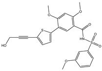
Table 1. 2D Chemical structures, computed docking scores, and intramolecular H-bond of the top 10 scoring streptomycetes NPs toward the DENV2 RdRp allosteric site a.
Compound Name/ID2D Chemical StructureDocking Score (kJ.mol−1)Intermolecular H-Bond
StandardExpensive
68TPharmaceuticals 18 01135 i001−35.6−35.6ARG729 (3.11 Å),
TRP795 (3.40 Å),
GLU802 (2.67 Å)
SDB9818Pharmaceuticals 18 01135 i002−46.9−46.9ARG737 (2.04; 2.62; 1.83 Å), SER796 (3.01 Å),
CYS709 (1.83 Å),
ASP664 (1.74 Å),
TRP795 (1.80 Å),
SER710 (1.70 Å)
SDB4806Pharmaceuticals 18 01135 i003−42.7−45.6GLU459 (1.63 Å),
ASP664 (1.84 Å),
ARG729 (2.16 Å),
ARG737 (2.84 Å),
THR794 (1.97; 2.44 Å), TRP795 (2.54 Å),
SER796 (2.83 Å)
SDB895Pharmaceuticals 18 01135 i004–41.8–45.2LYS461 (2.90 Å),
ASP664 (1.81 Å),
ARG737 (3.72 Å),
SER796 (2.22; 2.33; 2.08 Å),
SDB12947Pharmaceuticals 18 01135 i005–35.6–45.2ASP664 (1.88 Å),
HIS798 (3.18 Å),
SER796 (3.02; 3.18 Å),
CYS709 (2.19 Å)
SDB13026Pharmaceuticals 18 01135 i006–46.9–45.2LYS461 (2.09; 2.12; 2.22 Å),
ASP664 (2.45 Å),
ARG729 (2.33; 2.35 Å),
TYR766 (2.83 Å),
ARG737 (1.73 Å),
SDB9891Pharmaceuticals 18 01135 i007–42.3–45.2LYS461 (1.90 Å),
SER796 (1.72; 2.25; 3.15 Å),
TYR766 (2.02; 2.15 Å),
HIS798 (3.08 Å),
CYS709 (1.83 Å)
SDB10285Pharmaceuticals 18 01135 i008–41.8–42.3LYS461 (2.24 Å),
ARG472 (2.95 Å),
ARG737 (2.53 Å),
GLU802 (1.86; 2.36 Å)
SDB993Pharmaceuticals 18 01135 i009–41.8–42.3SER796 (2.65 Å),
ARG737 (2.01 Å)
SDB1014Pharmaceuticals 18 01135 i010–41.4–40.2LYS461 (2.60 Å),
SER796 (2.84 Å)
SDB827Pharmaceuticals 18 01135 i011–41.4–39.3ASP664 (2.13; 2.23 Å),
ARG729 (3.07 Å),
ARG737 (1.99, 2.57 Å),
TYR766 (2.09, 2.01 Å),
THR794 (2.41 Å),
SER796 (2.12, 2.19 Å)
a Streptomycetes NPs were ordered according to the expensive docking computations.
Table 2. The drug-like characteristics of the identified streptomycetes NPs and 68T as prospective anti-DENV2 drug candidates.
Compound Name/IDLogPMW (g/mol)HBDHBA
68T4.23487.5527
SDB98180.40423.4668
SDB48062.24511.52410
Table 3. The computed EHOMO, ELUMO, EFL, and Egap in eV for the most promising streptomycetes NPs and 68T.
Compound Name/IDEHOMOELUMOEFLEgap
68T−6.88−0.83−3.856.05
SDB9818−7.99−1.34−4.676.65
SDB4806−8.06−2.33−5.195.74
Table 4. Computed global descriptors for SDB4806, SDB9818, and 68T.
Compound Name/IDIP (eV)EA (eV)η (eV)S (eV−1)
68T6.880.833.030.33
SDB98187.991.343.320.30
SDB48068.062.332.870.35
Disclaimer/Publisher’s Note: The statements, opinions and data contained in all publications are solely those of the individual author(s) and contributor(s) and not of MDPI and/or the editor(s). MDPI and/or the editor(s) disclaim responsibility for any injury to people or property resulting from any ideas, methods, instructions or products referred to in the content.