Article

Novel 3,19-(N-Phenyl-3-(4-fluorophenyl)-pyrazole) Acetal of Andrographolide Promotes Cell Cycle Arrest and Apoptosis in MDA-MB-231 Breast Cancer Cells

by
Siva Kumar Rokkam
1,
Shahjalal Chowdhury
2,
Yashwanth Inabathina
2,
Lakshminath Sripada
1,
Srinivas Nanduri
3,
Balasubramanyam Karanam
2,* and
Nageswara Rao Golakoti
1,*
1
Department of Chemistry, Sri Sathya Sai Institute of Higher Learning, Puttaparthi 515134, Andhra Pradesh, India
2
Department of Biology and Cancer Research, Tuskegee University, Tuskegee, AL 36088, USA
3
Department of Chemical Sciences, National Institute of Pharmaceutical Education and Research, Balanagar, Hyderabad 500037, Telangana, India
*
Authors to whom correspondence should be addressed.
Pharmaceuticals 2025, 18(7), 1026; https://doi.org/10.3390/ph18071026
Submission received: 11 May 2025 / Revised: 24 June 2025 / Accepted: 4 July 2025 / Published: 10 July 2025
(This article belongs to the Special Issue Exploring Natural Products with Antioxidant and Anticancer Properties)

Abstract

Background: Natural products play a crucial role in cancer treatment due to their ability to selectively target cancer cells. Andrographolide, a major constituent of Andrographis paniculata, exhibits potential anticancer properties. Considering the pharmacological importance of nitrogen-based heteroaromatic scaffolds, particularly pyrazole motifs, this study aimed to integrate the pyrazole pharmacophore with the andrographolide scaffold to develop novel therapeutic candidates. Methods: Twenty novel 3,19-(N-phenyl-3-aryl-pyrazole) acetals of andrographolide and isoandrographolide were synthesized and characterized using UV-Vis, FT-IR, NMR, and HRMS. Initial anticancer screening was conducted by the National Cancer Institute (NCI), USA, against 60 human cancer cell lines. The most promising compound, 1f (R = 4-F), was selected for further biological evaluation in the MDA-MB-231 breast cancer cell line. Results: The MTT assay results demonstrated that compound 1f exhibited strong, dose-dependent anti-proliferative effects. The apoptosis analysis of 1f revealed a time-dependent increase in apoptotic cells, and cell cycle studies indicated S phase arrest in MDA-MB-231 cells. Antioxidant activity via the DPPH assay identified compounds 1b (R = 3-NO2) and 2b (R = 3-NO2) as the most effective radical scavengers. The most active compounds were also evaluated for drug-likeness using in silico Lipinski’s rule assessments. Conclusions: The synthesized 3,19-(N-phenyl-3-aryl-pyrazole) acetals of andrographolide and isoandrographolide exhibited promising anticancer and antioxidant properties. Among them, compound 1f showed the most significant activity, supporting its potential as a lead candidate for further anticancer drug development.

Graphical Abstract

1. Introduction

Cancer is a group of diseases that occur when cells divide uncontrollably and consistently [1]. Despite the achievements of oncology, cancer continues to be one of the most fatal diseases and the second leading cause of mortality in the world [2]. As stated by the GLOBOCAN 2020 global cancer statistical report, ~19.3 million new cancer cases were recorded and accounted for ~10 million deaths worldwide (barring non-melanoma skin cancer) [3]. Breast, lung, colon, prostate, skin, and stomach cancer are the most common cancer, and their fatality rate is considerably high [4]. The most prevalent cancer diagnosed currently is breast cancer, with an estimated 2 million new cases annually, and remains the 5th highest cause of cancer death, with over half a million deaths [5,6]. Approximately 28 million new cases of cancer are anticipated worldwide over the next twenty years, a 47% increase from 2020 [3,7]. Current anticancer drugs used in treatment often cause adverse effects such as weakened immune function, hair loss, cardiotoxicity, and infertility in women with active ovaries [8,9,10]. Consequently, alternative treatments involving natural products, which offer greater potential with fewer side effects for cancer patients, should be prioritized [11,12,13,14].
Natural products have been an excellent primary source of bioactive molecules. These bioactive molecules have acquired great value in cancer drug discovery due to their structural features and wide range of medicinal properties [15,16,17,18]. One such bioactive molecule is andrographolide (ADG). It is an active diterpenoidal constituent of Andrographis paniculata Nees (Figure 1), which belongs to the family Acanthaceae [19,20]. It is primarily accumulated in the leaves, while modest amounts can also be found in the flowering tops, stems, and roots [21]. It is considered an easily isolable compound with a high yield. Andrographis paniculata, also commonly known as the ‘King of Bitter’, is widely cultivated in tropical and subtropical Southeast Asia, China, and India [22]. In traditional Chinese and Indian medicine, the leaves and roots of this plant were used as a remedy for various ailments such as fever, common cold, mouth ulcers, skin infections, urinary tract infections, gastrointestinal disorders, to dispel the toxins from the body, venomous snakebites, etc. [23,24,25]. Several research groups investigated the extracts and compounds isolated from A. paniculata and reported that they displayed a wide range of pharmacological activities, including anticancer, antiviral, anti-inflammatory, antimalarial, antioxidant, immunostimulatory activities, etc. In addition, these investigations revealed that the observed anticancer properties of the methanolic extracts of the plant are mainly attributed to andrographolide [26,27,28,29,30,31,32,33,34].
In various human cancer cell lines, andrographolide was evaluated for its anticancer potential both in vitro and in vivo [35]. The studies disclosed that andrographolide targets several signaling pathways and holds significant promise for chemoprevention in various types of cancer. By interacting with a variety of targets, andrographolide affects several cancer-related characteristics, such as an increase in the expression of pro-apoptotic genes that lead to apoptosis by either intrinsic mitochondrial death or extrinsic TRAIL-related death, cell cycle arrest at different stages with the enhancement of pro-apoptotic gene and protein activities, anti-angiogenic properties, the inhibition of the VEGF pathway, NF- κβ inhibition, the ubiquitin-mediated proteasomal degradation of proteins, and the inhibition of IL-mediated signals [35,36,37,38,39]. Though it exhibited excellent therapeutic potential, it has limited oral bioavailability due to its quick elimination and high plasma protein-binding capacity [40]. This makes creating formulations for clinical treatment extremely difficult. However, owing to its unique structural features, several structural changes have been made to various parts of andrographolide to enhance its anticancer activity and oral bioavailability. Interestingly, these modifications resulted in a significant increment in the anticancer potential and bioavailability. Like andrographolide, pyrazole derivatives also exhibit remarkable anticancer effects via the inhibition of various kinds of enzymes, proteins, and receptors, such as CDKs, kinases, EGFR, VEGF, TGF-β, etc. [41,42,43,44,45].
In this context, we have synthesized twenty novel 3,19-(N-phenyl-3-aryl-pyrazole) acetals of andrographolide and isoandrographolide. Our main intention is to link the pyrazole scaffold to andrographolide due to its widespread pharmacological effects in medicinal chemistry, mainly as an anticancer and anti-inflammatory pharmacophore. As expected, the synthesized derivatives have displayed excellent anticancer activity, several-fold increased activity compared to andrographolide, against the breast cancer cell line MDA-MB-231.

2. Results and Discussion
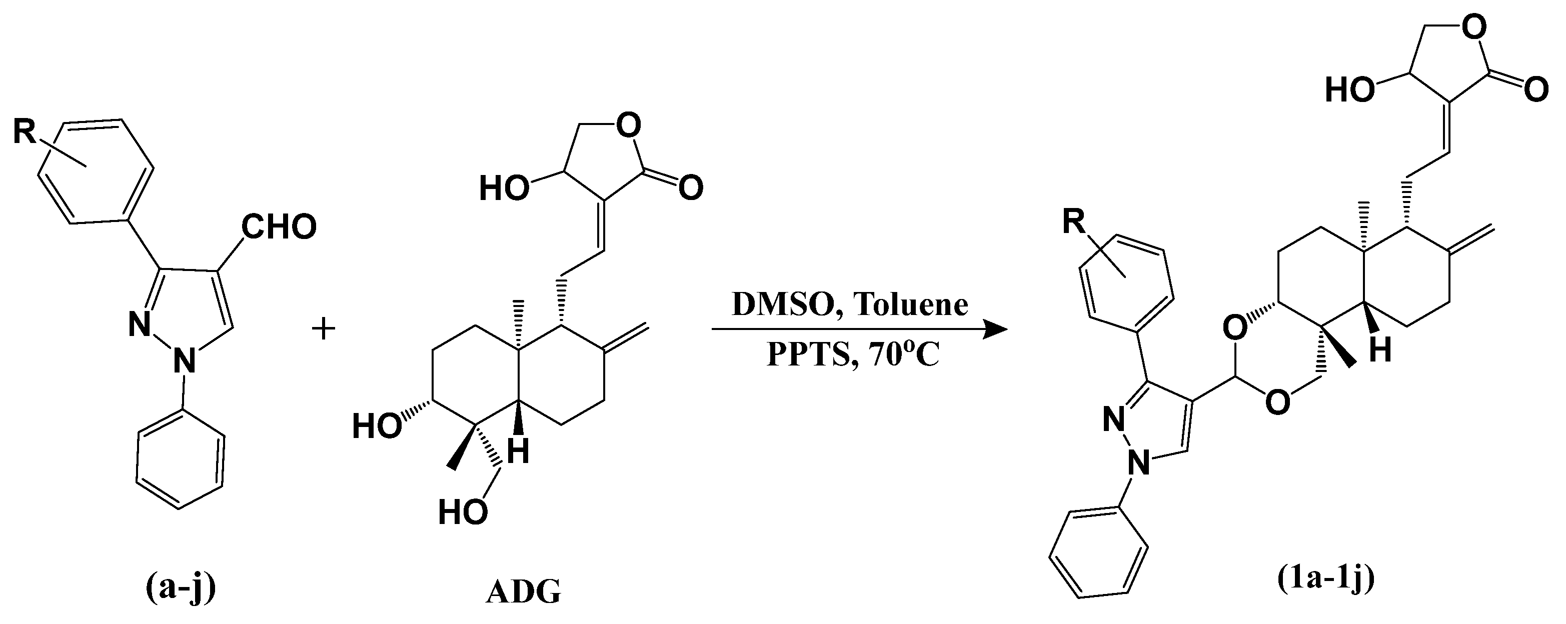
2.1. Chemistry

The synthesis of 3,19-(N-phenyl-3-aryl-pyrazole) acetals of andrographolide and isoandrographolide entails a series of consecutive steps. This synthesis encompasses three distinct steps, and the schematics detailing the adopted synthetic strategies are presented in Scheme 1, Scheme 2 and Scheme 3 [46]. In the initial step, ten 1-phenyl-2-(1-arylethylidene) hydrazines (AJ) were synthesized. Then, in the second step, these compounds were transformed into the corresponding N-aryl-pyrazole-4-carboxaldehydes (aj). Lastly, in the third step, these aldehydes were subjected to reactions with andrographolide (ADG) and isoandrographolide (ISOADG), resulting in the final attainment of 3,19-(N-phenyl-3-aryl-pyrazole) acetals of andrographolide and isoandrographolide (1a1j and 2a2j).

2.2. Characterization

The UV-Vis spectra exhibited bands between 275 and 280 nm for all the synthesized compounds (1a1j and 2a2j). In the IR spectra of 3,19-(N-phenyl-3-aryl-pyrazole) acetals of andrographolide (1a1j), peaks at ~3400 cm−1 and ~1672 cm−1 were observed for -OH and exocyclic C=C groups. These peaks were absent for 3,19-(N-phenyl-3-aryl-pyrazole) acetals of isoandrographolide (2a2j). The presence of the C-H stretching band within the 3089–3070 cm−1 range and the aromatic skeletal bands spanning from 1600 to 1450 cm−1 confirms the existence of the aromatic group. The band situated at 2948–2920 cm−1 corresponds to the sp3 C-H stretch originating from the diterpene moiety. The peak in the range of 1732–1759 cm−1 corresponds to the carbonyl group (C=O) of the butyrolactone ring. Distinct absorptions at approximately 1220 and 1100 cm−1 can be attributed to the C-O stretching. Compounds with aryl chloride have a band at 1071–1060 cm−1 (Ar-Cl stretch). Compounds with aryl fluorides have a band around 1245 and 1220 cm−1 (Ar-F stretch), and compounds with aryl bromides have a band between 1020 and 1011 cm−1 (Ar-Br stretch).
The attachment of the substituted pyrazole moiety is discernible in 1H NMR and 13C NMR spectra of all the acetals. In 1H NMR, the peak at ~δ 8.18 is due to the presence of only hydrogen in the pyrazole ring, whereas peaks around δ 8.76 to 6.99 correspond to the protons on the aromatic ring attached to the pyrazole ring. In 13C NMR, the carbons in the pyrazole ring and aromatic ring were identified in the ~δ 163 to 115 range. The bridged carbon, which was bonded to two oxygen atoms, provides strong evidence for the formation of the acetals, and its peak was observed at δ 90 in the 13C NMR spectrum, and the proton attached to the acetal carbon is observed at ~δ 5.87 in the 1H NMR. However, the carbonyl group (C=O) signals in the acetals appeared approximately within the range of δ 173–170. In 3,19-(N-phenyl-3-aryl-pyrazole) acetals of andrographolide, peaks at ~148 and 110 in the 13C NMR spectra correspond to an exocyclic C=C, which is absent in 3,19-(N-phenyl-3-aryl-pyrazole) acetals of isoandrographolide [47,48]. More detailed information on the characterization data is provided in the Supplementary Information.

2.3. In Vitro Anticancer Studies

2.3.1. NCI Screening

All the synthesized compounds (1a1j and 2a2j) were initially assessed for their anticancer potential at the National Cancer Institute (NCI, USA) against a diverse panel of 60 human cancer cell lines, representing nine distinct cancer types including leukemia, non-small-cell lung, colon, CNS, melanoma, ovarian, renal, prostate, and breast. The testing protocol employed by the NCI encompasses two stages: initial assessment at a single dose and subsequent evaluation at five doses. Compounds that demonstrated growth inhibition exceeding 32% at the single-dose stage were identified as active candidates (32% cutoff used by the NCI as a general guide to filter out inactive compounds).

2.3.2. Single-Dose Study

Initially, a comprehensive anticancer screening of all twenty compounds (1a1j and 2a2j), including the parent compounds ADG and ISOADG, was conducted using a 10 µM dose across all 60 cell lines (a standardized protocol followed for the initial assessment of test compounds by the NCI). In this assessment, a subset of ten compounds (1a1j) demonstrated impressive percent growth inhibition in most of the cell lines. As a result, these ten compounds were chosen for five-dose studies. Of particular interest is the observation that all ten compounds exhibiting notable activity across most of the cell lines are 3,19-(N-phenyl-3-aryl-pyrazole) acetals of andrographolide (1a1j). Conversely, 3,19-(N-phenyl-3-aryl-pyrazole) acetals of isoandrographolide (2a2j), ADG, and ISODG displayed a lower % growth inhibition, suggesting that these derivatives are less potent across the majority of cell lines. Consequently, compounds 2a2j, ADG, and ISODG did not meet the criteria for further advancement in the study.

2.3.3. Five-Dose Study

In the five-dose study, 3,19-(N-phenyl-3-aryl-pyrazole) acetals of andrographolide (1a1j) were tested at concentrations of 0.01 µM, 0.1 µM, 1 µM, 10 µM, and 100 µM against 60 cancer cell line panels. The growth inhibition 50% (GI50), total growth inhibition (TGI), and lethal concentration 50% (LC50) of the ten potent compounds on the cancer cell lines are given in Table 1.
It is interesting to observe that all ten compounds have exhibited excellent GI50 values across all the cell lines. However, among these compounds, 1f demonstrated the best activity, followed by 1j and 1h. Compounds 1i and 1b were slightly less active as compared to other compounds. In addition, we note that para-substituted derivatives (1f, 1g, 1h, 1i, and 1j) exhibited better GI50 values than meta-substituted derivatives (1b, 1c, 1d, and 1e). The observed anticancer activity order of these compounds is as follows: 1f > 1j > 1h > 1g > 1d > 1c > 1e > 1a > 1b > 1i.

2.3.4. GI50 Values of Compounds on Individual Cancer Types

Leukemia Cancer: The compound 1f displayed the most potent activity on most of the leukemia cancer cell lines. However, the best GI50 (0.40 µM) shown by 1f was against the cell line CCRF-CEM, followed by the compounds 1h (GI50: 0.48 µM), 1a (GI50: 0.59 µM), 1j (GI50: 0.64 µM), 1e (GI50: 0.70 µM), and 1b (GI50: 1.14 µM). However, the compounds 1c, 1d, 1g, and 1i showed better activity against the cell line MOLT-4.
Non-Small-Cell Lung Cancer: The compound 1g was observed to have the best GI50 (1.02 µM) against the NCI-H322M cell line. While the compounds 1a, 1f, 1i, and 1j were active against the HOP-92 cell line, compounds 1b, 1c, 1d, and 1e were active against the NCI-H522 cell line.
Colon Cancer: All the compounds except 1b, 1c, and 1i were found to be active against the HCT-116 cell line. Compound 1f demonstrated the best activity with GI50: 0.39 µM. Compounds 1b, 1c, and 1i showed the best GI50 against HCT-15 and HCC-2998 cell lines.
CNS Cancer: Among the CNS cancer cell lines, these compounds showed promising effects against three cell lines. All the para-substituted derivatives (1f, 1g, 1h, and 1i) except 1j displayed excellent growth inhibition against the SNB-19 cell line, whereas 1j, 1a, and 1d are more active against SNB-75, and 1d (GI50: 1.12 µM) is the best among all. On the other hand, meta-substituted derivatives (1b, 1c, and 1e) showed the best GI50s against SF-295.
Melanoma Cancer: As observed in CNS cancer, against the melanoma cancer cell line LOX IMVI, the para-substituted derivatives (1f, 1g, 1h, and 1i) demonstrated outstanding growth inhibition with GI50s of 1.32, 1.58, 1.55, and 1.43 µM, respectively. All other compounds displayed better activity on various melanoma cancer cell lines.
Ovarian Cancer: These compounds were observed to have very good activity against different ovarian cancer cell lines. However, among all, compound 1j (GI50: 1.35 µM) was found to be the most potent against OVCAR-8, followed by 1d (GI50: 1.39 µM) against the IGROV1 cell line.
Renal Cancer: As in the case of CNS and melanoma cancer, para-substituted derivatives (1f, 1g, 1h, and 1i), except 1j, exhibited the best activity with GI50s of 1.27, 1.33, 1.19, and 1.33 µM against TK-10. However, compound 1j (GI50: 0.89 µM) was the most active against RXF 393 amongst all renal cancer cell lines. The meta-substituted derivatives (1b, 1d, and 1e), except 1c (active against TK-10, GI50: 1.41 µM), displayed the best activity against RXF 393 with GI50s of 1.62, 1.05, and 1.18 µM.
Prostate Cancer: Between the two types of cancer cell lines (PC-3 and DU-145) against which the compounds were tested, four compounds 1a, 1b, 1f, and 1i showed the best GI50s against PC-3, while remaining six compounds 1c, 1d, 1e, 1g, 1h, and 1j showed the best GI50s against DU-145.
Breast Cancer: Including 1a and 1b (GI50: 1.42 and 1.76 µM), the para-substituted derivatives (1f (GI50: 1.19 µM), 1g (GI50: 1.39 µM), 1h (GI50: 1.18 µM), and 1i (GI50: 1.14 µM)) also demonstrated the highest growth inhibition against the T-47D cell line. However, the compounds 1j and 1d (GI50: 1.46 and 1.48 µM) against MCF-7 and 1c and 1e (GI50: 1.30 and 1.46 µM) against BT-549 were more active.

2.3.5. NCI Compare Analysis

The NCI compare analysis assesses and arranges compounds based on their similarity in response to the NCI 60 cell lines compared to marketed drugs [49]. This similarity is quantified using the Pearson Correlation Coefficient (PCC). When a marketed drug ranks high in this comparison alongside the compound being studied, it suggests that the studied compound might share a similar mechanism of action. The most promising compounds 1f, 1h, and 1j from the five-dose study were chosen for NCI compare analysis, and their GI50 values were compared to those of marketed drugs (Table 2).
The NCI compare analysis revealed that the three compounds exhibited correlations with various marketed drugs that are recognized for their ability to inhibit DNA synthesis and promote apoptosis. This observation strongly suggests that these compounds could potentially impede cancer growth through similar mechanisms.

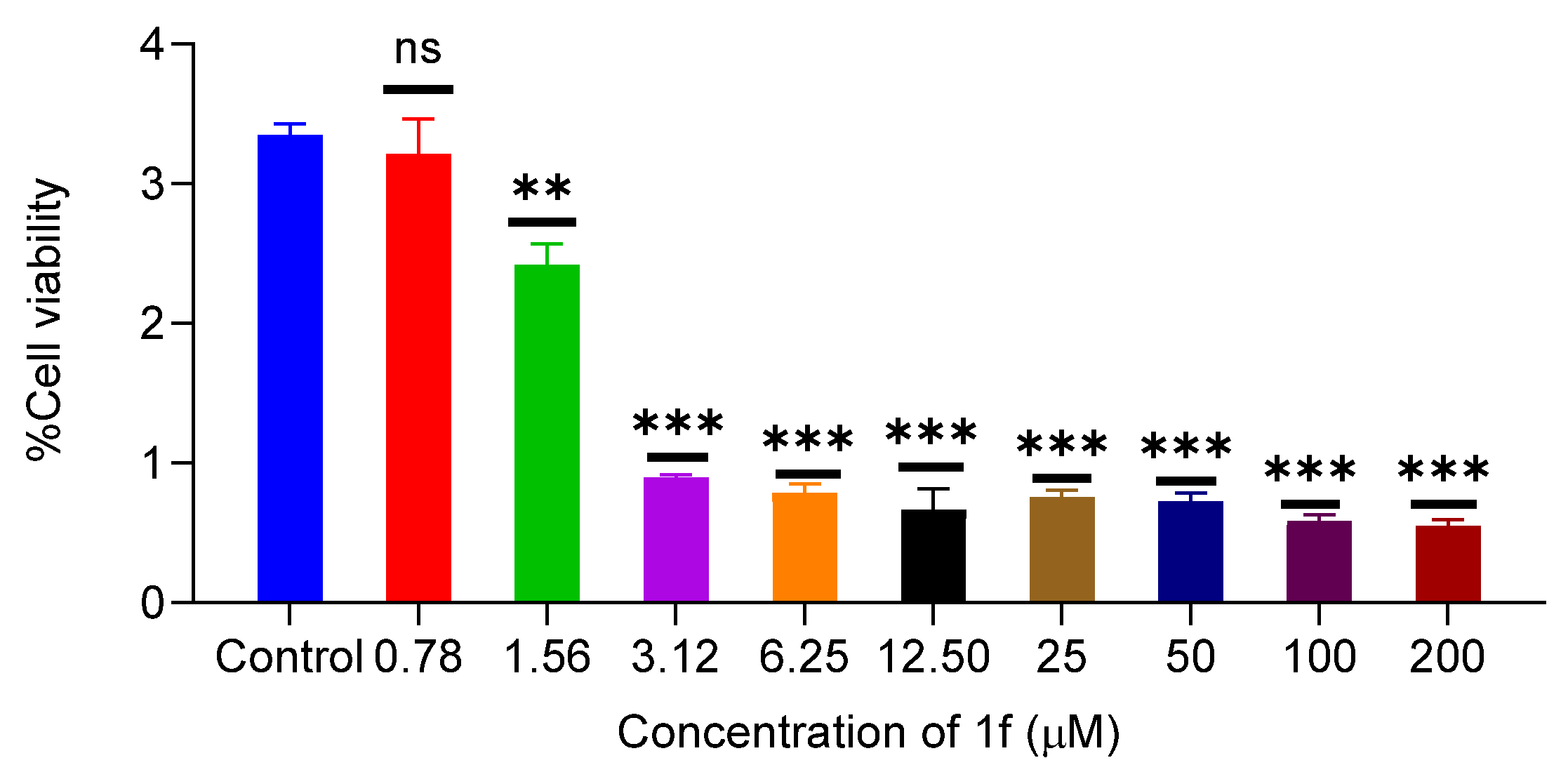
2.3.6. Effect of 1f on Proliferation of MCF-10a and MDA-MB-231

As compound 1f exhibited the best anticancer activity among all the screened compounds in the NCI study, its cytotoxicity on breast cells MCF-10a (normal breast cells) was examined. Compound 1f was found to have a low inhibitory concentration with a CC50 of 50.01 µM against the breast cells MCF-10a. Further, the MTT assay was performed for compound 1f against the MDA-MB-231 cell line to check its cytotoxicity. These MDA-MB-231 breast cancer cells were treated with 1f at different concentrations, i.e., 0, 1.56, 3.125, 6.25, 12.5, 25, 50, 100, and 200 µM, for 72 h. As shown in Figure 2, the absorbance decreased with the increasing concentration of the drug. The results indicate that 1f has the strong dose-dependent anti-proliferation activity against the MDA-MB-231 breast cancer cell line.

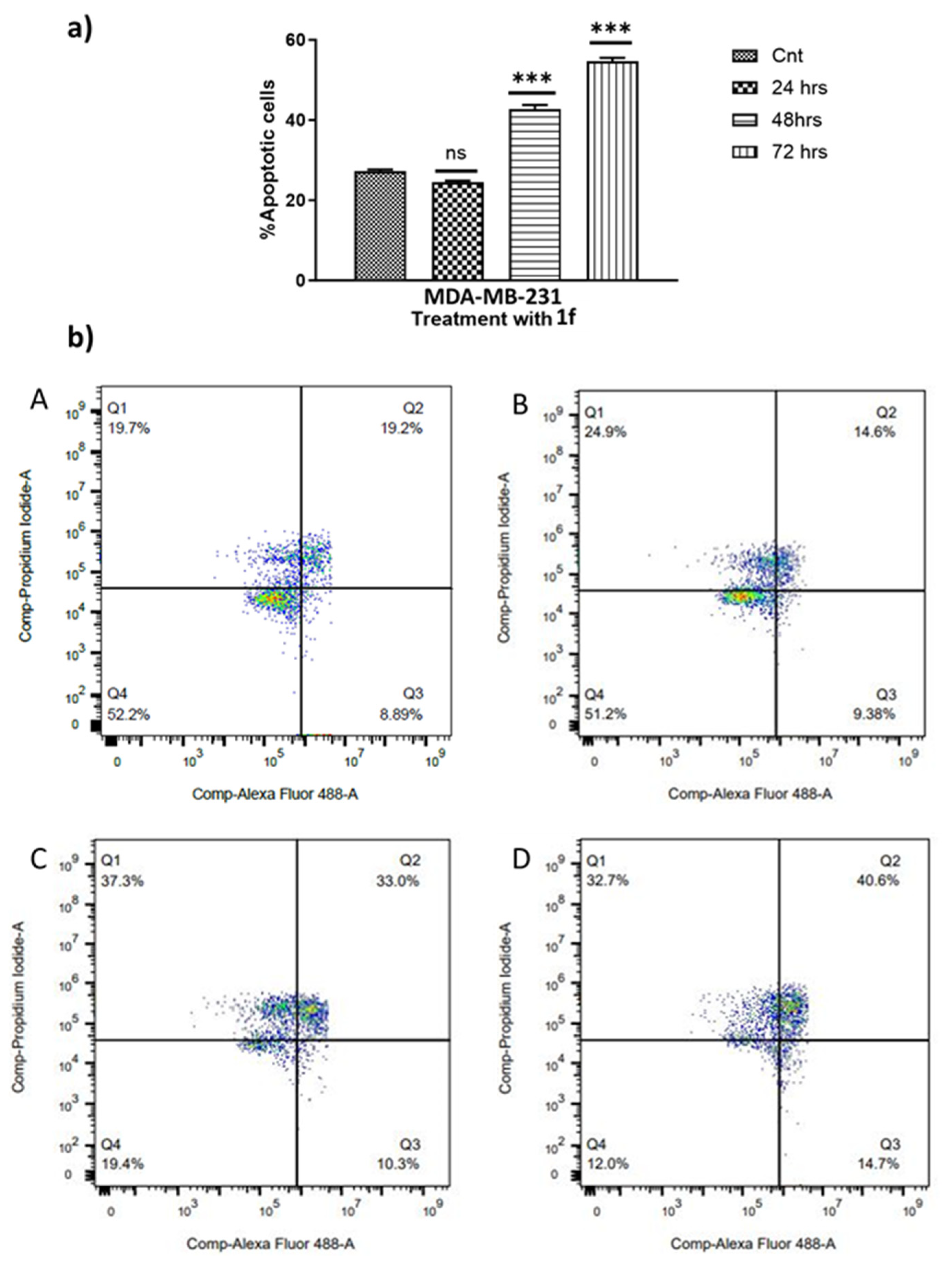
2.3.7. Effect of 1f on Cell Apoptosis Inducement

Apoptosis is considered a preventive mechanism against cancer dissemination, as it plays a crucial role in eliminating excessively proliferating and mutated cancer cells from the body [50,51]. Following the administration of cytotoxic drugs as treatment for various types of cancer, apoptosis is a significant contributor to cellular death [52]. Hence, an enhanced understanding of the processes and mechanisms underlying apoptosis can aid in the development of novel therapeutic drugs for cancer [53]. We proceeded to investigate whether compound 1f triggers apoptosis or not. The MDA-MB-231 breast cancer cell line was treated with 1f for 24, 48, and 72 h. A quantitative evaluation of apoptotic activity was performed using FACS.
As depicted in Figure 3, a notable rise in apoptotic cells was noticed in a time-dependent manner. We observed 28.09% apoptotic cells in the untreated control (8.89% in early apoptosis and 19.2% in late apoptosis). Upon treatment with 1f, while after 24 h apoptotic cells remained almost the same, we observed a gradual increase in apoptotic cells by 44% at 48 h (10.3% in early apoptosis and 33.0% at late apoptosis) and 55% at 72 h (14.7% in early apoptosis and 40.6% at late apoptosis), respectively, compared to the control at 0 h. Thus, confirming that compound 1f promoted apoptosis in MDA-MB-231 breast cancer cells.

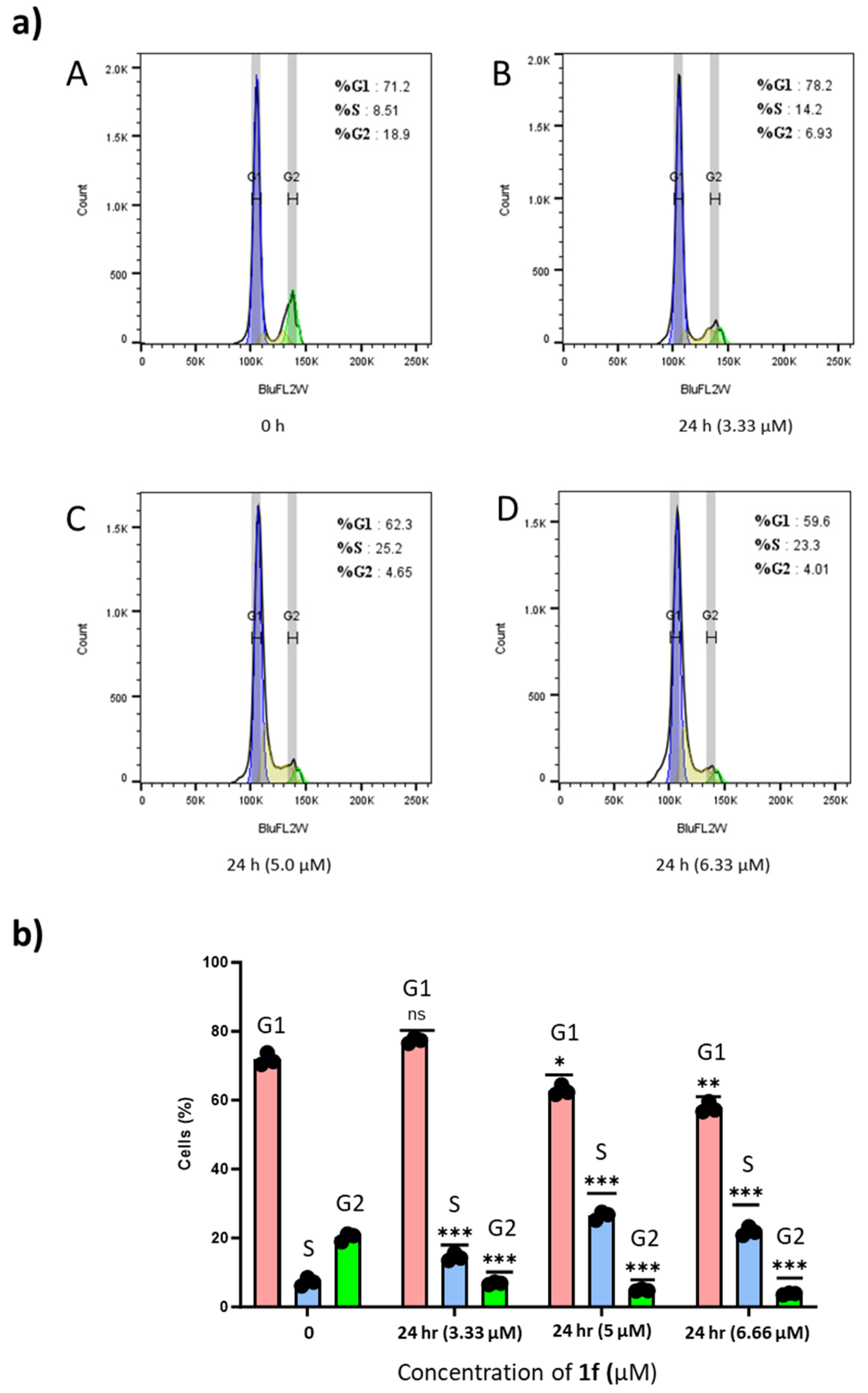
2.3.8. Effects of 1f on MDA-MB-231 Cell Cycle Distribution

Cell proliferation is well associated with the regulation of cell cycle progression. Therefore, the effect of 1f on cell cycle distribution was evaluated by flow cytometry in PI-stained cells. As shown in Figure 4, after treatment for 24 h with 1f at concentrations of 3.33, 5.0, and 6.66 µM, cells in G2/M distributions decreased from 18.9% to 4.01% compared to control cells (at 0 h), whereas the percentage of cells in the G1 phase decreased from 71.2% to 59.6% and cells in the S phase increased gradually from 8.51% to 14.2%, 25.2%, and 23.3%, respectively (Table 3). The result indicates that 1f could induce cell cycle arrest in the S phase.

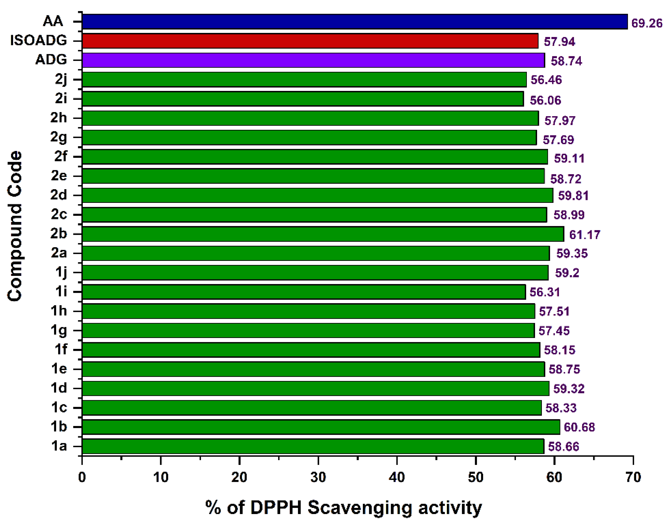
2.4. Antioxidant Activity

The onset and advancement of cancer have been associated with oxidative stress due to its role in escalating DNA mutations, triggering DNA impairment, fostering genome instability, and promoting cellular proliferation [54,55,56]. Therefore, to find out the antioxidant properties of the compounds, DPPH radical scavenging activity was performed. As shown in Figure 5, all the synthesized compounds showed a DPPH radical scavenging activity between 56.06% to 61.17% as compared to ADG (58.74%), ISOADG (57.94%), and the positive control ascorbic acid (AA, 69.26%). All the compounds demonstrated more or less similar antioxidant potential compared to ADG and ISOADG. However, the compounds with the strongest electron-withdrawing groups, i.e., 1b (R = 3-NO2, 60.68%) and 2b (R = 3-NO2, 61.17%), are the most active, whereas the compounds 1i (R = 4-CH3, 56.31%) and 2i (R = 4-CH3, 56.06 %) are the least active. Among the halogen-substituted derivatives, chloro-substituted compounds in both the series (1d, 59.32% and 2d, 59.81%) have shown superior activity. Another interesting observation was that all the meta-substituted derivatives displayed slightly better radical scavenging activity than all the para-substituted derivatives in both series, except in the case of 2c.

2.5. Drug-Likeness Studies for Active Compounds

Using the pkCSM tool, a computer-based assessment was conducted on the active compounds (1a1j) to assess their suitability as drug candidates by examining their Lipinski parameters [57].
The analysis of the results, as given in Table 4, revealed that in silico molecular properties of the compounds align with Lipinski’s rule of five (RO5), except for their molecular weights, which fall within the range of 580–658 Daltons, slightly surpassing the recommended limit (<500 Daltons). Nevertheless, it is worth noting that natural products often deviate from Lipinski’s rules regarding molecular weights [58]. The compounds exhibited a TPSA ranging from 78 to 126, whereas the Log p-value slightly exceeded the acceptable range for some compounds. Additionally, they have fewer than five N-H and O-H hydrogen bond donors and fewer than ten nitrogen and oxygen hydrogen bond acceptors (HBA). The parent compound ADG shows better predicted bioavailability than its derivatives, due to its lower molecular weight, LogP, and balanced polarity. However, some of the derivatives showed better anticancer activity than ADG. Overall, despite the slight deviation in molecular weights, the compounds largely adhere to Lipinski’s parameters, suggesting their suitability as potential anticancer agents.

3. Experimental

3.1. Synthesis

All the chemicals used in the experiments were procured from Merck and S.D. Fine chemicals (Bangalore, India). Andrographolide (98%) was obtained from Maysar Herbals (Faridabad, Haryana, India) and purified by recrystallization. Solvents such as methanol and dichloromethane were distilled before use.

3.1.1. Synthesis of Arylhydrazones (A–J)

To a solution of acetophenone (25 mmol) in methanol (80 mL), phenylhydrazine (25 mmol) was added and stirred at room temperature. To this, 2 mL acetic acid was added, and the mixture was refluxed for 24 h. The progress of the reaction was checked using thin-layer chromatography (TLC). After the completion of the reaction, crushed ice was added, and the solid formed was filtered and dried. The pure product obtained was then subjected to the next reaction [46].

3.1.2. Synthesis of 3-Aryl-4-pyrazole carboxaldehydes (a–j)

To a cold solution of N, N-dimethylformamide (40 mL), aliquot amounts of arylhydrazone (20 mmol) were added and stirred over 30 min. Then, POCl3 (100 mmol) was added dropwise to the solution while maintaining the temperature at 0–5 °C. After the addition, the reaction mixture was heated at 55 °C for 4 h. After the completion of the reaction, the resulting mixture was cooled to room temperature and poured into crushed ice and stirred for 20 min. The white solid formed was filtered, dried, and purified using column chromatography (hexane/ethyl acetate, 75:25). In the case the solid was not formed after being poured into crushed ice, the solution was neutralized with saturated Na2CO3 to obtain the solid [46].

3.1.3. Synthesis of 3,19-(N-phenyl-3-aryl-pyrazole) Acetals of Andrographolide (1a1j)

Andrographolide (1 mmol) in DMSO (2 mL) was added to 3-aryl-4-pyrazole carboxaldehydes (4 mmol) in toluene (3 mL) at room temperature. To this catalytic amount (0.08 mmol) of pyridinium para-toluene sulfonate (PPTs) were added and heated at 70 °C for 24 h. After the completion of the reaction, the mixture was cooled to room temperature and then neutralized with a freshly prepared, cold saturated sodium bicarbonate solution. The resulting solution was then extracted three times (15 mL each) with DCM, and the obtained organic layer was washed thoroughly with brine solution and dried over anhydrous sodium sulphate. DCM was removed using a rota evaporator. The crude product obtained was then purified using column chromatography (silica gel: 230–400 mesh, hexane/acetone, 80:20) [46].

3.1.4. Synthesis of Isoandrographolide and 3,19-(N-phenyl-3-aryl-pyrazole) Acetals of Isoandrographolide (2a–2j)

To synthesize isoandrographolide, andrographolide (8 g) was dissolved in concentrated hydrochloric acid (HCl, 150 mL) and stirred at room temperature for 24 h. After the completion of the reaction (monitored by TLC), the reaction mixture was poured into crushed ice and extracted with DCM. Then, the organic layer was thoroughly washed with brine and freshly prepared saturated sodium bicarbonate solution. The crude solid was then dried over anhydrous sodium sulphate and recrystallized from ethyl acetate to obtain a pure product [46].
3,19-(N-phenyl-3-aryl-pyrazole) acetals of isoandrographolide were synthesized as described in the synthesis of 3,19-(N-phenyl-3-aryl-pyrazole) acetals of andrographolide (in this case, isoandrographolide was used as a starting material instead of andrographolide).

3.2. Instrumentation

The 1H NMR and 13C NMR spectra were recorded using a Bruker Ascend NMR spectrometer (Bruker BioSpin AG, Faellanden, Switzerland) at 400 MHz and 100 MHz, respectively. CDCl3 was used as the solvent to dissolve the compounds. TMS was an internal standard. Agilent 6550 Q-TOF LC/MS (HRMS) (Agilent Technologies India Private Limited, Bangalore, India) was employed for the mass spectra. UV-Vis spectra were recorded in acetonitrile in the range of 200–600 nm using a Agilent Cary UV (Agilent Technologies, Santa Clara, CA, USA). An Agilent Cary 630 spectrophotometer (Agilent Technologies India Private Limited, Bangalore, India) was used to record FT-IR spectra between 400 and 4000 cm−1 using KBr pellets. For HPLC, the Agilent 1260 Infinity high-performance liquid chromatography system (Agilent Technologies India Private Limited, Bangalore, India), equipped with a quaternary solvent delivery system, inline degasser, autosampler, and photodiode array detector, was used. Chromatographic separation was carried out using an RP-HPLC Zorbax Extend-C18 (4.6 mm × 250 mm, 5 µm) column (Agilent Technologies India Private Limited, Bangalore, India). The mobile phase consisted of acetonitrile (25%) and water (75%) and was maintained at a flow rate of 1 mL/min. The temperature of the column was controlled at 25 °C with an injection volume of 10 µL. Detection wavelengths were set at the respective λmax for each compound, and a diode array detector was employed to detect the eluted peaks between 220 and 250 nm.

3.3. Biological Testing

3.3.1. Cell Proliferation Assay

MDA-MB-231 breast cancer cells and MCF-10A normal breast epithelial cells (from ATCC, Manassas, VA, USA) were seeded into 96-well plates at a density of 4 × 10³ cells per well. After overnight incubation at 37 °C, the cells were treated with varying concentrations of compound 1f (0, 1.56, 3.125, 6.25, 12.5, 25, 50, 100, and 200 µM) and incubated for an additional 72 h. Untreated cells served as the negative control. Subsequently, 10 µL of the MTT solution (5 mg/mL) was added to each well, and the plates were incubated for 3 h to assess cell viability. The medium was then discarded, and the absorbance was measured at 570 nm using a microplate reader (Promega GloMax, Madison, WI, USA).

3.3.2. Apoptotic Assay

To evaluate the pro-apoptotic effect of compound 1f, Annexin V-FITC/PI dual staining was conducted. Cells were seeded into 12-well plates at a density of 1 × 10⁵ cells per well and treated with the compound for 24, 48, and 72 h. Post-treatment, the cells were collected using 1X trypsin, including both floating and adherent cells, and centrifuged at 4000 rpm for 5 min. The resulting pellet was resuspended in 100 µL of 1X Annexin V binding buffer and stained with Annexin V-FITC, followed by a 15 min incubation in the dark at 4 °C. Cells were then washed, resuspended in fresh binding buffer, and stained with propidium iodide before immediate analysis via flow cytometry.

3.3.3. Cell Cycle Assay

Cell cycle distribution was assessed by propidium iodide (PI) staining followed by flow cytometric analysis. Cells (3 × 10⁵/well) were plated in 6-well plates and allowed to adhere for 24 h. To synchronize the cell cycle, the medium was replaced with serum-free medium for 8 h. After synchronization, cells were treated with various concentrations of compound 1f for 24 h. Following treatment, cells were harvested with 1X trypsin, washed with cold PBS, and fixed in 70% ethanol at 4 °C for 2 h. Fixed cells were washed twice with PBS, then incubated overnight in a staining solution containing RNase A (1 µL, 10 mg/mL) and PI (5 µL, 1 mg/mL) in 1X PBS. Cell cycle distribution was subsequently determined using flow cytometry.

3.4. DPPH Assay

A stable free radical DPPH (2,2-Diphenyl-1-picrylhydrazyl) assay was employed to test the antioxidant activity of all the synthesized compounds. Concisely, 25 µM of the test compounds were dissolved in acetonitrile and combined with 0.1 mM of DPPH (in acetonitrile) in a 1:2 ratio (80 µL and 160 µL). Then, the mixture was left undisturbed in the dark for 30 min at room temperature. After 30 min, the absorbance of the solutions was instantly measured at 517 nm using a Varioskan Multiplate Reader (Thermo Fisher Scientific, Mumbai, India) [59]. Ascorbic acid (in acetonitrile) was used as the positive control, and andrographolide and isoandrogapholide (in acetonitrile) were used as the standards. The following equation was used to determine the DPPH radical scavenging activity:
% Radical scavenging activity = (Ac − As)/Ac × 100
where Ac = the absorbance of the blank (DPPH solution), and As = the absorbance of the test samples

4. Conclusions

In conclusion, twenty novel 3,19-(N-phenyl-3-aryl-pyrazole) acetals of andrographolide and isoandrographolide (1a1j and 2a2j) were synthesized and characterized. All the synthesized compounds were initially screened for their anticancer activity against 60 human cancer cell lines at the NCI, USA. Single-dose study results revealed that all 3,19-(N-phenyl-3-aryl-pyrazole) acetals of andrographolide (1a1j) displayed superior anticancer activity compared to 3,19-(N-phenyl-3-aryl-pyrazole) acetals of isoandrographolide (2a2j). Therefore, only compounds 1a1j were selected by the NCI for five-dose studies to find out their GI50s, TGI, and LC50s. Among the compounds tested, compound 1f exhibited the best GI50s, TGI, and LC50s on most cell lines, followed by 1h and 1j, respectively. Therefore, compound 1f was further selected for detailed in vitro analysis on normal breast cells MCF-10a and the breast cancer cell line MDA-MB-231. The cytotoxicity study of compound 1f on MCF-10a was found to have a low inhibitory concentration. The MTT assay results demonstrated that compound 1f exhibited strong, dose-dependent anti-proliferative activity on the cells. In addition, the apoptotic analysis revealed that compound 1f induced apoptosis in the MDA-MB-231 cells and increased the apoptotic cells from 10% to 55% over 72 h. Furthermore, cell cycle analysis indicated that 1f caused S phase arrest in MDA-MB-231 cells. The antioxidant activity of the synthesized compounds was evaluated using the DPPH assay. Of all the compounds tested, 1b (R = 3-NO2, 60.68%) and 2b (R = 3-NO2, 61.17%) demonstrated the highest radical scavenging activity. Moreover, the in silico Lipinski’s analyses of the most active compounds indicate their potential to be developed as promising therapeutic leads for cancer treatment.

Supplementary Materials

The following supporting information can be downloaded at: https://www.mdpi.com/article/10.3390/ph18071026/s1. Supplementary data contains spectral characterization, i.e., the 1H-NMR, 13C-NMR, HRMS spectra, and the NCI in vitro results.

Author Contributions

Conceptualization, S.K.R., S.N. and N.R.G.; methodology, S.K.R.; validation, S.K.R.; formal analysis, S.K.R., S.C., Y.I., L.S., B.K. and N.R.G.; investigation, S.C., Y.I., B.K. and N.R.G.; writing—original draft, S.K.R. and N.R.G.; writing—review & editing, S.K.R., S.C., Y.I., L.S., S.N., B.K. and N.R.G.; supervision, L.S., S.N., B.K. and N.R.G.; project administration, N.R.G.; funding acquisition, N.R.G. and B.K. All authors have read and agreed to the published version of the manuscript.

Funding

Council of Scientific & Industrial Research (CSIR), and National Institute on Minority Health and Disparities funded this research. Grant numbers 02(0304)/17/EMR-II and 5U54MD007585-33. National Institute on Minority Health and Disparities funded the APC.

Institutional Review Board Statement

Not applicable.

Informed Consent Statement

Not applicable.

Data Availability Statement

Data is contained within the article or Supplementary Materials.

Acknowledgments

The authors dedicate this work to the Founder-Chancellor Bhagawan Sri Sathya Sai Baba. We thank the Central Research Instruments Facility-SSSIHL for providing the characterization facilities. The authors also thank Manohar Bhujel for his valuable inputs. N.R.G. and S.N. are grateful to the Council of Scientific & Industrial Research (CSIR) for funding this project (no. 02(0304)/17/EMR-II). B.K. is grateful to the National Institute on Minority Health and Disparities (no. 5U54MD007585-33).

Conflicts of Interest

The authors declare no conflicts of interest.

References

  1. Majérus, M.-A. The Cause of Cancer: The Unifying Theory. Adv. Cancer Biol.-Metastasis 2022, 4, 100034. [Google Scholar] [CrossRef]
  2. Cao, W.; Chen, H.-D.; Yu, Y.-W.; Li, N.; Chen, W.-Q. Changing Profiles of Cancer Burden Worldwide and in China: A Secondary Analysis of the Global Cancer Statistics 2020. Chin. Med. J. 2021, 134, 783–791. [Google Scholar] [CrossRef]
  3. Deo, S.V.S.; Sharma, J.; Kumar, S. GLOBOCAN 2020 Report on Global Cancer Burden: Challenges and Opportunities for Surgical Oncologists. Ann. Surg. Oncol. 2022, 29, 6497–6500. [Google Scholar] [CrossRef]
  4. Siegel, R.L.; Miller, K.D.; Fuchs, H.E.; Jemal, A. Cancer Statistics, 2021. CA Cancer J. Clin. 2021, 71, 7–33. [Google Scholar] [CrossRef] [PubMed]
  5. Houghton, S.C.; Hankinson, S.E. Cancer Progress and Priorities: Breast Cancer. Cancer Epidemiol. Biomark. Prev. 2021, 30, 822–844. [Google Scholar] [CrossRef] [PubMed]
  6. Hong, R.; Xu, B. Breast Cancer: An Up-to-date Review and Future Perspectives. Cancer Commun. 2022, 42, 913–936. [Google Scholar] [CrossRef]
  7. Bray, F.; Laversanne, M.; Sung, H.; Ferlay, J.; Siegel, R.L.; Soerjomataram, I.; Jemal, A. Global Cancer Statistics 2022: GLOBOCAN Estimates of Incidence and Mortality Worldwide for 36 Cancers in 185 Countries. CA Cancer J. Clin. 2024, 74, 229–263. [Google Scholar] [CrossRef]
  8. Kroschinsky, F.; Stölzel, F.; von Bonin, S.; Beutel, G.; Kochanek, M.; Kiehl, M.; Schellongowski, P. New Drugs, New Toxicities: Severe Side Effects of Modern Targeted and Immunotherapy of Cancer and Their Management. Crit. Care 2017, 21, 89. [Google Scholar] [CrossRef] [PubMed]
  9. Oun, R.; Moussa, Y.E.; Wheate, N.J. The Side Effects of Platinum-Based Chemotherapy Drugs: A Review for Chemists. Dalt. Trans. 2018, 47, 6645–6653. [Google Scholar] [CrossRef]
  10. Iwamoto, T. Clinical Application of Drug Delivery Systems in Cancer Chemotherapy: Review of the Efficacy and Side Effects of Approved Drugs. Biol. Pharm. Bull. 2013, 36, 715–718. [Google Scholar] [CrossRef]
  11. Huang, M.; Lu, J.-J.; Ding, J. Natural Products in Cancer Therapy: Past, Present and Future. Nat. Prod. Bioprospect. 2021, 11, 5–13. [Google Scholar] [CrossRef] [PubMed]
  12. Wang, Z.; Liu, Z.; Qu, J.; Sun, Y.; Zhou, W. Role of Natural Products in Tumor Therapy from Basic Research and Clinical Perspectives. Acta Mater. Medica 2024, 3, 163–206. [Google Scholar] [CrossRef]
  13. Ouyang, L.; Luo, Y.; Tian, M.; Zhang, S.; Lu, R.; Wang, J.; Kasimu, R.; Li, X. Plant Natural Products: From Traditional Compounds to New Emerging Drugs in Cancer Therapy. Cell Prolif. 2014, 47, 506–515. [Google Scholar] [CrossRef]
  14. Mukherjee, A.K.; Basu, S.; Sarkar, N.; Ghosh, A.C. Advances in Cancer Therapy with Plant Based Natural Products. Curr. Med. Chem. 2001, 8, 1467–1486. [Google Scholar] [CrossRef] [PubMed]
  15. Ali Abdalla, Y.O.; Subramaniam, B.; Nyamathulla, S.; Shamsuddin, N.; Arshad, N.M.; Mun, K.S.; Awang, K.; Nagoor, N.H. Natural Products for Cancer Therapy: A Review of Their Mechanism of Actions and Toxicity in the Past Decade. J. Trop. Med. 2022, 2022, 5794350. [Google Scholar] [CrossRef]
  16. Wang, Y.; Zhong, J.; Bai, J.; Tong, R.; An, F.; Jiao, P.; He, L.; Zeng, D.; Long, E.; Yan, J. The Application of Natural Products in Cancer Therapy by Targeting Apoptosis Pathways. Curr. Drug Metab. 2018, 19, 739–749. [Google Scholar] [CrossRef] [PubMed]
  17. Huang, M.; Lu, J.-J.; Huang, M.-Q.; Bao, J.-L.; Chen, X.-P.; Wang, Y.-T. Terpenoids: Natural Products for Cancer Therapy. Expert Opin. Investig. Drugs 2012, 21, 1801–1818. [Google Scholar] [CrossRef]
  18. Demain, A.L.; Vaishnav, P. Natural Products for Cancer Chemotherapy. Microb. Biotechnol. 2011, 4, 687–699. [Google Scholar] [CrossRef]
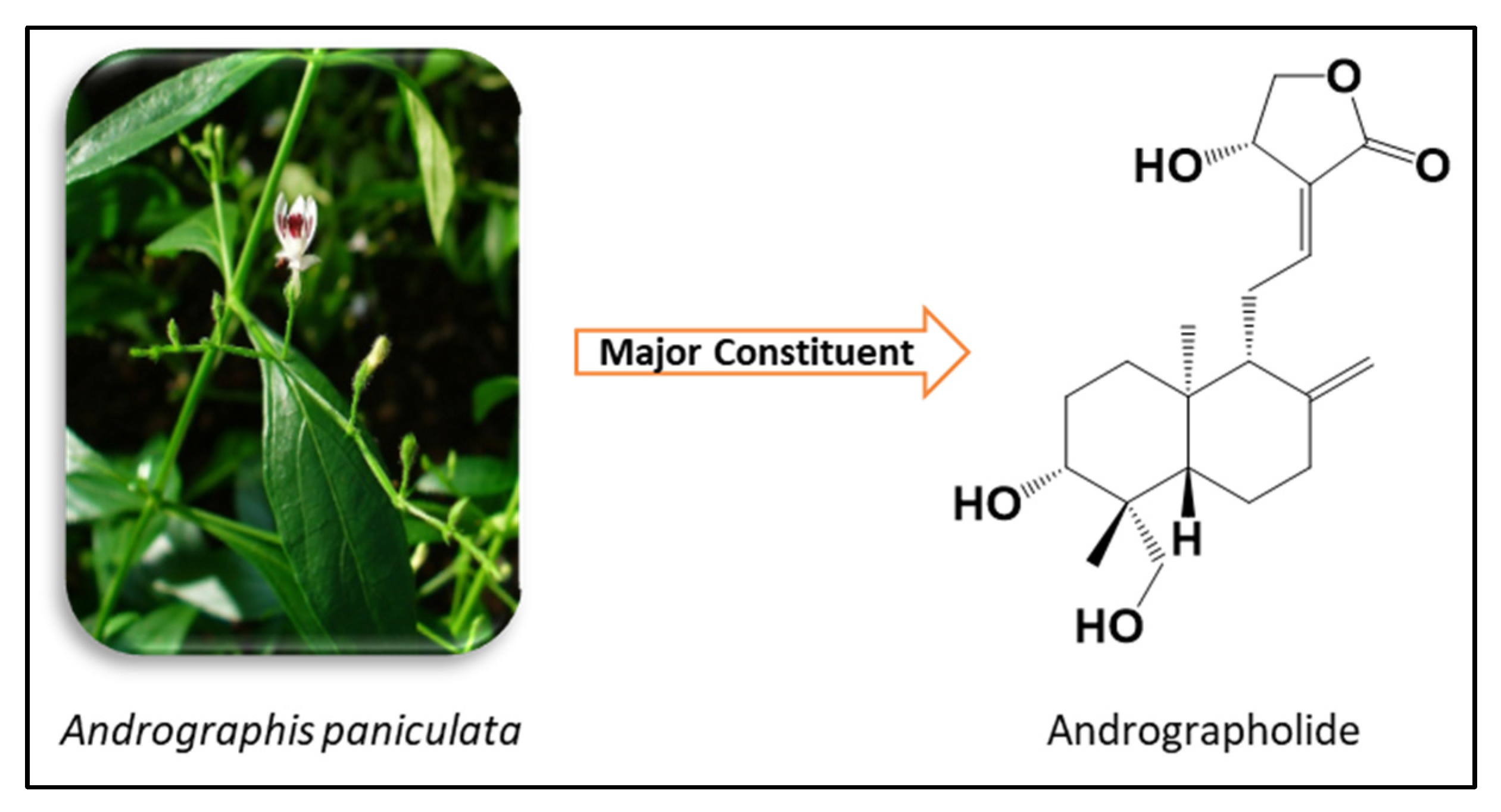
  19. Hossain, M.S.; Urbi, Z.; Sule, A.; Rahman, K.M.H. Andrographis paniculata (Burm. f.) Wall. Ex Nees: A Review of Ethnobotany, Phytochemistry, and Pharmacology. Sci. World J. 2014, 2014, 274905. [Google Scholar] [CrossRef]
  20. Jiang, M.; Sheng, F.; Zhang, Z.; Ma, X.; Gao, T.; Fu, C.; Li, P. Andrographis paniculata (Burm.f.) Nees and Its Major Constituent Andrographolide as Potential Antiviral Agents. J. Ethnopharmacol. 2021, 272, 113954. [Google Scholar] [CrossRef]
  21. Kandanur, S.G.S.; Tamang, N.; Golakoti, N.R.; Nanduri, S. Andrographolide: A Natural Product Template for the Generation of Structurally and Biologically Diverse Diterpenes. Eur. J. Med. Chem. 2019, 176, 513–533. [Google Scholar] [CrossRef]
  22. Jayakumar, T.; Hsieh, C.-Y.; Lee, J.-J.; Sheu, J.-R. Experimental and Clinical Pharmacology of Andrographis paniculata and Its Major Bioactive Phytoconstituent Andrographolide. Evid.-Based Complement. Altern. Med. 2013, 2013, 846740. [Google Scholar] [CrossRef]
  23. Bharati, B.D.; Sharma, P.K.; Kumar, N.; Dudhe, R.; Bansal, V. Pharmacological Activity of Andrographis paniculata: A Brief Review. Pharmacologyonline 2011, 2, 10. [Google Scholar]
  24. Okhuarobo, A.; Ehizogie Falodun, J.; Erharuyi, O.; Imieje, V.; Falodun, A.; Langer, P. Harnessing the Medicinal Properties of Andrographis paniculata for Diseases and beyond: A Review of Its Phytochemistry and Pharmacology. Asian Pacific J. Trop. Dis. 2014, 4, 213–222. [Google Scholar] [CrossRef]
  25. Jadhav, A.K.; Karuppayil, S.M. Andrographis paniculata (Burm. F) Wall Ex Nees: Antiviral Properties. Phyther. Res. 2021, 35, 5365–5373. [Google Scholar] [CrossRef] [PubMed]
  26. Wei, S.; Tang, Y.-B.; Hua, H.; Ohkoshi, E.; Goto, M.; Wang, L.-T.; Lee, K.-H.; Xiao, Z. Discovery of Novel Andrographolide Derivatives as Cytotoxic Agents. Bioorg. Med. Chem. Lett. 2013, 23, 4056–4060. [Google Scholar] [CrossRef]
  27. Suebsasana, S.; Pongnaratorn, P.; Sattayasai, J.; Arkaravichien, T.; Tiamkao, S.; Aromdee, C. Analgesic, Antipyretic, Anti-Inflammatory and Toxic Effects of Andrographolide Derivatives in Experimental Animals. Arch. Pharm. Res. 2009, 32, 1191–1200. [Google Scholar] [CrossRef] [PubMed]
  28. Liu, G.; Chu, H. Andrographolide Inhibits Proliferation and Induces Cell Cycle Arrest and Apoptosis in Human Melanoma Cells. Oncol. Lett. 2018, 15, 5301–5305. [Google Scholar] [CrossRef]
  29. Aromdee, C.; Sriubolmas, N.; Wiyakrutta, S.; Suebsasna, S.; Khunkitti, W. Effect of the Derivatives of Andrographolide on the Morphology of Bacillus subtilis. Arch. Pharm. Res. 2011, 34, 71–77. [Google Scholar] [CrossRef]
  30. Rajagopal, S.; Kumar, R.A.; Deevi, D.S.; Satyanarayana, C.; Rajagopalan, R. Andrographolide, a Potential Cancer Therapeutic Agent Isolated from Andrographis paniculata. J. Exp. Ther. Oncol. 2003, 3, 147–158. [Google Scholar] [CrossRef]
  31. Calabrese, C.; Berman, S.H.; Babish, J.G.; Ma, X.; Shinto, L.; Dorr, M.; Wells, K.; Wenner, C.A.; Standish, L.J. A Phase I Trial of Andrographolide in HIV Positive Patients and Normal Volunteers. Phyther. Res. 2000, 14, 333–338. [Google Scholar] [CrossRef]
  32. Chen, J.X.; Xue, H.J.; Ye, W.C.; Fang, B.H.; Liu, Y.H.; Yuan, S.H.; Yu, P.; Wang, Y.Q. Activity of Andrographolide and Its Derivatives against Influenza Virus in Vivo and in Vitro. Biol. Pharm. Bull. 2009, 32, 1385–1391. [Google Scholar] [CrossRef] [PubMed]
  33. Yu, B.C.; Chen, W.C.; Cheng, J.T. Antihyperglycemic Effect of Andrographolide in Streptozotocin-Induced Diabetic Rats. Planta Med. 2003, 69, 1075–1079. [Google Scholar] [PubMed]
  34. Wang, Z.; Yu, P.; Zhang, G.; Xu, L.; Wang, D.; Wang, L.; Zeng, X.; Wang, Y. Design, Synthesis and Antibacterial Activity of Novel Andrographolide Derivatives. Bioorg. Med. Chem. 2010, 18, 4269–4274. [Google Scholar] [CrossRef]
  35. Hu, J.; Li, Y.; Xie, X.; Song, Y.; Yan, W.; Luo, Y.; Jiang, Y. The Therapeutic Potential of Andrographolide in Cancer Treatment. Biomed. Pharmacother. 2024, 180, 117438. [Google Scholar] [CrossRef] [PubMed]
  36. Farooqi, A.A.; Attar, R.; Sabitaliyevich, U.Y.; Alaaeddine, N.; de Sousa, D.P.; Xu, B.; Cho, W.C. The Prowess of Andrographolide as a Natural Weapon in the War against Cancer. Cancers 2020, 12, 2159. [Google Scholar] [CrossRef]
  37. Mishra, S.K.; Tripathi, S.; Shukla, A.; Oh, S.H.; Kim, H.M. Andrographolide and Analogues in Cancer Prevention. Front. Biosci. 2015, 7, 255–266. [Google Scholar] [CrossRef]
  38. Malik, Z.; Parveen, R.; Parveen, B.; Zahiruddin, S.; Khan, M.A.; Khan, A.; Massey, S.; Ahmad, S.; Husain, S.A. Anticancer Potential of Andrographolide from Andrographis paniculata (Burm. f.) Nees and Its Mechanisms of Action. J. Ethnopharmacol. 2021, 272, 113936. [Google Scholar] [CrossRef]
  39. Kandanur, S.G.S.; Kundu, S.; Cadena, C.; Juan, H.S.; Bajaj, A.; Guzman, J.D.; Nanduri, S.; Golakoti, N.R. Design, Synthesis, and Biological Evaluation of New 12-Substituted-14-Deoxy-Andrographolide Derivatives as Apoptosis Inducers. Chem. Pap. 2019, 73, 1669–1675. [Google Scholar] [CrossRef]
  40. Elsheikh, M.A.; Rizk, S.A.; Elnaggar, Y.S.R.; Abdallah, O.Y. Nanoemulsomes for Enhanced Oral Bioavailability of the Anticancer Phytochemical Andrographolide: Characterization and Pharmacokinetics. AAPS PharmSciTech 2021, 22, 246. [Google Scholar] [CrossRef]
  41. Ebenezer, O.; Shapi, M.; Tuszynski, J.A. A Review of the Recent Development in the Synthesis and Biological Evaluations of Pyrazole Derivatives. Biomedicines 2022, 10, 1124. [Google Scholar] [CrossRef]
  42. Ansari, A.; Ali, A.; Asif, M. Biologically Active Pyrazole Derivatives. New J. Chem. 2017, 41, 16–41. [Google Scholar] [CrossRef]
  43. Bennani, F.E.; Doudach, L.; Cherrah, Y.; Ramli, Y.; Karrouchi, K.; Ansar, M.; Faouzi, M.E.A. Overview of Recent Developments of Pyrazole Derivatives as Anticancer Agents in Different Cell Lines. Bioorg. Chem. 2020, 97, 103470. [Google Scholar] [CrossRef] [PubMed]
  44. Kumar, H.; Saini, D.; Jain, S.; Jain, N. Pyrazole Scaffold: A Remarkable Tool in the Development of Anticancer Agents. Eur. J. Med. Chem. 2013, 70, 248–258. [Google Scholar] [CrossRef] [PubMed]
  45. Prabhu, V.V.; Guruvayoorappan, C. Anti-Inflammatory and Anti-Tumor Activity of the Marine Mangrove Rhizophora Apiculata. J. Immunotoxicol. 2012, 9, 341–352. [Google Scholar] [CrossRef]
  46. Rokkam, S.K.; Bhujel, M.; Jain, D.; Sripada, L.; Nanduri, S.; Bajaj, A.; Golakoti, N.R. Synthesis of Novel Pyrazole Acetals of Andrographolide and Isoandrographolide as Potent Anticancer Agents. RSC Adv. 2024, 14, 26625–26636. [Google Scholar] [CrossRef] [PubMed]
  47. Silverstein, R.M.; Webster, F.X.; Kiemle, D.J.; Bryce, D.L. Spectrometric Identification of Organic Compounds, 8th ed.; Wiley: Hoboken, NJ, USA, 2014. [Google Scholar]
  48. Pavia, D.L.; Lampman, G.M.; Kriz, G.S. Introduction to Spectroscopy—A Guide for Students of Organic Chemistry; Thomson Learning, Inc.: Chicago, IL, USA, 2001. [Google Scholar]
  49. Naasani, I. COMPARE Analysis, a Bioinformatic Approach to Accelerate Drug Repurposing against COVID-19 and Other Emerging Epidemics. SLAS Discov. Adv. Sci. Drug Discov. 2020, 26, 345–351. [Google Scholar] [CrossRef]
  50. Elmore, S. Apoptosis: A Review of Programmed Cell Death. Toxicol. Pathol. 2007, 35, 495–516. [Google Scholar] [CrossRef]
  51. Igney, F.H.; Krammer, P.H. Death and Anti-Death: Tumour Resistance to Apoptosis. Nat. Rev. Cancer 2002, 2, 277–288. [Google Scholar] [CrossRef]
  52. Hickman, J.A. Apoptosis Induced by Anticancer Drugs. Cancer Metastasis Rev. 1992, 11, 121–139. [Google Scholar] [CrossRef]
  53. Hengartner, M.O. The Biochemistry of Apoptosis. Nature 2000, 407, 770–776. [Google Scholar] [CrossRef] [PubMed]
  54. Klaunig, J.E. Oxidative Stress and Cancer. Curr. Pharm. Des. 2018, 24, 4771–4778. [Google Scholar] [CrossRef] [PubMed]
  55. Sosa, V.; Moliné, T.; Somoza, R.; Paciucci, R.; Kondoh, H.; Leonart, M.E. Oxidative Stress and Cancer: An Overview. Ageing Res. Rev. 2013, 12, 376–390. [Google Scholar] [CrossRef] [PubMed]
  56. Liou, G.Y.; Storz, P. Reactive Oxygen Species in Cancer. Free Radic. Res. 2010, 44, 479–496. [Google Scholar] [CrossRef] [PubMed]
  57. Pires, D.E.V.; Blundell, T.L.; Ascher, D.B. PkCSM: Predicting Small-Molecule Pharmacokinetic and Toxicity Properties Using Graph-Based Signatures. J. Med. Chem. 2015, 58, 4066–4072. [Google Scholar] [CrossRef]
  58. Ganesan, A. The Impact of Natural Products upon Modern Drug Discovery. Curr. Opin. Chem. Biol. 2008, 12, 306–317. [Google Scholar] [CrossRef]
  59. Kumar Rokkam, S.; Mas-Rosario, J.A.; Joshi, B.P.; Joshi, M.; Choudhury, A.R.; Kar, S.; Golakoti, N.R.; Farkas, M.E. Diarylidene-N-Methyl-4-Piperidones and Spirobibenzopyrans as Antioxidant and Anti-Inflammatory Agents. Chem. Biodivers. 2023, 20, e202300822. [Google Scholar] [CrossRef]
Figure 1. Andrographis paniculata and the chemical structure of andrographolide.
Pharmaceuticals 18 01026 g001
Scheme 1. Synthesis of 1–phenyl–2–(1–arylethylidene) hydrazines (AJ) and N–aryl–pyrazole–4–carboxaldehydes (aj).
Pharmaceuticals 18 01026 sch001
Scheme 2. Synthesis of 3,19–(N–phenyl–3–aryl–pyrazole) acetals of andrographolide (1a1j).
Pharmaceuticals 18 01026 sch002
Scheme 3. Synthesis of 3,19–(N–phenyl–3–aryl–pyrazole) acetals of isoandrographolide (2a2j).
Pharmaceuticals 18 01026 sch003
Figure 2. The dose-dependent cytotoxicity of compound 1f against the breast cancer cell line MDA-MB-231. t-Test was used to generate p-values in order to compute the difference between treated and untreated cells. Control (untreated) and treated cells with serially diluted concentrations. Statistical analysis between untreated and treated MDA-MB-231 cells. ** p ≤ 0.01, *** p ≤ 0.001, and ns p ≥ 0.05.
Pharmaceuticals 18 01026 g002
Figure 3. (a) The percentage of apoptotic cells at different time intervals following treatment with compound 1f. t-Test was used to generate p-values in order to compute the difference between treated and untreated cells. 0 h (untreated) and after treatment with 1f for 24 h, 48 h, and 72 h at a concentration of 3.33 µM. Statistical analysis between untreated and treated MDA-MB-231 cells. *** p ≤ 0.001, and ns p ≥ 0.05; (b) Total apoptotic cells induced by 1f: at 0 h (A), 24 h (B), 48 h (C), and 72 h (D). Colors in the figure indicate density of the cells: blue—lowest density, green—moderate density, yellow—high density, and red—highest density.
Pharmaceuticals 18 01026 g003
Figure 4. (a) The analysis of compound 1f on MDA-MB-231 cell cycle distribution: (A) control at 0 h; (B) after treatment for 24 h with 1f at a concentration of 3.33 µM; (C) after treatment for 24 h with 1f at a concentration of 5.0 µM; (D) after treatment for 24 h with 1f at a concentration of 6.66 µM. Colors in the figure indicate density of the cells at different phases: violet—G1 phase, yellow—S phase, green—G2 phase. (b) The statistical analysis of flow cytometry-obtained apoptosis (%) in MDA-MB-231 cell line. t-Test was used to generate p-values in order to compute the difference between treated and untreated scores. 0 h (untreated) and after treatment for 24 h with 1f at concentrations of 3.33 µM, 5.0 µM, and 6.66 µM. Statistical analysis between untreated and treated MDA-MB-231 cells. * p ≤ 0.05, ** p ≤ 0.01, *** p ≤ 0.001, and ns p ≥ 0.05.
Pharmaceuticals 18 01026 g004
Figure 5. The % DPPH radical scavenging activity of the compounds 1a1j and 2a2j.
Pharmaceuticals 18 01026 g005
Table 1. GI50s of compounds 1a1j against NCI human cancer cell line panel.
CancerSub PanelGI50 (µM)
1a1b1c1d1e1f1g1h1i1j
LeukemiaCCRF-CEM0.591.142.061.320.700.400.920.488.400.64
HL-60(TB)2.321.730.761.381.170.642.670.672.171.08
K-5622.972.162.142.412.892.343.672.073.821.90
MOLT-41.972.870.691.140.910.600.880.612.111.71
RPMI-82262.022.452.631.382.750.492.461.288.600.92
SR3.793.462.201.291.771.702.531.692.741.74
Non-Small-Cell LungA549/ATCC2.493.623.882.823.072.143.573.103.092.39
EKVX2.072.752.212.212.102.062.242.212.131.96
HOP-621.873.151.741.782.081.622.361.802.122.65
HOP-921.531.861.531.561.751.291.501.381.431.47
NCI-H2261.952.381.671.761.751.511.551.361.471.72
NCI-H231.691.831.791.751.901.701.731.711.761.71
NCI-H322M3.351.764.223.266.063.341.024.204.922.94
NCI-H4601.662.271.811.681.781.691.791.591.831.74
NCI-H5221.901.751.521.541.501.951.581.691.751.73
ColonCOLO 2052.012.412.061.982.091.482.301.701.971.95
HCC-29981.681.921.731.811.721.761.881.831.631.70
HCT-1161.611.993.481.041.240.391.200.943.911.40
HCT-151.721.741.571.581.601.611.591.601.591.58
HT292.371.821.701.491.662.381.692.161.901.70
KM122.061.992.261.671.831.842.291.683.011.66
SW-6201.732.215.711.531.724.49NTNT5.421.69
CNSSF-2683.207.652.271.602.022.362.392.112.611.67
SF-2951.641.681.521.421.361.551.501.541.581.43
SF-5391.841.741.771.631.781.771.721.761.791.69
SNB-191.853.64NT2.242.171.441.401.471.362.46
SNB-751.367.34NT1.121.571.661.691.511.711.15
U2511.841.741.811.591.692.362.392.112.611.66
MelanomaLOX IMVI1.541.721.441.611.641.321.581.551.431.57
MALME-3M1.731.781.871.471.871.581.821.602.461.54
M141.812.081.751.691.861.501.841.711.761.72
MDA-MB-4351.863.031.501.561.721.681.651.671.611.54
SK-MEL-21.741.781.791.571.781.691.771.761.791.57
SK-MEL-281.772.041.771.801.731.661.781.731.741.94
SK-MEL-51.401.631.651.631.541.621.701.601.591.54
UACC-2571.901.801.931.771.771.651.801.701.741.64
UACC-621.651.691.651.711.681.581.681.581.641.91
OvarianIGROV11.772.082.071.392.201.844.291.673.951.37
OVCAR-31.821.871.951.601.692.021.781.521.951.50
OVCAR-42.314.051.742.092.491.752.031.891.921.72
OVCAR-51.862.061.921.781.851.741.981.931.931.86
OVCAR-81.811.901.771.531.761.481.951.551.671.35
NCI/ADR-RES4.752.981.771.882.151.541.831.831.651.94
SK-OV-31.772.082.705.864.161.883.042.033.473.44
Renal786-02.033.041.782.312.591.601.992.071.772.30
A4982.341.904.132.904.333.315.204.354.032.63
ACHN1.641.891.801.641.611.661.892.071.831.52
CAKI-11.862.811.561.321.421.581.631.591.711.26
RXF 3931.401.621.641.051.181.601.791.711.490.89
SN12C1.872.931.591.651.571.551.671.511.661.98
TK-101.782.221.412.142.121.271.331.191.331.95
UO-311.351.821.781.301.451.601.992.071.771.22
ProstatePC-31.952.751.972.072.291.552.072.031.922.18
DU-1452.054.111.971.882.121.822.051.772.052.04
BreastMCF72.002.641.631.481.771.431.511.491.571.46
MDA-MB-2311.721.841.721.491.681.681.681.591.701.47
HS 578T2.312.691.661.911.941.511.751.521.851.86
BT-5491.641.941.301.571.461.311.491.471.201.65
T-47D1.421.761.391.632.291.191.311.181.141.80
MDA-MB-4681.691.761.631.741.821.532.001.931.851.71
Table 2. NCI compare analysis of compounds 1f, 1h, and 1j.
CompoundPearson Correlation Coefficient (PCC)Marketed DrugMechanism of Action
1fGI500.51NelarabineInhibits DNA elongation, apoptosis, and cellular destruction
1hGI500.60MelphalanInhibits DNA synthesis or transcription
1jGI500.50Trisenox 3DNA fragmentation characteristic of apoptosis in NB4 human promyelocytic leukemia cells
Table 3. Percentage of MDA-MB-231 cells in G1, S, and G2 phases before and after treatment with compound 1f.
TimeG1 (%)S (%)G2 (%)
0 h71.8 ± 1.457.29 ± 0.9720.2 ± 0.98
24 h (3.33 µM)77.36 ± 0.6914.4 ± 0.956.94 ± 0.28
24 h (5.00 µM)62.83 ± 1.226.5 ± 0.964.87 ± 0.26
24 h (6.66 µM)57.83 ± 1.221.9 ± 1.033.82 ± 0.20
Table 4. Lipinski parameters for active compounds.
Compound* MWLogPHBDHBATPSA
Ia580.295.691782.81
Ib625.285.55110125.95
Ic598.285.871782.81
Id614.256.281782.81
Ie658.206.381782.81
If598.286.351778.88
Ig614.256.271782.81
Ih658.206.381782.81
Ii594.316.091782.81
Ij610.305.711892.04
ADG350.451.9635148.93
* MW—molecular weight; LogP—partition coefficient; HBD—no. of hydrogen bond donors; HBA—no. of hydrogen bond acceptors; TPSA—Topological Polar Surface Area.
Disclaimer/Publisher’s Note: The statements, opinions and data contained in all publications are solely those of the individual author(s) and contributor(s) and not of MDPI and/or the editor(s). MDPI and/or the editor(s) disclaim responsibility for any injury to people or property resulting from any ideas, methods, instructions or products referred to in the content.

