Article

Radioiodinated Bicyclic RGD Peptide Derivatives for Enhanced Tumor Accumulation

1
Department of Biofunctional Analysis, Graduate School of Pharmaceutical Sciences, Osaka Medical and Pharmaceutical University, 4-20-1 Nasahara, Takatsuki 569-1094, Osaka, Japan
2
Division of Fundamental Technology Development, Near InfraRed Photo-ImmunoTherapy Institute, Kansai Medical University, 2-5-1 Shin-machi, Hirakata 573-1010, Osaka, Japan
*
Author to whom correspondence should be addressed.
Pharmaceuticals 2025, 18(4), 549; https://doi.org/10.3390/ph18040549
Submission received: 25 March 2025 / Revised: 4 April 2025 / Accepted: 7 April 2025 / Published: 8 April 2025
(This article belongs to the Special Issue Development of Novel Radiopharmaceuticals for SPECT and PET Imaging)

Abstract

:
Background/Objectives: Integrin αVβ3 plays a crucial role in tumor angiogenesis and cancer progression, making it a key target for radiolabeled probes used in imaging and therapy. A previously developed probe, [125I]bcRGD, exhibited high selectivity for αVβ3 but limited tumor accumulation due to rapid blood clearance. This study aimed to address this issue through two strategies: (1) conjugating albumin-binding molecules to enhance systemic circulation and (2) dimerizing RGD peptides to improve binding affinity via multivalency effects. Methods: Three [125I]bcRGD derivatives were synthesized: [125I]bcRGDpal (with palmitic acid), [125I]bcRGDiba (with 4-(p-iodophenyl)butyric acid), and [125I]bcRGDdimer (a dimeric bicyclic RGD peptide). Their physicochemical properties, αVβ3-selectivity, albumin-binding capacity, and biodistribution were assessed in vitro and in vivo using tumor-bearing mice. Tumor models included αVβ3-high U-87 MG and αVβ3-low A549 xenografts. Results: [125I]bcRGDpal and [125I]bcRGDiba exhibited prolonged blood retention (30-fold and 55-fold vs. [125I]bcRGD, respectively) and increased tumor accumulation (3.9% ID/g and 3.6% ID/g at 2 h, respectively). Despite improved systemic circulation, tumor-to-blood ratios remained low (<1), indicating limited tumor retention. [125I]bcRGDdimer achieved significantly greater tumor accumulation (4.2% ID/g at 2 h) and favorable tumor-to-blood (22) and tumor-to-muscle (14) ratios, with a 5.4-fold higher uptake in U-87 MG tumors compared to A549 tumors. Conclusions: Dimerization was more effective than albumin binding in enhancing bcRGD’s tumor-targeting potential. The dimeric probe demonstrated improved tumor accumulation, favorable pharmacokinetics, and preserved integrin selectivity. These findings provide a foundation for further structural optimization of bicyclic RGD peptides for integrin αVβ3-targeted imaging and therapy applications.

Graphical Abstract

1. Introduction

Integrin αVβ3 plays a pivotal role in tumor angiogenesis, as well as in cancer cell adhesion, migration, and invasion [1,2]. It is highly expressed on tumor endothelial cells, contributing to cancer progression and metastasis [3]. Overexpression of αVβ3 has been observed in various cancers, including breast, pancreatic, and cervical cancers, with expression levels correlating with tumor grade and prognosis [4,5].
Cyclic RGD peptides (cRGDs) are recognized ligands for integrin αVβ3 [6], leading to the development of numerous radiolabeled imaging probes [7,8,9]. Radiolabeled cRGD peptides are anticipated to play an important role in nuclear medicine imaging for the detection of αVβ3 and the evaluation of angiogenesis in cancer. Assessing cancer angiogenesis can aid in predicting the efficacy of angiogenesis inhibitors and in providing a more detailed understanding of cancer pathology [10,11]. However, cRGDs exhibit cross-reactivity with alternative integrin subtypes [10], particularly αVβ5, where structural homology impedes selective αVβ3 ligand development [12]. For instance, the inhibitor cilengitide, which is derived from the cRGD peptide and has undergone clinical investigation, inhibits both αVβ3 and αVβ5 [13]. Since integrins αVβ3 and αVβ5 are expressed in distinct regions within cancer tissues and play different biological roles, probes capable of distinguishing between these subtypes could greatly improve tumor characterization, prognosis prediction, and the development of targeted therapies [14,15,16]. This unmet need has driven the development of αVβ3-specific peptides [12,17] and engineered knottin-RGD constructs with enhanced binding kinetics [18].
Recent advances yielded a bicyclic RGD peptide (bcRGD) exhibiting remarkable αVβ3 selectivity [19]. The binding potency of bcRGD to αVβ3 was comparable to that of cyclo-(RGDfK), a classical cyclic RGD; however, the αVβ3Vβ5 selectivity ratio (ratio of corresponding IC50 values) was ≤0.003, significantly outperforming cyclo-(RGDfK) (0.2), cilengitide (4.7), and knottin-RGD (0.5) [19]. A radiolabeled probe based on this bicyclic peptide, [125I]bcRGD, was synthesized and shown to selectively accumulate in αVβ3-expressing cancer cells and xenograft models in mice [20]. However, like many low-molecular-weight peptides, [125I]bcRGD exhibited rapid blood clearance [21], resulting in minimal tumor accumulation in αVβ3-expressing tumors 120 min post-administration [20]. It was hypothesized that structural modifications to [125I]bcRGD, aimed at enhancing tumor accumulation, could significantly advance integrin αVβ3-targeted imaging and support the development of nuclear medicine therapeutics.
This study aimed to enhance tumor accumulation by employing two strategies. The first focused on prolonging blood retention, as extended systemic circulation increases radiopharmaceutical distribution to tumors. Incorporating albumin-binding molecules is a well-established approach for improving tumor accumulation by extending peptide half-life in circulation [22,23]. We selected 4-(p-iodophenyl)butyric acid (IBA) and palmitic acid (PAL), extensively studied as albumin-binding small molecules [24,25,26,27], and designed two [125I]-labeled peptides, [125I]bcRGDiba and [125I]bcRGDpal, with IBA or PAL at the N-terminal, whereas bcRGD retained an acetyl group (Figure 1 and Figure S1).
The second strategy utilized peptide dimerization to exploit the multivalency effect, where multiple binding sites significantly enhance receptor-binding affinity [28]. This approach has been shown to improve cancer cell targeting, with multimeric RGD peptides exhibiting superior binding affinity compared to monomeric counterparts [29,30]. In this study, bcRGD was modified at its N-terminal to introduce either an alkyne or azide group. Using a click reaction, a radiolabeled dimeric probe, [125I]bcRGDdimer, was synthesized, incorporating two bcRGD units (Figure 1 and Figure S1).
This study provides a foundational assessment of these novel radiolabeled probes to evaluate their potential for therapeutic and diagnostic applications targeting integrin αVβ3.

2. Results

2.1. Properties of Prepared Peptides

All peptides were synthesized with purities exceeding 99%. Electrospray ionization mass spectrometry (ESI-MS) data are presented in Table S1 and Figure S4a–h. Given the absence of other reactive functional groups—except for the ε-amino groups of the N-terminal Lys in bcRGDpal and bcRGDiba, and the amino group of the terminal β-alanine in bcRGDazide—these peptides were expected to undergo site-specific radioiodination by [125I]N-succinimidyl 3-iodobenzoate ([125I]SIB) (Figure 1). The radiochemical yield (RCY) for [125I]bcRGDpal and [125I] bcRGDiba from [125I]SIB was 59% and 70%, respectively, with radiochemical purities (RCP) exceeding 99% following high-performance liquid chromatography (HPLC) purification. The RCY for [125I]bcRGDazide from [125I]SIB was 40% with RCP > 99%, while the RCY for [125I]bcRGDdimer from [125I]bcRGDazide was 61% (total 24% from [125I]SIB) with RCP > 99%. [125I]bcRGDpal (retention time (TR) = 18.2 min, condition A), [125I]bcRGDiba (TR = 20.4 min, condition B), and [125I]bcRGDdimer (TR = 12.2 min, condition C). Each was fully separated from its unreacted precursor (bcRGDpal, TR = 13.1 min; bcRGDiba, TR = 12.4 min; bcRGDalkyne, TR = 10.8 min) by HPLC, allowing all radiolabeled peptides to be obtained in a non-carrier-added form, with molar activities estimated at 81 GBq/µmol.

2.2. In Vitro αVβ3 Selectivity

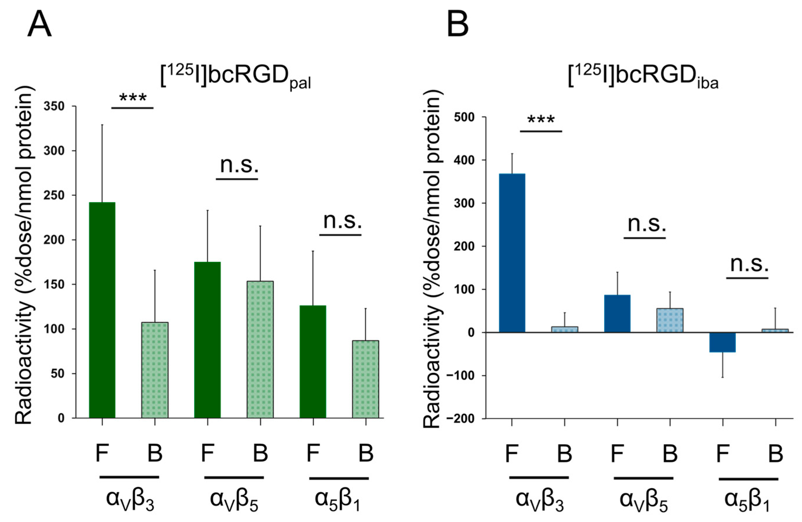
Both [125I]bcRGDpal and [125I]bcRGDiba exhibited high radioactivity accumulation in αVβ3, which was significantly inhibited by the broad-spectrum integrin inhibitory peptide cyclo-(RGDfK). No differences were observed in radioactivity accumulation for αVβ5 and α5β1 between treatment and inhibitor groups, confirming that [125I]bcRGDpal and [125I]bcRGDiba specifically recognized αVβ3 in vitro (Figure 2).

2.3. Octanol–Water Distribution Coefficient (Log D)

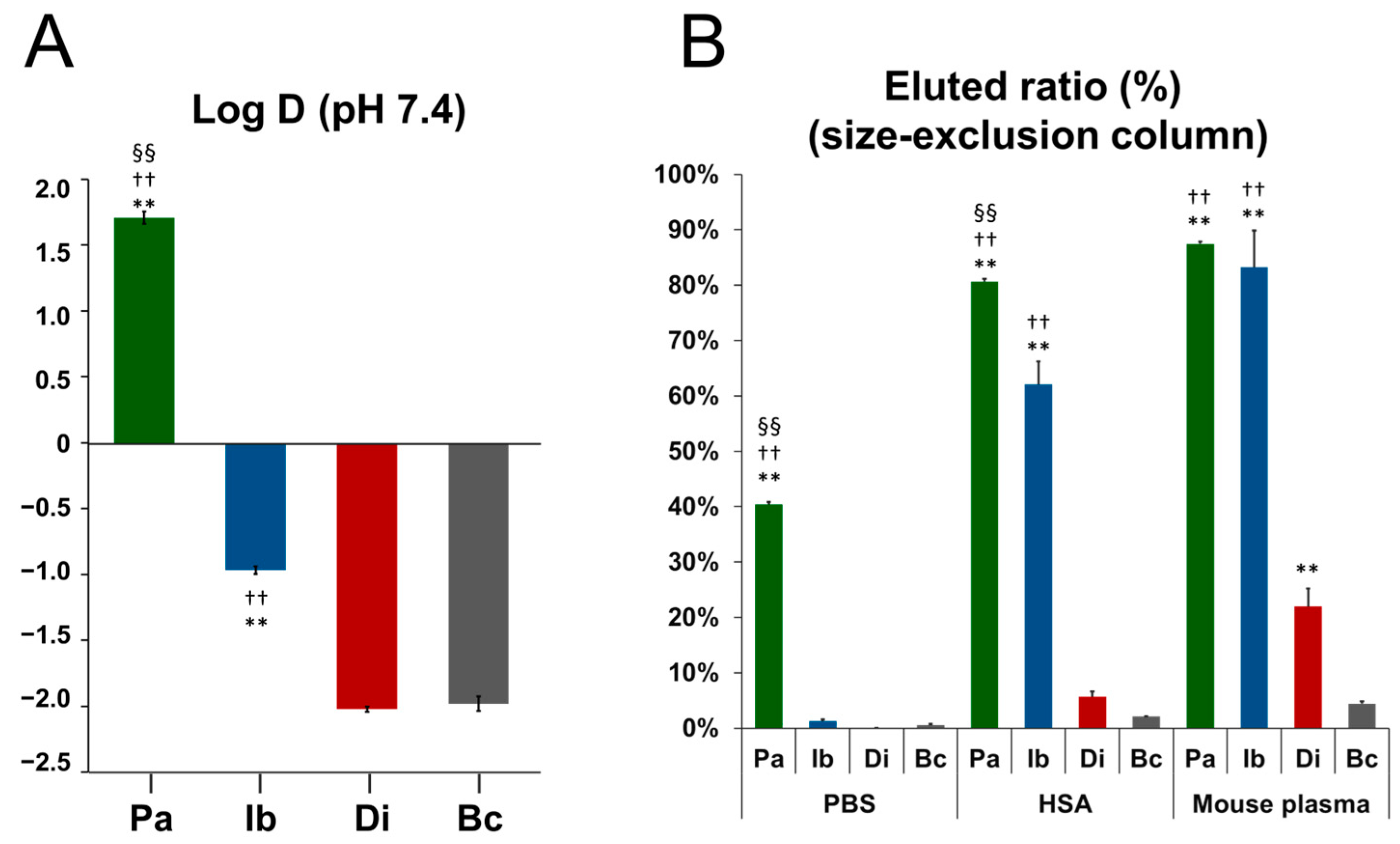
The log D values revealed that [125I]bcRGDpal (Log D = 1.71) and [125I]bcRGDiba (Log D = −0.97) were significantly higher than [125I]bcRGD (Log D = −2.02). [125I]bcRGDpal exhibited particularly high Log D values, significantly exceeding those of [125I]bcRGDiba, while [125I]bcRGDdimer (Log D = −1.98) showed no significant difference from [125I]bcRGD (Figure 3A).

2.4. Albumin Binding Property

Elution rates from the size-exclusion resin followed the order: PBS < human serum albumin (HSA) < mouse plasma (Figure 3B). In mouse plasma, [125I]bcRGD exhibited a negligible elution rate (2%), whereas [125I]bcRGDpal and [125I]bcRGDiba displayed high elution rates (>80%). [125I]bcRGDdimer showed moderate elution (22%), significantly higher than [125I]bcRGD. In HSA, [125I]bcRGDpal (80%) had a higher elution rate than [125I]bcRGDiba (60%), while [125I]bcRGDdimer exhibited a low elution rate (6%). In PBS, only [125I]bcRGDpal (40%) was eluted, with other peptides showing negligible elution.

2.5. In Vivo Study

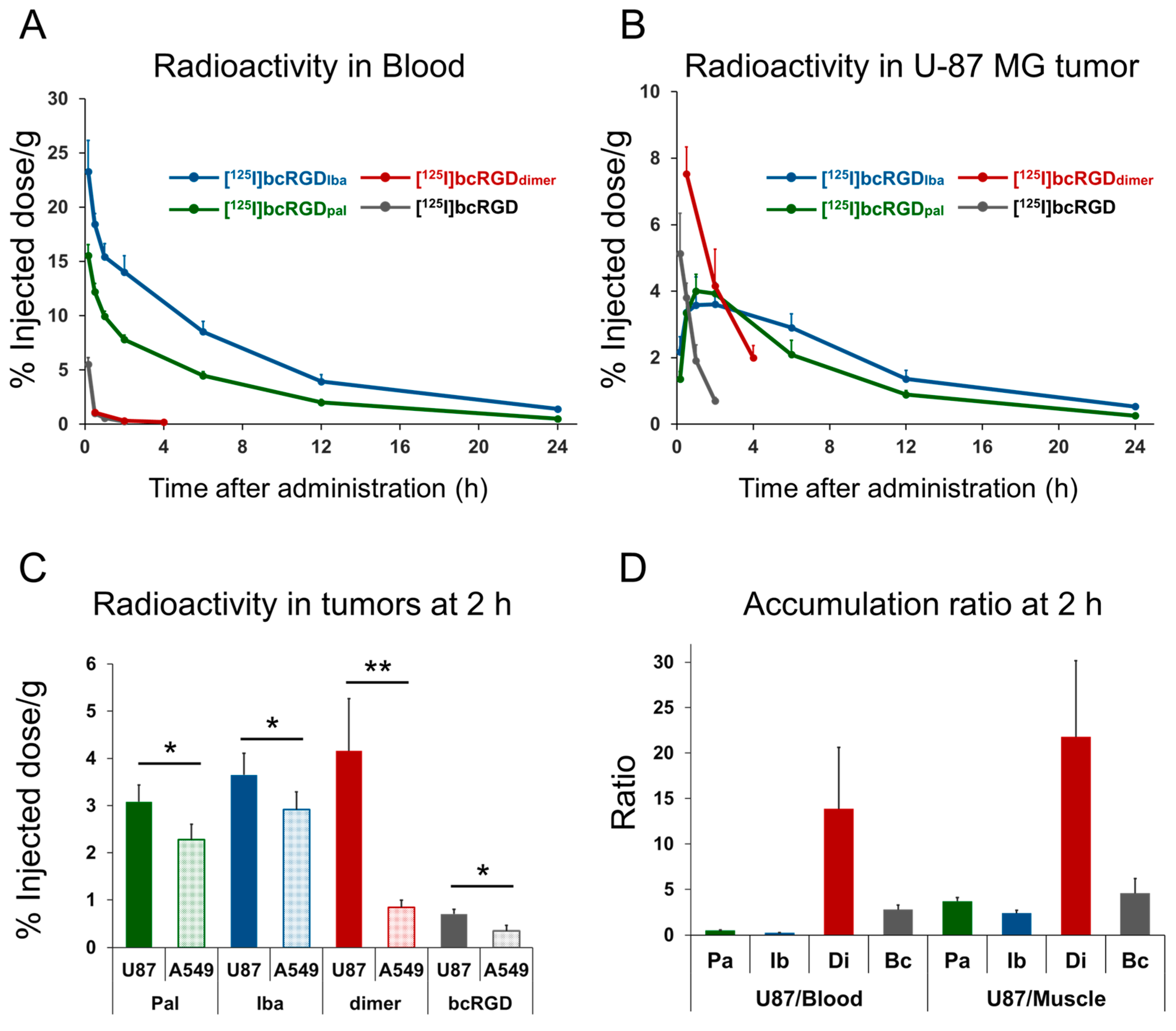
Immunohistochemical analysis confirmed high αVβ3 expression in U-87 MG cells and higher αVβ5 expression in A549 cells (Figure S2). Biodistribution data for [125I]bcRGDpal and [125I]bcRGDiba in αVβ3-positive U-87 MG-bearing mice are summarized in Table 1 and Table 2, respectively. Biodistribution data for [125I]bcRGDdimer in U-87 MG and A549 co-transplanted mice are presented in Table 3. Temporal comparisons of blood and tumor radioactivity post-administration are illustrated in Figure 4A,B.
Both [125I]bcRGDpal and [125I]bcRGDiba exhibited increased blood retention compared to [125I]bcRGD, with [125I]bcRGDiba demonstrating the longest retention. [125I]bcRGDdimer did not significantly differ from [125I]bcRGD in blood retention. At 2 h post-administration, blood radioactivity levels were 7.8% ID/g for [125I]bcRGDpal, 14% ID/g for [125I]bcRGDiba, 0.3% ID/g for [125I]bcRGDdimer, and 0.3% ID/g for [125I]bcRGD. [125I]bcRGDiba remained 55 times more radioactive in the blood than [125I]bcRGD, highlighting its prolonged systemic circulation.
Regarding tumor accumulation, [125I]bcRGDpal and [125I]bcRGDiba exhibited significantly higher radioactivity than [125I]bcRGD. At 2 h post-administration, tumor-accumulated radioactivity measured 3.9, 3.6, and 0.7% ID/g for [125I]bcRGDpal, [125I]bcRGDiba, and [125I]bcRGD, respectively. However, the tumor-to-blood radioactivity ratio remained below 1 at all time points for [125I]bcRGDpal and [125I]bcRGDiba. In contrast, [125I]bcRGDdimer demonstrated both greater tumor accumulation (4.2% ID/g at 2 h) and superior tumor-to-blood (22) and tumor-to-muscle (14) ratios compared to [125I]bcRGD (2.8 and 4.6, respectively). At 2 h post-administration, all probes showed significantly higher accumulation in αVβ3-high expressing U-87 MG tumors compared to low-expressing A549 tumors (Figure 4C,D). Notably, [125I]bcRGDdimer exhibited the highest accumulation ratio (U87/A549 = 5.4). With the exception of excretion-related organs, all peptides showed high accumulation in bone and lung, which are known to express integrin αVβ3 [31,32]. Minimal thyroid accumulation across all probes indicated negligible in vivo deiodination.

3. Discussion

Integrin αVβ3-targeted probes follow a defined trajectory from the plasma to the extracellular space, where they bind to integrin αVβ3 on tumor cell membranes and are occasionally internalized into tumor cells [33,34]. Enhancing tumor accumulation requires deliberate intervention in this process.
Our initial strategy to increase tumor accumulation focused on raising the concentration of radioactive probes in the plasma. Albumin binders, PAL and IBA, have been extensively explored as agents that prolong peptide circulation in the bloodstream [24,25,26,27]. PAL, a long-chain fatty acid, binds albumin at multiple sites, including subdomains IIIA and IIIB [35], while IBA is structurally optimized to bind tightly to Sudlow site II of HSA [36,37]. In our design, where PAL and IBA were conjugated to the N-terminal of bcRGD, both probes retained specific binding to αVβ3 in vitro and showed significantly higher accumulation in αVβ3-high expressing U-87 MG tumors compared to low-expressing A549 tumors in vivo, confirming preserved αVβ3 recognition (Figure 4C).
A key difference between [125I]bcRGDpal and [125I]bcRGDiba was their varied hydrophobicity, as reflected by their Log D values. [125I]bcRGDpal displayed the greatest lipophilicity (Log D > 1, Figure 3A), which may explain its nonspecific adsorption during in vitro protein-binding assays. This nonspecific interaction reduced the distinction in accumulation between the Free and Blocking groups, as well as between αVβ3 and other integrins (αVβ5 and α5β1). Additionally, size-exclusion chromatography indicated that [125I]bcRGDpal eluted as a high-molecular-weight fraction even in PBS (Figure 3B), suggesting possible self-assembly into larger structures in aqueous environments [38].
As anticipated, [125I]bcRGDpal and [125I]bcRGDiba showed a substantial increase in blood radioactivity (30-fold and 55-fold, respectively, compared to [125I]bcRGD at 2 h post-administration, Figure 4A). Although [125I]bcRGDpal exhibited higher protein-binding efficiency in vitro, in vivo data revealed that [125I]bcRGDiba achieved greater blood radioactivity retention. While detailed in vivo probe stability was not evaluated in this study, our results confirmed successful albumin binding and improved systemic exposure as designed. Tumor accumulation of both [125I]bcRGDpal and [125I]bcRGDiba also increased (5.6-fold and 5.1-fold vs. [125I]bcRGD at 2 h post-administration, Figure 4B); however, the tumor-to-blood radioactivity ratio remained persistently low. Unlike earlier studies utilizing albumin-binding molecules to improve tumor accumulation, which reported sustained tumor radioactivity even at later time points post-injection [24,25], our findings showed a marked decline in tumor accumulation for [125I]bcRGDpal and [125I]bcRGDiba at 24 h post-administration (0.3% ID/g and 0.5% ID/g, respectively), suggesting poor tumor retention.
The consistently low tumor-to-blood radioactivity ratio and reduced tumor accumulation at later time points imply that the binding equilibrium between αVβ3 and the probes may favor dissociation, pointing to an insufficient binding affinity for αVβ3. Additionally, limited intracellular radionuclide retention may contribute to this effect. Previous studies employed metallic radionuclides such as 68Ga and 177Lu [25,27,39], which demonstrated prolonged intracellular retention after internalization. In contrast, our study used non-metallic 125I, which is more prone to extracellular efflux than metallic radionuclides [40]. Future research may enhance tumor retention by labeling bcRGDpal or bcRGDiba with metallic radionuclides.
Our second strategy focused on enhancing the probe’s binding affinity to integrin αVβ3. Increased αVβ3 binding affinity is expected to improve tumor accumulation, tumor-to-blood ratio, and αVβ3-specific targeting [41]. Dimerization of integrin-targeted probes is a widely used approach to enhance affinity [42,43,44], and in this study, we explored the dimerization of bcRGD. This strategy ([125I]bcRGDdimer) resulted in minimal changes in pharmacokinetics but significantly elevated accumulation in αVβ3-positive U-87 MG tumors (Table 3). Importantly, [125I]bcRGDdimer demonstrated the highest accumulation ratio in U-87 MG tumors relative to A549 tumors, which exhibit low αVβ3 expression (Figure 4D), indicating that in vivo tumor accumulation correlates with integrin expression levels. In addition to the expected excretory organs (liver, kidneys, and intestine), notable radioactivity accumulation was observed in bone and lung tissues. This is consistent with the high expression of integrin αVβ3 in osteoclasts, where it regulates adhesion, migration, and bone resorption [31,45], as well as in pulmonary microvascular endothelial cells, where it contributes to inflammation and vascular permeability [32]. Under pathological conditions such as pulmonary fibrosis and acute lung injury, integrin αVβ3 is implicated in fibroblast activation and cytoskeletal remodeling—processes that promote tissue stiffening and fibrosis [46]. For [125I]bcRGDdimer, the lung-to-blood radioactivity ratio was 3.7 at 2 h post-administration, suggesting potential diagnostic value in these disease contexts.
This study represents the first attempt to dimerize bcRGD, resulting in enhanced tumor accumulation. Initially, we proposed a synthetic pathway where the peptide was dimerized via a click chemistry reaction before being radiolabeled with 125I; however, low overall yields necessitated a three-step synthesis approach (Figure S3). Optimization of reaction conditions will be crucial for improving yield in future studies. The observed improvement in tumor retention is likely due to an increased binding affinity to integrins. However, attempts to quantify this affinity using Ni-NTA beads with immobilized integrin proteins were unsuccessful, owing to nonspecific adsorption of [125I]bcRGDdimer to the beads. Future research should clarify the relationship between bcRGD multimerization and its binding affinity. Competitive inhibition assays between radiolabeled ligands (e.g., radiolabeled cRGD) and unlabeled bcRGDdimer would mitigate nonspecific adsorption interference and facilitate more precise affinity determination. Furthermore, multimerized bcRGD offers multiple avenues for structural refinement. For instance, strategies such as trimerization [29] and tetramerization [47], previously applied to cRGD probes, could be extended to bcRGD. Furthermore, as the spatial arrangement between binding motifs is critical for bivalency, optimizing the linker length remains an important consideration for the bcRGD dimer [48]. Incorporating metallic radionuclides via chelation may offer further benefits, including streamlined radiolabeling [49] and prolonged intracellular retention following internalization [40]. Combining these designs with albumin-binding molecules could further enhance tumor accumulation [50]. It is anticipated that the multimeric bcRGD, which demonstrated promising potential in this study, will evolve into more effective therapeutic and diagnostic probes through future structural refinements.
In conclusion, this study provides a comprehensive assessment of the physicochemical characteristics, pharmacokinetics, and tumor-targeting performance of PAL- and IBA-conjugated peptides, offering valuable insights into the structural engineering of bicyclic peptides for advanced applications. Moreover, this work constitutes the first report on bcRGD dimerization, detailing its pharmacokinetic profile and tumor accumulation behavior. These findings establish a foundation for further structural optimization of bcRGD, paving the way for its use in integrin αVβ3-targeted imaging and therapy. As such, the outcomes of this study may significantly contribute to progress in the diagnosis and treatment of integrin αVβ3-associated diseases.

4. Materials and Methods

4.1. Preparation of Peptides

All amino acids and coupling reagents were purchased from Watanabe Chemical Industries (Hiroshima, Japan) and used without further purification. The linear peptide sequences—palmitic acid-KPPPSG-Abz-SGCHPQcRGDc-NH2 (RGDpal; Abz, 4-amino-benzoic acid; c, D-Cys), 4-(p-iodophenyl)butyric acid-KPPPSG-Abz-SGCHPQcRGDc-NH2 (RGDiba), β-ala-γ-azidohomoalanine-KPPPSG-Abz-SGCHPQcRGDc-NH2 (RGDazide), and 4-pentynoic acid-KPPPSG-Abz-SGCHPQcRGDc-NH2 (RGDalkyne) were synthesized on Rink amide-PEG resin (A00213, Watanabe Chemical) using Fmoc solid-phase peptide synthesis. Peptides were cleaved from the resin using a cocktail of trifluoroacetic acid (TFA)/phenol/water/triisopropylsilane (88:5:5:2) and purified by reversed-phase HPLC (RP-HPLC) on a C18-reversed-phase column (COSMOSIL 5C18-AR-II, 10 mm ID × 250 mm; Nacalai Tesque, Kyoto, Japan) with UV detection to obtain the amount needed for the experiment. The mobile phase consisted of a linear gradient of 0.1% TFA in water and 0.1% TFA in acetonitrile, applied under three conditions: 70:30 to 10:90 (condition A), 90:10 to 30:70 (condition B), or 90:10 to 50:50 (condition C), over 30 min at a flow rate of 5.0 mL/min.
Purified linear peptides were dissolved in a 1:3 mixture of acetonitrile and water. To this solution, 1.1 equivalents of 1,3,5-tris(bromomethyl) benzene in acetonitrile were added, followed by homogenization. Ammonium carbonate (44 eq. in water) was then introduced, and the mixture was shaken for 60 min at room temperature. Bicyclic RGD peptides (bcRGDpal, bcRGDiba, bcRGDazide, and bcRGDalkyne) were subsequently purified via RP-HPLC. To synthesize I-bcRGDpal, I-bcRGDiba, and I-bcRGDazide, N-succinimidyl iodobenzoate (SIB) was prepared according to previous reports [51]. SIB (1.5 eq.) and bcRGD were dissolved in a 1:1 mixture of acetonitrile and borate buffer (0.1 M, pH 8.5), reacted at 40 °C for 1 h, and purified by RP-HPLC. For the synthesis of I-bcRGDdimer, equimolar amounts of I-bcRGDazide and bcRGDalkyne were dissolved in dry DMF with CuSO4·5H2O (50 eq.) and ascorbic acid (100 eq.), stirred for 1 h at room temperature, and purified by RP-HPLC.
The purified peptides were characterized by analytical HPLC under the same conditions as those used for semi-preparative HPLC, as well as by ESI-MS (LCMS-8045; Shimadzu, Kyoto, Japan). bcRGD was also synthesized following our previously reported method [20].

4.2. Radiolabeling

Na[125I]I was obtained from PerkinElmer Japan (Yokohama, Japan). [125I]SIB was prepared using previously described procedures in a no-carrier-added form (estimated molar activity: 81 GBq/μmol) [51]. For radiolabeling, bcRGDpal or bcRGDiba (200 μg) was dissolved in a mixture of borate buffer (100 μL, 0.1 M, pH 8.5) and acetonitrile (100 μL), and then added to [125I]SIB (10–20 MBq) dissolved in acetonitrile (100 μL). After 1 h incubation at 40 °C, the reaction mixture was purified by RP-HPLC to yield [125I]bcRGDpal or [125I]bcRGDiba.
To synthesize [125I]bcRGDdimer, [125I]bcRGDazide was prepared using the same procedure as for [125I]bcRGDpal and [125I]bcRGDiba. A reaction mixture consisting of [125I]bcRGDazide, CuSO4·5H2O (2.2 mg), ascorbic acid (3.1 mg), and bcRGDalkyne (200 μg) in dry DMF was stirred for 1 h at room temperature, followed by purification via RP-HPLC. The purified radiolabeled peptides were further analyzed by RP-HPLC to assess radiochemical purity.
The final purified solutions of [125I]bcRGDpal, [125I]bcRGDiba, and [125I]bcRGDdimer were dried under reduced pressure and reconstituted in an appropriate buffer for subsequent experiments. [125I]bcRGD was also synthesized according to our previously published method [20]. Detailed synthesis schemes and protocols for [125I]SIB and [125I]bcRGDdimer are provided in the Supplementary Information (Figure S3).

4.3. In Vitro Selectivity Assay

Binding assays were conducted following our previously reported method [20], using the Dynabeads™ His-Tag Isolation and Pulldown protocol (Thermo Fisher Scientific, Tokyo, Japan). Briefly, [125I]bcRGDpal or [125I]bcRGDiba (10 kBq/sample) was incubated at 37 °C for 2 h in 200 μL of incubation buffer (3.25 mM sodium phosphate, 70 mM NaCl, 0.01% Tween 20, pH 7.4) with 10 pmol of His-tagged integrin αVβ3, αVβ5, or α5β1 proteins (αVβ3: IT3-H52E3, ACRO Biosystems, Newark, DE, USA; αVβ5: 2528-AV, R&D Systems, Minneapolis, MN, USA; α5β1: CT014-H2508H, Sino Biological, Beijing, China). For the inhibition group, cyclo(RGDfK) (25 μM, S7834, Selleck, Houston, TX, USA) was preincubated with the integrin protein for 30 min at 37 °C. Next, A 2× binding buffer (100 mM sodium phosphate, 600 mM NaCl, 0.01% Tween 20, pH 8.0) was mixed with the solution at a 1:1 volume ratio and incubated with 5 μL of magnetic beads at 37 °C for 10 min to enable protein immobilization. Following immobilization, the beads were rinsed three times with the binding buffer, and radioactivity was measured using a NaI well-type scintillation counter (2480 Wizard2; PerkinElmer Japan, Yokohama, Japan). Protein binding was expressed as %dose/nmol protein.

4.4. Log D and Albumin Binding Measurement

For log D determination, [125I]bcRGD, [125I]bcRGDpal, [125I]bcRGDiba, or [125I]bcRGDdimer (40 kBq) was dissolved in 1.0 mL PBS(–) (pH 7.4) and mixed with 1.0 mL 1-octanol (1:1, v/v). The mixture was vortexed for 2 min and centrifuged at 5000 rpm for 5 min. Radioactivity in 0.5 mL of each phase was measured using a γ-counter, and log D values were calculated based on the radioactivity distribution (n = 4).
To assess albumin binding, mouse plasma was obtained by centrifugation of whole blood from male ddY mice (9 weeks old). [125I]bcRGD, [125I]bcRGDpal, [125I]bcRGDiba, or [125I]bcRGDdimer (37 kBq) in 50 μL PBS(–) was incubated at 37 °C for 10 min with 200 μL of PBS(–), mouse plasma, or human serum albumin (HSA, 45 mg/mL in PBS(–); Wako Pure Chemical Industries, Osaka, Japan). The mixtures were applied to gel filtration spin columns (Sephadex G-50 Fine; Cytiva, Tokyo, Japan), centrifuged (2000 rpm, 2 min), and radioactivity in the eluted fraction was measured with a γ-counter. The high molecular weight fraction was calculated as the elution ratio = (radioactivity in elution)/(added radioactivity) × 100 (%).

4.5. In Vivo Study

4.5.1. Cell Lines

Human glioblastoma U-87 MG cells, generously provided by Prof. Magata (Hamamatsu University School of Medicine), and human lung carcinoma A549 cells, sourced from ATCC (Manassas, VA, USA), were cultured in DMEM supplemented with 10% fetal bovine serum at 37 °C under a humidified 5% CO2 atmosphere. To assess αVβ3 and αVβ5 expression, the cells were fixed in 4% paraformaldehyde and incubated at room temperature for 2 h with either an anti-αVβ3 antibody (23C6, 5 μg/mL, Abcam) or an anti-αVβ5 antibody (P1F6, 5 μg/mL, Abcam, Cambridge, UK) as the primary antibody. After washing with PBS(−), the cells were exposed to an Alexa568-labeled secondary antibody (AB4600075, 2 μg/mL, Sigma-Aldrich, St. Louis, MO, USA) for 1 h at room temperature. For nuclear staining, Hoechst 33342 (5 μg/mL, Nacalai Tesque) was applied to the cells for 10 min at room temperature. Fluorescence images were acquired using a BZ-X810 fluorescence microscope (Keyence, Osaka, Japan).

4.5.2. Animal Preparation

Animal experiments were performed in compliance with institutional animal care regulations, with the study protocol approved by the Experimental Animal Committee of Osaka Medical and Pharmaceutical University (Approval Numbers: 21–76, 22–76, and 23–76). Four-week-old male BALB/c nu-nu mice (Japan SLC, Shizuoka, Japan) were housed under a 12 h light/12 h dark cycle with free access to food and water. Tumor-bearing mice were established by suspending U-87 MG cells (2 × 10⁶ cells/mouse) in PBS(–) and subcutaneously injecting 100 μL of the suspension into the right hind leg. For co-implantation models, A549 cells (2 × 10⁶ cells/mouse) were suspended in PBS(–) and inoculated (100 μL) into the left hind leg of the same mice already inoculated with U-87 MG cells. Mice with tumor size approximately 10 mm in diameter, 4–5 weeks after inoculation, were used for biodistribution studies. Animals in which either tumor was not viable were excluded from the experiment.

4.5.3. Biodistribution Study

U-87 MG-bearing mice (n = 42, 8–9 weeks old; 22–27 g) were randomly assigned into groups and administered [125I]bcRGDpal or [125I]bcRGDiba via intravenous injection (37 kBq in 100 μL of PBS containing 0.1% Tween 80 (PBS-T80)). At 5 min, 30 min, 1 h, 2 h, 6 h, 12 h, or 24 h post-injection, the mice were euthanized (n = 3 per time point). The heart, kidneys, liver, brain, pancreas, spleen, lung, small intestine, large intestine, muscle, skin, stomach, bone, and tumors were excised. Each organ’s weight and radioactivity were measured by a NaI well-type scintillation counter, and the % ID/g was calculated. To investigate the correlation between integrin expression and radioactivity accumulation, biodistribution at 2 h after administration of [125I]bcRGDpal or [125I]bcRGDiba was assessed in A549 and U-87 MG co-implantation mice (n = 5) using the same procedure. Additionally, co-implantation mice were injected with [125I]bcRGDdimer (37 kBq/100 μL PBS-T80) and euthanized at 30 min, 2 h, and 4 h post-injection (n = 4 per time point), and % ID/g values were calculated as described above. The minimum sample size needed for the biodistribution study in U-87 MG-bearing mice was calculated using G Power software version 3.1.9.7. The calculation was based on a two-tailed test, with an effect size of 3.9, a significance level (α) of 0.05, and a statistical power (1 − β) of 0.8. This analysis indicated that three animals per group were sufficient for the study. The minimum required sample size for the biodistribution study in A549 and U-87 MG co-implantation mice was also calculated to be four animals per group (effect size = 2.5, α = 0.05, 1 − β = 0.8). To guarantee objective results, the experiment was carried out in a blinded manner. Specifically, drug administration and radioactivity measurements were conducted by different investigators to eliminate potential bias.

4.6. Statistics

All data are expressed as means ± standard deviation. Statistical analyses were conducted using Tukey’s multiple comparisons test or unpaired t-test via GraphPad Prism 8 (GraphPad Software, Boston, MA, USA). Differences were considered statistically significant at the 95% confidence level (p < 0.05) unless otherwise indicated.

Supplementary Materials

The following supporting information can be downloaded at: https://www.mdpi.com/article/10.3390/ph18040549/s1, Figure S1: Chemical structures of peptides; Figure S2: Representative images of fluorescence immunostaining of αVβ3 and αVβ5 in U-87 MG and A549 cells; Figure S3: Radiosynthesis scheme of [125I]bcRGDdimer; Figure S4: Representative MS spectra for all newly synthesized peptides; Table S1: ESI-MS data of synthetic peptides; supplemental methods.

Author Contributions

Conceptualization, N.K.; Methodology, N.K.; Validation, N.K., M.K. and A.O.; Formal Analysis, N.K., A.M. and T.T.; Investigation, N.K., M.K., A.O., F.H. and A.M.; Resources, N.K., M.K. and A.O.; Data Curation, N.K. and T.T.; Writing—Original Draft Preparation, N.K.; Writing—Review and Editing, N.K., M.K., A.O., F.H., A.M. and T.T.; Visualization, N.K.; Supervision, T.T.; Project Administration, N.K. and T.T. All authors have read and agreed to the published version of the manuscript.

Funding

This research was funded by JSPS KAKENHI 19K17212 and 22K07732. The funding bodies had no role in the study design, data collection and analysis, decision to publish, or manuscript preparation.

Institutional Review Board Statement

Animal experiments were carried out according to the guidelines for animal experiments from the Osaka Medical and Pharmaceutical University. The study protocol was approved by the Experimental Animal Committee at Osaka Medical and Pharmaceutical University (Permission Number: 21–76, date of approval: 31 March 2021; 22–76, date of approval: 31 March 2022; and Permission Number: 23–76, date of approval: 31 March 2023).

Informed Consent Statement

Not applicable.

Data Availability Statement

The data supporting the results and findings of this study are available within the paper and the Supplementary Information Files. Additional raw data are available from the corresponding author upon request.

Acknowledgments

The authors thank Serina Mizuguchi for experimental assistance of radiolabeling.

Conflicts of Interest

The authors declare no conflicts of interest.

Abbreviations

The following abbreviations are used in this manuscript:
PALPalmitic acid
IBA4-(p-iodophenyl)butyric acid
RCYRadiochemical yield
RCPRadiochemical purity
HPLCHigh-performance liquid chromatography
ESI-MSElectrospray ionization mass spectrometry
PBSPhosphate-buffered saline
HSAHuman serum albumin

References

  1. Carmeliet, P.; Jain, R.K. Angiogenesis in cancer and other diseases. Nature 2000, 407, 249–257. [Google Scholar] [CrossRef] [PubMed]
  2. Takada, Y.; Ye, X.; Simon, S. The integrins. Genome Biol. 2007, 8, 215. [Google Scholar] [CrossRef] [PubMed]
  3. Liu, Z.; Wang, F.; Chen, X. Integrin αvβ3-Targeted Cancer Therapy. Drug Dev. Res. 2008, 69, 329–339. [Google Scholar] [CrossRef] [PubMed]
  4. Schnell, O.; Krebs, B.; Wagner, E.; Romagna, A.; Beer, A.J.; Grau, S.J.; Thon, N.; Goetz, C.; Kretzschmar, H.A.; Tonn, J.C.; et al. Expression of integrin αvβ3 in gliomas correlates with tumor grade and is not restricted to tumor vasculature. Brain Pathol. 2008, 18, 378–386. [Google Scholar] [CrossRef]
  5. Boger, C.; Warneke, V.S.; Behrens, H.M.; Kalthoff, H.; Goodman, S.L.; Becker, T.; Rocken, C. Integrins αvβ3 and αvβ5 as prognostic, diagnostic, and therapeutic targets in gastric cancer. Gastric Cancer 2015, 18, 784–795. [Google Scholar] [CrossRef]
  6. Danhier, F.; Le Breton, A.; Preat, V. RGD-based strategies to target αvβ3 integrin in cancer therapy and diagnosis. Mol. Pharm. 2012, 9, 2961–2973. [Google Scholar] [CrossRef]
  7. Krajcovicova, S.; Daniskova, A.; Bendova, K.; Novy, Z.; Soural, M.; Petrik, M. [68Ga]Ga-DFO-c(RGDyK): Synthesis and Evaluation of Its Potential for Tumor Imaging in Mice. Int. J. Mol. Sci. 2021, 22, 7391. [Google Scholar] [CrossRef]
  8. Ogawa, K.; Echigo, H.; Mishiro, K.; Hirata, S.; Washiyama, K.; Kitamura, Y.; Takahashi, K.; Shiba, K.; Kinuya, S. 68Ga- and 211At-Labeled RGD Peptides for Radiotheranostics with Multiradionuclides. Mol. Pharm. 2021, 18, 3553–3562. [Google Scholar] [CrossRef]
  9. Beer, A.J. Positron Emission Tomography Using [18F]Galacto-RGD Identifies the Level of Integrin αvβ3 Expression in Man. Clin. Cancer Res. 2006, 12, 3942–3949. [Google Scholar] [CrossRef]
  10. Chen, H.; Niu, G.; Wu, H.; Chen, X. Clinical Application of Radiolabeled RGD Peptides for PET Imaging of Integrin αvβ3. Theranostics 2016, 6, 78–92. [Google Scholar] [CrossRef]
  11. Haubner, R.; Maschauer, S.; Prante, O. PET radiopharmaceuticals for imaging integrin expression: Tracers in clinical studies and recent developments. BioMed Res. Int. 2014, 2014, 871609. [Google Scholar] [CrossRef]
  12. Mas-Moruno, C.; Beck, J.G.; Doedens, L.; Frank, A.O.; Marinelli, L.; Cosconati, S.; Novellino, E.; Kessler, H. Increasing αvβ3 selectivity of the anti-angiogenic drug cilengitide by N-methylation. Angew. Chem. Int. Ed. 2011, 50, 9496–9500. [Google Scholar] [CrossRef]
  13. Hariharan, S.; Gustafson, D.; Holden, S.; McConkey, D.; Davis, D.; Morrow, M.; Basche, M.; Gore, L.; Zang, C.; O’Bryant, C.L.; et al. Assessment of the biological and pharmacological effects of the αvβ3 and αvβ5 integrin receptor antagonist, cilengitide (EMD 121974), in patients with advanced solid tumors. Ann. Oncol. 2007, 18, 1400–1407. [Google Scholar] [CrossRef] [PubMed]
  14. Gvozdenovic, A.; Boro, A.; Meier, D.; Bode-Lesniewska, B.; Born, W.; Muff, R.; Fuchs, B. Targeting αvβ3 and αvβ5 integrins inhibits pulmonary metastasis in an intratibial xenograft osteosarcoma mouse model. Oncotarget 2016, 7, 55141–55154. [Google Scholar] [CrossRef]
  15. Fabricius, E.M.; Wildner, G.P.; Kruse-Boitschenko, U.; Hoffmeister, B.; Goodman, S.L.; Raguse, J.D. Immunohistochemical analysis of integrins αvβ3, αvβ5 and α5β1, and their ligands, fibrinogen, fibronectin, osteopontin and vitronectin, in frozen sections of human oral head and neck squamous cell carcinomas. Exp. Ther. Med. 2011, 2, 9–19. [Google Scholar] [CrossRef]
  16. Friedlander, M.; Brooks, P.C.; Shaffer, R.W.; Kincaid, C.M.; Varner, J.A.; Cheresh, D.A. Definition of two angiogenic pathways by distinct αv integrins. Science 1995, 270, 1500–1502. [Google Scholar] [CrossRef]
  17. Ma, Y.; Ai, G.; Zhang, C.; Zhao, M.; Dong, X.; Han, Z.; Wang, Z.; Zhang, M.; Liu, Y.; Gao, W.; et al. Novel Linear Peptides with High Affinity to αvβ3 Integrin for Precise Tumor Identification. Theranostics 2017, 7, 1511–1523. [Google Scholar] [CrossRef]
  18. Silverman, A.P.; Levin, A.M.; Lahti, J.L.; Cochran, J.R. Engineered cystine-knot peptides that bind αvβ3 integrin with antibody-like affinities. J. Mol. Biol. 2009, 385, 1064–1075. [Google Scholar] [CrossRef]
  19. Bernhagen, D.; Jungbluth, V.; Quilis, N.G.; Dostalek, J.; White, P.B.; Jalink, K.; Timmerman, P. Bicyclic RGD Peptides with Exquisite Selectivity for the Integrin αvβ3 Receptor Using a “Random Design” Approach. ACS Comb. Sci. 2019, 21, 198–206. [Google Scholar] [CrossRef]
  20. Kondo, N.; Wakamori, K.; Hirata, M.; Temma, T. Radioiodinated bicyclic RGD peptide for imaging integrin αvβ3 in cancers. Biochem. Biophys. Res. Commun. 2020, 528, 168–173. [Google Scholar] [CrossRef]
  21. Fani, M.; Maecke, H.R.; Okarvi, S.M. Radiolabeled peptides: Valuable tools for the detection and treatment of cancer. Theranostics 2012, 2, 481–501. [Google Scholar] [CrossRef] [PubMed]
  22. Zorzi, A.; Middendorp, S.J.; Wilbs, J.; Deyle, K.; Heinis, C. Acylated heptapeptide binds albumin with high affinity and application as tag furnishes long-acting peptides. Nat. Commun. 2017, 8, 16092. [Google Scholar] [CrossRef] [PubMed]
  23. Davis, R.A.; Hausner, S.H.; Harris, R.; Sutcliffe, J.L. A Comparison of Evans Blue and 4-(p-Iodophenyl)butyryl Albumin Binding Moieties on an Integrin αvβ6 Binding Peptide. Pharmaceutics 2022, 14, 745. [Google Scholar] [CrossRef]
  24. Xu, M.; Zhang, P.; Ding, J.; Chen, J.; Huo, L.; Liu, Z. Albumin Binder-Conjugated Fibroblast Activation Protein Inhibitor Radiopharmaceuticals for Cancer Therapy. J. Nucl. Med. 2022, 63, 952–958. [Google Scholar] [CrossRef]
  25. Eder, M.; Pavan, S.; Bauder-Wust, U.; van Rietschoten, K.; Baranski, A.C.; Harrison, H.; Campbell, S.; Stace, C.L.; Walker, E.H.; Chen, L.; et al. Bicyclic Peptides as a New Modality for Imaging and Targeting of Proteins Overexpressed by Tumors. Cancer Res. 2019, 79, 841–852. [Google Scholar] [CrossRef]
  26. Holtke, C.; Alsibai, W.; Grewer, M.; Stolting, M.; Geyer, C.; Eisenblatter, M.; Wildgruber, M.; Helfen, A. How Different Albumin-Binders Drive Probe Distribution of Fluorescent RGD Mimetics. Front. Chem. 2021, 9, 689850. [Google Scholar] [CrossRef]
  27. Zhang, P.; Xu, M.; Ding, J.; Chen, J.; Zhang, T.; Huo, L.; Liu, Z. Fatty acid-conjugated radiopharmaceuticals for fibroblast activation protein-targeted radiotherapy. Eur. J. Nucl. Med. Mol. Imaging 2022, 49, 1985–1996. [Google Scholar] [CrossRef]
  28. Deng, Y.; Efremov, A.K.; Yan, J. Modulating binding affinity, specificity, and configurations by multivalent interactions. Biophys. J. 2022, 121, 1868–1880. [Google Scholar] [CrossRef]
  29. Kaeopookum, P.; Petrik, M.; Summer, D.; Klinger, M.; Zhai, C.; Rangger, C.; Haubner, R.; Haas, H.; Hajduch, M.; Decristoforo, C. Comparison of 68Ga-labeled RGD mono- and multimers based on a clickable siderophore-based scaffold. Nucl. Med. Biol. 2019, 78–79, 1–10. [Google Scholar] [CrossRef]
  30. Liu, S. Radiolabeled Cyclic RGD Peptide Bioconjugates as Radiotracers Targeting Multiple Integrins. Bioconjug. Chem. 2015, 26, 1413–1438. [Google Scholar] [CrossRef]
  31. Nakamura, I.; Duong, L.T.; Rodan, S.B.; Rodan, G.A. Involvement of αvβ3 integrins in osteoclast function. J. Bone Miner. Metab. 2007, 25, 337–344. [Google Scholar] [CrossRef] [PubMed]
  32. Singh, B.; Fu, C.; Bhattacharya, J. Vascular expression of the αvβ3-integrin in lung and other organs. Am. J. Physiol. Lung Cell. Mol. Physiol. 2000, 278, L217–L226. [Google Scholar] [CrossRef] [PubMed]
  33. Menke-van der Houven van Oordt, C.W.; McGeoch, A.; Bergstrom, M.; McSherry, I.; Smith, D.A.; Cleveland, M.; Al-Azzam, W.; Chen, L.; Verheul, H.; Hoekstra, O.S.; et al. Immuno-PET Imaging to Assess Target Engagement: Experience from 89Zr-Anti-HER3 mAb (GSK2849330) in Patients with Solid Tumors. J. Nucl. Med. 2019, 60, 902–909. [Google Scholar] [CrossRef]
  34. Wang, T.; Deng, Y.; Wang, S.; He, J.; Wang, S. Kinetic 18F-FDG PET/CT imaging of hepatocellular carcinoma: A dual input four-compartment model. EJNMMI Phys. 2024, 11, 20. [Google Scholar] [CrossRef]
  35. Simard, J.R.; Zunszain, P.A.; Ha, C.E.; Yang, J.S.; Bhagavan, N.V.; Petitpas, I.; Curry, S.; Hamilton, J.A. Locating high-affinity fatty acid-binding sites on albumin by X-ray crystallography and NMR spectroscopy. Proc. Natl. Acad. Sci. USA 2005, 102, 17958–17963. [Google Scholar] [CrossRef]
  36. Yu, C.; Xiao, E.; Xu, P.; Lin, J.; Hu, L.; Zhang, J.; Dai, S.; Ding, Z.; Xiao, Y.; Chen, Z. Novel albumin-binding photothermal agent ICG-IBA-RGD for targeted fluorescent imaging and photothermal therapy of cancer. RSC Adv. 2021, 11, 7226–7230. [Google Scholar] [CrossRef]
  37. Busslinger, S.D.; Becker, A.E.; Vaccarin, C.; Deberle, L.M.; Renz, M.L.; Groehn, V.; Schibli, R.; Muller, C. Investigations Using Albumin Binders to Modify the Tissue Distribution Profile of Radiopharmaceuticals Exemplified with Folate Radioconjugates. Cancers 2023, 15, 4259. [Google Scholar] [CrossRef]
  38. Deshmukh, S.A.; Solomon, L.A.; Kamath, G.; Fry, H.C.; Sankaranarayanan, S.K. Water ordering controls the dynamic equilibrium of micelle-fibre formation in self-assembly of peptide amphiphiles. Nat. Commun. 2016, 7, 12367. [Google Scholar] [CrossRef]
  39. Choy, C.J.; Ling, X.; Geruntho, J.J.; Beyer, S.K.; Latoche, J.D.; Langton-Webster, B.; Anderson, C.J.; Berkman, C.E. 177Lu-Labeled Phosphoramidate-Based PSMA Inhibitors: The Effect of an Albumin Binder on Biodistribution and Therapeutic Efficacy in Prostate Tumor-Bearing Mice. Theranostics 2017, 7, 1928–1939. [Google Scholar] [CrossRef]
  40. Sugyo, A.; Tsuji, A.B.; Sudo, H.; Nagatsu, K.; Koizumi, M.; Ukai, Y.; Kurosawa, G.; Zhang, M.R.; Kurosawa, Y.; Saga, T. Evaluation of 89Zr-Labeled Human Anti-CD147 Monoclonal Antibody as a Positron Emission Tomography Probe in a Mouse Model of Pancreatic Cancer. PLoS ONE 2013, 8, e61230. [Google Scholar] [CrossRef]
  41. Tolmachev, V.; Tran, T.A.; Rosik, D.; Sjoberg, A.; Abrahmsen, L.; Orlova, A. Tumor targeting using affibody molecules: Interplay of affinity, target expression level, and binding site composition. J. Nucl. Med. 2012, 53, 953–960. [Google Scholar] [CrossRef] [PubMed]
  42. Pallarola, D.; Platzman, I.; Bochen, A.; Cavalcanti-Adam, E.A.; Axmann, M.; Kessler, H.; Geiger, B.; Spatz, J.P. Focal adhesion stabilization by enhanced integrin-cRGD binding affinity. BioNanoMaterials 2017, 18, 20160014. [Google Scholar] [CrossRef]
  43. Hedhli, J.; Czerwinski, A.; Schuelke, M.; Ploska, A.; Sowinski, P.; Hood, L.; Mamer, S.B.; Cole, J.A.; Czaplewska, P.; Banach, M.; et al. Synthesis, Chemical Characterization and Multiscale Biological Evaluation of a Dimeric-cRGD Peptide for Targeted Imaging of αvβ3 Integrin Activity. Sci. Rep. 2017, 7, 3185. [Google Scholar] [CrossRef] [PubMed]
  44. Echigo, H.; Mishiro, K.; Fuchigami, T.; Shiba, K.; Kinuya, S.; Ogawa, K. Synthesis and Evaluation of a Dimeric RGD Peptide as a Preliminary Study for Radiotheranostics with Radiohalogens. Molecules 2021, 26, 6107. [Google Scholar] [CrossRef]
  45. Nakamura, I.; Pilkington, M.F.; Lakkakorpi, P.T.; Lipfert, L.; Sims, S.M.; Dixon, S.J.; Rodan, G.A.; Duong, L.T. Role of αvβ3 integrin in osteoclast migration and formation of the sealing zone. J. Cell Sci. 1999, 112 Pt 22, 3985–3993. [Google Scholar] [CrossRef]
  46. Fiore, V.F.; Wong, S.S.; Tran, C.; Tan, C.; Xu, W.; Sulchek, T.; White, E.S.; Hagood, J.S.; Barker, T.H. αvβ3 Integrin drives fibroblast contraction and strain stiffening of soft provisional matrix during progressive fibrosis. JCI Insight 2018, 3, e97597. [Google Scholar] [CrossRef]
  47. Liolios, C.; Sachpekidis, C.; Kolocouris, A.; Dimitrakopoulou-Strauss, A.; Bouziotis, P. PET Diagnostic Molecules Utilizing Multimeric Cyclic RGD Peptide Analogs for Imaging Integrin αvβ3 Receptors. Molecules 2021, 26, 1792. [Google Scholar] [CrossRef]
  48. Ye, X.; Gaucher, J.F.; Hu, H.; Wang, L.; Broussy, S. Dimer Peptide Ligands of Vascular Endothelial Growth Factor: Optimizing Linker Length for High Affinity and Antiangiogenic Activity. J. Med. Chem. 2023, 66, 9753–9765. [Google Scholar] [CrossRef]
  49. Brandt, M.; Cardinale, J.; Aulsebrook, M.L.; Gasser, G.; Mindt, T.L. An Overview of PET Radiochemistry, Part 2: Radiometals. J. Nucl. Med. 2018, 59, 1500–1506. [Google Scholar] [CrossRef]
  50. Yang, G.; Gao, H.; Luo, C.; Zhao, X.; Luo, Q.; Shi, J.; Wang, F. Palmitic Acid-Conjugated Radiopharmaceutical for Integrin αvβ3-Targeted Radionuclide Therapy. Pharmaceutics 2022, 14, 1327. [Google Scholar] [CrossRef]
  51. Kondo, Y.; Kimura, H.; Fukumoto, C.; Yagi, Y.; Hattori, Y.; Kawashima, H.; Yasui, H. Copper-mediated radioiodination reaction through aryl boronic acid or ester precursor and its application to direct radiolabeling of a cyclic peptide. J. Label. Compd. Radiopharm. 2021, 64, 336–345. [Google Scholar] [CrossRef]
Figure 1. Schematic representation of radioiodinated bcRGD derivatives. Structure and color correspond in all figures; [125I]bcRGDpal (green), [125I]bcRGDiba (blue), [125I]bcRGDdimer (red), and [125I]bcRGD (gray).
Pharmaceuticals 18 00549 g001
Figure 2. Radioactivity accumulation in αVβ3, αVβ5, and α5β1 proteins after 2 h of incubation with [125I]bcRGDpal (A) and [125I]bcRGDiba (B). F (Free): without cyclo-(RGDfK); B (Blocking): with cyclo-(RGDfK) (25 μM). Data are expressed as mean ± standard deviation and analyzed using an unpaired t-test. *** p < 0.001 compared to the corresponding blocking group. n.s.: not significant.
Pharmaceuticals 18 00549 g002
Figure 3. Physicochemical properties of radioiodinated bcRGD derivatives. (A) Octanol–water distribution coefficient (log D) determined by radioactivity distribution. (B) Radioactivity elution ratio from a size-exclusion column after incubation with PBS, HSA, and mouse plasma. Data are expressed as mean ± standard deviation and analyzed using Tukey’s multiple comparison test. ** p < 0.01, compared to Bc, †† p < 0.01 compared to Di, §§ p < 0.01 compared to Ib. Pa: [125I]bcRGDpal; Ib: [125I]bcRGDiba; Di: [125I]bcRGDdimer; Bc: [125I]bcRGD.
Pharmaceuticals 18 00549 g003
Figure 4. Biodistribution of radioiodinated bcRGD derivatives in tumor-bearing mice following intravenous administration. Radioactivity levels in (A) blood and (B) U-87 MG tumors. (C) Radioactivity distribution in U-87 MG and A549 tumors at 2 h post-administration. Data are expressed as mean ± standard deviation and analyzed using an unpaired t-test. * p < 0.05, ** p < 0.01, compared to the corresponding accumulation in A549. (D) U-87 MG-to-blood and U-87 MG-to-muscle radioactivity ratios at 2 h post-administration. Data are presented as mean ± standard deviation.
Pharmaceuticals 18 00549 g004
Table 1. Biodistribution of radioactivity following [125I]bcRGDpal administration in tumor-bearing mice (% injected dose per gram of tissue).
Time After Administration
5 min30 min1 h2 h6 h12 h24 h
Blood15.6 ± 1.012.2 ± 0.89.9 ± 0.57.8 ± 0.44.5 ± 0.42.0 ± 0.30.5 ± 0.0
Heart6.6 ± 0.14.7 ± 0.44.3 ± 0.33.1 ± 0.21.7 ± 0.00.8 ± 0.10.2 ± 0.0
Lung11.8 ± 1.311.2 ± 1.09.6 ± 1.37.8 ± 1.04.4 ± 0.52.2 ± 0.30.6 ± 0.1
Liver17.6 ± 0.919.0 ± 2.115.8 ± 0.912.3 ± 0.36.0 ± 0.21.9 ± 0.20.5 ± 0.0
Kidneys9.0 ± 0.28.3 ± 0.97.7 ± 0.26.7 ± 0.64.2 ± 0.61.7 ± 0.20.4 ± 0.0
Stomach 0.5 ± 0.10.6 ± 0.10.7 ± 0.10.8 ± 0.10.4 ± 0.00.4 ± 0.30.1 ± 0.1
Small intestine2.3 ± 0.24.5 ± 0.67.0 ± 0.39.3 ± 0.94.0 ± 0.21.9 ± 0.50.3 ± 0.0
Large intestine0.9 ± 0.11.4 ± 0.21.4 ± 0.16.9 ± 1.111.8 ± 2.07.5 ± 0.82.3 ± 1.1
Pancreas2.8 ± 0.22.5 ± 0.31.9 ± 0.11.6 ± 0.11.2 ± 0.30.4 ± 0.00.1 ± 0.0
Spleen5.1 ± 0.45.9 ± 0.84.6 ± 0.63.7 ± 0.11.9 ± 0.20.8 ± 0.10.2 ± 0.0
Muscle1.4 ± 0.31.6 ± 0.21.4 ± 0.21.1 ± 0.10.6 ± 0.10.3 ± 0.00.1 ± 0.0
Bone1.4 ± 0.11.7 ± 0.61.1 ± 0.31.3 ± 0.40.7 ± 0.10.4 ± 0.00.1 ± 0.1
Brain0.5 ± 0.10.3 ± 0.10.3 ± 0.00.2 ± 0.00.1 ± 0.00.1 ± 0.00.0 ± 0.0
Thyroid 0.1 ± 0.00.1 ± 0.10.1 ± 0.10.1 ± 0.10.1 ± 0.00.0 ± 0.00.0 ± 0.0
U-87 MG1.4 ± 0.23.4 ± 0.54.0 ± 0.53.9 ± 0.22.1 ± 0.40.9 ± 0.10.3 ± 0.1
U87/Blood0.1 ± 0.00.3 ± 0.00.4 ± 0.10.5 ± 0.00.5 ± 0.10.4 ± 0.00.5 ± 0.1
U87/Muscle3.0 ± 0.12.1 ± 0.32.9 ± 0.23.7 ± 0.43.3 ± 0.83.4 ± 0.52.4 ± 0.4
Expressed as % injected dose. Data are expressed as mean ± standard deviation (n = 3).
Table 2. Biodistribution of radioactivity following [125I]bcRGDiba administration in tumor-bearing mice (% injected dose per gram of tissue).
Time After Administration
5 min30 min1 h2 h6 h12 h24 h
Blood23.3 ± 2.918.5 ± 1.015.4 ± 1.214.0 ± 1.58.5 ± 1.03.9 ± 0.71.4 ± 0.1
Heart7.4 ± 1.45.3 ± 0.54.5 ± 0.84.2 ± 0.12.7 ± 0.21.3 ± 0.10.5 ± 0.0
Lung10.7 ± 1.99.4 ± 0.38.2 ± 0.67.5 ± 0.45.3 ± 0.52.6 ± 0.31.2 ± 0.1
Liver7.6 ± 0.56.9 ± 0.25.7 ± 0.75.1 ± 0.43.4 ± 0.41.4 ± 0.20.6 ± 0.0
Kidneys7.8 ± 0.57.1 ± 1.47.3 ± 0.45.5 ± 0.33.9 ± 0.41.8 ± 0.10.8 ± 0.1
Stomach 0.4 ± 0.10.8 ± 0.20.7 ± 0.10.7 ± 0.30.8 ± 0.20.2 ± 0.10.2 ± 0.0
Small intestine2.3 ± 0.43.0 ± 0.14.8 ± 0.64.9 ± 0.35.6 ± 1.12.2 ± 0.50.9 ± 0.1
Large intestine1.5 ± 0.71.0 ± 0.11.0 ± 0.17.1 ± 2.011.3 ± 2.03.4 ± 1.02.2 ± 0.2
Pancreas2.3 ± 0.42.0 ± 0.21.5 ± 0.21.4 ± 0.21.0 ± 0.20.5 ± 0.10.2 ± 0.0
Spleen2.9 ± 0.53.0 ± 0.22.5 ± 0.22.3 ± 0.31.7 ± 0.50.9 ± 0.10.4 ± 0.0
Muscle1.4 ± 0.41.5 ± 0.21.5 ± 0.11.5 ± 0.21.2 ± 0.30.5 ± 0.10.3 ± 0.0
Bone1.7 ± 0.31.9 ± 0.61.6 ± 0.41.5 ± 0.31.3 ± 0.10.6 ± 0.20.3 ± 0.1
Brain0.6 ± 0.20.4 ± 0.10.4 ± 0.00.3 ± 0.00.2 ± 0.00.1 ± 0.00.0 ± 0.0
Thyroid 0.1 ± 0.00.1 ± 0.10.1 ± 0.10.1 ± 0.10.1 ± 0.00.0 ± 0.00.0 ± 0.0
U-87 MG2.2 ± 0.53.4 ± 0.53.6 ± 0.83.6 ± 0.72.9 ± 0.41.4 ± 0.30.5 ± 0.1
U87/Blood0.1 ± 0.00.2 ± 0.00.2 ± 0.00.3 ± 0.00.3 ± 0.00.4 ± 0.10.4 ± 0.1
U87/Muscle1.6 ± 0.32.3 ± 0.62.3 ± 0.42.4 ± 0.32.5 ± 0.32.6 ± 0.32.1 ± 0.4
Expressed as % injected dose. Data are expressed as mean ± standard deviation (n = 3).
Table 3. Biodistribution of radioactivity following [125I]bcRGDdimer administration in tumor-bearing mice (% injected dose per gram of tissue).
Time After Administration
30 min2 h4 h
Blood1.1 ± 0.10.3 ± 0.10.2 ± 0.1
Heart0.6 ± 0.10.2 ± 0.00.1 ± 0.0
Lung2.9 ± 0.51.2 ± 0.30.4 ± 0.1
Liver0.9 ± 0.10.5 ± 0.10.3 ± 0.1
Kidneys10.9 ± 2.62.0 ± 0.20.9 ± 0.2
Stomach 0.6 ± 0.50.2 ± 0.00.3 ± 0.3
Small intestine3.6 ± 0.62.3 ± 1.01.6 ± 0.4
Large intestine0.4 ± 0.25.5 ± 1.05.5 ± 1.4
Pancreas0.5 ± 0.10.2 ± 0.10.1 ± 0.0
Spleen0.7 ± 0.20.3 ± 0.10.2 ± 0.0
Muscle0.7 ± 0.10.2 ± 0.00.1 ± 0.0
Bone1.5 ± 0.30.9 ± 0.30.4 ± 0.0
Brain0.1 ± 0.00.0 ± 0.00.0 ± 0.0
Thyroid 0.0 ± 0.00.0 ± 0.00.0 ± 0.0
U-87 MG7.5 ± 0.84.2 ± 1.12.9 ± 0.4
A5491.7 ± 0.60.8 ± 0.20.4 ± 0.1
U87/Blood12.3 ± 4.021.8 ± 8.418.2 ± 8.0
U87/Muscle7.1 ± 1.813.9 ± 6.810.3 ± 2.5
Expressed as % injected dose. Data are expressed as mean ± standard deviation (n = 4).
Disclaimer/Publisher’s Note: The statements, opinions and data contained in all publications are solely those of the individual author(s) and contributor(s) and not of MDPI and/or the editor(s). MDPI and/or the editor(s) disclaim responsibility for any injury to people or property resulting from any ideas, methods, instructions or products referred to in the content.