Article

HDAC/σ1R Dual-Ligand as a Targeted Melanoma Therapeutic

by
Claudia Giovanna Leotta
1,2,†,
Carla Barbaraci
3,4,†,
Jole Fiorito
5,6,
Alessandro Coco
3,
Viviana di Giacomo
7,
Emanuele Amata
3,
Agostino Marrazzo
3,* and
Giovanni Mario Pitari
1,2,*
1
Dream Factory Lab, Vera Salus Ricerca S.r.l., 96100 Siracusa, Italy
2
J4Med Lab, Via Paolo Gaifami 9, 95126 Catania, Italy
3
Department of Drug and Health Sciences, University of Catania, 95125 Catania, Italy
4
Institut Català d’Investigació Química (ICIQ), Avinguda dels Països Catalans 16, 43007 Tarragona, Spain
5
Department of Biological and Chemical Sciences, New York Institute of Technology, Old Westbury, NY 11568, USA
6
Department of Medicine, Columbia University, New York, NY 10032, USA
7
Department of Pharmacy, University “G. d’Annunzio”, Chieti-Pescara, Via dei Vestini 31, 66100 Chieti, Italy
*
Authors to whom correspondence should be addressed.
These authors contributed equally to this work.
Pharmaceuticals 2025, 18(2), 179; https://doi.org/10.3390/ph18020179
Submission received: 24 December 2024 / Revised: 16 January 2025 / Accepted: 24 January 2025 / Published: 28 January 2025
(This article belongs to the Section Medicinal Chemistry)

Abstract

:
Background: In melanoma, multiligand drug strategies to disrupt cancer-associated epigenetic alterations and angiogenesis are particularly promising. Here, a novel dual-ligand with a single shared pharmacophore capable of simultaneously targeting histone deacetylases (HDACs) and sigma receptors (σRs) was synthesized and subjected to phenotypic in vitro screening. Methods: Tumor cell proliferation and spreading were investigated using immortalized human cancer and normal cell lines. Angiogenesis was also evaluated in mouse endothelial cells using a tube formation assay. Results: The dual-ligand compound exhibited superior potency in suppressing both uveal and cutaneous melanoma cell viability compared to other cancer cell types or normal cells. Melanoma selectivity reflected inhibition of the HDAC-dependent epigenetic regulation of tumor proliferative kinetics, without involvement of σR signaling. In contrast, the bifunctional compound inhibited the formation of capillary-like structures, formed by endothelial cells, and tumor cell spreading through the specific regulation of σ1R signaling, but not HDAC activity. Conclusions: Together, the present findings suggest that dual-targeted HDAC/σ1R ligands might efficiently and simultaneously disrupt tumor growth, dissemination and angiogenesis in melanoma, a strategy amenable to future clinical applications in precision cancer treatment.

Graphical Abstract

1. Introduction

Melanoma is a clinically aggressive cancer arising from the malignant transformation of melanocytes, pigment-producing cells resident at various tissue locations including the skin, mucosae, leptomeninges, eyes and inner ears [1,2]. The main risk factor for malignant melanocyte transformation is exposure to ultraviolet radiation and the progressive accumulation of genetic mutations [3]. The two most common clinical presentations for melanoma are cutaneous melanoma (CM) and uveal melanoma (UM) [4]. Although CM represents less than 10% of all malignant skin tumors, it is the deadliest form of skin cancer, mainly due to its high metastatic potential [4]. As a consequence, patient survival rates at 5 years from diagnosis range from 98% to 16% for localized and metastatic CM, respectively [5,6]. UM accounts instead for only 5% of all primary melanoma cases, and includes melanomas of the choroid, ciliary body and iris of the eye [7]. However, UM is the most common primary intraocular malignancy in adults [7], exhibiting a unique genetic mutational landscape and extremely aggressive behavior [7]. Thus, despite early diagnosis and treatment with conventional therapies, nearly 50% of all UM patients have poor outlooks and develop hepatic metastases within 1 year from diagnosis [8].
Epigenetic reprogramming via histone deacetylation has been identified as a crucial regulator of melanoma progression, driving the tumor mutation burden and the emergence of drug resistance [9]. In this regard, histone deacetylases (HDACs) are a class of enzymes that affect gene expression by removing acetyl groups from histone proteins, a process leading to chromatin condensation and transcriptional repression [10]. HDAC levels are often increased in malignant tumors, including CM and UM, and are inversely associated with favorable clinicopathological parameters and patient survival [11,12]. Moreover, experimental models of melanoma have unequivocally demonstrated HDAC involvement in the pathogenesis of the disease [13,14,15,16]. Presumably, HDAC deregulation in cancer promotes alterations in coordinated transcriptional programs that control key cellular processes, such as cell proliferation, apoptosis, tumor metabolism and immunogenicity [17]. Noteworthy, a variety of HDAC inhibitors (HDACi) are being evaluated in multiple solid and hematologic malignancies, with promising results in CM and UM [18,19,20].
Tumor angiogenesis and invasion are critically permissive factors for metastatic progression in CM and UM [21]. UM cells secrete elevated levels of vascular endothelial growth factor (VEGF), which promotes cancer cell migration, endothelial activation and neovascularization in tumors [21,22]. In addition, emerging targeted antiangiogenic therapies are proving utility in the potential management of patients with CM and UM. Among these, compounds targeting sigma receptors (σRs) are attracting interest for their ability to specifically disrupt motility and angiogenesis-dependent processes in UM cells [23]. A class of unique proteins, σRs are divided into two distinct subtypes: the σ1R situated at the mitochondria-associated endoplasmic reticulum (ER) membrane, and the σ2R in the ER-resident membrane [24,25] encoded by the TMEM97 gene [26]. Beyond their involvement in many physiological functions such as memory and brain plasticity, neurotransmission and pain [24], σRs are upregulated in distinct cancer types and may contribute to tumor growth, invasion and angiogenesis [27,28,29].
Previously, dual-ligand prodrugs encompassing HDACi and σ1R antagonist/σ2R agonist functional profiles were developed [30], which provided potent antiproliferative and antimigratory effects in UM and VEGF-stimulated human retinal endothelial cells [30]. Subsequently, non-esterified multiligand drugs with simultaneous HDAC/σR signaling regulation were obtained and confirmed as a favorable antiproliferative pharmacology in various cancers [23]. In the present work, this latter series of hybrid dual-function ligands was expanded in the quest for a highly potent and selective anti-melanoma drug. Thus, employing an original design, a novel dual-ligand small molecule concurrently targeting HDAC enzymes and σ1R was synthesized and subjected to functional characterization with respect to the proliferation and spreading of melanoma cells and endothelial-dependent angiogenic processes.

2. Results

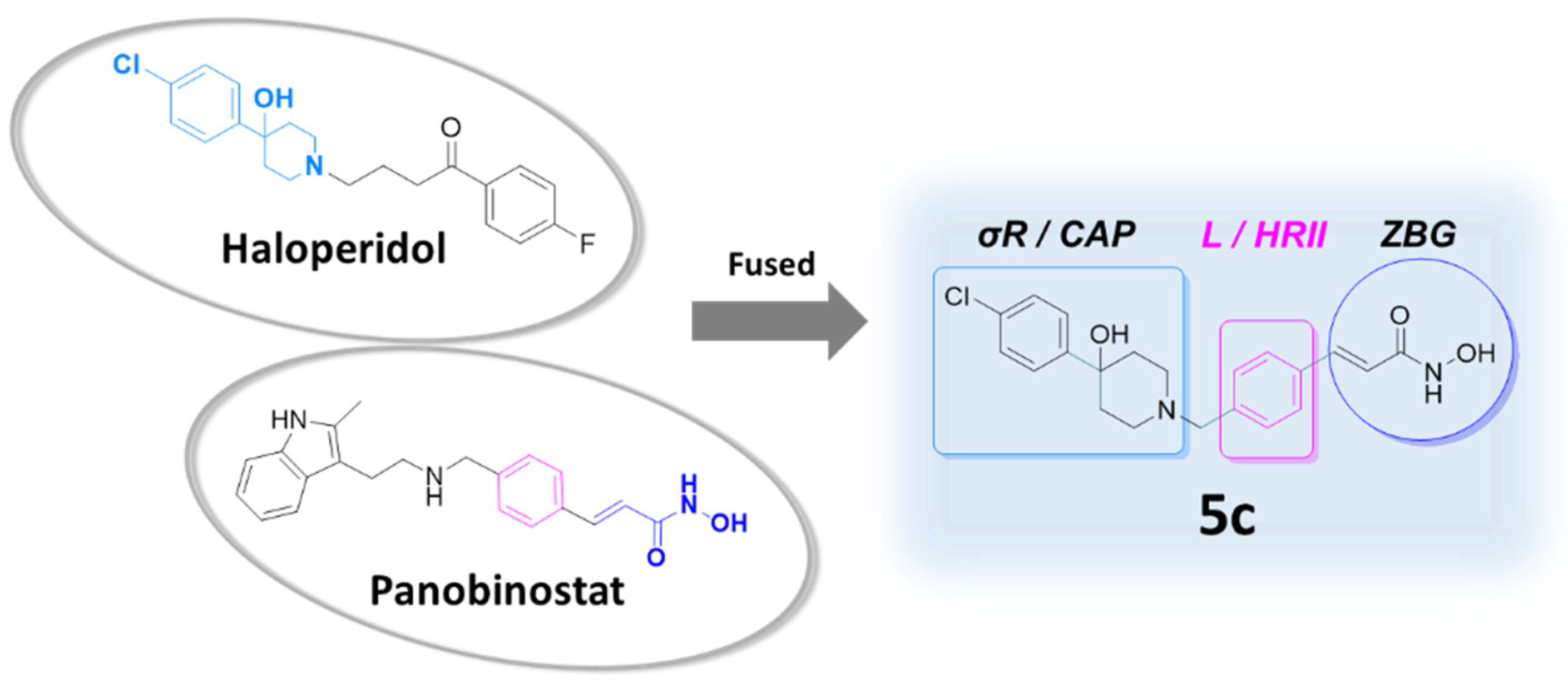
2.1. Design of the Dual-Target Anti-Melanoma Hybrid

To construct a novel HDAC/σR dual-ligand with meaningful anticancer and antiangiogenic actions, successful strategies of medicinal chemistry were employed, as earlier described [23,30]. Appropriate functional groups were selected to provide an original single pharmacophore capable of simultaneously targeting HDAC enzymes and σRs and anticipated to display excellent binding affinities [23,30]. Specifically, the novel dual hybrid molecule (compound 5c; original name, MRJF15) was conceived to exhibit three functionally distinct, chemical domains (Figure 1): (a) a σR binding moiety, formed by the hydrophobic region I and the piperidine ring of the σ1R antagonist/σ2R agonist haloperidol with its conserved nitrogen atom, which plays a crucial role in σR binding; (b) a central hydrophobic linker, comprising the inner benzene of the HDACi LBH-589 (panobinostat); and (c) a zinc binding group (ZBG), represented by the hydroxamic acid group of panobinostat, responsible for chelating the metal Zn2+ in the catalytic pocket of HDAC and inhibiting its activity [31,32].
The structural configuration of the new dual-ligand 5c afforded intentional, synergistic molecular features (Figure 1). Thus, the σR binding moiety, with its hydrophobic portion, might alternatively serve as the capping region (CAP) for the HDACi moiety of the molecule, required for optimal enzymatic inhibition by sealing the access to the catalytic pocket of HDACs. Also, the central benzene could coincidentally fulfill the role of the hydrophobic region II for the σR ligand moiety of the dual-target hybrid [33,34]. Importantly, the selected haloperidol portion should confer specific anti-melanoma and antiangiogenic properties by targeting σR signaling, mimicking its function in dual-target prodrugs [30]. Finally, the HDACi molecular end, formed by hydroxamic acid attached to the central benzene linker, should provide elevated tumor-selective antiproliferative pharmacology, as demonstrated in other HDAC/σR dual-function compounds [23].
In addition, the new dual-target hybrid is predicted to be a general HDACi but a selective σ1R ligand [23]. These functional characteristics may shed light on the precise contribution of σR signaling in dual-ligand drug pharmacology. Fundamentally, the new dual-ligand hybrid displayed the critical structures of both the σR-binding haloperidol core and the zinc-chelating nucleus of an established HDACi (Figure 1), rationally assembled in a compacted molecular configuration and providing a single pharmacophore for optimal therapeutic performance [23]. In this way, the design was aimed at obtaining a small molecule (386.88 Da) with improved ligand affinity, target selectivity and biological efficacy.

2.2. Chemical Synthesis

The synthetic route for the preparation of the dual-ligand 5c, an α,β-unsaturated hydroxamic acid derivative with a central benzene linker, involved four reaction steps (Scheme 1). In the first step, an esterification reaction was carried out between trans-4-formylcinnamic acid (compound 1) and methyl iodide in N,N-dimethylformamide (DMF) to obtain (E)-3-(4-formylphenyl)methyl acrylate (compound 2). Subsequently, a reductive amination reaction was performed between compound 2 and 4-(4-chlorophenyl)piperidin-4-ol in tetrahydrofuran (THF), in the presence of sodium triacetoxyborohydride [NaHB(OAc)3] and acetic acid, yielding compound 3c. In the third step, the ester group of compound 3c was hydrolyzed (in methanol) with 1 M lithium hydroxide (LiOH). Following hydrolysis, the carboxyl group was activated using ethyl chloroformate, in the presence of triethylamine in THF. Then, this activated intermediate underwent a nucleophilic reaction with O-(tetrahydro-2H-pyran-2-yl)hydroxylamine (NH2OTHP) in THF, to produce compound 4. Finally, the deprotection of the hydroxyamino group in compound 4 was achieved by hydrolysis with 0.6 N hydrochloric acid (HCl) aqueous solution, to obtain the final hybrid product 5c.

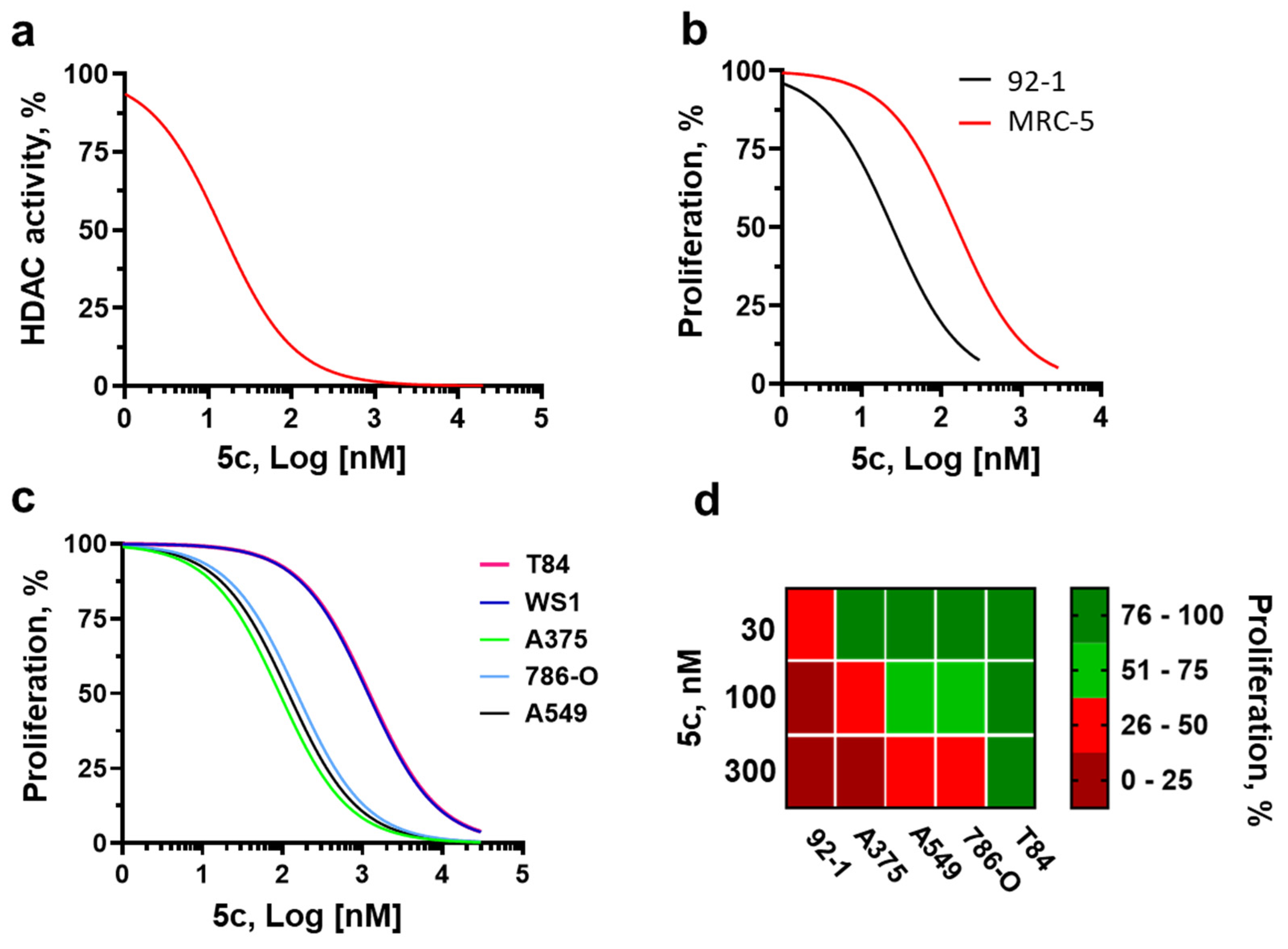
2.3. Molecular Pharmacology and Tumor Selectivity

Effective target engagements by the novel dual-function agent 5c were assessed. First, σR binding affinities were determined by employing a radioligand binding assay [23,30]. Dual-targeted 5c exhibited >100 fold higher affinity for σ1R (Ki, 19.0 nM) than σ2R (Ki, 2051.2 nM), suggesting the induction of selective σR signaling (Table 1) as for previous congeners [23]. Similarly, compound 3c, the hydroxamic-null precursor of 5c (Scheme 1) anticipated to lack HDACi activity, also behaved as a selective σ1R ligand (Table 1) with superior binding affinities for σ1R (Ki, 28.0 nM) than for σ2R (Ki, 1330.0 nM). These binding behaviors differentiated 3c and 5c from their parental chemical scaffold haloperidol, which bound to both σ1R and σ2R with high affinities (Table 1) and resembled those of the selective σ1R agonist pentazocine [35] (Table 1).
Next, the ability of 5c to inhibit HDACs was investigated, employing various cancer cell lysates as biological sources of diverse HDAC enzymatic activities [23], including colon (HCT116), breast (MCF7), prostate (PC3) and gastric (AGS) carcinoma cells. First, dose–response curves were generated for the HCT116 cell lysates (Figure 2a), which demonstrated that 5c reduces HDAC-dependent catalytic activity with a calculated 50% inhibitory concentration (IC50) of 14.6 nM. Importantly, HDACi activity by 5c was profound (~80% inhibition with respect to the vehicle control) and of a similar extent to that of the positive control trichostatin A (Figure S1), a potent and non-selective HDAC inhibitor [36]. The HDAC inhibitory effects of 5c were generalizable to other cancer cell types, with significant suppression of resident HDAC enzymatic activities compared to respective control conditions, which mirrored those of trichostatin A (Figure S1). Together, the molecular pharmacology studies of receptor engagements indicated that 5c may signal as a dual-target ligand with selective σ1R, but general HDACi actions.
To establish the anti-melanoma selective effects of the novel HDAC/σ1R hybrid, complete antiproliferative dose–response curves were generated by employing two in vitro human models, the UM 92-1 cells and normal lung fibroblasts MRC-5 (Figure 2b). The UM 92-1 model was intentionally chosen for these studies because 92-1 cells express all the molecular targets of interest, including HDAC proteins and both σ1R and σ2R [30,37]. Moreover, they exhibit sensitivity to anticancer actions by dual-target HDAC/σR prodrugs [30]. The lung fibroblasts MRC-5 were in turn interrogated as an experimental surrogate of normal systemic tissues, to ascertain in vitro safety. Of relevance, 5c appropriately discriminated between tumor and normal cells, with antiproliferative sigmoidal curves shifted to the left in 92-1 cells, compared to MRC-5 cells (Figure 2b). Estimates of antiproliferative IC50 confirmed that this hybrid exhibited significantly superior (~5 fold) potency in UM cells, compared to normal lung fibroblasts (Table 2). This latter finding is important because it suggests that 5c possesses favorable therapeutic profiles, reflecting selective anti-melanoma actions, in the context of safer behaviors in normal cells (Table 2).

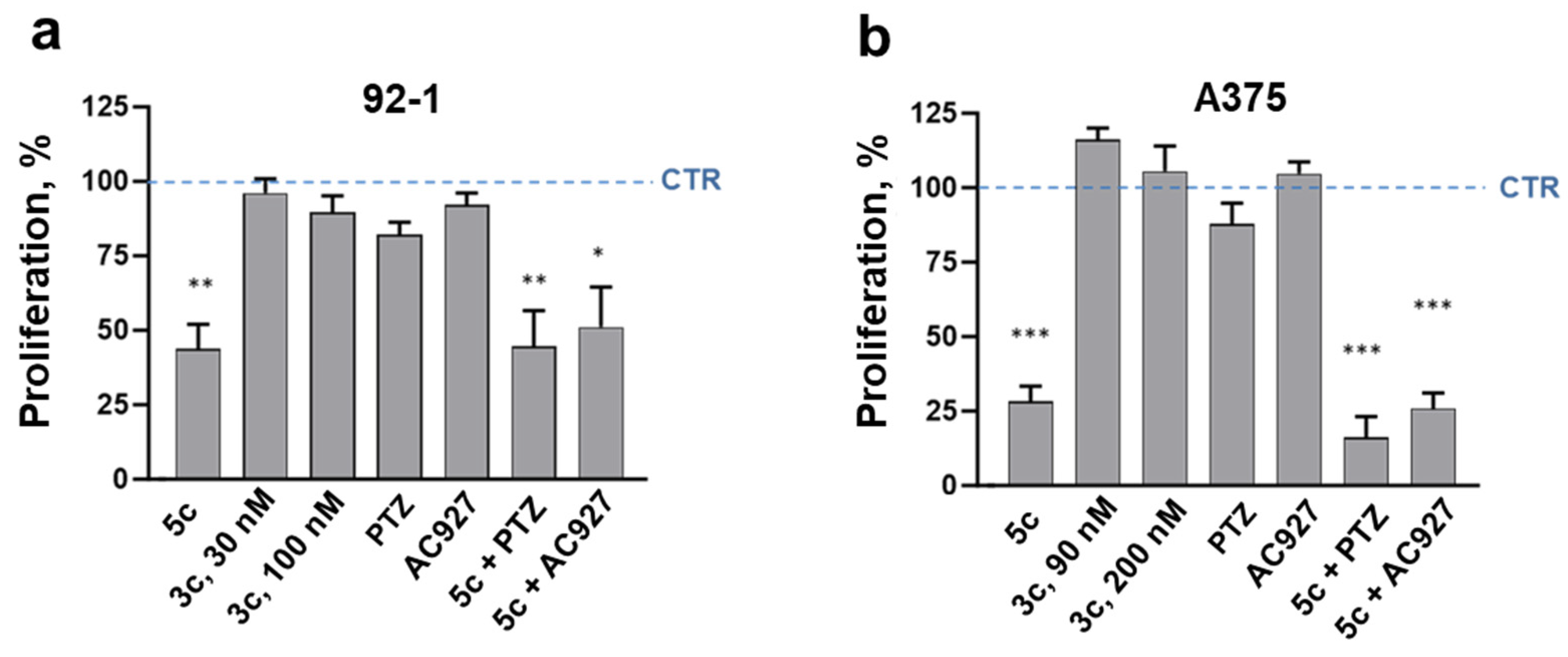
2.4. Cancer Specificity and Antiproliferative Signaling in Melanoma Cells

The tumor specificity of the test compound was investigated by employing immortalized human cells from four different cancers (CM A375, lung A549, renal 786-O and colorectal T84) and normal skin fibroblast WS1 (Figure 2c). Dual-ligand hybrid 5c efficiently and selectively reduced proliferation in three additional human cancers (CM, lung and renal adenocarcinomas) compared to normal skin fibroblasts, as indicated by the ~8–10 fold shift to the left in correspondent antiproliferative dose–response curves (Figure 2c). In contrast, colon cancer T84 cells were resistant to 5c antitumor actions (Figure 2c). IC50 calculations confirmed that compound 5c exhibited higher antiproliferative potencies in melanoma cells (both UM and CM), compared to other normal or cancer cell types (Table 2). Accordingly, a heat map representation of IC50 values from all cancer cells demonstrated a melanoma-specific sensitivity to 5c antiproliferation, with UM 92-1 and CM A375 cells ranking as the first and second most suppressed tumors, respectively, by the multiligand hybrid (Figure 2d).
Importantly, melanoma-specific antiproliferation by 5c reflected HDAC targeting and suppression of dependent epigenetic regulations, as demonstrated by the loss of activity upon disruption of the HDACi-enabling hydroxamic group (with precursor compound 3c; Scheme 1 and Figure 3). In contrast, exposure of UM (Figure 3a) and A375 (Figure 3b) cells to the σ1R agonist pentazocine or the σ2R antagonist AC927 [38] did not alter their proliferative kinetics, nor did it perturb 5c’s antiproliferative actions. Together, these data indicate that HDAC, but not σR, is the main pharmacological target for 5c-mediated antiproliferation in human melanoma cells.

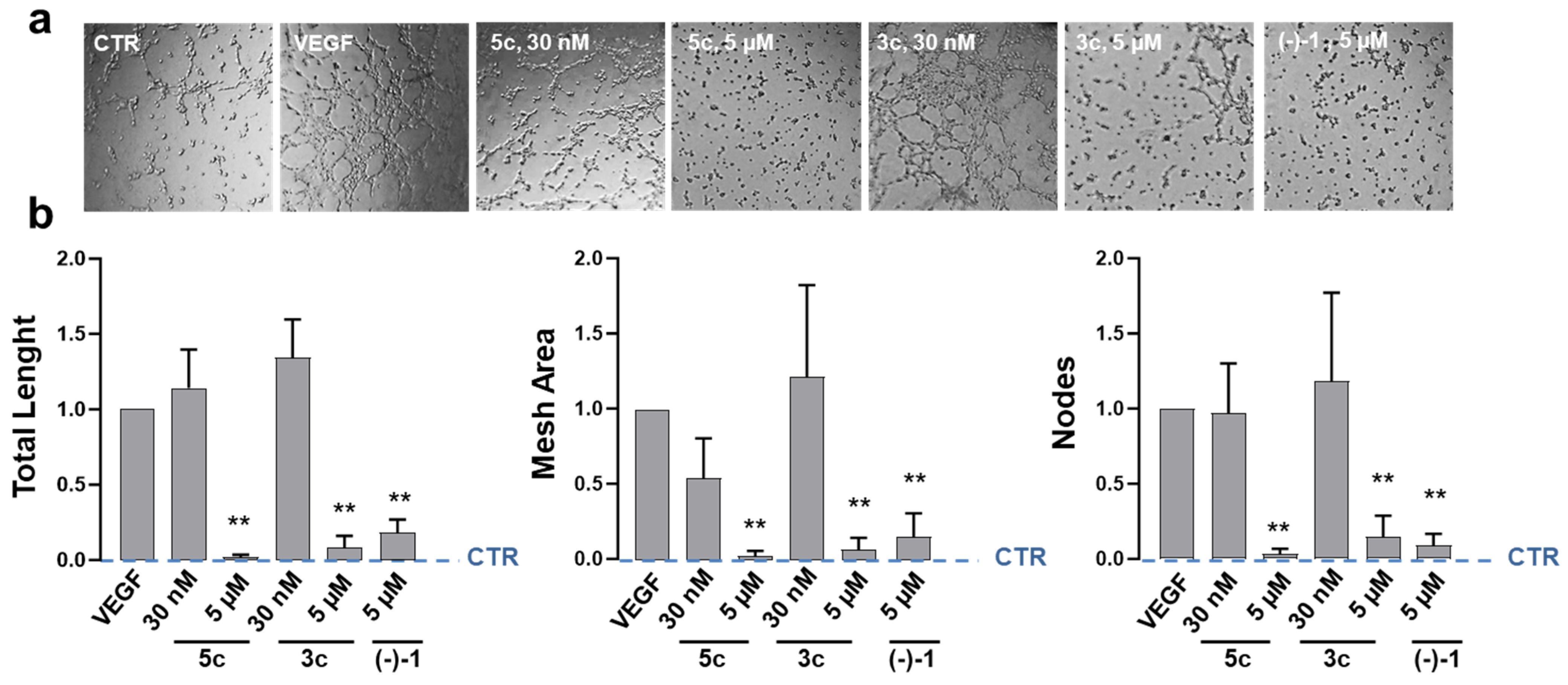
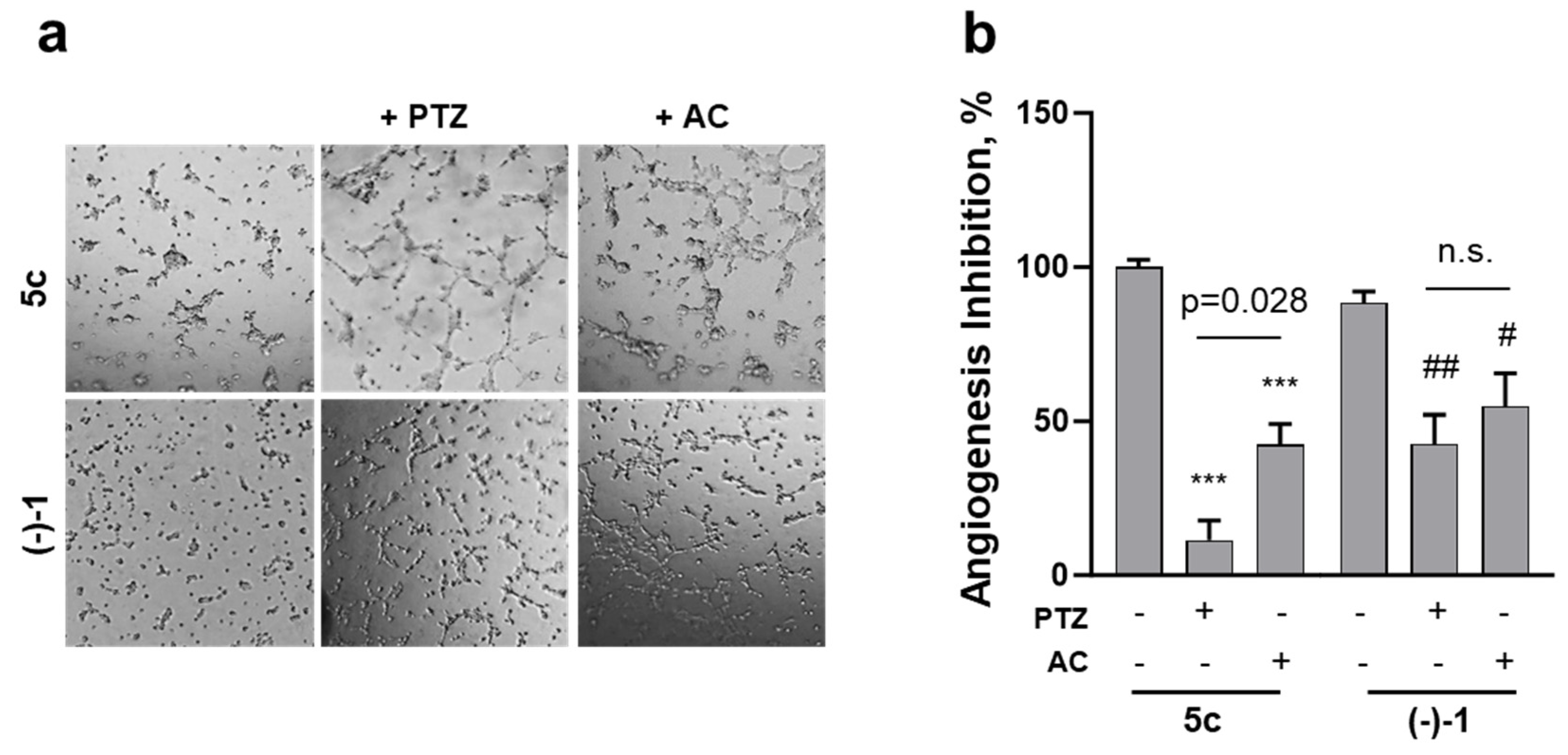
2.5. Effects on Endothelial-Dependent Angiogenic Processes

The ability of the dual-function hybrid 5c to affect endothelial-driven neoangiogenesis was explored using the tube formation assay with mouse endothelial C166 cells (Figure 4). To focus on the motility and phenotypic organization of capillary-like structures (Figure 4a) [39,40] away from the mechanistic contributions of cell proliferation, short observational periods (3 h) were selected upon C166 cell seeding onto basement membrane-like Matrigel and VEGF-A-stimulation [39]. As expected, VEGF-A activated the endothelial C166 cells and promoted the formation of vasculature-like networks to a greater degree than the vehicle control (Figure 4a,b). Also, the HDAC/σRs bifunctional prodrug (S)-(-)-MRJF22 [(-)-1], used as the positive control [30], significantly inhibited angiogenesis by VEGF-A in these cells (Figure 4a,b). Treatment with 5c at antiproliferative concentrations (30 nM; Figure 3) was without effects on VEGF-A-stimulated angiogenesis (Figure 4b). In contrast, C166 exposure to 5c at 5 µM, a concentration level capable of inducing σR-dependent antiangiogenesis [30], abrogated the ability of VEGF-A to induce neovascular networks (Figure 4a), with significant suppression of quantitative angiogenic parameters including tube lengths, mesh areas and node numbers (Figure 4b). Of relevance, the hydroxamic-null compound 3c mimicked the effects of 5c on endothelial-dependent angiogenesis (Figure 4b), demonstrating that HDACi is not involved in the disruption of VEGF-induced microvascular organizations by the novel dual-target hybrid.
Application of the σ1R agonist pentazocine or the σ2R antagonist AC927 inhibited the ability of 5c, 3c and (-)-1 to suppress the proangiogenic phenotype of C166 cells, although with different degrees of efficiency with respect to each ligand and the precise angiogenic parameter quantified (Figure S2). A comprehensive examination of 5c-dependent antiangiogenesis (Figure 5a), achieved by combining all parameters examined in one score, revealed that pentazocine completely prevented 5c’s effects on vascular tube formations (Figure 5b). AC927 also inhibited 5c’s actions on angiogenesis, but to a significantly lesser extent than pentazocine (Figure 5b). In contrast, inhibitory activities of VEGF-A-stimulated angiogenesis by (-)-1, a signaling ligand of both σ1R and σ2R [30], were partially and equally blocked by co-incubations with pentazocine or AC927 (Figure 5). All together, these observations suggest that the HDAC/σ1R dual-target 5c inhibits VEGF-A-stimulated vascular morphogenesis in endothelial C166 cells by acting as a selective σ1R antagonist, but without the involvement of HDAC-dependent pathways.

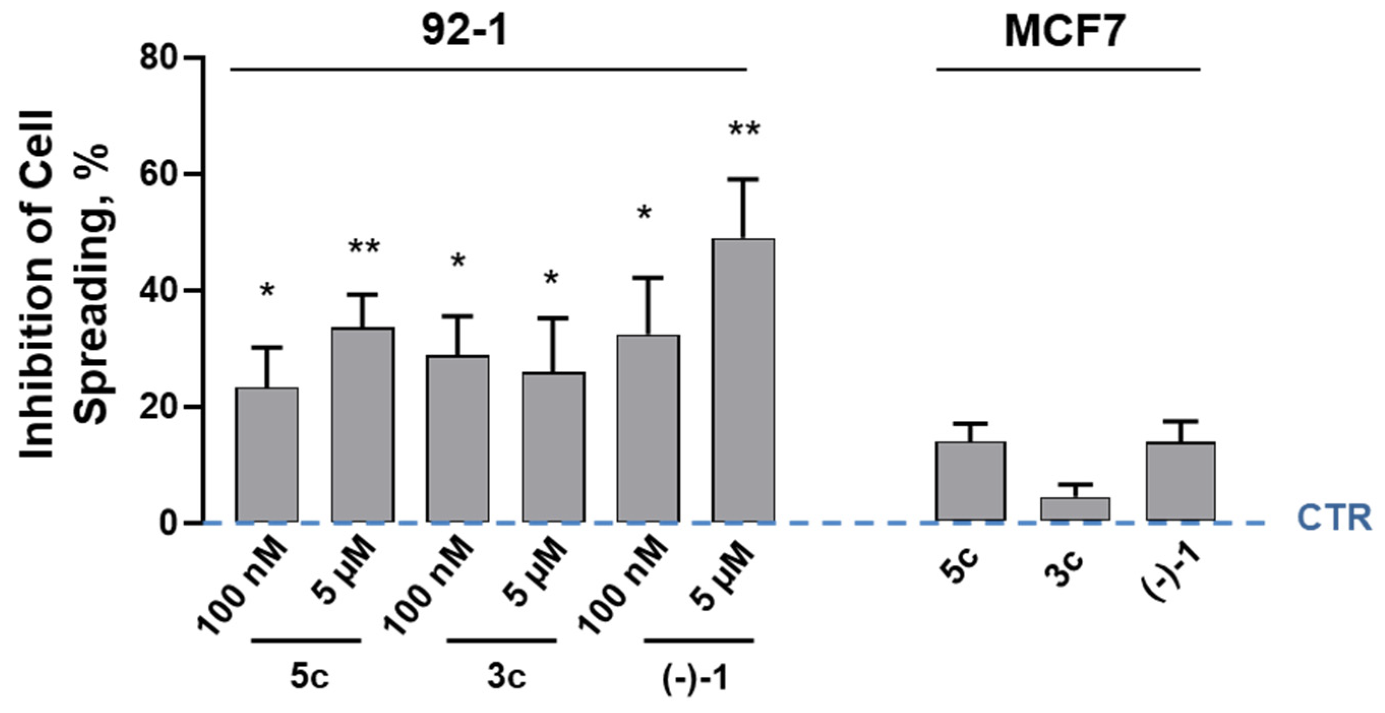
2.6. Receptor Signaling in Melanoma Cell Spreading

In solid tumors, changes in cancer cell shape (spreading) underlies migration, invasion and metastasis to distant organs [41]. The impact of the HDAC/σ1R dual-ligand 5c on locomotory organelle formation (lamellipodia, filopodia) in melanoma cells was investigated with the tumor cell spreading assay [42,43,44]. Quantification of the fraction of UM 92-1 cells extending membrane protrusions revealed that short 5c exposures (1 h) provoked a shrinkage of this migratory cell population, with a correspondent expansion of cells with rounded morphology (Table S1). In contrast to antiangiogenic effects (Figure 4), significant inhibition of the invasive cell shape by 5c was already apparent at low concentrations (100 nM; Figure 6), coincident with those eliciting anti-melanoma proliferation (Figure 3). The HDACi-null analog 3c also significantly suppressed membrane protrusion-driven 92-1 cell spreading, with nearly identical efficacy and potency as 5c (Figure 6). Although with similar efficacy, the reference HDAC/σR multiligand prodrug (-)-1, which does not discriminate between σ1R and σ2R [30], reduced UM cell spreading with seemingly higher potencies (Figure 6).
To establish the role of σ1R as the downstream molecular target for the dual-function hybrid 5c, locomotory-driven cell spreading was then examined in human breast cancer MCF7 cells, which exhibit low levels of endogenous σ1R protein and silenced σ1R-dependent biological pathways [45]. Treatment with 5c failed to suppress tumor cell spreading in MCF7 cells (Figure 6). Accordingly, the HDACi-null analog 3c and the reference prodrug (-)-1 were unable to significantly inhibit MCF7 cell spreading (Figure 6). Together, these findings support the hypothesis that HDAC/σ1R dual-target 5c disrupts locomotory organelle formation in melanoma cells by selectively targeting σ1R signaling pathways, without the involvement of HDAC regulation.

3. Discussion

Compounds with multi-target pharmacology are emerging as promising small molecules for the safer, more efficacious therapeutic management of multifactorial diseases such as cancer [23,30,31]. In this context, compared to multitherapy with distinct drugs, the simultaneous regulation of different molecular targets, endowed by the dual-ligand strategy, should enable low-dose exposures and improve patient compliance by reducing off-target effects [31]. Indeed, dual-ligands are superior therapeutics anticipated to target distinct biological sites of action in the same tumor cell, thereby maximizing treatment responses. Moreover, this innovative synthetic design is suited to enhance drug therapeutic potentials further, exploiting the synergistic opportunities offered by simultaneously targeting distinct pathways while minimizing the risk of target-based drug resistance [31].
Dual-target drugs could be particularly useful in melanoma, a disease with poor prognosis and high treatment failure rates [1,2,3]. Accordingly, HDAC/σR dual-ligand prodrugs reduced UM cell proliferation and motility while concurrently opposing angiogenic processes stimulated by VEGF [30]. In the present study, a novel dual-function HDAC/σR hybrid was synthesized by employing a pharmacophore fusion approach, wherein the σR ligand part of the molecule was directly attached to the central hydrophobic linker of the selected HDACi moiety (Figure 1). Importantly, the distinct structural components of the parental ligands simultaneously provided reciprocal permissive domains to the dual-target pharmacophore, with the HDACi’s hydrophobic linker acting as the hydrophobic region II for the σR ligand, and the distal hydrophobic end of the σR ligand as the CAP portion for HDACi (Figure 1). In this way, the key functional groups of the precursor drugs contributed to forming a single pharmacophore, simultaneously fitting HDAC and σR whilst maintaining excellent binding interactions.
Importantly, the newly developed dual hybrid 5c induced potent antiproliferative actions in cancers, which were tumor-selective and specific to UM and CM cells (Table 2). The melanoma selectivity reflected the inhibition of HDAC-dependent epigenetic regulation of proliferative kinetics, without the involvement of σR signaling, as demonstrated by the lack of inhibitory effects using the hydroxamic-null precursor 3c (Figure 3). Apart from the essential role of ZBG, though, it is conceivable to speculate that additional structural components of the hybrid 5c strengthen its antiproliferative activity. Among these, the hydrophobic benzene of the central linker (Figure 1) was previously selected as a biologically optimal molecular module, conferring potent anticancer activities on HDAC/σR dual-ligands [23]. Moreover, the CAP region for HDACi, covered in the novel hybrid by its σR moiety (Figure 1), seals the gate of the catalytic pocket and is instrumental in elevated HDAC inhibition.
HDACs comprise an important family of 18 isozymes that can induce profound epigenetic modifications in cells and are being pursued as drug targets for many types of disorders [9]. In cancers, these enzymes regulate the expression and activity of numerous proteins involved in tumor initiation and progression [46]. By removing acetyl groups from histones, HDACs create a non-permissive chromatin conformation that prevents the transcription of specific genes. However, in addition to histones, HDACs can bind to and deacetylate a variety of other protein targets, including transcription factors and other abundant cellular proteins implicated in the control of cell growth, differentiation and apoptosis [9]. As a result, HDACis have recently emerged as promising cancer therapeutics, including in UM and CM [46]. Since the dual-ligand 5c behaves as a non-selective HDACi in various tumor cells (Figure S1), it is predicated that its role as an anticancer agent could be generalized to other cancers, beyond its specific antiproliferative effects in melanomas.
Of note, the dual hybrid 5c provided a novel selective σ1R ligand, expanding the class of similar dual-ligand congeners [23]. Indeed, 5c exhibited different σR binding affinities than its chemical scaffold haloperidol, with similar σ1R affinity but disrupted σ2R binding ability, effects shared by the ester precursor 3c (Table 1). These original functional outcomes probably reflect the substantial pharmacophore remodeling of the novel dual-ligand compound (and its hydroxamic-null precursor), which comprises a differentially rearranged hydrophobic region II (Figure 1). Moreover, 5c functioned as a selective σ1R antagonist (Figure 5) whose biological activity was antagonized by pentazocine, a putative σ1R agonist (Table 1).
The σ1R is a unique integral membrane chaperone operating in mitochondria-associated ER domains [25,27]. Acting as a scaffolding protein that allosterically modulates the activity of its associated proteins, σ1R is increasingly regarded as a key regulatory signaling receptor implicated in many biological processes [47]. In cancer, σ1R ligands inhibited tumor migration, proliferation and survival [47]. Furthermore, σ1R has been demonstrated to suppress tumor growth, alleviate cancer-associated pain and exert immunomodulation [30]. Here, the bifunctional 5c compound inhibited VEGF-stimulated capillary morphogenesis by endothelial cells, through the specific regulation of σ1R signaling, but not HDAC (Figure 5). These results are in agreement with the modulation of angiogenesis by σ1R in various cell models [46] and support the utility of targeting this receptor to oppose hematogenous tumor cell dissemination.
Selective σ1R targeting with the dual-function hybrid 5c also disrupted invasive locomotory organelle formation in melanoma cells (Figure 6). In this context, invading tumor cells form membrane protrusions (e.g., lamellipodia, filopodia) to spread and migrate into neighboring tissues by extending their leading edges along the direction of the invasive front [43,45]. Thus, the inhibition of σ1R functions through pharmacological antagonism could afford the dual-ligand hybrid significant antimetastatic potential in melanoma. Accordingly, the colon cancer cell shape which promotes migration and invasion was suppressed by σ1R signaling through the human voltage-dependent K+ channel human ether-à-go-go-related gene [48]. Moreover, σ1R is often upregulated in tumors [49,50], and its expression potentiated invasive cancer behavior and angiogenesis in vivo, resulting in poor survival [48].
Taken collectively, the present findings suggest that dual-targeted HDAC/σ1R ligands could represent a novel paradigm for melanoma therapeutics, being versatile and highly potent. Indeed, they are predicted to efficiently and simultaneously oppose many cancer-permissive pathogenetic programs in melanoma, including tumor proliferation, migration, invasion, angiogenesis and metastasis.

4. Materials and Methods

4.1. Chemistry

4.1.1. General Procedure

Required chemicals were obtained from Merck (Darmstadt, Germany) or Fluorochem (Derbyshire, UK). All reagents and chemicals were processed, and reactions performed as previously detailed [30]. Flash chromatography purification was done on Merck silica gels 60 (230−400 mesh) and nuclear magnetic resonance spectra (1H NMR and 13C NMR recorded at 500 MHz) were acquired with Varian INOVA spectrometers, using CDCl3, CD3OD and DMSO-d6 with 0.03% of TMS as the internal standard (Figures S3 and S4) [30]. High Resolution Mass Spectrometry (HRMS) was conducted using Q-Exactive with ESI source (Thermo Scientific, Waltham, MA, USA). MS full scan acquisitions were performed in positive ion mode, in the m/z range 150.0–800.0. The HCD gas on fragmentation method was used, settled in enhanced resolution, with a maximum injection time of 50 ns (Figures S5 and S6).

4.1.2. Synthesis and Product Characterization

Methyl-(2E)-3-(4-formylphenyl)prop-2-enoate (2). The synthesis of intermediary 2 was performed as described in previous studies [23]. The obtained solid product (4.6 g) exhibited a yield of 85.4%. 1H NMR (500 MHz, CDCl3): δ 3.80 (s, 3H), 6.53 (d, J = 16.0 Hz, 1H), 7.63–7.90 (m, 5H), 10.01(s, 1H). 13C NMR (125 MHz, CDCl3): 53.41, 116.80, 128.52, 129.53, 134.90, 138.15, 142.28, 163.69, 191.22.
Reductive amination. Compound 2 (7.3 mmol) was added (dropwise; RT) to an anhydrous tetrahydrofuran (THF) solution of 4-(4-chlorophenyl)piperidin-4-ol (7.3 mmol) under a nitrogen atmosphere. After addition of AcOH (7.3 mmol), the reaction mixture was stirred (30 min) and added dropwise to a NaBH(OAc)3 solution (10.9 mmol) in 1.85 mL of anhydrous THF at RT in a nitrogen atmosphere. The reaction mixture was stirred overnight at RT. Subsequently, a saturated solution of NaHCO3 was added, and the aqueous phase was repeatedly extracted with CHCl3. The combined organic phases were dried with Na2SO4 anhydrous, filtered, and evaporated under vacuum. The crude was purified by MPLC using EtOAc as the eluent to obtain intermediate 3 (compound 3c):
Metil-(E)-3-(4-{[4-(4-clorofenil)-4-idrossipiperidin-1-il]methyl}phenyl)acrylate (3, compound 3c). White solid 4.78 g (yield 73%). 1H NMR (500 MHz, CDCl3) δ 7.85–7.60 (d, 1H, J = 15.0 Hz), 7.49–7.29 (m, 8H), 6.425 (d, 1H, J = 15.0 Hz), 3.80 (s, 3H), 3,58 (s, 2H), 2.77–2.75 (m, 2H), 2.49–2.44 (m, 2H), 2.15–2.09 (m, 2H), 1.72–1.65 (m, 2H). 13C NMR (500 MHz, CDCl3) δ 167.51, 146,84, 144.63, 133.24, 132.76, 129.56, 128.38, 128.04, 126.10, 117.35, 71.02, 62.75, 51.69, 49.36, 38.43. HRMS (ESI, m/z) calcd for [C22H25ClNO3]+ (M + H)+ 386.152, found. 386.144.
Synthesis of the hydroxyamide. An aqueous solution of LiOH 1 M (2 mmol) was added dropwise (at 50 °C) to a MeOH solution of intermediate 3 (compound 3c; 1 mmol), and the reaction mixture was stirred overnight. After solvent evaporation (in vacuum), residues were dissolved in THF/DMF (1:1) and supplemented first with TEA (2 mmol), and then with an ethyl chloroformate solution (4 mmol) in THF (added dropwise at 0 °C). Following 3 h stirring, O-(tetrahydro-2H-pyran-2-yl)-hydroxylamine (5 mmol) was added under nitrogen at RT, and the reaction stirred overnight. The resulting residue was washed with a saturated solution of NaHCO3, and the aqueous phase extracted with CHCl3. The combined organic phases were dried with anhydrous Na2SO4, filtered, and evaporated under vacuum. The crude was purified by MPLC to obtain the intermediate 4:
(E)-3-(4-{[4-(4-clorofenil)-4-idrossipiperidin-1-il]metil}fenil)-N-(tetrahydro-2H-pyran-2-yl-oxy) acrylamide (4). White solid 0.310 g (yield 65%). 1H NMR (DMSO-d6) δ 11.20 (s, 1H), 7.54–7.34 (m, 9H), 6.49 (d, 1H), 4.91–4.89 (m, 2H), 3.98–3.96 (m, 1H), 3.15 (s, 2H), 2.60–2.56 (m, 2H), 2.45–2.43 (m, 2H), 189–193 (m, 2H), 1.78–1.62 (m, 2H), 1.58–1.54 (m, 6H). 13C NMR (DMSO-d6) δ 165.55, 162.57, 149.03, 140.71, 139.25, 133.30, 130.75, 129.34, 127.77, 127.43, 126.80, 118.15, 100.98, 69.36, 61.85, 61.35, 48.86, 37.81, 27.77, 24.63, 18.26.
Then, a solution of HCl 1.25 M in EtOH was added (dropwise at 0 °C) to an EtOH solution of intermediate 4, and the reaction was stirred for 24 h (at RT). After solvent evaporation (in vacuum), the residue was dissolved in EtOH and precipitated with Et2O to obtain 5 (compound 5c):
(E)-4-(4-chlorophenyl)-4-hydroxy-1-{4-[3-(hydroxyamino)-3-oxoprop-1-en-1-yl]benzyl}piperidin-1-ium chloride (5, compound 5c). White solid 72 mg (yield 88%). 1H NMR (DMSO-d6) δ 11.24 (broad s, 1H), 7.73–7.41 (m, 9H), 6.55 (d, 1H, J = 15 Hz), 4.37–4.36 (m, 2H), 3.26–3.20 (m, 4H), 2.50 (s, 2H), 1.75–1.78 (m, 2H). 13C NMR (DMSO-d6) δ 162.36, 146.95, 137.34, 135.84, 132.02, 131.242, 130.86, 128.00, 127.62, 126.55, 120.32, 67.94, 58.22, 47.68, 34.65. Anal. calcd. for C21H23ClN2O3⸱HCl: C, 65.20; H, 7.24; N, 5.99; Found: C, 65.34; H, 6.98; N, 6.17. HRMS (ESI, m/z) calcd for [C21H24ClN2O3]+ (M + H)+ 387.148, found. 387.139.

4.2. Cell Cultures and Reagents

Unless otherwise specified, cell lines were obtained from the American Type Culture Collection (ATCC; Manassas, VA, USA). Human UM cell line 92-1 was maintained at 37 °C (5% CO2) in RPMI-1640 medium, containing 10% fetal bovine serum (FBS), 2 mM L-glutamine, 100 units/mL penicillin and 100 µg/mL streptomycin. Human renal adenocarcinoma cell line 786-O (ATCC® CRL-1932™) was cultured in RPMI with 10% FBS. Human cutaneous melanoma cells A375 (ATCC® CRL-1619™), human lung adenocarcinoma A549 (ATCC® CCL-185™) and mouse endothelial cell C166 (ATCC® CRL-2581™) were cultured in Dulbecco’s modified Eagle’s medium (DMEM) with 10% FBS, whereas human skin fibroblast WS1 (ATCC® CRL-1502™) and human lung fibroblast MRC-5 (ATCC® CCL-171™) in EMEM with 10% FBS. Human colorectal carcinoma cell line T84 (ATCC® CCL-248™) was cultured in DMEM/F12 medium with 10% FBS. Human breast cancer MCF7 (HTB-22™) and human colorectal carcinoma cell line HCT116 (CCL-247™) were maintained in DMEM-high glucose (Euroclone, Milan, Italy) supplemented with 10% FBS and 1% penicillin/streptomycin. Human gastric adenocarcinoma AGS cells and human prostate adenocarcinoma PC3 cells were purchased from the European Collection of Authenticated Cell Cultures (ECACC, Porton Down, Salisbury, UK) and maintained in HAM’s F12 medium supplemented with 10% FBS and 1% of penicillin/streptomycin. Cell media and general reagents were from Euroclone S.p.A. (Pero, Milan, Italy). The positive control (-)-1 was prepared as previously described [30]. Radioactive (+)-Pentazocine (Italian Minister of Health permit to produce and use SP/072 5 April 2019) and AC927 were prepared according to published methods [51,52].

4.3. Cell Proliferation

Cells were seeded in 96-well plates at a cell-specific density, following quantification of cell numbers on counting chambers after trypsinization and staining with trypan blue. After seeding (72 h), at a cell confluence of ~50%, the medium was removed, and treatments were started with fresh media. Treatments, carried out in quadruplicate, lasted for 48 h. Then, the cells were fixed with 4% paraformaldehyde in PBS for 20 min and, after washing, stained with either 1% crystal violet in water or an acridine orange solution (50 µg/mL) for an additional 20 min. At the end of the incubations, cells were washed (3 times) with 100 μL/well of bidistilled water. Finally, for crystal violet assays, 100 μL of 10% acetic acid was added to each well, and the absorbance (λ = 590 nm) was measured with a Synergy HTX Bioteck spectrophotometer (BioSPX, Beersel, Belgium). For acridine orange assays, cell staining was evaluated by measuring the fluorescence (excitation 485/20 nm, emission 528/20 nm) with the Synergy HTX Bioteck reader.

4.4. Radioligand Binding Assay

The σ1R and σ2R binding studies were performed as previously described [30,53,54], using guinea pig brain membranes and appropriate radioligands, including [3H]-(+)-pentazocine (45 Ci/mmol) for σ1R, and [3H]-1,3-di-o-tolylguanidine ([3H]-DTG; 31 Ci/mmol) for σ2R. Nonspecific binding was also assessed with unlabeled haloperidol or DTG. Radioactivity was determined with a Beckman LS6500 scintillation counter. Inhibition constants (Ki values) were calculated with the EBDA/LIGAND program (Elsevier/Biosoft).

4.5. HDAC Activity Assay

HDAC activity was determined in the indicated cell lysates (50 µg proteins; 37 °C) using the non-selective HDAC Activity Colorimetric Assay kit (BioVision, Cambridge, UK), following the procedures described in previous studies [23]. Samples were read at 405 nm with a microplate reader (Multiskan GO (Thermo Fisher Scientific, Waltham, MA, USA)), and the HDAC activity was expressed as the relative optical density (O.D.) values. The IC50 value of compound 5c (0–20 µM) in HCT116 cells was calculated using the GraphPad Prism® software (version 10.4.1).

4.6. Tube Formation Assay

The tube formation assay using mouse endothelial C166 cells was analyzed employing Matrigel (BD, Bedford, MA, USA), as previously described [30]. Briefly, 96-well plates coated with Matrigel (50 μL/well) were filled with 100 μL medium/well and maintained overnight. Endothelial cells were seeded at 2.5 × 10⁴ cells/well and incubated (for 3 h) with VEGF-A (80 ng/mL), in the presence of the indicated treatments. Then, capillary-like structures were imaged (×4 magnification) with a CCD camera connected with an inverted EVOS microscope. Finally, ImageJ software (NIH, Bethesda, MD, USA) was employed to quantify tube formations.

4.7. Cell Spreading

Adherent 92-1 and MCF-7 cells were harvested using 0.05% Trypsin-EDTA and plated in 6-well plates. After 1 h incubations with the indicated treatments, the number of cells that formed, or not, distinct protrusions (lamellipodia, filopodia) were scored with an inverted microscope (20×, EVOS cell imaging system, Thermo Fisher, Waltham, MA, USA). Results were reported as either the fraction of total cell number per microscopic field (≥400 cells evaluated for each condition) or the relative percentages of inhibition of cell spreading with respect to the vehicle controls.

4.8. Statistical Analysis

Unless otherwise specified, data were expressed as means ± SEM of ≥3 independent experiments, each conducted at least in duplicate. Student’s t-test or one-way ANOVA multiple comparisons test were employed for all statistical analyses. Differences between groups were considered significant at p values < 0.05.

5. Conclusions

A novel dual-target HDAC/σR hybrid was synthesized and investigated herein, exhibiting a single pharmacophore which simultaneously regulated HDAC and σ1R signaling in cells. The dual-ligand preferentially suppressed melanoma cell proliferation and membrane protrusions by inhibiting HDAC-mediated epigenetic regulations and σ1R activation, respectively. Occurrence of these effects in the same malignant cells, afforded by the dual-target strategy, is anticipated to provide synergistic opportunities for melanoma-specific eradication therapies. The novel bifunctional agent also restricted the formation of capillary-like structures in endothelial cells, an effect mediated by the selective regulation of σ1R, and not HDAC. Future studies should explore the mechanisms of action for the dual-ligand, focusing on molecular events downstream to target engagements that underlie individual biological outcomes in tumors.
The ability to concurrently disrupt many malignant processes, including tumor growth, invasion and angiogenesis, clearly represents an advantageous trait for the dual-function HDAC/σ1R ligand. In this way, it might provide the elusive curative, safe remedy that would shrink tumor volumes at primary sites, prevent cancer dissemination to distant organs and defeat the occurrence of tumor resistance. Translation of this therapeutic paradigm into clinical applications would contribute to the advancement of precision medicine in oncology.

Supplementary Materials

The following supporting information can be downloaded at: https://www.mdpi.com/article/10.3390/ph18020179/s1; Figure S1: Inhibition of HDAC activity in HCT116, MCF7, PC3 and AGS cells by 5c; Figure S2: Regulation of total tube length, nodes, and mesh area in angiogenesis assays; Figures S3 and S4: NMR spectra of synthesized compounds; Figures S5 and S6: HRMS spectra of synthesized compounds; Table S1: Cell spreading counting.

Author Contributions

Conceptualization, G.M.P. and A.M.; methodology, C.G.L., G.M.P., A.M., E.A. and C.B.; formal analysis, C.G.L. and G.M.P.; investigation, C.G.L., C.B., J.F., A.C., V.d.G. and E.A.; data curation, C.G.L., G.M.P., A.M. and C.B.; writing—original draft preparation, C.G.L., G.M.P. and C.B.; writing—review and editing, G.M.P. and A.M.; supervision, G.M.P. and A.M.; funding acquisition, G.M.P. and A.M. All authors have read and agreed to the published version of the manuscript.

Funding

This research was funded by PO FESR 2014–2020, Action 1.1.5, project titled “NuSTeO, Nuove Strategie Terapeutiche in Oftalmologia: Infezioni Batteriche, Virali e Microbiche”, CUP: G68I18000700007. Additional funding was provided by Italian MUR, PRIN–2022 PNRR program (Project code: P20224L3NK, CUP E53D23015810001, PE5) funded by the European Union—Next Generation EU, (D.D. 1409 del 14 September 2022 MUR) and PRIN-2022, Prot. 2022Z3BBPE_006, PE5.

Institutional Review Board Statement

Not applicable.

Informed Consent Statement

Not applicable.

Data Availability Statement

All data supporting the findings of the study are available from the corresponding authors, G.M.P. and A.M., upon reasonable request.

Acknowledgments

Authors thank the Institute of Biomolecular Chemistry of the Italian National Research Center (CNR) for general technical support.

Conflicts of Interest

C.G.L. is an employee at Vera Salus Ricerca S.r.l. G.M.P. is a shareholder and the Chief Science Officer at Vera Salus Ricerca S.r.l. All other authors declare no conflicts of interest. The company had no role in the design of the study; in the collection, analyses, or interpretation of data; in the writing of the manuscript; or in the decision to publish the results.

Abbreviations

CM, cutaneous melanoma; UM, uveal melanoma; HDAC, histone deacetylase; σR, sigma receptor; VEGF, vascular endothelial growth factor; HP, haloperidol; PTZ, pentazocine; ZBG, zinc binding group; CAP, capping region; IC50, 50% inhibitory concentration; FBS, fetal bovine serum; MPLC, medium pressure liquid chromatography; NMR, nuclear magnetic resonance; HRMS, high resolution mass spectrometry; RT, room temperature.

References

  1. Bolick, N.L.; Geller, A.C. Epidemiology of Melanoma. Hematol. Oncol. Clin. N. Am. 2021, 35, 57–72. [Google Scholar] [CrossRef] [PubMed]
  2. Leonardi, G.C.; Falzone, L.; Salemi, R.; Zanghì, A.; Spandidos, D.A.; Mccubrey, J.A.; Candido, S.; Libra, M. Cutaneous melanoma: From pathogenesis to therapy (Review). Int. J. Oncol. 2018, 52, 1071–1080. [Google Scholar] [CrossRef]
  3. Gilchrest, B.A.; Eller, M.S.; Geller, A.C.; Yaar, M. The pathogenesis of melanoma induced by ultraviolet radiation. N. Engl. J. Med. 1999, 340, 1341–1348. [Google Scholar] [CrossRef] [PubMed]
  4. Ahmed, B.; Qadir, M.I.; Ghafoor, S. Malignant Melanoma: Skin Cancer-Diagnosis, Prevention, and Treatment. Crit. Rev. Eukaryot. Gene Exp. 2020, 30, 291–297. [Google Scholar] [CrossRef] [PubMed]
  5. Clark, W.H.J.; From, L.; Bernardino, E.A.; Mihm, M.C. The histogenesis and biologic behavior of primary human malignant melanomas of the skin. Cancer Res. 1969, 29, 705–727. [Google Scholar]
  6. Garbe, C.; Amaral, T.; Peris, K.; Hauschild, A.; Arenberger, P.; Basset-Seguin, N.; Bastholt, L.; Bataille, V.; del Marmol, V.; Dréno, B.; et al. European consensus-based interdisciplinary guideline for melanoma. Part 1: Diagnostics: Update 2022. Eur. J. Cancer 2022, 170, 236–255. [Google Scholar] [CrossRef]
  7. Kaliki, S.; Shields, C.L. Uveal melanoma: Relatively rare but deadly cancer. Eye 2017, 31, 241–257. [Google Scholar] [CrossRef]
  8. Andreoli, M.T.; Mieler, W.F.; Leiderman, Y.I. Epidemiological trends in uveal melanoma. Br. J. Ophthalmol. 2015, 99, 1550–1553. [Google Scholar] [CrossRef]
  9. Palamaris, K.; Moutafi, M.; Gakiopoulou, H.; Theocharis, S. Histone Deacetylase (HDAC) Inhibitors: A Promising Weapon to Tackle Therapy Resistance in Melanoma. Int. J. Mol. Sci. 2022, 23, 3660. [Google Scholar] [CrossRef]
  10. Park, S.Y.; Kim, J.S. A Short Guide to Histone Deacetylases Including Recent Progress on Class II Enzymes. Exp. Mol. Med. 2020, 52, 204–212. [Google Scholar] [CrossRef]
  11. Wilmott, J.S.; Colebatch, A.J.; Kakavand, H.; Shang, P.; Carlino, M.S.; Thompson, J.F.; Long, G.V.; Scolyer, R.A.; Hersey, P. Expression of the Class 1 Histone Deacetylases HDAC8 and 3 Are Associated with Improved Survival of Patients with Metastatic Melanoma. Mod. Pathol. 2015, 28, 884–894. [Google Scholar] [CrossRef] [PubMed]
  12. Goutas, D.; Theocharis, S.; Tsourouflis, G. Unraveling the Epigenetic Role and Clinical Impact of Histone Deacetylases in Neoplasia. Diagnostics 2021, 11, 1346. [Google Scholar] [CrossRef] [PubMed]
  13. Gatti, L.; Sevko, A.; de Cesare, M.; Arrighetti, N.; Manenti, G.; Ciusani, E.; Verderio, P.; Ciniselli, C.M.; Cominetti, D.; Carenini, N.; et al. Histone Deacetylase Inhibitor-Temozolomide Co-Treatment Inhibits Melanoma Growth through Suppression of Chemokine (C-C Motif) Ligand 2-Driven Signals. Oncotarget 2014, 5, 4516–4528. [Google Scholar] [CrossRef]
  14. Souri, Z.; Jochemsen, A.G.; Versluis, M.; Wierenga, A.P.A.; Nemati, F.; van der Velden, P.A.; Kroes, W.G.M.; Verdijk, R.M.; Luyten, G.P.M.; Jager, M.J. HDAC Inhibition Increases HLA Class I Expression in Uveal Melanoma. Cancers 2020, 12, 3690. [Google Scholar] [CrossRef]
  15. Ny, L.; Jespersen, H.; Karlsson, J.; Alsén, S.; Filges, S.; All-Eriksson, C.; Andersson, B.; Carneiro, A.; Helgadottir, H.; Levin, M.; et al. The PEMDAC Phase 2 Study of Pembrolizumab and Entinostat in Patients with Metastatic Uveal Melanoma. Nat. Commun. 2021, 12, 5155. [Google Scholar] [CrossRef]
  16. Ibrahim, N.; Buchbinder, E.I.; Granter, S.R.; Rodig, S.J.; Giobbie-Hurder, A.; Becerra, C.; Tsiaras, A.; Gjini, E.; Fisher, D.E.; Hodi, F.S. A Phase I Trial of Panobinostat (LBH589) in Patients with Metastatic Melanoma. Cancer Med. 2016, 5, 3041–3050. [Google Scholar] [CrossRef]
  17. Gallinari, P.; Marco, S.; Jones, P.; Pallaoro, M.; Steinkühler, C. HDACs, histone deacetylation and gene transcription: From molecular biology to cancer therapeutics. Cell Res. 2007, 17, 195–211. [Google Scholar] [CrossRef]
  18. Souri, Z.; Jochemsen, A.G.; Wierenga, A.P.A.; Kroes, W.G.M.; Verdijk, R.M.; van der Velden, P.A.; Luyten, G.P.M.; Jager, M.J. Expression of Hdacs 1, 3 and 8 Is Upregulated in the Presence of Infiltrating Lymphocytes in Uveal Melanoma. Cancers 2021, 13, 4146. [Google Scholar] [CrossRef]
  19. Eckschlager, T.; Plch, J.; Stiborova, M.; Hrabeta, J. Histone Deacetylase Inhibitors as Anticancer Drugs. Int. J. Mol. Sci. 2017, 18, 1414. [Google Scholar] [CrossRef]
  20. Venugopal, B.; Baird, R.; Kristeleit, R.S.; Plummer, R.; Cowan, R.; Stewart, A.; Fourneau, N.; Hellemans, P.; Elsayed, Y.; McClue, S.; et al. A phase I Study of Quisinostat (JNJ-26481585), an Oral Hydroxamate Histone Deacetylase Inhibitor with Evidence of Target Modulation and Antitumor Activity, in Patients with Advanced Solid Tumors. Clin. Cancer Res. 2013, 19, 4262–4272. [Google Scholar] [CrossRef]
  21. Cho, W.C.; Jour, G.; Aung, P.P. Role of angiogenesis in melanoma progression: Update on key angiogenic mechanisms and other associated components. Semin. Cancer Biol. 2019, 59, 175–186. [Google Scholar] [CrossRef] [PubMed]
  22. Tarhini, A.A.; Frankel, P.; Margolin, K.A.; Christensen, S.; Ruel, C.; Shipe-Spotloe, J.; Gandara, D.R.; Chen, A.; Kirkwood, J.M. Aflibercept (VEGF Trap) in inoperable stage III or stage IV melanoma of cutaneous or uveal origin. Clin. Cancer Res. 2011, 17, 6574–6581. [Google Scholar] [CrossRef] [PubMed]
  23. Barbaraci, C.; di Giacomo, V.; Maruca, A.; Patamia, V.; Rocca, R.; Dichiara, M.; Di Rienzo, A.; Cacciatore, I.; Cataldi, A.; Balaha, M.; et al. Discovery of first novel sigma/HDACi dual-ligands with a potent in vitro antiproliferative activity. Bioorg. Chem. 2023, 140, 106794. [Google Scholar] [CrossRef] [PubMed]
  24. Rousseaux, C.G.; Greene, S.F. Sigma receptors [σRs]: Biology in normal and diseased states. J. Recept. Signal Transduct. Res. 2016, 36, 327–388. [Google Scholar] [CrossRef]
  25. Hanner, M.; Moebius, F.F.; Flandorfer, A.; Knaus, H.G.; Striessnig, J.; Kempner, E.; Glossmann, H. Purification, molecular cloning, and expression of the mammalian sigma1-binding site. Proc. Natl. Acad. Sci. USA 1996, 93, 8072–8077. [Google Scholar] [CrossRef]
  26. Alon, A.; Schmidt, H.R.; Wood, M.D.; Sahn, J.J.; Martin, S.F.; Kruse, A.C. Identification of the gene that codes for the σ 2 receptor. Proc. Natl. Acad. Sci. USA. 2017, 114, 7160–7165. [Google Scholar] [CrossRef]
  27. Borde, P.; Cosgrove, N.; Charmsaz, S.; Safrany, S.T.; Young, L. An investigation of Sigma-1 receptor expression and ligand-induced endoplasmic reticulum stress in breast cancer. Cancer Gene Ther. 2023, 30, 368–374. [Google Scholar] [CrossRef]
  28. Yang, K.; Zeng, C.; Wang, C.; Sun, M.; Yin, D.; Sun, T. Sigma-2 Receptor—A Potential Target for Cancer/Alzheimer’s Disease Treatment via Its Regulation of Cholesterol Homeostasis. Molecules 2020, 25, 5439. [Google Scholar] [CrossRef]
  29. Georgiadis, M.-O.; Karoutzou, O.; Foscolos, A.-S.; Papanastasiou, I. Sigma Receptor (σR) Ligands with Antiproliferative and Anticancer Activity. Molecules 2017, 22, 1408. [Google Scholar] [CrossRef]
  30. Barbaraci, C.; Giurdanella, G.; Leotta, C.G.; Longo, A.; Amata, E.; Dichiara, M.; Pasquinucci, L.; Turnaturi, R.; Prezzavento, O.; Cacciatore, I.; et al. Haloperidol Metabolite II Valproate Ester (S)-(-)-MRJF22: Preliminary Studies as a Potential Multifunctional Agent Against Uveal Melanoma. J. Med. Chem. 2021, 64, 13622–13632. [Google Scholar] [CrossRef]
  31. Longhitano, L.; Castracani, C.C.; Tibullo, D.; Avola, R.; Viola, M.; Russo, G.; Prezzavento, O.; Marrazzo, A.; Amata, E.; Reibaldi, M.; et al. Sigma-1 and Sigma-2 receptor ligands induce apoptosis and autophagy but have opposite effect on cell proliferation in uveal melanoma. Oncotarget 2017, 8, 91099–91111. [Google Scholar] [CrossRef] [PubMed]
  32. Miller, T.A.; Witter, D.J.; Belvedere, S. Histone deacetylase inhibitors. J. Med. Chem. 2003, 46, 5097–5116. [Google Scholar] [CrossRef] [PubMed]
  33. Mai, A.; Massa, S.; Rotili, D.; Pezzi, R.; Bottoni, P.; Scatena, R.; Meraner, J.; Brosch, G. Exploring the connection unit in the HDAC inhibitor pharmacophore model: Novel uracil-based hydroxamates. Bioorg. Med. Chem. Lett. 2005, 15, 4656–4661. [Google Scholar] [CrossRef] [PubMed]
  34. Glennon, R.A. Pharmacophore identification for sigma-1 (sigma1) receptor binding: Application of the “deconstruction-reconstruction-elaboration” approach. Mini Rev. Med. Chem. 2005, 5, 927–940. [Google Scholar]
  35. Gasparre, G.; Abate, C.; Carlucci, R.; Berardi, F.; Cassano, G. The σ1 receptor agonist (+)-pentazocine increases store-operated Ca2+ entry in MCF7σ1 and SK-N-SH cell lines. Pharmacol. Rep. 2017, 69, 542–545. [Google Scholar] [CrossRef]
  36. Vigushin, D.M.; Ali, S.; Pace, P.E.; Mirsaidi, N.; Ito, K.; Adcock, I.; Coombes, R.C. Trichostatin A is a histone deacetylase inhibitor with potent antitumor activity against breast cancer in vivo. Clin. Cancer Res. 2001, 7, 971–976. [Google Scholar]
  37. Levinzon, L.; Madigan, M.; Nguyen, V.; Hasic, E.; Conway, M.; Cherepanoff, S. Tumour Expression of Histone Deacetylases in Uveal Melanoma. Ocul. Oncol. Pathol. 2019, 5, 153–161. [Google Scholar] [CrossRef]
  38. Crawford, K.W.; Coop, A.; Bowen, W.D. Sigma(2)Receptors regulate changes in sphingolipid levels in breast tumor cells. Eur. J. Pharmacol. 2002, 443, 207–209. [Google Scholar] [CrossRef]
  39. McCollum, C.W.; Conde-Vancells, J.; Hans, C.; Vazquez-Chantada, M.; Kleinstreuer, N.; Tal, T.; Knudsen, T.; Shah, S.S.; Merchant, F.A.; Finnell, R.H.; et al. Identification of vascular disruptor compounds by analysis in zebrafish embryos and mouse embryonic endothelial cells. Reprod. Toxicol. 2017, 70, 60–69. [Google Scholar] [CrossRef]
  40. Ikawa, T.; Miyagawa, T.; Fukui, Y.; Toyama, S.; Omatsu, J.; Awaji, K.; Norimatsu, Y.; Watanabe, Y.; Yoshizaki, A.; Sato, S.; et al. Endothelial CCR6 expression due to FLI1 deficiency contributes to vasculopathy associated with systemic sclerosis. Arthritis Res. Ther. 2021, 23, 283. [Google Scholar] [CrossRef]
  41. Flavahan, W.A.; Gaskell, E.; Bernstein, B.E. Epigenetic Plasticity and the Hallmarks of Cancer. Science 2017, 357, eaal2380. [Google Scholar] [CrossRef] [PubMed]
  42. Lubbe, W.J.; Zhou, Z.Y.; Fu, W.; Zuzga, D.; Schulz, S.; Fridman, R.; Muschel, R.J.; Waldman, S.A.; Pitari, G.M. Tumor Epithelial Cell Matrix Metalloproteinase 9 Is a Target for Antimetastatic Therapy in Colorectal Cancer. Clin. Cancer Res. 2006, 12, 1876–1882. [Google Scholar] [CrossRef] [PubMed]
  43. Pihl, E.; Hughes, E.S.; McDermott, F.T.; Milne, B.J.; Price, A.B. Disease-free survival and recurrence after resection of colorectal carcinoma. J. Surg. Oncol. 1981, 16, 333–341. [Google Scholar] [CrossRef] [PubMed]
  44. McCawley, L.J.; Matrisian, L.M. Matrix metalloproteinases: They’re not just for matrix anymore! Curr. Opin. Cell Biol. 2001, 13, 534–540. [Google Scholar] [CrossRef]
  45. Wu, Y.; Bai, X.; Li, X.; Zhu, C.; Wu, Z.P. Overexpression of sigma-1 receptor in MCF-7 cells enhances proliferation via the classic protein kinase C subtype signaling pathway. Oncol. Lett. 2018, 16, 6763–6769. [Google Scholar] [CrossRef]
  46. Glozak, M.; Seto, E. Histone deacetylases and cancer. Oncogene 2007, 26, 5420–5432. [Google Scholar] [CrossRef]
  47. Oyer, H.M.; Sanders, C.M.; Kim, F.J. Small-Molecule Modulators of Sigma1 and Sigma2/TMEM97 in the Context of Cancer: Foundational Concepts and Emerging Themes. Front. Pharmacol. 2019, 10, 1141. [Google Scholar] [CrossRef]
  48. Crottès, D.; Rapetti-Mauss, R.; Alcaraz-Perez, F.; Tichet, M.; Gariano, G.; Martial, S.; Guizouarn, H.; Pellissier, B.; Loubat, A.; Popa, A.; et al. SIGMAR1 Regulates Membrane Electrical Activity in Response to Extracellular Matrix Stimulation to Drive Cancer Cell Invasiveness. Cancer Res. 2016, 76, 607–618. [Google Scholar] [CrossRef]
  49. Xu, Q.X.; Li, E.M.; Zhang, Y.F.; Liao, L.D.; Xu, X.E.; Wu, Z.Y.; Shen, J.H.; Xu, L.Y. Overexpression of sigma1 receptor and its positive associations with pathologic TNM classification in esophageal squamous cell carcinoma. J. Histochem. Cytochem. 2012, 60, 457–466. [Google Scholar] [CrossRef]
  50. Moody, T.W.; Leyton, J.; John, C. Sigma ligands inhibit the growth of small cell lung cancer cells. Life Sci. 2000, 66, 1979–1986. [Google Scholar] [CrossRef]
  51. Matsumoto, R.R.; Bowen, W.D.; Tom, M.A.; Vo, V.N.; Truong, D.D.; De Costa, B.R. Characterization of two novel sigma receptor ligands: Antidystonic effects in rats suggest sigma receptor antagonism. Eur. J. Pharmacol. 1995, 280, 301–310. [Google Scholar] [CrossRef] [PubMed]
  52. Anfuso, C.D.; Longo, A.; Distefano, A.; Amorini, A.M.; Salmeri, M.; Zanghi, G.; Giallongo, C.; Giurdanella, G.; Lupo, G. Uveal Melanoma Cells Elicit Retinal Pericyte Phenotypical and Biochemical Changes in an in Vitro Model of Coculture. Int. J. Mol. Sci. 2020, 21, 5557. [Google Scholar] [CrossRef] [PubMed]
  53. Rossi, M.; Petralla, S.; Protti, M.; Baiula, M.; Kobrlova, T.; Soukup, O.; Spampinato, S.M.; Mercolini, L.; Monti, B.; Bolognesi, M.L. Alpha-linolenic acid-valproic acid conjugates: Toward single-molecule polypharmacology for multiple sclerosis. ACS Med. Chem. Lett. 2020, 11, 2406–2413. [Google Scholar] [CrossRef] [PubMed]
  54. Mach, R.H.; Smith, C.R.; Childers, S.R. Ibogaine possesses a selective affinity for sigma 2 receptors. Life Sci. 1995, 57, PL57–PL62. [Google Scholar] [CrossRef]
Figure 1. Chemical scaffolds and molecular design for dual-function hybrid 5c.
Pharmaceuticals 18 00179 g001
Scheme 1. Synthesis of the dual-ligand compound 5c (5) and its precursor 3c (3). All reactions were performed at room temperature (RT). Principal reagents employed: (i) MeI, DMF, RT; (ii) AcOH, NaBH(OAc)3, THF, RT; (iii) 1. LiOH 1 M, MeOH, RT; 2. ClCO2Et, TEA, THF, RT; 3. NH2OTHP, THF, RT; (iv) HCl 1.25 M.
Pharmaceuticals 18 00179 sch001
Figure 2. Characterization of HDACi and the anticancer pharmacology by the dual-target hybrid 5c. (a) Dose–response curve of 5c on total HDAC activity in HCT116 cells. HDAC enzymatic activity was measured as described in Materials and Methods. (b) Antiproliferative dose–response curves of 5c in UM 92-1 (3, 10, 30, 100 and 300 nM) and MRC-5 (30, 100, 300, 1000 and 3000 nM) cells. (c) Antiproliferative dose–response curves of 5c (1, 3, 10, 30 and 100 nM) in various human cancer and normal cell lines. In (b,c), treatments were performed for 48 h, and proliferation was assessed using the crystal violet assay and spectrophotometry (with absorbance at 590 nm). Data were expressed as the percentage of the vehicle control (DMSO) and organized on a log scale. (d) Heat map representation of antiproliferative IC50 values calculated for 5c in each of the five cancer cell lines investigated.
Pharmaceuticals 18 00179 g002
Figure 3. Assessment of the pharmacological antiproliferative target for 5c in human melanoma cells. (a) Proliferation of UM 92-1 cells exposed to 5c (30 nM) alone or in combination with selective σ1R agonist pentazocine (PTZ, 2 μM) or σ2R antagonist AC927 (2 μM), and 3c (employed at 30 and 100 nM), the 5c precursor compound lacking the HDACi hydroxamic moiety. Results were quantified by the crystal violet assay and spectrophotometry (absorbance, 590 nm). (b) Proliferation of CM A375 cells exposed to 5c (90 nM) alone or in the presence of PTZ (2 μM) or AC927 (2 μM). Hydroxamic-null 3c was employed at 90 and 200 nM. Results were quantified by acridine orange staining and fluorescence spectroscopy (excitation, 490 nm; emission, 520 nm). Data in (a,b) are expressed as percentages of the vehicle DMSO control (CTR; indicated as dashed blue lines). *, p < 0.05; **, p < 0.01; and ***, p < 0.001 vs. CTR by Student’s t-test.
Pharmaceuticals 18 00179 g003
Figure 4. Antiangiogenic effects of the HDAC/σ1R dual-ligand 5c in mouse endothelial C166 cells. (a) Representative phase-contrast images (magnification, ×4) of the capillary tube-like structures formed upon incubation with VEGF-A (VEGF, 80 ng/mL) for 3 h. (b) Quantification of vascular network formations with the angiogenesis analyzer tool of the NIH-Image-J (software version 1.51k). Three parameters were measured: total length, total mesh area and number of nodes. Endothelial cells were treated with 30 nM or 5 µM of 5c and 3c, or (-)-1, the positive antiangiogenic control (5 µM). Data were normalized to the VEGF-A-stimulated effect over the vehicle (DMSO) control condition (CTR; indicated as dashed blue lines). **, p < 0.01 vs. VEGF by one-way ANOVA.
Pharmaceuticals 18 00179 g004
Figure 5. Antiangiogenic signaling by the HDAC/σ1R dual-ligand 5c in endothelial C166 cells. (a) Representative phase-contrast images (magnification, ×4) of vasculature-like organizations promoted by VEGF-A (80 ng/mL; 3 h). (b) Angiogenesis was quantified as described in Figure 4b, followed by the combination of all three parameters evaluated (lengths, mesh area and nodes; shown in Figure S2) into a single score (reflecting mean ± SEM of the three distinct values, from three independent experiments). Results were expressed as angiogenesis inhibition activity (%), calculated as 100–[(A/B) × 100], where A is the value observed in the presence of 5c or (-)-1 (alone or plus pentazocine/AC927) and B is the correspondent value observed in the VEGF-A condition. Treatments included 5 µM of 5c or (-)-1, alone or in the presence of pentazocine (PTZ, 2 μM) or AC927 (AC, 2 μM). ***, p < 0.001 vs. 5c; #, p < 0.05 and ##, p <0.01 vs. (-)-1 by one-way ANOVA.
Pharmaceuticals 18 00179 g005
Figure 6. Inhibition of membrane protrusion formation by the HDAC/σ1R dual-ligand 5c. Cell spreading was quantified as the ratio of cells extending membrane protrusions (filopodia and lamellipodia) over the total cell number per microscopic field (magnification, ×20) on an inverted microscope, and expressed as the percent of spreading inhibition [42]. In MCF7 cells, dual-ligands were employed at 100 nM each. In all tumor cells, treatments were performed for 1 h. Percent inhibition of cell spreading was calculated with respect to the vehicle (DMSO) control condition (CTR; indicated as a dashed blue line). *, p < 0.05 and **, p < 0.01 vs. CTR by one-way ANOVA.
Pharmaceuticals 18 00179 g006
Table 1. Ligand binding affinities toward σ1R and σ2R. HP, haloperidol; PTZ, pentazocine. Data are means ± SD.
CompoundKi (nM) ± SD
σ1Rσ2R
5c19.0 ± 1.62051.2 ± 737.1
3c28.0 ± 4.41330.0 ± 286.0
HP2.6 ± 0.477.0 ± 18.0
PTZ4.3 ± 0.51465.0 ± 224.0
Table 2. Antiproliferative IC50 of 5c on cancer 92.1, A375, A549, 786-O and T84 cells, and normal MRC-5 and WS1 fibroblasts. Data are expressed as means ± SD.
Human Cells5c, IC50 (nM)
92.1 (Uveal Melanoma)24.8 ± 5.8
A375 (Cutaneous Melanoma, CM)91.5 ± 11.4
A549 (Lung Carcinoma)118.1 ± 13.0
786-O (Renal Adenocarcinoma)150.4 ± 24.0
MRC-5 (Normal Skin Fibroblast)156.7 ± 55.2
T84 (Colorectal Carcinoma)1198.0 ± 380.6
WS1 (Normal Skin Fibroblast)1162.0 ± 186.7
Disclaimer/Publisher’s Note: The statements, opinions and data contained in all publications are solely those of the individual author(s) and contributor(s) and not of MDPI and/or the editor(s). MDPI and/or the editor(s) disclaim responsibility for any injury to people or property resulting from any ideas, methods, instructions or products referred to in the content.

