Article

Discovery of 1H-benzo[d]imidazole-(halogenated) Benzylidenebenzohydrazide Hybrids as Potential Multi-Kinase Inhibitors

by
Tebyan O. Mirgany
,
Hanadi H. Asiri
,
A. F. M. Motiur Rahman
* and
Mohammed M. Alanazi
*
Department of Pharmaceutical Chemistry, College of Pharmacy, King Saud University, Riyadh 11451, Saudi Arabia
*
Authors to whom correspondence should be addressed.
Pharmaceuticals 2024, 17(7), 839; https://doi.org/10.3390/ph17070839
Submission received: 2 June 2024 / Revised: 20 June 2024 / Accepted: 21 June 2024 / Published: 26 June 2024
(This article belongs to the Special Issue Heterocyclic Compounds in Medicinal Chemistry)

Abstract

:
In an effort to develop improved and effective targeted tyrosine kinase inhibitors (TKIs), a series of twelve novel compounds with the structural motif “(E)-4-(((1H-benzo[d]imidazol-2-yl)methyl)amino)-N′-(halogenated)benzylidenebenzohydrazide” were successfully synthesized in three steps, yielding high product yields (53–97%). Among this new class of compounds, 6c and 6h-j exhibited excellent cytotoxic effects against four different cancer cell lines, with half-maximal inhibitory concentration (IC50) values ranging from 7.82 to 21.48 μM. Notably, compounds 6h and 6i emerged as the most potent inhibitors, demonstrating significant activity against key kinases such as EGFR, HER2, and CDK2. Furthermore, compound 6h displayed potent inhibitory activity against AURKC, while 6i showed potent inhibitory effects against the mTOR enzyme, with excellent IC50 values comparable with well-established TKIs. The mechanistic study of lead compound 6i revealed its ability to induce cell cycle arrest and apoptosis in HepG2 liver cancer cells. This was accompanied by upregulation of pro-apoptotic caspase-3 and Bax and downregulation of anti-apoptotic Bcl-2. Additionally, molecular docking studies indicated that the binding interactions of compounds 6h and 6i with the target enzymes give multiple interactions. These results underscore the ability of compound 6i as a compelling lead candidate warranting further optimization and development as a potent multi-targeted kinase inhibitor, which could have significant implications for the treatment of various cancers. The detailed structural optimization, mechanism of action, and in vivo evaluation of this class of compounds warrant further investigation to assess their therapeutic potential.

1. Introduction

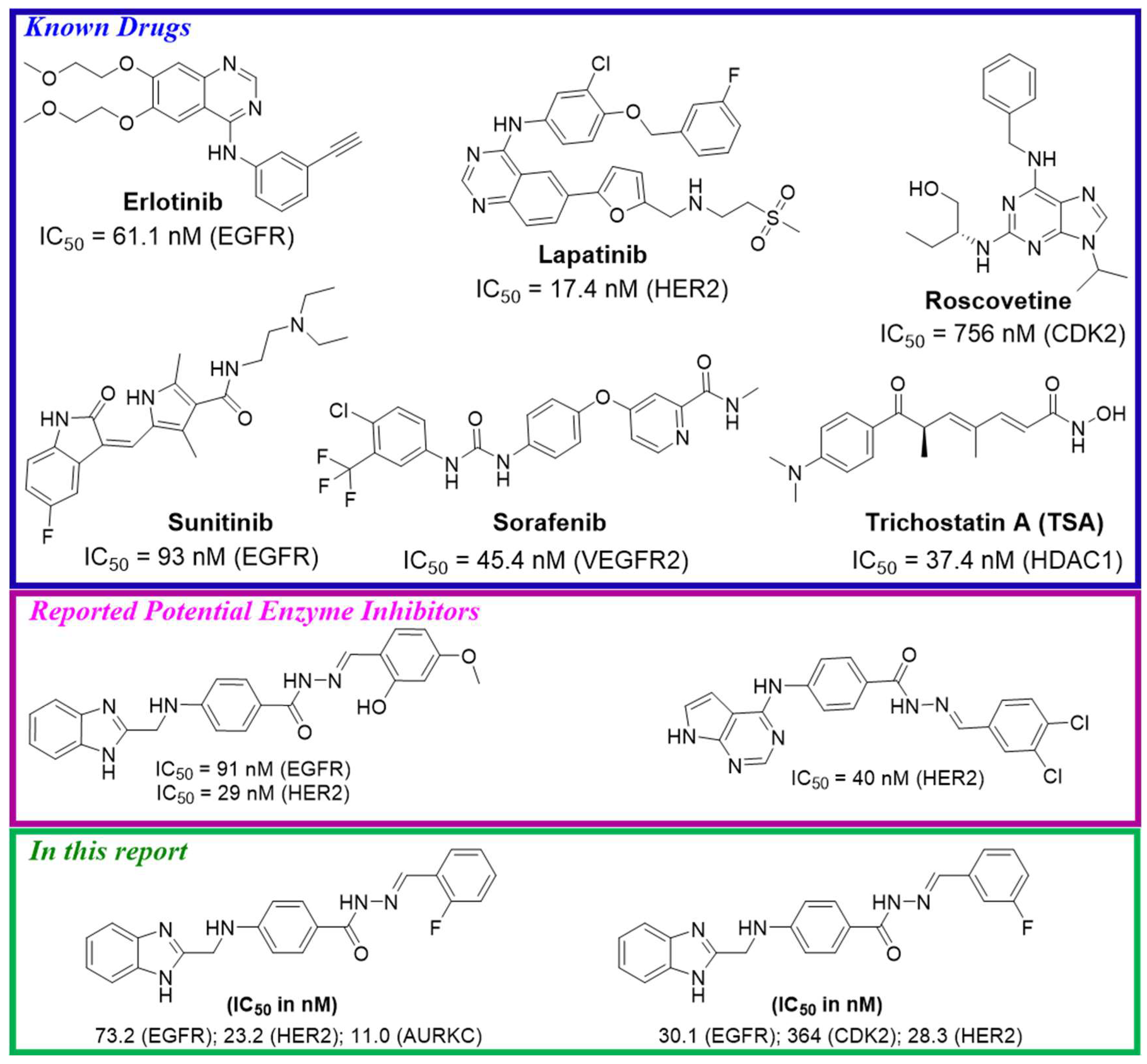
Cancer therapy continues to be a formidable task, demanding the discovery and development of effective treatments. Among the emerging classes of anticancer agents, halogenated TKIs have garnered significant attention owing to their unique chemical structures and enhanced pharmacological properties. Incorporating halogen atoms, such as fluorine, chlorine, bromine, or iodine, into the molecular backbone of TKIs offers a promising avenue to improve their therapeutic efficacy, selectivity, and potency [1]. The addition of halogen substituents in TKIs has been shown to exert a profound influence on their binding affinity to target kinases. Through their strategic placement, halogen atoms modulate the interactions between the TKIs and their kinase targets, potentially leading to enhanced therapeutic effects. These modifications can optimize the TKIs’ ability to bind to the kinase active site, resulting in increased potency and selectivity [1,2,3]. The halogenated TKI gefitinib, developed to precisely target the epidermal growth factor receptor (EGFR), has demonstrated its clinical utility in the care of non-small cell lung cancer (NSCLC) patients, leading to its adoption as a first-line standard of care therapy for advanced NSCLC cases harboring activating EGFR mutations. Furthermore, gefitinib offers advantages such as oral bioavailability and a comparatively lower toxicity profile, in contrast to conventional chemotherapy agents. Notably, it has shown promising outcomes in patients with EGFR mutations, which are frequently observed in Asian populations and allied with higher response rates to TKIs (Figure 1) [4,5,6,7,8]. Dasatinib, another halogenated TKI, exerts its therapeutic effects by targeting multiple tyrosine kinases, including BCR-ABL, SRC family kinases, and c-KIT. The incorporation of halogen substituents in dasatinib has been demonstrated to enhance its potency and selectivity, rendering it a valuable therapeutic option for various types of cancer. Notably, dasatinib has demonstrated efficacy in treating chronic myeloid leukemia (CML) and acute lymphoblastic leukemia (ALL), warranting its approval as a first-line or second-line therapy for these conditions. Additionally, dasatinib has demonstrated the capacity to overcome resistance to other TKIs, such as imatinib, which are commonly employed in the treatment of CML. However, it is crucial to recognize the potential adverse effects associated with dasatinib, which may include fluid retention, bleeding, and pulmonary arterial hypertension (PAH). Furthermore, the higher cost linked to the use of dasatinib is an important consideration [6]. Afatinib, a halogenated TKI with targets including EGFR, HER2, and HER4, has emerged as an effective therapy for NSCLC and head and neck cancer. Halogen substituents incorporated into afatinib have demonstrated the potential to enhance its potency and selectivity, thus improving its effectiveness in specific patient populations. Afatinib’s broad spectrum of activity against multiple members of the HER family of receptor tyrosine kinases positions it as a valuable treatment option for diverse cancer types. It has shown efficacy, particularly in patients with EGFR mutations, leading to its approval as a first-line therapy in this population. Additionally, afatinib has shown the capability of overwhelming resistance to other EGFR TKIs, including gefitinib and erlotinib. However, it is essential to consider potential adverse effects, such as diarrhea, rash, and mucositis, as well as the higher cost associated with afatinib [7,8]. Several halogenated TKIs have shown promising results in clinical trials and preclinical studies. Entrectinib, a halogenated TKI, has demonstrated efficacy in targeting ROS1 and NTRK fusions in solid tumors, including lung cancer and neuroblastoma [9]. Avitinib, another halogenated TKI, has shown potential in inhibiting the EGFR T790M mutation, which is allied with battling first-generation EGFR TKIs [10,11]. Saracatinib, a halogenated Src/Abl kinase inhibitor, has exhibited promising activity in solid tumors, particularly in pancreatic cancer [12,13]. Ponatinib, a halogenated TKI with potent activity against BCR-ABL and other tyrosine kinases, has been effective in treating chronic myeloid leukemia (CML) and Philadelphia chromosome-positive acute lymphoblastic leukemia (Ph+ ALL) [6,14] Foretinib, a halogenated TKI targeting MET, VEGFR, and other kinases, has shown efficacy in various solid tumors, including renal cell carcinoma and hepatocellular carcinoma [15]. Vandetanib, flumatinib, vemurafenib, sorafenib, cabozantinib, nilotinib, lapatinib, and selumetinib, all halogenated TKIs, have demonstrated efficacy in different cancer types by targeting specific kinases involved in tumor growth and progression [16,17,18,19,20,21,22,23,24]. These include VEGFR, BRAF, c-Met, RET, and HER2, among others. However, it is important to consider the potential adverse effects associated with these halogenated TKIs. These halogenated tyrosine kinase inhibitors may cause debilitating fatigue, cognitive deficits, and even life-threatening lung disease. Patients may also experience gastrointestinal issues, blood cell depletion, heart rhythm abnormalities, high blood pressure, painful skin, nail changes, and heightened photosensitivity. Vigilant monitoring and active management of these diverse adverse effects are crucial when prescribing these halogenated TKI therapies. Close monitoring and management of these side effects are crucial for ensuring patient safety and treatment effectiveness. Moreover, it is worth noting that some of these halogenated TKIs may have limitations, including high costs and limited accessibility in certain healthcare systems. The affordability and availability of these drugs are important factors to consider when evaluating their clinical utility and impact on patient care. However, halogenated TKIs have emerged as a promising class of anticancer agents, with multiple compounds demonstrating efficacy in various cancer types. While these drugs offer significant therapeutic potential, it is essential to carefully assess their adverse effects and limitations to optimize their clinical utility and ensure equitable access to effective treatments in cancer care. Further research and clinical investigations are warranted to elucidate the full potential of halogenated TKIs and their role in improving patient outcomes in oncology.
On the other hand, benzimidazole compounds have gained attention in drug development owing to their versatility and diverse applications. They exhibit antibacterial properties [25], potential as anti-tubercular agents [26], antifungal activities [27], and antiprotozoal effects [28]. Benzimidazole derivatives also show promise as antiviral agents [29] and as protein kinase inhibitors [30,31]. Studies have demonstrated the therapeutic action of benzimidazole-based compounds, such as nazartinib, in inhibiting EGFR and HER2 proteins [32,33,34,35]. Our previous research focused on hydrazone derivatives, which exhibited potent multi-kinase inhibition activity, including EGFR and HER2 [36,37,38,39]. To further explore their potential, we designed benzimidazole and (halogenated)benzylidene-benzohydrazide hybrids, namely, (E)-4-(((1H-benzo[d]imidazol-2-yl)methyl)amino)-N′-(halogenated)benzylidenebenzohydrazides, aiming for multi-kinase inhibitory activities. The synthesized compounds (6a-l) were evaluated for their in-vitro cytotoxicity against cancerous/normal cell lines, and their ability to inhibit multiple tyrosine kinases, investigated for their apoptosis-inducing ability, cell cycle-suppressing effects, drug-likeness properties, and in silico molecular docking, shed light on their selective mechanism of action and substantiated their potential as promising anticancer agents.
Figure 1. An example of some known TKIs [39], recently reported TKIs [40], and synthesized potential TKIs.
Pharmaceuticals 17 00839 g001

2. Results and Discussion

2.1. Chemistry

The synthesis of (E)-4-(((1H-benzo[d]imidazol-2-yl)methyl)amino)-N′-(halogenated)benzylidenebenzohydrazide derivatives (6a-l) was carried out using a straightforward method, as described in Scheme 1, following the multi-step procedure adopted for our previously reported method [39]. The use of a previously reported synthetic methodology, along with the straightforward nature of the individual steps, allowed for the efficient preparation of the desired series of halogenated benzylidenebenzohydrazide derivatives (6a-l) bearing the benzo[d]imidazole moiety. The structural elucidation of the synthesized compounds was carried out using a comprehensive analytical approach, employing a suite of spectroscopic techniques. These included infrared (IR) spectroscopy, mass spectrometry (MS), and proton and carbon nuclear magnetic resonance (1H-NMR and 13C-NMR) analyses (please see the Supplementary Materials for the spectra). Additionally, we also reported the physical properties of the compounds 6a-l, specifically their color and melting point ranges. The 1H-NMR spectroscopic analysis of the synthesized compounds 6a-l revealed characteristic signals for the secondary aromatic amine protons located at the 4-position of the benzohydrazide moiety. These amine proton signals were observed in the 7.01 to 7.06 ppm region of the 1H-NMR spectra as triplets, with coupling constant values ranging from 5.7 to 6.1 Hz. The observed chemical shift and coupling pattern of these amine proton signals provided evidence that the aromatic amine functionality was directly bonded to an aliphatic (CH2) carbon, specifically the methylene group derived from the condensation of compounds 1 and 2 during the synthetic sequence. These 1H-NMR data thus confirmed the successful formation of the desired compound 6a-l structures, with the secondary aromatic amine protons serving as a characteristic spectroscopic signature for the benzohydrazide moiety within the target molecules. The 1H-NMR spectra of the synthesized compounds 6a-l also exhibited characteristic signals for the benzylidene protons. These benzylidene proton signals were observed in the 8.35 to 8.38 ppm region of the 1H-NMR spectra, except for the compounds with substituents at the 2-position of the benzene ring. Specifically, compound 6b, which contained a bromo substituent at the 2-position, displayed a downfield shift of 0.38 ppm compared with the non-substituted compound 6a. Similarly, the 2-chloro substituted compound 6e exhibited a 0.43 ppm downfield shift, while the 2-fluoro substituted compound 6h showed a 0.27 ppm downfield shift relative to 6a. Furthermore, the compounds with 2,4-dihalogen substitution, 6k (2,4-dichloro) and 6l (2,5-difluoro), also demonstrated downfield shifts of 0.38 ppm and 0.22 ppm, respectively, compared with the unsubstituted analog 6a. These observed downfield shifts in the benzylidene proton signals for the 2-substituted and 2,4-disubstituted compounds can be attributed to the electronic effects of the halogen substituents, which influenced the chemical environment and deshielding of the benzylidene protons.
The structures of the synthesized compounds 6a-l were further proved by the observation of distinctive IR spectral signatures, such as the sharp peaks observed in the range of 1604–1613 cm−1, which were characteristic of the benzylidene hydrazone (-N=CH-) functional group present in all the compounds. N-H stretching vibration bonds of amide -NH (-CO-NH-) appeared between 3044 and 3464 cm−1 and around 1502–1568 cm−1. Carbonyl (C=O) vibrational bonds appeared at around 1642–1656 cm−1. Furthermore, each compound displayed distinct IR spectral features. Notably, the bromo-substituted analogs (6b-d) exhibited characteristic stretching bands in the range of 741–748 cm−1. In contrast, the chloro- and fluoro-substituted compounds (6e-l) showed distinctive stretching bands between 504 and 758 cm−1. Finally, the structures of compounds 6e-l were further confirmed through their mass spectral analysis data.

2.2. Biological Evaluation

2.2.1. In Vitro Cytotoxicity

The cytotoxic activities of compounds 6a-l were assessed using a standard MTT method, which was performed across three different cancer cell line models, namely human colon cancer (HCT-116), hepatocellular carcinoma (HepG2), mammary gland cancer (MCF-7), and normal (WI-38) cell lines. The results presented in Table 1 show that the cytotoxicity of the tested compounds was quantified by the concentration required to induce 50% inhibition of cancer cell viability. The cytotoxicity results provide a comprehensive evaluation of the synthesized compounds 6a-6l in comparison with the standard drugs sorafenib, doxorubicin, and sunitinib. Among the synthesized compounds, 6c (3-Br substituted) and 6i (3-F substituted) exhibited the most potent cytotoxic activity, with IC50 values ranging from 7.82 to 10.21 μM across the tested cancer cell lines. These values are comparable to the standard drugs, which have IC50 values between 4.17 and 24.06 μM. Compounds 6h (2-F), 6j (4-F), and 6d (4-Br) also demonstrated relatively strong cytotoxicity, with IC50 values generally below 30 μM in the cancer cell lines. The remaining compounds, including 6a, 6b, 6e, 6f, 6g, 6k, and 6l, exhibited moderate to weaker cytotoxic activity, with IC50 values mostly above 20 μM. When considering the selectivity toward cancer cells over the normal WI-38 fibroblast cell line, compounds 6c, 6i, and 6j stood out, showing significantly lower IC50 values in the cancer cell lines compared with the WI-38 cells, indicating a higher degree of selectivity. Other compounds, such as 6h and 6d, also exhibited relatively better selectivity toward the cancer cell lines. The structure–activity relationship analysis suggested that the presence of bromo (Br) or fluoro (F) substituents at the 3-position of the phenyl ring is favorable for potent and selective cytotoxic activity, as observed with compounds 6c and 6i. Substitution at the 4-position, as in the case of compounds 6d, 6g, and 6j, also seems beneficial, with fluoro (6j) showing the best results. In contrast, the disubstituted compounds 6k and 6l generally exhibited lower cytotoxic potency and selectivity compared with their monosubstituted counterparts. Overall, the cytotoxicity data provide valuable insights into the structure–activity relationships of the synthesized compounds and can guide further optimization efforts to develop potential anticancer agents with improved potency and selectivity.
The Selectivity Index (SI) values shown in Table 1 provide important information about the selectivity of the synthesized compounds (6a-l) against the various cancer cell lines tested. The SI values represent the ratio of the IC50 values for the normal WI-38 cell line compared with the respective cancer cell lines, with higher SI values indicating greater selectivity for the cancer cells over normal cells. Compounds 6c, 6h, and 6i exhibited the highest SI values against the SI cancer cell line, with SI values of 4.20–4.72 (HCT-116), 5.41–7.19 (HepG2), and 4.85–6.16 (MCF-7), respectively. The SI values for the standard drugs were 1.16–1.95 (sorafenib), 1.28–1.61 (doxorubicin), and 2.31–3.11 (sunitinib), except 6.64 against the HepG2 cell line for sunitinib 6.64, respectively. This suggests these compounds (6c, 6h, 6i) demonstrated the greatest selectivity for the SI cancer cells compared with the normal WI-38 cells.

2.2.2. In Vitro Protein Kinase Inhibition Assays

Based on the cytotoxicity analysis of the synthesized compounds (6a-l), four compounds (6c, 6h-j) were chosen for further enzymatic activity assessment against a range of kinase enzymes, including EGFR, Her2, VEGFR2, CDK2, AURKC, HDAC1, and mTOR (Table 2). To benchmark the activity of these compounds, several well-known kinase inhibitors were used as reference standards for the tested kinases, such as erlotinib for EGFR, lapatinib for Her2, sorafenib for VEGFR2, roscovetine for CDK2, TSA for AURKC and HDAC1, and rapamycin for mTOR. The synthesized compounds 6c and 6h-j demonstrated varying degrees of potent inhibition across the different protein kinases tested. For instance, compound 6h exhibited very high potency against EGFR (IC50 = 73.2 nM), which is almost 1-fold higher than the standard erlotinib (IC50 = 61.1 nM), and against Her2 (IC50 = 23.2 nM), which is also almost 1-fold higher than lapatinib (IC50 = 17.4 nM). Additionally, 6h showed high potency against CDK2 (IC50 = 284 nM), which is 2.5-fold higher than roscovetine (IC50 = 756 nM), and very high potency against AURKC (IC50 = 11 nM), which is 3-fold higher than the standard TSA (IC50 = 30.4 nM). Compound 6i showed excellent potency against EGFR (IC50 = 30.1 nM), which is almost 2-fold higher than the standard erlotinib, and against Her2 (IC50 = 28.3 nM), which is almost 1-fold higher than lapatinib. Additionally, 6i exhibited high potency against CDK2 (IC50 = 364 nM), which is 2-fold higher than roscovetine, and against mTOR (IC50 = 152 nM), which is 1-fold higher than the standard rapamycin (IC50 = 208 nM). In contrast, compound 6c gave disappointing results compared with the expectations based on the cytotoxicity against various cancer cell lines. Additionally, compound 6j showed very little inhibitory activity against the tested kinases. Compared with the standard drugs, the synthesized compounds 6h and 6i exhibited superior potency across multiple kinase targets. The low IC50 values for the synthesized compounds indicate their high binding affinity and inhibitory activity toward these clinically relevant protein kinases, suggesting their potential to be developed as potent and selective anticancer agents targeting multiple dysregulated signaling pathways in cancer. These findings underscore the promising potential of compounds 6h and 6i as candidates for further development as kinase inhibitors.

2.2.3. Cell Cycle Analysis

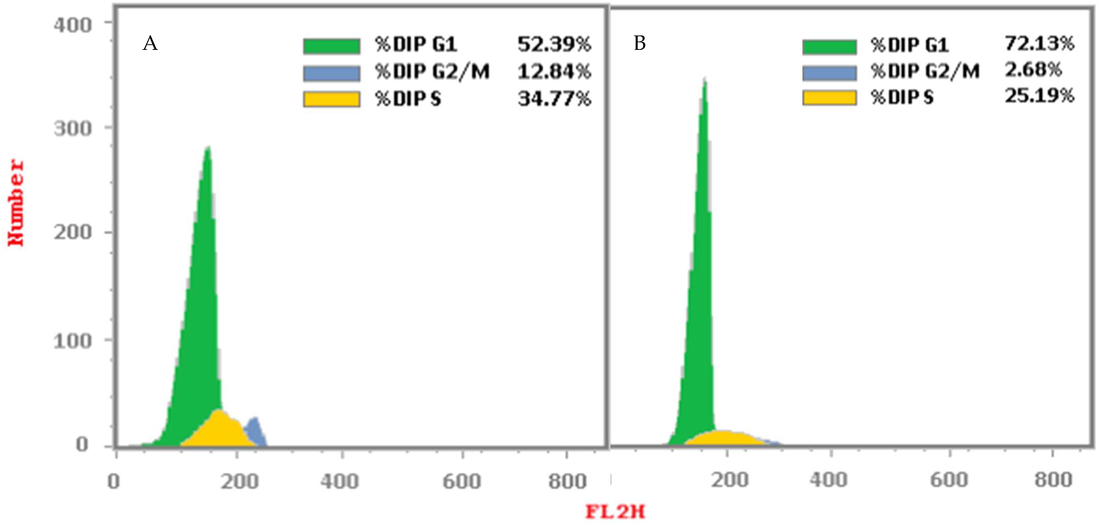
The cytotoxicity assay, selectivity index (SI) values, and enzyme inhibitory activities of the synthesized compounds 6a-l were evaluated, and based on these results, compound 6i was selected for further investigation to assess its impact on cell cycle progression in HepG2 cells. To examine the effect of compound 6i on the cell cycle, HepG2 cells were treated with the compound at its IC50 concentration for 24 h. Following the treatment, the cells were stained with propidium iodide and analyzed using flow cytometry to determine the cell cycle phase distribution. The cell cycle analysis results, presented in Table 3 and Figure 2, revealed that treatment with compound 6i elicited a distinct cell cycle arrest effect. Specifically, the data showed that the DNA content of cells treated with 6i increased in the G1 phase while decreasing in the S and G2/M phases. This indicates that compound 6i induced a significant cell cycle arrest at the G1 phase. The quantitative data further substantiated this observation. The percentage of cells in the G0-G1 phase increased from 52.39% in the untreated control to 72.13% upon treatment with compound 6i. Conversely, the proportion of cells in the S phase decreased from 34.77% to 25.19%, and the percentage of cells in the G2/M phase declined from 12.84% to 2.68%. These findings suggest that treatment with compound 6i substantially altered the cell cycle distribution of the HepG2 cancer cells. It led to a marked accumulation of cells in the G1 phase, accompanied by a concomitant reduction in the proportion of cells in the S and G2/M phases. The cell cycle arrest effect observed with compound 6i may be a key mechanistic aspect contributing to its anticancer potential. The observed cell cycle arrest at the G1 phase is a common mechanism of action for many anti-cancer agents, as it can prevent cell division and proliferation, ultimately leading to cell cycle arrest and potentially inducing apoptosis or other forms of cell death. These results, along with the previously reported cytotoxicity and kinase inhibitory activities of compound 6i, further highlight its potential as a promising anticancer candidate that warrants further investigation and optimization for targeted cancer therapy.
The cell cycle distribution index (CDI) is a metric that quantifies the rate of cell proliferation, calculated as CDI = (G2/M + S)/(G0 − G1), where the values represent the percentages of cells in each phase. In this study, the CDI values for the control and compound 6i-treated HepG2 cells were 0.91 and 0.39, respectively. A decrease in CDI indicates a reduced rate of cell proliferation, suggesting cell cycle arrest. Compound 6i was further evaluated for apoptosis analysis in HepG2 cell lines.

2.2.4. Apoptosis Analysis

Annexin-V/Propidium Iodide (PI) Staining Assay

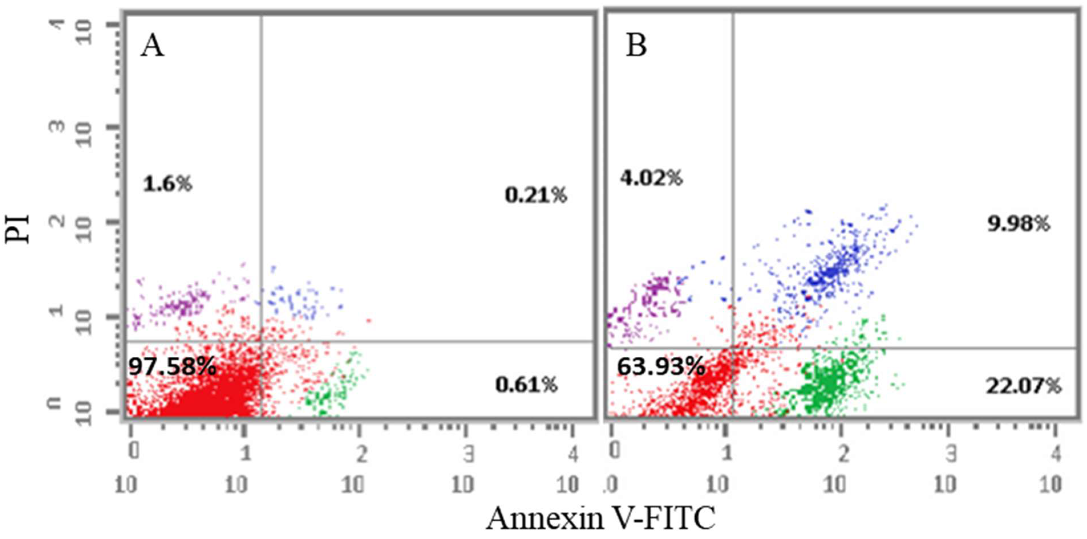
To investigate the mode of cell death induced by the synthesized compound 6i, flow cytometry analysis was performed. HepG2 cancer cells were treated with compound 6i at their respective IC50 concentrations and incubated for 24 h. Following the treatment, the cells were double-stained with Annexin V and propidium iodide (PI) to assess the levels of apoptosis and necrosis. The data presented in Table 4 reveal a significant decline in the percentage of viable cells in the compound 6i-treated group (63.93%) compared with the untreated control group (97.58%). In the early stages of the cell death process, the percentage of cells undergoing apoptosis was 0.61% in the control group, whereas it increased to 22.07% in the compound 6i-treated group. Additionally, in the late stages of apoptosis, the percentage of apoptotic cells rose from 0.21% in the control group to 9.98% in the compound 6i-treated group. These findings suggest that the antiproliferative effects of compound 6i against HepG2 cancer cells are at least partially mediated through the induction of apoptosis, particularly during the early stages of the cell death process. The flow cytometry data indicate that treatment with compound 6i leads to a substantial increase in the proportion of HepG2 cells undergoing both early and late stages of apoptosis. The observed increase in both early and late apoptotic cell populations upon treatment with compound 6i indicates that this compound may trigger the apoptotic signaling cascades, leading to programmed cell death. Additionally, the data show a slight increase in the percentage of necrotic cells from 1.6% in the control group to 4.02% in the compound 6i-treated group, suggesting that the compound may also induce some degree of necrosis in HepG2 cells. Taken together, these results highlight the pro-apoptotic and potentially cytotoxic effects of compound 6i on HepG2 cells, which could have important implications for its further development as a potential anticancer agent. The apoptosis levels in the control HepG2 cell lines and the cells treated with compound 6i are shown in the graphical representation provided in Figure 3.

Determination of Apoptotic Protein Levels

To further elucidate the mechanism by which compound 6i induces cell death, the expression levels of key apoptotic proteins were assessed in the HepG2 cell line. As shown in Table 5, treatment with compound 6i resulted in significant changes in the levels of critical proteins involved in the apoptotic signaling cascade in HepG2 cells. Specifically, the levels of the pro-apoptotic proteins caspase-3 and Bax were markedly higher in the compound 6i-treated group compared with the control. Caspase-3 levels were approximately 3.9-fold higher, while Bax levels were 7.22-fold higher in the treated cells. In contrast, the level of the anti-apoptotic protein Bcl-2 was 7.5-fold lower in the compound 6i-treated group compared with the control. These findings strongly suggest that the induction of apoptosis by compound 6i in HepG2 cells is mediated, at least in part, by the upregulation of the pro-apoptotic proteins caspase-3 and Bax, coupled with the downregulation of the anti-apoptotic protein Bcl-2. The magnitude of the changes observed in the levels of these key apoptotic regulators upon treatment with compound 6i was comparable to, or even exceeded, the effects seen with the known apoptosis-inducing agent staurosporine. These results further substantiate the pro-apoptotic mechanism of action of compound 6i and highlight its potential as a promising candidate for the development of novel therapies targeting diseases involving dysregulated cell death, such as cancer. Additional studies are warranted to elucidate the detailed molecular mechanisms underlying the apoptosis-inducing effects of compound 6i and to assess its therapeutic potential in relevant in vivo models.

2.3. In Silico Studies

Molecular Docking

In order to predict the potential binding interactions between compounds 6h and 6i and the investigated protein kinase enzymes, compound 6h was docked into the active site of Her2 (PDB: 7PCD) and AURKC (PDB: 6GR8), and 6i was docked into the active side of EGFR (PDB: 4HJO) and Her2 (PDB: 7PCD), respectively.
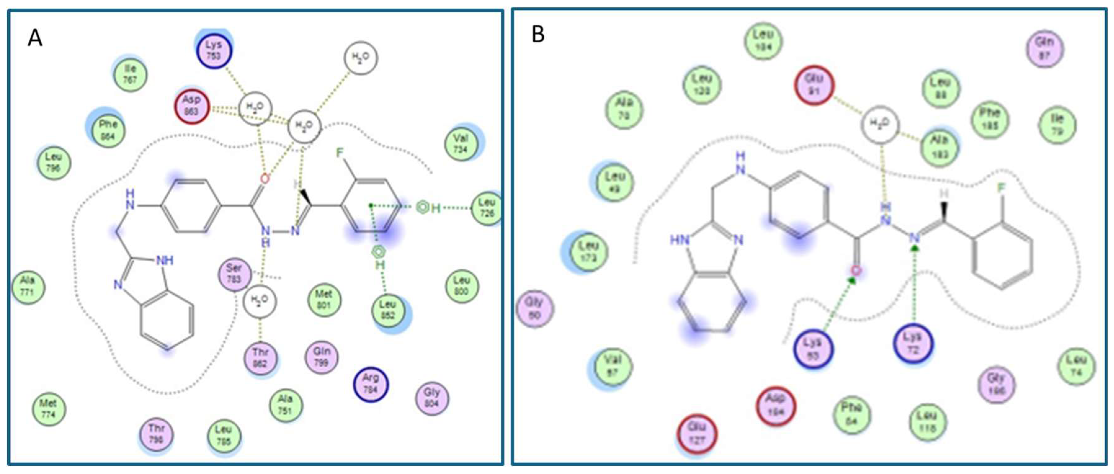
Based on the docking data of compound 6h with the HER2 kinase enzyme (PDB: 7PCD), the hydrogen bond network with Lys753, Asp863, and Thr862 through water molecules stabilized the compound inside the active site of the target enzyme (Figure 4A). Additionally, binding interactions between the benzylidene benzene moiety of compound 6h and the amino acid residues Leu726 and Leu852 were observed, involving H-π bonds (Figure 4A). However, when compound 6h was co-crystallized with the AURKC kinase enzyme (PDB: 6GR8), three hydrogen bonds were formed between the key amino acid residues and the hydrazide moiety, facilitating the fitting of the compound within the active site (Figure 4B). Specifically, one hydrogen bond was between the side chain of Lys35 and the carbonyl group, the second hydrogen bond was between Lys72 and a nitrogen atom, and the third hydrogen bond was part of a network involving Glu91 and Ala183, mediated by a water molecule (Figure 4B).
The docking study results of compound 6i with the EGFR (PDB: 4HJO) and the active site of HER2 kinase enzyme (PDB: 7PCD) are depicted in Figure 5. For the EGFR kinase enzyme, the benzimidazole moiety of compound 6i formed a hydrogen bonding network, mediated by water molecules, with side chains of Thr766 and Thr830 (Figure 5A). Additionally, the nitrogen atom of the imidazole ring participated in a hydrogen bond with the key amino acid residue Asp831, while another important hydrogen bond was formed between the backbone of Met769 and the carbonyl oxygen of the compound, facilitating its fitting within the active site. Furthermore, the presence of a fluorine atom in the meta position of the benzene ring enhanced its interaction with Lys704 through the formation of a cation-π bond compared with other derivatives (Figure 5A). For the Her2 active site, compound 6i made a hydrogen bond network with Lys753, Asp863, and Met501, stabilized by water molecules inside the active site (Figure 5B). Additionally, binding interactions between the benzylidene benzene moiety of compound 6i and the amino acid residues Leu726 and Leu852 were also observed, similar to the interactions seen for compound 6h.

3. Materials and Methods

3.1. General

All reagents and solvents used in the experiments were of commercial grade and employed without further purification. Barnstead electrothermal digital melting point apparatus (model IA9100, BIBBY scientific limited, Staffordshire, UK) was used to determine the melting points. A Jasco FT/IR-6600 spectrometer (Tokyo, Japan) was used for recording IR data. Bruker 700 MHz NMR spectrometry (Zurich, Switzerland) was used to obtain the NMR data. Mass spectra were taken using an Agilent 6320 ion trap mass spectrometer equipped with an ESI ion source (Agilent Technologies, Palo Alto, CA, USA).

3.2. Chemistry

3.2.1. Synthesis of Ethyl 4-(((1H-benzo[d]imidazol-2-yl)methyl)amino)benzoate (3)

Ethyl 4-(((1H-benzo[d]imidazol-2-yl)methyl)amino)benzoate (3) was prepared using a previously reported method [39]. White powder (65%), mp. 255 °C (lit. [39] mp. = 255 °C).

3.2.2. Synthesis of 4-(((1H-benzo[d]imidazol-2-yl)methyl)amino)benzohydrazide (4)

Additionally, 4-(((1H-Benzo[d]imidazol-2-yl)methyl)amino)benzohydrazide (4) was prepared using our previously reported method [39]. White powder (80%), p. 240 °C (lit. [39] mp. = 240 °C).

3.3. General Procedure for the Preparation of (E)-4-(((1H-benzo[d]imidazol-2-yl)methyl)amino)-N′-(substitutedbenzylidene)benzohydrazide (6a-l)

An equimolar mixture of hydrazide 4 and halogen-substituted benzaldehyde (5) was reacted to obtain the desired 1H-benzo[d]imidazole-(halogenated)benzylidenebenzohydrazide (6a-l) following the reported procedure [39,40,42].

3.3.1. (E)-4-(((1H-benzo[d]imidazol-2-yl)methyl)amino)-N′-benzylidenebenzohydrazide (6a)

White solid (53.43%), mp. 278 °C (lit. [39] mp. = 278 °C; lit [43] mp. = 246 °C), CAS registry number 76321-88-5.

3.3.2. (E)-4-(((1H-benzo[d]imidazol-2-yl)methyl)amino)-N′-(2-bromobenzylidene)benzohydrazide (6b)

White solid (105 mg, 0.234 mmol, 66%), mp. 284 °C. FT-IR (KBr): ν (cm−1) = 3437, 3174, 3135, 3044, 2976, 2829, 1926, 1893, 1649, 1604, 1558, 1522, 1431, 1350, 1294, 1268, 1187,1057, 1014, 832, 761, 739, 689, 647, and 608 cm−1. 1H-NMR (700 MHz, DMSO-d6), δ 1H NMR (700 MHz, DMSO), δ 12.35 (s, 1H), 11.76 (s, 1H), 8.75 (s, 1H), 7.99 (s, 1H), 7.75 (d, J = 8.3 Hz, 2H), 7.69 (d, J = 8.2 Hz, 1H), 7.58 (s, 1H), 7.46 (t, J = 7.7 Hz, 2H), 7.35 (t, J = 7.6 Hz, 1H), 7.15 (s, 2H), 7.04 (t, J = 6.1 Hz, 1H), 6.74 (d, J = 8.3 Hz, 2H), and 4.59 (d, J = 5.8 Hz, 2H) ppm. 13C-NMR (176 MHz, DMSO-d6), δ 163.54, 153.45, 152.11, 144.83, 143.78, 134.90, 133.98, 133.68, 131.93, 129.87, 128.62, 127.67, 123.87, 122.38, 121.66, 120.71, 118.98, 111.98, 111.80, and 41.85 ppm. Mass (ESI): m/z 448 [79(Br)M+H]+, 450 [81(Br)M+H]+.

3.3.3. (E)-4-(((1H-benzo[d]imidazol-2-yl)methyl)amino)-N′-(3-bromobenzylidene)benzohydrazide (6c)

White solid (97 mg, 0.216 mmol, 61%), mp. 278 °C. FT-IR (KBr): ν (cm−1) = 3388, 3096, 2963, 2907, 2845, 2790, 1963, 1656, 1606, 1522, 1438, 1255, 1073, 1021, 895, 822, 741,692, 640, and 478 cm−1. 1H-NMR (700 MHz, DMSO-d6), δ 1H NMR (700 MHz, DMSO), δ 12.35 (s, 1H), 11.62 (s, 1H), 8.35 (s, 1H), 7.89 (s, 1H), 7.73 (d, J = 8.3 Hz, 2H), 7.68 (d, J = 8.0 Hz, 1H), 7.60 (d, J = 8.2 Hz, 1H), 7.57 (s, 1H), 7.45 (s, 1H), 7.41 (t, J = 8.0 Hz, 1H), 7.15 (s, 2H), 7.04 (t, J = 6.1 Hz, 1H), 6.73 (d, J = 8.5 Hz, 2H), and 4.58 (d, J = 5.7 Hz, 2H) ppm. 13C-NMR (176 MHz, DMSO-d6), δ 163.46, 153.35, 151.97, 144.67, 143.67, 137.64, 134.79, 132.65, 131.45, 129.73, 129.29, 126.49, 122.62, 122.27, 121.56, 120.68, 118.87, 111.87, 111.70, and 41.75 ppm. Mass (ESI): m/z 448 [79(Br)M+H]+, 450 [81(Br)M+H]+.

3.3.4. (E)-4-(((1H-benzo[d]imidazol-2-yl)methyl)amino)-N′-(4-bromobenzylidene)benzohydrazide (6d)

White solid (124 mg, 0.276 mmol, 63%), mp. 287 °C (lit. [43] mp. = 230 °C), CAS registry number 76321-87-4. FT-IR (KBr): ν (cm−1) =3389, 3060, 2957, 2906, 2796, 1655, 1610, 1568, 1526, 1487, 1455, 1438, 1357, 1335, 1296, 1276, 1260, 1212, 1190, 1150, 1132, 1068, 1026, 1009, 956, 934, 825, 748, 695, 638, 515, and 503 cm−1. 1H-NMR (700 MHz, DMSO-d6), δ 12.35 (s, 1H), 11.56 (s, 1H), 8.36 (s, 1H), 7.72 (d, J = 8.6 Hz, 2H), 7.64 (s, 4H), 7.58 (s, 1H), 7.45 (s, 1H), 7.15 (s, 2H), 7.03 (s, 1H), 6.73 (d, J = 8.7 Hz, 2H), and 4.58 (d, J = 5.8 Hz, 2H) ppm. 13C-NMR (176 MHz, DMSO-d6), δ 163.40, 153.37, 151.93, 145.29, 143.68, 134.80, 134.43, 132.27, 129.70, 129.17, 123.31, 122.29, 121.56, 120.79, 118.88, 111.88, 111.71, and 41.76 ppm. Mass (ESI): m/z 448 [79(Br)M+H]+, 450 [81(Br)M+H]+.

3.3.5. (E)-4-(((1H-benzo[d]imidazol-2-yl)methyl)amino)-N′-(2-chlorobenzylidene)benzohydrazide (6e)

White solid (140 mg, 0.346 mmol, 97%), mp. 289 °C. FT-IR (KBr): ν (cm−1) = 1656, 1606, 1522, 1431, 1360, 1330, 1298, 1258, 1187, 1148, 1132, 1050, 1024, 936, 826, 744, 713, 692, 640, and 498 cm−1. 1H-NMR (700 MHz, DMSO-d6), δ 1H NMR (700 MHz, DMSO), δ 12.35 (s, 1H), 11.73 (s, 1H), 8.80 (s, 1H), 8.00 (s, 1H), 7.74 (d, J = 8.8 Hz, 2H), 7.58 (d, J = 7.6 Hz, 1H), 7.52 (d, J = 4.4 Hz, 1H), 7.43 (t, J = 8.4 Hz, 3H), 7.15 (s, 2H), 7.04 (t, J = 6.1 Hz, 1H), 6.74 (d, J = 8.7 Hz, 2H), and 4.58 (d, J = 5.7 Hz, 2H) ppm. 13C-NMR (176 MHz, DMSO-d6), δ 163.39, 158.77, 153.34, 152.01, 143.68, 142.37, 134.79, 133.37, 132.38, 131.57, 130.35, 129.75, 128.04, 127.18, 122.29, 121.55, 120.60, 118.87, 111.88, 111.70, and 41.74 ppm. Mass (ESI): m/z 404 [35(Cl)M+H]+, 406 [37(Cl)M+H]+.

3.3.6. (E)-4-(((1H-benzo[d]imidazol-2-yl)methyl)amino)-N′-(3-chlorobenzylidene)benzohydrazide (6f)

White solid (136 mg, 0.336 mmol, 94%), mp. 271 °C. FT-IR (KBr): ν (cm−1) = 3398, 3057, 3025, 2871, 2836, 1609, 1525, 1473, 1434, 1334, 1281, 1184, 1132, 1024, 920, 822, 739, 696, 621, and 556 cm−1. 1H-NMR (700 MHz, DMSO-d6), δ 1H NMR (700 MHz, DMSO), δ 12.35 (s, 1H), 11.62 (s, 1H), 8.37 (s, 1H), 7.74 (s, 1H), 7.73 (d, J = 8.8 Hz, 2H), 7.65 (s, 1H), 7.58 (d, J = 7.6 Hz, 1H), 7.47 (d, J = 5.2 Hz, 2H), 7.44 (d, J = 7.5 Hz, 1H), 7.18–7.12 (m, 2H), 7.04 (t, J = 5.8 Hz, 1H), 6.74 (d, J = 8.8 Hz, 2H), and 4.58 (d, J = 5.8 Hz, 2H) ppm. 13C-NMR (176 MHz, DMSO-d6), δ 163.49, 153.35, 151.97, 144.78, 137.41, 134.80, 134.09, 131.17, 129.77, 126.46, 126.06, 122.29, 121.55, 120.70, 118.87, 111.87, 111.70, and 41.76 ppm. Mass (ESI): m/z 404 [35(Cl)M+H]+, 406 [37(Cl)M+H]+.

3.3.7. (E)-4-(((1H-benzo[d]imidazol-2-yl)methyl)amino)-N′-(4-chlorobenzylidene)benzohydrazide (6g)

White solid (100 mg, 0.247 mmol, 69%), mp. 290 °C (lit. [43] mp. = 238 °C), CAS registry number 76321-91-0. FT-IR (KBr): ν (cm−1) = 3385, 3057, 2952, 2911, 2787, 2514, 1656, 1606, 1522, 1434, 1334, 1294, 1255, 1210, 1184, 1086, 1026, 934, 822, 741, 637, 504, and 436 cm−1. 1H-NMR (700 MHz, DMSO-d6), δ 12.35 (s, 1H), 11.55 (s, 1H), 8.38 (s, 1H), 7.72 (d, J = 8.5 Hz, 4H), 7.57 (s, 1H), 7.51 (d, J = 8.3 Hz, 2H), 7.44 (s, 1H), 7.14 (s, 2H), 7.02 (t, J = 5.8 Hz, 1H), 6.73 (d, J = 8.6 Hz, 2H), and 4.58 (d, J = 5.8 Hz, 2H) ppm. 13C-NMR (176 MHz, DMSO-d6), δ 163.38, 153.37, 151.92, 145.20, 143.69, 134.80, 134.54, 134.09, 129.69, 129.36, 128.92, 122.28, 121.55, 120.79, 118.87, 111.87, 111.70, and 41.76 ppm. Mass (ESI): m/z 404 [35(Cl)M+H]+, 406 [37(Cl)M+H]+.

3.3.8. (E)-4-(((1H-benzo[d]imidazol-2-yl)methyl)amino)-N′-(2-fluorobenzylidene)benzohydrazide (6h)

White solid (100 mg, 0.258 mmol, 72%), mp. 289 °C. FT-IR (KBr): ν (cm−1) = 3388, 3096, 2963, 2907, 2845, 2790, 1963, 1655, 1606, 1521, 1437, 1255, 1073, 1021, 894, 822, 741, 692, 640, and 478 cm−1. 1H-NMR (700 MHz, DMSO-d6), δ 12.35 (s, 1H), 11.62 (s, 1H), 8.64 (s, 1H), 7.92 (s, 1H), 7.73 (d, J = 8.7 Hz, 2H), 7.58 (d, J = 8.0 Hz, 1H), 7.47 (d, J = 6.1 Hz, 1H), 7.45 (d, J = 7.6 Hz, 1H), 7.29 (t, J = 8.5 Hz, 2H), 7.15 (s, 2H), 7.04 (s, 1H), 6.74 (d, J = 8.7 Hz, 2H), and 4.58 (d, J = 5.8 Hz, 2H) ppm. 13C-NMR (176 MHz, DMSO-d6), δ 163.37, 160.61 (1JC-F = 250 Hz), 153.36, 151.97, 143.67, 139.15, 134.79, 132.03 (3JC-F = 8.4 Hz), 129.71, 126.63 (4JC-F = 2.8 Hz), 125.37 (3JC-F = 3Hz), 122.66 (2JC-F = 9.8 Hz), 122.30, 121.56, 120.66, 118.88, 116.43 (2JC-F = 20.8 Hz), 111.90, 111.70, and 41.74 ppm. Mass (ESI): m/z 388 [(18F)M+H]+; 389 [(19F)M+H]+.

3.3.9. (E)-4-(((1H-benzo[d]imidazol-2-yl)methyl)amino)-N′-(3-fluorobenzylidene)benzohydrazide (6i)

Beige solid (151 mg, 0.309 mmol, 87%), mp. 278 °C. FT-IR (KBr): ν (cm−1) =3464, 3405, 3206, 3066, 2924, 2843, 1613, 1525, 1443, 1353, 1294, 1265, 1187, 1021, 936, 898, 872, 826, 758, 735, 683, and 521 cm−1. 1H-NMR (700 MHz, DMSO-d6), δ 12.35 (s, 1H), 11.60 (s, 1H), 8.39 (s, 1H), 7.73 (d, J = 8.4 Hz, 2H), 7.58 (d, J = 4.4 Hz, 1H), 7.52 (s, 1H), 7.49 (d, J = 7.6 Hz, 2H), 7.44 (d, J = 8.0 Hz, 1H), 7.25 (t, J = 8.6 Hz, 1H), 7.15 (s, 2H), 7.03 (s, 1H), 6.73 (d, J = 8.3 Hz, 2H), and 4.58 (d, J = 5.6 Hz, 2H) ppm. 13C-NMR (176 MHz, DMSO-d6), δ 163.49, 162.69 (1JC-F = 245 Hz), 153.36, 151.95, 145.13, 143.67, 137.76 (3JC-F = 7.8Hz), 134.79, 131.34 (3JC-F = 8.5 Hz), 129.72, 123.67, 122.29, 121.55, 120.72, 118.87, 116.88 (2JC-F = 21 Hz), 113.20 (2JC-F = 22 Hz), 111.87, 111.70, and 41.75 ppm. Mass (ESI): m/z 388 [(18F)M+H]+; 389 [(19F)M+H]+.

3.3.10. (E)-4-(((1H-benzo[d]imidazol-2-yl)methyl)amino)-N′-(4-fluorobenzylidene)benzohydrazide (6j)

White solid (70 mg, 0.180 mmol, 53%), mp. 291 °C. FT-IR (KBr): ν (cm−1) =3392, 2956, 2898, 2790, 2693, 1649,1604, 1502, 1438, 1298, 1187, 1145, 832, 751, 696, and 514 cm−1. 1H-NMR (700 MHz, DMSO-d6), δ 12.34 (s, 1H), 11.49 (s, 1H), 8.38 (s, 1H), 7.75 (s, 2H), 7.71 (d, J = 8.6 Hz, 2H), 7.55 (s, 1H), 7.46 (s, 1H), 7.28 (t, J = 8.7 Hz, 2H), 7.15 (s, 2H), 7.01 (t, J = 6.0 Hz, 1H), 6.73 (d, J = 8.6 Hz, 2H), and 4.57 (d, J = 5.8 Hz, 2H) ppm. 13C-NMR (176 MHz, DMSO-d6), δ 13C NMR (176 MHz, DMSO), δ 163.37, 163.35 (1JC-F = 247 Hz), 153.38, 151.87, 145.44, 131.75, 129.62, 129.42 (1JC-F = 8.5 Hz), 122.24, 121.58, 120.89, 118.87, 116.31 (2JC-F = 22 Hz), 111.87, 111.68, and 41.76 ppm. Mass (ESI): m/z 388 [(18F)M+H]+; 389 [(19F)M+H]+.

3.3.11. (E)-4-(((1H-benzo[d]imidazol-2-yl)methyl)amino)-N′-(2,4-dichlorobenzylidene)benzohydrazide (6k)

White solid (134 mg, 0.305 mmol, 86%), mp. 294 °C. FT-IR (KBr): ν (cm−1) = 3385, 3096, 2956, 2790, 1656, 1604, 1522l, 1441, 1330, 1298, 1258, 1184, 1044, 970, 822, 739, 692, 637, and 514 cm−1. 1H-NMR (700 MHz, DMSO-d6), δ 1H NMR (700 MHz, DMSO), δ 12.35 (s, 1H), 11.78 (s, 1H), 8.75 (s, 1H), 7.99 (s, 1H), 7.74 (d, J = 8.7 Hz, 2H), 7.71 (s, 1H), 7.57 (s, 1H), 7.51 (d, J = 8.8 Hz, 1H), 7.45 (s, 1H), 7.15 (s, 2H), 7.06 (t, J = 5.8 Hz, 1H), 6.74 (d, J = 8.7 Hz, 2H), and 4.59 (d, J = 5.7 Hz, 2H) ppm. 13C-NMR (176 MHz, DMSO-d6), δ 163.47, 153.42, 152.17, 143.77, 141.38, 135.20, 134.88, 134.11, 131.62, 129.89, 128.55, 128.46, 122.36, 121.68, 120.58, 118.99, 111.99, 111.80, and 41.83 ppm. Mass (ESI): m/z 438 [35(Cl)M+H]+, 440 [37(Cl)M+H]+.

3.3.12. (E)-4-(((1H-benzo[d]imidazol-2-yl)methyl)amino)-N′-(2,5-difluorobenzylidene)benzohydrazide (6l)

Cream solid (110 mg, 0.271 mmol, 84%), mp. 160 °C. FT-IR (KBr): ν (cm−1) =3444, 3206, 3139, 2963, 2826, 1642, 1606, 1519, 1486, 1428, 1353, 1298, 1184, 1090, 1060, 1014, 940, 898, 836, 813, 739, and 481 cm−1. 1H-NMR (700 MHz, DMSO-d6) δ 1H NMR (700 MHz, DMSO) δ 12.36 (s, 1H), 11.72 (s, 1H), 8.59 (s, 1H), 7.73 (d, J = 8.5 Hz, 2H), 7.63–7.59 (m, 1H), 7.51 (dt, J = 9.8, 4.8 Hz, 2H), 7.39–7.36 (m, 1H), 7.32 (dt, J = 8.8, 4.3 Hz, 1H), 7.15 (dt, J = 7.5, 3.7 Hz, 2H), 7.06 (t, J = 5.7 Hz, 1H), 6.74 (d, J = 8.4 Hz, 2H), and 4.58 (d, J = 5.7 Hz, 2H) ppm. 13C-NMR (176 MHz, DMSO-d6), δ 164.49, 158.00 (dd, 1JC-F = 277, 243 Hz), 157 (ddd, 1JC-F = 245, 50, 2.4Hz), 153.32, 152.07, 138.07, 129.77, 124.35 (dd, 1JC-F = 12.1, 7.8 Hz), 121.94, 121.75 (dd, 1JC-F = 24, 9 Hz), 120.45, 119.34 (dd, 1JC-F = 26, 8.4 Hz), 118.53 (dd, 1JC-F = 24.6, 9 Hz), 118.34 (dd, 1JC-F = 24, 8.4 Hz), 118.20, 118.05, 112.04, 112.03, 111.90, 111.88, and 41.72 ppm. Mass (ESI): m/z 406 [(18F)M+H]+; 407 [(19F)M+H]+.

3.4. Biological Evaluation

3.4.1. In Vitro Cytotoxicity Assay

The cytotoxic potential of compounds 6a-l was evaluated against a panel of cancer cell lines, including HCT-116, HepG2, and MCF-7, as well as a normal cell line, WI-38, using the reported methodology [44]. The cell lines utilized in this study were obtained from the American Type Culture Collection (ATCC) through the Holding Company for Biological Products and Vaccines (VACSERA) in Cairo, Egypt. These included: 1. A human colon cancer cell line (HCT-116); 2. Hepatocellular carcinoma (HepG2)—a liver cancer cell line; 3. Mammary gland breast cancer (MCF-7); and 4. A human lung fibroblast (WI-38)—a normal cell line.

3.4.2. In Vitro Enzyme Inhibitory Assays

The enzyme inhibitory activities of compounds 6c and 6h-j against EGFR, HER2, VEGFR2, CDK2, AURKC, HDAC1, and mTOR enzymes were assessed using recently reported methods [44].

3.4.3. Cell Cycle Analysis

Cell cycle distribution was evaluated using ab139418 Propidium Iodide (PI) flow cytometry kit/BD following a previously reported method [44,45] for the most potent compound 6i.

3.4.4. Annexin-V/Propidium Iodide (PI) Double Staining Assay

The apoptosis effect of compound 6i was studied using a previously reported method [44,45].

3.4.5. Determination of Apoptotic Protein Levels

Apoptotic protein levels for compound 6i were determined using an ELISA technique for caspase-3, BAX, and Bcl-2, applying a previously reported method [44,46,47].

3.5. In Silico Molecular Docking Studies

Molecular docking analysis of compounds 6h and 6i were conducted using the active sites of EGFR (PDB: 4HJO), Her2 (PDB: 7PCD), and AURKC (PDB: 6GR8) kinase enzymes. For docking studies, MOE 2020 software programs were employed.

4. Conclusions

The findings of this study highlight the significant potential of the novel class of halogenated compounds with the structural motif “(E)-4-(((1H-Benzo[d]imidazol-2-yl)methyl)amino)-N′-(halogenated)benzohydrazide (6a-l)” as promising candidates for the development of highly potent and effective targeted kinase inhibitors (TKIs). The in vitro evaluation of this compound series demonstrated excellent cytotoxic effects against a panel of diverse cancer cell lines, with select analogs, such as 6c and 6h-j, exhibiting remarkably low IC50 values in the range of 7.82 to 21.48 μM. Of particular note, compounds 6h and 6i emerged as the most potent inhibitors, displaying significant inhibitory activity against critical oncogenic kinases, including EGFR, HER2, CDK2, AURKC, and mTOR. The in-depth mechanistic investigation of lead compound 6i uncovered its remarkable capability to induce cell cycle arrest and programmed cell death (apoptosis) within HepG2 liver cancer cells. This was accompanied by a notable upregulation of pro-apoptotic proteins caspase-3 and Bax, coupled with a downregulation of the anti-apoptotic protein Bcl-2. Furthermore, computational molecular docking analyses suggested favorable binding interactions between compounds 6h and 6i and the relevant target enzymes. These findings collectively underscore the immense potential of this class of halogenated-1H-benzo[d]imidazol-2-yl)methyl)amino)-N′-benzohydrazide compounds as a promising avenue for the development of next-generation multi-targeted kinase inhibitors with enhanced potency and therapeutic efficacy. The ability of these compounds to modulate multiple oncogenic signaling pathways simultaneously presents a compelling opportunity to address the inherent complexity and adaptability of cancer cells, potentially overcoming the limitations associated with single-target kinase inhibitors. Moving forward, further optimization of the lead compounds, including structural modifications, in-depth mechanistic studies, and rigorous in vivo evaluations, will be crucial to fully elucidate the therapeutic potential of this compound class. Successful translation of these findings into clinical settings could pave the way for the development of innovative treatment strategies, ultimately benefiting patients with a wide range of cancers and other diseases characterized by deregulated kinase signaling.

Supplementary Materials

The following supporting information can be downloaded at https://www.mdpi.com/article/10.3390/ph17070839/s1: 1H-NMR, 13CNMR, and Mass spectra.

Author Contributions

Conceptualization, M.M.A. and A.F.M.M.R.; methodology, H.H.A. and T.O.M.; validation, A.F.M.M.R.; formal analysis, H.H.A. and T.O.M.; investigation, M.M.A. and A.F.M.M.R.; resources, M.M.A.; data curation, T.O.M. and A.F.M.M.R.; writing—original draft preparation, M.M.A. and A.F.M.M.R.; writing—review and editing, M.M.A. and A.F.M.M.R.; supervision, M.M.A. and A.F.M.M.R.; funding acquisition, M.M.A. All authors have read and agreed to the published version of the manuscript.

Funding

This work was funded by the Researchers Supporting Project number (RSPD2024R628), King Saud University, Riyadh, Saudi Arabia.

Institutional Review Board Statement

Not applicable.

Informed Consent Statement

Not applicable.

Data Availability Statement

Data are given in the Supplementary Materials and are available upon request.

Acknowledgments

The authors extend their appreciation to the Researchers Supporting Project number (RSPD2024R628), King Saud University, Riyadh, Saudi Arabia, for funding this work.

Conflicts of Interest

The authors declare no conflicts of interest.

References

  1. Hernandes, M.Z.; Cavalcanti, S.M.; Moreira, D.R.; de Azevedo Junior, W.F.; Leite, A.C. Halogen atoms in the modern medicinal chemistry: Hints for the drug design. Curr. Drug Targets 2010, 11, 303–314. [Google Scholar] [CrossRef] [PubMed]
  2. Tsang, J.E.; Urner, L.M.; Kim, G.; Chow, K.; Baufeld, L.; Faull, K.; Cloughesy, T.F.; Clark, P.M.; Jung, M.E.; Nathanson, D.A. Development of a Potent Brain-Penetrant EGFR Tyrosine Kinase Inhibitor against Malignant Brain Tumors. ACS Med. Chem. Lett. 2020, 11, 1799–1809. [Google Scholar] [CrossRef] [PubMed]
  3. Poznanski, J.; Winiewska-Szajewska, M.; Czapinska, H.; Poznańska, A.; Shugar, D. Halogen bonds involved in binding of halogenated ligands by protein kinases. Acta Biochim. Pol. 2016, 63, 203–214. [Google Scholar] [CrossRef] [PubMed]
  4. Nanda, R. Targeting the human epidermal growth factor receptor 2 (HER2) in the treatment of breast cancer: Recent advances and future directions. Rev. Recent. Clin. Trials 2007, 2, 111–116. [Google Scholar] [CrossRef] [PubMed]
  5. Zhou, C.; Wu, Y.L.; Chen, G.; Feng, J.; Liu, X.Q.; Wang, C.; Zhang, S.; Wang, J.; Zhou, S.; Ren, S.; et al. Erlotinib versus chemotherapy as first-line treatment for patients with advanced EGFR mutation-positive non-small-cell lung cancer (OPTIMAL, CTONG-0802): A multicentre, open-label, randomised, phase 3 study. Lancet Oncol. 2011, 12, 735–742. [Google Scholar] [CrossRef] [PubMed]
  6. Cortes, J.E.; Kim, D.W.; Pinilla-Ibarz, J.; le Coutre, P.; Paquette, R.; Chuah, C.; Nicolini, F.E.; Apperley, J.F.; Khoury, H.J.; Talpaz, M.; et al. A phase 2 trial of ponatinib in Philadelphia chromosome-positive leukemias. N. Engl. J. Med. 2013, 369, 1783–1796. [Google Scholar] [CrossRef] [PubMed]
  7. Sequist, L.V.; Yang, J.C.; Yamamoto, N.; O’Byrne, K.; Hirsh, V.; Mok, T.; Geater, S.L.; Orlov, S.; Tsai, C.M.; Boyer, M.; et al. Phase III Study of Afatinib or Cisplatin Plus Pemetrexed in Patients With Metastatic Lung Adenocarcinoma With EGFR Mutations. J. Clin. Oncol. 2023, 41, 2869–2876. [Google Scholar] [CrossRef] [PubMed]
  8. Cárdenas-Fernández, D.; Soberanis Pina, P.; Turcott, J.G.; Chávez-Tapia, N.; Conde-Flores, E.; Cardona, A.F.; Arrieta, O. Management of diarrhea induced by EGFR-TKIs in advanced lung adenocarcinoma. Ther. Adv. Med. Oncol. 2023, 15, 17588359231192396. [Google Scholar] [CrossRef] [PubMed]
  9. Drilon, A.; Laetsch, T.W.; Kummar, S.; DuBois, S.G.; Lassen, U.N.; Demetri, G.D.; Nathenson, M.; Doebele, R.C.; Farago, A.F.; Pappo, A.S.; et al. Efficacy of Larotrectinib in TRK Fusion-Positive Cancers in Adults and Children. N. Engl. J. Med. 2018, 378, 731–739. [Google Scholar] [CrossRef]
  10. Yang, J.C.; Shih, J.Y.; Su, W.C.; Hsia, T.C.; Tsai, C.M.; Ou, S.H.; Yu, C.J.; Chang, G.C.; Ho, C.L.; Sequist, L.V.; et al. Afatinib for patients with lung adenocarcinoma and epidermal growth factor receptor mutations (LUX-Lung 2): A phase 2 trial. Lancet Oncol. 2012, 13, 539–548. [Google Scholar] [CrossRef]
  11. Wu, Q.; Jiang, H.; Wang, S.; Dai, D.; Chen, F.; Meng, D.; Geng, P.; Tong, H.; Zhou, Y.; Pan, D.; et al. Effects of avitinib on the pharmacokinetics of osimertinib in vitro and in vivo in rats. Thorac. Cancer 2020, 11, 2775–2781. [Google Scholar] [CrossRef] [PubMed]
  12. Foran, J.; Ravandi, F.; Wierda, W.; Garcia-Manero, G.; Verstovsek, S.; Kadia, T.; Burger, J.; Yule, M.; Langford, G.; Lyons, J.; et al. A phase I and pharmacodynamic study of AT9283, a small-molecule inhibitor of aurora kinases in patients with relapsed/refractory leukemia or myelofibrosis. Clin. Lymphoma Myeloma Leuk. 2014, 14, 223–230. [Google Scholar] [CrossRef] [PubMed]
  13. Thompson, S.L.; Gianessi, C.A.; O’Malley, S.S.; Cavallo, D.A.; Shi, J.M.; Tetrault, J.M.; DeMartini, K.S.; Gueorguieva, R.; Pittman, B.; Krystal, J.H.; et al. Saracatinib Fails to Reduce Alcohol-Seeking and Consumption in Mice and Human Participants. Front. Psychiatry 2021, 12, 709559. [Google Scholar] [CrossRef] [PubMed]
  14. Massaro, F.; Molica, M.; Breccia, M. Ponatinib: A Review of Efficacy and Safety. Curr. Cancer Drug Targets 2018, 18, 847–856. [Google Scholar] [CrossRef] [PubMed]
  15. Fujino, T.; Suda, K.; Koga, T.; Hamada, A.; Ohara, S.; Chiba, M.; Shimoji, M.; Takemoto, T.; Soh, J.; Mitsudomi, T. Foretinib can overcome common on-target resistance mutations after capmatinib/tepotinib treatment in NSCLCs with MET exon 14 skipping mutation. J. Hematol. Oncol. 2022, 15, 79. [Google Scholar] [CrossRef] [PubMed]
  16. Wells, S.A., Jr.; Robinson, B.G.; Gagel, R.F.; Dralle, H.; Fagin, J.A.; Santoro, M.; Baudin, E.; Elisei, R.; Jarzab, B.; Vasselli, J.R.; et al. Vandetanib in patients with locally advanced or metastatic medullary thyroid cancer: A randomized, double-blind phase III trial. J. Clin. Oncol. 2012, 30, 134–141. [Google Scholar] [CrossRef] [PubMed]
  17. Zhang, L.; Meng, L.; Liu, B.; Zhang, Y.; Zhu, H.; Cui, J.; Sun, A.; Hu, Y.; Jin, J.; Jiang, H.; et al. Flumatinib versus Imatinib for Newly Diagnosed Chronic Phase Chronic Myeloid Leukemia: A Phase III, Randomized, Open-label, Multi-center FESTnd Study. Clin. Cancer Res. 2021, 27, 70–77. [Google Scholar] [CrossRef] [PubMed]
  18. Chapman, P.B.; Hauschild, A.; Robert, C.; Haanen, J.B.; Ascierto, P.; Larkin, J.; Dummer, R.; Garbe, C.; Testori, A.; Maio, M.; et al. Improved survival with vemurafenib in melanoma with BRAF V600E mutation. N. Engl. J. Med. 2011, 364, 2507–2516. [Google Scholar] [CrossRef]
  19. Cheng, A.L.; Kang, Y.K.; Chen, Z.; Tsao, C.J.; Qin, S.; Kim, J.S.; Luo, R.; Feng, J.; Ye, S.; Yang, T.S.; et al. Efficacy and safety of sorafenib in patients in the Asia-Pacific region with advanced hepatocellular carcinoma: A phase III randomised, double-blind, placebo-controlled trial. Lancet Oncol. 2009, 10, 25–34. [Google Scholar] [CrossRef]
  20. Yakes, F.M.; Chen, J.; Tan, J.; Yamaguchi, K.; Shi, Y.; Yu, P.; Qian, F.; Chu, F.; Bentzien, F.; Cancilla, B.; et al. Cabozantinib (XL184), a novel MET and VEGFR2 inhibitor, simultaneously suppresses metastasis, angiogenesis, and tumor growth. Mol. Cancer Ther. 2011, 10, 2298–2308. [Google Scholar] [CrossRef]
  21. Kantarjian, H.; Giles, F.; Wunderle, L.; Bhalla, K.; O’Brien, S.; Wassmann, B.; Tanaka, C.; Manley, P.; Rae, P.; Mietlowski, W.; et al. Nilotinib in imatinib-resistant CML and Philadelphia chromosome-positive ALL. N. Engl. J. Med. 2006, 354, 2542–2551. [Google Scholar] [CrossRef] [PubMed]
  22. Geyer, C.E.; Forster, J.; Lindquist, D.; Chan, S.; Romieu, C.G.; Pienkowski, T.; Jagiello-Gruszfeld, A.; Crown, J.; Chan, A.; Kaufman, B.; et al. Lapatinib plus capecitabine for HER2-positive advanced breast cancer. N. Engl. J. Med. 2006, 355, 2733–2743. [Google Scholar] [CrossRef] [PubMed]
  23. Xu, B.; Yan, M.; Ma, F.; Hu, X.; Feng, J.; Ouyang, Q.; Tong, Z.; Li, H.; Zhang, Q.; Sun, T.; et al. Pyrotinib plus capecitabine versus lapatinib plus capecitabine for the treatment of HER2-positive metastatic breast cancer (PHOEBE): A multicentre, open-label, randomised, controlled, phase 3 trial. Lancet Oncol. 2021, 22, 351–360. [Google Scholar] [CrossRef] [PubMed]
  24. Carvajal, R.D.; Sosman, J.A.; Quevedo, J.F.; Milhem, M.M.; Joshua, A.M.; Kudchadkar, R.R.; Linette, G.P.; Gajewski, T.F.; Lutzky, J.; Lawson, D.H.; et al. Effect of selumetinib vs chemotherapy on progression-free survival in uveal melanoma: A randomized clinical trial. Jama 2014, 311, 2397–2405. [Google Scholar] [CrossRef] [PubMed]
  25. Tunçbilek, M.; Kiper, T.; Altanlar, N. Synthesis and in vitro antimicrobial activity of some novel substituted benzimidazole derivatives having potent activity against MRSA. Eur. J. Med. Chem. 2009, 44, 1024–1033. [Google Scholar] [CrossRef] [PubMed]
  26. Shingalapur, R.V.; Hosamani, K.M.; Keri, R.S. Synthesis and evaluation of in vitro anti-microbial and anti-tubercular activity of 2-styryl benzimidazoles. Eur. J. Med. Chem. 2009, 44, 4244–4248. [Google Scholar] [CrossRef] [PubMed]
  27. Zhang, H.Z.; Damu, G.L.; Cai, G.X.; Zhou, C.H. Design, synthesis and antimicrobial evaluation of novel benzimidazole type of Fluconazole analogues and their synergistic effects with Chloromycin, Norfloxacin and Fluconazole. Eur. J. Med. Chem. 2013, 64, 329–344. [Google Scholar] [CrossRef] [PubMed]
  28. Hernández-Luis, F.; Hernández-Campos, A.; Castillo, R.; Navarrete-Vázquez, G.; Soria-Arteche, O.; Hernández-Hernández, M.; Yépez-Mulia, L. Synthesis and biological activity of 2-(trifluoromethyl)-1H-benzimidazole derivatives against some protozoa and Trichinella spiralis. Eur. J. Med. Chem. 2010, 45, 3135–3141. [Google Scholar] [CrossRef]
  29. Luo, Y.; Yao, J.P.; Yang, L.; Feng, C.L.; Tang, W.; Wang, G.F.; Zuo, J.P.; Lu, W. Design and synthesis of novel benzimidazole derivatives as inhibitors of hepatitis B virus. Bioorg Med. Chem. 2010, 18, 5048–5055. [Google Scholar] [CrossRef]
  30. Garuti, L.; Roberti, M.; Bottegoni, G. Benzimidazole derivatives as kinase inhibitors. Curr. Med. Chem. 2014, 21, 2284–2298. [Google Scholar] [CrossRef]
  31. Singla, P.; Luxami, V.; Paul, K. Benzimidazole-biologically attractive scaffold for protein kinase inhibitors. RSC Adv. 2014, 4, 12422–12440. [Google Scholar] [CrossRef]
  32. Woolley, D.W. Some biological effects produced by benzimidazole and their reversal by purines. J. Biol. Chem. 1944, 152, 225–232. [Google Scholar] [CrossRef]
  33. Demirel, S.; Ayhan Kilcigil, G.; Kara, Z.; Güven, B.; Onay Beşikci, A. Synthesis and Pharmacologic Evaluation of Some Benzimidazole Acetohydrazide Derivatives as EGFR Inhibitors. Turk. J. Pharm. Sci. 2017, 14, 285–289. [Google Scholar] [CrossRef] [PubMed]
  34. Akhtar, M.J.; Siddiqui, A.A.; Khan, A.A.; Ali, Z.; Dewangan, R.P.; Pasha, S.; Yar, M.S. Design, synthesis, docking and QSAR study of substituted benzimidazole linked oxadiazole as cytotoxic agents, EGFR and erbB2 receptor inhibitors. Eur. J. Med. Chem. 2017, 126, 853–869. [Google Scholar] [CrossRef] [PubMed]
  35. Jia, Y.; Juarez, J.; Li, J.; Manuia, M.; Niederst, M.J.; Tompkins, C.; Timple, N.; Vaillancourt, M.T.; Pferdekamper, A.C.; Lockerman, E.L.; et al. EGF816 Exerts Anticancer Effects in Non-Small Cell Lung Cancer by Irreversibly and Selectively Targeting Primary and Acquired Activating Mutations in the EGF Receptor. Cancer Res. 2016, 76, 1591–1602. [Google Scholar] [CrossRef]
  36. Brown, J.R. 3 Adriamycin and Related Anthracycline Antibiotics. In Progress in Medicinal Chemistry; Ellis, G.P., West, G.B., Eds.; Elsevier: Amsterdam, The Netherlands, 1978; Volume 15, pp. 125–164. [Google Scholar]
  37. Daunorubicin. Available online: https://go.drugbank.com/drugs/DB00694 (accessed on 11 December 2023).
  38. Zorubicin. Available online: https://en.wikipedia.org/wiki/Zorubicin (accessed on 11 December 2023).
  39. Mirgany, T.O.; Rahman, A.F.M.M.; Alanazi, M.M. Design, synthesis, and mechanistic evaluation of novel benzimidazole-hydrazone compounds as dual inhibitors of EGFR and HER2: Promising candidates for anticancer therapy. J. Mol. Struct. 2024, 1309, 138177. [Google Scholar] [CrossRef]
  40. Indrayanto, G.; Putra, G.S.; Suhud, F. Chapter Six—Validation of in-vitro bioassay methods: Application in herbal drug research. In Profiles of Drug Substances, Excipients and Related Methodology; Al-Majed, A.A., Ed.; Academic Press: Cambridge, MA, USA, 2021; Volume 46, pp. 273–307. [Google Scholar]
  41. Alotaibi, A.A.; Asiri, H.H.; Rahman, A.F.M.M.; Alanazi, M.M. Novel pyrrolo[2,3-d]pyrimidine derivatives as multi-kinase inhibitors with VEGFR-2 selectivity. J. Saudi Chem. Soc. 2023, 27, 101712. [Google Scholar] [CrossRef]
  42. Alotaibi, A.A.; Alanazi, M.M.; Rahman, A.F.M.M. Discovery of New Pyrrolo[2,3-d]pyrimidine Derivatives as Potential Multi-Targeted Kinase Inhibitors and Apoptosis Inducers. Pharmaceuticals 2023, 16, 1324. [Google Scholar] [CrossRef]
  43. Bahadur, S.; Pandey, K.K. Synthesis of p-alkyl-(2-benzimidazolyl)-methyl-aminobenzoates and corresponding hydrazides as possible antimalarial agents. J. Indian. Chem. Soc. 1980, 57, 447–448. [Google Scholar]
  44. Alanazi, A.S.; Mirgany, T.O.; Alsfouk, A.A.; Alsaif, N.A.; Alanazi, M.M. Antiproliferative Activity, Multikinase Inhibition, Apoptosis- Inducing Effects and Molecular Docking of Novel Isatin–Purine Hybrids. Medicina 2023, 59, 610. [Google Scholar] [CrossRef]
  45. Eldehna, W.M.; Hassan, G.S.; Al-Rashood, S.T.; Al-Warhi, T.; Altyar, A.E.; Alkahtani, H.M.; Almehizia, A.A.; Abdel-Aziz, H.A. Synthesis and in vitro anticancer activity of certain novel 1-(2-methyl-6-arylpyridin-3-yl)-3-phenylureas as apoptosis-inducing agents. J. Enzym. Inhib. Med. Chem. 2019, 34, 322–332. [Google Scholar] [CrossRef] [PubMed]
  46. Sabt, A.; Abdelhafez, O.M.; El-Haggar, R.S.; Madkour, H.M.F.; Eldehna, W.M.; El-Khrisy, E.; Abdel-Rahman, M.A.; Rashed, L.A. Novel coumarin-6-sulfonamides as apoptotic anti-proliferative agents: Synthesis, in vitro biological evaluation, and QSAR studies. J. Enzym. Inhib. Med. Chem. 2018, 33, 1095–1107. [Google Scholar] [CrossRef] [PubMed]
  47. Alanazi, M.M.; Aldawas, S.; Alsaif, N.A. Design, Synthesis, and Biological Evaluation of 2-Mercaptobenzoxazole Derivatives as Potential Multi-Kinase Inhibitors. Pharmaceuticals 2023, 16, 97. [Google Scholar] [CrossRef] [PubMed]
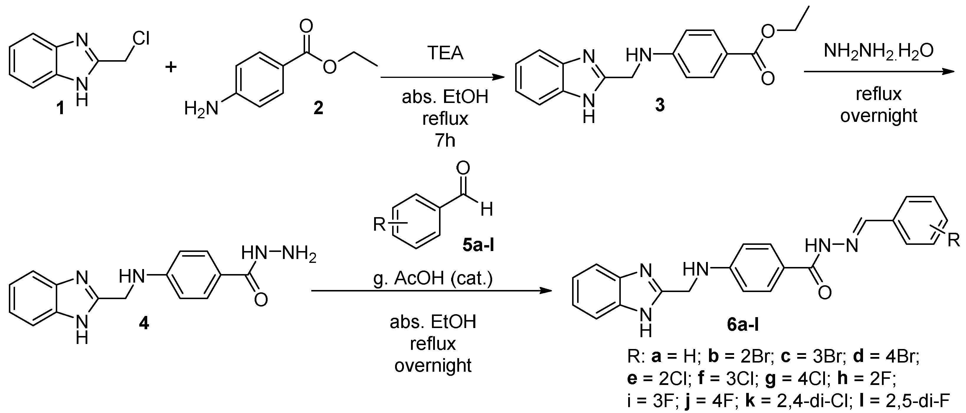
Scheme 1. Synthesis of (E)-4-(((1H-benzo[d]imidazol-2-yl)methyl)amino)-N’-(substituted)benzylidenebenzohydrazide (6a-l).
Pharmaceuticals 17 00839 sch001
Figure 2. Cell cycle phases of HepG2 cells following treatment with compound 6i: (A) Control HepG2 cell lines; (B) HepG2 cells treated with compound 6i.
Pharmaceuticals 17 00839 g002
Figure 3. Apoptotic cell distributions: (A) Control HepG2 cells; (B) HepG2 cells treated with compound 6i. The colors red, green, blue and purple are representing alive cells, early apoptosis, late apoptosis and necrosis, respectively.
Pharmaceuticals 17 00839 g003
Figure 4. Docking study of compound 6h showing different types of binding interaction with the key amino acid residues: (A) 6h with Her2; and (B) 6h with AURKC kinase enzymes.
Pharmaceuticals 17 00839 g004
Figure 5. Docking study of compound 6i showing different types of binding interaction with the key amino acid residues: (A) compound 6i with EGFR; and (B) compound 6i with Her2 kinase enzymes.
Pharmaceuticals 17 00839 g005
Table 1. In vitro cytotoxicity of compounds 6a-l against selected cancer cell lines.
CompoundIn Vitro Cytotoxicity IC50 (µM)
No.RHCT-116SIHepG2SIMCF-7SIWI-38
6aH24.62 ± 1.92.9531.76 ± 2.22.2926.31 ± 1.92.7672.60 ± 4.0
6b2-Br42.90 ± 2.51.9626.16 ± 1.93.2231.82 ± 2.12.6484.15 ± 4.3
6c3-Br10.21 ± 0.84.728.90 ± 0.65.417.82 ± 0.66.1648.17 ± 2.7
6d4-Br30.26 ± 2.11.7917.78 ± 1.33.0525.18 ± 1.82.1654.30 ± 3.1
6e2-Cl46.67 ± 2.71.3328.80 ± 1.72.1536.16 ± 2.31.7161.86 ± 3.5
6f3-Cl63.72 ± 3.51.5734.79 ± 2.12.8745.61 ± 2.52.19>100
6g4-Cl35.29 ± 2.32.0224.90 ± 1.72.8622.51 ± 1.63.1671.19 ± 3.5
6h2-F21.48 ± 1.64.3312.94 ± 1.07.1916.31 ± 1.25.7092.98 ± 4.6
6i3-F13.44 ± 1.24.209.39 ± 0.86.0111.64 ± 0.94.8556.46 ± 3.0
6j4-F18.72 ± 1.42.0814.02 ± 1.22.788.31 ± 0.74.7039.03 ± 2.3
6k2,4-di-Cl67.83 ± 3.81.4744.78 ± 2.42.2348.64 ± 2.72.06>100
6l2,5-di-F39.84 ± 2.21.9520.02 ± 1.53.8829.81 ± 2.02.6077.61 ± 4.1
Sorafenib5.47 ± 0.31.959.18 ± 0.61.167.26 ± 0.31.4710.65 ± 0.8
Doxorubicin5.23 ± 0.31.284.50 ± 0.21.494.17 ± 0.21.616.72 ± 0.5
Sunitinib17.91 ± 1.33.118.38 ± 0.56.6424.06 ± 2.02.3155.63 ± 3.3
IC50 values are the mean ± SD of triplicate measurements; SI = Selectivity Index = IC50 of normal cell line (WI-38)/IC50 of cancer cell line [41].
Table 2. In vitro protein kinase inhibition of compounds 6c and 6h-j against EGFR, Her2, VEGFR2, CDK2, AURKC, HDAC1, and mTOR.
CompoundIn Vitro Protein Kinase Inhibition IC50 (nM)
EGFRHer2VEGFR2CDK2AURKCHDAC1mTOR
6c125.2 ± 0.04155.6 ± 0.023604.5 ± 0.022938 ± 0.03994.4 ± 0.0362263 ± 0.0771461 ± 0.05
6h73.2 ± 0.00423.2 ± 0.001194.5 ± 0.007284 ± 0.01211 ± 0.004151.1 ± 0.005413 ± 0.014
6i30.1 ± 0.0328.3 ± 0.001172.2 ± 0.006364 ± 0.01174.5 ± 0.00396.6 ± 0.003152 ± 0.005
6j166.4 ± 0.008204.7 ± 0.009307.2 ± 0.0111448 ± 0.062589.4 ± 0.022473.3 ± 0.0161305 ± 0.044
Erlotinib61.1 ± 0.002------
Lapatenib-17.4 ± 0.001-----
Sorafenib--45.4 ± 0.002----
Roscovetine---756 ± 0.032---
TSA----30.4 ± 0.00137.4 ± 0.001-
Rapamycin------208 ± 0.007
IC50 values are the mean ± SD of triplicate measurements.
Table 3. Results of cell cycle analysis of HepG2 cells treated with compound 6i.
Compound/Cell LineDNA Content (%)Cell Cycle Distribution Index (CDI)
%G0-G1%S %G2/M
Cont. HepG252.3934.7712.840.91
Compound 6i/HepG272.1325.192.680.39
Table 4. Apoptotic cell distributions of HepG2 cells treated with compound 6i.
SampleApoptosisNecrosis
Alive Cell (%)EarlyLate
Cont. HepG297.580.610.211.6
Compound 6i/HepG263.9322.079.984.02
Table 5. Expression changes in apoptotic protein following treatment with compound 6i.
Kinase ProteinProtein Expression (Pg/mL) (Folds)
Caspase-3BaxBcl-2
Control HepG299.904 ± 3.88 (1)71.075 ± 2.762 (1)15.668 ± 0.53 (1)
6i/HepG2388.497 ± 15.09 (3.9)513.731 ± 19.96 (7.22)2.073 ± 0.07 (0.132)
Saurosporine/HepG2541.162 ± 21.02 (5.4)386.743 ± 15.03 (5.44)3.336 ± 0.11 (0.212)
Protein expression values are the mean ± SD of triplicate measurements.
Disclaimer/Publisher’s Note: The statements, opinions and data contained in all publications are solely those of the individual author(s) and contributor(s) and not of MDPI and/or the editor(s). MDPI and/or the editor(s) disclaim responsibility for any injury to people or property resulting from any ideas, methods, instructions or products referred to in the content.