

Gallium-68-Labeled KISS1-54 Peptide for Mapping KISS1 Receptor via PET: Initial Evaluation in Human Tumor Cell Lines and in Tumor-Bearing Mice

Abstract
1. Introduction
2. Results
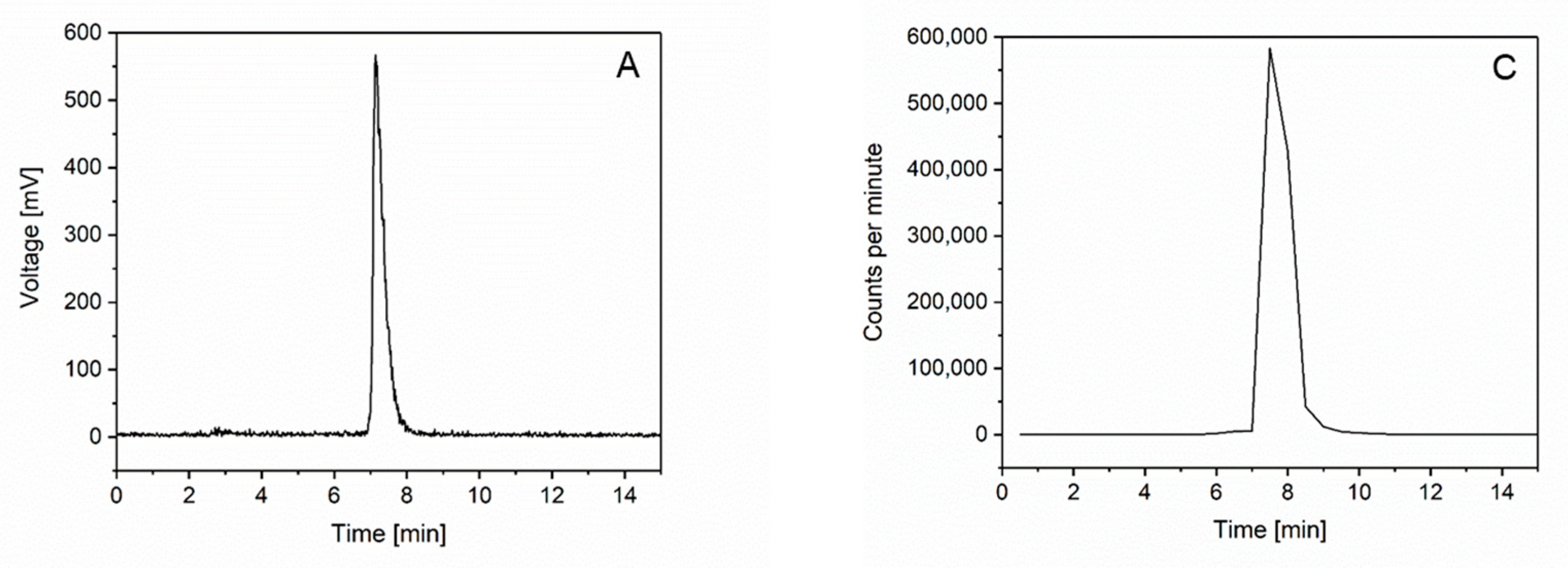
2.1. Radiochemistry
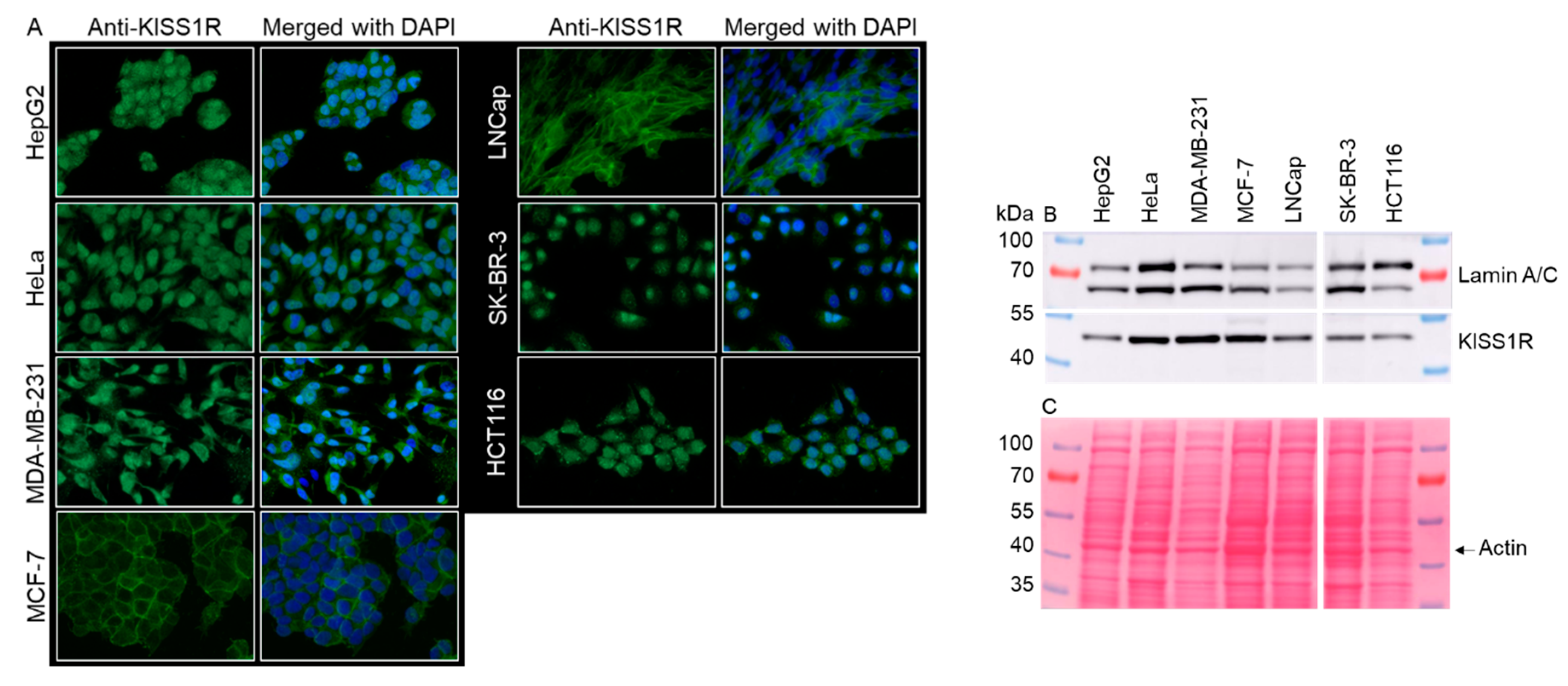
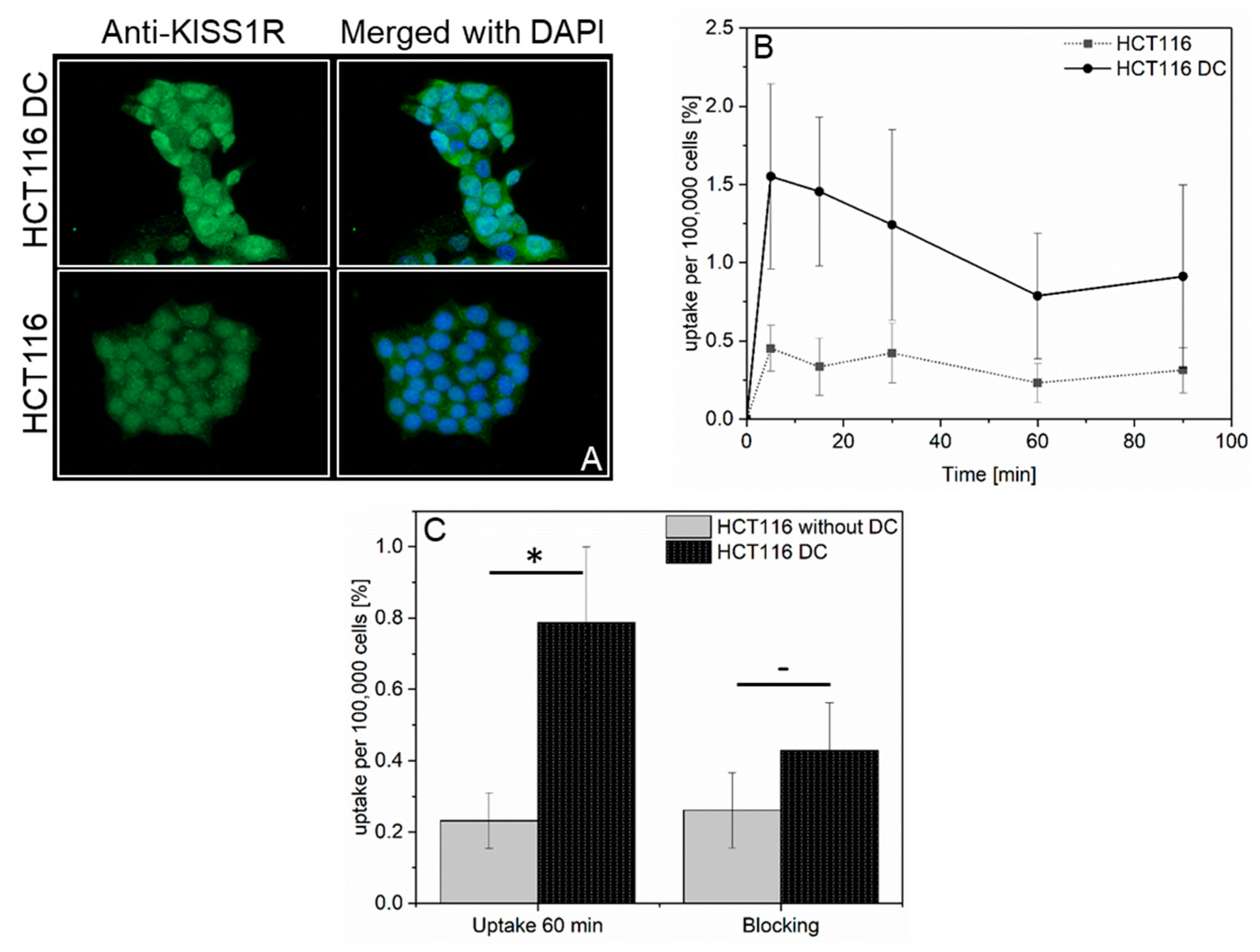
2.2. Examination of KISS1R Expression by Immunofluorescence and Western Blot
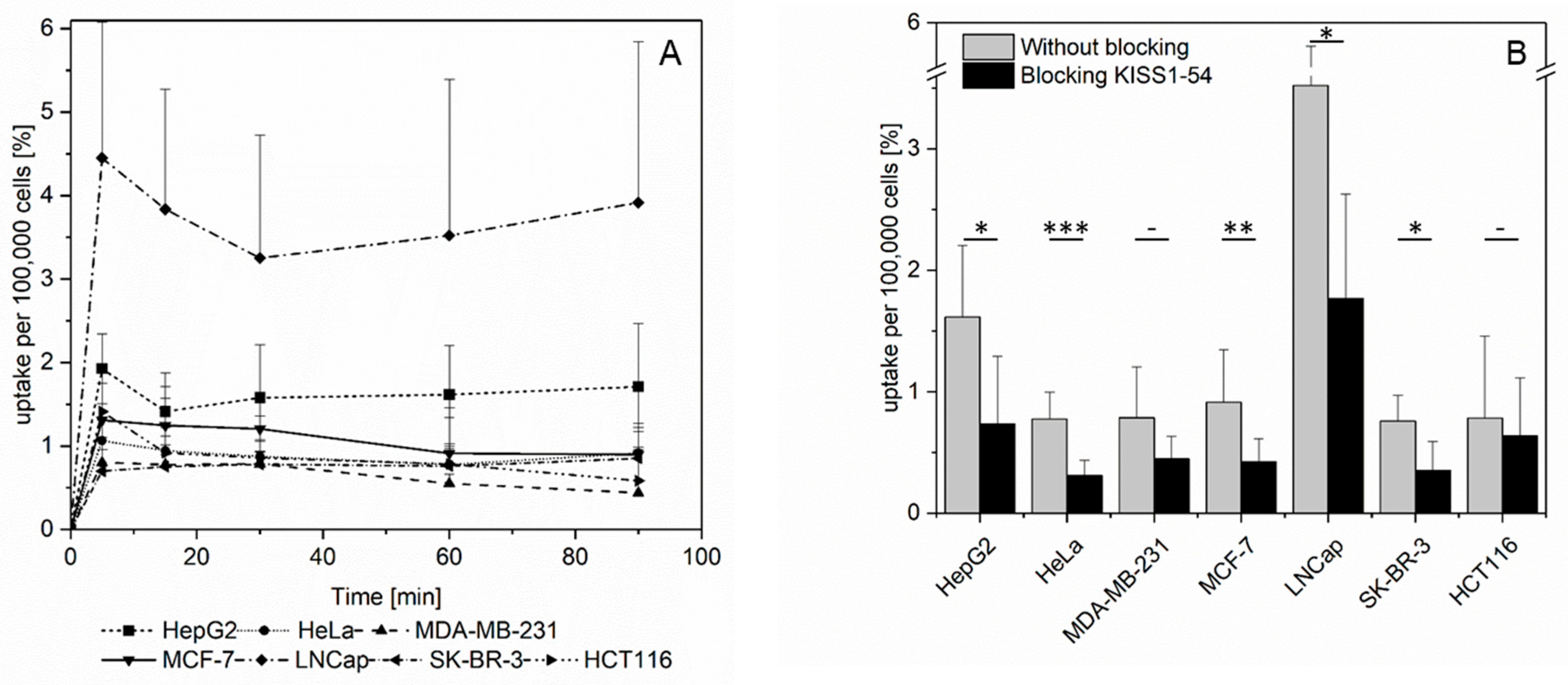
2.3. Investigation of [68Ga]KISS1-54 in Human Tumor Cell Lines
2.4. In Vivo Studies in Mice
3. Discussion
4. Material and Methods
4.1. Materials
4.2. Radiosynthesis of [68Ga]KISS1-54
4.3. In Vivo Stability
4.4. Cell Lines and Cell Culture
4.5. Immunofluorescence
4.6. Western Blot
4.7. In Vitro Cell Uptake
4.8. Animal Studies
4.9. µ-PET Imaging
4.10. Statistical Analysis
5. Conclusions
Supplementary Materials
Author Contributions
Funding
Institutional Review Board Statement
Informed Consent Statement
Data Availability Statement
Acknowledgments
Conflicts of Interest
Abbreviations
| BSA | bovine serum albumin |
| DAPI | 4′,6-diamidin-2-phenylindol |
| DC | 5-Aza-2′-Deoxycytidine (Decitabine) |
| DMEM | Dulbecco’s Modified Eagle Medium |
| FBS | fetal bovine serum |
| GPR54 | G protein-coupled receptor |
| HCl | hydrochloric acid |
| HEPES | 4-(2-hydroxyethyl)piperazine-1-ethanesulfonic acid |
| HPLC | high-performance liquid chromatography |
| IF | immunofluorescence |
| i.v. | intravenous |
| KISS1R | KISS1 receptor |
| KISS1-54 | KISS1 peptide with 54 amino acids |
| KP | kisspeptin |
| NaCl | sodium chloride |
| NEAA | nonessential amino acids |
| NODAGA | 1,4,7-triazacyclononane-1-glutaric acid-4,7-acetic acid |
| PBS | phosphate-buffered saline |
| PET | positron emission tomography |
| p.i. | post-injection |
| ROI | region of interest |
| RPMI | Roswell Park Memorial Institute |
| SD | standard deviation |
| TRIS | tris(hydroxymethyl)aminomethane |
| [18F]FDG | 2-[18F]fluoro-2-deoxyglucose |
| [68Ga]KISS1-54 | NODAGA-KISS1-54 peptide radiolabeled with 68Ga |
| 68GaCl3 | [68Ga]gallium chloride |
References
- Kotani, M.; Detheux, M.; Vandenbogaerde, A.; Communi, D.; Le Poul Vanderwinden, J.M.; Brezillon, E.S.; Tyldesley, R.; Suarez-Huerta, N.; Vandeput, F. The Metastasis Suppressor Gene Kiss-1 Encodes Kisspeptins, the Natural Ligands of the Orphan G Protein-Coupled Receptor Gpr54. J. Biol. Chem. 2001, 276, 34631–34636. [Google Scholar] [CrossRef] [PubMed]
- Ohtaki, T.Y.; Shintani, S.; Honda, H.; Matsumoto, A.; Hori, K.; Kanehashi, Y.; Terao, S.; Kumano, Y.; Takatsu, Y.; Masuda, Y.; et al. Metastasis Suppressor Gene Kiss-1 Encodes Peptide Ligand of a G-Protein-Coupled Receptor. Nature 2000, 411, 613–617. [Google Scholar] [CrossRef] [PubMed]
- Lee, J.H.; Miele, M.E.; Hicks, D.J.; Phillips, K.K.; Trent, J.M.; Weissman, B.E.; Welch, D.R. Kiss-1, a Novel Human Malignant Melanoma Metastasis-Suppressor Gene. J. Natl. Cancer Inst. 1996, 88, 1731–1737. [Google Scholar] [CrossRef] [PubMed]
- Cebrian, V.M.; Fierro, E.; Orenes-Pinero, L.; Grau, P.; Moya, T.; Ecke, M.; Alvarez, M.; Gil, F.; Algaba, J.; Bellmunt, J.; et al. Kiss1 Methylation and Expression as Tumor Stratification Biomarkers and Clinical Outcome Prognosticators for Bladder Cancer Patients. Am. J. Pathol. 2011, 179, 540–546. [Google Scholar] [CrossRef] [PubMed]
- Ikeguchi, M.; Yamaguchi, K.; Kaibara, N. Clinical Significance of the Loss of Kiss-1 and Orphan G-Protein-Coupled Receptor (Hot7t175) Gene Expression in Esophageal Squamous Cell Carcinoma. Clin. Cancer Res. 2004, 10, 1379–1383. [Google Scholar] [CrossRef] [PubMed]
- Kostakis, I.D.G.; Agrogiannis, A.G.; Vaiopoulos, E.; Mylona, E.; Patsouris, G.K.; Koutsilieris, M. Kiss1 and Kiss1r Expression in Gastric Cancer. J. BUON 2018, 23, 79–84. [Google Scholar]
- McNally, L.R.; Welch, D.R.; Beck, B.H.; Stafford, L.J.; Long, J.W.; Sellers, J.C.; Huang, Z.Q.; Grizzle, W.E.; Stockard, C.R.; Nash, K.T. Kiss1 over-Expression Suppresses Metastasis of Pancreatic Adenocarcinoma in a Xenograft Mouse Model. Clin. Exp. Metastasis 2010, 27, 591–600. [Google Scholar] [CrossRef]
- Okugawa, Y.Y.; Inoue, K.; Tanaka, Y.; Toiyama, T.; Shimura, M.; Okigami, A.; Kawamoto, J.; Hiro, S.; Saigusa, Y.; Mohri, Y.; et al. Loss of the Metastasis Suppressor Gene Kiss1 Is Associated with Lymph Node Metastasis and Poor Prognosis in Human Colorectal Cancer. Oncol. Rep. 2013, 30, 1449–1454. [Google Scholar] [CrossRef][Green Version]
- Prentice, L.M.; Klausen, C.; Kalloger, S.; Köbel, M.; McKinney, S.; Santos, J.L.; Kenney, C.; Mehl, E.; Gilks, C.B.; Leung, P.; et al. Kisspeptin and Gpr54 Immunoreactivity in a Cohort of 518 Patients Defines Favourable Prognosis and Clear Cell Subtype in Ovarian Carcinoma. BMC Med. 2007, 5, 33. [Google Scholar] [CrossRef]
- Corno, C.; Perego, P. Kiss1 in Regulation of Metastasis and Response to Antitumor Drugs. Drug Resist. Updat. 2019, 42, 12–21. [Google Scholar] [CrossRef]
- Castaño, J.P.; Martínez-Fuentes, A.J.; Gutiérrez-Pascual, E.; Vaudry, H.; Tena-Sempere, M.; Malagón, M.M. Intracellular Signaling Pathways Activated by Kisspeptins through Gpr54: Do Multiple Signals Underlie Function Diversity? Peptides 2009, 30, 10–15. [Google Scholar] [CrossRef]
- Chen, Y.; Yusenko, M.V.; Kovacs, G. Lack of Kiss1r Expression Is Associated with Rapid Progression of Conventional Renal Cell Carcinomas. J. Pathol. 2011, 223, 46–53. [Google Scholar] [CrossRef] [PubMed]
- Hata, K.; Dhar, D.K.; Watanabe, Y.; Nakai, H.; Hoshiai, H. Expression of Metastin and a G-Protein-Coupled Receptor (Axor12) in Epithelial Ovarian Cancer. Eur. J. Cancer 2007, 43, 1452–1459. [Google Scholar] [CrossRef] [PubMed]
- Ikeguchi, M.; Hirooka, Y.; Kaibara, N. Quantitative Reverse Transcriptase Polymerase Chain Reaction Analysis for Kiss-1 and Orphan G-Protein-Coupled Receptor (Hot7t175) Gene Expression in Hepatocellular Carcinoma. J. Cancer Res. Clin. Oncol. 2003, 129, 531–535. [Google Scholar] [CrossRef] [PubMed]
- Xoxakos, I.; Petraki, C.; Msauel, P.; Armakolas, A.; Grigorakis, A.; Stefanakis, S.; Koutsilieris, M. Expression of Kisspeptin (Kiss1) and Its Receptor Gpr54 (Kiss1r) in Prostate Cancer. Anticancer. Res. 2020, 40, 709–718. [Google Scholar] [CrossRef] [PubMed]
- Sanchez-Carbayo, M.; Capodieci, P.; Cordon-Cardo, C. Tumor Suppressor Role of Kiss-1 in Bladder Cancer: Loss of Kiss-1 Expression Is Associated with Bladder Cancer Progression and Clinical Outcome. Am. J. Pathol. 2003, 162, 609–617. [Google Scholar] [CrossRef] [PubMed]
- Matthaiou, S.; Kostakis, I.D.; Kykalos, S.; Machairas, N.; Spartalis, E.; Ntikoudi, E.; Lambropoulou, M.; Karayiannakis, A.; Machairas, N. Kiss1 and Kiss1r Expression in Primary and Metastatic Colorectal Cancer. J. BUON 2018, 23, 598–603. [Google Scholar]
- Nicolle, G.; Comperat, E.; Nicolaïew, N.; Cancel-Tassin, G.; Cussenot, O. Metastin (Kiss-1) and Metastin-Coupled Receptor (Gpr54) Expression in Transitional Cell Carcinoma of the Bladder. Ann. Oncol. 2007, 18, 605–607. [Google Scholar] [CrossRef]
- Cvetkovic, D.; Babwah, A.V.; Bhattacharya, M. Kisspeptin/Kiss1r System in Breast Cancer. J. Cancer 2013, 4, 653–661. [Google Scholar] [CrossRef]
- Goertzen, C.G.; Dragan, M.; Turley, E.; Babwah, A.V.; Bhattacharya, M. Kiss1r Signaling Promotes Invadopodia Formation in Human Breast Cancer Cell Via Beta-Arrestin2/Erk. Cell. Signal. 2016, 28, 165–176. [Google Scholar] [CrossRef]
- Gomaa, S.H.; Zaki, M.A.; Sultan, M.H.; Dwedar, F.I.; Zidan, E.H. Evaluation of Kiss1 Receptor Gene Expression in Egyptian Female Patients with Breast Cancer. Asian Pac. J. Cancer Prev. 2022, 23, 961–969. [Google Scholar] [CrossRef] [PubMed]
- Sun, Y.B.; Xu, S. Expression of Kiss1 and Kiss1r (Gpr54) May Be Used as Favorable Prognostic Markers for Patients with Non-Small Cell Lung Cancer. Int. J. Oncol. 2013, 43, 521–530. [Google Scholar] [CrossRef] [PubMed][Green Version]
- Kang, H.S.; Baba, T.; Mandai, M.; Matsumura, N.; Hamanishi, J.; Kharma, B.; Kondoh, E.; Yoshioka, Y.; Oishi, S.; Fujii, N.; et al. Gpr54 Is a Target for Suppression of Metastasis in Endometrial Cancer. Mol. Cancer Ther. 2011, 10, 580–590. [Google Scholar] [CrossRef]
- Cvetković, D.; Dragan, M.; Leith, S.J.; Mir, Z.M.; Leong, H.S.; Pampillo, M.; Lewis, J.D.; Babwah, A.V.; Bhattacharya, M. Kiss1r Induces Invasiveness of Estrogen Receptor-Negative Human Mammary Epithelial and Breast Cancer Cells. Endocrinology 2013, 154, 1999–2014. [Google Scholar] [CrossRef] [PubMed]
- Marot, D.; Bieche, I.; Aumas, C.; Esselin, S.; Bouquet, C.; Vacher, S.; Lazennec, G.; Perricaudet, M.; Kuttenn, F.; Lidereau, R.; et al. High Tumoral Levels of Kiss1 and G-Protein-Coupled Receptor 54 Expression Are Correlated with Poor Prognosis of Estrogen Receptor-Positive Breast Tumors. Endocr. Relat. Cancer 2007, 14, 691–702. [Google Scholar] [CrossRef] [PubMed]
- Israel, I.; Elflein, K.; Schirbel, A.; Chen, K.; Samnick, S. A Comparison of the Monomeric [(68)Ga]Nodaga-Ngr and Dimeric [(68)Ga]Nota-(Ngr)(2) as Aminopeptidase N Ligand for Positron Emission Tomography Imaging in Tumor-Bearing Mice. Eur. J. Pharm. Sci. 2021, 166, 105964. [Google Scholar] [CrossRef] [PubMed]
- Ziegler, E.; Olbrich, T.; Emons, G.; Gründker, C. Antiproliferative Effects of Kisspeptin-10 Depend on Artificial Gpr54 (Kiss1r) Expression Levels. Oncol. Rep. 2013, 29, 549–554. [Google Scholar] [CrossRef] [PubMed]
- Blake, A.; Dragan, M.; Tirona, R.G.; Hardy, D.B.; Brackstone, M.; Tuck, A.B.; Babwah, A.V.; Bhattacharya, M. G Protein-Coupled Kiss1 Receptor Is Overexpressed in Triple Negative Breast Cancer and Promotes Drug Resistance. Sci. Rep. 2017, 7, 46525. [Google Scholar] [CrossRef] [PubMed]
- Shao, Y.; Liang, W.; Kang, F.; Yang, W.; Ma, X.; Li, G.; Zong, S.; Chen, K.; Wang, J. 68ga-Labeled Cyclic Ngr Peptide for Micropet Imaging of Cd13 Receptor Expression. Molecules 2014, 19, 11600–11612. [Google Scholar] [CrossRef]
- Yan, Y.; Chen, K.; Yang, M.; Sun, X.; Liu, S.; Chen, X. A New 18f-Labeled Bbn-Rgd Peptide Heterodimer with a Symmetric Linker for Prostate Cancer Imaging. Amino Acids 2011, 41, 439–447. [Google Scholar] [CrossRef]
- Dotterweich, J.; Tower, R.J.; Brandl, A.; Müller, M.; Hofbauer, L.C.; Beilhack, A.; Ebert, R.; Glüer, C.C.; Tiwari, S.; Schütze, N. The Kiss1 Receptor as an in Vivo Microenvironment Imaging Biomarker of Multiple Myeloma Bone Disease. PLoS ONE 2016, 11, e0155087. [Google Scholar] [CrossRef] [PubMed]
- Nash, K.T.; Phadke, P.A.; Navenot, J.M.; Hurst, D.R.; Accavitti-Loper, M.A.; Sztul, E.; Vaidya, K.S.; Frost, A.R.; Kappes, J.C.; Peiper, S.C. Requirement of Kiss1 Secretion for Multiple Organ Metastasis Suppression and Maintenance of Tumor Dormancy. J. Natl. Cancer Inst. 2007, 99, 309–321. [Google Scholar] [CrossRef] [PubMed]
- Liu, X.; Herbison, A.E. Kisspeptin Regulation of Neuronal Activity Throughout the Central Nervous System. Endocrinol. Metab. 2016, 31, 193–205. [Google Scholar] [CrossRef] [PubMed]
- Israel, I.; Richter, D.; Stritzker, J.; van Ooschot, M.; Donat, U.; KBuck, A.; Samnick, S. Pet Imaging with [(6)(8)Ga]Nota-Rgd for Prostate Cancer: A Comparative Study with [(1)(8)F]Fluorodeoxyglucose and [(1)(8)F]Fluoroethylcholine. Curr. Cancer Drug Targets 2014, 14, 371–379. [Google Scholar] [CrossRef]
- Schadt, F.; Israel, I.; Beez, A.; Alushi, K.; Weiland, J.; Ernestus, R.I.; Westermaier, T.; Samnick, S.; Lilla, N. Analysis of Cerebral Glucose Metabolism Following Experimental Subarachnoid Hemorrhage over 7 Days. Sci. Rep. 2023, 13, 427. [Google Scholar] [CrossRef]
- Maier, P.; Riehl, G.; Israel, I.; Samnick, S. Synthesis and Preliminary Evaluations of [(18)F]Fluorinated Pyridine-2- Carboxamide Derivatives for Targeting Pd-L1 in Cancer. Curr. Cancer Drug Targets 2023, 23, 412–424. [Google Scholar]


Disclaimer/Publisher’s Note: The statements, opinions and data contained in all publications are solely those of the individual author(s) and contributor(s) and not of MDPI and/or the editor(s). MDPI and/or the editor(s) disclaim responsibility for any injury to people or property resulting from any ideas, methods, instructions or products referred to in the content. |
© 2023 by the authors. Licensee MDPI, Basel, Switzerland. This article is an open access article distributed under the terms and conditions of the Creative Commons Attribution (CC BY) license (https://creativecommons.org/licenses/by/4.0/).
Share and Cite
Israel, I.; Riehl, G.; Butt, E.; Buck, A.K.; Samnick, S. Gallium-68-Labeled KISS1-54 Peptide for Mapping KISS1 Receptor via PET: Initial Evaluation in Human Tumor Cell Lines and in Tumor-Bearing Mice. Pharmaceuticals 2024, 17, 44. https://doi.org/10.3390/ph17010044
Israel I, Riehl G, Butt E, Buck AK, Samnick S. Gallium-68-Labeled KISS1-54 Peptide for Mapping KISS1 Receptor via PET: Initial Evaluation in Human Tumor Cell Lines and in Tumor-Bearing Mice. Pharmaceuticals. 2024; 17(1):44. https://doi.org/10.3390/ph17010044
Chicago/Turabian StyleIsrael, Ina, Gabriele Riehl, Elke Butt, Andreas K. Buck, and Samuel Samnick. 2024. "Gallium-68-Labeled KISS1-54 Peptide for Mapping KISS1 Receptor via PET: Initial Evaluation in Human Tumor Cell Lines and in Tumor-Bearing Mice" Pharmaceuticals 17, no. 1: 44. https://doi.org/10.3390/ph17010044
APA StyleIsrael, I., Riehl, G., Butt, E., Buck, A. K., & Samnick, S. (2024). Gallium-68-Labeled KISS1-54 Peptide for Mapping KISS1 Receptor via PET: Initial Evaluation in Human Tumor Cell Lines and in Tumor-Bearing Mice. Pharmaceuticals, 17(1), 44. https://doi.org/10.3390/ph17010044