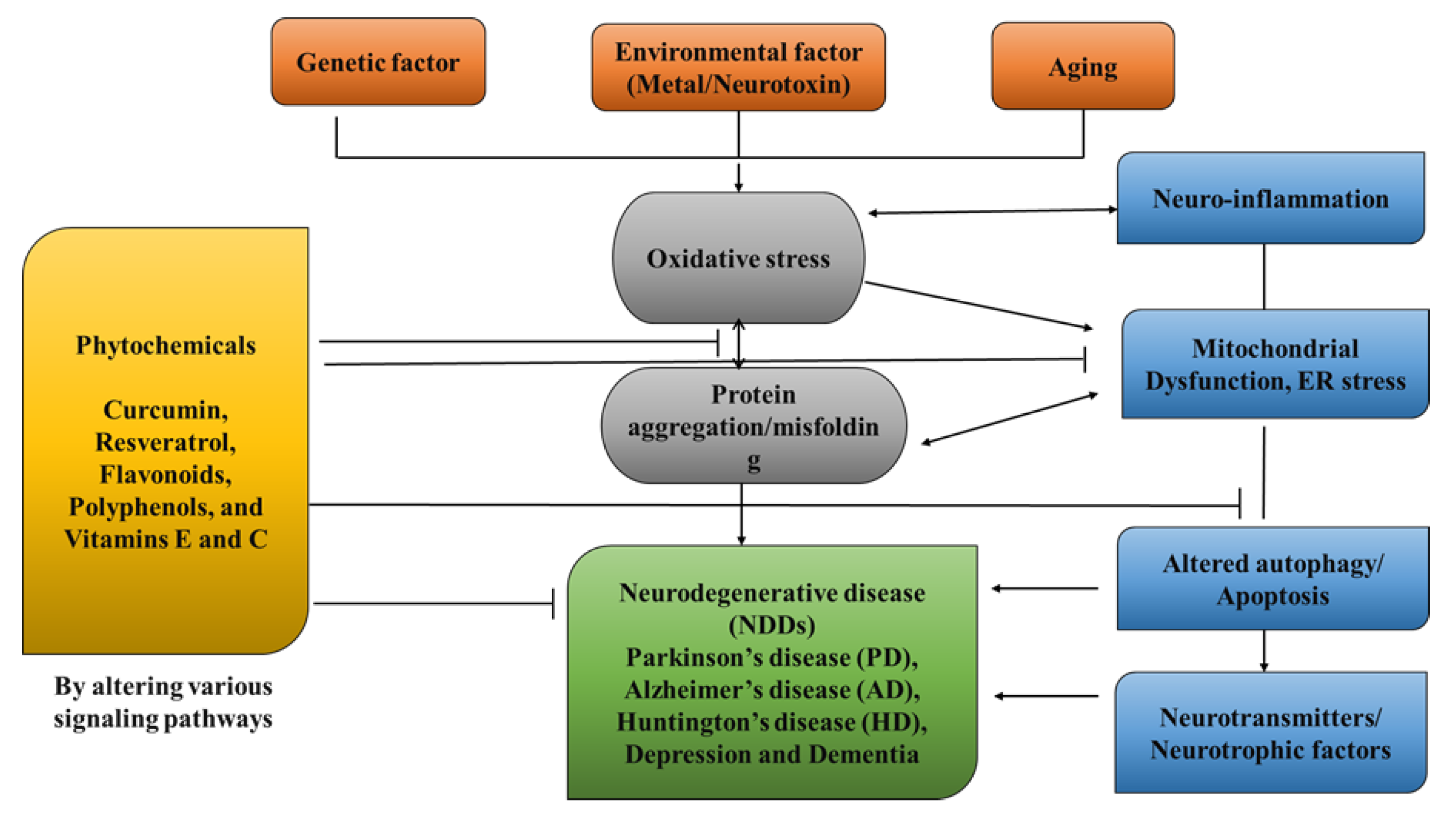
Review

A Review on Natural Antioxidants for Their Role in the Treatment of Parkinson’s Disease

1
Chitkara College of Pharmacy, Chitkara University, Rajpura 140401, India
2
Ganpati Institute of Pharmacy, Bilaspur 135102, India
3
Department of Food and Nutrition, BioNanocomposite Research Center, Kyung Hee University, 260 Kyunghee-daero, Seoul 02447, Republic of Korea
4
Nondestructive Bio-Sensing Laboratory, Department of Biosystems Machinery Engineering, College of Agriculture and Life Science, Chungnam National University, 99 Daehak-ro, BLDG# E10-2, RM# 2213, Daejeon 34134, Republic of Korea
5
Department of Pharmacognosy, Orlean College of Pharmacy, Dr. A.P.J. Abdul Kalam Technical University, 42, Knowledge Park—III, Greater Noida 201308, India
6
Department of Pharmacognosy, HIMT College of Pharmacy, Dr. A.P.J. Abdul Kalam Technical University, 8, Institutional Area, Knowledge Park—I, Greater Noida 201301, India
7
Department of Biology, College of Science, Imam Mohammad Ibn Saud Islamic University (IMSIU), Riyadh 11623, Saudi Arabia
8
Department of Pharmacy, Mohammed Al-Mana College for Medical Sciences, Dammam 34222, Saudi Arabia
9
Department of Biochemistry, College of Medicine, Imam Mohammad Ibn Saud Islamic University (IMSIU), Riyadh 11623, Saudi Arabia
*
Authors to whom correspondence should be addressed.
Pharmaceuticals 2023, 16(7), 908; https://doi.org/10.3390/ph16070908
Submission received: 22 May 2023 / Revised: 8 June 2023 / Accepted: 14 June 2023 / Published: 21 June 2023

Abstract

:
The neurodegenerative condition known as Parkinson’s disease (PD) is brought on by the depletion of dopaminergic neurons in the basal ganglia, which is the brain region that controls body movement. PD occurs due to many factors, from which one of the acknowledged effects of oxidative stress is pathogenic pathways that play a role in the development of Parkinson’s disease. Antioxidants, including flavonoids, vitamins E and C, and polyphenolic substances, help to reduce the oxidative stress brought on by free radicals. Consequently, this lowers the risk of neurodegenerative disorders in the long term. Although there is currently no cure for neurodegenerative illnesses, these conditions can be controlled. The treatment of this disease lessens its symptoms, which helps to preserve the patient’s quality of life. Therefore, the use of naturally occurring antioxidants, such as polyphenols, which may be obtained through food or nutritional supplements and have a variety of positive effects, has emerged as an appealing alternative management strategy. This article will examine the extent of knowledge about antioxidants in the treatment of neurodegenerative illnesses, as well as future directions for research. Additionally, an evaluation of the value of antioxidants as neuroprotective agents will be provided.

1. Introduction

NDDs (neurodegenerative disorders) are a collection of disorders with various clinical implications and etiology. ALS (amyotrophic lateral sclerosis), cerebellar illness, Huntington’s chorea, Parkinson’s disease, Alzheimer’s, dementia, and schizophrenia are all examples of NDDs [1,2,3,4]. Age groups, hereditary diseases, non-enzymatic antioxidants, excitotoxicity, cytoskeletal abnormalities, autoimmunity, asymmetry, toxicity, elevated blood pressure, oxidative stress, and peripheral vascular diseases are subject to both experimental and epidemiological research. Some of the risk variables that have been discovered through the clinical manifestations of NDDs are associated with free radical toxicity, radical-mediated alteration, oxidase dysfunction, and endoplasmic reticulum stress caused by perinatal genetic abnormalities. The most important common symptoms are disturbances in balance, breathing, movement, reflexes, motor skills, or cardiac activity. Antioxidants such as flavonoids, polyphenols, and vitamins E and C can help to prevent these symptoms [5,6,7]. Antioxidants have a great impact on human health, as they can fight free radicals and, thus, halt the aging process and decrease the effects of oxidative damage caused by, for example, an unbalanced diet [8,9]. As a result, the long-term risk of neurodegenerative diseases is reduced (Figure 1). Although they can be treated, there is currently no cure for neurodegenerative diseases. Treatment of this disease relieves the symptoms in order to preserve the quality of life. Natural antioxidants, such as polyphenols, are becoming more popular because they can be obtained from foods and supplements and offer a range of health benefits [10]. Although it affects both males and females with increase in their age but males tend to be affected more frequently [11]. It is considered an international disease that does not distinguish between social class or race. The disease is estimated to affect 1% of people over the age of 65 worldwide, accounting for up to two-thirds of all people with movement disorders [12].
As the population ages, PD has become more prevalent, with those over 85 years old reaching 2.6%. The non-motor symptoms that patients with PD encounter include pain, exhaustion, autonomic dysfunction, changed mood, sleep disturbance, and cognitive abnormalities [13]. PD is referred to asa synucleinopathy because Lewy bodies, a crucial clinical characteristic of the disease, accumulate due to the misfolding of β-synuclein as a major feature. Furthermore, β-synuclein appears to be connected to both idiopathic and inherited forms of Parkinson’s disease and has a special role in the disease’s pathogenesis. It is worth noting that β-synuclein buildup has been closely linked to posttranslational modifications, systemic inflammation, oxidative stress, mitochondrial biogenesis, changed mitochondrial physical properties, synapse dysfunction, glycolipids, ER stress, and metal complexes. The overproduction of ROS and years of age breakdown the antioxidant defense system and increase oxidative stress in certainbrain areas, which can contribute to the misfolding of β-synuclein being the catalyst for the aging process in PD [14,15]. Although it is not a cure, levodopa has emerged as an efficacious drug for PD’s first motor symptoms. Despite the fact that stiffness and bradykinesia respond the best, tremors may only be somewhat reduced by levodopa. Other symptoms, such as balance issues, may worsen. L-dopa, however, cannot be used to treat Lewy disease, non-motor symptoms, or neuronal loss [16]. Patients require greater L-dopa doses over time, which is accompanied by a rise in side effects such as dyskinesias. Amantadine, an antiviral medication, appears to alleviate motor problems as well. Medicines based on dopamine agonists are also administered to address a range of neuromotor symptoms that are associated with disease progression. The long-term use of conventional PD medications can lead to adverse effects, such as dyskinesias and motor fluctuations (Table 1). As a result, novel therapeutic techniques used to prevent neurodegeneration, non-motor symptoms, Lewy disease accumulation, or synuclein aggregation in the brain are required [17,18].
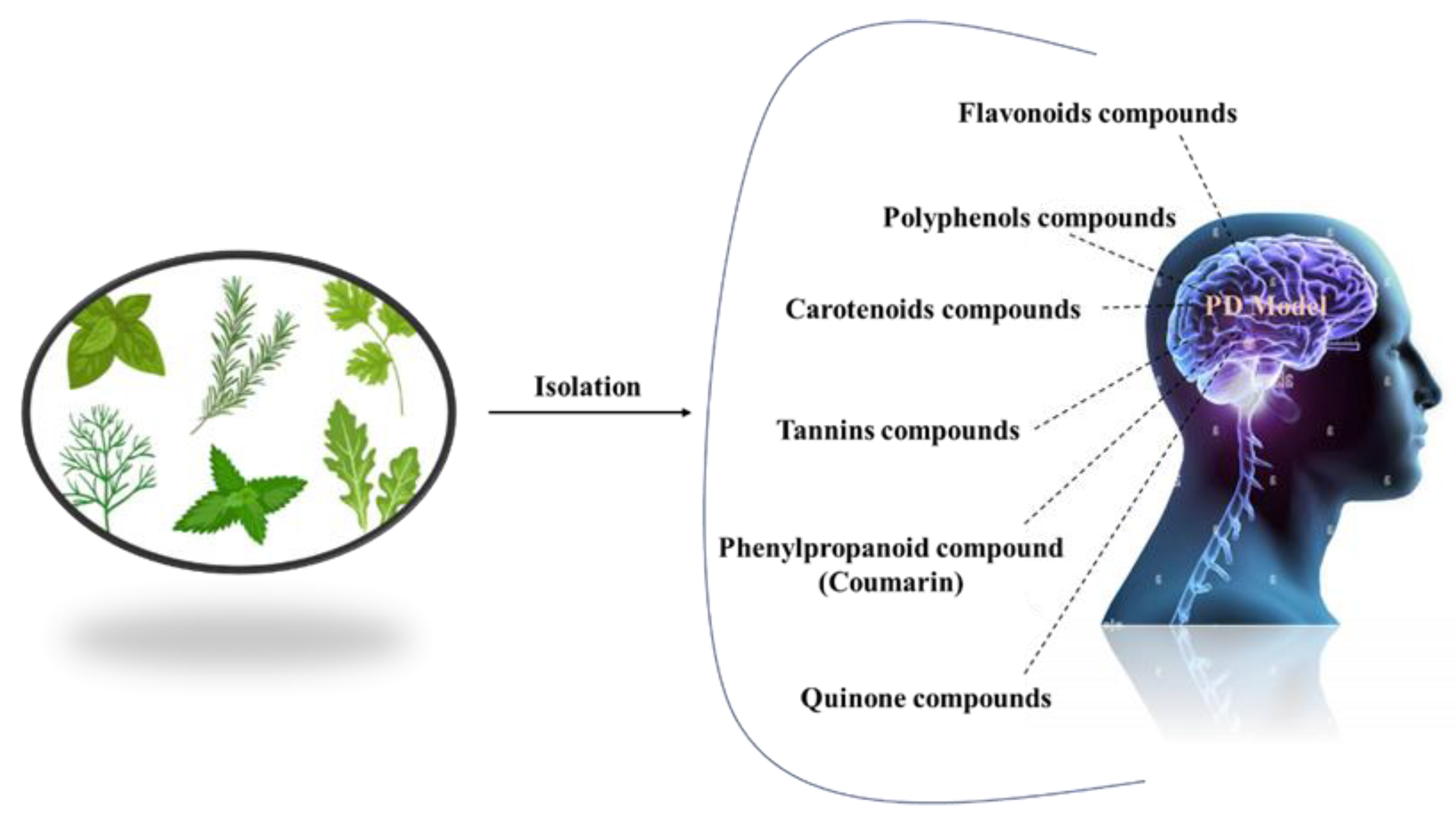
Complementary Therapies for PD
Complementary therapies and phytonutrients derived from plant sources have been proposed as treatments for Parkinson’s disease [24]. Numerous natural phytochemicals have emerged as therapeutically interesting compounds, drug entities, and phytochemicals for the treatment of inflammatory disorders [25]. Additionally, numerous pharmacological studies have shown that phytochemicals are useful in treating neurodegenerative diseases (NDDs), depression, and dementia [26,27,28,29,30]. Physiologically active phytochemicals are important therapeutically because they serve as the antioxidant defense system’s primary and secondary metabolites, which protect against a variety of stress-related disorders and clinical symptoms [31]. These phytochemicals’ positive and therapeutic effects include antioxidant capacity, pathogen prevention, immune system activation, and nutritional support for healthy living cells [32]. Botanical substances, with their active phytonutrients, efficiently salvage oxygen-based free radicals, influencing the antioxidative defense system and contributing to major cognitive impairments such as dyskinesias, residual tremors, muscle stiffness, and instability [33]. This review holds prime importance, as it addresses the treatment gaps in PD and explores the potential of natural antioxidants as a therapeutic approach. By summarizing the current literature on natural antioxidants and their effects on PD, this review aims to provide a comprehensive overview that can guide further research and clinical trials. Understanding the role of natural antioxidants in PD treatment may lead to the development of novel therapeutic strategies that can modify the disease course, mitigate the symptoms, and improve the quality of life for individuals living with PD. In this review, we address the natural antioxidants that can be used to treat Parkinson’s disease, as well as their mode of action (Figure 2).
Pathogenesis
Dopamine levels in the caudal nucleus, striatum, and expression medium fall as a result of the death of dopaminergic neurons in the substantia nigra pars compacta, which is associated with Parkinson’s disease. The progressive loss of cholinergic neurons is the most significant pathogenic discovery in Parkinson’s disease sufferers’ brains. For this reason, with the loss of these neurons, the dopamine levels fall. When 50–60% of the dopamine pathway is shut down and striatal dopamine levels are lowered by 80–85%, symptoms of the disease arise. However, Parkinson’s disease may have multiple etiological factors, including mitochondrial dysfunction, neuroinflammation, oxidative stress, and the formation of protein kinases called Lewy bodies in the cytosol (Figure 3) [34].
Phytochemicals and Parkinson’s Disease
Neurodegenerative diseases, including Parkinson’s disease, contributes to N increase in nicotinamide adenine dinucleotide phosphate oxidase and intracellular RNS, and, thus, result in cell damage due to oxidative stress [35].
Free radicals also damage cell membranes by oxidizing proteins, lipids, and nucleic acids, causing peptide cross-links [36]. The antioxidant and chelating effects of flavonoids and terpenoids are believed to be accountable for their therapeutic effects. Terpenoids, phenols, and flavonoids show important neuroprotective effects, due to their oxidative capacity [37]. By releasing extra protons, flavonoids scavenge superoxide, hydroxyl, and peroxyl radicals by donating an extra proton. This prevents ROS generation by the formation of complex structures containing dihydroxy groups, copper, multiple ions of transition metals, and iron [38].
Glutathione reductase, septo-optic dysplasia, and glutathione-S-transferase (GST) are the polyphenols that activate antioxidant enzymes. Polyphenols can raise the antioxidant levels by enhancing cell-signaling pathways through these enzymes. By generating unsaturated carbon–carbon double bonds with lipid bilayers, these phenolic acids inhibit lipid peroxidation and, thus, work as a powerful therapeutic and preventative agent [39].
The lack of oxygenation of the rings is assumed to be responsible for chrysin’s chemical capabilities, which include anti-inflammatory and antioxidant potential [40]. Diverse structures of flavones have been discovered to increase the capacity of antioxidant enzymes and also work in inhibiting COX-2, which is a pro-inflammatory mediator. Experiments with xanthine oxidase demonstrate that chrysin dramatically reduces ROS production. It is worth noting that rodent models require polyphenol dosages ranging from 10 µM to 100 µM to exercise their putative antioxidant and anti-inflammatory properties [41].
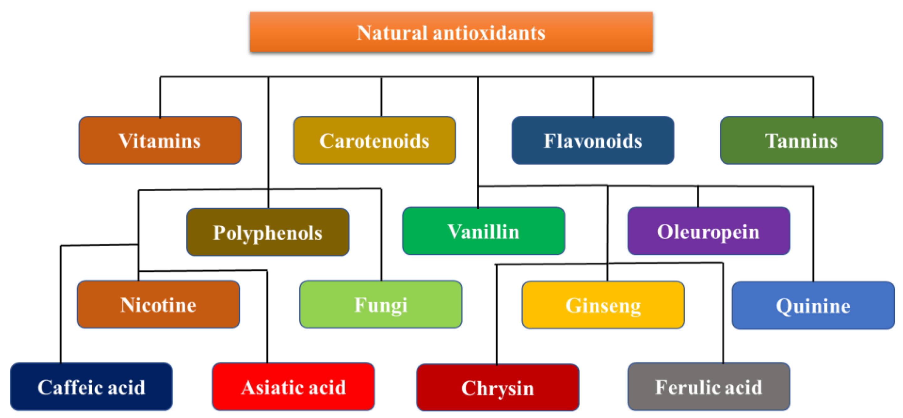
Neuroprotective Dietary Antioxidants
Antioxidants are neuroprotective compounds that protect or repair cellular components from oxidative damage. They efficiently halt neuronal lipid, protein, and DNA breakdown. Vitamins, flavonoids, carotenoids, tannins, and polyphenols are all antioxidants [42]. Oxidative stress damage is a prominent manifestation of neurodegenerative disorders. However, few efficacious agents have been found for clinical use, and even fewer have been successful because of their toxicity and cancer risks [43]. Evidence suggests that neuroprotection is a potential pharmaceutical target for neurodegenerative diseases, suggesting that it is possible. The use of relatively harmless antioxidant molecules found in food as remedies for many ailments, on the other hand, is appealing; however, it is limited by the difficulty of creating positive feedback in the brain. Polyphenols, a class of antioxidants, have been shown to have neuroprotective properties. Many biological processes, such as interactions with metal complexes, the scavenging of free radicals, changes in the activity of different enzymes, and impacts on intracellular signaling pathways and gene expression are all biological processes that contribute to this protection. A diet that is high in antioxidants is essential to prevent many diseases [44]. A reduction in the incidence of stroke, neurological disease, heart disease, cancer, and high blood pressure has been associated with these chemicals, which are primarily present in vegetables and fruit. The body’s natural antioxidant system needs help from the exogenous antioxidants that are found in fruits, nuts, vegetables, and other foods. Exogenous antioxidants are often classified as vitamins and non-vitamins. This section reviews the specific properties and roles of the key exogenous antioxidants in neuroprotection (Figure 4) [45].
Vitamin E
Four tocopherols, trocochromanols or four tocotrienols, and major fat-soluble antioxidants are collectively referred to as vitamin E [46]. The human body cannot produce this vitamin itself, therefore, it must be obtained from food (Table 2).
Each vitamin E isoform has complex antioxidant properties, and it is currently unknown how these effects work. It is suspected that they work by stopping the chain process of lipid peroxidation, therefore, important cellular components can be protected [47]. Vitamin E bioavailability varies with many factors, including the dietary matrix, genetics, and metabolic fat, and ranges from 10 to 33%. Preclinical studies in Parkinson’s disease have shown conflicting results regarding the efficacy of vitamin E as a disease-modifying agent [48]. Vitamin E deficiency increased substantia nigra MPTP toxicity, while partially protecting against neurotransmitter and metabolite deficiencies caused by striatal MPTP. Dopamine degradation could not be stopped, even by pre-MPTP injection therapy with β-tocopherol. However, substantial amounts of vitamin E may partially protect dopaminergic neurons from MPTP-mediated toxicity [37]. During the Deprenyl and Tocopherol antioxidative treatment of Parkinsonism study, patients with early or untreated Parkinson’s disease were given deprenyl (10 micrograms daily) and tocopherol as a treatment. The results of the study demonstrated that the monoamine oxidase inhibitor (deprenyl) slowed the progression of the disorders.However, the modest improvement in motor function gradually diminished when deprenyl was discontinued [49]. In individuals with Parkinson’s disease with a recent diagnosis, vitamin C and alpha-tocopherol supplementation delayed the initiation of levodopa therapy by 25 years.
Table 2. Some other potent activities of Vitamin E.
Compound NameDiseaseImpactRef.
Vitamin EAlzheimer’s disease (AD)Effective against AD when consumed with vitamin C in the earlier phase of AD.[50]
Cell membrane dysfunctionEffective in the protection of the cell membrane[51]
Cardiovascular disorderPrevents the clot formation and buildup of bad cholesterol[52]
Ascorbic Acid
Ascorbate, or ascorbic acid, is a vitamin that is soluble in water that is necessary for catecholamine production and other enzyme processes. Ascorbic acid shows its antioxidant capabilities by reduction inO2, alkoxyl (RO), hydroxyl (HO), peroxyl (ROO), and other free radicals in body. When vitamin E is oxidized while scavenging free radicals from the lipid membranes, the radical tocopheroxyl is formed, which interacts with vitamin C in order to replenish vitamin E. Vitamin C absorption, distribution, and metabolism are more complex than those of most lighter weight molecular compounds [53]. A family of sodium-dependent vitamin C transporters contains the key proteins that are involved in substance distribution and tissue absorption. The brain contains one of the highest concentrations of vitamin C in the body. Concentrations vary per brain region, with the motor cortex having the lowest concentration [54]. Different transporters are present in the brain for reduced and oxidized forms of vitamin C (SVCT2 or GLUT family). Individuals whosuffer from neurodegenerative conditions such as Parkinson’s disease (PD) exhibit low plasma exogenous antioxidant concentrations (including vitamin C). A lack of vitamin C has also been related to a higher incidence of Parkinson’s disease. It has been proposed that the level of vitamin C in lymphocytes could serve as a diagnostic indicator of Parkinson’s disease progression. Vitamin C supplementation is one of the medicinal methods used to cure Parkinson’s disease [55] (Table 3).
Flavonoids
It is the largest polyphenol class and has a backbone that is made up of one heterocyclic ring and two phenyl rings. The side chain configuration and quantity of hydroxyl groups influence the biological features, such as antioxidant activity and the modification of enzyme activity [40].
By reacting with free radicals and serving as metal catalysts, flavonoids act as radical chain terminators. In order to inhibit oxidation, phenolic antioxidants (PhOH) provide hydrogen atoms to the free radicals. There are several different types of flavonoids present in legumes, plants, and fruits (obtained from celery, citrus peels, chamomile, paprika, ginkgo biloba, parsley leaves, and mint). Crops, flowers, and leaves contain flavones as glucosides [59].
Kaempferol, quercetin, ferulic acid, and myricetin are examples of flavonols. In 6-OHDA-treated PC12 cells, 20 µM of quercetin boosted the mitochondrial activity, decreased the oxidative stress, and decreased β-synuclein protein synthesis [60]. Furthermore, oral quercetin administration improved the motor performance in arat model by lowering the mitochondrial oxidative stress, β-synuclein aggregation formation, and neuronal cell death [46].
Chalcone, isoflavonoids (such as daidzein and genistein), water-soluble anthocyanidin pigments (such as delphinidin, pelargonidin, peonidin, and cyanidin), and aromatic colorless flavanones (such as hesperetin, naringin, and hesperidin) are the other class of flavonoids that can be useful in the treatment of PD (Table 4) [61].
Green Tea Polyphenols
Green tea polyphenols, notably EGCG, exhibit a broad range of pharmacological properties, involving antimutagenic and anticarcinogenic properties. Green tea polyphenols’ strong antioxidant capacity is responsible for these positive effects. They are potent antioxidants that combat free radicals such as lipid radicals, phenolic radicals, superoxide anion radicals, and hydroxide ions [65]. When they are taken orally, the flavonoids and green tea’s polyphenols have been demonstrated to prevent aging. Tea catechins (TC) are commonly thought to be radical scavengers, although other components of tea have numerous applications, possibly protecting against 6-OHDP-induced apoptosis. In MTT assays, 6-OHDA treatment decreased the cell viability in a concentration-dependent manner, whereas flow cytometry, fluorescence microscopy, and DNA fragmentation confirmed apoptosis in PC12 cells. TC substantially reduces PC12 cell death. EGC and EC had lower efficacy, but EGCG and ECG were more efficacious compared to TC. The protective effect increased from 50 µM to 400 µM, and, at these concentrations, EGCG outperformed the green tea polyphenols. They inhibit at 83.2% and 84.9%, respectively, while EGCG inhibitory concentration is at 88.4% and 90.2% [54]. According to flow cytometry data, 200–400 µM of green tea polyphenols and EGCG dramatically decrease the number of apoptotic cells. Notably, EGCG-protected PC12 cells abolished nuclear changes indicative of apoptosis [66] (Table 5).
Isoflavones
Genistein, the most active soy isoflavone, has estrogen receptor affinity, antioxidant characteristics, increased cytoplasmic glutathione peroxidase, PTK inhibition, and other physiological effects. Soy isoflavones have protective efficacy against a variety of illnesses, including atherosclerosis, the consequences of estrogen deficiency in menopause, and hormone-dependent breast and prostate cancer (Table 6) [70]. It has been found that it exhibits neuroprotection by antagonizing the toxic effect of the critical Aβ protein [71]. As a result, it can be used for PD management.
Nicotine
In hippocampus cultures, nicotine significantly inhibits apoptosis induced by Ab and increases caspase activity. Nicotine reduced the number of free radicals accumulated by the Ab. The cholinergic antagonist mecamylamine decreased nicotine’s capacity to protect cells from Ab-induced caspase-3 activation and ROS buildup [75]. According to research, nicotine receptors play a major role in nicotine’s protective potential. The findings imply that nicotine may assist in averting the emergence of neurodegenerative diseases such as PD and Alzheimer’s. Although smoking has been linked to a lower incidence of Parkinson’s disease, the specific mechanism for this is unknown. Nicotine has been discovered to suppress cytochrome C release from intact mitochondria, as well as the high-amplitude mitochondrial swelling that is caused by calcium and N-methyl-4-phenylpyridine (MPPt). The redox state within mitochondria was also maintained by nicotine, which was likelyas a result of decreased mitochondrial permeability. Nicotine did not prevent the reduction in mitochondrial membrane potential by MPPt or calcium, but decreased electron leakage at sites of respiratory chain complex I and 6-OHDA, causing cytochrome C release and mitochondrial enlargement in SH-SY5Y cells [66].
This suggests that nicotine has receptor-independent neuroprotective effects. These findings imply that, when evaluating nicotine’s neuroprotective benefits, it is critical to consider both its interaction with the mitochondrial respiratory chain and its antioxidant activity. It is not entirely clear how nicotine attenuates β-amyloidosis and prevents PD.
In one study, after nicotine treatment, pure zincand copper metal concentrations in amyloid plaques and neuropils dropped considerably. The density of zinc and copper distribution in the hippocampus CA1 area is likewise lowered after nicotine administration. Researchers examined how nicotine affected the homeostasis of metals in SH-SY5Y cells that were overexpressing the Swedish variant of human APP (APPsw). These effects are not dependent on nicotinic acetylcholine receptor activation and are mediated by a decrease in intracellular copper concentrations with nicotine therapy, as well as a reduction in Ab-induced neurotoxicity, helped by copper addition [76] (Table 7).
Carotenoids
There are about 850 fat-soluble tetraterpenes in the class of carotenoids that are produced by photosynthetic plants, algae, and bacteria. They support plants in both photosynthesis and photo-defense. In addition to fruits and vegetables, such as beets, cherries, melons, and pumpkins, foods such as microalgae, fish, and shrimp also contain yellow, orange, and red pigments called carotenoids [80]. Serum lycopene, β, and α-carotene levels are considerably lower in Parkinson’s disease patients, and lower serum carotenoid levels are also linked to motor impairment. Carotenoids act as antioxidants by eliminating ROS via chemical and physical reactions; for example, β-carotene absorbs thermal energy from singlet oxygen and/or peroxynitrite, therefore, inhibiting tyrosine nitration. Carotenoids protect neurons against neurodegenerative diseases in multiple ways, including ROS quenching, the activation of antioxidant enzyme systems, and anti-neuroinflammatory effects [81] (Table 8).
Resveratrol
Resveratrol is a phenolic compound chemical that is accessible in several plants, including berries and grapes. In animal studies of Parkinson’s disease, they help with movement deficits, oxidative stress, and the elimination of TH neurons [85]. Resveratrol inhibits chromatin condensation and mitochondrial expansion, while decreasing COX-2 and TNF gene output [86]. This phenol has a short oral survival period due to its low water solubility and short half-life (10 min). Furthermore, resveratrol derivatives triethylsilyl and ditriisopropylsilyl show superior action in invitro anti-inflammatory and neuroprotective properties when compared to pure resveratrol. Resveratrol is employed because it has shown increased biocompatibility and neuroprotective properties in zebrafish embryos. Also selected from the prodrugs, this chemical reduced clinical scores and the severity of motor impairment in a 3-nitropropionic acid PD mousemodel of experimental autoimmune encephalomyelitis [87] (Table 9).
Tannins
These phenolic compounds have enough hydroxyl and carboxyl groups and high molecular weight to form a complex with biomolecules (0.5–20 kDa). They are also water-soluble. Tannins are unique in their ability to precipitate and link to alkaloids, proteins, and amino acids.There are several methods for removing tannins, however, acetone solvent extraction is the most effective for reducing tannin–protein complexes [91]. Tannic acid was chosen as the most optimistic molecule to minimize α-synuclein fibrosis without significantly raising toxicity levels [92] (Table 10).
Curcumin Polyphenol
Curcumin (10 µM) therapy also lowers oxidation-related protein changes such as carbonylation and nitrotyrosine production, which help to sustain dopaminergic cells. Curcumin, an antioxidant, effectively protects against oxidative-stress-induced mitochondrial damage [96,97]. In MPTP-induced mouse striatum, curcumin inhibits TH-positive cell death and DA depletion. In addition, it reduces inflammatory markers such as total nitrite, cytokines, and inducible nitric oxide synthase [98] (Table 11).
Herbs’ most prevalent chemical ingredients are ginkgolic acids, terpenoids, and flavonoids. Quercetin, isorhamnetin, and kaempferol are the most prevalent flavonoids in a registered standardized extract of ginkgo biloba (EGb 761) produced by solid–liquid extraction using aqueous acetone. Terpene lactones account for 6% of the extract (3.1% ginkgolides and 2.9% bilobalides) and other components (e.g., glucose and organic acids).
The long-term injection of EGb761 reduced dopaminergic nerve terminal degeneration caused by 1-methyl-4-phenyl-1,2,3,6-tetrahydropyridine in a Parkinson’s disease rat model (MPTP) [102]. Before and after treatment, EGb761 has been shown to guard against MPTP-induced dopaminergic neurotoxicity. Furthermore, EGb761 reduced the neurotoxicity of levodopa in the Parkinson’s disease 6-hydroxydopamine (6-OHDA) model (PD). Monoamine oxidase, an enzyme that is involved in dopamine metabolism and the production of free radicals that damage nigrostriatal neurons, was either inhibited or lowered by EGb761 [103] (Table 12).
Pomegranate
Punica granatum, belonging to the Punica family, and the earliest known edible fruit, is endemic to Iran. It is high in polyphenolic chemicals. Pomegranate juice contains soluble polyphenolic components such as catechins, anthocyanins, ellagitannins, gallic acid, and ellagic acid. The anti-inflammatory, antiapoptotic, and antioxidant efficacy of the juice are supported by ellagic acid, punicalin, punicalagin, pedunculagin, gallic acid, glucose esters of ellagic acid, and their metabolites. Ellagitannins are primarily responsible for the neuroprotective properties [107]. In a dose-dependent way, the oral pre-administration of pomegranate extract standardized to 40% ellagic acid shields adult rats against the neuronal damage that is brought on by cerebral ischemia-reperfusion brain injury. The neuroprotective antiapoptotic effect of the extract was in addition to oxidative stress-induced apoptosis, reduce levels of TNF and caspase-3-attributed nuclear factor NF-B p65, and increased interleukin-10 and brain ATP [108] (Table 13).
Baicalein
A substance called baicalein (Lamiaceae) is created from the dehydrated roots of Scutellaria baicalensis. When it was tested for rotenone-induced neurotoxicity, baicalein decreased ROS generation, apoptosis, ATP deficit, and mitochondrial transmembrane breach PC12 cells [112]. Baicalein treatment raises and maintains dopamine and 5-hydroxytryptamine levels in the basal ganglia. In Hela and SH-SY5Y cells, baicalein reduced α-synuclein oligomerization and aggregation [113] (Table 14).
Peganum harmara
Peganum harmara (Nitralaceae) reduced muscle stiffness, prevented the oxidation of brain proteins and lipids, and stopped the deterioration of dopaminergic neurons. The herb’s ability to reduce angiotensin II activity is thought to provide neuroprotective effects. The opaminergic neurons are protected and oxidative damage is reduced [116] (Table 15).
Carthamus tinctorius L. (Safflower)
Safflower (Asteraceae), which contains flavonoids, is widely used in China as a traditional remedy for diseases of the cerebrovascular system [112]. In addition to increasing DA levels, DJ-1 and DA transporter expression were also improved. Safflower can reduce the reactive astrogliosis, overexpression, or aggregation of β-synuclein [120] (Table 16).
Pueraria lobata
Puerarin (legume family) has been found to inhibit ubiquitin-binding protein accumulation, proteasome malfunction, and the creation of other potentially hazardous proteins. Contrarily, puerarin lowers the ratio of caspase-3 activity to that of bcl-2/bax [124]. Puerarin, is a drug that protects DA, and also safeguards tyrosine hydroxylase (TH)-positive neurons from the damage that is caused by 6-OHDA [125] (Table 17).
Ginseng
Rb1 and Rg1 ginsenosides are most likely the primary ginseng mediators (Araliaceae). In SNK-SH cells, MPTP-induced cell death was prevented by ginsenosides Rb1 and Rg1 (neuroblastoma cell line). By the upregulation of Bcl-2 and Bcl-xl, the downregulation of Bax and iNOS, and the inhibition of caspase-3 activation, Rg1 protects the cells from MPTP-induced apoptosis. Ginsenosides protect cells by increasing the antioxidant activity, lowering intracellular reactive oxygen species (ROS), maintaining complex I activity, and increasing intracellular adenosine triphosphate (ATP) [129].
In mice that were given MPTP, Rg1 enhanced their movement and boosted dopaminergic neurons in the striatum and substantia nigra (SN). Increased ginsenoside Rb1 levels can also prevent α-synuclein polymerization and disintegrate fibrils [130] (Table 18).
Passionflower
Passionflower, or Passiflora incarnata, contains glycosides, flavonoids, alkaloids, and phenolic chemicals. It has the potential to treat a variety of conditions such as anxiety, epilepsy, restlessness, and muscle spasms [134]. Tacrine, a well-known animal model for Parkinson’s tremors, induces jaw movements that can be reduced by passionflower extract. The duration of haloperidol-induced catalepsy in animals was significantly shortened, and cognitive performance improved. The antioxidant activity of this herb is responsible for its PD effects [135] (Table 19).
St. John’s Wort
The active ingredients in this plant include naphthodianthrone, phloroglucinol, flavonoids, and essential oils. St. John’s wort (Hypericaceae) also has antioxidant and neuroprotective properties [138]. St. John’s wort extract decreased the neurotoxicity that was produced by long-term rotenone therapy in rats. St. John’s wort reduces the damage caused to neurons and also prevents apoptosis by lowering the Bax levels. After dopaminergic neuron lesioning in rats, it lowered striatal malondialdehyde levels, catalase activity, glutathione (GSH) content, tumor necrosis factor-alpha (TNF-) expression, and DNA fragmentation [139] (Table 20).
Bacopa monnieri
The efficacy of B. monnieri against Parkinson’s disease was studied in vitro, as well as in animal models of the neurodegenerative disease. B. monnieri’s antioxidant and neuroprotective characteristics result in anti-parkinsonian effects linked toreduced synuclein protein aggregation and selective dopaminergic neurodegeneration. In Ayurvedic medicine, this herb is also often used as a brain stimulant. Because of its antioxidant capacity and restoration of mitochondrial ETC complex activity, B. monnieri extract has been demonstrated to greatly diminish paraquat-induced Parkinson’s disease in both Drosophila and mice models [143] (Table 21).
Mushrooms
Metabolites from sea Mushrooms may have anti-PD neuroprotective effects. Neoechinulin A is an isoprene quinone alkaloid that can be isolated from two Aspergillus sp. red algal-based fungi, as well as Microsporum [148]. By addressing mitochondrial complex I malfunction, it can protect PC12 cells from neuronal cell death caused by MPP+ and peroxynitrite [149].
Secalonic acid A is a natural substance derived from two species of marine fungi, Aspergillus ochraceous and Paecilomyces [150]. By blocking p38 phosphorylation, JNK, calcium entry, and caspase-3 activation in colchicine-induced mortality of cortical neurons were significantly reduced at concentrations of 3–10 µM [151] (Table 22).
Sea Cucumbers
Nutrient-dense foods include sea cucumbers and sea mollusks. In many oriental countries, sea cucumbers are considered a restorative and conventional therapy for neurological diseases. These compounds dramatically reduce the 6-OHDA-induced loss of DA neurons in Caenorhabditis elegans [155]. These extracts may also increase lipid levels and inhibit synuclein aggregation. Cucumaria frondosa sea cucumber extract has high levels of sphingolipids SCG-1, SCG-2, and SCG-3, which raises TrkA phosphorylation and BDNF expression [156], thus, promoting their potent anti-PD action (Table 23).
Oleuropein
Oleuropein is made up of the following three components: elenolic acid, glucose, and hydroxytyrosol. It is a well-known phenolic component that is present in olive oil [160]. Although oleuropein is the most abundant, olive oil also contains aglycones and oleurosides, as well as other oleuropein derivatives, such as beta-6-sulfonic acid, and a radical scavenger of 2,2-diphenyl-1-picrylhydrazyl radicals. The use of oleuropein and its derivatives has been found to minimize ROS accumulation [161,162] (Table 24).
Theaflavin
Polyphenol theaflavin (TF) improves the flavor and color of black tea [166]. Because of its antioxidant capabilities, which include the capacity to minimize excessive free radical formation and metal chelation, it is widely known for having a wide range of positive effects against many disorders [167]. According to recently released scholarly research, it may have neuroprotective properties. Theaflavin has been shown to be similarly as efficient as EGCG in reducingamyloid- and α-synuclein-induced neurotoxicity because of its potential antioxidant properties [168] (Table 25).
Caffeic Acid
Caffeic acid (CA), a phenol molecule that is present in a variety of vegetation types, is prevalent in caffeine and wines, as well as brews. It contains therapeutic qualities that are similar to propolis, whose remarkable antioxidant potential has been well studied [173]. It also contains pharmacological potential properties, such as anti-cancer, anti-inflammatory, and neuroprotective qualities [174]. It has been discovered that it can interact with the peroxy radicals that are responsible for lipid peroxidation, allowing it to successfully cure a variety of disorders. It has been demonstrated that caffeine increases the activity of antioxidant enzymes such SOD, GPx, GSH, and CAT, and also reduces the overproduction of ROS and RNS [175]. Furthermore, after delivery, striatal DA levels increased significantly, while inflammatory mediators dropped [176] (Table 26).
Chrysin
Chrysin is a flavonoid, a naturally produced polyphenolic substance. A wide range of foods, including fruits, honey, vegetables, mushrooms, plants, and blue passionflowers contain chrysin [180].
Chrysin’s anti-inflammatory and antioxidant abilities, and variety of pharmacological characteristics, have been researched for their neuroprotective effects. The bioavailability and amount that can be attained in rat cells and target tissues influence its adaptability of therapeutic efficacy. Increased DA levels are inversely linked with dopaminergic neuron mortality in both in vivo and in vitro research, supporting chrysin’s neuroprotective action. It also boosted DA levels in PD mousemodels and CGN cells treated with MPP+ and MPTP by inhibiting monoamine oxidase B [181].
Furthermore, in Barnes maze, rotational behavior models, chrysin greatly corrected motor and cognitive deficiencies in such animals [182]. Chrysin is thus a potential disease modulator that can also assist in the decrease of Parkinson’s disease symptoms (Table 27).
Vanillin
Vanillin, a phenolic aldehyde molecule, is a popular flavoring component that is found all over the world. It is employed by various plant species and is frequently used in food, drinks, pharmaceuticals, aromatherapy, and personal care items. Vanillin’s structure and key biological functions, such as vanillyl alcohol and vanillic acid, are well known [186]. It can simply pass the blood–brain barrier, preventing oxidative damage to the brain [187] (Table 28).
Asiatic Acid
As a neuroprotective drug, asiatic acid, a naturally occurring pentacyclic triterpenoid, has several pharmacological effects. Many biological roles of asiatic acid have been demonstrated, including antioxidative, anticarcinogenic, and neuroprotective characteristics. It protects against Parkinson’s disease by reducing mitochondrial oxidative stress [191] (Table 29).
Thymoquinone
Black cumin, as well as other Labiatae plants’ seeds, contains the pharmacologically active compound thymoquinone (TQ) [197]. Thymoquinone therapy reduced the mortality of primary dopaminergic neurons in a rotenone-induced Parkinson’s disease model, confirming the compound’s neuroprotective effect [198].
Thymoquinone dramatically increased the expression of neuroprotective proteins, decreased NF-B activation, and significantly reduced the expression of pro-inflammatory cytokines in LPS/IFN-activated BV-2 microglial cells. In addition, it reduces oxidative stress via the Nrf2/ARE signaling pathway, which delays dopaminergic neurodegeneration [199] (Table 30).
Ferulic Acid
Ferulic acid (FA) is a naturally occurring phenolic phytochemical that is present in a number of foods. Eating vegetables, fruits, and wholegrains has been shown to help prevent illnesses such as malignancy, obesity, coronary heart disease, and Parkinson’s disease [203]. It contains immunomodulatory, oxidative, chemotherapeutic, and therapeutic properties [204]. It also has a low toxicity level. Through decreasing lipid peroxidation and ROS production through the action of its phenolic hydroxyl group, ferulic acid is able to carry out these functions. It also suppresses the expression of enzymes that promote inflammation [203] (Table 31 and Table 32).
Recent Clinical Trials
Below are some of the phytoconstituents that have been used in various disorders that shown their effect in different clinical trials (Table 33).
Phytochemical-Based Formulations
Below are some phytochemical-based market formulations for the treatment of neurodegenerative disorders (Table 34).
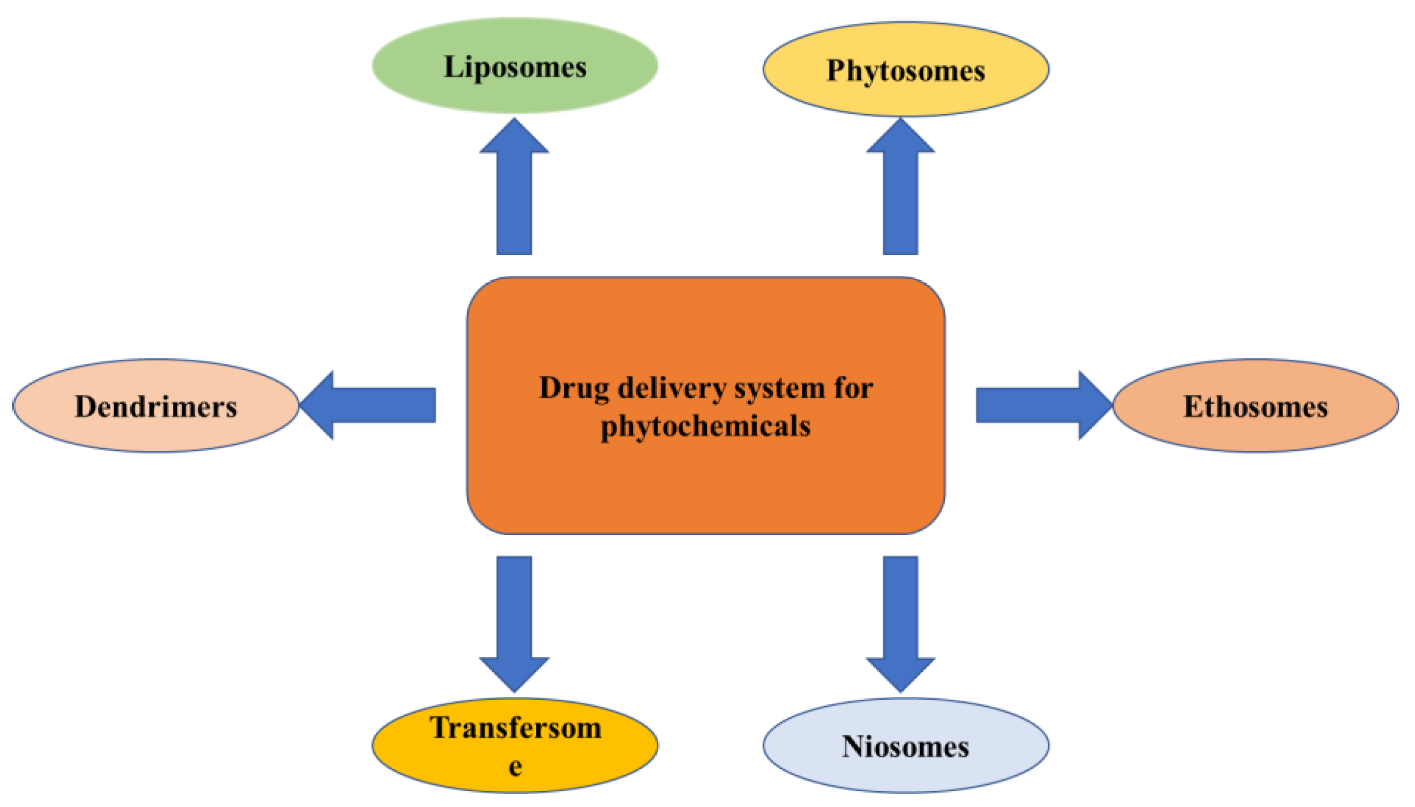
Novel Drug Delivery Approaches for Phytoconstituents
The novel techniques for the drug delivery of phytochemicals use liposomes, phytosomes, niosomes, transferosomes, ethosomes, etc., which are discussed below (Figure 5).
Role of Nanotechnology in Various Neurodegenerative Disorders
The therapy of neurodegenerative disorders such as AD, PD, stroke, epilepsy, and Huntington’s disease has been improved by the development of newer medication delivery strategies such as nanoparticles. The nanoparticles that are given have the ability to cross the blood–brain barrier and then target a particular cell or signaling pathway (Figure 6).
Nanotechnology-Based Brain Targeting via Alteration of Blood–Brain Barrier
The BBB is a selective permeability barrier that works as a local gateway for circulating antigens and foreign particles. This is the most complex gateway to be crossed and acts as a major challenge for the drug delivery vehicles to target the drug molecules to the brain. Emerging nanotechnologies, involve a number of fascinating concepts that have the potential to serve as the answer to this persistent challenge.
The advancement of nanotechnology through interdisciplinary collaboration will lead to new understandings of how neural circuits function, as well as the methods for the detection and treatment of brain disorders. A new drug delivery platform that makes it simple to transfer therapeutic compounds to the brain can be developed by utilizing the unique qualities of nanomaterials, such as their smaller size, biocompatibility, longer blood circulation, and lack of toxicity. Both specialized and general targeting strategies for brain locations are used in nanotechnology-mediated medication delivery systems. The formulation of nano-vehicles for the delivery of drugs, such as nanoparticles, liposomes, dendrimers, micelles, and carbon nanotubes, has received a lot of attention recently. These vehicles can deliver pharmaceuticals, peptides, proteins, vaccines, or nucleic acids [220].
Alzheimer’s Disease—Researchers have developed a wide variety of nano-formulations in the hopes that they will have a positive effect on AD patients. The studies that were conducted in vitro on the SHSY-5Y human neuroblastoma cell line showed that phospholipid-based PEG-stabilized nano-micelles inhibit Aβaggregation and reduce Aβ-induced neurotoxicity [230]. The phytochemical curcumin was shown to have the power to decrease Aβ oligomerization and cell proliferation in the in vitro study; however, when it was injected into mice, it showed poor bioavailability [231]. Curcumin was formulated by using nanoliposomes, which improved the bioavailability without compromising the drug’s ability to prevent Aβ aggregation [232].
Parkinson’s Disease—According to one study, L-Dopa and nerve-growth-factor-bound PBCA nanoparticles can cross the BBB and treat the primary signs and symptoms of Parkinson’s disease (PD) [233].
In addition, in another study, SA (Schisantherin A), which is used for the treatment of PD, was encapsulated in nanoparticles, which increasedits circulation in the blood stream and also its uptake in the brain. This showed that delivery with nanoparticles increases the efficacy of SA and makesit a potent compound for the treatment of PD [234].
Huntington’s Disease—Nitrendipine, a calcium channel blocker, has been demonstrated to significantly lower the incidence of dementia in HD patients over the duration of two years. However, this drug has permeability issues, due to its hydrophilic nature, and, as a result, it crosses the BBB very poorly. A study was conducted in which SLNs of nitrendipine were prepared, and a comparison of the uptake of nano-formulation and the bulk drug was carried out. According to the findings, the drug was taken in at a higher rate when it was encapsulated in SLNs [235].
Epilepsy—It has been discovered that solid lipid nanoparticles packed with carbamazepine and PLGA nanoparticles filled with β-carotene have the ability to have a greater anticonvulsant impact compared to polysorbate-80 and carbamazepine-packed, nanoemulged-covered nanoparticles [236].
In a rat model, subcutaneous injection of ethosuximide-encapsulated chitosan nano-capsules was found to reduce the spike-wave discharge. This finding was made by researchers using the rat as a model. Because they can provide a consistent release of the medication, these nanoparticles can be developed as depot drug delivery systems, enabling the long-term usage of antiepileptic drugs [237].
Advantages of Various Novel Drug Delivery Methods
There are various advantages of novel approaches for drug delivery using nanoparticles (Table 35).

2. Conclusions

Phytochemicals that are naturally derived and have been shown to be effective against a number of age-related neurodegenerative illnesses have been the focus of a growing number of studies in recent years. Because the treatment options for such illnesses with synthetic drugs in clinical studies have been demonstrated to be challenging, due to their toxicity and capacity to induce numerous different serious disorders, treatments with natural antioxidants, such as polyphenols via food or nutraceuticals, have become an excellent option to fight against the oxidative damage caused by free radicals to the neuronal cells. Although no major side effects of the phytochemicals that are now available have been observed clinically, there is a paucity of research in the field. In addition, Parkinson’s disease has been treated successfully with natural products; in particular, the phytochemicals obtained from naturally occurring foods have the ability to act as antioxidants and may serve as a dependable source of medication. However, naturally occurring lipophilic phytochemicals are able to enter the brain and cross the blood–brain barrier with relative ease. These phytochemicals offer enhanced bioavailability, a faster metabolism, and a better affinity to the receptors. Therefore, they can be taken on a consistent basis if one wishes to accelerate the process of neuronal function and reversal of the disease. In addition, when multiple neurotoxic models of Parkinson’s disease are combined, this showed that herbal therapies may be utilized in the development of novel Parkinson’s disease treatments. However, in the future, research should be conducted in real life to investigate the efficacy of plant extracts and the active components of those extracts in PD models. In addition, there is an ongoing demand for more in-depth explanations of the ingredients of herbal extracts, as well as the mechanisms through which they exert their effects. Furthermore, for the management of Parkinson’s disease (PD) by the use of natural products, methodological changes need to be implemented in order to guarantee high levels of reproducibility, increase the therapeutic effect, and reduce the likelihood of any potential harm.

3. Materials and Methods

A literature search was accomplished with electronic databases PubMed, Scopus, and Google Scholar, from which only English articles published until January 2023 were included. The succeeding search terms and Boolean connectors were used in the following search: “PD OR Parkinson’s disease OR neurodegenerative disorders OR neurodegenerative diseases OR NDD” AND “antioxidant OR antioxidant compounds OR natural antioxidant OR natural antioxidants”. These terms were searched in the abstract, title, or keywords. From the searched results, examples of natural antioxidant compounds had been studied, which were effective in neurodegenerative disorders, and the most were selected. The inclusion criteria for the articles were as follows: (i) research that uses natural antioxidants in the management of neurodegenerative disorders, including PD; (ii) their mechanism of action; (iii) invitrostudies performed; (iv) phytochemical-based formulations; (v) research that includes the novel approaches for the drug delivery of phytochemicals; (vi) recent clinical trials on phytochemicals; and (vii) the chemical structure of phytochemical used in PD. Studies without protein or mechanism analysis were excluded. However, in this review, we have tried to include phytochemicals that can be used to manage Parkinson’s disease.

Author Contributions

Conceptualization, S.D. and S.C.; methodology, P.M.; software, N.G.; validation, N.G., W.A. and T.B.; formal analysis, T.G.S.; investigation, H.A.R.; resources, M.A.-Z.; data curation, A.A.C.; writing—original draft preparation, S.D.; writing—review and editing, S.D., N.G. and S.C.; visualization, S.U.-D.K.; supervision, S.C. and P.M.; project administration, S.M.; funding acquisition, M.A. All authors have read and agreed to the published version of the manuscript.

Funding

The authors extend their appreciation to the Deanship of Scientific Research, Imam Mohammad Ibn Saud Islamic University (IMSIU), Saudi Arabia, for funding this work through grant number 221412037.

Institutional Review Board Statement

Not applicable.

Informed Consent Statement

Not applicable.

Data Availability Statement

Not applicable.

Conflicts of Interest

The authors declare no conflict of interest.

References

  1. Chauhan, S.; Gupta, S.; Yasmin, S.; Saini, M. Antihyperglycemic and Antioxidant Potential of Plant Extract of Litchi chinensis and Glycine max. Int. J. Nutr. Pharmacol. Neurol. Dis. 2021, 11, 225–233. [Google Scholar]
  2. Chauhan, S.; Kaur, N.; Kishore, L.; Singh, R. Pharmacological evaluation of anti-inflammatory and analgesic potential of Litchi chinensis gaertn.(sonn.). Group 2014, 10, 100. [Google Scholar]
  3. Karunaratne, T.B.; Okereke, C.; Seamon, M.; Purohit, S.; Wakade, C.; Sharma, A. Niacin and butyrate: Nutraceuticals targeting dysbiosis and intestinal permeability in parkinson’s disease. Nutrients 2020, 13, 28. [Google Scholar] [CrossRef]
  4. Chattopadhyaya, I.; Gupta, S.; Mohammed, A.; Mushtaq, N.; Chauhan, S.; Ghosh, S. Neuroprotective effect of Spirulina fusiform and amantadine in the 6-OHDA induced Parkinsonism in rats. BMC Complement. Altern. Med. 2015, 15, 296. [Google Scholar]
  5. Sławińska, N.; Olas, B. Selected Seeds as Sources of Bioactive Compounds with Diverse Biological Activities. Nutrients 2023, 15, 187. [Google Scholar] [CrossRef]
  6. Sharma, A.; Gupta, S.; Chauhan, S.; Nair, A.; Sharma, P. Astilbin: A promising unexplored compound with multidimensional medicinal and health benefits. Pharmacol. Res. 2020, 158, 104894. [Google Scholar] [CrossRef]
  7. Sharma, C.; Chauhan, S.; Gupta, S.; Devi, A.; Nair, A. Role of whole plant extract of nelumbo nucifera gaertn in the treatment of thrombolysis. Cardiovasc. Hematol. Agents Med. Chem. 2019, 17, 115–124. [Google Scholar] [CrossRef] [PubMed]
  8. Knight, E.; Geetha, T.; Burnett, D.; Babu, J.R. The Role of Diet and Dietary Patterns in Parkinson’s Disease. Nutrients 2022, 14, 4472. [Google Scholar] [CrossRef]
  9. Sindhu, R.K.; Kaur, P.; Kaur, P.; Singh, H.; Batiha, G.E.-S.; Verma, I. Exploring multifunctional antioxidants as potential agents for management of neurological disorders. Environ. Sci. Pollut. Res. 2022, 29, 24458–24477. [Google Scholar] [CrossRef] [PubMed]
  10. Evans, J.A.; Mendonca, P.; Soliman, K.F. Neuroprotective effects and therapeutic potential of the citrus flavonoid hesperetin in neurodegenerative diseases. Nutrients 2022, 14, 2228. [Google Scholar] [CrossRef]
  11. Savica, R.; Grossardt, B.R.; Rocca, W.A.; Bower, J.H. Parkinson disease with and without dementia: A prevalence study and future projections. Mov. Disord. 2018, 33, 537–543. [Google Scholar] [CrossRef]
  12. Choo, X.Y.; Lim, S.-Y.; Chinna, K.; Tan, Y.J.; Yong, V.W.; Lim, J.L.; Lau, K.F.; Chung, J.Y.; Em, J.M.; Tan, H.T. Understanding patients’ and caregivers’ perspectives and educational needs in Parkinson’s disease: A multi-ethnic Asian study. Neurol. Sci. 2020, 41, 2831–2842. [Google Scholar] [CrossRef]
  13. Zeng, X.-S.; Geng, W.-S.; Jia, J.-J. Neurotoxin-induced animal models of Parkinson disease: Pathogenic mechanism and assessment. ASN Neuro 2018, 10, 1759091418777438. [Google Scholar] [CrossRef] [Green Version]
  14. Lashuel, H.A.; Overk, C.R.; Oueslati, A.; Masliah, E. The many faces of α-synuclein: From structure and toxicity to therapeutic target. Nat. Rev. Neurosci. 2013, 14, 38–48. [Google Scholar] [CrossRef] [PubMed] [Green Version]
  15. Kaur, I.; Behl, T.; Sehgal, A.; Singh, S.; Sharma, N.; Aleya, L.; Bungau, S. Connecting the dots between mitochondrial dysfunction and Parkinson’s disorder: Focus mitochondria-targeting therapeutic paradigm in mitigating the disease severity. Environ. Sci. Pollut. Res. 2021, 28, 37060–37081. [Google Scholar] [CrossRef] [PubMed]
  16. Chagraoui, A.; Boulain, M.; Juvin, L.; Anouar, Y.; Barrière, G.; De Deurwaerdère, P. L-DOPA in Parkinson’s disease: Looking at the “false” neurotransmitters and their meaning. Int. J. Mol. Sci. 2019, 21, 294. [Google Scholar] [CrossRef] [Green Version]
  17. Poewe, W.; Seppi, K.; Tanner, C.M.; Halliday, G.M.; Brundin, P.; Volkmann, J.; Schrag, A.-E.; Lang, A.E. Parkinson disease. Nat. Rev. Dis. Prim. 2017, 3, 17013. [Google Scholar] [CrossRef] [PubMed]
  18. Ghosh, S.; Haldar, S.; Gupta, S.; Bisht, A.; Chauhan, S.; Kumar, V.; Roy, P.; Lahiri, D. Anisotropically conductive biodegradable scaffold with coaxially aligned carbon nanotubes for directional regeneration of peripheral nerves. ACS Appl. Bio Mater. 2020, 3, 5796–5812. [Google Scholar] [CrossRef]
  19. Montastruc, J.; Rascol, O.; Senard, J.; Rascol, A. A randomised controlled study comparing bromocriptine to which levodopa was later added, with levodopa alone in previously untreated patients with Parkinson’s disease: A five year follow up. J. Neurol. Neurosurg. Psychiatry 1994, 57, 1034–1038. [Google Scholar] [CrossRef]
  20. Ahlskog, J.E.; Muenter, M.D.; Bailey, P.A.; Stevens, P.M. Dopamine agonist treatment of fluctuating parkinsonism: D-2 (controlled-release MK-458) vs combined D-1 and D-2 (pergolide). Arch. Neurol. 1992, 49, 560–568. [Google Scholar] [CrossRef]
  21. Kumar, B.; Prakash Gupta, V.; Kumar, V. A perspective on monoamine oxidase enzyme as drug target: Challenges and opportunities. Curr. Drug Targets 2017, 18, 87–97. [Google Scholar] [CrossRef] [PubMed]
  22. Mintzer, J.; Burns, A. Anticholinergic side-effects of drugs in elderly people. J. R. Soc. Med. 2000, 93, 457–462. [Google Scholar] [CrossRef] [PubMed]
  23. Singer, C. Adverse effects in the treatment of Parkinson’s disease. Expert Rev. Neurother. 2002, 2, 105–118. [Google Scholar] [CrossRef] [PubMed]
  24. Hussain, G.; Rasul, A.; Anwar, H.; Aziz, N.; Razzaq, A.; Wei, W.; Ali, M.; Li, J.; Li, X. Role of plant derived alkaloids and their mechanism in neurodegenerative disorders. Int. J. Biol. Sci. 2018, 14, 341. [Google Scholar] [CrossRef] [Green Version]
  25. Farah, S.; Anderson, D.G.; Langer, R. Physical and mechanical properties of PLA, and their functions in widespread applications—A comprehensive review. Adv. Drug Deliv. Rev. 2016, 107, 367–392. [Google Scholar] [CrossRef] [Green Version]
  26. Zhang, Y.-J.; Gan, R.-Y.; Li, S.; Zhou, Y.; Li, A.-N.; Xu, D.-P.; Li, H.-B. Antioxidant phytochemicals for the prevention and treatment of chronic diseases. Molecules 2015, 20, 21138–21156. [Google Scholar] [CrossRef] [Green Version]
  27. Mohseni, M.; Sahebkar, A.; Askari, G.; Johnston, T.P.; Alikiaii, B.; Bagherniya, M. The clinical use of curcumin on neurological disorders: An updated systematic review of clinical trials. Phytother. Res. 2021, 35, 6862–6882. [Google Scholar] [CrossRef]
  28. Pearson, P.; Lewis, S.; Britton, J.; Fogarty, A. Vitamin E supplements in asthma: A parallel group randomised placebo controlled trial. Thorax 2004, 59, 652–656. [Google Scholar] [CrossRef] [Green Version]
  29. Gao, W.; Tang, H.; Wang, D.; Zhou, X.; Song, Y.; Wang, Z. Effect of short-term vitamin D supplementation after nonsurgical periodontal treatment: A randomized, double-masked, placebo-controlled clinical trial. J. Periodontal Res. 2020, 55, 354–362. [Google Scholar] [CrossRef]
  30. Vieregge, A.; Sieberer, M.; Jacobs, H.; Hagenah, J.; Vieregge, P. Transdermal nicotine in PD: A randomized, double-blind, placebo-controlled study. Neurology 2001, 57, 1032–1035. [Google Scholar] [CrossRef]
  31. Chmiel, M.; Stompor-Gorący, M. The Spectrum of Pharmacological Actions of Syringetin and Its Natural Derivatives—A Summary Review. Nutrients 2022, 14, 5157. [Google Scholar] [CrossRef] [PubMed]
  32. Isah, T. Stress and defense responses in plant secondary metabolites production. Biol. Res. 2019, 52, 39. [Google Scholar] [CrossRef] [PubMed] [Green Version]
  33. Hannan, M.A.; Dash, R.; Sohag, A.A.M.; Haque, M.N.; Moon, I.S. Neuroprotection against oxidative stress: Phytochemicals targeting TrkB signaling and the Nrf2-ARE antioxidant system. Front. Mol. Neurosci. 2020, 13, 116. [Google Scholar] [CrossRef]
  34. Lees, A.J. Impact Commentaries. A modern perspective on the top 100 cited JNNP papers of all time: The relevance of the Lewy body to the pathogenesis of idiopathic Parkinson’s disease: Accuracy of clinical diagnosis of idiopathic Parkinson’s disease. J. Neurol. Neurosurg. Psychiatry 2012, 83, 954–955. [Google Scholar] [CrossRef] [Green Version]
  35. Mischley, L.K.; Standish, L.J.; Weiss, N.S.; Padowski, J.M.; Kavanagh, T.J.; White, C.C.; Rosenfeld, M.E. Glutathione as a biomarker in Parkinson’s disease: Associations with aging and disease severity. Oxidative Med. Cell. Longev. 2016, 2016, 9409363. [Google Scholar] [CrossRef] [Green Version]
  36. Pospíšil, P.; Prasad, A.; Rác, M. Mechanism of the formation of electronically excited species by oxidative metabolic processes: Role of reactive oxygen species. Biomolecules 2019, 9, 258. [Google Scholar] [CrossRef] [Green Version]
  37. Yi, L.; Ma, S.; Ren, D. Phytochemistry and bioactivity of Citrus flavonoids: A focus on antioxidant, anti-inflammatory, anticancer and cardiovascular protection activities. Phytochem. Rev. 2017, 16, 479–511. [Google Scholar] [CrossRef]
  38. Chu, K.O.; Chan, S.-O.; Pang, C.P.; Wang, C.C. Pro-oxidative and antioxidative controls and signaling modification of polyphenolic phytochemicals: Contribution to health promotion and disease prevention? J. Agric. Food Chem. 2014, 62, 4026–4038. [Google Scholar] [CrossRef] [PubMed]
  39. Zhang, Y.; Yu, W.; Zhang, L.; Wang, M.; Chang, W. The Interaction of Polyphenols and the Gut Microbiota in Neurodegenerative Diseases. Nutrients 2022, 14, 5373. [Google Scholar] [CrossRef]
  40. Fadaka, A.O.; Ajiboye, B.O.; Adewale, I.; Ojo, O.A.; Oyinloye, B.E.; Okesola, M.A. Significance of antioxidants in the treatment and prevention of neurodegenerative diseases. J. Phytopharm. 2019, 8, 75–83. [Google Scholar] [CrossRef]
  41. Muszyńska, B.; Łojewski, M.; Rojowski, J.; Opoka, W.; Sułkowska-Ziaja, K. Natural products of relevance in the prevention and supportive treatment of depression. Psychiatr. Pol. 2015, 49, 435–453. [Google Scholar] [CrossRef]
  42. Pignolo, A.; Mastrilli, S.; Davì, C.; Arnao, V.; Aridon, P.; dos Santos Mendes, F.A.; Gagliardo, C.; D’Amelio, M. Vitamin D and Parkinson’s disease. Nutrients 2022, 14, 1220. [Google Scholar] [CrossRef]
  43. Elfawy, H.A.; Das, B. Crosstalk between mitochondrial dysfunction, oxidative stress, and age related neurodegenerative disease: Etiologies and therapeutic strategies. Life Sci. 2019, 218, 165–184. [Google Scholar] [CrossRef]
  44. Caioni, G.; Cimini, A.; Benedetti, E. Food Contamination: An Unexplored Possible Link between Dietary Habits and Parkinson’s Disease. Nutrients 2022, 14, 1467. [Google Scholar] [CrossRef]
  45. Teleanu, R.I.; Chircov, C.; Grumezescu, A.M.; Volceanov, A.; Teleanu, D.M. Antioxidant therapies for neuroprotection—A review. J. Clin. Med. 2019, 8, 1659. [Google Scholar] [CrossRef] [PubMed] [Green Version]
  46. Naoi, M.; Shamoto-Nagai, M.; Maruyama, W. Neuroprotection of multifunctional phytochemicals as novel therapeutic strategy for neurodegenerative disorders: Antiapoptotic and antiamyloidogenic activities by modulation of cellular signal pathways. Future Neurol. 2019, 14, FNL9. [Google Scholar] [CrossRef] [Green Version]
  47. Yap, H.-M.; Lye, K.-L. An insight of vitamin E as neuroprotective agents. Prog. Microbes Mol. Biol. 2020, 3, a0000071. [Google Scholar] [CrossRef]
  48. Schirinzi, T.; Martella, G.; Imbriani, P.; Di Lazzaro, G.; Franco, D.; Colona, V.L.; Alwardat, M.; Sinibaldi Salimei, P.; Mercuri, N.B.; Pierantozzi, M. Dietary vitamin E as a protective factor for Parkinson’s disease: Clinical and experimental evidence. Front. Neurol. 2019, 10, 148. [Google Scholar] [CrossRef] [PubMed] [Green Version]
  49. Elizan, T. Treatment of Parkinson’s Disease. In Inhibitors of Monoamine Oxidase B: Pharmacology and Clinical Use in Neurodegenerative Disorders; Springer: Basel, Switzerland, 2019; p. 277. [Google Scholar]
  50. Eckert, G.P. The role of vitamin E in aging and Alzheimer’s disease. In Vitamin E in Human Health; Humana: Cham, Switzerland, 2019; pp. 325–344. [Google Scholar]
  51. Lewis, E.D.; Meydani, S.N.; Wu, D. Regulatory role of vitamin E in the immune system and inflammation. IUBMB Life 2019, 71, 487–494. [Google Scholar] [CrossRef] [PubMed]
  52. Garg, A.; Lee, J.C.-Y. Vitamin E: Where are we now in vascular diseases? Life 2022, 12, 310. [Google Scholar] [CrossRef] [PubMed]
  53. Lykkesfeldt, J. On the effect of vitamin C intake on human health: How to (mis) interprete the clinical evidence. Redox Biol. 2020, 34, 101532. [Google Scholar] [CrossRef] [PubMed]
  54. Baiano, A.; Del Nobile, M.A. Antioxidant compounds from vegetable matrices: Biosynthesis, occurrence, and extraction systems. Crit. Rev. Food Sci. Nutr. 2016, 56, 2053–2068. [Google Scholar] [CrossRef] [PubMed]
  55. Ciulla, M.; Marinelli, L.; Cacciatore, I.; Stefano, A.D. Role of dietary supplements in the management of Parkinson’s disease. Biomolecules 2019, 9, 271. [Google Scholar] [CrossRef] [PubMed] [Green Version]
  56. Morris-Blanco, K.C.; Chokkalla, A.K.; Kim, T.; Bhatula, S.; Bertogliat, M.J.; Gaillard, A.B.; Vemuganti, R. High-dose vitamin C prevents secondary brain damage after stroke via epigenetic reprogramming of neuroprotective genes. Transl. Stroke Res. 2022, 13, 1017–1036. [Google Scholar] [CrossRef] [PubMed]
  57. Maciag, M.C.; Phipatanakul, W. Prevention of asthma: Targets for intervention. Chest 2020, 158, 913–922. [Google Scholar] [CrossRef] [PubMed]
  58. Lim, J.C.; Caballero Arredondo, M.; Braakhuis, A.J.; Donaldson, P.J. Vitamin C and the lens: New insights into delaying the onset of cataract. Nutrients 2020, 12, 3142. [Google Scholar] [CrossRef]
  59. Panche, A.N.; Diwan, A.D.; Chandra, S.R. Flavonoids: An overview. J. Nutr. Sci. 2016, 5, e47. [Google Scholar] [CrossRef] [Green Version]
  60. Bayazid, A.B.; Lim, B.O. Quercetin is an Active Agent in Berries against Neurodegenerative Diseases Progression through Modulation of Nrf2/HO1. Nutrients 2022, 14, 5132. [Google Scholar] [CrossRef]
  61. Wang, W.-W.; Han, R.; He, H.-J.; Li, J.; Chen, S.-Y.; Gu, Y.; Xie, C. Administration of quercetin improves mitochondria quality control and protects the neurons in 6-OHDA-lesioned Parkinson’s disease models. Aging 2021, 13, 11738. [Google Scholar] [CrossRef]
  62. Pan, M.-H.; Lai, C.-S.; Ho, C.-T. Anti-inflammatory activity of natural dietary flavonoids. Food Funct. 2010, 1, 15–31. [Google Scholar] [CrossRef]
  63. Górniak, I.; Bartoszewski, R.; Króliczewski, J. Comprehensive review of antimicrobial activities of plant flavonoids. Phytochem. Rev. 2019, 18, 241–272. [Google Scholar] [CrossRef] [Green Version]
  64. Prasad, K.N.; Xie, H.; Hao, J.; Yang, B.; Qiu, S.; Wei, X.; Chen, F.; Jiang, Y. Antioxidant and anticancer activities of 8-hydroxypsoralen isolated from wampee [Clausena lansium (Lour.) Skeels] peel. Food Chem. 2010, 118, 62–66. [Google Scholar] [CrossRef]
  65. Nie, G.; Wei, T.; Shen, S.; Zhao, B. Polyphenol protection of DNA against damage. In Methods in Enzymology; Elsevier: Amsterdam, The Netherlands, 2001; Volume 335, pp. 232–244. [Google Scholar]
  66. Zhao, B. Natural antioxidants protect neurons in Alzheimer’s disease and Parkinson’s disease. Neurochem. Res. 2009, 34, 630–638. [Google Scholar] [CrossRef] [PubMed]
  67. Mukhtar, H.; Ahmad, N. Tea polyphenols: Prevention of cancer and optimizing health. Am. J. Clin. Nutr. 2000, 71, 1698S–1702S. [Google Scholar] [CrossRef] [PubMed] [Green Version]
  68. Wang, Q.-M.; Gong, Q.-Y.; Yan, J.-J.; Tang, J.-J.; Wang, M.-W.; Yang, Z.-J.; Wang, L.-S. Association between green tea intake and coronary artery disease in a Chinese population. Circ. J. 2010, 74, 294–300. [Google Scholar] [CrossRef] [Green Version]
  69. Baumann, C.R. Epidemiology, diagnosis and differential diagnosis in Parkinson’s disease tremor. Park. Relat. Disord. 2012, 18, S90–S92. [Google Scholar] [CrossRef]
  70. Popa, D.-S.; Rusu, M.E. Isoflavones: Vegetable sources, biological activity, and analytical methods for their assessment. In Superfood and Functional Food: The Development of Superfoods and Their Roles as Medicine; BoD: Norderstedt, Germany, 2017; pp. 133–153. [Google Scholar]
  71. Duan, X.; Li, Y.; Xu, F.; Ding, H. Study on the neuroprotective effects of Genistein on Alzheimer’s disease. Brain Behav. 2021, 11, e02100. [Google Scholar] [CrossRef]
  72. Wada, K.; Nakamura, K.; Tamai, Y.; Tsuji, M.; Kawachi, T.; Hori, A.; Takeyama, N.; Tanabashi, S.; Matsushita, S.; Tokimitsu, N. Soy isoflavone intake and breast cancer risk in Japan: From the Takayama study. Int. J. Cancer 2013, 133, 952–960. [Google Scholar] [CrossRef]
  73. Wei, P.; Liu, M.; Chen, Y.; Chen, D.-C. Systematic review of soy isoflavone supplements on osteoporosis in women. Asian Pac. J. Trop. Med. 2012, 5, 243–248. [Google Scholar] [CrossRef] [PubMed] [Green Version]
  74. Tikkanen, M.J.; Adlercreutz, H. Dietary soy-derived isoflavone phytoestrogens: Could they have a role in coronary heart disease prevention? Biochem. Pharmacol. 2000, 60, 1–5. [Google Scholar] [CrossRef]
  75. Nicholatos, J.W.; Francisco, A.B.; Bender, C.A.; Yeh, T.; Lugay, F.J.; Salazar, J.E.; Glorioso, C.; Libert, S. Nicotine promotes neuron survival and partially protects from Parkinson’s disease by suppressing SIRT6. Acta Neuropathol. Commun. 2018, 6, 120. [Google Scholar] [CrossRef] [PubMed]
  76. Zhang, J.; Liu, Q.; Chen, Q.; Liu, N.-Q.; Li, F.-L.; Lu, Z.-B.; Qin, C.; Zhu, H.; Huang, Y.-Y.; He, W. Nicotine attenuates the β-amyloid neurotoxicity through regulating metal homeostasis. FASEB J. 2006, 20, 1212–1214. [Google Scholar] [CrossRef]
  77. Anan, J.; Hijioka, M.; Kurauchi, Y.; Hisatsune, A.; Seki, T.; Katsuki, H. Cortical hemorrhage-associated neurological deficits and tissue damage in mice are ameliorated by therapeutic treatment with nicotine. J. Neurosci. Res. 2017, 95, 1838–1849. [Google Scholar] [CrossRef]
  78. Zarrindast, M.-R.; Khakpai, F. The modulatory role of nicotine on cognitive and non-cognitive functions. Brain Res. 2019, 1710, 92–101. [Google Scholar] [CrossRef] [PubMed]
  79. Quik, M.; Boyd, J.T.; Bordia, T.; Perez, X. Potential therapeutic application for nicotinic receptor drugs in movement disorders. Nicotine Tob. Res. 2019, 21, 357–369. [Google Scholar] [CrossRef]
  80. Maoka, T. Carotenoids as natural functional pigments. J. Nat. Med. 2020, 74, 1–16. [Google Scholar] [CrossRef] [PubMed] [Green Version]
  81. Britton, G.; Liaaen-Jensen, S.; Pfander, H. Carotenoids: Natural Functions; Springer Science & Business Media: Berlin/Heidelberg, Germany, 2008; Volume 4. [Google Scholar]
  82. Freedman, N.D.; Park, Y.; Subar, A.F.; Hollenbeck, A.R.; Leitzmann, M.F.; Schatzkin, A.; Abnet, C.C. Fruit and vegetable intake and head and neck cancer risk in a large United States prospective cohort study. Int. J. Cancer 2008, 122, 2330–2336. [Google Scholar] [CrossRef] [PubMed]
  83. Przybylska, S. Lycopene–a bioactive carotenoid offering multiple health benefits: A review. Int. J. Food Sci. Technol. 2020, 55, 11–32. [Google Scholar] [CrossRef]
  84. Palombo, P.; Fabrizi, G.; Ruocco, V.; Ruocco, E.; Fluhr, J.; Roberts, R.; Morganti, P. Beneficial long-term effects of combined oral/topical antioxidant treatment with the carotenoids lutein and zeaxanthin on human skin: A double-blind, placebo-controlled study. Ski. Pharmacol. Physiol. 2007, 20, 199–210. [Google Scholar] [CrossRef]
  85. Lu, K.-T.; Ko, M.-C.; Chen, B.-Y.; Huang, J.-C.; Hsieh, C.-W.; Lee, M.-C.; Chiou, R.Y.; Wung, B.-S.; Peng, C.-H.; Yang, Y.-L. Neuroprotective effects of resveratrol on MPTP-induced neuron loss mediated by free radical scavenging. J. Agric. Food Chem. 2008, 56, 6910–6913. [Google Scholar] [CrossRef]
  86. Jin, F.; Wu, Q.; Lu, Y.-F.; Gong, Q.-H.; Shi, J.-S. Neuroprotective effect of resveratrol on 6-OHDA-induced Parkinson’s disease in rats. Eur. J. Pharmacol. 2008, 600, 78–82. [Google Scholar] [CrossRef]
  87. Peng, K.; Tao, Y.; Zhang, J.; Wang, J.; Ye, F.; Dan, G.; Zhao, Y.; Cai, Y.; Zhao, J.; Wu, Q. Resveratrol regulates mitochondrial biogenesis and fission/fusion to attenuate rotenone-induced neurotoxicity. Oxidative Med. Cell. Longev. 2016, 2016, 6705621. [Google Scholar] [CrossRef] [Green Version]
  88. Jang, J.-H.; Surh, Y.-J. Protective effects of resveratrol on hydrogen peroxide-induced apoptosis in rat pheochromocytoma (PC12) cells. Mutat. Res./Genet. Toxicol. Environ. Mutagen. 2001, 496, 181–190. [Google Scholar] [CrossRef]
  89. Kitada, M.; Koya, D. Renal protective effects of resveratrol. Oxidative Med. Cell. Longev. 2013, 2013, 568093. [Google Scholar] [CrossRef] [Green Version]
  90. Sun, A.Y.; Wang, Q.; Simonyi, A.; Sun, G.Y. Resveratrol as a therapeutic agent for neurodegenerative diseases. Mol. Neurobiol. 2010, 41, 375–383. [Google Scholar] [CrossRef] [PubMed] [Green Version]
  91. Sepperer, T.; Hernandez-Ramos, F.; Labidi, J.; Oostingh, G.J.; Bogner, B.; Petutschnigg, A.; Tondi, G. Purification of industrial tannin extract through simple solid-liquid extractions. Ind. Crops Prod. 2019, 139, 111502. [Google Scholar] [CrossRef]
  92. Hideshima, M.; Kimura, Y.; Aguirre, C.; Kakuda, K.; Takeuchi, T.; Choong, C.-J.; Nabekura, K.; Yamaguchi, K.; Nakajima, K.; Baba, K. Two-step screening method to identify α-synuclein aggregation inhibitors for Parkinson’s disease. Sci. Rep. 2022, 12, 351. [Google Scholar] [CrossRef] [PubMed]
  93. Kumari, M.; Jain, S. Tannins: An antinutrient with positive effect to manage diabetes. Res. J. Recent Sci. 2012, 2277, 2502. [Google Scholar]
  94. Zarin, M.A.; Wan, H.Y.; Isha, A.; Armania, N. Antioxidant, antimicrobial and cytotoxic potential of condensed tannins from Leucaena leucocephala hybrid-Rendang. Food Sci. Hum. Wellness 2016, 5, 65–75. [Google Scholar] [CrossRef] [Green Version]
  95. Hussain, G.; Huang, J.; Rasul, A.; Anwar, H.; Imran, A.; Maqbool, J.; Razzaq, A.; Aziz, N.; Makhdoom, E.U.H.; Konuk, M. Putative roles of plant-derived tannins in neurodegenerative and neuropsychiatry disorders: An updated review. Molecules 2019, 24, 2213. [Google Scholar] [CrossRef] [Green Version]
  96. Buratta, S.; Chiaradia, E.; Tognoloni, A.; Gambelunghe, A.; Meschini, C.; Palmieri, L.; Muzi, G.; Urbanelli, L.; Emiliani, C.; Tancini, B. Effect of curcumin on protein damage induced by rotenone in dopaminergic PC12 cells. Int. J. Mol. Sci. 2020, 21, 2761. [Google Scholar] [CrossRef] [Green Version]
  97. Sumeet, G.; Rachna, K.; Samrat, C.; Ipshita, C.; Vikas, J.; Manu, S. Anti inflammatory and anti arthritic activity of different milk based formulation of curcumin in rat model. Curr. Drug Deliv. 2018, 15, 205–214. [Google Scholar] [CrossRef] [PubMed]
  98. Ojha, R.P.; Rastogi, M.; Devi, B.P.; Agrawal, A.; Dubey, G. Neuroprotective effect of curcuminoids against inflammation-mediated dopaminergic neurodegeneration in the MPTP model of Parkinson’s disease. J. Neuroimmune Pharmacol. 2012, 7, 609–618. [Google Scholar] [CrossRef] [PubMed]
  99. Gupta, A.; Vij, G.; Sharma, S.; Tirkey, N.; Rishi, P.; Chopra, K. Curcumin, a polyphenolic antioxidant, attenuates chronic fatigue syndrome in murine water immersion stress model. Immunobiology 2009, 214, 33–39. [Google Scholar] [CrossRef]
  100. Mythri, R.B.; Srinivas Bharath, M.M. Curcumin: A potential neuroprotective agent in Parkinson’s disease. Curr. Pharm. Des. 2012, 18, 91–99. [Google Scholar] [CrossRef] [PubMed] [Green Version]
  101. Ataie, A.; Sabetkasaei, M.; Haghparast, A.; Moghaddam, A.H.; Kazeminejad, B. Neuroprotective effects of the polyphenolic antioxidant agent, Curcumin, against homocysteine-induced cognitive impairment and oxidative stress in the rat. Pharmacol. Biochem. Behav. 2010, 96, 378–385. [Google Scholar] [CrossRef] [PubMed]
  102. Niesche, R.; Haase, M. Emotions and ethics: A Foucauldian framework for becoming an ethical educator. Educ. Philos. Theory 2012, 44, 276–288. [Google Scholar] [CrossRef]
  103. DeFeudis, F.; Drieu, K. Ginkgo biloba extract (EGb 761) and CNS functions basic studies and clinical applications. Curr. Drug Targets 2000, 1, 25–58. [Google Scholar] [CrossRef] [Green Version]
  104. Mahady, G.B. Ginkgo biloba for the prevention and treatment of cardiovascular disease: A review of the literature. J. Cardiovasc. Nurs. 2002, 16, 21–32. [Google Scholar] [CrossRef]
  105. Martínez-Solís, I.; Acero, N.; Bosch-Morell, F.; Castillo, E.; González-Rosende, M.E.; Muñoz-Mingarro, D.; Ortega, T.; Sanahuja, M.A.; Villagrasa, V. Neuroprotective potential of Ginkgo biloba in retinal diseases. Planta Med. 2019, 85, 1292–1303. [Google Scholar] [CrossRef] [Green Version]
  106. Singh, S.K.; Srivastav, S.; Castellani, R.J.; Plascencia-Villa, G.; Perry, G. Neuroprotective and antioxidant effect of Ginkgo biloba extract against AD and other neurological disorders. Neurotherapeutics 2019, 16, 666–674. [Google Scholar] [CrossRef]
  107. Seeram, N.P.; Lee, R.; Heber, D. Bioavailability of ellagic acid in human plasma after consumption of ellagitannins from pomegranate (Punica granatum L.) juice. Clin. Chim. Acta 2004, 348, 63–68. [Google Scholar] [CrossRef] [PubMed] [Green Version]
  108. Cerdá, B.; Llorach, R.; Cerón, J.J.; Espín, J.C.; Tomás-Barberán, F.A. Evaluation of the bioavailability and metabolism in the rat of punicalagin, an antioxidant polyphenol from pomegranate juice. Eur. J. Nutr. 2003, 42, 18–28. [Google Scholar] [CrossRef] [PubMed]
  109. Bassiri-Jahromi, S. Punica granatum (Pomegranate) activity in health promotion and cancer prevention. Oncol. Rev. 2018, 12, 345. [Google Scholar] [CrossRef] [PubMed] [Green Version]
  110. Banihani, S.; Swedan, S.; Alguraan, Z. Pomegranate and type 2 diabetes. Nutr. Res. 2013, 33, 341–348. [Google Scholar] [CrossRef] [PubMed]
  111. Al-Muammar, M.N.; Khan, F. Obesity: The preventive role of the pomegranate (Punica granatum). Nutrition 2012, 28, 595–604. [Google Scholar] [CrossRef]
  112. Amro, M.; Teoh, S.; Norzana, A.; Srijit, D. The potential role of herbal products in the treatment of Parkinson’s disease. La Clin. Ter. 2018, 169, e23–e33. [Google Scholar]
  113. Lu, J.H.; Ardah, M.T.; Durairajan, S.S.K.; Liu, L.F.; Xie, L.X.; Fong, W.F.D.; Hasan, M.Y.; Huang, J.D.; El-Agnaf, O.M.; Li, M. Baicalein Inhibits Formation of α-Synuclein Oligomers within Living Cells and Prevents Aβ Peptide Fibrillation and Oligomerisation. Chembiochem 2011, 12, 615–624. [Google Scholar] [CrossRef] [PubMed]
  114. Dinda, B.; Dinda, S.; DasSharma, S.; Banik, R.; Chakraborty, A.; Dinda, M. Therapeutic potentials of baicalin and its aglycone, baicalein against inflammatory disorders. Eur. J. Med. Chem. 2017, 131, 68–80. [Google Scholar] [CrossRef]
  115. Zhou, L.; Tan, S.; Shan, Y.-L.; Wang, Y.-G.; Cai, W.; Huang, X.-H.; Liao, X.-Y.; Li, H.-Y.; Zhang, L.; Zhang, B.-J. Baicalein improves behavioral dysfunction induced by Alzheimer’s disease in rats. Neuropsychiatr. Dis. Treat. 2016, 12, 3145–3152. [Google Scholar] [CrossRef] [Green Version]
  116. Rezaei, M.; Nasri, S.; Roughani, M.; Niknami, Z.; Ziai, S.A. Peganum Harmala L. extract reduces oxidative stress and improves symptoms in 6-Hydroxydopamine-induced Parkinson’s disease in rats. Iran. J. Pharm. Res. 2016, 15, 275. [Google Scholar]
  117. Khlifi, D.; Sghaier, R.M.; Amouri, S.; Laouini, D.; Hamdi, M.; Bouajila, J. Composition and anti-oxidant, anti-cancer and anti-inflammatory activities of Artemisia herba-alba, Ruta chalpensis L. and Peganum harmala L. Food Chem. Toxicol. 2013, 55, 202–208. [Google Scholar] [CrossRef]
  118. Osman, N.N.; Alanbari, K.H.; Al-Shreef, H.A. Evaluation of the possible antioxidant effects of Peganum harmala and Ginkgo biloba in ameliorating Alzheimer’s disease in rat model. Inter. J. Pharm. Sci. Res. 2018, 9, 3189–3198. [Google Scholar]
  119. Akhtar, M.F.; Raza, S.A.; Saleem, A.; Hamid, I.; Ashraf Baig, M.M.F.; Sharif, A.; Sohail, K.; Javaid, Z.; Saleem, U.; Rasul, A. Appraisal of anti-arthritic and anti-inflammatory potential of folkloric medicinal plant Peganum harmala. Endocr. Metab. Immune Disord. Drug Targets 2022, 22, 49–63. [Google Scholar] [CrossRef] [PubMed]
  120. Ren, R.; Shi, C.; Cao, J.; Sun, Y.; Zhao, X.; Guo, Y.; Wang, C.; Lei, H.; Jiang, H.; Ablat, N. Neuroprotective effects of a standardized flavonoid extract of safflower against neurotoxin-induced cellular and animal models of Parkinson’s disease. Sci. Rep. 2016, 6, 22135. [Google Scholar] [CrossRef] [Green Version]
  121. Asgary, S.; Rahimi, P.; Mahzouni, P.; Madani, H. Antidiabetic effect of hydroalcoholic extract of Carthamus tinctorius L. in alloxan-induced diabetic rats. J. Res. Med. Sci. 2012, 17, 386. [Google Scholar] [PubMed]
  122. Mahadik, V.; Mali, N. Antioxidant activity in safflower (Carthamus tinctorius L.) cultivars under the pathogenesis of foliar fungal disease complex. Indian J. Agric. Res. 2018, 52, 76–80. [Google Scholar] [CrossRef] [Green Version]
  123. Hiramatsu, M.; Takahashi, T.; Komatsu, M.; Kido, T.; Kasahara, Y. Antioxidant and neuroprotective activities of Mogami-benibana (safflower, Carthamus tinctorius Linne). Neurochem. Res. 2009, 34, 795–805. [Google Scholar] [CrossRef]
  124. Cheng, Y.-F.; Zhu, G.-Q.; Wang, M.; Cheng, H.; Zhou, A.; Wang, N.; Fang, N.; Wang, X.-C.; Xiao, X.-Q.; Chen, Z.-W. Involvement of ubiquitin proteasome system in protective mechanisms of Puerarin to MPP+-elicited apoptosis. Neurosci. Res. 2009, 63, 52–58. [Google Scholar] [CrossRef]
  125. Zhu, G.; Wang, X.; Chen, Y.; Yang, S.; Cheng, H.; Wang, N.; Li, Q. Puerarin protects dopaminergic neurons against 6-hydroxydopamine neurotoxicity via inhibiting apoptosis and upregulating glial cell line-derived neurotrophic factor in a rat model of Parkinson’s disease. Planta Med. 2010, 76, 1820–1826. [Google Scholar] [CrossRef] [Green Version]
  126. Lertpatipanpong, P.; Janpaijit, S.; Park, E.-Y.; Kim, C.-T.; Baek, S.J. Potential anti-diabetic activity of Pueraria lobata flower (Flos Puerariae) extracts. Molecules 2020, 25, 3970. [Google Scholar] [CrossRef]
  127. He, H.; Peng, S.; Song, X.; Jia, R.; Zou, Y.; Li, L.; Yin, Z. Protective effect of isoflavones and triterpenoid saponins from Pueraria lobata on liver diseases: A review. Food Sci. Nutr. 2022, 10, 272–285. [Google Scholar] [CrossRef] [PubMed]
  128. Jin, S.E.; Son, Y.K.; Min, B.-S.; Jung, H.A.; Choi, J.S. Anti-inflammatory and antioxidant activities of constituents isolated from Pueraria lobata roots. Arch. Pharmacal Res. 2012, 35, 823–837. [Google Scholar] [CrossRef]
  129. Jiang, W.; Wang, Z.; Jiang, Y.; Lu, M.; Li, X. Ginsenoside Rg1 ameliorates motor function in an animal model of Parkinson’s disease. Pharmacology 2015, 96, 25–31. [Google Scholar] [CrossRef] [PubMed]
  130. Rahman, M.M.; Rahaman, M.S.; Islam, M.R.; Hossain, M.E.; Mannan Mithi, F.; Ahmed, M.; Saldías, M.; Akkol, E.K.; Sobarzo-Sánchez, E. Multifunctional therapeutic potential of phytocomplexes and natural extracts for antimicrobial properties. Antibiotics 2021, 10, 1076. [Google Scholar] [CrossRef] [PubMed]
  131. Kim, J.-H. Pharmacological and medical applications of Panax ginseng and ginsenosides: A review for use in cardiovascular diseases. J. Ginseng Res. 2018, 42, 264–269. [Google Scholar] [CrossRef]
  132. Huu Tung, N.; Uto, T.; Morinaga, O.; Kim, Y.H.; Shoyama, Y. Pharmacological effects of ginseng on liver functions and diseases: A minireview. Evid. Based Complement. Altern. Med. 2012, 2012, 173297. [Google Scholar] [CrossRef] [PubMed] [Green Version]
  133. Heo, J.H.; Lee, S.T.; Chu, K.; Oh, M.; Park, H.J.; Shim, J.Y.; Kim, M. An open-label trial of Korean red ginseng as an adjuvant treatment for cognitive impairment in patients with Alzheimer’s disease. Eur. J. Neurol. 2008, 15, 865–868. [Google Scholar] [CrossRef]
  134. Dhawan, K.; Dhawan, S.; Sharma, A. Passiflora: A review update. J. Ethnopharmacol. 2004, 94, 1–23. [Google Scholar] [CrossRef]
  135. Ingale, S.P.; Kasture, S.B. Protective effect of standardized extract of Passiflora incarnata flower in Parkinson’s and Alzheimer’s disease. Anc. Sci. Life 2017, 36, 200. [Google Scholar] [CrossRef]
  136. Nojoumi, M.; Ghaeli, P.; Salimi, S.; Sharifi, A.; Raisi, F. Effects of Passion Flower Extract, as an Add-on Treatment to Sertraline, on Reaction Time in Patients with Generalized Anxiety Disorder: A Double-blind Placebo-Controlled Study. Iran. J. Psychiatry 2016, 11, 191. [Google Scholar]
  137. Janda, K.; Wojtkowska, K.; Jakubczyk, K.; Antoniewicz, J.; Skonieczna-Żydecka, K. Passiflora incarnata in neuropsychiatric disorders—A systematic review. Nutrients 2020, 12, 3894. [Google Scholar] [CrossRef]
  138. Barnes, J.; Anderson, L.A.; Phillipson, J.D. St John’s wort (Hypericum perforatum L.): A review of its chemistry, pharmacology and clinical properties. J. Pharm. Pharmacol. 2001, 53, 583–600. [Google Scholar] [CrossRef] [Green Version]
  139. Kiasalari, Z.; Baluchnejadmojarad, T.; Roghani, M. Hypericum perforatum hydroalcoholic extract mitigates motor dysfunction and is neuroprotective in intrastriatal 6-Hydroxydopamine rat model of Parkinson’s disease. Cell. Mol. Neurobiol. 2016, 36, 521–530. [Google Scholar] [CrossRef]
  140. Müller, T.; Mannel, M.; Murck, H.; Rahlfs, V.W. Treatment of somatoform disorders with St. John’s wort: A randomized, double-blind and placebo-controlled trial. Psychosom. Med. 2004, 66, 538–547. [Google Scholar] [CrossRef]
  141. Apaydin, E.A.; Maher, A.R.; Shanman, R.; Booth, M.S.; Miles, J.N.; Sorbero, M.E.; Hempel, S. A systematic review of St. John’s wort for major depressive disorder. Syst. Rev. 2016, 5, 148. [Google Scholar] [CrossRef] [Green Version]
  142. Hofrichter, J.; Krohn, M.; Schumacher, T.; Lange, C.; Feistel, B.; Walbroel, B.; Heinze, H.-J.; Crockett, S.; Sharbel, T.F.; Pahnke, J. Reduced Alzheimer’s disease pathology by St. John’s Wort treatment is independent of hyperforin and facilitated by ABCC1 and microglia activation in mice. Curr. Alzheimer Res. 2013, 10, 1057–1069. [Google Scholar] [CrossRef]
  143. Hosamani, R. Neuroprotective efficacy of Bacopa monnieri against rotenone induced oxidative stress and neurotoxicity in Drosophila melanogaster. Neurotoxicology 2009, 30, 977–985. [Google Scholar] [CrossRef] [PubMed]
  144. Goswami, S.; Kumar, N.; Thawani, V.; Tiwari, M.; Thawani, M. Effect of Bacopa monnieri on cognitive functions in Alzheimer’s disease patients. Int. J. Collab. Res. Intern. Med. Public Health 2011, 3, 285–293. [Google Scholar]
  145. Jadiya, P.; Khan, A.; Sammi, S.R.; Kaur, S.; Mir, S.S.; Nazir, A. Anti-Parkinsonian effects of Bacopa monnieri: Insights from transgenic and pharmacological Caenorhabditis elegans models of Parkinson’s disease. Biochem. Biophys. Res. Commun. 2011, 413, 605–610. [Google Scholar] [CrossRef] [PubMed]
  146. Vishnupriya, P.; Padma, V.V. A review on the antioxidant and therapeutic potential of Bacopa monnieri. React. Oxyg. Spec. 2017, 3, 111–120. [Google Scholar] [CrossRef]
  147. Chaudhary, R.; Gupta, S.; Chauhan, S. Protein Uncoupling as an Innovative Practice in Diabetes Mellitus Treatment: A Metabolic Disorder. Endocr. Metab. Immune Disord. Drug Targets 2022, 23, 494–502. [Google Scholar] [CrossRef]
  148. Li, Y.; Li, X.; Kang, J.S.; Choi, H.D.; Son, B.W. New radical scavenging and ultraviolet-A protecting prenylated dioxopiperazine alkaloid related to isoechinulin A from a marine isolate of the fungus Aspergillus. J. Antibiot. 2004, 57, 337–340. [Google Scholar] [CrossRef] [PubMed] [Green Version]
  149. Kajimura, Y.; Aoki, T.; Kuramochi, K.; Kobayashi, S.; Sugawara, F.; Watanabe, N.; Arai, T. Neoechinulin A protects PC12 cells against MPP+-induced cytotoxicity. J. Antibiot. 2008, 61, 330–333. [Google Scholar] [CrossRef] [PubMed] [Green Version]
  150. Huang, C.; Zhang, Z.; Cui, W. Marine-derived natural compounds for the treatment of Parkinson’s disease. Mar. Drugs 2019, 17, 221. [Google Scholar] [CrossRef] [PubMed] [Green Version]
  151. Zhai, A.; Zhang, Y.; Zhu, X.; Liang, J.; Wang, X.; Lin, Y.; Chen, R. Secalonic acid A reduced colchicine cytotoxicity through suppression of JNK, p38 MAPKs and calcium influx. Neurochem. Int. 2011, 58, 85–91. [Google Scholar] [CrossRef]
  152. Ayeka, P.A. Potential of mushroom compounds as immunomodulators in cancer immunotherapy: A review. Evid. Based Complement. Altern. Med. 2018, 2018, 7271509. [Google Scholar] [CrossRef]
  153. Bhushan, A.; Kulshreshtha, M. The medicinal mushroom Agaricus bisporus: Review of phytopharmacology and potential role in the treatment of various diseases. J. Nat. Sci. Med. 2018, 1, 4. [Google Scholar]
  154. Moro, C.; Palacios, I.; Lozano, M.; D’Arrigo, M.; Guillamón, E.; Villares, A.; Martínez, J.A.; García-Lafuente, A. Anti-inflammatory activity of methanolic extracts from edible mushrooms in LPS activated RAW 264.7 macrophages. Food Chem. 2012, 130, 350–355. [Google Scholar] [CrossRef]
  155. Chalorak, P.; Jattujan, P.; Nobsathian, S.; Poomtong, T.; Sobhon, P.; Meemon, K. Holothuria scabra extracts exhibit anti-Parkinson potential in C. elegans: A model for anti-Parkinson testing. Nutr. Neurosci. 2018, 21, 427–438. [Google Scholar] [CrossRef]
  156. Wang, X.; Cong, P.; Liu, Y.; Tao, S.; Chen, Q.; Wang, J.; Xu, J.; Xue, C. Neuritogenic effect of sea cucumber glucocerebrosides on NGF-induced PC12 cells via activation of the TrkA/CREB/BDNF signalling pathway. J. Funct. Foods 2018, 46, 175–184. [Google Scholar] [CrossRef]
  157. Farshadpour, F.; Gharibi, S.; Taherzadeh, M.; Amirinejad, R.; Taherkhani, R.; Habibian, A.; Zandi, K. Antiviral activity of Holothuria sp. a sea cucumber against herpes simplex virus type 1 (HSV-1). Eur. Rev. Med. Pharmacol. Sci. 2014, 18, 333–337. [Google Scholar] [PubMed]
  158. Ghanbari, R.; Zarei, M.; Ebrahimpour, A.; Abdul-Hamid, A.; Ismail, A.; Saari, N. Angiotensin-I converting enzyme (ACE) inhibitory and anti-oxidant activities of sea cucumber (Actinopyga lecanora) hydrolysates. Int. J. Mol. Sci. 2015, 16, 28870–28885. [Google Scholar] [CrossRef] [PubMed] [Green Version]
  159. Olivera-Castillo, L.; Davalos, A.; Grant, G.; Valadez-Gonzalez, N.; Montero, J.; Barrera-Perez, H.A.M.; Chim-Chi, Y.; Olvera-Novoa, M.A.; Ceja-Moreno, V.; Acereto-Escoffie, P. Correction: Diets Containing Sea Cucumber (Isostichopus badionotus) Meals Are Hypocholesterolemic in Young Rats. PLoS ONE 2015, 10, e0125451. [Google Scholar] [CrossRef]
  160. Omar, S.H. Oleuropein in olive and its pharmacological effects. Sci. Pharm. 2010, 78, 133–154. [Google Scholar] [CrossRef] [Green Version]
  161. Mohammad-Beigi, H.; Aliakbari, F.; Sahin, C.; Lomax, C.; Tawfike, A.; Schafer, N.P.; Amiri-Nowdijeh, A.; Eskandari, H.; Møller, I.M.; Hosseini-Mazinani, M. Oleuropein derivatives from olive fruit extracts reduce α-synuclein fibrillation and oligomer toxicity. J. Biol. Chem. 2019, 294, 4215–4232. [Google Scholar] [CrossRef]
  162. Lambert de Malezieu, M.; Courtel, P.; Sleno, L.; Abasq, M.-L.; Ramassamy, C. Synergistic properties of bioavailable phenolic compounds from olive oil: Electron transfer and neuroprotective properties. Nutr. Neurosci. 2021, 24, 660–673. [Google Scholar] [CrossRef] [PubMed]
  163. Susalit, E.; Agus, N.; Effendi, I.; Tjandrawinata, R.R.; Nofiarny, D.; Perrinjaquet-Moccetti, T.; Verbruggen, M. Olive (Olea europaea) leaf extract effective in patients with stage-1 hypertension: Comparison with Captopril. Phytomedicine 2011, 18, 251–258. [Google Scholar] [CrossRef]
  164. Nediani, C.; Ruzzolini, J.; Romani, A.; Calorini, L. Oleuropein, a bioactive compound from Olea europaea L., as a potential preventive and therapeutic agent in non-communicable diseases. Antioxidants 2019, 8, 578. [Google Scholar] [CrossRef] [Green Version]
  165. Esmailidehaj, M.; Bajoovand, S.; Rezvani, M.E.; Sherifidehaj, M.; Hafezimoghadam, Z.; Hafizibarjin, Z. Effect of oleuropein on myocardial dysfunction and oxidative stress induced by ischemic-reperfusion injury in isolated rat heart. J. Ayurveda Integr. Med. 2016, 7, 224–230. [Google Scholar] [CrossRef]
  166. Schuh, C.; Schieberle, P. Characterization of the key aroma compounds in the beverage prepared from Darjeeling black tea: Quantitative differences between tea leaves and infusion. J. Agric. Food Chem. 2006, 54, 916–924. [Google Scholar] [CrossRef] [PubMed]
  167. He, J.; Xu, L.; Yang, L.; Sun, C. Anti-oxidative effects of catechins and theaflavins on glutamate-induced HT22 cell damage. RSC Adv. 2019, 9, 21418–21428. [Google Scholar] [CrossRef] [PubMed] [Green Version]
  168. Grelle, G.; Otto, A.; Lorenz, M.; Frank, R.F.; Wanker, E.E.; Bieschke, J. Black tea theaflavins inhibit formation of toxic amyloid-β and α-synuclein fibrils. Biochemistry 2011, 50, 10624–10636. [Google Scholar] [CrossRef]
  169. Gao, Y.; Yin, J.; Tu, Y.; Chen, Y.C. Theaflavin-3, 3′-digallate suppresses human ovarian carcinoma OVCAR-3 cells by regulating the checkpoint kinase 2 and p27 kip1 pathways. Molecules 2019, 24, 673. [Google Scholar] [CrossRef] [PubMed] [Green Version]
  170. Lemieux, I. Energy partitioning in gluteal-femoral fat: Does the metabolic fate of triglycerides affect coronary heart disease risk? 2004, 24, 795–797.
  171. Hamdaoui, M.H.; Snoussi, C.; Dhaouadi, K.; Fattouch, S.; Ducroc, R.; Le Gall, M.; Bado, A. Tea decoctions prevent body weight gain in rats fed high-fat diet; black tea being more efficient than green tea. J. Nutr. Intermed. Metab. 2016, 6, 33–40. [Google Scholar] [CrossRef] [Green Version]
  172. Chauhan, S.; Chalotra, R.; Rathi, A.; Saini, M.; Deol, S.; Lard, M.; Gupta, S. Current Approaches in Healing of Wounds in Diabetes and Diabetic Foot Ulcers. Curr. Bioact. Compd. 2023, 19, 104–121. [Google Scholar]
  173. Aladedunye, F.; Catel, Y.; Przybylski, R. Novel caffeic acid amide antioxidants: Synthesis, radical scavenging activity and performance under storage and frying conditions. Food Chem. 2012, 130, 945–952. [Google Scholar] [CrossRef]
  174. Rodak, K.; Kokot, I.; Kratz, E.M. Caffeine as a factor influencing the functioning of the human body—Friend or foe? Nutrients 2021, 13, 3088. [Google Scholar] [CrossRef]
  175. Tolba, M.F.; Azab, S.S.; Khalifa, A.E.; Abdel-Rahman, S.Z.; Abdel-Naim, A.B. Caffeic acid phenethyl ester, a promising component of propolis with a plethora of biological activities: A review on its anti-inflammatory, neuroprotective, hepatoprotective, and cardioprotective effects. IUBMB Life 2013, 65, 699–709. [Google Scholar] [CrossRef]
  176. Miyazaki, I.; Asanuma, M.; Kikkawa, Y.; Takeshima, M.; Murakami, S.; Miyoshi, K.; Sogawa, N.; Kita, T. Astrocyte-derived metallothionein protects dopaminergic neurons from dopamine quinone toxicity. Glia 2011, 59, 435–451. [Google Scholar] [CrossRef]
  177. Lu, D.-Y.; Huang, B.-R.; Yeh, W.-L.; Lin, H.-Y.; Huang, S.-S.; Liu, Y.-S.; Kuo, Y.-H. Anti-neuroinflammatory effect of a novel caffeamide derivative, KS370G, in microglial cells. Mol. Neurobiol. 2013, 48, 863–874. [Google Scholar] [CrossRef]
  178. Weng, C.-J.; Yen, G.-C. Chemopreventive effects of dietary phytochemicals against cancer invasion and metastasis: Phenolic acids, monophenol, polyphenol, and their derivatives. Cancer Treat. Rev. 2012, 38, 76–87. [Google Scholar] [CrossRef] [PubMed]
  179. Wang, X.; Lim, L.; Preedy, V. Coffee in Health and Disease Prevention; Academic Press: Cambridge, MA, USA, 2015. [Google Scholar]
  180. Mani, R.; Natesan, V. Chrysin: Sources, beneficial pharmacological activities, and molecular mechanism of action. Phytochemistry 2018, 145, 187–196. [Google Scholar] [CrossRef] [PubMed]
  181. Guo, B.; Zheng, C.; Cai, W.; Cheng, J.; Wang, H.; Li, H.; Sun, Y.; Cui, W.; Wang, Y.; Han, Y. Multifunction of chrysin in Parkinson’s model: Anti-neuronal apoptosis, neuroprotection via activation of MEF2D, and inhibition of monoamine oxidase-B. J. Agric. Food Chem. 2016, 64, 5324–5333. [Google Scholar] [CrossRef]
  182. Del Fabbro, L.; Goes, A.R.; Jesse, C.R.; de Gomes, M.G.; Souza, L.C.; Ladd, F.V.L.; Ladd, A.A.L.; Arantes, R.V.N.; Simionato, A.R.; Oliveira, M.S. Chrysin protects against behavioral, cognitive and neurochemical alterations in a 6-hydroxydopamine model of Parkinson’s disease. Neurosci. Lett. 2019, 706, 158–163. [Google Scholar] [CrossRef]
  183. Ciftci, O.; Ozdemir, I.; Aydin, M.; Beytur, A. Beneficial effects of chrysin on the reproductive system of adult male rats. Andrologia 2012, 44, 181–186. [Google Scholar] [CrossRef] [PubMed]
  184. Satyanarayana, K.; Sravanthi, K.; Shaker, I.A.; Ponnulakshmi, R.; Selvaraj, J. Role of chrysin on expression of insulin signaling molecules. J. Ayurveda Integr. Med. 2015, 6, 248. [Google Scholar] [CrossRef] [PubMed] [Green Version]
  185. Zeinali, M.; Rezaee, S.A.; Hosseinzadeh, H. An overview on immunoregulatory and anti-inflammatory properties of chrysin and flavonoids substances. Biomed. Pharmacother. 2017, 92, 998–1009. [Google Scholar] [CrossRef]
  186. Kundu, A. Vanillin biosynthetic pathways in plants. Planta 2017, 245, 1069–1078. [Google Scholar] [CrossRef]
  187. Makni, M.; Chtourou, Y.; Barkallah, M.; Fetoui, H. Protective effect of vanillin against carbon tetrachloride (CCl4)-induced oxidative brain injury in rats. Toxicol. Ind. Health 2012, 28, 655–662. [Google Scholar] [CrossRef]
  188. Scipioni, M.; Kay, G.; Megson, I.L.; Lin, P.K.T. Synthesis of novel vanillin derivatives: Novel multi-targeted scaffold ligands against Alzheimer’s disease. MedChemComm 2019, 10, 764–777. [Google Scholar] [CrossRef]
  189. Yan, X.; Liu, D.-F.; Zhang, X.-Y.; Liu, D.; Xu, S.-Y.; Chen, G.-X.; Huang, B.-X.; Ren, W.-Z.; Wang, W.; Fu, S.-P. Vanillin protects dopaminergic neurons against inflammation-mediated cell death by inhibiting ERK1/2, P38 and the NF-κB signaling pathway. Int. J. Mol. Sci. 2017, 18, 389. [Google Scholar] [CrossRef] [PubMed]
  190. Gupta, S.; Sharma, B. Pharmacological benefits of agomelatine and vanillin in experimental model of Huntington’s disease. Pharmacol. Biochem. Behav. 2014, 122, 122–135. [Google Scholar] [CrossRef]
  191. Chen, D.; Zhang, X.-Y.; Sun, J.; Cong, Q.-J.; Chen, W.-X.; Ahsan, H.M.; Gao, J.; Qian, J.-J. Asiatic acid protects dopaminergic neurons from neuroinflammation by suppressing mitochondrial ROS production. Biomol. Ther. 2019, 27, 442. [Google Scholar] [CrossRef]
  192. Soumyanath, A.; Zhong, Y.P.; Yu, X.; Bourdette, D.; Koop, D.R.; Gold, S.A.; Gold, B.G. Centella asiatica accelerates nerve regeneration upon oral administration and contains multiple active fractions increasing neurite elongation in-vitro. J. Pharm. Pharmacol. 2005, 57, 1221–1229. [Google Scholar] [CrossRef]
  193. Libby, P. Inflammatory mechanisms: The molecular basis of inflammation and disease. Nutr. Rev. 2007, 65, S140–S146. [Google Scholar] [CrossRef] [PubMed]
  194. Kaur, N.; Kaur, S.; Gupta, S.; Chauhan, S.; Chattopadhyaya, I. Article details anti-inflammatory and analgesic activity of curcumin with different NSAIDs. Indian Drugs 2018, 55, 72–76. [Google Scholar] [CrossRef]
  195. Zhang, L.; Chen, J.; Gong, Y.; Liu, J.; Zhang, L.; Hua, W.; Sun, H. Synthesis and biological evaluation of asiatic acid derivatives as inhibitors of glycogen phosphorylases. Chem. Biodivers. 2009, 6, 864–874. [Google Scholar] [CrossRef]
  196. Gupta, S.; Burman, S.; Nair, A.B.; Chauhan, S.; Sircar, D.; Roy, P.; Dhanwat, M.; Lahiri, D.; Mehta, D.; Das, R. Brassica oleracea Extracts Prevent Hyperglycemia in Type 2 Diabetes Mellitus. Prev. Nutr. Food Sci. 2022, 27, 50. [Google Scholar] [CrossRef]
  197. Goyal, S.N.; Prajapati, C.P.; Gore, P.R.; Patil, C.R.; Mahajan, U.B.; Sharma, C.; Talla, S.P.; Ojha, S.K. Therapeutic potential and pharmaceutical development of thymoquinone: A multitargeted molecule of natural origin. Front. Pharmacol. 2017, 8, 656. [Google Scholar] [CrossRef]
  198. Ebrahimi, S.S.; Oryan, S.; Izadpanah, E.; Hassanzadeh, K. Thymoquinone exerts neuroprotective effect in animal model of Parkinson’s disease. Toxicol. Lett. 2017, 276, 108–114. [Google Scholar] [CrossRef] [PubMed]
  199. Cobourne-Duval, M.K.; Taka, E.; Mendonca, P.; Soliman, K.F. Thymoquinone increases the expression of neuroprotective proteins while decreasing the expression of pro-inflammatory cytokines and the gene expression NFκB pathway signaling targets in LPS/IFNγ-activated BV-2 microglia cells. J. Neuroimmunol. 2018, 320, 87–97. [Google Scholar] [CrossRef] [PubMed]
  200. Pottoo, F.H.; Ibrahim, A.M.; Alammar, A.; Alsinan, R.; Aleid, M.; Alshehhi, A.; Alshehri, M.; Mishra, S.; Alhajri, N. Thymoquinone: Review of its potential in the treatment of neurological diseases. Pharmaceuticals 2022, 15, 408. [Google Scholar] [CrossRef] [PubMed]
  201. Farkhondeh, T.; Samarghandian, S.; Shahri, A.M.P.; Samini, F. The neuroprotective effects of thymoquinone: A review. Dose-Response 2018, 16, 1559325818761455. [Google Scholar] [CrossRef]
  202. Chan, P.H. Reactive oxygen radicals in signaling and damage in the ischemic brain. J. Cereb. Blood Flow Metab. 2001, 21, 2–14. [Google Scholar] [CrossRef]
  203. Zhao, Z.; Moghadasian, M.H. Chemistry, natural sources, dietary intake and pharmacokinetic properties of ferulic acid: A review. Food Chem. 2008, 109, 691–702. [Google Scholar] [CrossRef]
  204. Barone, E.; Calabrese, V.; Mancuso, C. Ferulic acid and its therapeutic potential as a hormetin for age-related diseases. Biogerontology 2009, 10, 97–108. [Google Scholar] [CrossRef]
  205. Paiva, L.B.D.; Goldbeck, R.; Santos, W.D.D.; Squina, F.M. Ferulic acid and derivatives: Molecules with potential application in the pharmaceutical field. Braz. J. Pharm. Sci. 2013, 49, 395–411. [Google Scholar] [CrossRef] [Green Version]
  206. Weng, J.-R.; Lin, C.-S.; Lai, H.-C.; Lin, Y.-P.; Wang, C.-Y.; Tsai, Y.-C.; Wu, K.-C.; Huang, S.-H.; Lin, C.-W. Antiviral activity of Sambucus Formosana Nakai ethanol extract and related phenolic acid constituents against human coronavirus NL63. Virus Res. 2019, 273, 197767. [Google Scholar] [CrossRef]
  207. Balasubashini, M.S.; Rukkumani, R.; Viswanathan, P.; Menon, V.P. Ferulic acid alleviates lipid peroxidation in diabetic rats. Phytother. Res. Int. J. Devoted Pharmacol. Toxicol. Eval. Nat. Prod. Deriv. 2004, 18, 310–314. [Google Scholar] [CrossRef]
  208. Dhankhar, S.; Chauhan, S.; Mehta, D.K.; Saini, K.; Saini, M.; Das, R.; Gupta, S.; Gautam, V. Novel targets for potential therapeutic use in Diabetes mellitus. Diabetol. Metab. Syndr. 2023, 15, 17. [Google Scholar] [CrossRef]
  209. Gupta, S.C.; Patchva, S.; Koh, W.; Aggarwal, B.B. Discovery of curcumin, a component of golden spice, and its miraculous biological activities. Clin. Exp. Pharmacol. Physiol. 2012, 39, 283–299. [Google Scholar] [CrossRef]
  210. Haldar, S.; Sharma, A.; Gupta, S.; Chauhan, S.; Roy, P.; Lahiri, D. Bioengineered smart trilayer skin tissue substitute for efficient deep wound healing. Mater. Sci. Eng. C 2019, 105, 110140. [Google Scholar] [CrossRef] [PubMed]
  211. Juneja, K.; Mishra, R.; Chauhan, S.; Gupta, S.; Roy, P.; Sircar, D. Metabolite profiling and wound-healing activity of Boerhavia diffusa leaf extracts using in vitro and in vivo models. J. Tradit. Complement. Med. 2020, 10, 52–59. [Google Scholar] [CrossRef]
  212. Paravati, S.; Rosani, A.; Warrington, S.J. Physiology, Catecholamines; StatPearls Publishing: Treasure Island, FL, USA, 2018. [Google Scholar]
  213. Pehlivan, F.E. Vitamin C: An antioxidant agent. Vitam. C 2017, 2, 23–35. [Google Scholar]
  214. Pangeni, R.; Sharma, S.; Mustafa, G.; Ali, J.; Baboota, S. Vitamin E loaded resveratrol nanoemulsion for brain targeting for the treatment of Parkinson’s disease by reducing oxidative stress. Nanotechnology 2014, 25, 485102. [Google Scholar] [CrossRef]
  215. Mattera, R.; Benvenuto, M.; Giganti, M.G.; Tresoldi, I.; Pluchinotta, F.R.; Bergante, S.; Tettamanti, G.; Masuelli, L.; Manzari, V.; Modesti, A. Effects of polyphenols on oxidative stress-mediated injury in cardiomyocytes. Nutrients 2017, 9, 523. [Google Scholar] [CrossRef] [Green Version]
  216. Kaur, S.; Greaves, P.; Cooke, D.N.; Edwards, R.; Steward, W.P.; Gescher, A.J.; Marczylo, T.H. Breast cancer prevention by green tea catechins and black tea theaflavins in the C3 (1) SV40 T, t antigen transgenic mouse model is accompanied by increased apoptosis and a decrease in oxidative DNA adducts. J. Agric. Food Chem. 2007, 55, 3378–3385. [Google Scholar] [CrossRef] [PubMed]
  217. Sato, Y.; Itagaki, S.; Kurokawa, T.; Ogura, J.; Kobayashi, M.; Hirano, T.; Sugawara, M.; Iseki, K. In vitro and in vivo antioxidant properties of chlorogenic acid and caffeic acid. Int. J. Pharm. 2011, 403, 136–138. [Google Scholar] [CrossRef] [PubMed]
  218. Srinivasan, M.; Sudheer, A.R.; Menon, V.P. Ferulic acid: Therapeutic potential through its antioxidant property. J. Clin. Biochem. Nutr. 2007, 40, 92–100. [Google Scholar] [CrossRef] [Green Version]
  219. Fidan, O.; Ren, J.; Zhan, J. Engineered production of bioactive natural products from medicinal plants. World J. Tradit. Chin. Med. 2022, 8, 59–76. [Google Scholar] [CrossRef]
  220. Peña-Altamira, E.; Petralla, S.; Massenzio, F.; Virgili, M.; Bolognesi, M.L.; Monti, B. Nutritional and pharmacological strategies to regulate microglial polarization in cognitive aging and Alzheimer’s disease. Front. Aging Neurosci. 2017, 9, 175. [Google Scholar] [CrossRef] [PubMed]
  221. Sun, X.; Feinberg, M.W. Vascular endothelial senescence: Pathobiological insights, emerging long noncoding RNA targets, challenges and therapeutic opportunities. Front. Physiol. 2021, 12, 693067. [Google Scholar] [CrossRef] [PubMed]
  222. Manzine, P.R.; Ettcheto, M.; Cano, A.; Busquets, O.; Marcello, E.; Pelucchi, S.; Di Luca, M.; Endres, K.; Olloquequi, J.; Camins, A. ADAM10 in Alzheimer’s disease: Pharmacological modulation by natural compounds and its role as a peripheral marker. Biomed. Pharmacother. 2019, 113, 108661. [Google Scholar] [CrossRef] [PubMed]
  223. Luz-Veiga, M.; Azevedo-Silva, J.; Fernandes, J.C. Beyond Pain Relief: A Review on Cannabidiol Potential in Medical Therapies. Pharmaceuticals 2023, 16, 155. [Google Scholar] [CrossRef] [PubMed]
  224. Xu, J.; Wang, A.; Meng, X.; Yalkun, G.; Xu, A.; Gao, Z.; Chen, H.; Ji, Y.; Xu, J.; Geng, D. Edaravone dexborneol versus edaravone alone for the treatment of acute ischemic stroke: A phase III, randomized, double-blind, comparative trial. Stroke 2021, 52, 772–780. [Google Scholar] [CrossRef]
  225. Mayor, D.; Tymianski, M. 10 Neuroprotection. In Neuroprotection in Critical Care and Perioperative Medicine; Oxford University Press: Oxford, UK, 2017. [Google Scholar]
  226. Zhang, F.; Yan, C.; Wei, C.; Yao, Y.; Ma, X.; Gong, Z.; Liu, S.; Zang, D.; Chen, J.; Shi, F.-D. Vinpocetine inhibits NF-κB-dependent inflammation in acute ischemic stroke patients. Transl. Stroke Res. 2018, 9, 174–184. [Google Scholar] [CrossRef]
  227. Mehla, J.; Gupta, P.; Pahuja, M.; Diwan, D.; Diksha, D. Indian medicinal herbs and formulations for Alzheimer’s disease, from traditional knowledge to scientific assessment. Brain Sci. 2020, 10, 964. [Google Scholar] [CrossRef]
  228. Yadav, K.D.; Reddy, K.; Kumar, V. Beneficial effect of Brahmi Ghrita on learning and memory in normal rat. Ayu 2014, 35, 325. [Google Scholar]
  229. Parle, M.; Vasudevan, M. Memory enhancing activity of Abana®: An Indian ayurvedic poly-herbal formulation. J. Health Sci. 2007, 53, 43–52. [Google Scholar] [CrossRef] [Green Version]
  230. Pai, A.S.; Rubinstein, I.; Önyüksel, H. PEGylated phospholipid nanomicelles interact with β-amyloid (1–42) and mitigate its β-sheet formation, aggregation and neurotoxicity in vitro. Peptides 2006, 27, 2858–2866. [Google Scholar] [CrossRef]
  231. Yang, F.; Lim, G.P.; Begum, A.N.; Ubeda, O.J.; Simmons, M.R.; Ambegaokar, S.S.; Chen, P.P.; Kayed, R.; Glabe, C.G.; Frautschy, S.A. Curcumin inhibits formation of amyloid β oligomers and fibrils, binds plaques, and reduces amyloid in vivo. J. Biol. Chem. 2005, 280, 5892–5901. [Google Scholar] [CrossRef] [PubMed] [Green Version]
  232. Taylor, M.; Moore, S.; Mourtas, S.; Niarakis, A.; Re, F.; Zona, C.; La Ferla, B.; Nicotra, F.; Masserini, M.; Antimisiaris, S.G. Effect of curcumin-associated and lipid ligand-functionalized nanoliposomes on aggregation of the Alzheimer’s Aβ peptide. Nanomed. Nanotechnol. Biol. Med. 2011, 7, 541–550. [Google Scholar] [CrossRef] [PubMed]
  233. Mohanraj, K.; Sethuraman, S.; Krishnan, U.M. Development of poly (butylene succinate) microspheres for delivery of levodopa in the treatment of Parkinson’s disease. J. Biomed. Mater. Res. Part B Appl. Biomater. 2013, 101, 840–847. [Google Scholar] [CrossRef] [PubMed]
  234. Chen, T.; Li, C.; Li, Y.; Yi, X.; Wang, R.; Lee, S.M.-Y.; Zheng, Y. Small-sized mPEG–PLGA nanoparticles of Schisantherin A with sustained release for enhanced brain uptake and anti-parkinsonian activity. ACS Appl. Mater. Interfaces 2017, 9, 9516–9527. [Google Scholar] [CrossRef]
  235. Manjunath, K.; Venkateswarlu, V. Pharmacokinetics, tissue distribution and bioavailability of nitrendipine solid lipid nanoparticles after intravenous and intraduodenal administration. J. Drug Target. 2006, 14, 632–645. [Google Scholar] [CrossRef]
  236. Yusuf, M.; Khan, R.A.; Khan, M.; Ahmed, B. Plausible antioxidant biomechanics and anticonvulsant pharmacological activity of brain-targeted β-carotene nanoparticles. Int. J. Nanomed. 2012, 7, 4311–4322. [Google Scholar]
  237. Hsiao, M.-H.; Larsson, M.; Larsson, A.; Evenbratt, H.; Chen, Y.-Y.; Chen, Y.-Y.; Liu, D.-M. Design and characterization of a novel amphiphilic chitosan nanocapsule-based thermo-gelling biogel with sustained in vivo release of the hydrophilic anti-epilepsy drug ethosuximide. J. Control. Release 2012, 161, 942–948. [Google Scholar] [CrossRef] [Green Version]
  238. Lather, V.; Sharma, D.; Pandita, D. Proniosomal gel-mediated transdermal delivery of bromocriptine: In vitro and ex vivo evaluation. J. Exp. Nanosci. 2016, 11, 1044–1057. [Google Scholar] [CrossRef] [Green Version]
  239. Vyas, S.P.; Khar, R.K. Targeted & Controlled Drug Delivery: Novel Carrier Systems; CBS Publishers & Distributors: New Delhi, India, 2004. [Google Scholar]
  240. Nimbalwar, M.; Upadhye, K.; Dixit, G. Fabrication and evaluation of ritonavir proniosomal transdermal gel as a vesicular drug delivery system. Pharmacophore 2016, 7, 82–95. [Google Scholar]
Figure 1. Role of Phytochemicals in the treatment of neurological diseases.
Pharmaceuticals 16 00908 g001
Figure 2. Different natural antioxidants used in the treatment of PD.
Pharmaceuticals 16 00908 g002
Figure 3. Pathogenesis of Parkinson’s Disease.
Pharmaceuticals 16 00908 g003
Figure 4. Neuroprotective dietary antioxidants.
Pharmaceuticals 16 00908 g004
Figure 5. Novel drug delivery system for phytochemicals.
Pharmaceuticals 16 00908 g005
Figure 6. Role of nanoparticles in neurodegenerative disorders.
Pharmaceuticals 16 00908 g006
Table 1. Side effects of synthetic drugs.
Synthetic DrugToxicityReferences
LevodopaMotor complications, dyskinesias, motor fluctuations[19]
Dopamine AgonistsHallucinations, impulse control disorders, excessive daytime sleepiness[20]
MAO-B InhibitorsPotential hypertensive crises, interactions with medications and certain foods[21]
Anticholinergic DrugsCognitive decline, memory impairment, dry mouth, urinary retention[22]
COMT InhibitorsHepatotoxicity, liver dysfunction[23]
Table 3. Some other potent activities of Ascorbic acid.
Compound NameDiseaseImpactRef.
Ascorbic acidHeart disorderPrevents stroke[56]
Respiratory disorderPrevents asthma[57]
CataractPrevents the occurrence of cataracts[58]
Table 4. Some other potent activities of Flavonoids.
Compound NameDiseaseImpactRef.
FlavonoidsInflammationPrevents diseases such as sclerosis, atherosclerosis, psoriasis, and ileitis/rheumatoid arthritis[62]
Bacterial infectionInactivates cell envelope transport proteins[63]
CancerTreats cancer by interfering in the pathway of signal transduction[64]
Table 5. Some other potent activities of Green tea polyphenols.
Compound NameDiseaseImpactRef.
Green Tea PolyphenolsCancerDefends against malignancies of the skin, breasts, colon, liver, lungs, esophagus, duodenum, and pancreas that are brought on by chemical carcinogens[67]
Cardiovascular diseasesReduces the risk of coronary artery disease[68]
Neurodegenerative disordersDecreases cognitive disorders[69]
Table 6. Some other potent activities of Isoflavones.
Compound NameDiseaseImpactRef.
IsoflavonesCancerShows protective effect against postmenopausal breast cancer[72]
OsteoporosisRecovers bone mineral density[73]
Coronary heart diseaseReduces LDL levels[74]
Table 7. Some other potent activities of Nicotine.
Compound NameDiseaseImpactRef.
NicotineNeurodegenerative disordersPrevents motor functions[77]
Alzheimer’s diseaseRaises cognitive functions[78]
Parkinson’s diseaseImproves Parkinson’s disease symptoms[79]
Table 8. Some other potent activities of Carotenoids.
Compound NameDiseaseImpactRef.
CarotenoidsCancerReduces the cancer risk[82]
Heart disordersShows a reduction in myocardial infarction[83]
PhotosensitivityPrevents pigmentation, telangiectasia, dryness, and skin inelasticity[84]
Table 9. Some other potent activities of Resveratrol.
Compound NameDiseaseImpactRef.
ResveratrolCardiovascular disordersShows pleiotropic effects[88]
Renal disordersPrevents kidney diseases[89]
StrokePrevents stroke[90]
Table 10. Some other potent activities of Tannins.
Compound NameDiseaseImpactRef.
TanninsDiabetesEnhances glucose uptake and inhibits adipogenesis[93]
Bacterial infectionShowsan antimicrobial impact on a range of Gram-positive andGram-negative bacteria[94]
Neurodegenerative and neuropsychiatric diseasesShows anti-inflammatory, antioxidative, and anti-cholinesteraseeffects[95]
Table 11. Some other potent activities of Curcumin polyphenol.
Compound NameDiseaseImpactRef.
Curcumin PolyphenolChronic fatigue syndromeSignificantly decreases immobility time and hyperalgesia[99]
Parkinson’s DiseaseShows antioxidant, anti-inflammatory, and anti-cancer effects[100]
Cognitive impairmentIn the rat hippocampus, curcumin, at low or high dosages, could prevent neurotoxicity[101]
Table 12. Some other potent activities of Ginkgo.
Compound NameDiseaseImpactRef.
Ginkgo bilobaCardiovascular diseaseDecreases platelet aggregation and thrombin formation[104]
Retinal diseasesShows an antioxidant effect for the treatment of retinal diseases associated with neurodegeneration[105]
Neurological disordersEffectively treatsa variety of human illnesses, such as those impacting brain function[106]
Table 13. Some other potent activities of Pomegranate.
Compound NameDiseaseImpactRef.
PomegranateCancerIt demonstrates anti-metastatic, anti-invasive, and antiproliferative effects on cancer[109]
Type 2 diabetesManagestype 2 diabetesby reducingoxidative stress and lipid peroxidation[110]
ObesityIncreases PPAR-a, and decreases oxidative stress and lipid peroxidation[111]
Table 14. Some other potent activities of Baicalein.
Compound NameDiseaseImpactRef.
BaicaleinParkinson’s DiseaseDecreases excitotoxicity, promotes neurogenesis and cell differentiation, prevents the accumulation of amyloid proteins that are associated with illness, and has antiapoptotic actions[114]
Alzheimer’sThe total times of rotating on the rotarod, T-maze, and cage activity, were all significantly reduced by baicalein. In addition, histopathology proves the effectiveness of baicalein against Alzheimer’s[115]
Inflammatory disordersUp-regulation of the relevant cytokines and chemokines from the target sites will improve antioxidant status and reduce oxidative stress in damaged tissues[114]
Table 15. Some other potent activities of Peganum harmara.
Compound NameDiseaseImpactRef.
Peganum harmaraCancer disorderExhibits anti-inflammatory and anti-cancer capacities[117]
Alzheimer’s DiseaseEnhances endogenous antioxidant enzymes, decreasesthe ACTH effect in the brain, and inhibits lipid peroxidation to improve neurodegeneration[118]
ArthritisProstaglandin-E2 and TNF levels in the serum of polyarthritic rats were significantly improved by the plant extract[119]
Table 16. Some other potent activities of Carthamus tinctorius L.
Compound NameDiseaseImpactRef.
Carthamus tinctorius L. (Safflower)DiabetesThe plant extract stimulates the secretion of insulin from the beta cells[121]
Foliar fungalOxidative stress resistance was induced to fight the foliar fungal disease complex[122]
Parkinson’sdiseaseMogamiflower benibana’s extract exhibits neuroprotective and free-radical-scavenging properties[123]
Table 17. Some other potent activities of Pueraria lobata.
Compound NameDiseaseImpactRef.
Pueraria lobataDiabetesIncreased expression of glucose transporter type 4 enhances glucose absorption in L6 cells and boosts glucose utilization activity (GLUT4)[126]
Liver diseasesControls lipid metabolism, inflammatory response, and oxidative stress[127]
InflammationLupenone and lupeol efficiently reduced the production of NO and the expression of iNOS and/or COX-2 in LPS-stimulated RAW 264.7 cells[128]
Table 18. Some other potent activities of Ginseng.
Compound NameDiseaseImpactRef.
GinsengCardiovascular diseasesGinseng has the capacity to manage cardiac conditions by controlling vasomotor function, enhancing lipid profiles, modulating ion channels and signal transduction, adjusting blood pressure, and reducing platelet adhesion, according to in vitro and in vivo investigations[131]
Liver diseaseGinseng suppresses the signals of nuclear factor-kappa-B (NF-kB) and mitogen-activated protein kinase (MAPK)[132]
Alzheimer’sWhen compared to the control group, ginseng-treated groups showed improvement from the baseline MMSE[133]
Table 19. Some other potent activities of Passionflower.
Compound NameDiseaseImpactRef.
PassionflowerAnxietyA significant reductionwas foundafter one monthin auditory omission errors in the passionflower group[136]
Parkinson’s diseaseDue to anti-Parkinson’s activity, there is a significant decrease in the tacrine-induced jaw and haloperidol-induced catalepsy movements[135]
Neuropsychiatric disordersThe passionflower plant may be able to treat problems with neuropsychiatric roots[137]
Table 20. Some other potent activities of St. John’s Wort.
Compound NameDiseaseImpactRef.
St. John’s Wort (SJW)Somatoform disordersSt. John Wort extract is effective and safe for the treatment of somatoform disorder[140]
DepressionFor treating mild to moderately severe depressive disorders, extracts are more effective than a placebo[141]
Alzheimer’sA highly elevated export activity in the blood–brain barrier results in a reduction in soluble A42 species[142]
Table 21. Some other potent activities of Bacopa monnieri.
Compound NameDiseaseImpactRef.
Bacopa monnieriAlzheimer’sThe clinical investigation demonstrates that taking the extract for six months improves several elements of cognitive function in elderly Alzheimer’s patients[144]
Parkinson’s diseaseDecreases alpha synuclein aggregation, restores the lipid content in nematodes, and prevents dopaminergic neurodegeneration[145]
DiabetesRat models treated with an ethanolic extract of B. monnieri exhibited significant improvements in body weight and glycemic index compared to the control rats[146,147]
Table 22. Some other potent activities of Mushrooms.
Compound NameDiseaseImpactRef.
MushroomsCancerInhibiting cancer cell metastasis and growth[152]
Skin disorderEffective in the treatment of skin disorders such as vitiligo[153]
InflammationIn activated macrophages, Mushroom extracts reduced iNOS expression and NO generation[154]
Table 23. Some other potent activities of Sea cucumbers.
Compound NameDiseaseImpactRef.
Sea CucumbersViralAntiviral potential[157]
HypertensionInhibit ACE and regulate blood pressure[158]
HyperlipidaemiaReduce high blood cholesterol levels[159]
Table 24. Some other potent activities of Oleuropein.
Compound NameDiseaseImpactRef.
OleuropeinHypertensionLowers blood pressure[163]
CancerInhibits inflammation and initiates apoptosis[164]
OleuropeinCardioprotective against ischemic—reperfusion injury[165]
Table 25. Some other potent activities of Theaflavin.
Compound NameDiseaseImpactRef.
TheaflavinCancerPromotes programmed cell death[169]
ObesityReduces function of adipocytes[170]
DiabetesReduces glucose levels[171,172]
Table 26. Some other potent activities of Caffeic acid.
Compound NameDiseaseImpactRef.
Caffeic AcidInflammationInhibits inflammatory mediators[177]
CancerDecreases metastases in tumor cells[178]
PhotoagingIncreased MMP-1 and MMP-9 expression in human skin fibroblasts is prevented by blocking UVB exposure[179]
Table 27. Some other potent activities of Chrysin.
Compound NameDiseaseImpactRef.
ChrysinCancerInhibits cancer cell growth[183]
DiabetesPrevents poor glucose tolerance and insulin signaling molecules[184]
InflammationReduces the level of interleukin-1β (IL-1β), TNF-α, and interferon gamma (IFN-γ), IL-12[185]
Table 28. Some other potent activities of Vanillin.
Compound NameDiseaseImpactRef.
VanillinAlzheimer’s diseaseInhibits Aβ amyloid aggregation[188]
PDProvides neuroprotection and anti-inflammatory action[189]
Huntington’s DiseaseReduces weight loss and impaired motor coordination caused by 3-NPA[190]
Table 29. Some other potent activities of Asiatic acid.
Compound NameDiseaseImpactRef.
Asiatic AcidWoundsHelps to heal wounds and increases the speed of nerve regeneration[192]
InflammationDisplays an anti-inflammatory effect[193,194]
DiabetesAdvanced glycation end products’ emergence is retarded[195,196]
Table 30. Some other potent activities of Thymoquinone.
Compound NameDiseaseImpactRef.
ThymoquinoneInflammationReduces inflammation in brain cells[200]
ADImproves learning and memory[201]
IschemiaProvides protection against transient
forebrain ischemia
[202]
Table 31. Some other potent activities of Ferulic acid.
Compound NameDiseaseImpactRef.
Ferulic AcidCardiovascular disordersReduces hypercholesterolemia and hyperglycemia[205]
Viral diseasesProduces antibodies against the influenza virus[206]
DiabetesExhibits antidiabetic action[207,208]
Table 32. Role of different natural antioxidants with their chemical structure and origin.
Chemical NameChemical StructurePlant ExtractMechanism of ActionRef.
BaicaleinPharmaceuticals 16 00908 i001Roots of Scutellaria baicalensis and Scutellarin laterifloraReducesoxidative stress, inhibiting aggregation of amyloid proteins[112]
CurcuminPharmaceuticals 16 00908 i002Curcuma longaThey have anti-inflammatory, chemotherapeutic, antioxidant, antiproliferative, wound-healing, and antiparasitic properties[209,210,211]
ResveratrolPharmaceuticals 16 00908 i003Polygonum cuspidatumImproves motor impairments and reduces oxidative stress[85]
GinkgolidesPharmaceuticals 16 00908 i004Ginkgo bilobaIs an ntioxidant and decreases oxidative stress[112]
L-DOPAPharmaceuticals 16 00908 i005Mucuna pruriensBoth the brain and peripheral areas convert it to dopamine[212]
NicotinePharmaceuticals 16 00908 i006Nicotiana tabacumNicotine reduces SIRT6 levels in brain tissue and neural cells[75]
St. John’s WortPharmaceuticals 16 00908 i007Hypericum perforatumShows antioxidant and antidepressant effects[138]
Vitamin CPharmaceuticals 16 00908 i008Citrus limonAntioxidant[213]
Vitamin EPharmaceuticals 16 00908 i009Prunus amygdalusAntioxidant[214]
IsoflavonePharmaceuticals 16 00908 i010ChamomileAn antioxidant, preventing mitochondrial oxidative stress[215]
PunicalinPharmaceuticals 16 00908 i011Punica granatumShows antioxidative, anti-inflammatory, and antiapoptotic activity[108]
OleuropeinPharmaceuticals 16 00908 i012Olive oilInhibits the buildup of ROS[161,162]
TheaflavinPharmaceuticals 16 00908 i013Black teaRemoves excess free radical production and metal chelation[167,216]
Caffeic acidPharmaceuticals 16 00908 i014CoffeeEliminates excess ROS/RNS production[217]
Ferulic acidPharmaceuticals 16 00908 i015PeanutsInhibitslipid peroxidation and ROS production[203,218]
Table 33. Recent clinical trials.
CompoundsUsesIdTrial PhaseRef.
PinocembrinIschemic strokeNCT02059785II[219]
EGCGIschemic stroke, Alzheimer’s disease, and Huntington’s diseaseNCT00951834, NCT01357681II[220]
QuercetinReduces brain injuries through a variety of methods, such as antiapoptosis, autophagy advancement, and significant metabolic regulationNCT01376011, NCT01376011I[221]
CurcuminAlzheimer’s, cerebral infarction, and brain edemaNCT00164749, NCT01001637II[222]
CannabidiolMotor/tremor improvementNCT03582137, NCT02818777II[223]
BorneolCan bring back consciousness when the brain has been injuredNCT03495206I[224]
Ginsenoside RdHelpful in neuroprotection NCT00591084, NCT00815763III[225]
VinpocetineEffect on preventing strokes caused by ischemiaNCT02878772III[226]
Table 34. Marketed formulations.
FormulationCompositionFunctionDoseRef
Mentat (BR-16A)Brahmi, Ashwagandha, Mandookaparni, Shankapushpi, Jatamansi, Vach, Tagar, Badam, Salap, Lavang, Pearl, Malkangni, SonthMemory enhancement, cognitive deformities100 mg/kg/day[227]
TrasinaWithania somnifera, Ocimum sanctum, Eclipta alba, Tinospora cordifolia, Picrorrhiza kurroa, ShilajitEnhance cholinergic markers and memory200 and 500 mg/kg[227]
MemorinMandookparni, Shankhpushpi, Jatamansi, Yashtimadhu, Smruti sagarBoost your memory200 mg/day/kg[227]
Bramhi GhritaBacopa monneri, Evolvulus alsinoids, Acorus calamus, Saussurea lappa, Cow’s gheeImprove learning ability30, 50, and 100 mg/kg[228]
AbanaTerminalia arjuna, Withania somnifera, Nepeta hindostana, Dashamoola, Tinospora cordifolia, Phyllanthus emblica, Terminalia chebula, Eclipta alba, Glycyrrhiza glabra, Asparagus racemosus, Boerhaavia diffusa, Shilajeet, Centella asiatica, Convolvulus pluricaulis, Ocimum sanctum, Nardostachys jatamansi, Piper longum, Carum copticum, Zingiber officinale, Shankh bhasma, Makardhwaj, Cyperus rotundus, Acorus calamus, Embelia ribes, Syzygium aromaticum, Celastrus paniculatus, Santalum album, Elettaria cardamomum, Foeniculum vulgare, Rosa damascene, Cinnamomum cassia, Jaharmohra, Abhrak bhasma, Akik pishti, Yeshab pishti, Yakut pishti, Praval pishti, Crocus sativusMemory enhancement200 mg/kg[229]
Table 35. Advantages of various novel drug delivery methods.
Drug Delivery MethodAdvantagesReferences
LiposomesCapable of transportingboth hydrophillic and hydrophobicdrugs.
Easily transport cytotoxic materials.
Easily carry sustained and controlled release medicaments.
[229]
PhytosomesEnhance the bioavailability of hydrophilic phytoconstituents, enhance the drug entrapment power, and also enhance absorption.
Utilizable in the cosmetics industry.
[238]
NiosomesCost less than liposomes.
Easy to transport
[239]
ProniosomesLess likely to degrade during the process of storing and sterilizing.
Simple in terms of transfer and distribution.
[239]
EthosomesIncrease absorption through the skin.
Deliver large quantities of drugs.
[240]
Disclaimer/Publisher’s Note: The statements, opinions and data contained in all publications are solely those of the individual author(s) and contributor(s) and not of MDPI and/or the editor(s). MDPI and/or the editor(s) disclaim responsibility for any injury to people or property resulting from any ideas, methods, instructions or products referred to in the content.

