Review

Advanced Therapy Medicinal Products for Age-Related Macular Degeneration; Scaffold Fabrication and Delivery Methods

by
Hanieh Khalili
1,2,*,
Hamid Heidari Kashkoli
1,†,
David Edward Weyland
1,†,
Sama Pirkalkhoran
1 and
Wiktoria Roksana Grabowska
1
1
School of Biomedical Science, University of West London, London W5 5RF, UK
2
School of Pharmacy, University College London, London WC1N 1AX, UK
*
Author to whom correspondence should be addressed.
These authors contributed equally to this work.
Pharmaceuticals 2023, 16(4), 620; https://doi.org/10.3390/ph16040620
Submission received: 27 February 2023 / Revised: 5 April 2023 / Accepted: 14 April 2023 / Published: 20 April 2023
(This article belongs to the Special Issue Recent Advances in Ocular Drug Delivery Systems)

Abstract

:
Retinal degenerative diseases such as age-related macular degeneration (AMD) represent a leading cause of blindness, resulting in permanent damage to retinal cells that are essential for maintaining normal vision. Around 12% of people over the age of 65 have some form of retinal degenerative disease. Whilst antibody-based drugs have revolutionised treatment of neovascular AMD, they are only effective at an early stage and cannot prevent eventual progression or allow recovery of previously lost vision. Hence, there is a clear unmet need to find innovative treatment strategies to develop a long-term cure. The replacement of damaged retinal cells is thought to be the best therapeutic strategy for the treatment of patients with retinal degeneration. Advanced therapy medicinal products (ATMPs) are a group of innovative and complex biological products including cell therapy medicinal products, gene therapy medicinal products, and tissue engineered products. Development of ATMPs for the treatment of retinal degeneration diseases has become a fast-growing field of research because it offers the potential to replace damaged retinal cells for long-term treatment of AMD. While gene therapy has shown encouraging results, its effectiveness for treatment of retinal disease may be hampered by the body’s response and problems associated with inflammation in the eye. In this mini-review, we focus on describing ATMP approaches including cell- and gene-based therapies for treatment of AMD along with their applications. We also aim to provide a brief overview of biological substitutes, also known as scaffolds, that can be used for delivery of cells to the target tissue and describe biomechanical properties required for optimal delivery. We describe different fabrication methods for preparing cell-scaffolds and explain how the use of artificial intelligence (AI) can aid with the process. We predict that combining AI with 3D bioprinting for 3D cell-scaffold fabrication could potentially revolutionise retinal tissue engineering and open up new opportunities for developing innovative platforms to deliver therapeutic agents to the target tissues.

1. Introduction

Age-related macular degeneration (AMD) is a progressive, multifactorial neurodegenerative disease of the macula and is a leading cause of irreversible blindness, affecting 1 in 8 people 60 years of age or older in the Western world [1]. AMD affects over 200 million individuals worldwide and is expected to affect close to 300 million people by 2040, which presents a significant public health concern and a substantial economic burden [1]. As global life expectancy increases, the socioeconomic burden of AMD is expected to become even greater in the future [2]. AMD is classified into three clinical stages of early, intermediate and late (or advanced) stage. The early and intermediate stages of the AMD disease are usually asymptomatic and characterised by the accumulation of insoluble extracellular aggregates called drusen (yellowish subretinal deposits made of proteins, lipids, and cellular debris), and the presence of abnormal pigments [3]. Late stage AMD is an advanced form of the disease that is characterised by geographic atrophy (GA) and neovascularisation [4].
AMD is further classified into two groups based on the absence or presence of neovascularisation: non-exudative or non-neovascular (‘dry’) AMD and exudative or neovascular (‘wet’) AMD [5]. Wet AMD is an advanced form of dry AMD which is characterised by the formation of fragile new blood vessels from the choroid into the retina. Several therapeutic options have been explored for the treatment of both wet and dry AMD, but only a few of them have entered clinical trials. These therapeutic approaches include antioxidant therapy, drug treatment targeting multiple pathways (angiogenesis, complement and inflammatory pathways), advanced therapy medicine (such as cell and gene therapy), as well as retinal implants [6].
The aim of this article is to provide an up-to-date review of recent progresses in development of advanced therapy medicinal products (ATMPs) for the treatment of AMD, including both scaffold and non-scaffold-based approaches. The properties of a scaffold such as porosity, permeability, biocompatibility, and fabrication methods have a major influence on the control of cell migration and growth [7]. Therefore, we aim to provide an overview of various methods and biomaterials that have been used in scaffold fabrication. Figure 1 shows an overview of scaffold fabrication and the delivery methods of cell-based and gene-based approaches for retinal degenerative disease. We will first describe the pathogenesis of AMD and available treatment options. We will then discuss different strategies for fabricating 3D scaffold-based cell therapies. We believe that 3D bioprinting offers superior benefits over other fabrication methods, and it is possible to apply artificial intelligence (AI) in scaffold optimization, which could eventually revolutionise retinal tissue engineering.

2. Pathogenesis of AMD and Treatment Options

Several factors are believed to play a role in the pathogenesis of dry AMD including genetics, oxidative stress, inflammatory [8], environmental [9] and ischaemic factors [10]. The formation of drusen is, however, considered as the hallmark for the earlier stages of dry AMD [11]. If drusen become enlarged and undergo confluence, it may transform into drusenoid which can cause detachment of the retinal pigment epithelium (RPE) (Figure 2) [12,13].
RPE is a monolayer of non-regenerative polygonal cells functioning as a barrier between the underlying Bruch’s membrane/choriocapillaris complex and the photoreceptors [15]. The RPE plays a crucial role in maintaining the integrity of photoreceptors (PR) and its dysfunction is implicated in a broad spectrum of degenerative retinal disorders such as AMD. The RPE cells keep retinal ganglion cells alive by facilitating the exchange of nutrients and water [16]. Photoreceptor cell death occurs as a result of separation of the photoreceptor cells from the underlying RPE cells and choroidal vessels [17] leading to irreversible blindness, given that the retina does not have endogenous stem cells to replace these damaged cells [18].
Drusen contains several proinflammatory factors such as complement components and tumour necrosis factors (TNF-α) [19]. Activation of complement pathway and the membrane attack complex (MAC) is suggested to play a critical role in the development of AMD and GA [20]. Elevated level of MAC in the outer layer of Bruch’s membrane has been identified as another hallmark of early stage AMD [21]. In addition, oxidative damage arising as a result of smoking, UV light exposure, oxidative stress, and mitochondrial damage to the retina, have all been suggested as key players in driving the progression of AMD [22]. Production of reactive oxygen species (ROS) and free radicals such as superoxide, hydrogen peroxide and hydroxyl radicals in the RPE lead to chronic oxidative stress and hence damage to the retina, however, the mechanisms have not been fully understood yet [23].
Accumulation of ROS results in overproduction of lipofuscin and β -amyloid [24]. A vicious circle is initiated where overexpression of inflammatory factors lead to increased production of reactive oxygen intermediates which reduce the bioavailability of antioxidants that are considered as a therapeutic option [25]. Anti-inflammatory agents such as immunosuppressants (e.g., corticosteroids or methotrexate), anti-TNF-α biologics and nonsteroidal anti-inflammatory drugs (NSAIDs) have also been considered as therapy options to inhibit progression of dry-AMD [26].
Furthermore, clinical studies have demonstrated that dietary antioxidants including zinc, carotenoids, flavonoids, and resveratrol, may serve as potential therapeutic interventions to prevent damage induced by ROS or even reverse vision loss [27]. As described in the age-related eye disease study (AREDS), a dietary intake with high levels of antioxidant vitamins and minerals, has the potential to decrease the risk of progression to advanced AMD in certain patients [28]. Results from several observational studies in AREDS suggest that increased dietary intake of omega-3 fatty acids and/or lutein and zeaxanthin is associated with an increase in macular pigment optical density [29].
Experimental evidence on different ω3 Polyunsaturated fatty acids (PUFAs) has documented the beneficial effects of docosahexaenoic acid (DHA) in modulating antioxidant gene expression in RPE cells under high glucose-like conditions [30]. In another study, DHA induced a potent antioxidant response by reducing lipases and β-oxidation enzymes and activating the Nrf2/Nqo1 signalling cascade, which is involved in the formation of reductive coenzyme NADH [31]. However, the AREDS2 randomized clinical trial suggested that adding DHA to the ARDES formula did not significantly reduce the progression of AMD [32,33].
Idebenone (IDB) has anti-apoptotic and cytoprotective effects on RPE cells under oxidative stress conditions, as confirmed by in vitro studies. These studies suggest that pre-treatment with IDB mediates the overexpression of Nrf-2 by increasing the level of Bcl-2 and decreasing the level of mitochondrial ROS levels, thus protecting against oxidative damage [34]. OT-551 is another novel molecule with antioxidant properties that protects against light-induced degeneration in RPE cells by downregulating the overexpression of the protein complex nuclear factor (NF)-kappa B [35].
The thickness of the Bruch’s membrane and the associated hypoxic conditions lead to overexpression of VEGF, which can give rise to angiogenesis and neovascularisation [34]. Currently, a variety of intravitreal anti-VEGF biologics, including monoclonal antibodies, antibody fragments, and bispecific antibodies have been used in the clinic for early stages of wet and/or neovascular AMD (nAMD) [36]. The approved anti-VEGF drugs include ranibizumab (Fab fragment, Lucentis®), aflibercept (Fc-fusion protein, Eylea®) and brolucizumab (scFV fragment, Beovu®) and faricimab (bispecific antibody consists of Fab fragment and modified Fc region, VabysmoTM). Bevacizumab, anti-VEGF full antibody, has also been used off-label to treat wet-AMD [37]. Although antibody-based medicines targeting VEGF, have revolutionised the treatment of neovascular (or ‘wet) AMD, anti-VEGF therapy is still far from perfect due to pharmacokinetic and compliance issues [38]. Currently, there is no effective treatment available to prevent or treat non-exudative (or ‘dry’) AMD [39].
To summarise, several therapeutic approaches are being investigated focusing on: (1) disease prevention, including antioxidant and visual cycle modulators, (2) halting disease progression, including drugs with anti-inflammatory, oxidative stress, mitochondrial enhancer, β-amyloid inhibitor and neuroprotective properties, and/or (3) vision restoration, including cell and gene therapy. While there are extensive reviews describing therapeutic options to address (1) and (2) in detail, which are listed in Table 1, we focus on vision restoration and the use of advanced therapy medicine in this review.

3. Advanced Therapy Medicines for Treatment of AMD

Although various pharmacological treatments are available for the treatment of AMD, the ideal treatment still does not exist. Advanced therapy medicinal products (ATMPs) are a group of complex and innovative biological products made for human use, consisting of cell therapy medicinal products (CTMPs), gene therapy medicinal products (GTMPs) and tissue engineered products (TEPs) [56]. The eye represents an ideal target for the application of ATMPs for several reasons. Firstly, it has relatively small dimensions, meaning that a small quantity of ATMP may be sufficient for effective treatment. Secondly, its compartmentalized anatomical structure can limit the distribution of therapeutic agents to non-target tissues. Thirdly, different administration methods to deliver ATMPs and examination of therapeutic outcomes is possible. Lastly, eye’s immunologically privileged status limits the movement of immune cells and molecules from the blood into the eye and aids in the development of ATMPS because it allows for the introduction of foreign substances (i.e., cells and gene) into the eye without triggering an immune response [56]. The field of ATMP development for ocular diseases is currently at the forefront of innovation as it offers the potential to identify novel therapeutic approaches for the treatment of eye conditions which were previously considered to be untreatable. Table 2 contains a summary of the cell- and gene-based approaches currently undergoing clinical trials for treatment of AMD.
Cell therapy medicinal products are being developed to treat AMD by exploiting the immune-privileged environment of the subretinal space. Two mechanisms have been suggested, including replacing or regenerating the damaged RPE cells and introducing cells that can exert a supportive paracrine effect on photoreceptor function and survival [57,58]. Current research is focused on determining optimal transplantation targets (RPE, photoreceptors, choroidal endothelial cells), time and methods of administartion. Induced pluripotent stem cells (iPSCs), human embryonic stem cells (hESCs) and human umbilical cord (hUC)-derived cells are being used as cell sources [59]. Intravitreal injection of cell-based therapies has been studied, but the results have been inconclusive [60]. Sub-retinal injection of cell suspensions or cells cultured into a monolayer in vitro, has also been attempted as a method for delivering cells to the back of the eye. RPE cells injected in the form of a suspension have several drawbacks, such as tendency of RPE towards de-differentiation, rosette formation and efflux of cells into the vitreous cavity, problems that are not present in scaffold-based implants [59].
Transplantation of healthy RPE cells seeded in carefully crafted biomimetic scaffolds can better mimic morphology of native tissues and aid in the restoration of visual function [61]. The ideal scaffold will have to be non-immunogenic, mechanically robust but also sufficiently thin to allow exchange of nutrients and metabolites between the retina and choriocapillaris [61]. A scaffold-based approach can also enable cells to maintain a basal and apical polarisation via tight junctions before they are implanted [62]. Transplantations using various types of scaffolds have demonstrated enhanced cell survival and improved organisation of RPE cell populations [58]. A wide range of biomaterials have been used to engineer scaffolds for retinal tissue engineering. Although a scaffold-based approach may ensure better cellular performance in terms of physiology and cell survival (when compared to cell suspensions), some limitations remain. Delivery of RPE-scaffold materials into the subretinal space will necessitate the use of carefully designed instruments to minimise trauma [63]. In addition, formation of scars by microglia on various scaffolds has also been documented as a result of change in RPE cell’s phenotype and behaviour through the SMAD3 pathway [64]. Furthermore, several scaffolds have not been evaluated in vivo, and managing batch-to-batch variability and biomechanical properties can be challenging [61]. One significant challenge in retinal tissue engineering is establishing appropriate neural connections between the RPE implant and the native cellular environment. Recent developments in biomaterials science and stem cell research, as well as feedback from clinical trials, can assist in tackling the problems involved in creating clinical-grade CTMPs for AMD.
A number of clinical trials are currently underway to evaluate the effectiveness of cell-based therapy for the treatment of AMD. One such therapy is OpRegen®, in Phase1/2a for patients with progressive dry-AMD, which composed of RPE cells derived from human embryonic stem cells. The therapy is administered as a cell suspension in a single surgery to the subretinal space [65]. The primary goals of this study are to assess the proportion of patients in whom OpRegen® can be delivered to the subretinal region, and to evaluate the safety of the procedure [65]. Another trial sponsored by the National Eye Institute, is investigating an autologous induced pluripotent stem cell (iPSC) therapy for the treatment of dry AMD. This approach involves generating iPSCs from somatic cells extracted from a patient with geographic atrophy, differentiating the iPSCs into RPE grown on a poly lactic-co-glycolic acid (PLGA) scaffold in vitro, and transplanting the RPE/scaffold construct into a small region in the subretinal space of the same patient, with the goal of rescuing the overlying neurosensory retina from further degeneration [66]. However, the results of these trials are yet to be published based on long-term assessments. Cell-based therapy has shown promise as an alternative treatment for preventing the progression of macular degeneration, but further trials are needed to determine their safety and efficacy. The use of hESCs for such therapies carries the risk of uncontrolled replication and teratoma formation [67]. Additionally, immunosuppressants are required to prevent implant rejection [68]. Human umbilical cord-derived cells (hUC) have limited differentiation capacity, while induced pluripotent stem cells (iPSCs) pose logistical challenges [38]. Surgical implantation procedures also carry an additional element of risk and there are safety concerns that need to be addressed. Assessing the effectiveness of transplants in cell therapy trials is a major challenge [67].
Gene therapy medicinal products (GTMPs) have also attracted the attention of researchers in the field of ocular therapeutics. Gene therapy involves introducing healthy genes to replace non-functioning or deficient proteins in patients’ cells using biological gene delivery vehicles or “vectors”. The therapeutic genes can be injected underneath the retina or directly into the vitreous body [69]. Gene-based therapy has the potential to offer sustained delivery of therapeutic agents in a single treatment [69]. For gene therapy to be effective, the vector must not cause an immune response or toxicity and must be able to sustain transgene expression [70]. Concerns about potential immune response to the vector used, are a significant issue in the development of GTMPs as it could negatively impact native tissues and lead to poor therapeutic outcomes. A variety of viral and non-viral vectors have been used in gene therapy; recombinant viral vectors remain the preferred delivery vehicledue to their stability and therapeutic efficacy [71]. However, the use of lentivirus-mediated gene therapy is limited due to the risk of insertional mutagenesis and poor transduction of retinal cells [72]. Recombinant adeno-associated viral vectors (AAVs) are widely recognised as versatile vectors for gene-based therapy, especially for retinal gene supplementation. They have a prolonged expression profile and high efficiency in transduction of multiple cell types [57], with low immunogenicity and vector-related toxicity [57]. Unlike lentiviral vectors, AAVs are predominantly non-integrating [73] and their small, single-stranded DNA genome of approximately 4.7 kb and capsid organisation greatly facilitate genetic modification, thereby enabling customisation of their properties [70]. AAVs are also available in multiple serotypes and their genome can persist as an episomal concatemer in transduced cells, resulting in long-lasting expression of the transgene in non-dividing retinal cells [57]. Early-phase gene therapy clinical trials for AMD using AAVs have shown promising results, and more clinical trials are expected in the near future.
A biotechnology company called SparingVision is currently developing SPVN06, an AAV-based gene therapy product that could potentially counteract the degeneration of cone photoreceptors in dry age-related AMD by restoring RdCVF, a neurotrophic factor secreted by photoreceptor cells in the retina, and by promoting RdCVFL, a potent antioxidant which protects retinal cone cells against damage caused by oxidative stress [74]. SparingVision has recently obtained FDA clearance for its IND application for SPVN06 and has also submitted a clinical authorisation application to the French regulator (ANSM). Several other gene therapies are also currently undergoing clinical trials. Phase 1 of a clinical trial for a gene therapy called HMR59, sponsored by Janssen Research & Development LLC, was recently completed for the treatment of both wet and dry AMD. This gene therapy can permanently alter retinal cells to upregulate expression of a soluble form of CD59. The soluble recombinant version of CD59 is capable of protecting retinal cells from the destructive effects of membrane attacking complex (MAC) [75]. Another example of an ongoing gene therapy trial for AMD is GT005, which is currently being sponsored by Gyroscope Therapeutics. Similar to HMR59, this therapy also targets MACs, however it relies on upregulation of complement factor I (CFI) protein to stabilise an overactive complement system [76].
Whilst gene therapy represents an ideal option for treating a debilitating condition like AMD, a number of challenges still remain in identifying therapeutic targets and designing efficient gene delivery vectors. The route of vector administration is also a major determinant of the efficacy of gene therapies [70]. Three principal modes of delivery are routinely used, namely intravitreal, subretinal and suprachoroidal. Although the overwhelming majority of research groups utilise AAV vectors, a number of concerns have been raised over their ability to successfully transduce cells after intravitreal injection [77]. Although intravitreal injection is routinely used in clinical practice to deliver anti-angiogenic agents to treat neovascularisation associated with wet AMD, the majority of currently available AAVs have poor rates of penetration into the outer retina when administered intravitreally [72]. Subretinal delivery may be more effective for diseases affecting the RPE, however, it carries additional risks such as cataract formation, retinal damage and potentially vision loss [75]. Subretinal injection can separate the photoreceptor layer from the supporting RPE layer, leading to impaired function and reduced rate of survival even in healthy retinas [76]. Furthermore, overall effectiveness may be constrained by the fact that subretinally injected AAV vectors only transduce a low proportion of outer retinal layer cells that are closely in contact with the subretinal bleb [76]. It must also be taken into account that the current capacity of AAV vectors is limited to 4.7 kb, unless a dual/triple vector is employed [77]. Suprachoroidal delivery has also been investigated; in this method the therapeutic agent is injected into the suprachoroidal space between the choroid and the sclera [72]. This approach holds great promise for less invasive delivery to retina, however there are concerns about undesirable immune responses and penetration through multiple layers of tissue [72].
Several early-phase clinical trials have shown promising results, however, translating gene and cell therapies from the laboratory to the clinic remains a significant challenge. While gene therapies could potentially have a substantial impact on the treatment of AMD, their long-term effectiveness has been hindered by the body’s response and issues related to ocular inflammation which could lead to vision loss. Currently, AAV-based gene therapies are limited to targeting long-lived cells [73]. Moreover, evaluating AAV vector performance using in vitro models is not ideal, and results obtained from small animal models may not be reproducible in human trials [78]. Bioprocessing and economical production of gene and cell therapies with adequate critical quality attributes also pose major challenges to getting such products to patients and realising the full potential of advanced therapy medicinal products.
Table 2. List of ATMPs currently undergoing R&D and clinical trials for treatment of AMD. Cell-based therapy includes both scaffold-based and scaffold-free approaches. While subretinal route is the main route of administration for both cell and gene therapy, other delivery methods have also been investigated.
MethodsProduct NameStudy PhaseTherapeutic Agent (s)Method of Delivery
Cell-Based TherapyScaffold-freeOpRegen [65]Phase IIaHuman embryonic stem cell (hESC)-derived RPE cellsSubretinal administration as a cell suspension either in ophthalmic Balanced Salt Solution Plus (BSS Plus) or in CryoStor® 5 (Thaw-and-Inject, TAI)
RPESC-RPE-4W [79]Phase I/IIaAllogeneic RPE stem cell (RPESC)-derived RPE cellsSubretinal administration; RPESC-RPE cell obtained after 4 weeks of differentiation (RPESC-RPE-4W). The RPESC-RPE-4W progenitor stage cell has shown increased engraftment and vision rescue compared to more mature RPE cell products
AlloRx [80]Phase ICultured allogeneic adult umbilical cord derived mesenchymal stem cellsIntravenous and sub-tenon administration; It has the potential to reduce inflammation through activation of anti-inflammatory biochemical and cellular pathways
Scaffold-based iPSC-derived RPE/PLGA transplantation
[66]
Phase I/IIaiPSC-derived RPE Subretinal administration; iPSCs are differentiated into RPE, which is grown as a monolayer on a thin poly lactic-co-glycolic acid (PLGA) scaffold
CPCB-RPE1 [81]Phase I/IIHuman embryonic stem cell (hESC)-derived RPE cells Subretinal administration; Implant is designed to replace the RPE and Bruch’s membrane in the eye that degenerate in AMD
PF-05206388 [82]Phase IHuman embryonic stem cell derived retinal pigment epitheliumSubretinal administration; Monolayer of RPE cells immobilized on a polyester membrane It is a living tissue equivalent, which is designed to remain in situ life-long
Gene-Based TherapyADVM-022 [83]Phase IAAV.7m8 gene vector carrying a coding sequence for afliberceptIntravitreal administration: One-time IVT administration of ADVM-022 provides durable expression of therapeutic levels of intraocular anti-VEGF protein (aflibercept)
FT-003 [84]Phase IAAV vectorIntravitreal administration FT-003 has the potential to treat AMD by providing durable expression of therapeutic levels of intraocular protein
4D-150 IVT [85]Phase I/IIAAV-based gene therapy carrying miRNA targeting VEGF-C and codon-optimized sequence encoding afliberceptIntravitreal administration: Dual-transgene gene therapy designed to inhibit four distinct angiogenic factors to prevent angiogenesis and reduce vascular permeability.
BD311 [86]Phase IIntegration-deficient lentiviral vector (IDLV) expressing VEGFA antibodySuprachoroidal administration: Gene is delivered to the RPE cells to express the VEGFA antibody to neutralizes the VEGFA activity in the posterior segment
RGX-314 [87]Phase IIAAV8 vector that contains a gene to encode for a monoclonal antibody fragment to neutralizes VEGF Subretinal administration: RGX-314 is being developed as a potential one-time treatment for wet AMD

4. Fabrication Strategies for Scaffold-Based Retinal Implants

Although cell and gene therapies have demonstrated great promise, their translation from bench to bedside remains a significant challenge. Tissue engineering (TE) offers an alternative approach for treating AMD by developing tissue engineered products (TEPs) that can replace dysfunctional RPE and restore its function, thus halting the progression of the disease [88]. TE for RPE regeneration requires scaffolds with suitable properties that allow cellular interactions, proliferation, and differentiation to take place, whilst preventing undesirable host immune responses and inflammation. Designing a 3D scaffold with adequate structural parameters and bioactivity is integral to achieving this goal [89]. Intuitively, the scaffold should be able to mimic the native extracellular matrix (ECM) activity of target tissues, provide structural support and a temporary matrix [90]. Table 3 provides a summary of the most important properties of an ideal scaffold: biocompatibility, biodegradability, tuneability and self-healing properties [91]. Scaffolds may be seeded with different types of cells (e.g., stem cells, progenitor cells, differentiated cells, etc.) or can also be implanted directly to guide functional tissue regeneration in dysfunctional tissues. From an engineering perspective, it is essential that scaffold properties and their regulatory functions are integrated within scaffold design in a reproducible manner, so that tissue regeneration can be induced at the site of implantation [92]. Due to the highly complex nature of biological systems, replicating natural processes and bioengineering tissues with the aid of a scaffold is a challenging task.
A thorough understanding of fundamental scaffold characteristics and functions is crucial for developing an effective scaffold fabrication strategy. The type of biomaterial used for scaffold fabrication is another important parameter in scaffold design. The selection of biomaterials plays a key role in determining the biomechanical properties of the scaffold and its ability to interact with native tissues in a biologically appropriate manner, rather than acting as an ‘inert body’ [101]. Natural, syntheticand hybrid biomaterials have been used to fabricate scaffolds for retinal tissue engineering. Natural biomaterials such as collagen, gelatin, alginate and hyaluronic acid [61] are commonly used. Synthetic materials including PCL, PDLLA PLGA, PTMC Parylene-C, have also been explored [62]. Decellularised tissues and thermo-responsive polymers are being investigated as well [102]. However, selection an appropriate fabrication technology for engineering 3D scaffolds remains a significant challenge [103].
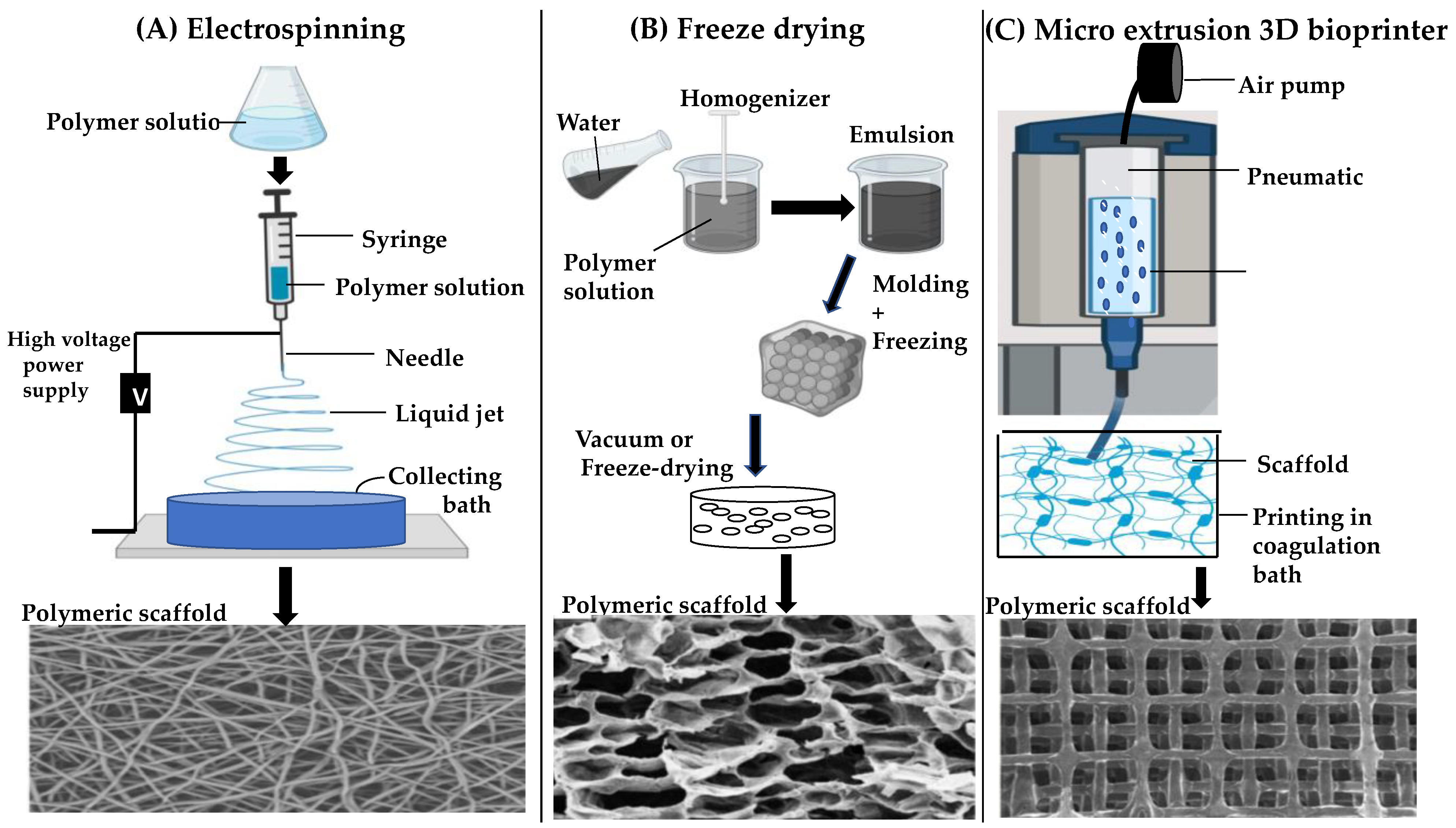
Various techniques have been developed for manufacturing 3D scaffolds, including electrospinning, freeze-drying and 3Dbioprinting [104]. The electrospinning technique has the advantage of providing an environment with fibre diameters down to the nanometre scale to facilitate cell attachment. Electrospinning requires a syringe pump, electrostatic force, and metallic collector to generate fibres [105]. As shown in Figure 3, a very high voltage is applied simultaneously to a metallic needle filled with polymeric solution that is placed at the tip of the syringe via surface tension. The electric field and the potential difference between terminals result in the repulsion of electric charges, followed by drawing out of polymers and deposition onto a collector [105]. A variety of synthetic and natural 3D nanofibrous polymers, such as a biomimetic nanofibrous scaffold comprising type I collagen with 1,1,1,3,3,3-hexafluoro-2-propanol, have been fabricated using the electrospinning method [106]. Despite its advantages, electrospinning still has some major limitations such as inadequate mechanical properties, use of toxic solvents, poor control over cell density, insufficient cell infiltration and non-homogenous cell distribution [107].
The freeze-drying (lyophilization) method is another commonly used technique in regenerative medicine for creating water-soluble polymer scaffolds through the process of sublimation [110]. The process of freeze-drying consists of three main stages: (1) pre-freezing, which involves forming an interpenetrating network of ice crystals by freezing a suspension of the water-soluble polymer at extremely low temperatures in the range of −70 °C to −80 °C, (2) primary drying, during which the bulk of ice crystals is removed under low pressure by sublimation (3) secondary drying, which involves the extraction of unfreezable, bound and associated water via a desorption process, leading to the formation of a highly porous scaffold (Figure 3) [111]. Freeze-drying is a convenient and simple technique to fabricate porous cell scaffolds, however, construction of a suitable scaffold shape with controllable microarchitecture from natural biomaterials remains a significant challenge with this method. In addition, the high rate of rehydration during freeze-drying process could lead to changes in cell diameter due to swelling of the cell wall, resulting in dramatic structural changes [112].
More recently, 3D bioprinting has shown great potential in tissue engineering for scaffold fabrication, by integrating living cells (bioink) and biomaterials directly or indirectly together using layer-by-layer approach [113]. To date, 3D bioprinted constructs have mainly been classified into two forms: acellular and cellular constructs. In acellular bioprinting, scaffold fabrication occurs without cellular material, which is only incorporated after the printing process is complete. In contrast, cellular bioprinting involves incorporating cells and other biological agents within the biomaterial during the fabrication process [114]. The bioink is a critical aspect of 3D bioprinting, as it must be highly biocompatible and bioprintable [108]. Three distinct approaches have been used for fabricating artificial Bruch’s membrane in TE using various 3D bioprinting methods: extrusion-based (EBB), droplet-based and laser-assisted bioprinting [111]. Compared to other techniques, EBB benefits from greater versatility, less process-induced cell damage, and the ability to precisely deposit very high cell densities through computer-aided design (CAD), making it ideal for printing more geometrically complex scaffolds with ease of customisation [115].

5. Conclusions and Future Perspective

The human retina is a highly complex tissue that serves as a key component of both the eye and central nervous system. Any damage to retinal cells including photoreceptor cells, can lead to permanent vision loss because the retina lacks an endogenous stem cell population to replace degenerated and/or damaged cells. AMD is a retinal degenerative disease that affects around 20 million people in the US alone, and it is projected that approximately 300 million worldwide will have the disease by 2040. Although treatment options exist for the neovascular form of AMD to prevent and halt progression of the disease, opportunities for restoring vision are limited once photoreceptor cells become dysfunctional. Advanced therapies represent an emerging field of research which seek to develop cell- and gene-based therapies to treat and potentially cure degenerative diseases such as AMD. Different products for both cell- and gene-based approaches are currently in clinical trials for treatment of both AMD and GA, however, no product has so far reached the market and received FDA approval. This must be due to challenges in selecting the appropriate gene and/or cell types, designing suitable delivery vehicles and ensuring ocular tolerability. Natural polymers also have their own limitations in terms of processability and mechanical properties. Changes in the constituents of natural polymers can lead to accumulation of debris in Bruch’s membrane and eventual cell death.
Recently, scaffold-based approaches for treating retinal degenerative diseases have provided a promising avenue to halt or prevent disease progression in AMD. Nevertheless, different biomaterials and methods of scaffold fabrication can have a significant impact on product success. The freeze-drying method is a primary step that is simple and convenient for making porous structures but has limitations such as (i) poor reproducibility, (ii) poor mechanical properties and (iii) less controllable microstructures and properties. Electrospinning technology has the ability to control fibre diameter and provide a high surface area for cell attachment. However, this method requires the use of toxic organic solvents during processing which can have an adverse effect on cell viability. 3D-bioprinting represents a more attractive scaffold fabrication method with potential advantages of superior physical characteristics, cell adhesion, better control of scaffold microstructures and overall improved mechanical properties, compared to freeze drying.
Multi-objective optimisations are, however, required to identify suitable printing conditions for 3D bioprinting (e.g.: print speed, pressure, layering and spacing) and material composition (percentage of cells to bioink) to achieve optimal mechanical and porous constructions properties. This requires extensive experimentation time that is resource-demanding. Artificial intelligence (AI) and machine learning (ML) methods have already been applied to solve problems in scientific research and represent potentially transformative resources to support researchers in the field of regenerative medicine. In the context of 3D bioprinting, an ML-based supervised learning framework could take the material composition and the printing parameters as input to (i) to enable optimisation of scaffold properties, (ii) assess the quality of the prints and (iii) optimise printability of the material. Given the appropriate training datasets, a machine learning algorithm could generate an accurate prediction model for efficient scaffold fabrication and provide a quantitative evaluation of scaffold printability for specific anatomical sites, such as the human retina. We predict that combining AI with 3D bioprinting for 3D cell-scaffold fabrication will revolutionise retinal tissue engineering and open up a world of new opportunities for developing novel drug delivery platforms for the treatment of retinal diseases.

Author Contributions

Conceptualization, all authors, writing—original draft preparation, D.E.W., H.H.K. and H.K.; writing—review and editing, all authors; supervision, H.K. All authors have read and agreed to the published version of the manuscript.

Funding

This research received no external funding.

Data Availability Statement

Data sharing not applicable.

Conflicts of Interest

The authors declare no conflict of interest.

References

  1. Vyawahare, H.; Shinde, P. Age-related macular degeneration: Epidemiology, pathophysiology, diagnosis, and treatment. Cureus 2022, 14, e29583. [Google Scholar] [CrossRef] [PubMed]
  2. Guo, J.; Huang, X.; Dou, L.; Yan, M.; Shen, T.; Tang, W.; Li, J. Aging and aging-related diseases: From molecular mechanisms to interventions and treatments. Signal Transduct. Target. Ther. 2022, 7, 391. [Google Scholar] [CrossRef]
  3. Stahl, A. The diagnosis and treatment of age-related macular degeneration. Dtsch. Arztebl. Int. 2020, 117, 513–520. [Google Scholar] [CrossRef] [PubMed]
  4. Sutton, J.; Menten, M.J.; Riedl, S.; Bogunović, H.; Leingang, O.; Anders, P.; Hagag, A.M.; Waldstein, S.; Wilson, A.; Cree, A.J.; et al. Developing and validating a multivariable prediction model which predicts progression of intermediate to late age-related macular degeneration-the PINNACLE trial protocol. Eye 2023, 37, 1275–1283. [Google Scholar] [CrossRef] [PubMed]
  5. Bhutto, I.; Lutty, G. Understanding age-related macular degeneration (AMD): Relationships between the photoreceptor/retinal pigment epithelium/Bruch’s membrane/choriocapillaris complex. Mol. Asp. Med. 2012, 33, 295–317. [Google Scholar] [CrossRef]
  6. Galindo-Camacho, R.M.; Blanco-Llamero, C.; da Ana, R.; Fuertes, M.A.; Señoráns, F.J.; Silva, A.M.; García, M.L.; Souto, E.B. Therapeutic approaches for age-related macular degeneration. Int. J. Mol. Sci. 2022, 23, 11769. [Google Scholar] [CrossRef]
  7. Prakoso, A.T.; Basri, H.; Adanta, D.; Yani, I.; Ammarullah, M.I.; Akbar, I.; Ghazali, F.A.; Syahrom, A.; Kamarul, T. The effect of tortuosity on permeability of porous scaffold. Biomedicines 2023, 11, 427. [Google Scholar] [CrossRef]
  8. Datta, S.; Cano, M.; Ebrahimi, K.; Wang, L.; Handa, J.T. The impact of oxidative stress and inflammation on RPE degeneration in non-neovascular AMD. Prog. Retin. Eye Res. 2017, 60, 201–218. [Google Scholar] [CrossRef]
  9. Cho, E.; Hung, S.; Willett, W.C.; Spiegelman, D.; Rimm, E.B.; Seddon, J.M.; Colditz, G.A.; Hankinson, S.E. Prospective study of dietary fat and the risk of age-related macular degeneration. Am. J. Clin. Nutr. 2001, 73, 209–218. [Google Scholar] [CrossRef]
  10. Feigl, B. Age-related maculopathy—Linking aetiology and pathophysiological changes to the ischaemia hypothesis. Prog. Retin. Eye Res. 2009, 28, 63–86. [Google Scholar] [CrossRef]
  11. Dehghan, S.; Mirshahi, R.; Shoae-Hassani, A.; Naseripour, M. Human-induced pluripotent stem cells-derived retinal pigmented epithelium, a new horizon for cells-based therapies for age-related macular degeneration. Stem Cell Res. Ther. 2022, 13, 217. [Google Scholar] [CrossRef] [PubMed]
  12. van Lookeren Campagne, M.; LeCouter, J.; Yaspan, B.L.; Ye, W. Mechanisms of age-related macular degeneration and therapeutic opportunities: Pathology, genetics, animal models, and therapeutic rationale of AMD. J. Pathol. 2014, 232, 151–164. [Google Scholar] [CrossRef] [PubMed]
  13. Ruan, Y.; Jiang, S.; Gericke, A. Age-related macular degeneration: Role of oxidative stress and blood vessels. Int. J. Mol. Sci. 2021, 22, 1296. [Google Scholar] [CrossRef]
  14. Fabre, M.; Mateo, L.; Lamaa, D.; Baillif, S.; Pagès, G.; Demange, L.; Ronco, C.; Benhida, R. Recent advances in age-related macular degeneration therapies. Molecules 2022, 27, 5089. [Google Scholar] [CrossRef] [PubMed]
  15. Tarau, I.-S.; Berlin, A.; Curcio, C.A.; Ach, T. The cytoskeleton of the retinal pigment epithelium: From normal aging to age-related macular degeneration. Int. J. Mol. Sci. 2019, 20, 3578. [Google Scholar] [CrossRef]
  16. Choi, E.H.; Suh, S.; Einstein, D.E.; Leinonen, H.; Dong, Z.; Rao, S.R.; Fliesler, S.J.; Blackshaw, S.; Yu, M.; Peachey, N.S.; et al. An inducible Cre mouse for studying roles of the RPE in retinal physiology and disease. JCI Insight 2021, 6, e146604. [Google Scholar] [CrossRef] [PubMed]
  17. Chirco, K.R.; Sohn, E.H.; Stone, E.M.; Tucker, B.A.; Mullins, R.F. Structural and molecular changes in the aging choroid: Implications for age-related macular degeneration. Eye 2017, 31, 10–25. [Google Scholar] [CrossRef]
  18. Stern, J.H.; Tian, Y.; Funderburgh, J.; Pellegrini, G.; Zhang, K.; Goldberg, J.L.; Ali, R.R.; Young, M.; Xie, Y.; Temple, S. Regenerating eye tissues to preserve and restore vision. Cell Stem Cell 2018, 22, 834–849. [Google Scholar] [CrossRef]
  19. Tan, W.; Zou, J.; Yoshida, S.; Jiang, B.; Zhou, Y. The role of inflammation in age-related macular degeneration. Int. J. Biol. Sci. 2020, 16, 2989–3001. [Google Scholar] [CrossRef]
  20. Armento, A.; Ueffing, M.; Clark, S.J. The complement system in age-related macular degeneration. Cell. Mol. Life Sci. 2021, 78, 4487–4505. [Google Scholar] [CrossRef]
  21. Somasundaran, S.; Constable, I.J.; Mellough, C.B.; Carvalho, L.S. Retinal pigment epithelium and age-related macular degeneration: A review of major disease mechanisms. Clin. Exp. Ophthalmol. 2020, 48, 1043–1056. [Google Scholar] [CrossRef] [PubMed]
  22. Bellezza, I. Oxidative stress in age-related macular degeneration: Nrf2 as therapeutic target. Front. Pharmacol. 2018, 9, 1280. [Google Scholar] [CrossRef] [PubMed]
  23. Domènech, E.B.; Marfany, G. The relevance of oxidative stress in the pathogenesis and therapy of retinal dystrophies. Antioxidants 2020, 9, 347. [Google Scholar] [CrossRef] [PubMed]
  24. Rohowetz, L.J.; Kraus, J.G.; Koulen, P. Reactive oxygen species-mediated damage of retinal neurons: Drug development targets for therapies of chronic neurodegeneration of the retina. Int. J. Mol. Sci. 2018, 19, 3362. [Google Scholar] [CrossRef]
  25. Ruan, Y.; Jiang, S.; Musayeva, A.; Gericke, A. Oxidative stress and vascular dysfunction in the retina: Therapeutic strategies. Antioxidants 2020, 9, 761. [Google Scholar] [CrossRef]
  26. Cho, Y.-K.; Park, D.-H.; Jeon, I.-C. Medication trends for age-related macular degeneration. Int. J. Mol. Sci. 2021, 22, 11837. [Google Scholar] [CrossRef]
  27. Hanneken, A.; Lin, F.-F.; Johnson, J.; Maher, P. Flavonoids protect human retinal pigment epithelial cells from oxidative-stress–induced death. Investig. Ophthalmol. Vis. Sci. 2006, 47, 3164. [Google Scholar] [CrossRef]
  28. Csader, S.; Korhonen, S.; Kaarniranta, K.; Schwab, U. The effect of dietary supplementations on delaying the progression of age-related macular degeneration: A systematic review and meta-analysis. Nutrients 2022, 14, 4273. [Google Scholar] [CrossRef]
  29. Eisenhauer, B.; Natoli, S.; Liew, G.; Flood, V.M. Lutein and zeaxanthin-food sources, bioavailability and dietary variety in age-related macular degeneration protection. Nutrients 2017, 9, 120. [Google Scholar] [CrossRef]
  30. Bianchetti, G.; Clementi, M.E.; Sampaolese, B.; Serantoni, C.; Abeltino, A.; De Spirito, M.; Sasson, S.; Maulucci, G. Investigation of DHA-induced regulation of redox homeostasis in retinal pigment epithelium cells through the combination of metabolic imaging and molecular biology. Antioxidants 2022, 11, 1072. [Google Scholar] [CrossRef]
  31. Bianchetti, G.; Clementi, M.E.; Sampaolese, B.; Serantoni, C.; Abeltino, A.; De Spirito, M.; Sasson, S.; Maulucci, G. Metabolic imaging and molecular biology reveal the interplay between lipid metabolism and DHA-induced modulation of redox homeostasis in RPE cells. Antioxidants 2023, 12, 339. [Google Scholar] [CrossRef] [PubMed]
  32. Group A-REDSR. Lutein + zeaxanthin and omega-3 fatty acids for age-related macular degeneration: The Age-Related Eye Disease Study 2 (AREDS2) randomized clinical trial. JAMA 2013, 309, 2005–2015. [Google Scholar] [CrossRef] [PubMed]
  33. Van Asten, C.Y.; Chiu, E.; Agrón, T.E.; Clemons, R.; Ratnapriya, A.; Swaroop, M.L.; Klein, M.L.; Fan, R.; Chew, E.Y.; Age-Related Eye Disease Study 2 Research Group. No CFH or ARMS2 interaction with omega-3 fatty acids, low versus high zinc, or β-carotene versus lutein and zeaxanthin on progression of age-related macular degeneration in the age-related eye disease study 2: Age. Ophthalmology 2019, 126, 1541–1548. [Google Scholar] [CrossRef] [PubMed]
  34. Tanito, M.; Li, F.; Anderson, R.E. Protection of retinal pigment epithelium by OT-551 and its metabolite TEMPOL-H against light-induced damage in rats. Exp. Eye Res. 2010, 91, 111–114. [Google Scholar] [CrossRef]
  35. Clementi, M.E.; Pizzoferrato, M.; Bianchetti, G.; Brancato, A.; Sampaolese, B.; Maulucci, G.; Tringali, G. Cytoprotective effect of idebenone through modulation of the intrinsic mitochondrial pathway of apoptosis in human retinal pigment epithelial cells exposed to oxidative stress induced by hydrogen peroxide. Biomedicines 2022, 10, 503. [Google Scholar] [CrossRef]
  36. Chen, E.; Kaiser, P. Therapeutic Potential of the Ranibizumab Port Delivery System in the Treatment of AMD: Evidence to Date. Clin. Ophthalmol. 2020, 14, 1349–1355. [Google Scholar] [CrossRef]
  37. Xu, M.; Fan, R.; Fan, X.; Shao, Y.; Li, X. Progress and challenges of anti-VEGF agents and their sustained-release strategies for retinal angiogenesis. Drug Des. Dev. Ther. 2022, 16, 3241–3262. [Google Scholar] [CrossRef]
  38. Rubido, M.; Sadikhov, S.; Szczesny, P.; Schwab, D.; Nogoceke, E.; Weikert, R. Safety and Efficacy of Different Doses and Regimens of Faricimab vs Ranibizumab in Neovascular Age-Related macular degeneration: The AVENUE phase 2 randomized clinical trial. JAMA Ophthalmol. 2020, 138, 955–963. [Google Scholar]
  39. Wu, A.; Lu, R.; Lee, E. Tissue engineering in age-related macular degeneration: A mini-review. J. Biol. Eng. 2022, 16, 11. [Google Scholar] [CrossRef]
  40. Cabral de Guimaraes, T.A.; Daich Varela, M.; Georgiou, M.; Michaelides, M. Treatments for dry age-related macular degeneration: Therapeutic avenues, clinical trials and future directions. Br. J. Ophthalmol. 2022, 106, 297–304. [Google Scholar] [CrossRef]
  41. Lin, J.B.; Murakami, Y.; Miller, J.W.; Vavvas, D.G. Neuroprotection for age-related macular degeneration. Ophthalmol. Sci. 2022, 2, 100192. [Google Scholar] [CrossRef] [PubMed]
  42. Rosenfeld, P.J.; Dugel, P.U.; Holz, F.G.; Heier, J.S.; Pearlman, J.A.; Novack, R.L.; Csaky, K.G.; Koester, J.M.; Gregory, J.K.; Kubota, R. Emixustat hydrochloride for geographic atrophy secondary to age-related macular degeneration. Ophthalmology 2018, 125, 1556–1567. [Google Scholar] [CrossRef] [PubMed]
  43. Racz, B.; Varadi, A.; Kong, J.; Allikmets, R.; Pearson, P.G.; Johnson, G.; Cioffi, C.L.; Petrukhin, K. A non-retinoid antagonist of retinol-binding protein 4 rescues phenotype in a model of Stargardt disease without inhibiting the visual cycle. J. Biol. Chem. 2018, 293, 11574–11588. [Google Scholar] [CrossRef] [PubMed]
  44. Saad, L.; Washington, I. Can vitamin A be improved to prevent blindness due to age-related macular degeneration, stargardt disease and other retinal dystrophies? In Retinal Degenerative Diseases; Springer International Publishing: Cham, Switzerland, 2016. [Google Scholar]
  45. Zelek, W.M.; Morgan, B.P. Monoclonal antibodies capable of inhibiting complement downstream of C5 in multiple species. Front. Immunol. 2020, 11, 612402. [Google Scholar] [CrossRef] [PubMed]
  46. Holz, F.G.; Sadda, S.R.; Busbee, B.; Chew, E.Y.; Mitchell, P.; Tufail, A.; Brittain, C.; Ferrara, D.; Gray, S.; Honigberg, L.; et al. Efficacy and safety of lampalizumab for geographic atrophy due to age-related macular degeneration: Chroma and spectri phase 3 randomized clinical trials. JAMA Ophthalmol. 2018, 136, 666. [Google Scholar] [CrossRef]
  47. Jaffe, G.J.; Westby, K.; Csaky, K.G.; Monés, J.; Pearlman, J.A.; Patel, S.S.; Joondeph, B.C.; Randolph, J.; Masonson, H.; Rezaei, K.A. C5 inhibitor avacincaptad pegol for geographic atrophy due to age-related macular degeneration: A randomized pivotal phase 2/3 trial. Ophthalmology 2021, 128, 576–586. [Google Scholar] [CrossRef]
  48. Liao, D.S.; Grossi, F.V.; El Mehdi, D.; Gerber, M.R.; Brown, D.M.; Heier, J.S.; Wykoff, C.C.; Singerman, L.J.; Abraham, P.; Grassmann, F.; et al. Complement C3 inhibitor pegcetacoplan for geographic atrophy secondary to age-related macular degeneration: A randomized phase 2 trial. Ophthalmology 2020, 127, 186–195. [Google Scholar] [CrossRef]
  49. Halawa, O.A.; Lin, J.B.; Miller, J.W.; Vavvas, D.G. A review of completed and ongoing complement inhibitor trials for geographic atrophy secondary to age-related macular degeneration. J. Clin. Med. 2021, 10, 2580. [Google Scholar] [CrossRef]
  50. Shaw, L.T.; Mackin, A.; Shah, R.; Jain, S.; Jain, P.; Nayak, R.; Hariprasad, S.M. Risuteganib-a novel integrin inhibitor for the treatment of non-exudative (dry) age-related macular degeneration and diabetic macular edema. Expert Opin. Investig. Drugs 2020, 29, 547–554. [Google Scholar] [CrossRef]
  51. Nhu, N.T.; Xiao, S.-Y.; Liu, Y.; Kumar, V.B.; Cui, Z.-Y.; Lee, S.-D. Neuroprotective Effects of a Small Mitochondrially-Targeted Tetrapeptide Elamipretide in Neurodegeneration. Front. Integr. Neurosci. 2022, 15, 747901. [Google Scholar] [CrossRef]
  52. Rosenfeld, P.J.; Berger, B.; Reichel, E. A randomized phase 2 study of an anti- amyloid β monoclonal antibody in geographic atrophy secondary to age-related macular degeneration. Ophthalmol. Retin. 2018, 2, 1028–1040. [Google Scholar] [CrossRef] [PubMed]
  53. Hanus, J.; Zhao, F.; Wang, S. Current therapeutic developments in atrophic age-related macular degeneration. Br. J. Ophthalmol. 2016, 100, 122–127. [Google Scholar] [CrossRef] [PubMed]
  54. Li, S.; Sato, K.; Gordon, W.C.; Sendtner, M.; Bazan, N.G.; Jin, M. Ciliary neurotrophic factor (CNTF) protects retinal cone and rod photoreceptors by suppressing excessive formation of the visual pigments. J. Biol. Chem. 2018, 293, 15256–15268. [Google Scholar] [CrossRef] [PubMed]
  55. Tamhane, M.; Luu, K.T.; Attar, M. Ocular pharmacokinetics of brimonidine drug delivery system in monkeys and translational modeling for selection of dose and frequency in clinical trials. J. Pharmacol. Exp. Ther. 2021, 378, 207–214. [Google Scholar] [CrossRef]
  56. López-Paniagua, M.; de la Mata, A.; Galindo, S.; Blázquez, F.; Calonge, M.; Nieto-Miguel, T. Advanced therapy medicinal products for the eye: Definitions and regulatory framework. Pharmaceutics 2021, 13, 347. [Google Scholar] [CrossRef] [PubMed]
  57. Rubner, R.; Li, K.V.; Canto-Soler, M.V. Progress of clinical therapies for dry age-related macular degeneration. Int. J. Ophthalmol. 2022, 15, 157–166. [Google Scholar] [CrossRef]
  58. Rizzolo, L.J.; Nasonkin, I.O.; Adelman, R.A. Retinal cell transplantation, biomaterials, and in vitro models for developing next-generation therapies of age-related macular degeneration. Stem Cells Transl. Med. 2022, 11, 269–281. [Google Scholar] [CrossRef]
  59. Jemni-Damer, N.; Guedan-Duran, A.; Fuentes-Andion, M.; Serrano-Bengoechea, N.; Alfageme-Lopez, N.; Armada-Maresca, F.; Guinea, G.V.; Perez-Rigueiro, J.; Rojo, F.; Gonzalez-Nieto, D.; et al. Biotechnology and biomaterial-based therapeutic strategies for age-related Macular Degeneration. Part II: Cell and tissue engineering therapies. Front. Bioeng. Biotechnol. 2020, 8, 588014. [Google Scholar] [CrossRef]
  60. Wang, Z.; Gao, F.; Zhang, M.; Zheng, Y.; Zhang, F.; Xu, L.; Cao, L.; He, W. Intravitreal injection of human retinal progenitor cells for treatment of retinal degeneration. Med. Sci. Monit. 2020, 26, e921184. [Google Scholar] [CrossRef]
  61. Nair, D.S.R.; Seiler, M.J.; Patel, K.H.; Thomas, V.; Camarillo, J.C.M.; Humayun, M.S.; Thomas, B.B. Tissue engineering strategies for retina regeneration. Appl. Sci. 2021, 11, 2154. [Google Scholar] [CrossRef]
  62. Kashani, A.H.; Lebkowski, J.S.; Rahhal, F.M.; Avery, R.L.; Salehi-Had, H.; Dang, W.; Lin, C.-M.; Mitra, D.; Zhu, D.; Thomas, B.B.; et al. A bioengineered retinal pigment epithelial monolayer for advanced, dry age-related macular degeneration. Sci. Transl. Med. 2018, 10, eaao4097. [Google Scholar] [CrossRef] [PubMed]
  63. Fernandes, R.A.B.; Stefanini, F.R.; Falabella, P.; Koss, M.J.; Wells, T.; Diniz, B.; Ribeiro, R.; Schor, P.; Maia, M.; Penha, F.M.; et al. Development of a new tissue injector for subretinal transplantation of human embryonic stem cell derived retinal pigmented epithelium. Int. J. Retin. Vitr. 2017, 3, 41. [Google Scholar] [CrossRef] [PubMed]
  64. Gullapalli, V.K.; Zarbin, M.A. New prospects for retinal pigment epithelium transplantation. Asia Pac. J. Ophthalmol. 2022, 11, 302–313. [Google Scholar] [CrossRef] [PubMed]
  65. Safety and Efficacy Study of OpRegen for Treatment of Advanced Dry-Form Age-Related Macular Degeneration. Available online: https://clinicaltrials.gov/ct2/show/NCT02286089 (accessed on 24 March 2023).
  66. Autologous Transplantation of Induced Pluripotent Stem Cell-Derived Retinal Pigment Epithelium for Geographic Atrophy Associated with Age-Related Macular Degeneration. Available online: https://clinicaltrials.gov/ct2/show/NCT04339764 (accessed on 24 March 2023).
  67. Hinkle, J.W.; Mahmoudzadeh, R.; Kuriyan, A.E. Cell-based therapies for retinal diseases: A review of clinical trials and direct to consumer “cell therapy” clinics. Stem Cell Res. Ther. 2021, 12, 538. [Google Scholar] [CrossRef]
  68. Ru, L.; Wu, N.; Wei, K.; Zeng, Y.; Li, Q.; Weng, C.; Ren, C.; Ren, B.; Huo, D.; Li, Y.; et al. Improving cell survival and engraftment in vivo via layer-by-layer nanocoating of hESC-derived RPE cells. Stem Cell Res. Ther. 2020, 11, 495. [Google Scholar] [CrossRef]
  69. Arabi, A.; Shahraki, T. Novel treatments and genetics of age-related macular degeneration-a narrative review. Ann. Eye Sci. 2021, 6, 38. [Google Scholar] [CrossRef]
  70. Guimaraes, T.A.C.; de Georgiou, M.; Bainbridge, J.W.B.; Michaelides, M. Gene therapy for neovascular age-related macular degeneration: Rationale, clinical trials and future directions. Br. J. Ophthalmol. 2021, 105, 151–157. [Google Scholar] [CrossRef]
  71. Hill, A.B.; Chen, M.; Chen, C.-K.; Pfeifer, B.A.; Jones, C.H. Overcoming gene-delivery hurdles: Physiological considerations for nonviral vectors. Trends Biotechnol. 2016, 34, 91–105. [Google Scholar] [CrossRef]
  72. Crane, R.; Conley, S.M.; Al-Ubaidi, M.R.; Naash, M.I. Gene therapy to the retina and the cochlea. Front. Neurosci. 2021, 15, 652215. [Google Scholar] [CrossRef]
  73. Muhuri, M.; Levy, D.I.; Schulz, M.; McCarty, D.; Gao, G. Durability of transgene expression after rAAV gene therapy. Mol. Ther. 2022, 30, 1364–1380. [Google Scholar] [CrossRef]
  74. SparingVision’s Lead Product, SPVN06, Is a Breakthrough Gene Therapy Approach Targeting Inherited Retinal Diseases; SpringVision: Paris, France, 2022.
  75. AAVCAGsCD59 for the Treatment of Wet AMD. Available online: https://clinicaltrials.gov/ct2/show/NCT03585556 (accessed on 24 March 2023).
  76. FOCUS: First in Human Study to Evaluate the Safety and Efficacy of GT005 Administered in Subjects with Dry AMD. Available online: https://clinicaltrials.gov/ct2/show/NCT03846193 (accessed on 24 March 2023).
  77. Becker, J.; Fakhiri, J.; Grimm, D. Fantastic AAV gene therapy vectors and how to find them-random diversification, rational design and machine learning. Pathogens 2022, 11, 756. [Google Scholar] [CrossRef] [PubMed]
  78. Bulcha, J.T.; Wang, Y.; Ma, H.; Tai, P.W.L.; Gao, G. Viral vector platforms within the gene therapy landscape. Signal Transduct. Target. Ther. 2021, 6, 1–24. [Google Scholar] [CrossRef]
  79. Safety and Tolerability of RPE Stem Cell-Derived RPE(RPESC-RPE) Transplantation in Patients with Dry Age-Related Macular Degeneration (AMD). Available online: https://clinicaltrials.gov/ct2/show/NCT04627428 (accessed on 24 March 2023).
  80. Safety of Cultured Allogeneic Adult Umbilical Cord Derived Mesenchymal Stem Cells for Eye Diseases. Available online: https://clinicaltrials.gov/ct2/show/NCT05147701 (accessed on 24 March 2023).
  81. Regenerative Patch Technologies LLC. Study of Subretinal Implantation of Human Embryonic Stem Cell-Derived RPE Cells in Advanced Dry AMD. ClinicalTrials.gov Identifier: NCT02590692 Updated. 2020, 29. Available online: https://clinicaltrials.gov/ct2/show/NCT02590692 (accessed on 24 March 2023).
  82. A Study of Implantation of Retinal Pigment Epithelium in Subjects with Acute Wet Age Related Macular Degeneration. Available online: https://clinicaltrials.gov/ct2/show/NCT01691261 (accessed on 24 March 2023).
  83. Safety and Efficacy of ADVM-022 in Treatment-Experienced Patients with Neovascular Age-Related Macular Degeneration [LUNA]. Available online: https://clinicaltrials.gov/ct2/show/NCT05536973 (accessed on 24 March 2023).
  84. Gene Therapy for Wet AMD. Available online: https://clinicaltrials.gov/ct2/show/NCT05611424 (accessed on 24 March 2023).
  85. 4D-150 in Patients With Neovascular (Wet) Age-Related Macular Degeneration. Available online: https://clinicaltrials.gov/ct2/show/NCT05197270 (accessed on 24 March 2023).
  86. VEGFA-Targeting Gene Therapy to Treat Retinal and Choroidal Neovascularization Diseases. Available online: https://clinicaltrials.gov/ct2/show/NCT05099094 (accessed on 24 March 2023).
  87. RGX-314 Gene Therapy Pharmacodynamic Study for Neovascular Age-related Macular Degeneration (nAMD). Available online: https://clinicaltrials.gov/ct2/show/NCT04832724 (accessed on 24 March 2023).
  88. Maddalena, A.; Tornabene, P.; Tiberi, P.; Minopoli, R.; Manfredi, A.; Mutarelli, M.; Rossi, S.; Simonelli, F.; Naggert, J.K.; Cacchiarelli, D.; et al. Triple vectors expand AAV transfer capacity in the retina. Mol. Ther. 2018, 26, 524–541. [Google Scholar] [CrossRef] [PubMed]
  89. Ikada, Y. Challenges in tissue engineering. J. R. Soc. Interface 2006, 3, 589–601. [Google Scholar] [CrossRef] [PubMed]
  90. Abbott, R.D.; Kaplan, D.L. Engineering biomaterials for enhanced tissue regeneration. Curr. Stem Cell Rep. 2016, 2, 140–146. [Google Scholar] [CrossRef]
  91. Nikolova, M.P.; Chavali, M.S. Recent advances in biomaterials for 3D scaffolds: A review. Bioact. Mater. 2019, 4, 271–292. [Google Scholar] [CrossRef]
  92. Bitar, K.N.; Zakhem, E. Design strategies of biodegradable scaffolds for tissue regeneration. Biomed. Eng. Comput. Biol. 2014, 6, BECB.S10961. [Google Scholar] [CrossRef]
  93. Wintermantel, E.; Mayer, J.; Blum, J.; Eckert, K.L.; Lüscher, P.; Mathey, M. Tissue engineering scaffolds using superstructures. Biomaterials 1996, 17, 83–91. [Google Scholar] [CrossRef]
  94. Mikos, A.G.; McIntire, L.V.; Anderson, J.M.; Babensee, J.E. Host response to tissue engineered devices. Adv. Drug Deliv. Rev. 1998, 33, 111–139. [Google Scholar] [CrossRef]
  95. Brown, B.N.; Valentin, J.E.; Stewart-Akers, A.M.; McCabe, G.P.; Badylak, S.F. Macrophage phenotype and remodeling outcomes in response to biologic scaffolds with and without a cellular component. Biomaterials 2009, 30, 1482–1491. [Google Scholar] [CrossRef]
  96. Bonferoni, M.C.; Caramella, C.; Catenacci, L.; Conti, B.; Dorati, R.; Ferrari, F.; Genta, I.; Modena, T.; Perteghella, S.; Rossi, S.; et al. Biomaterials for soft tissue repair and regeneration: A focus on Italian research in the field. Pharmaceutics 2021, 13, 1341. [Google Scholar] [CrossRef]
  97. Mastrullo, V.; Cathery, W.; Velliou, E.; Madeddu, P.; Campagnolo, P. Angiogenesis in Tissue Engineering: As Nature Intended? Front. Bioeng. Biotechnol. 2020, 8, 188. [Google Scholar] [CrossRef] [PubMed]
  98. White, C.E.; Olabisi, R.M. Scaffolds for retinal pigment epithelial cell transplantation in age-related macular degeneration. J. Tissue Eng. 2017, 8, 2041731417720841. [Google Scholar] [CrossRef] [PubMed]
  99. Bružauskaitė, I.; Bironaitė, D.; Bagdonas, E.; Bernotienė, E. Scaffolds and cells for tissue regeneration: Different scaffold pore sizes-different cell effects. Cytotechnology 2016, 68, 355–369. [Google Scholar] [CrossRef] [PubMed]
  100. Goldenberg, D.; McLaughlin, C.; Koduru, S.V.; Ravnic, D.J. Regenerative engineering: Current applications and future perspectives. Front. Surg. 2021, 8, 731031. [Google Scholar] [CrossRef]
  101. Reddy, M.S.B.; Ponnamma, D.; Choudhary, R.; Sadasivuni, K.K. A comparative review of natural and synthetic biopolymer composite scaffolds. Polymers 2021, 13, 1105. [Google Scholar] [CrossRef]
  102. Lin, K.T.; Wang, A.; Nguyen, A.B.; Iyer, J.; Tran, S.D. Recent advances in hydrogels: Ophthalmic applications in cell delivery, vitreous substitutes, and ocular adhesives. Biomedicines 2021, 9, 1203. [Google Scholar] [CrossRef]
  103. Parisi, L.; Toffoli, A.; Ghiacci, G.; Macaluso, G.M. Tailoring the interface of biomaterials to design effective scaffolds. J. Funct. Biomater. 2018, 9, 50. [Google Scholar] [CrossRef]
  104. Krishani, M.; Shin, W.Y.; Suhaimi, H.; Sambudi, N.S. Development of scaffolds from bio-based natural materials for tissue regeneration applications: A review. Gels 2023, 9, 100. [Google Scholar] [CrossRef]
  105. Xue, J.; Wu, T.; Dai, Y.; Xia, Y. Electrospinning and electrospun nanofibers: Methods, materials, and applications. Chem. Rev. 2019, 119, 5298–5415. [Google Scholar] [CrossRef]
  106. Hong, J.; Yeo, M.; Yang, G.H.; Kim, G. Cell-electrospinning and its application for tissue engineering. Int. J. Mol. Sci. 2019, 20, 6208. [Google Scholar] [CrossRef] [PubMed]
  107. Beachley, V.; Wen, X. Polymer nanofibrous structures: Fabrication, biofunctionalization, and cell interactions. Prog. Polym. Sci. 2010, 35, 868–892. [Google Scholar] [CrossRef] [PubMed]
  108. Zhang, L.; Leong, K.; Fisher, J. 3D Bioprinting and Nanotechnology in Tissue Engineering and Regenerative Medicine; Academic Press: Cambridge, MA, USA, 2022. [Google Scholar]
  109. Wang, Z.; Wang, Y.; Yan, J.; Zhang, K.; Lin, F.; Xiang, L.; Deng, L.; Guan, Z.; Cui, W.; Zhang, H. Pharmaceutical electrospinning and 3D printing scaffold design for bone regeneration. Adv. Drug Deliv. Rev. 2021, 174, 504–534. [Google Scholar] [CrossRef] [PubMed]
  110. Collins, M.N.; Ren, G.; Young, K.; Pina, S.; Reis, R.L.; Oliveira, J.M. Scaffold fabrication technologies and structure/function properties in bone tissue engineering. Adv. Funct. Mater. 2021, 31, 2010609. [Google Scholar] [CrossRef]
  111. Garg, T.; Singh, O.; Arora, S.; Murthy, R. Scaffold: A novel carrier for cell and drug delivery. Crit. Rev. Ther. Drug Carr. Syst. 2012, 29, 1–63. [Google Scholar] [CrossRef]
  112. Capuana, E.; Lopresti, F.; Carfì Pavia, F.; Brucato, V.; La Carrubba, V. Solution-based processing for scaffold fabrication in tissue engineering applications: A brief review. Polymers 2021, 13, 2041. [Google Scholar] [CrossRef]
  113. Yang, P.; Ju, Y.; Hu, Y.; Xie, X.; Fang, B.; Lei, L. Emerging 3D bioprinting applications in plastic surgery. Biomater. Res. 2023, 27, 1. [Google Scholar] [CrossRef]
  114. Yazdanpanah, Z.; Johnston, J.D.; Cooper, D.M.L.; Chen, X. 3D bioprinted scaffolds for bone tissue engineering: State-of-the-art and emerging technologies. Front. Bioeng. Biotechnol. 2022, 10, 824156. [Google Scholar] [CrossRef]
  115. Gu, Z.; Fu, J.; Lin, H.; He, Y. Development of 3D bioprinting: From printing methods to biomedical applications. Asian J. Pharm. Sci. 2020, 15, 529–557. [Google Scholar] [CrossRef]
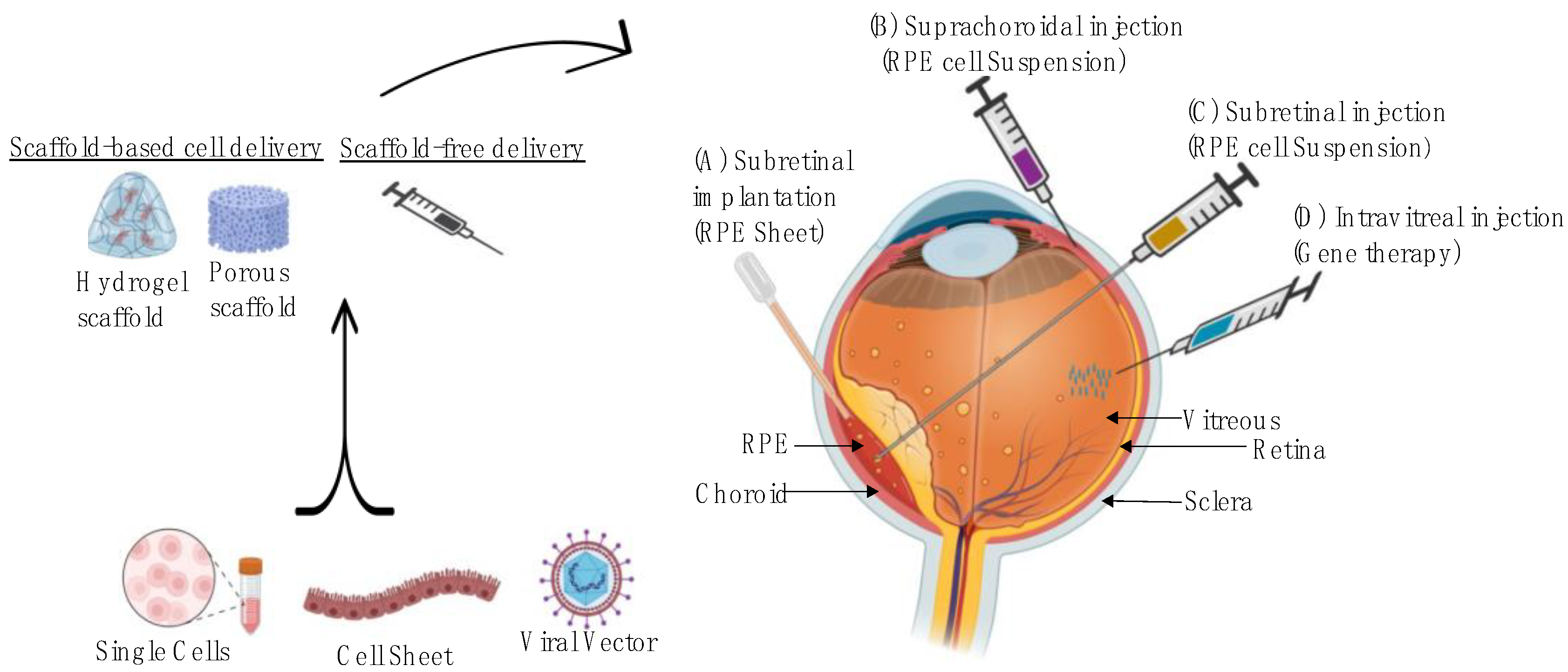
Figure 1. Overview of scaffold fabrication and the delivery methods of cells-based and gene-based approaches for the retinal degeneration disease. (A) Subretinal implantations, performed in the operating room, allows for direct delivery of RPE sheet as a cell suspension into the subretinal space, (B) Suprachoroidal injection is a nonsurgical method to deliver therapeutic suspension into a virtual space between the sclera and choroid, (C) Subretinal injection delivers RPE cell suspension under the sensory retina using a micro-needle and (D) Intravitreal injection method is applied in an office setting to deliver vectors to the vitreous cavity.
Pharmaceuticals 16 00620 g001
Figure 2. Diagram showing the structure of the retina, the RPE and choroid in the dry and wet forms of AMD. Late AMD is characterised by drusen formation, neovascularisation, disruption of Bruch’s membrane as well as RPE cell damage and death [14].
Pharmaceuticals 16 00620 g002
Figure 3. Schematic illustration of different techniques for manufacturing 3D tissue scaffold and optical, SEM images of the printing constructs [108,109,110].
Pharmaceuticals 16 00620 g003
Table 1. List of potential treatment options in clinical trials for dry-AMD, focusing on disease prevention and halting disease progression.
Disease PreventionProduct CategoryProduct NameStudy PhaseMechanism of ActionMethod of Delivery
Antioxidant AREDS
[28]
Phase IIIvitamin supplement Oral
OT-551
[40,41]
Phase IIvitamin supplementEye drop
Visual cycle modulators
(Prevent drusen formation)
ACU-4429
[42]
Phase II/IIIInhibits the formation of 11-cis-retinal to slow the rate of retinoid metabolism and A2E generationOral
Fenretinide [43]Phase IISynthetic retinoid (vitamin A); reduce accumulation of lipofuscin through binding to its carrier proteinOral
C20-D3-vitamin A (ALK-001)
[44]
Phase IIIA modified form of Vitamin A to decrease toxic by-product formation through reducing A2E biosynthesis Oral
Halting Disease ProgressionAnti-inflammatory drugs
(anti-complement pathways)
Eculizumab
[45]
Phase IIIA monoclonal antibody to inhibit the complement protein C5, preventing MAC formationIV
Lampalizumab
[46]
Phase IIIA monoclonal antibody to inhibit complement factor DIntravitreal
Avacincaptad pegol (Zimura)
[47]
Phase II/IIIAnti-complement factor 5, preventing MAC formationIntravitreal
Pegcetacoplan (APL-2)
[48]
Phase IIComplement C3 inhibitor and prevents downstream activation of C3b Intravitreal
LFG316
[49]
Phase IIA monoclonal antibody to inhibit the complement protein C5Intravitreal
Oxidative stressRisuteganib
[50]
Phase IIAn integrin inhibitor of αVβ3/αVβ5 and α5β1 to target multiple oxidative stress factors Intravitreal
Mitochondrial enhancerElamipretide
[51]
Phase IIIA small mitochondrially targeted tetrapeptide to reduce the production of toxic ROS and stabilize cardiolipin levelsSubcutaneous
β-amyloid inhibitorsGSK933776
[52]
Phase IIAn anti-amyloid β monoclonal antibody IV
RN6G
[53]
Phase IIA humanized antibody to inhibit accumulation of amyloid β-40 and β-42IV
NeuroprotectionCiliary nerve trophic factor
[54]
Phase IIProtects rod photoreceptors and retinal cones by improving morphology of photoreceptor mitochondria and reduingoxygen consumptionIntravitreal
Brimonidine tartrate
[55]
Phase IIAn alpha2-adrenergic receptor agonistIntravitreal
Table 3. Desirable characteristics of a tissue-engineered scaffold.
Scaffold PropertyBiological SignificanceRefs
Biocompatibility
(1)
Provide a normally functioning matrix to enable cell adhesion, proliferation and ECM generation
(2)
Maintain cellular viability within scaffold and at the site of implantation
(3)
Following implantation, TE construct must not give rise to adverse immunological response (e.g., inflammation, fibrotic scarring, immune rejection, impaired healing)
[93,94]
Biodegradability
(1)
Regeneration of fully functional tissue should coincide in time with the complete degradation and resorption of the scaffold
(2)
The rate of degradation should kinetically match with the changing microenvironment during the regeneration process
(3)
Biodegradation can have a critical role in the provision of pathways for angiogenesis and metabolite diffusion, as well as release of therapeutic agents loaded into the scaffold
[95,96,97]
Mechanical properties
(1)
Scaffold must fulfil key mechanical functions of tissue that is being replaced
(2)
Mechanical properties should ideally be consistent with implantation site to enable mechanotransduction and minimise native tissue damage
(3)
Scaffold should have sufficient mechanical integrity to allow surgical handling during implantation (unless delivered by injection)
[96,97]
Scaffold architecture
(1)
Scaffold should mimic the architecture of the native tissue (e.g., Bruch’s membrane) as closely as possible to allow remodelling to take place
(2)
Architecture should display sufficient interconnectivity and porosity to allow the transfer of nutrients/waste products and facilitate adequate cellular penetration and migration
[98,99,100]
Manufacturing technology
(1)
The ideal scaffold needs to be manufacturable to GMP standards
(2)
Amenable to scalable and cost-effective bioprocessing
[100]
Disclaimer/Publisher’s Note: The statements, opinions and data contained in all publications are solely those of the individual author(s) and contributor(s) and not of MDPI and/or the editor(s). MDPI and/or the editor(s) disclaim responsibility for any injury to people or property resulting from any ideas, methods, instructions or products referred to in the content.