Cytotoxic Activity of α-Aminophosphonic Derivatives Coming from the Tandem Kabachnik–Fields Reaction and Acylation
Abstract
1. Introduction
2. Results and Discussion
2.1. Synthesis of α-Aminophosphonate Derivatives
2.2. In Vitro Cytostatic Effect of α-Aminophosphonic Derivatives on Human Tumor Cell Lines
2.3. In Silico Target Assessment of the Major Cytotoxic Hit Compounds
- Furin
- Prostatic Acid Phosphatase
- Tyrosine Phosphatase Enzyme Family
3. Materials and Methods
3.1. Synthesis of α-Aminophosphonate Derivatives
3.2. Cell Lines and Culture Conditions—In Vitro Cytotoxicity Assays of Carcinoma Cell Lines
4. Conclusions
Author Contributions
Funding
Institutional Review Board Statement
Informed Consent Statement
Data Availability Statement
Acknowledgments
Conflicts of Interest
References
- Quin, L.D. A Guide to Organophosphorus Chemistry; Wiley & Sons: New York, NY, USA, 2000; ISBN 978-0-471-31824-8. [Google Scholar]
- Keglevich, G. (Ed.) Organophosphorus Chemistry—Novel Developments; De Gruyter: Berlin, Germany, 2018; ISBN 978-3-11-053453-5. [Google Scholar]
- Abdou, M.M. Synopsis of recent synthetic methods and biological applications of phosphinic acid derivatives. Tetrahedron 2020, 76, 131251. [Google Scholar] [CrossRef]
- Rodriguez, J.B.; Gallo-Rodriguez, C. The role of the phosphorus atom in drug design. ChemMedChem 2019, 14, 190–216. [Google Scholar] [CrossRef]
- Sevrain, C.M.; Berchel, M.; Couthon, H.; Jaffres, P.-A. Phosphonic acid: Preparation and applications. Beilstein J. Org. Chem. 2017, 13, 2186–2213. [Google Scholar] [CrossRef]
- Mucha, A.; Kafarski, P.; Berlicki, L. Remarkable potential of the α-aminophosphonate/phosphinate structural motif in medicinal chemistry. J. Med. Chem. 2011, 54, 5955–5980. [Google Scholar] [CrossRef] [PubMed]
- Kafarski, P.; Lejczak, B. Aminophosphonic acids of potential medical importance. Curr. Med. Chem. Anticancer Agents 2001, 1, 301–312. [Google Scholar] [CrossRef]
- Kang, S.-U.; Shi, Z.-D.; Worthy, K.M.; Bindu, L.K.; Dharmawardana, P.G.; Choyke, S.J.; Bottaro, D.P.; Fisher, R.J.; Burke, T.R., Jr. Examination of phosphoryl-mimicking functionalities within a macrocyclic Grb2 SH2 domain-binding platform. J. Med. Chem. 2005, 48, 3945–3948. [Google Scholar] [CrossRef]
- Robbins, B.L.; Srinivas, R.V.; Kim, C.; Bischofberger, N.; Fridland, A. Anti-human immunodeficiency virus activity and cellular 372 metabolism of a potential prodrug of the acyclic nucleoside phosphonate 9-R-(2-phosphonomethoxypropyl)adenine (PMPA), bis(isopropyloxymethylcarbonyl)PMPA. Antimicrob. Agents Chemother. 1998, 42, 612–617. [Google Scholar] [CrossRef] [PubMed]
- Lassaux, P.; Hamel, M.; Gulea, M.; Delbruck, H.; Mercuri, P.S.; Horsfall, L.; Dehareng, D.; Kupper, M.; Frere, J.-M.; Hoffmann, K.; et al. Mercaptophosphonate compounds as broad-spectrum inhibitors of the metallo-β-lactamases. J. Med. Chem. 2010, 53, 4862–4876. [Google Scholar] [CrossRef] [PubMed]
- Haemers, T.; Wiesner, J.; Van Poecke, S.; Goeman, J.; Henschker, D.; Beck, E.; Jomaa, H.; Van Calenbergh, S. Synthesis of α-substituted fosmidomycin analogues as highly potent Plasmodium falciparum growth inhibitors. Bioorg. Med. Chem. Lett. 2006, 16, 1888–1891. [Google Scholar] [CrossRef] [PubMed]
- Maryanoff, B.E. Inhibitors of serine proteases as potential therapeutic agents: The road from thrombin to tryptase to cathepsin G. J. Med. Chem. 2004, 47, 769–787. [Google Scholar] [CrossRef]
- Sienczyk, M.; Oleksyszyn, J. Irreversible inhibition of serine proteases—Design and in vivo activity of diaryl alpha-aminophosphonate derivatives. Curr. Med. Chem. 2009, 16, 1673–1687. [Google Scholar] [CrossRef]
- Combs, A.P. Recent advances in the discovery of competitive protein tyrosine phosphatase 1B inhibitors for the treatment of diabetes, obesity, and cancer. J. Med. Chem. 2010, 53, 2333–2344. [Google Scholar] [CrossRef]
- Li, Y.J.; Wang, C.Y.; Ye, M.Y.; Yao, G.Y.; Wang, H.S. Novel coumarin-containing aminophosphonatesas antitumor agent: Synthesis, cytotoxicity, DNA-binding and apoptosis evaluation. Molecules 2015, 20, 14791–14809. [Google Scholar] [CrossRef]
- Loredo-Calderón, E.L.; Velázquez-Martínez, C.A.; Ramírez-Cabrera, M.A.; Her-nández-Fernández, E.; Rivas-Galindo, V.M.; Arredondo Espinoza, E.; López-Cortina, S.T. Synthesis of novel α-aminophosphonates under microwave irradiation, biological evaluation as antiproliferative agents and apoptosis inducers. Med. Chem. Res. 2019, 28, 2067–2078. [Google Scholar] [CrossRef]
- Yu, Y.C.; Kuang, W.B.; Huang, R.Z.; Fang, Y.L.; Zhang, Y.; Chen, Z.F.; Ma, X.L. Design, synthesis and pharmacological evaluation of new 2-oxo-quinoline derivatives containing α-aminophosphonates as potential antitumor agents. MedChemComm 2017, 8, 1158–1172. [Google Scholar] [CrossRef] [PubMed]
- Bahrami, F.; Panahi, F.; Daneshgar, F.; Yousefi, R.; Shahsavani, M.B.; Khalafi-Nezhad, A. Synthesis of new α-aminophosphonate derivatives incorporating benzimidazole, theophylline and adenine nucleobases using l-cysteine functionalized magnetic nanoparticles (LCMNP) as magnetic reusable catalyst: Evaluation of their anticancer properties. RSC Adv. 2016, 6, 5915–5924. [Google Scholar] [CrossRef]
- Deshmukh, S.U.; Kharat, K.R.; Yadav, A.R.; Shisodia, S.U.; Damale, M.G.; Sangshetti, J.N.; Pawar, R.P. Synthesis of Novel α-Aminophosphonate Derivatives, Biological Evaluation as Potent Antiproliferative Agents and Molecular Docking. ChemistrySelect 2018, 3, 5552–5558. [Google Scholar] [CrossRef]
- Kukhar, V.P.; Hudson, H.R. Aminophosphonic and Aminophosphinic Acids: Chemistry and Biological Activity; John Wiley & Sons: Chichester, UK, 2000; ISBN 0471891495. [Google Scholar]
- Shastri, R.A. Review on the synthesis of α-aminophosphonate derivatives. Chem. Sci. Trans. 2019, 8, 359–367. [Google Scholar] [CrossRef]
- Keglevich, G.; Bálint, E. The Kabachnik–Fields reaction; mechanism and synthetic use. Molecules 2012, 17, 12821–12835. [Google Scholar] [CrossRef]
- Varga, P.R.; Keglevich, G. Synthesis of α-aminophosphonates and related derivatives; The last decade of the Kabachnik–Fields reaction. Molecules 2021, 26, 2511. [Google Scholar] [CrossRef] [PubMed]
- Kafarski, P.; Gorniak, M.G.; Andrasiak, I. Kabachnik-Fields reaction under green conditions—A critical overview. Curr. Green Chem. 2015, 2, 218–222. [Google Scholar] [CrossRef]
- Ranu, B.C.; Hajra, A. A simple and green procedure for the synthesis of α-aminophosphonate by a one-pot three-component condensation of carbonyl compound, amine and diethyl phosphite without solvent and catalyst. Green Chem. 2002, 4, 551–554. [Google Scholar] [CrossRef]
- Kabachnik, M.M.; Zobnina, E.V.; Beletskaya, I.P. Catalyst-free microwave-assisted synthesis of α-aminophosphonates in a three-component system: R1C(O)R2-(EtO)2P(O)H-RNH2. Synlett 2005, 2005, 1393–1396. [Google Scholar] [CrossRef]
- Mu, X.-J.; Lei, M.-Y.; Zou, J.-P.; Zhang, W. Microwave-assisted solvent-free and catalyst-free Kabachnik–Fields reactions for α-amino phosphonates. Tetrahedron Lett. 2006, 47, 1125–1127. [Google Scholar] [CrossRef]
- Keglevich, G.; Szekrényi, A. Eco-friendly accomplishment of the extended Kabachnik–Fields reaction; a solvent- and catalyst-free microwave-assisted synthesis of α-aminophosphonates and α-aminophosphine oxides. Lett. Org. Chem. 2008, 5, 616–622. [Google Scholar] [CrossRef]
- Bálint, E.; Fazekas, E.; Pinter, G.; Szöllősy, A.; Holczbauer, T.; Czugler, M.; Keglevich, G. Synthesis and utilization of the bis(> P(O)CH2)amine derivatives obtained by the double Kabachnik–Fields reaction with cyclohexylamine; Quantum chemical and X-ray study of the related bidentate chelate platinum complexes. Curr. Org. Chem. 2012, 16, 547–554. [Google Scholar] [CrossRef]
- Bálint, E.; Fazekas, E.; Pongrácz, P.; Kollár, L.; Drahos, L.; Holczbauer, T.; Czugler, M.; Keglevich, G. N-Benzyl and N-aryl bis(phospha-Mannich adducts): Synthesis and catalytic activity of the related bidentate chelate platinum complexes in hydroformylation. J. Organomet. Chem. 2012, 717, 75–82. [Google Scholar] [CrossRef]
- Bálint, E.; Fazekas, E.; Drahos, L.; Keglevich, G. The synthesis of N,N-bis(dialkoxyphosphinoylmethyl)- and N,N-bis(diphenylphosphinoylmethyl)glycine esters by the microwave-assisted double Kabachnik–Fields reaction. Heteroatom Chem. 2013, 24, 510–515. [Google Scholar] [CrossRef]
- Bálint, E.; Tripolszky, A.; Hegedűs, L.; Keglevich, G. Microwave-assisted synthesis of N,N-bis(phosphinoylmethyl)amines and N,N,N-tris(phosphinoylmethyl)amines bearing different substituents on the phosphorus atoms. Belstein J. Org. Chem. 2019, 15, 469–473. [Google Scholar] [CrossRef]
- Varga, P.R.; Karaghiosoff, K.; Sári, É.V.; Simon, A.; Hegedűs, L.; Drahos, L.; Keglevich, G. New N-acyl-, as well as N-phosphonoylmethyl- and N-phoshinoylmethyl-α-amino-benzylphosphonates by acylation and a tandem Kabachnik–Fields protocol. Org. Biomol. Chem. 2023, 21, 1709–1718. [Google Scholar] [CrossRef]
- Varga, P.R.; Dinnyési, E.; Tóth, S.; Szakács, G.; Keglevich, G. Optimized synthesis and cytotoxic activity of α-aminophosphonates against a multidrug resistant uterine sarcoma cell line. Lett. Drug Des. Discov. 2023, 20, 365–371. [Google Scholar] [CrossRef]
- Cailleau, R.; Olivé, M.; Cruciger, Q.V. Long-term human breast carcinoma cell lines of metastatic origin: Preliminary characterization. In Vitro 1978, 14, 911–915. [Google Scholar] [CrossRef]
- Haigler, H.; Ash, J.F.; Singer, S.J.; Cohen, S. Visualization by fluorescence of the binding and internalization of epidermal growth factor in human carcinoma cells A-431. Proc. Natl. Acad. Sci. USA 1978, 75, 3317–3321. [Google Scholar] [CrossRef]
- Kaighn, M.E.; Narayan, K.S.; Ohnuki, Y.; Lechner, J.F.; Jones, L.W. Establishment and characterization of a human prostatic carcinoma cell line (PC-3). Investig. Urol. 1979, 17, 16–23. [Google Scholar]
- Fujita, T.; Kiyama, M.; Tomizawa, Y.; Kohno, T.; Yokota, J. Comprehensive analysis of p53 gene mutation characteristics in lung carcinoma with special reference to histological subtypes. Int. J. Oncol. 1999, 15, 927–961. [Google Scholar] [CrossRef]
- Slater, T.F.; Sawyerand, B.; Strauli, U. Studies on succinate-tetrazolium reductase systems: III. Points of coupling of four different tetrazolium salts III. Points of coupling of four different tetrazolium salts. Biochim. Biophys. Acta 1963, 77, 383–393. [Google Scholar] [CrossRef] [PubMed]
- Liu, Y.B.; Peterson, D.A.; Kimuraand, H.; Schubert, D.J. Mechanism of cellular 3-(4,5-dimethylthiazol-2-yl)-2,5-diphenyltetrazolium bromide (MTT) reduction. J. Neurochem. 1997, 69, 581–593. [Google Scholar] [CrossRef] [PubMed]
- Altman, F.P. Tetrazolium salts and formazans. Prog. Histochem. Cytochem. 1976, 9, 1–56. [Google Scholar] [CrossRef] [PubMed]
- Denizot, F.; Lang, R.J. Rapid colorimetric assay for cell growth and survival. Modifications to the tetrazolium dye procedure giving improved sensitivity and reliability. J. Immunol. Methods 1986, 89, 271–277. [Google Scholar] [CrossRef]
- Orsini, F.; Sello, G.; Sisti, M. Aminophosphonic acids and derivatives. Synthesis and biological applications. Curr. Med. Chem. 2010, 17, 264–289. [Google Scholar] [CrossRef]
- Kraicheva, I.; Bogomilova, A.; Tsacheva, I.; Momekov, G.; Troev, K. Synthesis, NMR characterization and in vitro antitumor evaluation of new aminophosphonic acid diesters. Eur. J. Med. Chem. 2009, 44, 3363–3367. [Google Scholar] [CrossRef] [PubMed]
- Naydenova, E.; Topashka-Ancheva, M.; Todorov, P.; Yordanova, T.; Troev, K. Novel α-aminophosphonic acids. Design, characterization, and biological activity. Bioorg. Med. Chem. 2006, 14, 2190–2196. [Google Scholar] [CrossRef] [PubMed]
- Varga, P.R.; Belovics, A.; Bagi, P.; Tóth, S.; Szakács, G.; Bősze, S.; Szabó, R.; Drahos, L.; Keglevich, G. Efficient synthesis of acylated, dialkyl α-hydroxy-benzylphosphonates and their anticancer activity. Molecules 2022, 27, 2067. [Google Scholar] [CrossRef] [PubMed]
- Rádai, Z.; Bagi, P.; Czugler, M.; Karaghiosoff, K.; Keglevich, G. Optical resolution of dimethyl α-hydroxy-arylmethylphosphonates via diastereomer complex formation using calcium hydrogen O,O’-dibenzoyl-(2R,3R)-tartrate; X-ray analysis of the complexes and products. Symmetry 2020, 12, 758. [Google Scholar] [CrossRef]
- Jenkins, J.L.; Bender, A.; Davies, J.W. In silico target fishing: Predicting biological targets from chemical structure. Drug Discov. Today 2006, 3, 413–421. [Google Scholar] [CrossRef]
- Nicola, G.; Liu, T.; Hwang, L.; Gilson, M. BindingDB: A protein-ligand database for drug discovery. Biophys. J. 2012, 102, 61a. [Google Scholar] [CrossRef]
- Venugopala, K.N.; Uppar, V.; Chandrashekharappa, S.; Abdallah, H.H.; Pillay, M.; Deb, P.K.; Morsy, M.A.; Aldhubiab, B.E.; Attimarad, M.; Nair, A.B.; et al. Cytotoxicity and antimycobacterial properties of pyrrolo [1, 2-a] quinoline derivatives: Molecular target identification and molecular docking studies. Antibiotics 2020, 9, 233. [Google Scholar] [CrossRef] [PubMed]
- Ur Rehman, N.; Halim, S.A.; Khan, M.; Hussain, H.; Yar Khan, H.; Khan, A.; Abbas, G.; Rafiq, K.; Al-Harrasi, A. Antiproliferative and carbonic anhydrase II inhibitory potential of chemical constituents from Lycium shawii and aloe vera: Evidence from in silico target fishing and in vitro testing. Pharmaceuticals 2020, 13, 94. [Google Scholar] [CrossRef]
- Poirier, M.; Awale, M.; Roelli, M.A.; Giuffredi, G.T.; Ruddigkeit, L.; Evensen, L.; Stooss, A.; Calarco, S.; Lorens, J.B.; Charles, R.P.; et al. Identifying lysophosphatidic acid acyltransferase β (LPAAT-β) as the target of a nanomolar angiogenesis inhibitor from a phenotypic screen using the polypharmacology browser PPB2. ChemMedChem 2019, 14, 224–236. [Google Scholar] [CrossRef]
- Piazza, R.; Ramazzotti, D.; Spinelli, R.; Pirola, A.; De Sano, L.; Ferrari, P.; Magistroni, V.; Cordani, N.; Sharma, N.; Gambacorti-Passerini, C. OncoScore: A novel, Internet-based tool to assess the oncogenic potential of genes. Sci. Rep. 2017, 7, s46290. [Google Scholar] [CrossRef]
- He, Z.; Khatib, A.M.; Creemers, J.W. The proprotein convertase furin in cancer: More than an oncogene. Oncogene 2022, 41, 1252–1262. [Google Scholar] [CrossRef]
- Alpert, E.; Akhavan, A.; Gruzman, A.; Hansen, W.J.; Lehrer-Graiwer, J.; Hall, S.C.; Johansen, E.; McAllister, S.; Gulati, M.; Lin, M.F.; et al. Multifunctionality of prostatic acid phosphatase in prostate cancer pathogenesis. Biosci. Rep. 2021, 41, BSR20211646. [Google Scholar] [CrossRef]
- Ortlund, E.; LaCount, M.W.; Lebioda, L. Crystal structures of human prostatic acid phosphatase in complex with a phosphate ion and α-benzylaminobenzylphosphonic acid update the mechanistic picture and offer new insights into inhibitor design. Biochemistry 2003, 42, 383–389. [Google Scholar] [CrossRef]
- Bollu, L.R.; Mazumdar, A.; Savage, M.I.; Brown, P.H. Molecular pathways: Targeting protein tyrosine phosphatases in cancertargeting protein tyrosine phosphatases in cancer. Clin. Cancer Res. 2017, 23, 2136–2142. [Google Scholar] [CrossRef] [PubMed]
- Barr, A.J. Protein tyrosine phosphatases as drug targets: Strategies and challenges of inhibitor development. Future Med. Chem. 2010, 2, 1563–1576. [Google Scholar] [CrossRef] [PubMed]
- Sivaganesh, V.; Sivaganesh, V.; Scanlon, C.; Iskander, A.; Maher, S.; Lê, T.; Peethambaran, B. Protein tyrosine phosphatases: Mechanisms in cancer. Int. J. Mol. Sci. 2021, 22, 12865. [Google Scholar] [CrossRef] [PubMed]
- Xu, R.; Yu, Y.; Zheng, S.; Zhao, X.; Dong, Q.; He, Z.; Liang, Y.; Lu, Q.; Fang, Y.; Gan, X.; et al. Overexpression ofShp2 tyrosine phosphatase is implicated in leukemogenesis in adult human leukemia. Blood 2005, 106, 3142–3149. [Google Scholar] [CrossRef]
- Nunes-Xavier, C.E.; Martín-Pérez, J.; Elson, A.; Pulido, R. Protein tyrosine phosphatases as novel targets in breast cancer therapy. Biochim. Biophys. Acta Rev. Cancer. 2013, 1836, 11–226. [Google Scholar] [CrossRef] [PubMed]
- Lin, C.C.; Suen, K.M.; Jeffrey, P.A.; Wieteska, L.; Lidster, J.A.; Bao, P.; Curd, A.P.; Stainthorp, A.; Seiler, C.; Koss, H.; et al. Receptor tyrosine kinases regulate signal transduction through a liquid-liquid phase separated state. Mol. Cell. 2022, 82, 1089–1106. [Google Scholar] [CrossRef]
- Song, Y.; Wang, S.; Zhao, M.; Yang, X.; Yu, B. Strategies targeting protein tyrosine phosphatase SHP2 for cancer therapy. J. Med. Chem. 2022, 65, 3066–3079. [Google Scholar] [CrossRef] [PubMed]


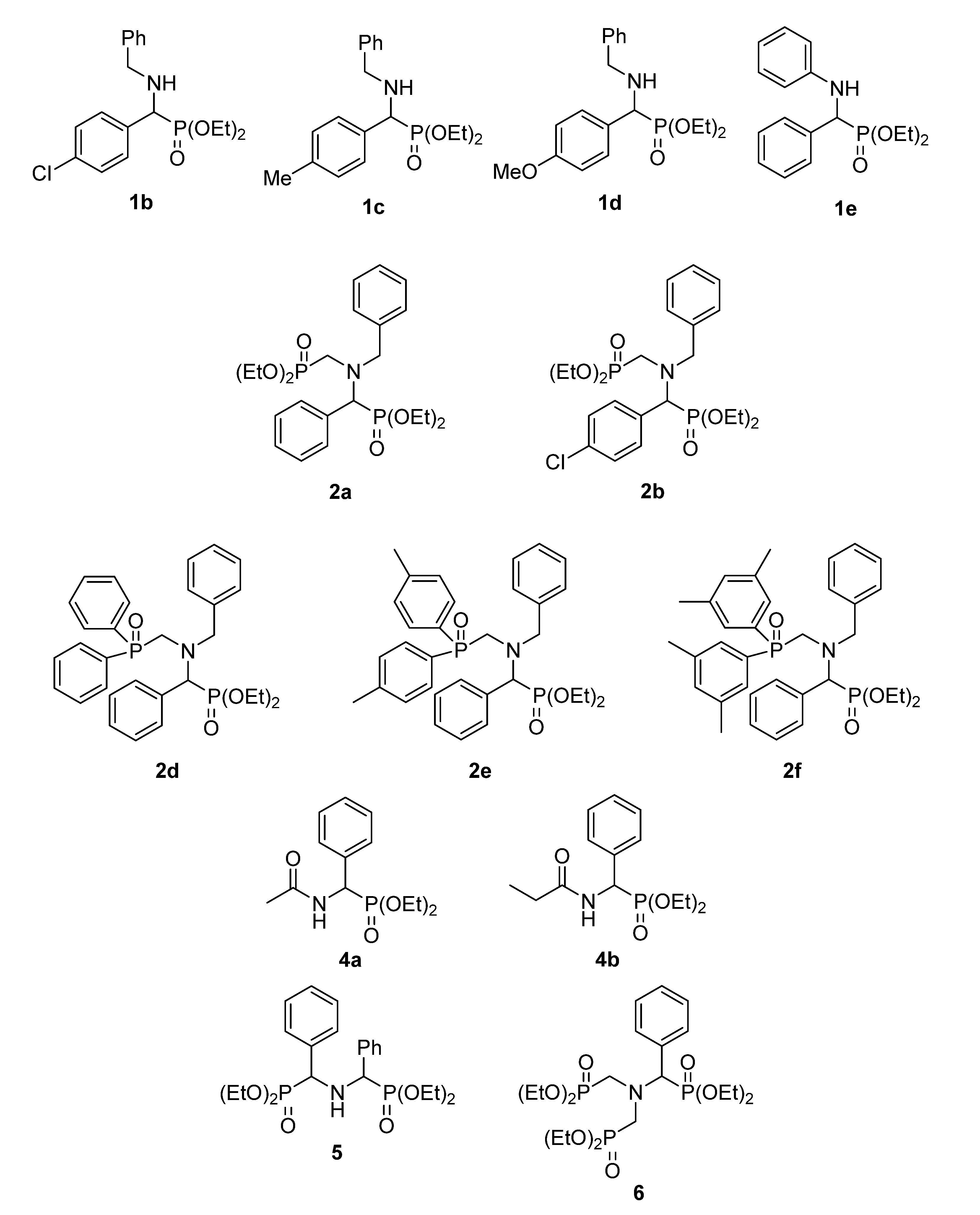
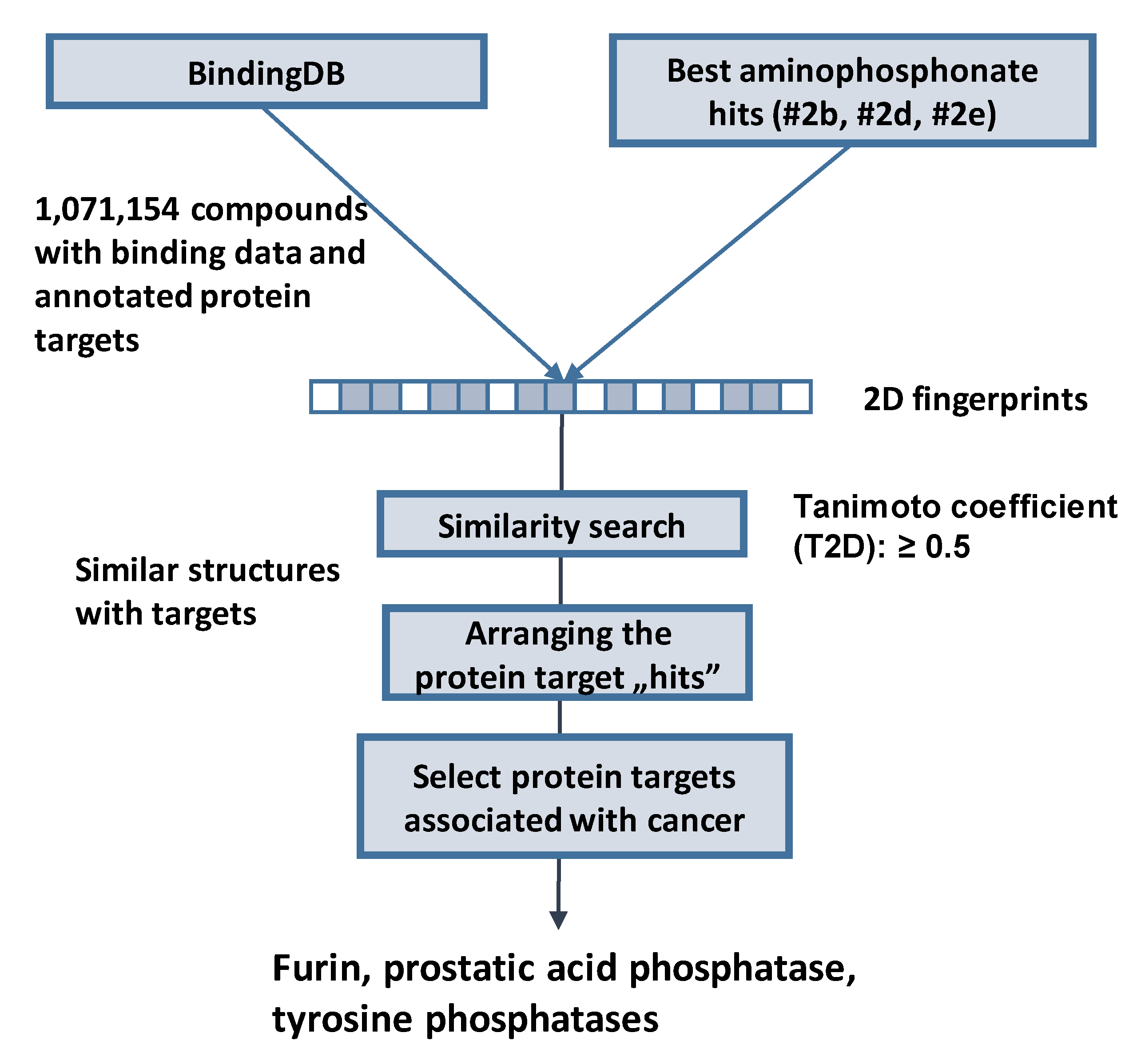
| Compound | Cytostasis (%) at c = 50 µM | ||||
|---|---|---|---|---|---|
| Cell Line | |||||
| MDA-MB 231 | PC-3 | Ebc-1 | A431 | ||
| 1b | |||||
| 1c | |||||
| 1d | |||||
| 1e | <10% | ||||
| 2a | 10–20% | ||||
| 2b | 20–30% | ||||
| 2d | 30–40% | ||||
| 2e | 40–50% | ||||
| 2f | >50% | ||||
| 4a | |||||
| 4b | |||||
| 5 | |||||
| Cell Line | Compound | 2a | 2b | 2d | 2e | 2f | 4a | 4b | 5 |
|---|---|---|---|---|---|---|---|---|---|
 |  | 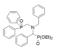 | 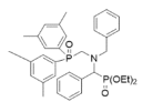 |  |  |  |  | ||
| c (µM) | Cytostasis (%) ± SD | ||||||||
| MDA-MB 231 | 2 | −4.5 ± 8.9 | −8.0 ± 3.3 | 36.7 ± 4.5 | 31.1 ± 1.2 | 38.8 ± 0.8 | 5.5 ± 5.9 | 17.9 ± 16.2 | 23.8 ± 13.4 |
| 10 | −9.0 ± 0.9 | 34.3 ± 6.9 | 28.7 ± 1.9 | 31.1 ± 3.5 | 32.5 ± 3.3 | 34.6 ± 6.2 | 21.7 ± 18.2 | 26.7 ± 9.6 | |
| 50 | 3.7 ± 1.6 | 29.5 ± 1.6 | 0.00 ± 3.3 | 10.3 ± 4.7 | 25.7 ± 1.6 | 26.9 ± 4.6 | 22.4 ± 3.4 | 21.5 ± 4.9 | |
| 250 | 58.8 ± 3.2 | 39.0 ± 0.6 | 90.5 ± 0.8 | 90.2 ± 0.6 | 90.6 ± 0.3 | n.d. | n.d. | n.d. | |
| PC-3 | 2 | n.d. | 32.3 ± 3.8 | 10.9 ± 8.3 | 6.9 ± 6.9 | −0.3 ± 2.8 | 8.9 ± 3.8 | 24.3 ± 4.3 | 16.1 ± 9.7 |
| 10 | 7.1 ± 5.1 | 21.1 ± 8.2 | 0.4 ± 6.0 | 3.4 ± 10.1 | −4.5 ± 5.4 | 24.5 ± 6.8 | 19.3 ± 2.3 | 16.9 ± 2.9 | |
| 50 | 10.3 ± 4.8 | 12.3 ± 4.9 | n.d. | 89.3 ± 0.9 | n.d. | 33.0 ± 4.6 | 20.7 ± 3.7 | 13.0 ± 2.4 | |
| 250 | n.d. | 69.0 ± 3.1 | 89.8 ± 0.5 | 89.5 ± 0.7 | 87.3 ± 0.7 | n.d. | n.d. | n.d. | |
| Ebc-1 | 2 | 32.2 ± 6.6 | 17.6 ± 2.7 | 11.8 ± 4.0 | 11.6 ± 2.5 | 26.2 ± 7.8 | 4.2 ± 5.9 | 9.8 ± 8.5 | 3.6 ± 6.3 |
| 10 | 23.8 ± 2.8 | 18.6 ± 2.4 | 9.1 ± 4.6 | 21.9 ± 1.8 | 37.0 ± 1.3 | 5.8 ± 7.5 | 12.0 ± 10.0 | 8.5 ± 8.5 | |
| 50 | 39.0 ± 1.8 | 37.8 ± 6.8 | 17.3 ± 1.4 | 19.1 ± 6.0 | 88.5 ± 0.3 | 14.2 ± 5.6 | 6.8 ± 0.4 | 11.1 ± 0.6 | |
| 250 | 86.3 ± 1.1 | 88.5 ± 0.2 | 57.4 ± 1.8 | 54.9 ± 2.7 | 90.4 ± 0.3 | n.d. | n.d. | n.d. | |
| A431 | 2 | n.d. | 11.1 ± 0.2 | 11.9 ± 6.3 | 14.7 ± 5.2 | 9.1 ± 8.7 | 3.3 ± 3.8 | −10.0 ± 9.2 | 2.7 ± 10.5 |
| 10 | 17.3 ± 6.4 | 9.2 ± 6.7 | 7.8 ± 2.1 | 18.7 ± 5.4 | 0.3 ± 14.7 | 2.1 ± 4.0 | −5.1 ± 11.1 | 6.6 ± 10.1 | |
| 50 | 43.7 ± 3.5 | 45.1 ± 6.9 | 25.8 ± 3.3 | 31.1 ± 6.3 | 92.8 ± 0.3 | 8.7 ± 2.6 | 5.6 ± 1.2 | −0.4 ± 7.8 | |
| 250 | 89.1 ± 1.5 | 91.2 ± 0.4 | 52.4 ± 0.4 | 32.2 ± 0.9 | 93.3 ± 0.4 | n.d. | n.d. | n.d. | |
| Compound | Cell Line | |||
|---|---|---|---|---|
| MDA-MB 231 | PC-3 | Ebc-1 | A431 | |
| IC50 (µM) | ||||
| 2a | 169.2 | >250 | >250 | 87.5 |
| 2b | >250 | 73.8 | 107.1 | 53.2 |
| 2d | 45.8 | 114.8 | 166.6 | 143.8 |
| 2e | 55.1 | 29.4 | 138.6 | >250 |
| 2f | 82.6 | 128.2 | 97.9 | 115.6 |
| Cancer-Related Targets | Non-Cancer-Related Targets |
|---|---|
| Furin | Tyrosyl-DNA phosphodiesterase 1 |
| Prostatic acid phosphatase | Corticotropin-releasing-factor binding protein |
| Tyrosine phosphatase non-receptor type 2 (TCPTP) | Muscarinic acetylcholine receptor M1/2 |
| Tyrosine phosphatase non-receptor type 6 (SHP1) | Solute carrier family 22 member 1 |
| Tyrosine phosphatase non-receptor type 9 (PTP-MEG2) | C-C chemokine receptor type 3 |
| Tyrosine phosphatase non-receptor type 11 (SHP2) | Acetylcholinesterase |
| Cancer-Associated Protein + A1:D5 | Expression/ Overexpression in Cancer | Similar Structure | Ref. |
|---|---|---|---|
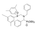
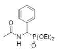
| Furin | breast, head/neck, gastric cancer | 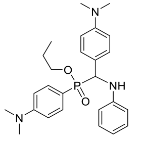 weak inhibitor | [54] |
| Prostatic acid phosphatase (PAP) | prostate cancer | 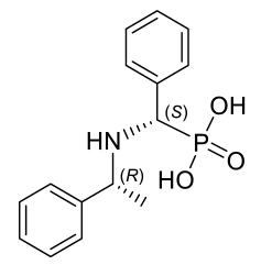 highly potent inhibitor | [55,56] |
| Tyrosine phosphatase non-receptor type 11 (SHP2), type 2 (TCPTP) | breast, skin cancer, leukemia | 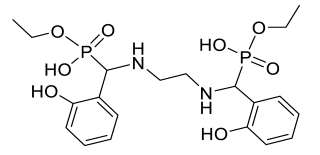 inhibitor of TCPTP and SHP2 in various affinities | [57,58,59,60,61,62,63] |
| Tyrosine phosphatase non-receptor type 11 (SHP2), type 2 (TCPTP), type 9 (PTP-MEG2) | breast, skin cancer, leukemia | 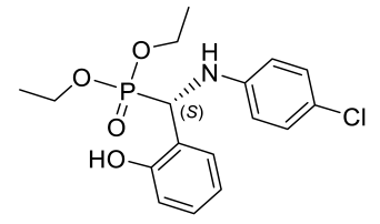 inhibitor of TCPTP, SHP2 and PTP-MEG2 in various affinities | [57,58,59,60,61,62,63] |
Disclaimer/Publisher’s Note: The statements, opinions and data contained in all publications are solely those of the individual author(s) and contributor(s) and not of MDPI and/or the editor(s). MDPI and/or the editor(s) disclaim responsibility for any injury to people or property resulting from any ideas, methods, instructions or products referred to in the content. |
© 2023 by the authors. Licensee MDPI, Basel, Switzerland. This article is an open access article distributed under the terms and conditions of the Creative Commons Attribution (CC BY) license (https://creativecommons.org/licenses/by/4.0/).
Share and Cite
Varga, P.R.; Szabó, R.O.; Dormán, G.; Bősze, S.; Keglevich, G. Cytotoxic Activity of α-Aminophosphonic Derivatives Coming from the Tandem Kabachnik–Fields Reaction and Acylation. Pharmaceuticals 2023, 16, 506. https://doi.org/10.3390/ph16040506
Varga PR, Szabó RO, Dormán G, Bősze S, Keglevich G. Cytotoxic Activity of α-Aminophosphonic Derivatives Coming from the Tandem Kabachnik–Fields Reaction and Acylation. Pharmaceuticals. 2023; 16(4):506. https://doi.org/10.3390/ph16040506
Chicago/Turabian StyleVarga, Petra R., Rita Oláhné Szabó, György Dormán, Szilvia Bősze, and György Keglevich. 2023. "Cytotoxic Activity of α-Aminophosphonic Derivatives Coming from the Tandem Kabachnik–Fields Reaction and Acylation" Pharmaceuticals 16, no. 4: 506. https://doi.org/10.3390/ph16040506
APA StyleVarga, P. R., Szabó, R. O., Dormán, G., Bősze, S., & Keglevich, G. (2023). Cytotoxic Activity of α-Aminophosphonic Derivatives Coming from the Tandem Kabachnik–Fields Reaction and Acylation. Pharmaceuticals, 16(4), 506. https://doi.org/10.3390/ph16040506


