Article

Oral Wound Healing Potential of Polygoni Cuspidati Rhizoma et Radix Decoction—In Vitro Study

by
Jakub Hadzik
1,*,
Anna Choromańska
2,
Bożena Karolewicz
3,
Adam Matkowski
4,
Marzena Dominiak
1,
Adrianna Złocińska
5 and
Izabela Nawrot-Hadzik
4,*
1
Department of Dental Surgery, Wroclaw Medical University, 50-425 Wroclaw, Poland
2
Department of Molecular and Cellular Biology, Faculty of Pharmacy, Wroclaw Medical University, 50-556 Wroclaw, Poland
3
Department of Drug Form Technology, Wroclaw Medical University, 50-556 Wroclaw, Poland
4
Department of Pharmaceutical Biology and Biotechnology, Faculty of Pharmacy, Wroclaw Medical University, 50-556 Wroclaw, Poland
5
Laboratory of Elemental Analysis and Structural Research, Faculty of Pharmacy, Wroclaw Medical University, 50-556 Wroclaw, Poland
*
Authors to whom correspondence should be addressed.
Pharmaceuticals 2023, 16(2), 267; https://doi.org/10.3390/ph16020267
Submission received: 6 December 2022 / Revised: 15 January 2023 / Accepted: 8 February 2023 / Published: 10 February 2023

Abstract

:
Polygoni Cuspidati Rhizoma et Radix (syn. rhizomes of Reynoutria japonica Houtt.) is a pharmacopoeial raw material in Europe and China. In traditional medicine, one of the applications for Reynoutria japonica rhizomes is wound healing. In a recent in vitro study, we demonstrated that ethanol and acetone extracts from this herbal drug have the potential to heal oral gum wounds. However, considering that a majority of herbal medicines have been traditionally administered as water decoctions, in the present study, a decoction of Reynoutria japonica rhizomes was prepared and detailed tests to determine its in vitro gingival wound healing activity were conducted. We used the primary human gingival fibroblasts (HGF) incubated with a decoction to determine cell viability (MTT assay), cell proliferation (the confocal laser scanning microscope—CLSM), and cell migration (wound healing assay). Moreover, the collagen type III expression was examined using immunocytochemical staining. The studied decoction was qualitatively and quantitatively characterized using the validated HPLC/DAD/ESI-HR-QTOF-MS method. The Folin–Ciocalteu assay was used to determine the total phenols and tannins content. Additionally, HPLC-RI analysis of decoction and the previously obtained ethanol and acetone extracts was used to determine the composition of saccharides. Low concentration (from 50 to 1000 µg/mL) of decoction after 24 h incubation caused a significant increase in HGF cell viability. No cytotoxic effect was observed at any tested concentration (up to 2000 µg/mL). The lowest active concentration of decoction (50 µg/mL) was selected for further experiments. It significantly stimulated human gingival fibroblasts to proliferate, migrate, and increase the synthesis of collagen III. Phytochemical analysis showed significantly fewer polyphenols in the decoction than in the ethanol and acetone extracts tested earlier. In contrast, high levels of polysaccharides were observed. In our opinion, they may have a significant effect on the oral wound healing parameters analyzed in vitro. The results obtained encourage the use of this raw material in its traditional, safe form—decoction.

1. Introduction

The rhizome with roots of Reynoutria japonica Houtt. is a traditional Chinese medicinal herb (hu zhang in pinyin Chinese) listed in the European Pharmacopoeia under the name Polygoni cuspidati rhizoma et radix. This herbal medicine was included in the European Pharmacopoeia in 2017. According to Pharmacopoeia, a minimum of 1.0% emodin content and 1.5% of polydatin content in the dried herb are required as a quality check [1]. Traditionally, hu zhang has been used in East Asia against many diseases, such as hyperlipemia, inflammation, infection, and for wound healing [2]. According to current knowledge, the most important bioactive compounds in Polygoni cuspidati rhizoma et radix are stilbenes (with a high concentration of resveratrol), anthraquinones, procyanidins, phenylpropanoid disaccharide esters, and other polyphenols [2,3,4,5]. To obtain a high content of these constituents, different percentages of ethanol/methanol or acetone are used in the extraction process [3,6].
In the previous in vitro study, our research team reported the gingival wound healing activity of 25% and 40% ethanolic as well as 60% acetone extracts from the Reynoutria japonica rhizomes [7]. The extracts stimulated human gingival fibroblasts (HGF) to proliferate and migrate, as well as increasing the synthesis of collagen III, but with different potency. The highest stimulation of proliferative and migratory activity was observed after incubation with 25% EtOH extract. This effect may have been related to the highest resveratrol content and the favorable composition of procyanidins. Currently, our attention has been drawn to the fact that although the overwhelming majority of Chinese herbal medicines (CHMs) are traditionally administered as water decoctions (tangs), relatively little research is done on them [8].
This experiment was designed to test whether a traditionally used Polygoni cuspidati rhizoma et radix decoction could have a stimulating effect on the oral wound healing process. The intraoral healing process is different from skin wound healing, so a different therapeutic approach is required. Intraoral wound healing aids are desirable for patients whose natural wound healing process is impaired, i.e., immunocompromised, post-transplant, post-radiotherapy or chemotherapy patients, and persons with systemic diseases [9,10]. Also, periodontal diseases, including periodontal and peri-implant disease [11], predispose to impaired wound healing due to high levels of oxidative stress, which impairs the proliferation and migration of HGFs [12,13]. Also, older age affects oral wound healing by impairing fibroblast proliferation, migration, and differentiation, and reducing collagen synthesis [14]. Moreover, prolonged wound healing in the oral cavity predisposes to bacterial invasion and wound infection, which can further inhibit the proper wound healing process. Due to the unsatisfactory results of currently available treatments, there is a need to search for new, effective therapies that promote intraoral healing [15,16].
In this study, we have prepared a decoction of Reynoutria japonica rhizomes according to the traditional recipe. We conducted a detailed study to determine its activity in healing gingival wounds in vitro using the same raw material and the same research methods as in our previous study [7]. Additionally, an HPLC-RI analysis of the decoction and the previously obtained extracts was used to determine the composition of saccharides.

2. Results and Discussion

2.1. Cell Viability—MTT Assay

The Reynoutria japonica rhizome decoction was not cytotoxic to HGF cells at any tested concentrations and incubation time (Figure 1). It was different from the previously tested ethanol and acetone extracts, which at high concentrations—from 1000 µg/mL to 2000 µg/mL—showed a significant reduction in the viability of fibroblasts after 24 h of incubation [7]. The decoction at concentrations from 50 µg/mL to 1000 µg/mL, after 24 h of incubation, significantly increased the viability of HGF cells, up to 124% compared with that of the control. This stimulating effect on cellular metabolism has also been observed in previous studies with ethanol and acetone extracts from R. japonica rhizomes [5,7].
To check if fibroblasts proliferate after treatment with the decoction, we used a confocal laser scanning microscope to visualize changes in histone 3 expression. The lowest concentration (50 µg/mL) demonstrating a significant stimulation of the fibroblasts’ viability in the MTT test was selected for further studies (the same as in our previous research with solvent extracts [7]).

2.2. Confocal Laser Microscopy Study

Microphotographs of cells with immunofluorescent staining of histone H3 after 24 h incubation with tested decoction (at a concentration of 50 µg/mL) and without decoction (untreated cells) are presented in Figure 2. The primary rabbit polyclonal antibody anti-phospho-histone H3 was used to present an abundance of phosphorylation of serine 10 on histone (H3S10ph), the increase of which is observed during cell division [17,18]. Figure 3 shows the mean fluorescence intensity in arbitrary units for treated cells (22,488) and for untreated cells (4.515). The results indicate that the studied decoction significantly stimulated human gingival fibroblasts to divide. Importantly, the fluorescence intensity after incubation with the decoction was even higher than with 25% EtOH extract, which was the most active in our previous study [7].

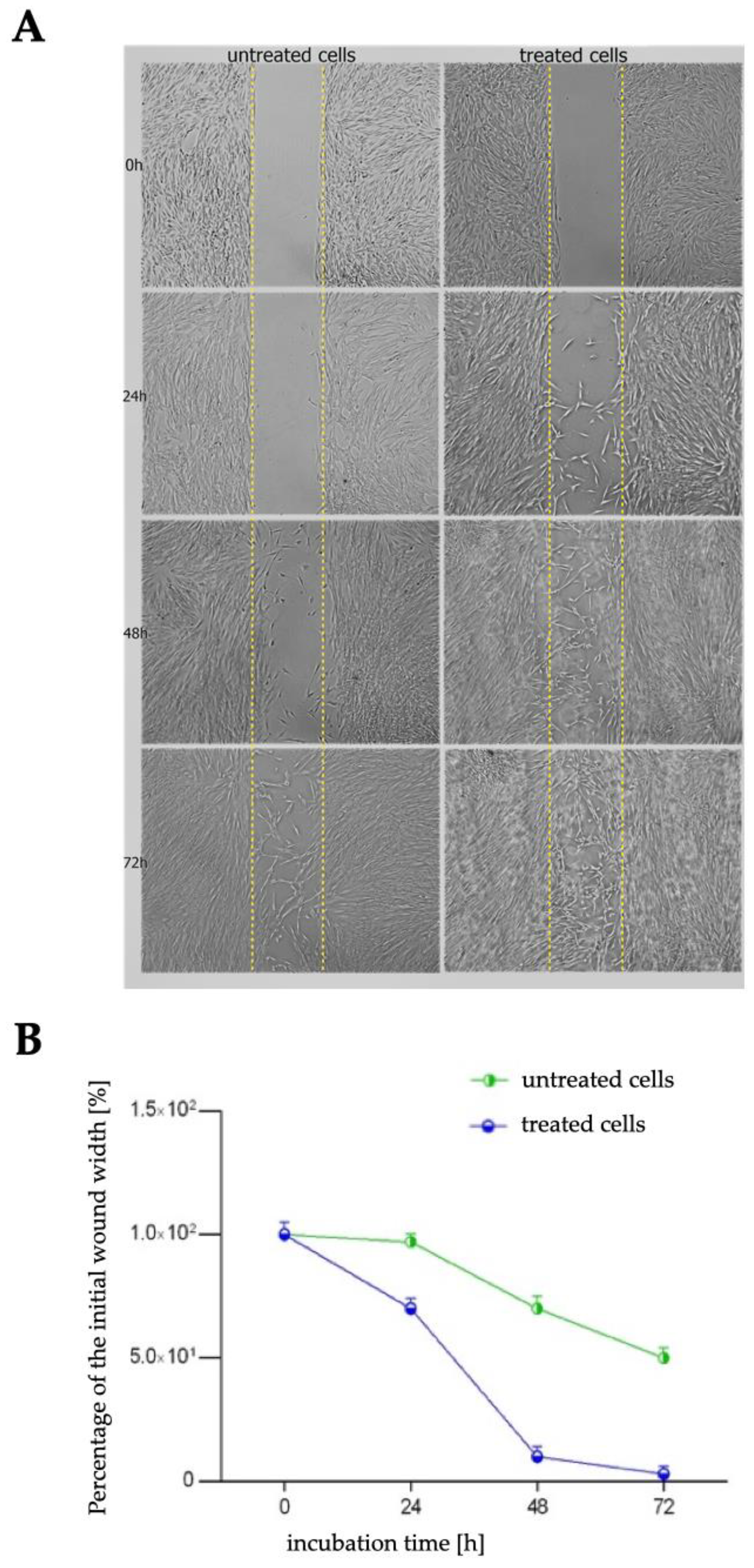
2.3. Wound Healing Assay

Proper proliferation and migration of gingival fibroblast cells play a significant role in the healing of oral cavity wounds. Using a cell migration-based wound healing assay, we analyzed the motility of the fibroblasts after incubation with 50 µg/mL of R. japonica decoction. Figure 4A shows the changes in the cell-covered area (gap closure) over time. Figure 4B illustrates the percentage of a healed wound as a function of time. Table 1 presents the percentage of wound (gap) closure over time.

2.4. Immunocytochemical Staining

Gingival fibroblasts synthesize and secrete collagen type III, a high expression of which is observed during wound healing. Over time, type III collagen is reabsorbed and replaced by type I collagen, a key component of the extracellular matrix. Microphotographs (Figure 5) of HGF with immunocytochemically stained collagen type III show the influence of R. japonica decoction on expression of collagen type III. According to Table 2, which shows the semi-quantitative results, the 50 µg/mL of R. japonica decoction had a similar stimulating effect on collagen III production as 2 µM betulinic acid, used as the positive control.
In comparison to our previous study, the effect of R. japonica decoction on stimulation of fibroblasts to produce collagen III was similar to 60% acetone extract, slightly weaker than 40% EtOH, but stronger than 25% EtOH extract [7].

2.5. HPLC/DAD/ESI-HR-TOF-MS Analysis

To determine the composition of the compounds that contributed to the observed effects, qualitative and quantitative HPLC/DAD/ESI-HR-QTOF-MS analyses of decoction was performed. The UHPLC-QTOF-MS analysis revealed a total of 37 different compounds (Table 3, Figure 6) that belong to carbohydrates, stilbenes, flavan-3-ols, procyanidins, anthraquinones, organic acids, and naphthalenes.
Among all of the detected chromatographic peaks, seven remained unassigned and without a clear indication of their chemical nature. Two peaks were tentatively defined as carbohydrates. Most of the identified compounds were previously detected in extracts and fractions from rhizomes of R. japonica, as described in our previous reports [3,5,7,19]. The decoction was less diverse in terms of the content of compounds than the ethanol and acetone extracts analyzed in previous studies. It did not contain phenylpropanoids. Moreover, the trace amount of resveratrol was only noticed after extraction from the raw chromatogram. Stilbene glycosides and anthraquinone glycosides were present in greater amounts than their aglycones. In contrast, compound 31 with a high peak in the BPC (base peak chromatogram), labeled as unknown, with a deprotonated ion at m/z 253.0513 [M∓H] and predicted formula of C15H9O4 was not observed in the previously studied extracts. Product ions at m/z 225.0545 (C14H9O3, 0.3 ppm), m/z 224.0481 (C14H8O3, 1.26 ppm), m/z 209.0611 (C14H9O2, −1.2 ppm), m/z 197.0606 (C13H9O2, 0.9 ppm), m/z 169.0664 (C12H9O, −3.0 ppm), and m/z 135.009 (C7H3O3, −1.5 ppm) suggest that it may be some kind of anthraquinone, i.e., rubiadin (HMDB0257354). In order to determine the structure of this compound, it must be isolated and subjected to detailed studies.
A previously developed, validated analytical method was used to quantify the selected compounds [19]. Piceid was detected in much lower amounts in the decoction than in previously tested ethanol or acetone extracts (Table 4). There was almost six times less piceid in the decoction than in the previously tested 25% EtOH extract and over ten times less than in the 60% acetone extract.
Resveratrol was not detectable without extraction from the chromatogram and was not quantifiable. Vanicoside A and B were not detected even in trace amounts Also, emodin and physcion were detected in much lower amounts than in ethanol and acetone extracts. Their content was sufficient for detection but not for correct quantification.

2.6. Total Polyphenols and Tannins Content

According to the modified Folin–Ciocalteu assay (Figure 7), total polyphenols and tannins make up a small part of the decoction.

2.7. HPLC-RI Analysis

The low content of polyphenols, including tannins, stilbenes, anthraquinones, phenylpropanoids, as well as the viscous consistency of the decoction encouraged us to examine its saccharide content. To compare the decoction with ethanol and acetone extracts from the previous study, the latter were also analyzed. The composition of saccharides in the studied samples was determined by high-pressure liquid chromatography with a refractometric detector (Figure 8A). All the extracts tested previously—25% EtOH (B), 40% EtOH (C), and 60% acetone (D) showed similar HPLC-RI chromatograms, which revealed the presence of three main compounds—glucose, xylose, and an unknown compound 1. The HPLC-RI chromatogram of the decoction differs from the extracts by the presence of a much larger unknown peak number 1 which also appears at a shorter retention time. To find out what kind of compound the unknown peak 1 is, multiple sugars were tested, among others: two monosaccharides (xylose (A) and glucose (B)), a disaccharide (maltose (C)), trisaccharide (maltotriose (D)), and polysaccharide (pectins (E)) (Figure 8B).
As shown above, under the chosen analysis conditions, the analytes flow from the Rezex ROA—Organic Acid H + column (Phenomenex) in the following order: first the most complex saccharides, such as the polysaccharide pectins, followed by trisaccharides, then disaccharides, and finally monosaccharides. We also confirmed this dependence on other compounds belonging to these sugar groups (data not presented). This is in accordance with the product specification of the Rezex ROA—Organic Acid H + column (Phenomenex). Our research shows that the decoction contained the greatest amount of polysaccharides. The exact identification of the polysaccharides and other saccharides present in the decoction requires further extensive research. At this point, however, it is important to consider whether they may have affected the activities under study.
The term polysaccharides covers a large group of compounds that are composed of monosaccharides (glucose, mannose, galactose, fructose, etc.). Depending on the type of monosaccharide, the polysaccharides can be divided into a homo-polysaccharide containing one type of monosaccharide and a hetero-polysaccharide containing two or more different types of monosaccharides. Monosaccharides are linked by glycosidic bonds to form long linear or branched structures. The literature can confirm the influence of polysaccharides on wound healing. There are reviews available [20,21] that show the skin wound healing activity of polysaccharides, most often obtained by hot water extraction, among others from such plants as Trigonella foenum-graecum, Hammada scoparia, Linus usitatissimum, Avena sativa, Caesalpinia pulcherrima, Sanguisorba officinalis, Glycyrrhiza uralensis, Pimpinella anisum as well as Bletilla striata, Konjac, Eucommia ulmoides, and Mesona procumbens [22]. Most of these studies have not established the exact chemical structure of the polysaccharides. Fewer data are available on the activity of polysaccharides in gingival wound healing. However, one of the polysaccharides with known gingival wound healing properties is acemannan from Aloe vera. Acemannan, a β-(1,4)-acetylated soluble polymannose has gingival wound healing properties proven in vitro and in vivo as well as in clinical studies [23]. A study by Jettanacheawchankit et al. [24] showed that acemannan induces proliferation and upregulation of growth factors (KGF-1, VEGF), and type I collagen expression in gingival fibroblasts as good reduction of wound area of experimental animals at day 7 after treatment. Another polysaccharide with a proven wound healing effect is β -glucan (glucose polymers linked by 1,3; 1,4 or 1,6 β-glycosidic bonds), which is a constituent of grains, yeast, and other fungi. In vitro studies have shown that β-glucans induced the proliferation and migration of keratinocytes and fibroblasts through specific receptors such as Dectin-1, CR3 or TLR. Preclinical animal studies have confirmed that it is effective as a wound healing agent [25].
The biological activity of the studied Polygoni cuspidati rhizoma et radix decoction was higher than that demonstrated by the ethanol and acetone extracts in our previous study, even though the decoction contained significantly fewer polyphenols [7]. On the other hand, the content of carbohydrates was significantly higher in the decoction than in either organic solvent extract. According to our best contemporary knowledge, there are no reports in the literature on polysaccharide contents in rhizomes of R. japonica and their biological activity. Notwithstanding, the methods used in this study were insufficient to determine the identity of the carbohydrates. Further research is therefore required to confirm our assumptions that polysaccharides in the decoction are largely responsible for stimulating gingival fibroblast cells to proliferate, migrate, and increase collagen III synthesis.

3. Materials and Methods

The present study was designed as an in vitro study on the human fibroblast cell line. The experimental protocol of a study “influence of extracts from medicinal plants Reynoutria on the functions of oral fibroblasts” was submitted to a Bioethics Commission of Wrocław Medical University (Wrocław, Poland) by a study principal investigator JH, and approved by the commission with the number KB-134/2020. The gingival biopsies for the cell study were provided by the Dental Clinical and Teaching facility of Dental Surgery Department at Wrocław Medical University. The study was conducted in full compliance with the GCP ICH: E6 (R2) and Declaration of Helsinki.

3.1. Plant Material and Decoction Preparation

Reynoutria japonica rhizomes harvested in Wroclaw (Poland) were used in the study. Details on the identification of the plant species, location, time of collection of raw material, and storage were presented in our previous study [7]. The same raw material was used in our previous study to prepare ethanol and acetone extracts [7]. Fifty grams of air-dried and powdered rhizomes of R. japonica were flooded with 500 mL of cold water and boiled under reflux for 20 min. After cooling down, the decoction was filtered through a paper filter and the solvent was evaporated under reduced pressure. An amount of 2.41 g of dry decoction was obtained. Stock solutions were prepared from the dried decoction by dissolving 100 mg of decoction in 1 mL of dimethyl sulfoxide (DMSO, Sigma-Aldrich, St. Louis, MO, USA). Different concentrations of the decoction (5–2000 µg/mL) were tested by taking the appropriate amount of the stock solution. As a solvent to prepare different concentrations of decoction, the cell culture medium was used.

3.2. Cell Culture

The study was carried out on the primary human gingival fibroblasts (HGF) obtained from the connective tissue of a dental patient’s hard palate. The details of the cell culture study were presented in our previous paper [7].

3.3. Cell Viability Assay

Fibroblast cell viability was assessed using the MTT (3-(4,5-dimethylthiazol-2-yl)-2,5-diphenyltetrazolium bromide) colorimetric assay (Sigma-Aldrich, Merck Group, Darmstadt, Germany). The assay was carried out as described in our previous work [7], except that a decoction was used instead of ethanol or acetone extracts. The decoction was used in the concentration range from 5 µg/mL to 2000 µg/mL. The concentration of DMSO in the samples was equal to or less than 2%, therefore, also 2% DMSO was used as a control in the study. For the experiment, the cells were seeded in 96-well microculture plates at 1 × 10⁴ cells/well. After the incubation with selected concentrations of decoction, the experiments were conducted according to the manufacturer’s protocol. The absorbance was determined at 570 nm.

3.4. Confocal Laser Microscopy Study

A confocal laser scanning microscope (CLSM, Olympus FluoView FV1000, Tokyo, Japan) was used to visualize histone 3 expression change after incubation with 50 µg/mL decoction. The study was performed exactly as described in our previous article [7], except that a decoction was used instead of acetone and ethanol extracts.

3.5. In Vitro Wound Healing Assay

Decoction at 50 µg/mL concentration was used for the wound healing assay as a fibroblast migration capacity test. The study was performed exactly as described in our previous article [7], except that a decoction was used instead of acetone and ethanol extracts.

3.6. Immunocytochemical Staining

Immunocytochemical staining was used to evaluate the expression of collagen type III after 24 h incubation with decoction at 50 µg/mL. Betulinic acid (2 µM) was used as a positive control. The study was performed exactly as described in our previous paper [7], except that a decoction was used instead of acetone and ethanol extracts. The intensity of immunohistochemical staining was evaluated as negative (−), weak (+), moderate (++), or strong (+++), as in the previous article [7,26].

3.7. HPLC/DAD/ESI-HR-QTOF-MS Analysis

The obtained decoction was prepared for the qualitative and quantitative analysis. An amount of 50 mg of dried decoction was dissolved in 80% methanol (MeOH, Merck / MilliporeSigma, Darmstadt, Germany) in a volumetric flask to obtain a 5 mg/mL concentration. After filtering the prepared solutions through a 0.22 μm syringe membrane (Chromafil, Macherey-Nagel, Düren, Germany) into vials, 4 μL of the sample was injected by autosampler into the high-pressure liquid chromatography (HPLC) system. The same HPLC-DAD-MS system was used as well as the same qualitative analysis conditions as in our previous study [7].
A previously developed, validated analytical method was used to quantify the selected compounds [19]. Linearity, the LOD (limit of detection), and LOQ (limit of quantification) for all quantified compounds were presented in our previous study [19].

3.8. Total Polyphenols and Tannins Content

The content of polyphenols and tannins was determined using the modified Folin–Ciocalteu assay based on Singleton and Rossi method [27]. The study was conducted in the same way as described in our previous articles [28,29].

3.9. HPLC-RI Analysis

The composition of saccharides in the studied samples was determined by high-pressure liquid chromatography (HPLC) according to the procedure described in the previous paper [30]. Briefly, the chromatographic method was performed using the Dionex Ultimate 3000 system (Thermo Fisher Scientific, Waltham, MA, USA). The content of saccharides was determined using a Rezex ROA—Organic Acid H+ column with the following dimensions: 300 mm × 7.8 mm i.d. and 8% cross-linked H + (Phenomenex, Torrance, CA, USA). The column was kept at 60 °C. The mobile phase was 5 mM sulfuric acid, previously filtered through a 0.45 mm membrane filter (Millipore). The flow rate was 0.6 mL/min. A refractometric detector ERC RefractoMax 520 (DataApex, Prague, Czech Republic) was used for analyte detection. All analytical determinations were performed in triplicate and mean values are given. An amount of 50 µL of decoction or extract (at 2.5 mg/mL concentration) dissolved in the mobile phase and filtered through a 0.22 µm Chromafil PTFE hydrophilic syringe membrane was injected into the HPLC system by an autosampler. Among others, the following compounds were analyzed as standards: glucose, xylose, maltose, maltotriose, and pectins (all from Merck/MilliporeSigma, Darmstadt, Germany). The standards were dissolved in the mobile phase in a volumetric flask to obtain a concentration of 1 mg/mL.

3.10. Statistical Analysis

All assays were performed in at least triplicate and results are presented as the mean of the replicates ± SD. The Shapiro–Wilk test was used to evaluate the distribution of results. Two-way ANOVA and Tukey’s multiple comparisons tests (GraphPad Prism v. 9, San Diego, CA, USA) were used to evaluate significant differences between the obtained values. In studies where only the means between the control and treatments were compared, the t-test was used.

4. Conclusions

The decoction of Reynoutria japonica rhizomes prepared according to a traditional recipe stimulated gingival fibroblasts to proliferate, migrate, and increase the synthesis of collagen III. Based on the phytochemical research (HPLC/DAD/ESI-HR-QTOF-MS, HPLC-RI analysis, and Folin–Ciocalteu assay), we can conclude that the high content of polysaccharides observed in the decoction may have an impact on their high wound-healing activity in vitro. Because this is the first published report on the presence of polysaccharides in this plant material, further research is needed to confirm our assumptions. Importantly, the decoction was found to be noncytotoxic to HGF cells at any tested concentrations and incubation time. The safety and high activity of the studied Polygoni cuspidati rhizoma et radix decoction encourage its future use in oral wound healing.

Author Contributions

Conceptualization, J.H. and I.N.-H.; methodology, J.H., I.N.-H., A.C. and A.Z.; software, J.H.; validation, J.H. and I.N.-H.; formal analysis, J.H. and I.N.-H.; investigation, J.H., I.N.-H., A.C. and A.Z.; resources, J.H., I.N.-H. and A.C.; data curation, I.N.-H. and A.C.; writing—original draft preparation, J.H. and I.N.-H.; writing—review and editing, I.N.-H., A.M., B.K. and M.D.; visualization, J.H.; supervision, I.N.-H.; project administration, J.H. and I.N.-H.; funding acquisition, J.H. All authors have read and agreed to the published version of the manuscript.

Funding

The study was supported by grant No. SUBK.B040.22.015 “Optimization of the extract from the medicinal plant of the genus Reynoutria for use in regenerative dentistry” granted by the Wroclaw Medical University, project manager JH and Wroclaw Medical University SUBZ.B040.23.048

Institutional Review Board Statement

The protocol for this study was submitted to the Bioethics Commission of Wrocław Medical University (Wrocław, Poland) and approved by the commission with the number KB-134/2020. The study has been conducted in full compliance with the GCP ICH: E6 (R2) and Declaration of Helsinki.

Informed Consent Statement

The study has been conducted in full compliance with the GCP ICH: E6 (R2) and Declaration of Helsinki. All patients gave consent to participate in the study.

Data Availability Statement

Data is contained within the article.

Acknowledgments

We would like to thank Hanna Czapor-Irzabek for the collection of LC/MS data. The measurements were carried out in the Laboratory of Elemental Analysis and Structural Research at Wroclaw Medical University.

Conflicts of Interest

The authors declare no conflict of interest.

References

  1. European Pharmacopoeia, 9th ed.; European Director- ate for the Quality of Medicines: Strasburg, France, 2017; pp. 1481–1483.
  2. Peng, W.; Qin, R.; Li, X.; Zhou, H. Botany, phytochemistry, pharmacology, and potential application of Polygonum cuspidatum Sieb.et Zucc.: A review. J. Ethnopharmacol. 2013, 148, 729–745. [Google Scholar] [CrossRef] [PubMed]
  3. Nawrot-Hadzik, I.; Ślusarczyk, S.; Granica, S.; Hadzik, J.; Matkowski, A. Phytochemical Diversity in Rhizomes of Three Reynoutria Species and their Antioxidant Activity Correlations Elucidated by LC-ESI-MS/MS Analysis. Molecules 2019, 24, 1136. [Google Scholar] [CrossRef]
  4. Nawrot-Hadzik, I.; Zmudzinski, M.; Matkowski, A.; Preissner, R.; Kęsik-Brodacka, M.; Hadzik, J.; Drag, M.; Abel, R. Reynoutria Rhizomes as a Natural Source of SARS-CoV-2 Mpro Inhibitors–Molecular Docking and In Vitro Study. Pharmaceuticals 2021, 14, 742. [Google Scholar] [CrossRef]
  5. Nawrot-Hadzik, I.; Hadzik, J.; Fleischer, M.; Choromańska, A.; Sterczała, B.; Kubasiewicz-Ross, P.; Saczko, J.; Gałczyńska-Rusin, M.; Gedrange, T.; Matkowski, A. Chemical composition of east Asian invasive knotweeds, their cytotoxicity and antimicrobial efficacy against cariogenic pathogens: An in-vitro study. Med. Sci. Monit. 2019, 25, 3279–3287. [Google Scholar] [CrossRef] [PubMed]
  6. Alperth, F.; Melinz, L.; Fladerer, J.P.; Bucar, F. Uhplc analysis of reynoutria japonica houtt. Rhizome preparations regarding stilbene and anthranoid composition and their antimycobacterial activity evaluation. Plants 2021, 10, 1809. [Google Scholar] [CrossRef]
  7. Nawrot-Hadzik, I.; Matkowski, A.; Pitułaj, A.; Sterczała, B.; Olchowy, C.; Szewczyk, A.; Choromańska, A. In Vitro Gingival Wound Healing Activity of Extracts from Reynoutria japonica Houtt Rhizomes. Pharmaceutics 2021, 13, 1764. [Google Scholar] [CrossRef]
  8. Sheridan, H.; Krenn, L.; Jiang, R.; Sutherland, I.; Ignatova, S.; Marmann, A.; Liang, X.; Sendker, J. The potential of metabolic fingerprinting as a tool for the modernisation of TCM preparations. J. Ethnopharmacol. 2012, 140, 482–491. [Google Scholar] [CrossRef]
  9. Buranasukhon, W.; Athikomkulchai, S.; Tadtong, S.; Chittasupho, C. Wound healing activity of Pluchea indica leaf extract in oral mucosal cell line and oral spray formulation containing nanoparticles of the extract. Pharm. Biol. 2017, 55, 1767–1774. [Google Scholar] [CrossRef]
  10. Field, E.A.; Allan, R.B. Review article: Oral ulceration--aetiopathogenesis, clinical diagnosis and management in the gastrointestinal clinic. Aliment. Pharmacol. Ther. 2003, 18, 949–962. [Google Scholar] [CrossRef] [PubMed]
  11. Caton, J.G.; Armitage, G.; Berglundh, T.; Chapple, I.L.C.; Jepsen, S.; Kornman, K.S.; Mealey, B.L.; Papapanou, P.N.; Sanz, M.; Tonetti, M.S. A new classification scheme for periodontal and peri-implant diseases and conditions—Introduction and key changes from the 1999 classification. J. Clin. Periodontol. 2018, 45, S1–S8. [Google Scholar] [CrossRef]
  12. Buranasin, P.; Mizutani, K.; Iwasaki, K.; Mahasarakham, C.P.N.; Kido, D.; Takeda, K.; Izumi, Y. High glucose-induced oxidative stress impairs proliferation and migration of human gingival fibroblasts. PLoS ONE 2018, 13, e0201855. [Google Scholar] [CrossRef]
  13. Nawrot-Hadzik, I.; Matkowski, A.; Kubasiewicz-Ross, P.; Hadzik, J. Proanthocyanidins and Flavan-3-ols in the Prevention and Treatment of Periodontitis—Immunomodulatory Effects, Animal and Clinical Studies. Nutrients 2021, 13, 239. [Google Scholar] [CrossRef]
  14. Cáceres, M.; Oyarzun, A.; Smith, P.C. Defective wound-healing in aging gingival tissue. J. Dent. Res. 2014, 93, 691–697. [Google Scholar] [CrossRef]
  15. Toma, A.I.; Fuller, J.M.; Willett, N.J.; Goudy, S.L. Oral wound healing models and emerging regenerative therapies. Transl. Res. 2021, 236, 17–34. [Google Scholar] [CrossRef] [PubMed]
  16. Kubasiewicz-Ross, P.; Fleischer, M.; Pitułaj, A.; Hadzik, J.; Nawrot-Hadzik, I.; Bortkiewicz, O.; Dominiak, M.; Jurczyszyn, K. Evaluation of the three methods of bacterial decontamination on implants with three different surfaces. Adv. Clin. Exp. Med. 2020, 29, 177–182. [Google Scholar] [CrossRef] [PubMed]
  17. Clément, C.; Orsi, G.A.; Gatto, A.; Boyarchuk, E.; Forest, A.; Hajj, B.; Miné-Hattab, J.; Garnier, M.; Gurard-Levin, Z.A.; Quivy, J.P.; et al. High-resolution visualization of H3 variants during replication reveals their controlled recycling. Nat. Commun. 2018, 9, 3181. [Google Scholar] [CrossRef] [PubMed]
  18. Komar, D.; Juszczynski, P. Rebelled epigenome: Histone H3S10 phosphorylation and H3S10 kinases in cancer biology and therapy. Clin. Epigenetics 2020, 12, 147. [Google Scholar] [CrossRef] [PubMed]
  19. Nawrot-Hadzik, I.; Granica, S.; Domaradzki, K.; Pecio, Ł.; Matkowski, A. Isolation and Determination of Phenolic Glycosides and Anthraquinones from Rhizomes of Various Reynoutria Species. Planta Med. 2018, 84, 1118–1126. [Google Scholar] [CrossRef]
  20. Albuquerque, P.B.S.; de Oliveira, W.F.; dos Santos Silva, P.M.; dos Santos Correia, M.T.; Kennedy, J.F.; Coelho, L.C.B.B. Skincare application of medicinal plant polysaccharides—A review. Carbohydr. Polym. 2022, 277, 118824. [Google Scholar] [CrossRef]
  21. Li, Q.; Niu, Y.; Xing, P.; Wang, C. Bioactive polysaccharides from natural resources including Chinese medicinal herbs on tissue repair. Chin. Med. 2018, 13, 7. [Google Scholar] [CrossRef] [Green Version]
  22. Fan, S.L.; Lin, J.A.; Chen, S.Y.; Lin, J.H.; Lin, H.T.; Chen, Y.Y.; Yen, G.C. Effects of Hsian-tsao (: Mesona procumbens Hemsl.) extracts and its polysaccharides on the promotion of wound healing under diabetes-like conditions. Food Funct. 2021, 12, 119–132. [Google Scholar] [CrossRef]
  23. Liu, C.; Cui, Y.; Pi, F.; Cheng, Y.; Guo, Y.; Qian, H. Extraction, purification, structural characteristics, biological activities and pharmacological applications of acemannan, a polysaccharide from aloe vera: A review. Molecules 2019, 24, 1554. [Google Scholar] [CrossRef]
  24. Jettanacheawchankit, S.; Sasithanasate, S.; Sangvanich, P.; Banlunara, W.; Thunyakitpisal, P. Acemannan stimulates gingival fibroblast proliferation; expressions of keratinocyte growth factor-1, vascular endothelial growth factor, and type I collagen; and wound healing. J. Pharmacol. Sci. 2009, 109, 525–531. [Google Scholar] [CrossRef] [PubMed]
  25. Majtan, J.; Jesenak, M. β-Glucans: Multi-functional modulator of wound healing. Molecules 2018, 23, 806. [Google Scholar] [CrossRef] [PubMed]
  26. Sterczala, B.; Kulbacka, J.; Saczko, J.; Dominiak, M. The effect of dental gel formulation on human primary fibroblasts—An in vitro study. Folia Histochem. Cytobiol. 2020, 58, 156–161. [Google Scholar] [CrossRef]
  27. Singleton, V.L.; Rossi, J.A. Colorimetry of Total Phenolics with Phosphomolybdic-Phosphotungstic Acid Reagents. Am. J. Enol. Vitic. 1965, 16, 144–158. [Google Scholar]
  28. Cieslak, A.; Zmora, P.; Matkowski, A.; Nawrot-Hadzik, I.; Pers-Kamczyc, E.; El-Sherbiny, M.; Bryszak, M.; Szumacher-Strabel, M. Tannins from sanguisorba officinalis affect in vitro rumen methane production and fermentation. J. Anim. Plant Sci. 2016, 26, 54–62. [Google Scholar]
  29. Nawrot-Hadzik, I.; Granica, S.; Abel, R.; Czapor-Irzabek, H.; Matkowski, A. Analysis of antioxidant polyphenols in loquat leaves using HPLC-based activity profiling. Nat. Prod. Commun. 2017, 12, 163–166. [Google Scholar] [CrossRef]
  30. Pińkowska, H.; Krzywonos, M.; Wolak, P.; Złocińska, A. Pectin and Neutral Monosaccharides Production during the Simultaneous Hydrothermal Extraction of Waste Biomass from Refining of Sugar—Optimization with the Use of Doehlert Design. Molecules 2019, 24, 472. [Google Scholar] [CrossRef] [Green Version]
Figure 1. HGF viability after 5 min, 6 h, and 24 h incubation with the different concentrations of the decoction. Presented error bars are means ± SD for n ≥ 6. * Statistically significant compared to control at p ≤ 0.05, ** for p ≤ 0.01, *** for p ≤ 0.001, and **** for p ≤ 0.0001.
Pharmaceuticals 16 00267 g001
Figure 2. Microphotographs of HGF with histone H3 staining after 24 h of incubation with tested decoction (at a concentration of 50 µg/mL) and without decoction (untreated cells). Presented magnification ×600. Alexa Fluor 488 was used for histone-3 labeled, and DAPI was used for the cell nucleus labeled.
Pharmaceuticals 16 00267 g002
Figure 3. Fluorescence intensity (in arbitrary units) of histone H3 after 24 h incubation with tested decoction in fibroblast cells. Error bars shown in this figure are means ± SD for n ≥ 5. * Statistically significant at p < 0.0001 according to t-test.
Pharmaceuticals 16 00267 g003
Figure 4. Effect of R. japonica rhizome decoction (at 50 µg/mL) on fibroblast motility investigated with wound healing assay in comparison to untreated cells. (A) Presentation of the migrating cells that invade the wound and (B) healed wound (%) as a function of time. ImageJ analysis software (NIH, Bethesda, MD, USA) was used. Data are mean ± SD (n = 3 replicates).
Pharmaceuticals 16 00267 g004
Figure 5. Microphotographs of HGF cells with immunocytochemically stained type III collagen after 24 h incubation with R. japonica decoction (at a concentration of 50 µg/mL). Positive control: betulinic acid (2 µM). Magnification on panel: ×200.
Pharmaceuticals 16 00267 g005
Figure 6. ESI-MS (negative mode) chromatograms (above) and UV-HPLC chromatograms (below) of decoction R. japonica rhizomes with detection at 298 nm. Key to peak identity as in Table 3.
Pharmaceuticals 16 00267 g006
Figure 7. Total polyphenols and tannins content in studied decoction of Reynoutria japonica rhizomes. Error bars shown in this figure are means ± SD for n ≥ 3. * for statistically significant with p ≤ 0.05.
Pharmaceuticals 16 00267 g007
Figure 8. (A) HPLC-RI chromatograms of decoction (black) and previously studied extracts: 25% EtOH (blue), 40% EtOH (pink), 60% acetone (red). 1—unknown, 2—glucose, and 3—xylose. (B) HPLC-RI chromatogram of monosaccharide-xylose. (C) HPLC-RI chromatogram of monosaccharide-glucose. (D) HPLC-RI chromatogram of disaccharide-maltose. (E) HPLC-RI chromatogram of trisaccharide-maltotriose. (F) HPLC-RI chromatogram of polysaccharide-pectins.
Pharmaceuticals 16 00267 g008aPharmaceuticals 16 00267 g008b
Table 1. Wound (gap) closure percentage. Data obtained in ImageJ software.
Time of the Incubation
24 h48 h72 h
untreated cells3%30%50%
treated cells30%90%97%
Table 2. The intensity of immunocytochemical reaction in human fibroblast cells using the antibodies against type III collagen after 24 h of exposure to the decoction (at a concentration of 50 µg/mL). Betulinic acid (2 µM) was used as positive control.
SampleIntensity of Staining HGF Stained [% ± SD]
untreated cells96 ± 2
positive control++95 ± 1.5 *
decoction++99 ± 1 *
* p < 0.05 with reference to the control cells (Student’s t-test). The intensity of immunohistochemical staining was evaluated as negative (−) and moderate (++).
Table 3. The table shows the compounds identified in Polygoni Cuspidati Rhizoma et Radix decoction and their retention times, UV λ max, MS data, and ionic formulas.
Nr.CompoundRt. [min]UV Max [nm]m/z [M-H]Error (ppm)Ion FormulaMS2 Main-Ion
(Relative Intensity %)
MS2 Fragments
(Relative Intensity %)
References
1Unknown carbohydrate1.0ND341.1093−1.0C12H21O11113.0239(100)119(80), 101(65), 179 (28), 173(21)HMDB0000258
2Unknown carbohydrate1.05ND719.20281.8C30H39O20377.0864(100)379(26), 341(13), 215 (1.9), 179(0.2)-
3Unknown1.1ND439.08154.5C26H15O796.9632(100)241(2)-
4Organic acid e.g., malic acid1.15ND133.0145−2.2C4H5O5115.0058(100) HMDB0000156
5Organic acid e.g., citric acid1.3ND191.0200−1.4C6H7O7111.0109(100) HMDB0000094
6Unknown1.57225, 280443.1931−1.9C21H31O10189.0588(100)119(7), 113(7), 101 (7)-
7Procyanidin dimer, Type B1.6–1.8225, 280577.1358−1.1C30H25O12289.0737(100)407(60), 125(30), 425(18) [3]
8Procyanidin trimer, Type B1.8–1.9225, 280865.1971.8C45H37O18575(100)287(92), 577(79), 695(57)[3]
9Procyanidin dimer, Type B1.9–2.0225, 280577.13490.4C30H25O12289.0719(100)407(68), 125(42), 425(20)[3]
10Procyanidin trimer, Type B2.0–2.1225, 280865.19830.3C45H37O18577.1353(100)287(90), 575(87), 865(75), 425(58), 289(45), 695(44), 713(42)[3]
11Catechin2.1225, 280289.0721−1.1C15H13O6123.0452(100)109(92), 125(70), 245(35)[3]
12Procyanidin dimer, Type B2.2225, 280577.1356−0.8C30H25O12289.0744(100)407(60), 125(34), 425(19)[3]
13Procyanidin dimer, Type B2.4225, 280577.1363−1.9C30H25O12289.0744(100)407(61), 125(39), 425(16)[3]
14Epicatechin2.8225, 280289.0723−0.8C15H13O6109.0292(100)123(93), 221(80), 125(76)[3]
15Procyanidin trimer, Type B3.15225, 280865.19662.3C45H37O18577.1344(100)287(85), 575(77), 865(64), 425(55), 713(44), 413(41), 695(40)[3]
16Unknown3.25280499.12932.3C18H27O16499.1298(100)97(10), 111(6)-
17Unknown3.38280499.12912.6C18H27O16499.1299(100)97(9), 111(5)-
18Piceatannol glucoside3.4220, 290, 318405.11870.9C20H21O9243.0671(100)244(15), 245(2), 201(1)[19]
19Unknown3.6282, 325439.10851.8C16H23O14439.1089(100)97(21), 424(2)-
20Procyanidin dimer monogallate3.7225, 280729.14481.8C37H29O16407.0787(100)289(46), 577(29), 451(21)[3]
21Resveratrolside3.8220, 304, 315389.1237, 435.1291 [M+COO]1.2C20H21O8227.0713(100)228(16), 225(9), 185(2)[19]
22Procyanidin dimer monogallate4.1225, 280729.14462.1C37H29O16407.0777(100)289(71), 577(47), 441(31), 451(27)[3]
23Unknown5.5220, 275269.01471.3C7H9O11189.0584 (100) -
24Piceid6.0220, 304, 315389.1245−0.8C20H21O8227.0716(100)228(13), 185(2), 225(0.5)[19]
25Epicatechin-3-O-gallate6.3220, 279441.08250.6C22H17O10169.0145(100)289(51), 125(22), 245(14)[19]
26Procyanidin trimer monogallate6.7–6.8225, 2801017.2127−3.1C52H41O22729.1431(100)577(31), 865(30), 287(28), 441(19)[3]
27Procyanidin tetramer monogallate7.3225, 280652.1307
[M−2H]2−, 1305.2673
−6.2C67H53O28125.0241(100)169(94), 289(52), 407(25), 451(11), 577(7), 729(6)[3]
28Resveratrol hexoside7.8220, 304, 315389.1242−0,1C20H21O8227.0712(100)228(11), 185(1), 143(0.5)[19]
29Resveratrol derivative8.4220, 282, 325431.1356−2.0C22H23O9227.0722(100)228(14), 185(1)[19]
30Resveratrol hexoside8.7220, 304, 315389.1249−1.9C20H21O8227.0718(100)228(15), 185(2), 143(0.5)[19]
31Unknown11.2220, 279, 307253.0513−2.5C15H9O4253.0513(100)254 (17), 224(9), 209(7), 197(3), 135(3)-
32Torachrysone- hexoside12.3226, 266, 325sh407.1359−1.1C20H23O9245.0845(100)246(14), 230(11)[19]
33Emodin-glucoside12.8221, 269, 281, 423431.099−1.5C21H19O10269.0465(100)431(50),311(5)[19]
34Emodin-8-O-(6′-O-malonyl)-glucoside14.2220, 281, 423517.0991−0.7C24H21O13473.1098(100)269(68), 311(3)[19]
35Sulfonyl torachrysone14.8220, 312325.0395−2.3C14H13O7S245.0829(100)230 (34)[6]
36Emodin25.4221, 248, 267, 288, 430269.0464−3.1C15H9O5269.0464(100)225(29), 241(10), 197(2), 181(1)[19]
37Physcion *29.5222, 266, 288, 430285.0757
[M-H]+
−1.9C16H13O5
[M-H]+
- [19]
HMDB ID: The Human Metabolome Database, Physcion*: observed only in the positive mode.
Table 4. Content of analyzed compounds in studied decoction.
AnalyteR. japonica Decoction
(μg/mL of Liquid Decoction)%CV(mg/g of Dry Decoction)%CV(mg/g Dry Plant Material)
Piceid22.600.234.520.230.83
Resveratrol-----
Vanicoside B-----
Vanicoside A-----
Emodin1.41 *0.400.280.400.05
Physcion1.26 *2.310.252.310.05
*—level below LOQ but above LOD.
Disclaimer/Publisher’s Note: The statements, opinions and data contained in all publications are solely those of the individual author(s) and contributor(s) and not of MDPI and/or the editor(s). MDPI and/or the editor(s) disclaim responsibility for any injury to people or property resulting from any ideas, methods, instructions or products referred to in the content.

